Definition and Quantification of Three-Dimensional Imaging Targets to Phenotype Pre-Eclampsia Subtypes: An Exploratory Study



Abstract
1. Introduction
2. Results
2.1. Imaging Targets in Placental Villous Tissue
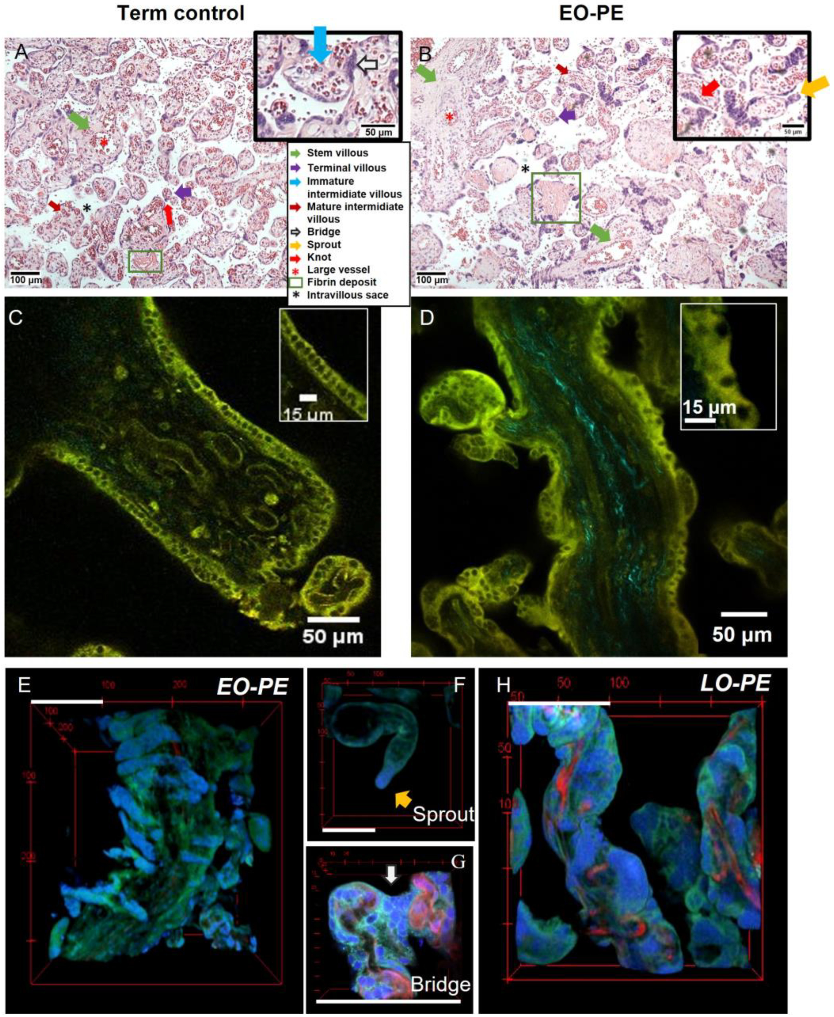
2.1.1. Trophoblast and Fibrotic Regions: Disorganised Trophoblast in Pre-Eclamptic Placenta
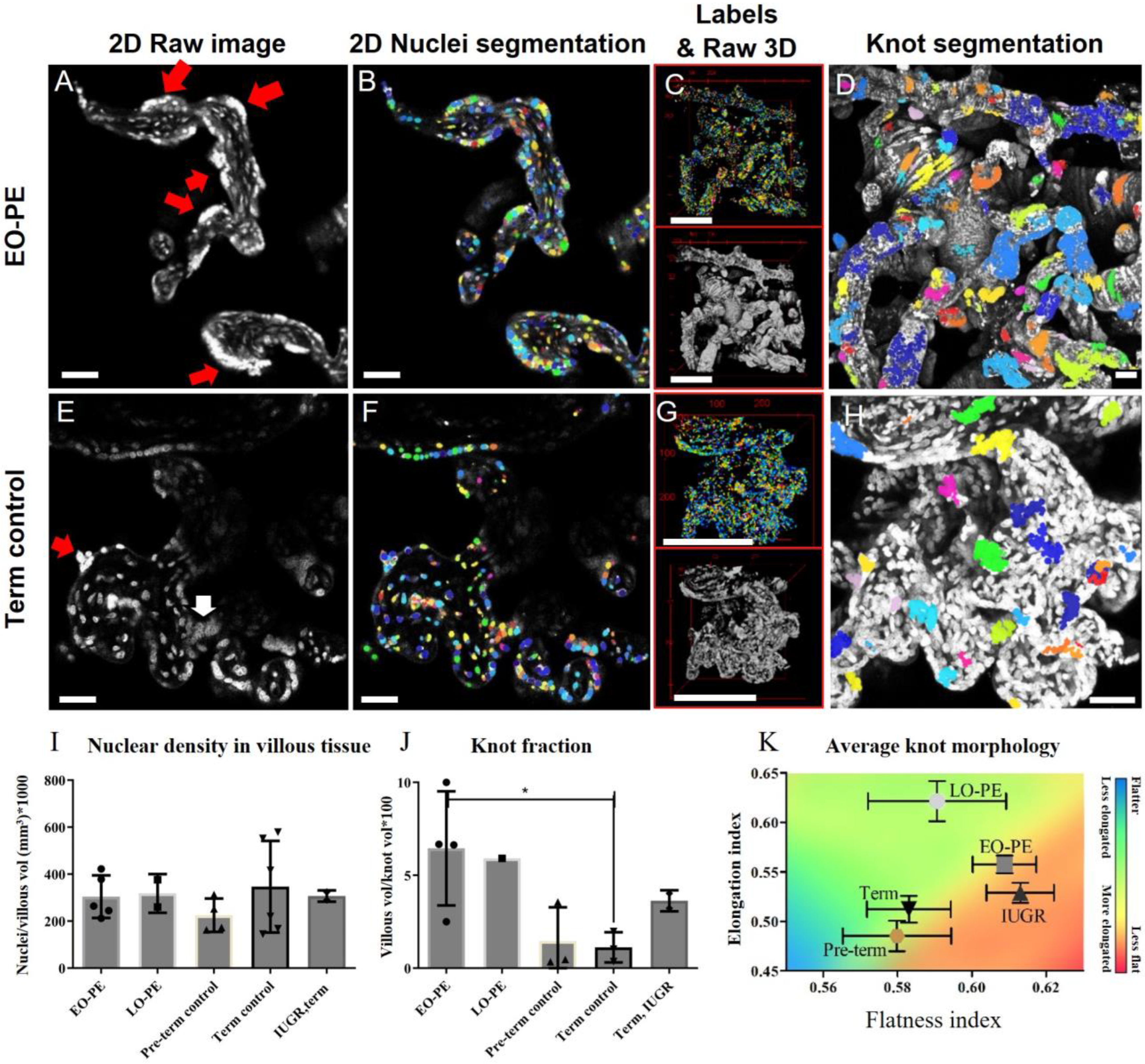
2.1.2. Nuclear 3D Architecture: Syncytial Knots Have Distinct 3D Organisations in PE
2.1.3. Villous Morphology and Vasculature
2.2. Quantification
2.2.1. Quantification of Nuclei and Their Architecture: MODEL Performance
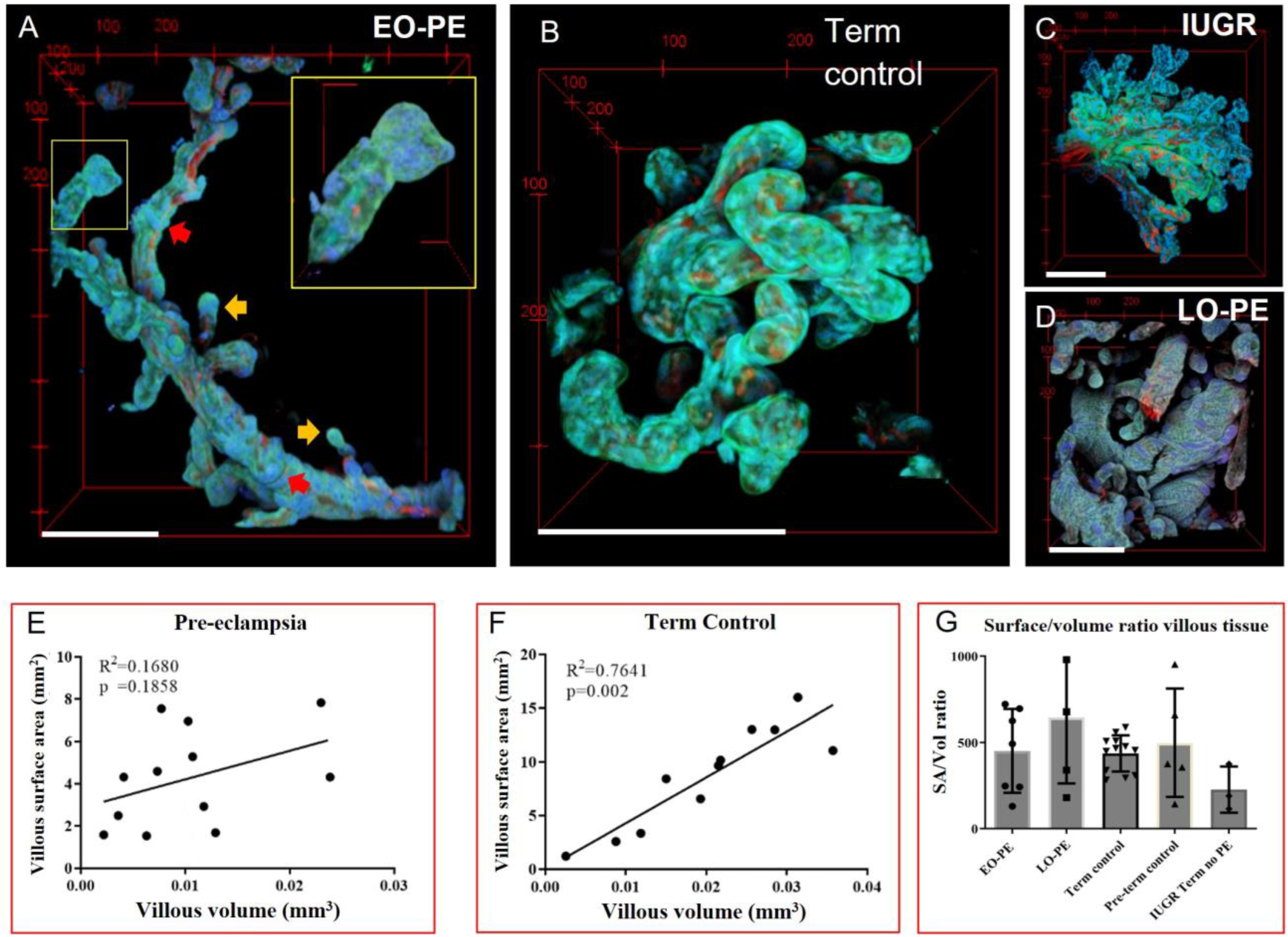
2.2.2. 3D Native Villous Tree Organisation: Altered Surface–Volume Relations in Pre-Eclampsia
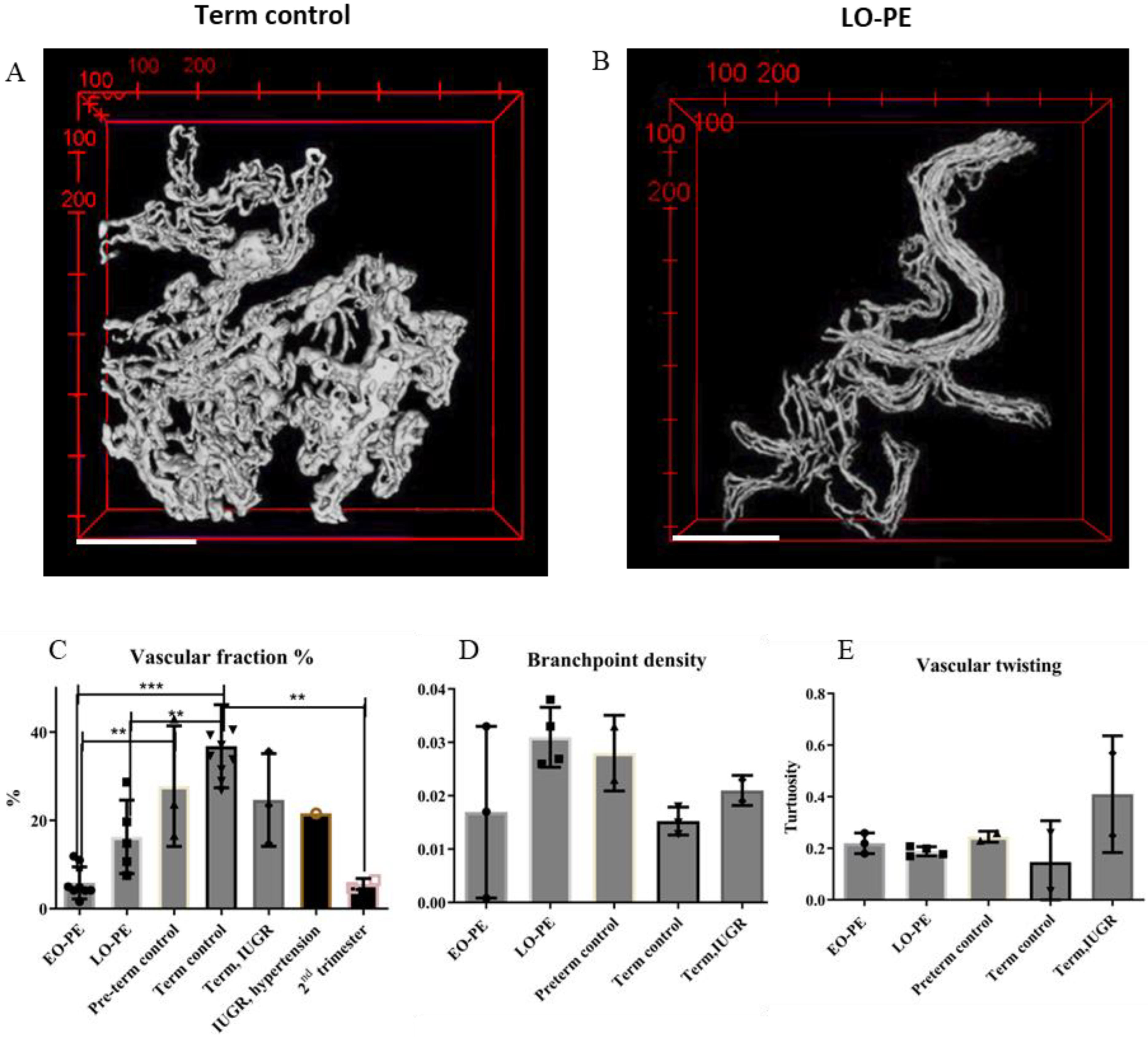
2.2.3. 3D Placental Microvasculature: A Reduced Vascular Fraction in Pre-Eclampsia
3. Discussion
3.1. Trophoblast and Knots
3.2. Placental Villous Tree Morphology: Exchange Surface and Volume
3.3. Placental Villous 3D Microvasculature: Volume and Network Quantification
3.4. Fibrosis
3.5. Strengths and Limitations
4. Methods
4.1. Tissue Collection and Study Population
4.2. Tissue Dissection and Fixation
4.3. Nuclear and Vascular Fluorescent Dyes
4.4. Mounting and MPM Imaging
4.5. Image processing and Quantitative Evaluation
4.5.1. Segmentation and Volume Quantification of Placental Villous Tissue and Vessels
4.5.2. Quantifying Vascular Network Characteristics
4.5.3. Nuclear Segmentation and Quantification
4.6. Statistical Analysis
5. Conclusions
Supplementary Materials
Author Contributions
Funding
Institutional Review Board Statement
Informed Consent Statement
Data Availability Statement
Acknowledgments
Conflicts of Interest
References
- Abalos, E.; Cuesta, C.; Grosso, A.L.; Chou, D.; Say, L. Global and regional estimates of preeclampsia and eclampsia: A systematic review. Eur. J. Obstet. Gynecol. Reprod. Biol. 2013, 170, 1–7. [Google Scholar] [CrossRef] [Scilit]
- Rosenberg, K.R.; Trevathan, W.R. An anthropological perspective on the evolutionary context of preeclampsia in humans. J. Reprod. Immunol. 2007, 76, 91–97. [Google Scholar] [CrossRef] [Scilit]
- Duley, L. The Global Impact of Pre-eclampsia and Eclampsia. Semin. Perinatol. 2009, 33, 130–137. [Google Scholar] [CrossRef] [Scilit] [PubMed]
- Hakim, J.; Senterman, M.K.; Hakim, A.M. Preeclampsia Is a Biomarker for Vascular Disease in Both Mother and Child: The Need for a Medical Alert System. Int. J. Pediatr. 2013, 2013, 953150. [Google Scholar] [CrossRef] [Scilit] [PubMed]
- Davis, E.F.; Lazdam, M.; Lewandowski, A.J.; Worton, S.A.; Kelly, B.; Kenworthy, Y.; Adwani, S.; Wilkinson, A.R.; McCormick, K.; Sargent, I.; et al. Cardiovascular risk factors in children and young adults born to preeclamptic pregnancies: A systematic review. Pediatrics 2012, 129, e1552–e1561. [Google Scholar] [CrossRef] [Scilit]
- Phipps, E.A.; Thadhani, R.; Benzing, T.; Karumanchi, S.A. Pre-eclampsia: Pathogenesis, novel diagnostics and therapies. Nat. Rev. Nephrol. 2019, 15, 275–289. [Google Scholar] [CrossRef] [Scilit] [PubMed]
- Capriglione, S.; Plotti, F.; Terranova, C.; Gulino, F.A.; Di Guardo, F.; Lopez, S.; Scaletta, G.; Angioli, R. Preeclampsia and the challenge of early prediction: Reality or utopia? State of art and critical review of literature. J. Matern. Neonatal Med. 2020, 33, 677–686. [Google Scholar] [CrossRef] [Scilit]
- Raymond, D.; Peterson, E. A critical review of early-onset and late-onset preeclampsia. Obstet. Gynecol. Surv. 2011, 66, 497–506. [Google Scholar] [CrossRef] [Scilit]
- Staff, A.C. The two-stage placental model of preeclampsia: An update. J. Reprod. Immunol. 2019, 134–135, 1–10. [Google Scholar] [CrossRef] [Scilit]
- Severens-Rijvers, C.A.H. Placental Syndrome: Early Pregnancy Adaptation and Placental Development. Ph.D. Thesis, Maastricht University, Maastricht, The Netherlands, 2018. [Google Scholar]
- Vangrieken, P.; Vanterpool, S.F.; van Schooten, F.J.; Al-Nasiry, S.; Andriessen, P.; Degreef, E.; Alfer, J.; Kramer, B.W.; von Rango, U. Histological villous maturation in placentas of complicated pregnancies. Histol. Histopathol. 2020, 35, 849–862. [Google Scholar]
- Corrêa, R.R.M.; Gilio, D.B.; Cavellani, C.L.; Paschoini, M.C.; Oliveira, F.A.; Peres, L.C.; Reis, M.A.; Teixeira, V.P.A.; Castro, E.C.C. Placental morphometrical and histopathology changes in the different clinical presentations of Hypertensive Syndromes in Pregnancy. Arch. Gynecol. Obstet. 2008, 277, 201–206. [Google Scholar] [CrossRef] [Scilit]
- Fogarty, N.M.E.; Ferguson-Smith, A.C.; Burton, G.J. Syncytial knots (Tenney-parker changes) in the human placenta: Evidence of loss of transcriptional activity and oxidative damage. Am. J. Pathol. 2013, 183, 144–152. [Google Scholar] [CrossRef] [Scilit] [PubMed]
- Askar, E.; Selim, S.; Sibai, H. Histological changes of human placenta in early intrauterine growth restriction with and without preeclampsia. J. Med. Histol. 2019, 3, 65–76. [Google Scholar] [CrossRef] [Scilit]
- Brosens, I.; Pijnenborg, R.; Vercruysse, L.; Romero, R. The “great Obstetrical Syndromes” are associated with disorders of deep placentation. Am. J. Obstet. Gynecol. 2011, 204, 193–201. [Google Scholar] [CrossRef] [Scilit]
- Ridder, A.; Giorgione, V.; Khalil, A.; Thilaganathan, B. Preeclampsia: The relationship between uterine artery blood flow and trophoblast function. Int. J. Mol. Sci. 2019, 20, 3263. [Google Scholar] [CrossRef] [Scilit] [PubMed]
- Sargent, J.A.; Roberts, V.H.J.; Gaffney, J.E.; Frias, A.E. Clarification and confocal imaging of the nonhuman primate placental micro-anatomy. Biotechniques 2019, 66, 79–84. [Google Scholar] [CrossRef] [Scilit]
- Mayo, R.P.; Abbas, Y.; Charnock-Jones, D.S.; Burton, G.J.; Marom, G. Three-dimensional morphological analysis of placental terminal villi. Interface Focus 2019, 9, 20190037. [Google Scholar] [CrossRef] [Scilit]
- Merz, G.; Schwenk, V.; Shah, R.; Salafia, C.; Necaise, P.; Joyce, M.; Villani, T.; Johnson, M.; Crider, N. Three-dimensional rendering and analysis of immunolabeled, clarified human placental villous vascular networks. J. Vis. Exp. 2018, 2018, 57099. [Google Scholar]
- Resta, L.; Capobianco, C.; Marzullo, A.; Piscitelli, D.; Sanguedolce, F.P.; Schena, F.; Gesualdo, L. Confocal Laser Scanning Microscope Study of Terminal Villi Vessels in Normal Term and Pre-eclamptic Placentas. Placenta 2006, 27, 735–739. [Google Scholar] [CrossRef] [Scilit]
- Baergen, R.N. Manual of Pathology of the Human Placenta; Springer Science & Business Media: Berlin/Heidelberg, Germany, 2011. [Google Scholar]
- Helmchen, F.; Denk, W. Deep tissue two-photon microscopy. Nat. Methods 2005, 2, 932–940. [Google Scholar] [CrossRef] [Scilit]
- Kapsokalyvas, D.; Cicchi, R.; Bruscino, N.; Alfieri, D.; Prignano, F.; Massi, D.; Lotti, T.; Pavone, F.S.S.F.S. In-vivo imaging of psoriatic lesions with polarization multispectral dermoscopy and multiphoton microscopy. Biomed. Opt. Express 2014, 5, 2405–2419. [Google Scholar] [CrossRef] [Scilit] [PubMed]
- Richardson, L.; Vargas, G.; Brown, T.; Ochoa, L.; Trivedi, J.; Kacerovský, M.; Lappas, M.; Menon, R. Redefining 3Dimensional placental membrane microarchitecture using multiphoton microscopy and optical clearing. Placenta 2017, 53, 66–75. [Google Scholar] [CrossRef] [Scilit] [PubMed]
- Richardson, L.S.; Vargas, G.; Brown, T.; Ochoa, L.; Sheller-Miller, S.; Saade, G.; Taylor, R.N.; Menon, R. Discovery and Characterization of Human Amniochorionic Membrane Microfractures. Am. J. Pathol. 2017, 187, 2821–2830. [Google Scholar] [CrossRef] [Scilit]
- Kaufmann, P.; Huppertz, B. Tenney–parker changes and apoptotic versus necrotic shedding of trophoblast in normal pregnancy and pre-eclampsia. In Pre-Eclampsia: Etiology and Clinical Practice; Cambridge University Press: Cambridge, UK, 2007; pp. 152–163. [Google Scholar]
- Coleman, S.J.; Gerza, L.; Jones, C.J.P.; Sibley, C.P.; Aplin, J.; Heazell, A.E.P. Syncytial nuclear aggregates in normal placenta show increased nuclear condensation, but apoptosis and cytoskeletal redistribution are uncommon. Placenta 2013, 34, 449–455. [Google Scholar] [CrossRef] [Scilit] [PubMed]
- Mayhew, T.M. Turnover of human villous trophoblast in normal pregnancy: What do we know and what do we need to know? Placenta 2014, 35, 229–240. [Google Scholar] [CrossRef] [Scilit] [PubMed]
- Sankar, K.D.; Bhanu, P.S.; Kiran, S.; Ramakrishna, B.A.; Shanthi, V. Vasculosyncytial membrane in relation to syncytial knots complicates the placenta in preeclampsia: A histomorphometrical study. Anat. Cell Biol. 2012, 45, 86–91. [Google Scholar] [CrossRef] [Scilit]
- Weigert, M.; Schmidt, U.; Haase, R.; Sugawara, K.; Myers, G. Star-convex polyhedra for 3D object detection and segmentation in microscopy. In Proceedings of the 2020 IEEE Winter Conference on Applications of Computer Vision WACV 2020, Snowmass Village, CO, USA, 1–5 March 2020; pp. 3655–3662. [Google Scholar]
- Tatsuzuki, A.; Ezaki, T.; Makino, Y.; Matsuda, Y.; Ohta, H. Characterization of the sugar chain expression of normal term human placental villi using lectin histochemistry combined with immunohistochemistry. Arch. Histol. Cytol. 2009, 72, 35–49. [Google Scholar] [CrossRef] [Scilit]
- Huppertz, B. The Critical Role of Abnormal Trophoblast Development in the Etiology of Preeclampsia. Curr. Pharm. Biotechnol. 2018, 19, 771–780. [Google Scholar] [CrossRef] [Scilit]
- Tomas, S.Z.; Prusac, I.K.; Roje, D.; Tadin, I. Trophoblast apoptosis in placentas from pregnancies complicated by preeclampsia. Gynecol. Obstet. Investig. 2011, 71, 250–255. [Google Scholar] [CrossRef] [Scilit]
- Vargas, A.; Toufaily, C.; LeBellego, F.; Rassart, É.; Lafond, J.; Barbeau, B. Reduced expression of both syncytin 1 and syncytin 2 correlates with severity of preeclampsia. Reprod. Sci. 2011, 18, 1085–1091. [Google Scholar] [CrossRef] [Scilit]
- Arnholdt, H.; Meisel, F.; Fandrey, K.; Löhrs, U. Proliferation of villous trophoblast of the human placenta in normal and abnormal pregnancies. Virchows Arch. B Cell Pathol. Incl. Mol. Pathol. 1991, 60, 365–372. [Google Scholar] [CrossRef] [Scilit]
- Can, M.; Guven, B.; Bektas, S.; Arikan, I. Oxidative stress and apoptosis in preeclampsia. Tissue Cell 2014, 46, 477–481. [Google Scholar] [CrossRef] [Scilit] [PubMed]
- Prusac, I.K.; Tomas, S.Z.; Roje, D. Apoptosis, proliferation and Fas ligand expression in placental trophoblast from pregnancies complicated by HELLP syndrome or pre-eclampsia. Acta Obstet. Gynecol. Scand. 2011, 90, 1157–1163. [Google Scholar] [CrossRef] [Scilit]
- Heazell, A.E.P.; Moll, S.J.; Jones, C.J.P.; Baker, P.N.; Crocker, I.P. Formation of Syncytial Knots is Increased by Hyperoxia, Hypoxia and Reactive Oxygen Species. Placenta 2007, 28, S33–S40. [Google Scholar] [CrossRef] [Scilit]
- Fitzgerald, B.; Kingdom, J.; Keating, S. Distal villous hypoplasia. Diagn. Histopathol. 2012, 18, 195–200. [Google Scholar] [CrossRef] [Scilit]
- Mayhew, T.M. Patterns of villous and intervillous space growth in human placentas from normal and abnormal pregnancies. Eur. J. Obstet. Gynecol. Reprod. Biol. 1996, 68, 75–82. [Google Scholar] [CrossRef] [Scilit] [PubMed]
- Mayhew, T.M.; Manwani, R.; Ohadike, C.; Wijesekara, J.; Baker, P.N. The Placenta in Pre-eclampsia and Intrauterine Growth Restriction: Studies on Exchange Surface Areas, Diffusion Distances and Villous Membrane Diffusive Conductances. Placenta 2007, 28, 233–238. [Google Scholar] [CrossRef] [Scilit] [PubMed]
- Mayhew, T.M.; Ohadike, C.; Baker, P.N.; Crocker, I.P.; Mitchell, C.; Ong, S.S. Stereological investigation of placental morphology in pregnancies complicated by pre-eclampsia with and without intrauterine growth restriction. Placenta 2003, 24, 219–226. [Google Scholar] [CrossRef] [Scilit]
- Yin, G.; Chen, M.; Li, J.; Zhao, X.; Yang, S.; Li, X.; Yuan, Z.; Wu, A. Vascular corrosion casting of normal and pre-eclamptic placentas. Exp. Ther. Med. 2017, 14, 5535–5539. [Google Scholar] [CrossRef] [Scilit]
- Devisme, L.; Merlot, B.; Ego, A.; Houfflin-Debarge, V.; Deruelle, P.; Subtil, D. A case-control study of placental lesions associated with pre-eclampsia. Int. J. Gynecol. Obstet. 2013, 120, 165–168. [Google Scholar] [CrossRef] [Scilit]
- Mayhew, T.M.; Charnock-Jones, D.S.; Kaufmann, P. Aspects of human fetoplacental vasculogenesis and angiogenesis. III. Changes in complicated pregnancies. Placenta 2004, 25, 127–139. [Google Scholar] [CrossRef] [Scilit] [PubMed]
- Kaufmann, P.; Mayhew, T.M.; Charnock-Jones, D.S. Aspects of human fetoplacental vasculogenesis and angiogenesis. II. Changes during normal pregnancy. Placenta 2004, 25, 114–126. [Google Scholar] [CrossRef] [Scilit] [PubMed]
- Han, H.-C. Twisted blood vessels: Symptoms, etiology and biomechanical mechanisms. J. Vasc. Res. 2012, 49, 185–197. [Google Scholar] [CrossRef] [Scilit] [PubMed]
- Richardson, D.S.; Lichtman, J.W. Clarifying Tissue Clearing. Cell 2015, 162, 246–257. [Google Scholar] [CrossRef] [Scilit]
- Power, R.M.; Huisken, J. A guide to light-sheet fluorescence microscopy for multiscale imaging. Nat. Methods 2017, 14, 360–373. [Google Scholar] [CrossRef] [Scilit]
- Orabona, R.; Donzelli, C.M.; Falchetti, M.; Santoro, A.; Valcamonico, A.; Frusca, T. Placental histological patterns and uterine artery Doppler velocimetry in pregnancies complicated by early or late pre-eclampsia. Ultrasound Obstet. Gynecol. 2016, 47, 580–585. [Google Scholar] [CrossRef] [Scilit]
- Schindelin, J.; Arganda-Carreras, I.; Frise, E.; Kaynig, V.; Longair, M.; Pietzsch, T.; Preibisch, S.; Rueden, C.; Saalfeld, S.; Schmid, B.; et al. Fiji: An open-source platform for biological-image analysis. Nat. Methods 2012, 9, 676–682. [Google Scholar] [CrossRef] [Scilit]
- Arzt, M.; Deschamps, J.; Schmied, C.; Pietzsch, T.; Schmidt, D.; Tomancak, P.; Haase, R.; Jug, F. LABKIT: Labeling and Segmentation Toolkit for Big Image Data. Front. Comput. Sci. 2022, 4, 10. [Google Scholar] [CrossRef] [Scilit]
- Ollion, J.; Cochennec, J.; Loll, F.; Escudé, C.; Boudier, T. TANGO: A generic tool for high-throughput 3D image analysis for studying nuclear organization. Bioinformatics 2013, 29, 1840–1841. [Google Scholar] [CrossRef] [Scilit]
- Kikinis, R.; Pieper, S.D.; Vosburgh, K.G. 3D Slicer: A Platform for Subject-Specific Image Analysis, Visualization, and Clinical Support. In Intraoperative Imaging Image-Guided Therapy; Springer: Berlin/Heidelberg, Germany, 2014; pp. 277–289. [Google Scholar]
- Antiga, L.; Piccinelli, M.; Botti, L.; Ene-Iordache, B.; Remuzzi, A.; Steinman, D.A. An image-based modeling framework for patient-specific computational hemodynamics. Med. Biol. Eng. Comput. 2008, 46, 1097–1112. [Google Scholar] [CrossRef] [Scilit]
- Mikut, R.; Bartschat, A.; Doneit, W.; Ordiano, J.Á.G.; Schott, B.; Stegmaier, J.; Waczowicz, S.; Reischl, M. The MATLAB Toolbox SciXMiner: User’s Manual and Programmer’s Guide. arXiv 2017, arXiv:1704.03298. [Google Scholar]
- Hypertension in pregnancy. Report of the American College of Obstetricians and Gynecologists’ Task Force on Hypertension in Pregnancy. Obstet Gynecol. 2013, 122, 1122–1131.
- Ives, C.W.; Sinkey, R.; Rajapreyar, I.; Tita, A.T.; Oparil, S. Preeclampsia—Pathophysiology and Clinical Presentations: JACC State-of-the-Art Review. J. Am. Coll. Cardiol. 2020, 76, 1690–1702. [Google Scholar] [CrossRef] [Scilit] [PubMed]
- Ester, M.; Kriegel, H.-P.; Sander, J.; Xu, X. (Eds.) A Density-Based Algorithm for Discovering Clusters in Large Spatial Databases with Noise; Kdd: Washington, DC, USA, 1996; pp. 226–231. [Google Scholar]
- Zingg, T. Beitrag zur Schotteranalyse; ETH Zurich: Zürich, Switzerland, 1935. [Google Scholar]
- Ahmed, A.; Dunk, C.; Ahmad, S.; Khaliq, A. Regulation of placental vascular endothelial growth factor (VEGF) and placenta growth factor (PIGF) and soluble Flt-1 by oxygen—A review. Placenta 2000, 21 (Suppl. A), S16–S24. [Google Scholar] [CrossRef] [Scilit] [PubMed]


| Placenta | Group | Maternal Age (Years) | Gravida/Para | GA | Placental Weight (g) | Birthweight (g) | Collection Method |
|---|---|---|---|---|---|---|---|
| 1 | Term control | 30 | 1/0 | 40 w 2 d | NA | 3750 | Fresh |
| 2 | Term control | 38 | 2/1 | 40 w 4 d | NA | 4040 | Fresh |
| 3 | Term control | 33 | 2/1 | 39 w 2 d | NA | 3150 | Fresh |
| 4 | EO-PE | 33 | 1/0 | 30 w 1 d | 190 incomplete | 1080 | Fresh |
| 5 | EO-PE | 26 | 1/0 | 32 w 3 d | 233 | 1400 | Archive |
| 6 | LO-PE | 26 | 2/0 | 37 w 5 d | 421 | 2058 | Archive |
| 7 | SGA/IUGR | 36 | 3/1 | 40 w 0 d | NA | 3065 | Fresh |
| 8 | Preterm control | 20 | 2/0 | 32 w 5 d | 328 | 2089 | Archive |
| 9 | Preterm control | 21 | 2/1 | 31 w 5 d | 362,5 | 2134 | Archive |
| 10 | Maternal hypertension + IUGR | 26 | 1/0 | 37 w 5 d | 282 | 1895 | Archive |
| 11 | 2nd trimester placenta | 21 | 2/1 | 22 w 6 d | 73 | 532 | Archive |
Disclaimer/Publisher’s Note: The statements, opinions and data contained in all publications are solely those of the individual author(s) and contributor(s) and not of MDPI and/or the editor(s). MDPI and/or the editor(s) disclaim responsibility for any injury to people or property resulting from any ideas, methods, instructions or products referred to in the content. |
© 2023 by the authors. Licensee MDPI, Basel, Switzerland. This article is an open access article distributed under the terms and conditions of the Creative Commons Attribution (CC BY) license (https://creativecommons.org/licenses/by/4.0/).
Share and Cite
Hermans, S.; Pilon, J.; Eschweiler, D.; Stegmaier, J.; Severens–Rijvers, C.A.H.; Al-Nasiry, S.; van Zandvoort, M.; Kapsokalyvas, D. Definition and Quantification of Three-Dimensional Imaging Targets to Phenotype Pre-Eclampsia Subtypes: An Exploratory Study. Int. J. Mol. Sci. 2023, 24, 3240. https://doi.org/10.3390/ijms24043240
Hermans S, Pilon J, Eschweiler D, Stegmaier J, Severens–Rijvers CAH, Al-Nasiry S, van Zandvoort M, Kapsokalyvas D. Definition and Quantification of Three-Dimensional Imaging Targets to Phenotype Pre-Eclampsia Subtypes: An Exploratory Study. International Journal of Molecular Sciences. 2023; 24(4):3240. https://doi.org/10.3390/ijms24043240
Chicago/Turabian StyleHermans, Sammy, Jacob Pilon, Dennis Eschweiler, Johannes Stegmaier, Carmen A. H. Severens–Rijvers, Salwan Al-Nasiry, Marc van Zandvoort, and Dimitrios Kapsokalyvas. 2023. "Definition and Quantification of Three-Dimensional Imaging Targets to Phenotype Pre-Eclampsia Subtypes: An Exploratory Study" International Journal of Molecular Sciences 24, no. 4: 3240. https://doi.org/10.3390/ijms24043240
APA StyleHermans, S., Pilon, J., Eschweiler, D., Stegmaier, J., Severens–Rijvers, C. A. H., Al-Nasiry, S., van Zandvoort, M., & Kapsokalyvas, D. (2023). Definition and Quantification of Three-Dimensional Imaging Targets to Phenotype Pre-Eclampsia Subtypes: An Exploratory Study. International Journal of Molecular Sciences, 24(4), 3240. https://doi.org/10.3390/ijms24043240