Communication

Detection and Genotypic Analysis of Anaplasma bovis and A. phagocytophilum in Horse Blood and Lung Tissue

1
College of Veterinary Medicine, Kyungpook National University, 80 Daehak-ro, Buk-gu, Daegu 41566, Republic of Korea
2
National Institute of Health, Korea Disease Control and Prevention Agency, 212 Osongsaengmyeong 2-ro, Cheongju, Chungbuk 28160, Republic of Korea
*
Author to whom correspondence should be addressed.
Int. J. Mol. Sci. 2023, 24(4), 3239; https://doi.org/10.3390/ijms24043239
Submission received: 27 December 2022 / Revised: 21 January 2023 / Accepted: 27 January 2023 / Published: 7 February 2023
(This article belongs to the Special Issue Human Animal Tick-Borne Diseases)

Abstract

:
A clinical case of Anaplasma bovis was reported for the first time in our previous study (2019) in a horse, a nondefinitive host. Although A. bovis is a ruminant and not a zoonotic pathogen, it is responsible for persistent infections in horses. In this follow-up study, the prevalence of Anaplasma spp., including A. bovis, was assessed in horse blood and lung tissue samples to fully understand Anaplasma spp. pathogen distribution and the potential risk factors of infection. Among 1696 samples, including 1433 blood samples from farms nationwide and 263 lung tissue samples from horse abattoirs on Jeju Island, a total of 29 samples (1.7%) tested positive for A. bovis and 31 (1.8%) samples tested positive for A. phagocytophilum, as determined by 16S rRNA nucleotide sequencing and restriction fragment length polymorphism. This study is the first to detect A. bovis infection in horse lung tissue samples. Further studies are needed to clarify the comparison of sample types within cohorts. Although the clinical significance of Anaplasma infection was not evaluated in this study, our results emphasize the need to clarify the host tropism and genetic divergence of Anaplasma to enable the development of effective prevention and control measures through broad epidemiological studies.

1. Introduction

Anaplasma phagocytophilum, a Gram-negative intracellular bacterium, is the etiologic agent of the tick-borne disease, equine granulocytic anaplasmosis (EGA; formerly known as equine granulocytic ehrlichiosis) [1]. The classic signs of EGA include anorexia, fever, depression, ataxia, petechia, peripheral edema, and reluctance to move [2]. A. phagocytophilum belongs to the order Rickettsiales and includes several variants that have distinct host specificities. Despite such preferences, these variants are classified as a single species based on their genetic similarities [1].
To date, multiple Anaplasma spp. have been detected in the Republic of Korea using PCR, including A. phagocytophilum in dogs [3]; A. phagocytophilum, A. bovis, and A. centrale in ticks [4]; A. capra, A. bovis, and Anaplasma sp. in ticks from water deer [5]; A. capra, A. bovis, and A. phagocytophilum-like Anaplasma spp. (APL) clade A and B in ticks from cattle [6]; A. bovis [7], A. phagocytophilum, APL clade A [8], A. capra, and A. bovis in cattle [9]; A. phagocytophilum and A. bovis in wild Korean water deer [10]; A. phagocytophilum in a single horse [11]; and a clinical case of A. bovis infection in another single horse [12].
Reportedly, 27,829 horses were reared in the Republic of Korea in 2017. Of these, 15,234 (54.7%) were raised on Jeju Island [13] where disease control is of paramount importance for horse farms. Recently, a clinical case of A. bovis was reported in a horse, a nondefinitive host, in the Republic of Korea for the first time [12], warranting a follow-up study across the nation, including on Jeju Island. Blood samples are typically used to detect Anaplasma spp.; however, in persistently infected animals with intermittent or low-level bacteremia, other tissue samples, such as the liver, lung, lymph nodes, bone marrow, and skin biopsies, are also suitable [14]. Anaplasma spp. have been detected in the lung tissues of humans, horses, sheep, cattle, red foxes, and raccoon dogs in several previous studies [15,16,17,18]. Therefore, this follow-up study aimed to detect Anaplasma spp. (including A. bovis) in both horse blood and lung tissue samples and to elucidate pathogen distribution and potential risk factors in the Republic of Korea.

2. Results

2.1. Nested PCR and Restriction Fragment Length Polymorphism

In this study, the results of nested PCR (nPCR) amplification of the 16S rRNA gene fragments using the EE1/EE2 and EE3/EE4 primer pairs indicated that 3.5% (60/1696; 53 lung tissue samples and 7 blood samples) of the horses were positive for Anaplasma spp. Additional nPCR analyses were conducted to amplify the 16S rRNA fragments of A. phagocytophilum and A. bovis for species identification. Upon nPCR, horse lung samples that were positive for A. phagocytophilum produced fragments that were 641 bp in length (Figure 1A, lane 4), whereas those positive for A. bovis generated fragments that were 551 bp in length (Figure 1A, lanes 5 and 6). The samples that were negative for Anaplasma spp. (Figure 1A, lanes 2 and 3) did not generate fragments during nPCR. The housekeeping gene (HKG) amplicons from all horse samples generated fragments that were 204 bp in length (Figure 1B, lanes 2 to 10). A sample containing no horse DNA was used as the negative control (Figure 1B, lane 11).
Species-specific PCR indicated that 0.2% (3/1433, 95% confidence interval, CI: 0–0.4) of the blood samples and 9.9% (26/263, 95% CI: 6.3–13.5) of the lung tissue samples were positive for A. bovis (Table 1). Moreover, 0.3% (4/1433, 95% CI: 0–0.6) of the blood samples and 10.3% (27/263, 95% CI: 6.6–13.9) of the lung tissue samples were positive for A. phagocytophilum (Table 1). When data were analyzed according to the sample groups, a statistically significant difference (p = 0.0253) was observed only in geographical regions with A. phagocytophilum infections.
The restriction fragment length polymorphism (RFLP) assay was performed by digesting 16S rRNA amplicons (924–926 bp) with AleI for additional discrimination between A. bovis and A. phagocytophilum. Upon digestion, A. bovis amplicons from the horse blood and lung samples generated fragments that were 660 and 264 bp in length, respectively (Figure 2A, lanes 3 and 5), whereas the A. phagocytophilum amplicons from the horse lung tissue were not digested by the enzyme (Figure 2A, lane 7). Digestion of the amplicons with BtgZI was also performed to distinguish A. bovis from A. phagocytophilum. The A. bovis amplicons from the horse blood and lung tissue samples could not be digested by the enzyme (Figure 2B, lanes 3 and 5), whereas the A. phagocytophilum amplicon from the horse lung tissue samples generated two fragments of 707 and 223 bp (Figure 2B, lane 7). Interestingly, no samples were co-infected with these or any other Anaplasma species.

2.2. Cloning, Sequencing, and Phylogeny

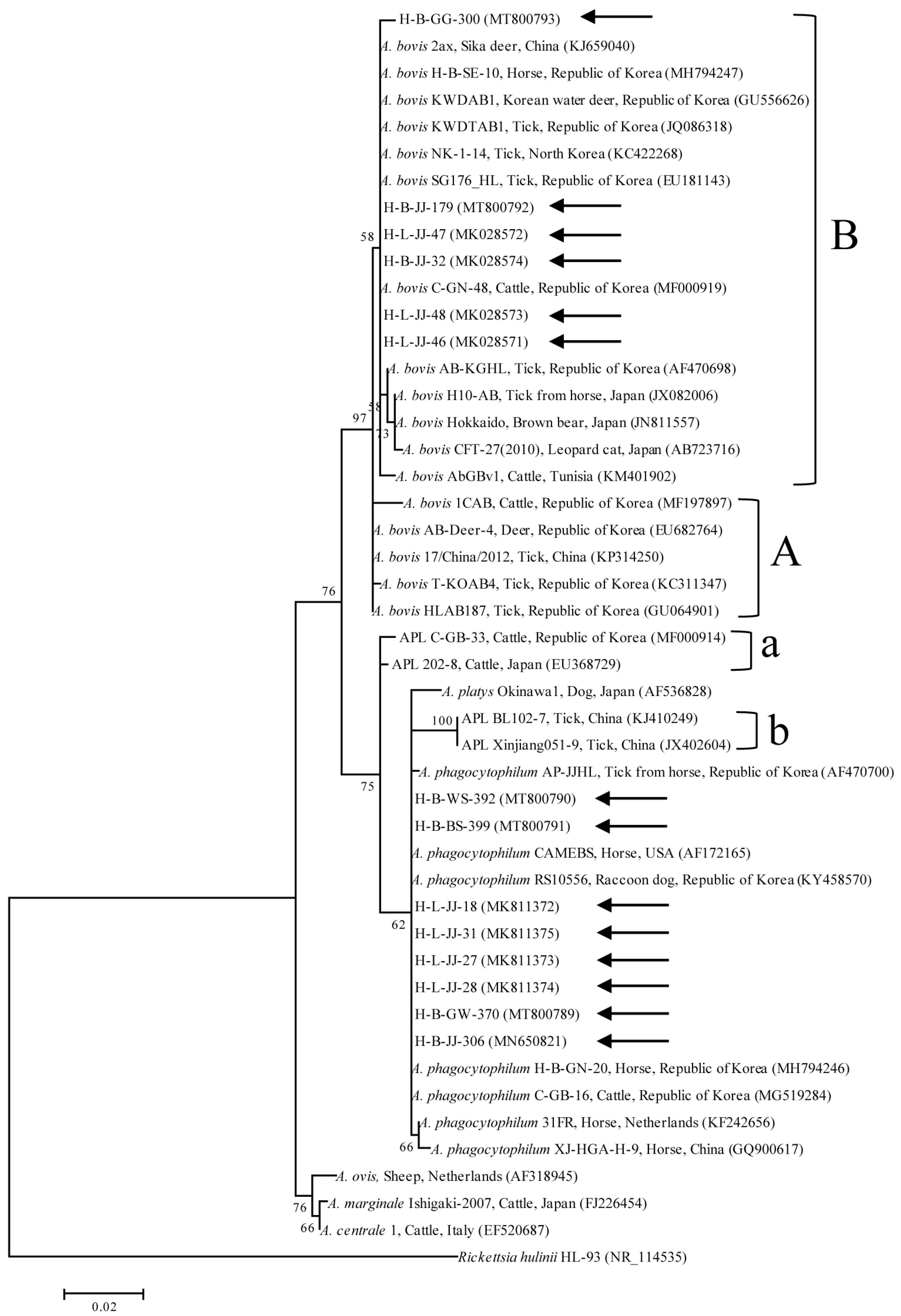
The RFLP assay data were confirmed by the sequencing of the amplicons from the species-specific nPCR of 16S rRNA. All positive samples were confirmed via RFLP. As the A. bovis sequences shared 99.7–100% identity (differences in 0–3 nucleotide positions), representative lung tissue sequences were randomly selected for phylogenetic analysis. The included samples were as follows: blood (H-B-JJ-32, H-B-JJ-179, and H-B-GG-300; GenBank accession numbers MK028574, MT800792, and MT800793, respectively) and lung (H-L-JJ-46, H-L-JJ-47, and H-L-JJ-48; accession numbers MK028571, MK028572, and MK028573, respectively). Phylogenetic analysis revealed that the A. bovis sequences (Figure 3) were clustered with previously documented sequences.
Nucleotide sequencing of the amplicons revealed that four blood and 27 lung tissue samples were positive for A. phagocytophilum. As the A. phagocytophilum 16S rRNA sequences shared 99.4–100% identity (differences in 0–5 nucleotide positions), representative lung tissue sequences were randomly selected for the phylogenetic analyses. The analyzed samples were as follows: blood (H-B-JJ-306, H-B-GW-370, H-B-WS-392, and H-B-BS-399; accession numbers MN650821, MT800789, MT800790, and MT800791, respectively) and lung (H-L-JJ-18, H-L-JJ-27, H-L-JJ-28, and H-L-JJ-31; accession numbers MK811372, MK811373, MK811374, and MK811375, respectively). The phylogenetic analysis revealed that the sequences of A. phagocytophilum (Figure 3) clustered with previously documented sequences.
Six A. bovis samples (H-B-JJ-32, H-B-JJ-179, H-B-GG-300, H-L-JJ-46, H-L-JJ-47, and H-L-JJ-48) belonging to clade B shared 94.1–100% sequence identity with previously identified isolates collected from cattle in Tunisia (94.1%, KM401902), a horse in Japan (95.2%, JX082006), a sika deer in China (99.8%, KJ659040), a tick in the Republic of Korea (99.1%, AF470698), and a horse in the Republic of Korea (100%, MH794247). Samples clustered in clade A shared 98.1–99.9% sequence identity; these samples included isolates from cattle in the Republic of Korea (98.1%, MF197897), a tick in China (99.5%, KP314250), and a tick in the Republic of Korea (99.9%, GU064901).
Eight A. phagocytophilum samples (H-B-JJ-306, H-B-GW-370, H-B-WS-392, H-B-BS-399, H-L-JJ-18, H-L-JJ-27, H-L-JJ-28, and H-L-JJ-31) shared 98.9–99.9% sequence identity with previously collected A. phagocytophilum isolates from a horse in the Netherlands (98.9%, KF242656), a horse in the United States (99.2%, AF172165), a horse in China (99.6%, GQ900617), and a horse in the Republic of Korea (99.9%, MH794246).

3. Discussion

A previous molecular study identified A. phagocytophilum infection in a horse in the Republic of Korea [11]. A clinical case of A. bovis infection in a horse, a nondefinitive host, was also identified [12]. In this study, we screened horses for the presence of A. bovis; 3 (0.2%) blood and 26 (9.9%) lung tissue samples from 1696 animals were found to be positive based on a species-specific nPCR analysis of 16S rRNA gene fragments. A. phagocytophilum was also detected in horse blood (n = 4; 0.3%) and lung tissue (n = 27; 10.3%) samples. These positive samples were further confirmed to be A. bovis and A. phagocytophilum via the RFLP assay.
Cross-contamination is a major problem associated with nPCR. Reducing the potential for contamination and ensuring the differentiation between true- and false-positive results necessitates the incorporation of reference genes as internal controls [20]. Reference genes, commonly known as HKG, are stably expressed in cells and tissues. An internal reference gene helps to ensure the accurate interpretation of the data [21]. Although several HKGs have been used, the 18S rRNA gene is considered as one of the most stable genes [19] that shows limited changes in expression under different experimental conditions [22]. Moreover, 18S rRNA sequences are highly conserved among eukaryotes, and a single assay can be used for an HKG measurement even in studies involving cells from several species [22]. In this study, horse 18S rRNA was used as the internal positive control and Coxiella burnetii was used as the internal negative control to ensure that cross-contamination did not occur and to improve the accuracy of the data.
Equine anaplasmosis is typically caused by A. phagocytophilum, the causative zoonotic pathogen of granulocytic anaplasmosis and tick-borne fever [23]. However, in this study, we detected A. bovis in the lung tissues of horses for the first time. A. bovis is widespread in tropical and subtropical regions [24] and is a common ruminant pathogen infecting buffalo and cattle in Africa and Asia [25]. Infections in other hosts have been rarely detected, such as in a leopard in the Republic of Korea [26], a Hokkaido brown bear in Japan [27], a sika deer and a wild boar in Japan [28], a raccoon in Japan [29]; moreover, human cases have been reported in China [30].
Unfortunately, the lung tissue samples collected from abattoirs were not associated with any demographic information. Nonetheless, as the blood and lung tissue samples obtained from Jeju Island tested positive for A. bovis and A. phagocytophilum, we surmised that Anaplasma spp. may be widespread on Jeju Island. This hypothesis is supported by multiple reports of the molecular detection of Anaplasma spp. in animals on Jeju Island. The prevalence has been reported for A. phagocytophilum (1.9%, 27/1395), A. bovis (0.4%, 5/1395), and A. centrale (0.1%, 1/1395) in the tick Haemaphysalis longicornis [4]; A. bovis (13.9%, 10/72) in H. longicornis from grazing cattle [31]; Anaplasma spp. (18%, 7/39) in native Korean goats [32]; and A. bovis (4.2%, 3/71) in cattle [7].
The prevalence of Anaplasma spp. has been reported to significantly differ between geographic locations and is associated with tick habitats and distribution [33,34]. Prevalence has also been reported to vary according to biogeoclimatic conditions. Cattle from sub-humid areas have been reported to be more susceptible to Anaplasma infection compared to cattle from semi-arid regions [34]. The climate of the Korean peninsula is steadily turning subtropical; Jeju Island is particularly vulnerable to this change because of its lower latitude. Haemaphysalis longicornis is the most commonly identified tick species in Korea and is more abundantly distributed across Jeju Island than the mainland [4]. The horses in this environment are allowed to graze freely in grassland areas, thereby increasing their exposure to ticks compared with those living on the mainland, which are kept in restricted areas. Biogeoclimatic differences may thus affect the prevalence of ticks as well as tick-borne infections. On the mainland, the southern region of the Republic of Korea exhibits by far the highest prevalence of A. phagocytophilum. Moreover, the horse industry continues to grow in the Republic of Korea each year. Therefore, it is extremely important to mitigate or prevent the spread of zoonotic diseases, such as EGA, between horses and humans on Jeju Island.
Blood smears from animals experiencing persistent infections may yield negative infection results, even though 400 granulocytes are adequate to detect infected leukocytes in ruminants with a history of recent illness [35,36]. Moreover, a negative result does not preclude infection. Microscopy in addition to supplementary diagnostic laboratory methods and the screening of other sample types are recommended if persistent infection is suspected [14]. In postmortem evaluations, tissue impressions or smears from the liver, spleen, kidneys, lungs, heart, or blood vessels can be used to visualize erythrocytotropic Anaplasma spp. [37]. In this study, blood sample analyses demonstrated a lower prevalence of both A. bovis (0.2%) and A. phagocytophilum (0.3%), whereas lung tissue samples had a higher prevalence of both A. bovis (9.9%) and A. phagocytophilum (10.3%). Unfortunately, the collection of blood and lung samples from different animals precludes the comparison of the prevalence of these pathogens between sample types. Further studies are needed to compare the sample types within cohorts. At the time of our investigation, the lung tissue samples were rich in blood, which might have resulted in the improved detection rates of A. bovis and A. phagocytophilum. These results suggest that A. bovis and A. phagocytophilum cause frequent and persistent infections in horses.
Although Anaplasma spp. usually infects blood cells, their presence in several other tissues has been reported. A. phagocytophilum has been detected in the kidney, thymus, sternal bone marrow, small intestine, mediastinal lymph node, and bladder wall tissue of persistently infected sheep [36]. In another study on humans, horses, and sheep, A. phagocytophilum was detected in the lungs, spleen, and liver; moreover, large numbers of infected neutrophils were detected in blood vessel lumens, mainly in the microvasculature of the lungs or in the sinusoids of the red pulp of the spleen [15]. A. phagocytophilum was also detected in the lung tissues of red foxes and raccoon dogs [18] and in the lung, spleen, liver, and heart tissue of cattle [16]. In another case, A. marginale was detected in the lymph node, spleen, heart, lung, and ear skin of cattle [16]. In humans, anaplasmosis caused by A. phagocytophilum presents as atypical pneumonitis and histopathological changes in the lungs [38]. Consistent with these reports, A. bovis and A. phagocytophilum were both detected in horse lung tissue samples in the current study. A. phagocytophilum infection in tissue has been associated with infected circulatory neutrophils rather than infected tissue cells [36]. Similarly, A. bovis detected in the current study might have infected circulatory monocytes within the lung tissue.
Based on phylogenetic analyses using 16S rRNA sequences, A. bovis was classified into clades A and B. The A. bovis sequences identified in this study belonged to clade B, which includes the strains identified in Tunisia, the Republic of Korea, the Democratic People’s Republic of Korea, China, and Japan. The A. bovis sequences of clade A contain strains from East Asia (China and the Republic of Korea). These results are consistent with an earlier study in which a genotypic analysis of Anaplasma spp. revealed a high degree of identity with species isolated from neighboring countries [39]. The sequences of 16S rRNA genes from Anaplama spp. have been reported for several species on Jeju Island: A. bovis in H. longicornis (EU181143, clade B; GU064901, clade A), A. bovis in cattle (MF197897, clade A), and A. phagocytophilum in H. longicornis from a horse (AF470700). The horse-derived A. bovis sequences in this study showed high sequence identities with those previously reported on Jeju Island.
To the best of our knowledge, this is the first study to perform a molecular detection of A. bovis in horse lung tissues. Recently, infected nondefinitive hosts were reported as threats for the spread of several diseases to humans. Indeed, human cases of goat-derived A. capra in China [40], sheep-derived A. ovis in one patient in Cyprus [41], and cattle-derived A. bovis in two patients in China [30] have been reported previously. A. bovis is a ruminant pathogen and, unlike A. phagocytophilum, it is not considered a zoonotic pathogen. However, A. bovis may cause frequent and persistent infections in horses (a nondefinitive host) reared on Jeju Island. Our study can be used as a reference for further investigating the clinical significance of Anaplasma infections. This study highlights the need for broad epidemiological studies to clarify the host–pathogen relationship and genetic divergence of Anaplasma spp. to aid in the development of effective preventive and control measures.

4. Materials and Methods

4.1. Ethical Approval

This study was conducted between 2017 and 2019 and did not need the approval of the Institutional Animal Care and Use Committee at Kyungpook National University as this is only required for research involving laboratory animals kept in indoor facilities, not outdoor animals. Practicing veterinarians collected whole blood samples during treatment or at regular medical check-ups, after obtaining verbal consent from the farmers.

4.2. Sample Size Determination and Sample Collection

Using a simple random sampling strategy and considering an expected disease prevalence of 10%, accepted absolute error of 5%, and confidence level of 95%, the sample size was calculated using the following formula [42]:
n = 1.96 2 p e x p 1 p e x p d 2
where n represents the required sample size, d represents the desired absolute precision, and pexp represents the expected prevalence.
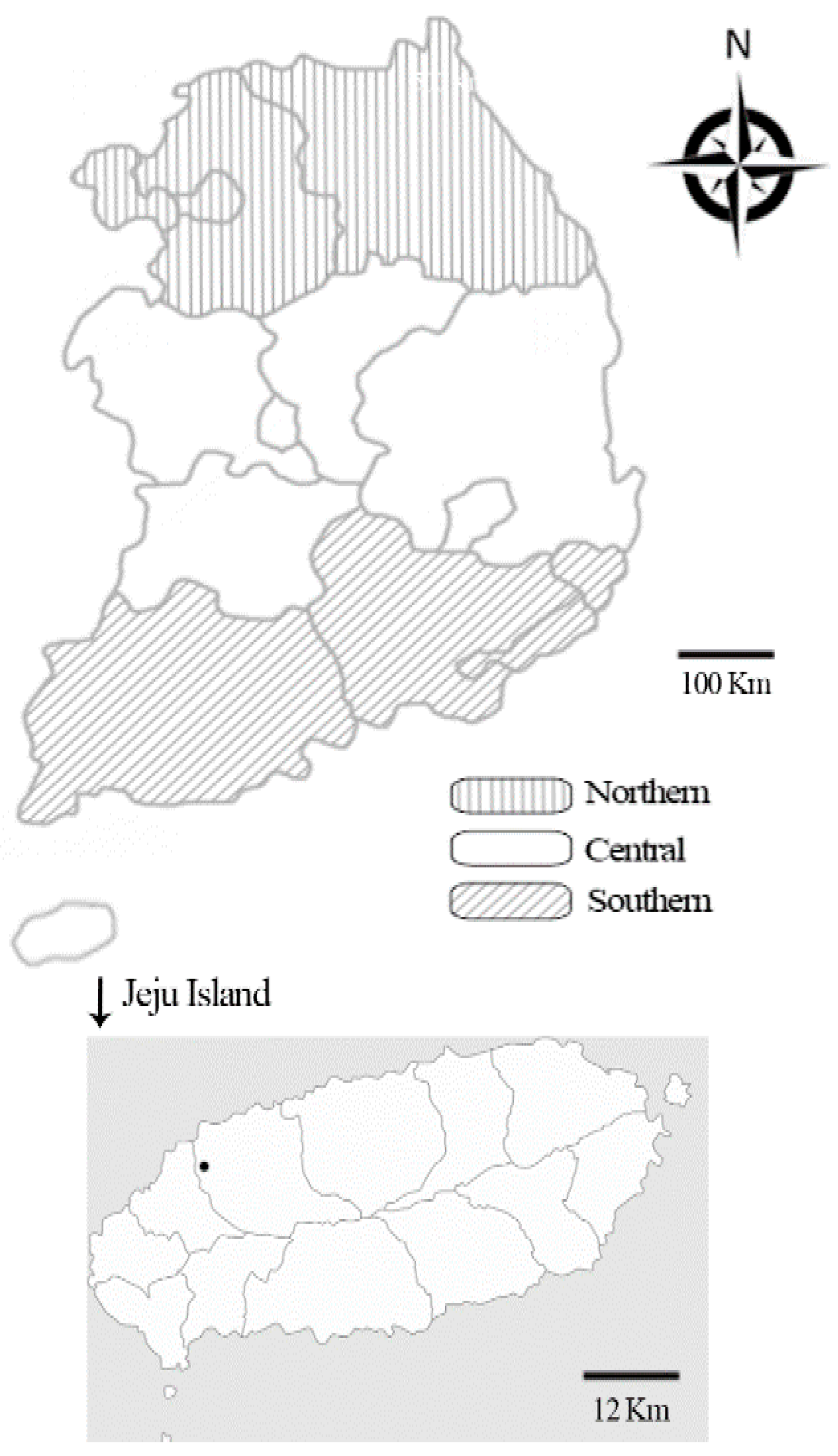
The formula indicated that the statistical power required a minimum of 138 samples. In this study, we collected 1696 samples (1433 blood and 263 lung tissue samples) from horses across the country, including Jeju Island, between 2017 and 2019 (Figure 4). Whole blood samples were collected from horse farms nationwide, whereas lung tissue samples were randomly collected from horse abattoirs on Jeju Island. Horse meat is a popular delicacy on Jeju Island and raw horse meat is regularly consumed here. Data on age, sex, location, activity, and breed were recorded for each blood sample; no additional data were recorded for the lung tissue samples.

4.3. DNA Extraction and PCR

Genomic DNA was extracted from the whole blood and lung tissue samples using a DNeasy Blood & Tissue Kit (Qiagen, Melbourne, Australia) following the manufacturer’s instructions. The extracted DNA was kept at −20 °C before use. PCR amplification was carried out using an AccuPower HotStart PCR Premix Kit (Bioneer, Daejeon, Republic of Korea). First, infection with Anaplasma spp. was screened via the amplification of 16S rRNA fragments using nPCR with the primer pairs EE1/EE2 and EE3/EE4 to obtain an expected amplicon of 924–926 bp in length [8,9]. For species identification, the 16S rRNA genes of A. phagocytophilum and A. bovis were identified by re-amplifying the PCR-positive samples using the primer pairs EE1/EE2 and SSAP2f/SSAP2r and EE1/EE2 and AB1f/AB1r, respectively [9], to obtain the expected amplicons of 641 and 551 bp in length, respectively. The positive control for each PCR reaction comprised the A. phagocytophilum [11] and A. bovis [12] strains that were previously identified in horses from mainland Korea. For each PCR reaction, a sample with distilled water and PCR reagents but no DNA was used as the negative control.
We addressed concerns regarding the amplicon contamination of nPCR using a distinct positive control sequence to ensure the differentiation of true positive results from those caused by possible contamination. The HKG of the 18S rRNA expressed with high stability in horse tissue and cultured cells [19] was used as an internal positive control. To identify the 18S rRNA HKG, horse DNA samples were amplified using previously published primer sequences to obtain the expected 204 bp long amplicons [19]. A horse blood sample infected with C. burnetii [43] was used as the internal negative control using the primer sets EE1/EE2 and SSAP2f/SSAP2r and EE1/EE2 and AB1f/AB1r.
All primers and amplification conditions used in the present study are presented in Supplementary Table S1.

4.4. RFLP

A. phagocytophilum and A. bovis were identified by digesting the 16S rRNA nPCR products of 868–870 bp in length (the PCR amplicon of 924–926 bp without primer sequences) using two restriction enzymes for the RFLP assay [9]. The restriction enzymes AleI and BtgZI were used for the RFLP assay conducted using the CLC Main Workbench 6.7.2 (CLC Bio, Qiagen, Aarhus, Denmark). The solution subjected restriction digestion comprised 10 μL of PCR product, 5 μL of buffer (10×, 1 μL of AleI (10,000 U/mL; New England Biolabs, Hitchin, UK) or 2 μL of BtgZI (5000 U/mL; New England Biolabs), and distilled water to obtain a final volume of 50 μL. For BtgZI or AleI, the reactions were incubated for 1 h at 60 °C or 30 °C, respectively. The restricted fragments were separated through electrophoresis on a 3% agarose gel in TAE solution at 100 V for 60 min. The gel was then stained with ethidium bromide and subjected to UV visualization.

4.5. DNA Cloning

The QIAquick Gel Extraction Kit (Qiagen) was used to purify the PCR products from the positive reactions produced using the 16S rRNA primers EE3/EE4. Using the pGEM-T Easy vector (Promega, Madison, WI, USA), the purified products were ligated as per the manufacturer’s recommendations. Competent Escherichia coli DH5α cells were transformed from the ligation product and were incubated at 37 °C overnight. As directed by the manufacturer, plasmid DNA was extracted using a plasmid miniprep kit (Qiagen).

4.6. DNA Sequencing and Phylogenetic Analysis

A few recombinant clones were chosen and delivered to Macrogen (Seoul, Republic of Korea) for sequencing. CLUSTAL Omega (v. 1.2.1, http://www.clustal.org/omega/, accessed on 1 September 2022) was used to align and evaluate the sequences; then, the alignment was modified using BioEdit (v. 7.2.5, http://www.mbio.ncsu.edu/BioEdit/bioedit.html, accessed on 1 September 2022). Phylogenetic analysis was conducted using MEGA (v. 6.0, https://megasoftware.net, accessed on 1 September 2022) through the maximum likelihood technique. The aligned sequences were examined by developing a similarity matrix, and a bootstrap approach with 1,000 repeats was used to determine the stability of the trees.

4.7. Statistical Analysis

Pearson’s chi-squared test was used to examine the significant differences between the groups. A p-value of <0.05 was considered as statistically significant. All statistical calculations were performed using the statistical analysis program GraphPad Prism (v. 5.04; GraphPad Soft-ware Inc., La Jolla, CA, USA). All estimations were given a 95% CI.

Supplementary Materials

The following is available online at https://www.mdpi.com/article/10.3390/ijms24043239/s1.

Author Contributions

Conceptualization, M.-G.S. and I.-O.O.; validation, M.-G.S. and D.K.; formal analysis, M.-G.S. and I.-O.O.; writing—original draft preparation, M.-G.S.; writing—review and editing, I.-O.O. and D.K.; supervision, D.K.; funding acquisition, D.K. All authors have read and agreed to the published version of the manuscript.

Funding

This research was supported by a grant (Grant No. NRF-2016R1D1A1B02015366) from the Basic Science Research Program through the National Research Foundation, Republic of Korea (NRF).

Institutional Review Board Statement

Not applicable.

Informed Consent Statement

Not applicable.

Data Availability Statement

Data supporting the conclusions of this article are included within the article. The newly generated sequences were submitted to the GenBank database under the accession numbers MT800789-93, MK028571-74, MN650821, and MK811372-75. The datasets used and/or analyzed during the present study are available from the corresponding author upon reasonable request.

Conflicts of Interest

The authors declare no conflict of interest.

References

  1. Dumler, J.S.; Barbet, A.F.; Bekker, C.P.; Dasch, G.A.; Palmer, G.H.; Ray, S.C.; Rikihisa, Y.; Rurangirwa, F.R. Reorganization of genera in the families Rickettsiaceae and Anaplasmataceae in the order Rickettsiales: Unification of some species of Ehrlichia with Anaplasma, Cowdria with Ehrlichia and Ehrlichia with Neorickettsia, descriptions of six new species combinations and designation of Ehrlichia equi and ‘HGE agent’ as subjective synonyms of Ehrlichia phagocytophila. Int. J. Syst. Evol. Microbiol. 2001, 51, 2145–2165. [Google Scholar] [CrossRef] [PubMed]
  2. Madigan, J.E.; Pusterla, N. Ehrlichial diseases. Vet. Clin. N. Am. Equine Pract. 2000, 16, 487–499. [Google Scholar] [CrossRef]
  3. Seo, M.G.; Kwon, O.D.; Kwak, D. Molecular detection and phylogenetic analysis of canine tick-borne pathogens from Korea. Ticks Tick Borne Dis. 2020, 11, 101357. [Google Scholar] [CrossRef] [PubMed]
  4. Oh, J.; Moon, B.-C.; Bae, B.; Shin, E.H.; Ko, Y.-H.; Kim, Y.-J.; Park, Y.; Chae, J.-S. Genetic identification and phylogenetic analysis of Anaplasma and Ehrlichia species in Haemaphysalis longicornis collected from Jeju Island, Korea. J. Bacteriol. Virol. 2009, 39, 257–267. [Google Scholar] [CrossRef]
  5. Seo, M.G.; Kwon, O.D.; Kwak, D. Molecular detection of Rickettsia raoultii, Rickettsia tamurae, and associated pathogens from ticks parasitizing water deer (Hydropotes inermis argyropus) in South Korea. Ticks Tick Borne Dis. 2021, 12, 101712. [Google Scholar] [CrossRef] [PubMed]
  6. Seo, M.G.; Kwon, O.D.; Kwak, D. Genotypic analysis of piroplasms and associated pathogens from ticks infesting cattle in Korea. Microorganisms 2020, 8, 728. [Google Scholar] [CrossRef]
  7. Park, J.; Han, D.G.; Ryu, J.H.; Chae, J.B.; Chae, J.S.; Yu, D.H.; Park, B.K.; Kim, H.C.; Choi, K.S. Molecular detection of Anaplasma bovis in Holstein cattle in the Republic of Korea. Acta Vet. Scand. 2018, 60, 15. [Google Scholar] [CrossRef]
  8. Seo, M.G.; Ouh, I.O.; Kwon, O.D.; Kwak, D. Molecular detection of Anaplasma phagocytophilum-like Anaplasma spp. and pathogenic A. Phagocytophilum in cattle from South Korea. Mol. Phylogenet. Evol. 2018, 126, 23–30. [Google Scholar] [CrossRef]
  9. Seo, M.G.; Ouh, I.O.; Lee, H.; Geraldino, P.J.L.; Rhee, M.H.; Kwon, O.D.; Kwak, D. Differential identification of Anaplasma in cattle and potential of cattle to serve as reservoirs of Anaplasma capra, an emerging tick-borne zoonotic pathogen. Vet. Microbiol. 2018, 226, 15–22. [Google Scholar] [CrossRef]
  10. Lee, M.; Seo, M.G.; Lee, S.H.; Ouh, I.O.; Kim, Y.H.; Kim, J.K.; Goo, Y.K.; Rhee, M.H.; Kim, T.H.; Kwon, O.D.; et al. Molecular detection and phylogenetic analysis of tick-borne pathogens in wild Korean water deer and farmed elk in Gyeongbuk and Gangwon Provinces of Korea. J. Vet. Med. Sci. 2018, 80, 1473–1478. [Google Scholar] [CrossRef] [Green Version]
  11. Seo, M.G.; Ouh, I.O.; Choi, E.; Kwon, O.D.; Kwak, D. Molecular Detection and Phylogenetic Analysis of Anaplasma phagocytophilum in Horses in Korea. Korean J. Parasitol. 2018, 56, 559–565. [Google Scholar] [CrossRef]
  12. Seo, M.G.; Kwon, O.D.; Kwak, D. Anaplasma bovis infection in a horse: First clinical report and molecular analysis. Vet. Microbiol. 2019, 233, 47–51. [Google Scholar] [CrossRef] [PubMed]
  13. Ministry of Agriculture, Food and Rural Affairs, South Korea (MAFRA). Report of “A Fact Finding Survey of Horse Industry in South Korea during 2017”; Ministry of Agriculture, Food and Rural Affairs: Sejong-Si, Republic of Korea, 2018.
  14. Silaghi, C.; Santos, A.S.; Gomes, J.; Christova, I.; Matei, I.A.; Walder, G.; Domingos, A.; Bell-Sakyi, L.; Sprong, H.; von Loewenich, F.D.; et al. Guidelines for the direct detection of Anaplasma spp. in diagnosis and epidemiological studies. Vector Borne Zoonot. Dis. 2017, 17, 12–22. [Google Scholar] [CrossRef] [PubMed]
  15. Lepidi, H.; Bunnell, J.E.; Martin, M.E.; Madigan, J.E.; Stuen, S.; Dumler, J.S. Comparative pathology, and immunohistology associated with clinical illness after Ehrlichia phagocytophila-group infections. Am. J. Trop. Med. Hyg. 2000, 62, 29–37. [Google Scholar] [CrossRef] [PubMed]
  16. Hoar, B.R.; Nieto, N.C.; Rhodes, D.M.; Foley, J.E. Evaluation of sequential coinfection with Anaplasma phagocytophilum and Anaplasma marginale in cattle. Am. J. Vet. Res. 2008, 69, 1171–1178. [Google Scholar] [CrossRef]
  17. Wamsley, H.L.; Alleman, A.R.; Johnson, C.M.; Barbet, A.F.; Abbott, J.R. Investigation of endothelial cells as an in vivo nidus of Anaplasma marginale infection in cattle. Vet. Microbiol. 2011, 153, 264–273. [Google Scholar] [CrossRef]
  18. Härtwig, V.; von Loewenich, F.D.; Schulze, C.; Straubinger, R.K.; Daugschies, A.; Dyachenko, V. Detection of Anaplasma phagocytophilum in red foxes (Vulpes vulpes) and raccoon dogs (Nyctereutes procyonoides) from Brandenburg, Germany. Ticks Tick Borne Dis. 2014, 5, 277–280. [Google Scholar] [CrossRef]
  19. Zhang, Y.W.; Davis, E.G.; Bai, J. Determination of internal control for gene expression studies in equine tissues and cell culture using quantitative RT-PCR. Vet. Immunol. Immunopathol. 2009, 130, 114–119. [Google Scholar] [CrossRef]
  20. Brito, A.B.; Lima, J.S.; Brito, D.C.; Santana, L.N.; Costa, N.N.; Miranda, M.S.; Ohashi, O.M.; Santos, R.R.; Domingues, S.F. Validation of reference genes for ovarian tissue from capuchin monkeys (Cebus apella). Zygote 2013, 21, 167–171. [Google Scholar] [CrossRef]
  21. Huggett, J.; Dheda, K.; Bustin, S.; Zumla, A. Real-time RT-PCR normalisation; strategies and considerations. Genes Immun. 2005, 6, 279–284. [Google Scholar] [CrossRef] [Green Version]
  22. Figueiredo, M.D.; Salter, C.E.; Andrietti, A.L.; Vandenplas, M.L.; Hurley, D.J.; Moore, J.N. Validation of a reliable set of primer pairs for measuring gene expression by real-time quantitative RT-PCR in equine leukocytes. Vet. Immunol. Immunopathol. 2009, 131, 65–72. [Google Scholar] [CrossRef] [PubMed]
  23. Stuen, S.; Granquist, E.G.; Silaghi, C. Anaplasma phagocytophilum—A widespread multi-host pathogen with highly adaptive strategies. Front. Cell Infect. Microbiol. 2013, 3, 31. [Google Scholar] [CrossRef] [PubMed]
  24. Dong, X.; Chen, X.P.; Liu, N.; Dumler, S.J.; Zhang, Y.Z. Co-circulation of multiple species of Rickettsiales bacteria in one single species of hard ticks in Shenyang, China. Ticks Tick Borne Dis. 2014, 5, 727–733. [Google Scholar] [CrossRef]
  25. Parola, P.; Paddock, C.D.; Raoult, D. Tick-borne rickettsioses around the world: Emerging diseases challenging old concepts. Clin. Microbiol. Rev. 2005, 18, 719–756. [Google Scholar] [CrossRef]
  26. Hwang, J.; Oh, D.H.; Lee, H.; Chun, M.S. Anaplasma sp. and hemoplasma infection in leopard cats (Prionailurus bengalensis euptilurus) from Korea. J. Vet. Sci. 2015, 16, 385–388. [Google Scholar] [CrossRef] [PubMed]
  27. Ybañez, A.P.; Sashika, M.; Inokuma, H. The phylogenetic position of Anaplasma bovis and inferences on the phylogeny of the genus Anaplasma. J. Vet. Med. Sci. 2014, 76, 307–312. [Google Scholar] [CrossRef] [PubMed]
  28. Masuzawa, T.; Uchishima, Y.; Fukui, T.; Okamoto, Y.; Pan, M.J.; Kadosaka, T.; Takada, N. Detection of Anaplasma phagocytophilum and Anaplasma bovis in small wild mammals from Taichung and Kinmen Island, Taiwan. Jpn. J. Infect. Dis. 2014, 67, 111–114. [Google Scholar] [CrossRef] [PubMed]
  29. Sashika, M.; Abe, G.; Matsumoto, K.; Inokuma, H. Molecular survey of Anaplasma and Ehrlichia infections of feral raccoons (Procyon lotor) in Hokkaido, Japan. Vector Borne Zoonot. Dis. 2011, 11, 349–354. [Google Scholar] [CrossRef]
  30. Lu, M.; Chen, Q.; Qin, X.; Lyu, Y.; Teng, Z.; Li, K.; Yu, L.; Jin, X.; Chang, H.; Wang, W.; et al. Anaplasma bovis infection in fever and thrombocytopenia patients—Anhui Province, China, 2021. China CDC Wkly. 2022, 4, 249–253. [Google Scholar] [CrossRef]
  31. Doan, H.T.; Noh, J.H.; Choe, S.E.; Yoo, M.S.; Kim, Y.H.; Reddy, K.E.; Quyen, D.V.; Nguyen, L.T.; Nguyen, T.T.; Kweon, C.H.; et al. Molecular detection and phylogenetic analysis of Anaplasma bovis from Haemaphysalis longicornis feeding on grazing cattle in Korea. Vet. Parasitol. 2013, 196, 478–481. [Google Scholar] [CrossRef]
  32. Seong, G.; Han, Y.J.; Chae, J.B.; Chae, J.S.; Yu, D.H.; Lee, Y.S.; Park, J.; Park, B.K.; Yoo, J.G.; Choi, K.S. Detection of Anaplasma sp. in Korean native goats (Capra aegagrus hircus) on Jeju Island, Korea. Korean J. Parasitol. 2015, 53, 765–769. [Google Scholar] [CrossRef] [PubMed]
  33. Liu, Z.; Ma, M.; Wang, Z.; Wang, J.; Peng, Y.; Li, Y.; Guan, G.; Luo, J.; Yin, H. Molecular survey and genetic identification of Anaplasma species in goats from central and southern China. Appl. Environ. Microbiol. 2012, 78, 464–470. [Google Scholar] [CrossRef] [PubMed]
  34. Belkahia, H.; Ben Said, M.; Alberti, A.; Abdi, K.; Issaoui, Z.; Hattab, D.; Gharbi, M.; Messadi, L. First molecular survey and novel genetic variants’ identification of Anaplasma marginale, A. centrale and A. bovis in cattle from Tunisia. Infect. Genet. Evol. 2015, 34, 361–371. [Google Scholar] [CrossRef] [PubMed]
  35. Stuen, S.; Van De Pol, I.; Bergström, K.; Schouls, L.M. Identification of Anaplasma phagocytophila (formerly Ehrlichia phagocytophila) variants in blood from sheep in Norway. J. Clin. Microbiol. 2002, 40, 3192–3197. [Google Scholar] [CrossRef]
  36. Stuen, S.; Casey, A.N.; Woldehiwet, Z.; French, N.P.; Ogden, N.H. Detection by the polymerase chain reaction of Anaplasma phagocytophilum in tissues of persistently infected sheep. J. Comp. Pathol. 2006, 134, 101–104. [Google Scholar] [CrossRef]
  37. Kocan, K.M.; de la Fuente, J.; Blouin, E.F.; Coetzee, J.F.; Ewing, S.A. The natural history of Anaplasma marginale. Vet. Parasitol. 2010, 167, 95–107. [Google Scholar] [CrossRef]
  38. Remy, V.; Hansmann, Y.; De Martino, S.; Christmann, D.; Brouqui, P. Human anaplasmosis presenting as atypical pneumonitis in France. Clin. Infect. Dis. 2003, 37, 846–848. [Google Scholar] [CrossRef]
  39. Kang, J.G.; Ko, S.; Smith, W.B.; Kim, H.C.; Lee, I.Y.; Chae, J.S. Prevalence of Anaplasma, Bartonella and Borrelia Species in Haemaphysalis longicornis collected from goats in North Korea. J. Vet. Sci. 2016, 17, 207–216. [Google Scholar] [CrossRef]
  40. Li, H.; Zheng, Y.C.; Ma, L.; Jia, N.; Jiang, B.G.; Jiang, R.R.; Huo, Q.B.; Wang, Y.W.; Liu, H.B.; Chu, Y.L.; et al. Human infection with a novel tick-borne Anaplasma species in China: A surveillance study. Lancet Infect. Dis. 2015, 15, 663–670. [Google Scholar] [CrossRef]
  41. Chochlakis, D.; Ioannou, I.; Tselentis, Y.; Psaroulaki, A. Human anaplasmosis and Anaplasma ovis variant. Emerg. Infect. Dis. 2010, 16, 1031–1032. [Google Scholar] [CrossRef]
  42. Thrusfield, M.V.; Christley, R. Veterinary Epidemiology, 4th ed.; Wiley: Hoboken, NJ, USA, 2018. [Google Scholar]
  43. Seo, M.G.; Lee, S.H.; VanBik, D.; Ouh, I.O.; Yun, S.H.; Choi, E.; Park, Y.S.; Lee, S.E.; Kim, J.W.; Cho, G.J.; et al. Detection and genotyping of Coxiella burnetii and Coxiella-like bacteria in horses in South Korea. PLoS ONE 2016, 11, e0156710. [Google Scholar] [CrossRef] [PubMed] [Green Version]
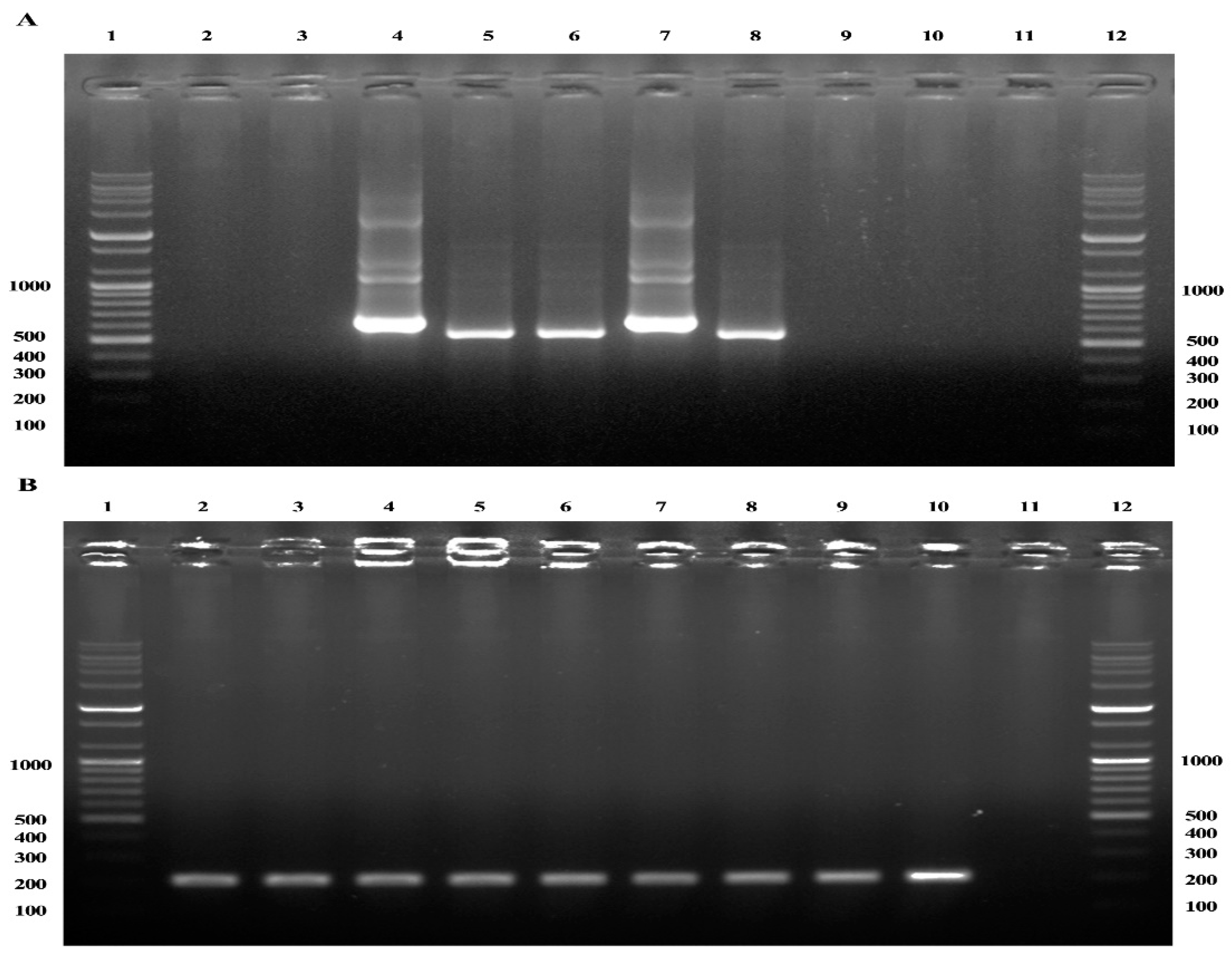
Figure 1. PCR detection of Anaplasma 16S rRNA and horse housekeeping gene (HKG) 18S rRNA in horse blood and lung tissue samples. (A) Nested PCR was used to identify the 16S rRNA genes of A. phagocytophilum and A. bovis using the primer sets EE1/EE2 and SSAP2f/SSAP2r and EE1/EE2 and AB1f/AB1r, respectively. Lanes 1 and 12: 100 bp ladder; lanes 2 and 3: horse blood and lung tissue samples negative for Anaplasma spp. using the primers EE1/EE2 and SSAP2f/SSAP2r or AB1f/AB1r, respectively; lane 4: A. phagocytophilum PCR product (641 bp) from horse lung sample; lane 5: A. bovis PCR product (551 bp) from horse blood samples; lane 6: A. bovis PCR product (551 bp) from horse lung tissue samples; lane 7: PCR product (641 bp) of A. phagocytophilum previously detected in a horse (positive control); lane 8: PCR product (551 bp) of A. bovis previously detected in a horse (positive control); lanes 9 and 10: internal negative control samples of Coxiella burnetii previously detected in a horse using the primer sets EE1/EE2 and SSAP2f/SSAP2r and EE1/EE2 and AB1f/AB1r, respectively; lane 11: lack of an amplicon from horse DNA generated using primers EE1/EE2 and SSAP2f/SSAP2r/AB1f/AB1r. (B) Single-round PCR detection using primers specific to horse HKG 18S rRNA. Lanes are in the same order as described in (A) but previously published primer sequences were used, producing expected amplicons of 204 bp [19].
Ijms 24 03239 g001
Figure 2. Restriction fragment length polymorphism (RFLP) assay and Anaplasma species identification. (A) RFLP analysis using AleI enzyme for PCR products. Lanes 1 and 13: 100 bp ladder; lanes 2 and 3: A. bovis PCR products from horse blood samples before (924 bp) and after (660 and 264 bp) AleI restriction digestion; lanes 4 and 5: A. bovis PCR products from horse lung tissue samples before (924 bp) and after (660 and 264 bp) AleI restriction digestion; lanes 6 and 7: A. phagocytophilum PCR products from horse lung tissue samples before (926 bp) and after (926 bp) AleI restriction digestion; lanes 8 and 9: PCR products of A. bovis detected in horse (positive control) before (924 bp) and after (660 and 264 bp) AleI restriction digestion; lanes 10 and 11: PCR products of A. phagocytophilum detected in horse (positive control) before (926 bp) and after (926 bp) AleI restriction digestion; lane 12: negative control. (B) RFLP analysis using the BtgZI enzyme for PCR products. Lanes 1 and 13: 100 bp ladder; lanes 2 and 3: A. bovis PCR products from horse blood samples before (924 bp) and after (924 bp) BtgZI restriction digestion; lanes 4 and 5: A. bovis PCR products from horse lung tissue samples before (924 bp) and after (924 bp) BtgZI restriction digestion; lanes 6 and 7: A. phagocytophilum PCR products from horse lung tissue samples before (926 bp) and after (707 and 223 bp) BtgZI restriction digestion; lanes 8 and 9: PCR products of A. bovis detected in horse (positive control) before (924 bp) and after (924 bp) BtgZI restriction digestion; lanes 10 and 11: PCR products of A. phagocytophilum detected in horse (positive control) before (926 bp) and after (707 and 223 bp) BtgZI restriction digestion; lane 12: negative control.
Ijms 24 03239 g002
Figure 3. Phylogenetic tree created using the maximum likelihood technique with 16S rRNA nucleotide sequences from Anaplasma. The sequences analyzed in this study are marked with arrows. The GenBank accession numbers of the additional sequences are listed adjacent to their respective sequence names. Branch numbers indicate bootstrap support (1000 replicates). The scale bar represents the phylogenetic distance between the sequences. A. bovis clusters are denoted using “A” and “B.” A. phagocytophilum-like Anaplasma spp. clusters are denoted using “a” and “b.”
Ijms 24 03239 g003
Figure 4. Map of the Republic of Korea. Horse blood samples were collected throughout the nation; however, lung tissue samples were only collected from Jeju Island. The dot denotes the location of the abattoir on Jeju Island from which the horse lung samples were collected. The regions of sample collection were divided into four groups: northern, central, southern, and Jeju Island.
Ijms 24 03239 g004
Table 1. Prevalence of Anaplasma bovis and A. phagocytophilum as determined by the analysis of 16S rRNA genes in horse samples from the Republic of Korea between 2017 and 2019.
SampleGroupNo. TestedA. bovisA. phagocytophilum
No. Positive (%)95% CI p-ValueNo. Positive (%)95% CI p-Value
BloodSexFemale7562 (0.3)0–0.60.60403 (0.4)0–0.80.5021
Male3540000
Castrated3231 (0.3)0–0.91 (0.3)0–0.9
Age
(year)
<5460000.3204000.3752
5–105371 (0.2)0–0.62 (0.4)0–0.9
>104362 (0.5)0–1.12 (0.5)0–1.1
BreedThoroughbred4562 (0.4)0–1.00.43243 (0.7)0–1.40.2361
Warmblood3081 (0.3)0–1.01 (0.3)0–1.0
Native Korean Pony4160000
Mixed2530000
Type of activityBroodmare5492 (0.4)0–0.90.67301 (0.2)0–0.50.2843
Leisure4521 (0.2)0–0.73 (0.7)0–1.4
Race2100000
Breeding2220000
RegionNorthern3011 (0.3)0–1.00.80161 (0.3)0–1.00.0253 *
Central2160000
Southern123002 (1.6)0–3.9
Jeju Island7932 (0.3)0–0.61 (0.1)0–0.4
Subtotal14333 (0.2)0–0.4 4 (0.3)0–0.6
Lung26326 (9.9)6.3–13.5 27 (10.3)6.6–13.9
Total169629 (1.7)1.1–2.3 31 (1.8)1.2–2.5
CI: Confidence interval. * Statistical significance was observed.
Disclaimer/Publisher’s Note: The statements, opinions and data contained in all publications are solely those of the individual author(s) and contributor(s) and not of MDPI and/or the editor(s). MDPI and/or the editor(s) disclaim responsibility for any injury to people or property resulting from any ideas, methods, instructions or products referred to in the content.