Review

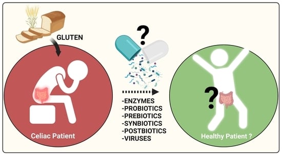
Celiac Disease and Possible Dietary Interventions: From Enzymes and Probiotics to Postbiotics and Viruses

by
Sandip K. Wagh
1,2,
Karen M. Lammers
3,
Manohar V. Padul
4,
Alfonso Rodriguez-Herrera
5 and
Veronica I. Dodero
1,*
1
Department of Organic and Bioorganic Chemistry, Bielefeld University, 33615 Bielefeld, Germany
2
Department of Biochemistry, Dr. Babasaheb Ambedkar Marathwada University, Aurangabad 431004, India
3
Tubascan Ltd., 1098 XG Amsterdam, The Netherlands
4
Department of Biochemistry, The Institute of Science, Dr. Homi Bhabha State University, Mumbai 400032, India
5
Pediatrics, Saint Luke’s Hospital, University College Dublin, R95 FY71 Kilkenny, Ireland
*
Author to whom correspondence should be addressed.
Int. J. Mol. Sci. 2022, 23(19), 11748; https://doi.org/10.3390/ijms231911748
Submission received: 10 August 2022 / Revised: 20 September 2022 / Accepted: 29 September 2022 / Published: 4 October 2022
(This article belongs to the Special Issue Pro-inflammatory Nutrients: Focus on Gliadin and Celiac Disease 2.0)

Abstract

:
Celiac Disease (CeD) is a chronic small intestinal immune-mediated enteropathy caused by the ingestion of dietary gluten proteins in genetically susceptible individuals. CeD is one of the most common autoimmune diseases, affecting around 1.4% of the population globally. To date, the only acceptable treatment for CeD is strict, lifelong adherence to a gluten-free diet (GFD). However, in some cases, GFD does not alter gluten-induced symptoms. In addition, strict adherence to a GFD reduces patients’ quality of life and is often a socio-economic burden. This narrative review offers an interdisciplinary overview of CeD pathomechanism and the limitations of GFD, focusing on current research on possible dietary interventions. It concentrates on the recent research on the degradation of gluten through enzymes, the modulation of the microbiome, and the different types of “biotics” strategies, from probiotics to the less explored “viromebiotics” as possible beneficial complementary interventions for CeD management. The final aim is to set the context for future research that may consider the role of gluten proteins and the microbiome in nutritional and non-pharmacological interventions for CeD beyond the sole use of the GFD.

Graphical Abstract

1. Introduction

1.1. Celiac Disease: Gluten Proteins and the Triggers of Disease

Celiac Disease (CeD) is a chronic inflammatory, autoimmune-mediated disease affecting the duodenum [1]. It is accepted that gluten proteins found in wheat, rye, and barley are responsible for this autoimmune enteropathy. Celiac disease is remarkably ubiquitous, with epidemiologic data now available for every continent; unfortunately, data about Asia and Africa are limited, but the condition is present. Globally, the pooled seroprevalence of celiac disease is 1.4% (95% CI 1.1–1.7%). The prevalence of biopsy-confirmed celiac disease is 0.7% (95% CI 0.5–0.9%) [2,3]. CeD could be developed at any age where women are affected more frequently than men, with a ratio of 3:1 [4,5]. The typical clinical symptoms of CeD are chronic diarrhea, anemia, osteoporosis, and intestinal cramps. If left untreated, the disease may result in complications such as intestinal lymphoma and a slightly increased risk of early death [6,7,8]. Furthermore, the coexistence of CeD with mental disorders, such as schizophrenia [9] and type 1 diabetes mellitus, has also been reported [10,11]. If unaddressed, CeD can lead to villous atrophy and malabsorption of nutrients and, consequently, can cause symptoms such as anemia and osteoporosis [12].
CeD has emerged as a model disease to decipher how the interplay between environmental and genetic factors can predispose one to autoimmunity. It is still a challenge to integrate a full explanation of the considerable variability in disease penetrance, severity, and presentation of CeD. Many authors postulate gaps in the current knowledge and missing actors in the chain of pathophysiological events. An excellent comprehensive review of CeD immunopathogenesis has been recently published by Levescot et al. [13].
The keystone event in CeD pathogenesis is the activation of a gluten-specific immune response that is driven by molecular interactions between gluten-derived peptides, as the indispensable environmental factor, the HLA-DQ2/8 locus, as the main predisposing genetic factor, and the enzyme transglutaminase 2 (tTG2), the identified CeD-specific autoantigen.
The human gastrointestinal tract lacks luminal endo-prolylpeptidases, so the proline-rich gluten proteins are incompletely digested and can progress through the intestinal lumen. As a consequence, large immunogenic peptides can interact with the enzyme transglutaminase 2, which converts neutral and polar, glutamine residues into negatively charged glutamic acids. This transformation allows the positively charged amino acids in the HLA-DQ2 or HLA-DQ8 molecules at the surface of the antigen-presenting cells to bind with higher affinity to the gluten peptides harboring negative charges. These interactions trigger an expansion of gluten-specific CD4+ T cells that produce pro-inflammatory cytokines such as interleukins and interferon-gamma (IFN γ). The epithelial damage in the intestinal mucosa requires the activation of CD8+ cytotoxic intraepithelial lymphocytes (IEL).
Gluten proteins contain proline (P) and glutamine (Q) rich proteins, and due to repetitive PQ sequences, they are difficult to degrade for mammalian digestive tract enzymes [14,15]. The wheat protein gliadin (α/β-, γ- and ω-gliadins) is the primary toxic component of gluten [16] and the most studied protein related to CeD. Like gluten, gliadin is only partially degraded by human digestive enzymes [17,18]. Gliadins form different protein aggregates under the physiologically relevant pH of 3.0 and 7.0, enabling the enzymes’ accessibility to other possible degradation positions [19,20,21]. Recently, it was demonstrated that the gliadin peptides obtained after pepsin proteolysis behave as an amyloid-like structure associated with inflammatory cytokine production and pro-apoptotic mRNA expression [22]. The gliadin peptides that resist the human digestive and intestinal enzymes interact with enterocytes, opening the tight junctions that facilitate their transport to the lamina propria [23]. The pathogenic gliadin peptides are classified as “toxic” or “immunogenic,” depending on their behavior. If they induce intestinal damage in vivo and/or in vitro, they are designed as toxic peptides, and if they activate T cells, they are considered immunogenic peptides [24,25,26]. It is hypothesized that gliadin peptides in the lamina propria interact with tTG2 [27,28], the enzyme responsible for converting glutamine residues to glutamic acid. These deamidated peptides represent antigens interacting with increased affinity with the HLA (human leukocyte antigen)-DQ2 or HLA-DQ8 molecules. These molecules are part of the MHC (major histocompatibility complex) class II. The MHC II is an antigen-presenting receptor system by which antigen-presenting cells, like dendritic cells, present antigens (for instance, deamidated gliadin peptides) to T cells to elicit an immune response and build a memory of that specific antigen. The most studied immunodominant gliadin fragment is a 33 amino acids fragments, LQLQPFPQPQLPYPQPQLPYPQ-PQLPYPQPQPF, named 33-mer [29].
The amount of 33-mer that remains after proteolysis in different wheat cultivars was quantified as 91 to 603μg g−1 in wheat flour [30]. The 33-mer sequence is responsible for the adaptive immune response in CeD because it contains six partially overlapping copies of canonical T-cell epitopes [31], three copies of the DQ2.5-glia-α1- (PF/YPQPQLPY), and the DQ2.5-glia-α2 (PQPQLPYPQ) epitope [32]. It is accepted that the 33-mer reaches the lamina propria, where dendritic cells process and present it in the context of HLA-DQ2/8 to CD4+ T lymphocytes [33,34]. In recent years, it has been shown that the 33-mer forms dynamic small oligomers and large aggregates depending only on peptide concentration with a concomitant conformational transition to the parallel beta structure, which is the signature of amyloid proteins [35,36]. In vitro, the large 33-mer oligomers can trigger a Toll-like (TLR-2 and -4) receptor-mediated innate immune response in macrophages, showing the relevance of such structures in activating inflammation [37].
On the other hand, a sequestering polymer and non-absorbable (polyhydroxy ethyl methacrylate-co-styrene sodium sulfonate (P(HEMA-co-SS)) have been used to form larger complex gliadin particles to prevent the enzymatic action from stopping CeD activation [38]. It was proposed that the polymer-sequestered gliadin peptides would be discharged from the body, inhibiting their absorption and transport to the bloodstream [39,40]. This last research highlights the role of inhibiting the entrance of gluten peptides to the host as a therapeutic strategy.
A strong focus on the microbiota has been made, considering it first as an element involved in the CeD pathogenesis and, therefore, recently considered a potential intervention. Among the proposed mechanisms is the potential role of a positive feedback loop that may amplify the activation of nearby lymphocytes (IEL) induced by a type I interferon response triggered by viruses. Another hypothetical scenario is that a pathogen or a commensal bacterium might trigger a T cell cross-reaction with gluten peptide epitopes and drive an expansion of a cross-reactive T cell repertoire with epitope spreading [41]. An excellent example was recently reported where many mimics of HLA-DQ2.5-restricted gliadin determinants were found in the commensal bacterium Pseudomonas fluorescens that activate reactive T cells isolated from CeD patients. This report is major proof of the concept that a molecular mimicry mechanism may trigger CeD. Recently, high sequence similarity was found between 33-mer gliadin peptide and pathogen-derived proteins, e.g., extracellular proteins from Streptococcus pneumoniae and Granulicatella sp., by stringent BLASTp search [42]. Since Granulicatella sp. is found in the gut and has been reported in CeD patients, it would be interesting to investigate the role of this pathogen in the development of CeD by molecular mimicry mechanisms. Beyond T-cell activation, it has been hypothesized that gluten proteins have functional similarities with non-replicative pathogens such as prions [43]. It was also hypothesized that since gluten peptides share structural/morphological similarities with pathogens, they possess latent pathogenicity. Therefore, although initially innocuous to the host, their accumulation and oligomerization with the conformational transition toward amyloid-type structures could trigger their recognition by the host’s innate immunity [42].
Several viruses have been involved in CeD pathogenesis: the adenovirus 12 E1A, Enterovirus, Hepatitis C virus, and Rotavirus. However, their role was later dismissed, it has been described, for example, that the risk of CeD was increased in a cohort of children who combined being Enterovirus positive with a high gluten intake, indicating a cumulative effect of these two factors in the development of disease in genetically at-risk children [44]. Recently, Reovirus, which causes mostly asymptomatic infections, is the last virus to be suggested to have a role in CeD pathogenesis. Reovirus would break the tolerance to protein antigens [45]. The underlying pathogenetic mechanisms of early-life infections and CeD are not described but could provide new insights into the prevention of celiac disease.

1.2. Is a Gluten-Free Diet Enough for CeD Treatment?

Currently, the only treatment available for CeD is the strict elimination of gluten from the diet. This so-called gluten-free diet (GFD) usually results in the alleviation of symptoms and the improvement of small-bowel mucosal damage [46]. Several antibodies based on ELISAs are available to check for gluten traces in food products. R5 and G12 antibody-based ELISAs are frequently used to detect the threshold quantity of gluten (>20 mg/kg). The R5 monoclonal antibody (mAb) strongly recognizes the most toxic fragments of gliadin as QQPFP, QQQFP, LQPFP, and QLPFP sequences [47]. The G12 is a highly sensitive mAb antibody against the α2-gliadin 33-mer toxic peptide of the gliadin [48]. It has been proven to be efficient in measuring prolamin concentrations in native and partially hydrolyzed cereals. When both were compared, they showed no significant difference in the mean gluten concentration detected [6]. Each one offers different limitations depending on the food matrix analyzed.
Gluten exposure damages the intestinal epithelium, causing atrophy of the villous, which subsequently induces poor digestion and absorption of a range of macro and micronutrients. The classic picture of CeD was of skinny children with diarrhea. The current picture includes patients who are obese or show persistent constipation. Silvester et al. have compiled some myths and fallacies about the GFD. We know now that not all patients show the same level of response to a GFD. The poor response may happen because of hidden gluten exposure or coexisting conditions such as small intestinal bacterial overgrowth (SIBO) or autoimmune enteropathy. Other factors associated with symptomatic persistent villous atrophy include age > 70 years old and the use of proton-pump inhibitors (PPI), non-steroidal anti-inflammatory drugs, or selective serotonin reuptake inhibitors. Moreover, mucosal recovery on a GFD is not universal among those responding clinically. It was published that only one-third of adults have normal villi (healed and healthy intestine) after two years on a GFD. Only 2/3 will get it after five years on a GFD [49]. Of note, the data of some studies point to a constitutive defect of the intestinal barrier function. Diosdado et al. found an increased expression of innate and adaptive immune genes and markers of increased neutrophil activity in biopsies from CeD patients (active and in-remission) versus non-celiac individuals. Neutrophil recruitment was also visualized by immunohistochemistry. Although they observed a gradual change in immune gene expression after implementing a GFD until complete normalization, increased neutrophil recruitment was observed in both active and remission CeD patients. This result led the authors to conclude that despite clinical, histologic, and serologic normalization in remission, there was an ongoing activated innate immunity, and a link was proposed between the ongoing neutrophil recruitment and impaired barrier function [50]. Recently, [51] in cultured organoids from CeD patients, it wasfound various molecular markers of increased permeability, particularly decreased claudin-18, ZO-1, mucin components, trefoil factors, and an increased expression of claudin-2. In addition, they determined that there was a higher permeability at baseline while gluten was absent.
Eliminating all dietary gluten may be considered more of an aspirational goal than a fact, as this is difficult to attain even for highly motivated patients [52]. The GFD adherence rates are generally above 80–90% [53]. However, due to the wide use of wheat in most food ingredients, gluten can be difficult to avoid, resulting in accidental gluten exposure [54], as summarized in Table 1. These difficulties of the GFD were highlighted by the Determination of Gluten Grams Ingested and Excreted By Adults Eating Gluten-free (DOGGIEBAG) study. It involved 18 adults with biopsy-confirmed celiac disease who were on a GFD for 24 months and collected food (25% portions in a “doggie bag”), urine, and stool samples over 10 days. Although non-intentional gluten intake was reported, two-thirds had at least one sample that tested positive for immunogenic gluten peptides. This can be a reason that following a GFD, up to 30% of patients have persistent problems [46], and the therapeutic small-bowel villous atrophy is reported in 60% of patients, and the enteropathy may even persist [9,55].
A GFD has been reported to be more burdensome than treatments for type 1 diabetes, irritable bowel syndrome (IBS), and congestive heart failure [49]. A life-long strict GFD is a challenging task for the elderly, people with reading or language barriers, those with psychological impairment, and those with limited financial means. Dining out, social events, or using a school or professional canteen become real-life obstacles daily [56].
Surveys suggest that most CeD patients would be interested in medical therapy, not based on diet alone [57]. The goal of these future therapies will not be to act as adjuncts to a GFD for people with non-responsive CeD or refractory CeD but aim to allow those patients to consume gluten-without harm. Different interventions and pharmacological treatments for CeD have been developed in recent years. The comprehensive review completed by Segura et al. focuses on phase III and II clinical trials; it includes the developments in the degradation of gluten in the intestinal lumen, regulation of the immune response, modulation of intestinal permeability, and induction of immunological tolerance [58]. If there is interest in knowing about the non-dietary strategies for CeD, please refer to recent reviews [58,59]. Developing suitable, safe drugs for autoimmune disorders is complicated, and CeD is no exception. Since certain microorganisms can degrade gluten, creating nutritional supplements such as prolyl-endopeptidases (PEPs) seems to be an easy dietary intervention. Although, until now, there have been no such efficient products, it constitutes the mainstream strategy for nutritional interventions in CeD. Herein, we are interested in the emerging dietary approaches based on PEPs and extending them to modulating/shaping the patients’ microbiota with their potential benefit for the management and/or prevention of CeD. The selection of examples was based, in some cases, on historical relevance and, for others, was based on novelty to provide a generalist overview and promote new areas of research at the interface of the disciplines.

2. Dietary Interventions for Complementing the GFD and Beyond

2.1. Enzymes as a Nutritional Supplement Therapy for CeD

Several proteases and peptidases have been proven to degrade gluten in vitro and/or in vivo [60,61]. As aforementioned, mammalian gastrointestinal proteases partially digested immunogenic gluten sequences [33,62]. Therefore, the detoxification of gluten can theoretically be achieved by proteolytic fragmentation by oral enzymatic therapy. The idea is to inactivate gluten peptides in the human gastrointestinal tract by peptidase supplementation, thereby minimizing the amount of gluten peptides reaching the small intestine. The gluten-hydrolyzing enzymes produced by the Rothia mucilaginosa were have been identified as two structurally closely related subtilisins [63]. Previously, some of us reported the significant hydrolysis of wheat gliadin by Peptidase S9, isolated from the B. tequilensis strain [64]. Several gluten-detoxifying peptidases have been isolated from probiotic preparations involving lactobacilli [65,66], other microorganisms [63,67], and germinating cereals [68].
The withdrawal or modification of celiac peptides during food processing using enzymes is already commercialized. For example, a dietary supplement based on Aspergillus niger-Prolyl endopeptidase (PEP) can degrade gluten at a particular stage. However, it is not currently a treatment for CeD because it does not entirely break down gluten, and the resulting accumulation of gluten peptides in the duodenum has not been determined [69]. Another commercialized product is based on caricain, a proteolytic enzyme obtained from the papaya plant and papain. Previous studies have reported that caricain has the potential specificity to target gluten amino acid sequence and helps reduce gluten concentration during food processing [70,71]. However, to date, all the commercialized enzymatic cocktails are not prescribed for CeD patients.
Other prolyl endopeptidases (PEPs) isolated from Myxococcus xanthus and Flavobacterium meningosepticum showed the ability to hydrolyze toxic gliadin peptides significantly. However, the presence of immunopeptides has not been determined [72,73,74,75]. PEPs from Sphingomonas capsulate, showed complete hydrolysis of immunogenic gluten peptides after mixing with barley cysteine endoproteases [17,76]. Another interesting PEP is latiglutenase, in which experiments in subjects receiving 900 mg of latiglutenase led to improvements (p-values) in the severity of symptoms relative to placebo-dosed subjects for week 12. The reduction in symptoms trended higher for more symptomatic patients [77]. However, previous randomized phase 2 trials were conducted with latiglutenase (IMGX003, formerly ALV003) (ClinicalTrials.gov, NCT03585478), and they reported contradictory findings regarding its effect on villous atrophy and clinical symptoms, showing only 88% gluten hydrolysis efficiency [78,79]. Nowadays, a phase I clinical study is being conducted to evaluate the bacterial endopeptidase TAK-062 that simultaneously targets proline and glutamine peptide motifs in the stomach (ClinicalTrials.gov Identifier: NCT05353985). TAK-062 is a second generation of the engineered endopeptidase kuma030 [80]. When healthy individuals ingested TAK-062 before a complex meal containing 1–6 g gluten, it was observed that after 20–65 min post-TAK-062 ingestion, 97–99% of the gluten was degraded as a measure in aspirate samples from the stomach [81]. The calculated remaining gluten showed a median amount of up to 38 mg. To our knowledge, this is the first glutenase that showed this high gliadin hydrolysis efficiency in vivo. This has a potential clinical relevance since amounts as low as 10 mg of gluten may be able to trigger the immunological cascade [82,83]. Yet, these data showed the high efficiency of TAK-062, and further studies in CeD patients are in progress to test the efficiency of Tak-062 in degrading inadvertent gluten exposure (ClinicalTrials.gov Identifier: NCT05353985).

2.2. Human Microbiota and Dysbiosis in CeD

During the co-evolution of humans and microbes, thousands of bacterial species have colonized the human body. The vast amount of microbial presence in the host’s body is termed “normal flora,” “microbiota,” or “microflora” [84,85,86]. The microflora consists of bacteria accompanied by fungi, archaea, viruses, and protozoans [87,88,89]. This colonization occurs at birth, covering every human body surface, including the ear, oral cavity, respiratory tract, genitourinary tract, and gastrointestinal (GI) tract [86,90]. The GI tract is loaded with a plethora of molecules providing nutrition to microbes, facilitating heavy colonization of harmful and beneficial microbes.
The indigenous gut bacteria maintain themselves and protect the host against freshly ingested microbes, including pathogens. It is an essential immune mechanism in the host, referred to as the “barrier effect” or “colonization resistance” [91,92]. Indigenous microbes present in the gut microflora were also reported to regulate the development of the structure and morphology of the GI tract.
Each healthy individual has a unique gut microbiota [93]. The two major bacterial phyla are Firmicutes (Bacillota) and Bacteroidetes (Bacteroidota), which are 90% of the whole gut microbiota [94]. The Firmicutes species is composed of ≥ 200 different genera, and Clostridium genera are 95% of the Firmicutes phyla. Bacteroidetes consist of predominant genera such as Bacteroides and Prevotella. Actinobacteria, Proteobacteria, Fusobacteria, and Verrucomicrobia are the next most numerous phyla, which are reported in a “healthy gut microbiota composition” [95].
Recently, many findings have reported that gluten metabolism is closely related to the GI microbiota [96,97,98]. The detailed mechanisms of microorganisms that play a protective role in CeD pathogenesis are broad. They comprise the metabolism of the triggering antigen (e.g., gliadin), increased intestinal barrier permeability, and inflection of innate and adaptive immune responses [99]. In 2016, Caminero et al. reported that the bacteria in the human GI tract could hydrolyze gluten in vivo and efficiently reduce its immunogenicity [100]. Faecalibacterium prausnitzii, Roseburia intestinalis, and Eubacterium hallii demonstrated a capability to restore and improve intestinal permeability [101]. Furthermore, orally administered bacteria, Lactococcus lactis, has been reported to induce antigen-specific tolerance in an experimental animal model [102]. Moreover, gluten hydrolyzing actions by dental plaque bacteria were reported [103], showing that the host’s indigenous bacteria could be able to degrade gluten.
Interestingly, microbial dysbiosis has been identified in patients with active CeD, which was exquisitely reviewed by Girvoban A. et al. in 2017 [104]. Their main conclusion was that both duodenal and colonic dysbiosis are associated with CeD. They reported that the most frequent Gram-negative bacterial species isolated from CeD patients were: Bacteroides spp., Salmonella spp., Shighellaspp, Klebsiella spp., Neisseria spp., and Prevotella spp. Although CeD is associated with a decrease in the number of Gram-positive bacteria, pathogenic Gram-positive species, such as Clostridium spp., Staphylococcus spp., and Actinomyces spp., were isolated from CeD patients. Of note is that bacterial virulence features are considered higher in CeD patients. Among them, it was reported that a peculiar Neisseria flavescens strain was identified in adults affected by CeD [105,106], using the 16S rRNA technique for duodenal and oropharyngeal samples from celiac patients and control subjects. This Neisseria flavescens strain, isolated from the CeD patients, induced an immune-inflammatory response in human and murine dendritic cells, both in CaCo-2 cells and in ex vivo duodenal mucosal explants of control subjects, thereby suggesting that it could play a role in CeD [105]. Leonard et al. reported that intestinal dysbiosis is associated with CeD onset in infants. They performed a prospective metagenomic analysis of the gut microbiota of infants at risk of CeD to track shifts in the microbiota before CeD development. The cross-sectional analysis at CeD onset identified an altered abundance of six microbial strains of B. longum and several metabolites between cases and controls but no change in microbial species or pathway abundance [83]. One of the main findings was the dysregulated interaction between the genus Bifidobacteria and butyrate-producing bacteria Faecalibacteriumprausnitzii, and Clostridium clostridioforme which could be critical in the development of CeD. Additionally, they reported new microbes (e.g., Porphyromonas sp.), pathways (e.g., high mannose–typeN-glycan biosynthesis), and metabolites (e.g., serine) that can be CeD-specific biomarkers. In another study, it was found that the stool microbiota of children with CeD active showed a significant abundance of Bacteroides-Prevotella, Akkermansia, and Staphylococcaceae compared with healthy controls. Interestingly, at the symptom level, the authors found a significantly increased mean relative abundance of Bacillaceae and Enterobaeriaceae in patients with abdominal pain. Meanwhile, those patients with diarrhea had a significantly reduced mean relative abundance, particularly of Akkermansia. The main conclusion was that CeD active patients’ microbiota differed from controls, where a pro-inflammatory microflora was found. Following the microbiota of such patients in GFD could shed light on the role of gluten in the observed disbalance [107].
In this direction, a recent report from Palmieri et al. described that adherence to GFD restored the alpha biodiversity, a measure applicable to a single sample of the gut microbiota in celiac people, showing a non-dysbiotic microbial composition. However, the microbial composition at beta diversity, a measure of the similarity or dissimilarity of two communities, resulted in differences from healthy controls. In concrete, the authors found that the microbial composition of the CeD subjects in GFD was decreased in several taxa, namely Bifidobacterium longum and several belonging to the Lachnospiraceae family. In contrast, the Bacteroides genus was found to be more abundant. Predicted metabolic pathways among the CeD bacterial communities revealed an important role in tetrapyrrole biosynthesis [108].

2.3. Probiotics

Probiotics are live microorganisms that have demonstrated beneficial effects on human health after being administered in adequate amounts by restoring the composition of the gut microbiome to prevent gut microbiota dysbiosis and improve immunity [109,110,111,112]. In this regard, probiotic bacteria are constantly being studied, and their applications are also being considered in promising adjuvant treatments for various intestinal diseases, including CeD [113,114]. Most of the probiotic bacteria belong to the genus Lactobacillus and Bifidobacterium. They are considered “Generally Recognized As Safe’’ (GRAS) by the United States Food and Drug Administration (USFDA) [115]. However, some researchers reported that several Bacillus sp. also fulfill the essential probiotic characteristics, such as resistance to antibiotics as well as acid, bile salt, and sodium chloride tolerance, and produce a group of antimicrobial peptides with a broader inhibition spectrum [116]. Probiotic Lactobacillus sp. and Bacillus sp. isolated from different sources are mainly used as probiotic candidates because they are generally safe and cost-effective [117]. Both of these species are usually found in abundance in the upper GI tracts of both humans and animals. De Angelis et al. reported the formulation of commercial enzymes with microbial consortia of Lactobacillus and Bacillus, named consortia I: Lactobacillus (Lp.) plantarum, (Lc.) paracasei, Bacillus subtilis, Bacillus pumilus, and consortia II: Lp. plantarum, Lc. Paracasei, Limosilactobacillusreuteri, Bacillus megaterium, B. pumilus, showed hydrolysis of gluten to non-immunogenic and non-toxic peptides under GI conditions. These findings state that both microbial consortia can detoxify immunogenic gluten peptides and may be used to improve the intestinal digestion of CeD and gluten-sensitive patients [113].
A curative measure of probiotics can help by preventing and treating conditions like IBD (e.g., Crohn’s disease and ulcerative colitis), autoimmune diseases (e.g., rheumatoid arthritis), CeD and lactose intolerance, IBS, vaginal infections (e.g., candida or thrush), and atopic dermatitis [118]. Probiotic consumption also helps to reduce diarrhea and allergies. Probiotics found in dairy and meats reduced low-density lipoprotein (LDL) levels, killed the bacteria that caused tooth decay, and lessened the harmful effects of gingivitis. Probiotics also stimulate, modulate, and regulate the host’s immune response, gastrointestinal hormone release, and brain-behavior through bidirectional neuronal signaling [119,120]. Probiotics have physiological functions that improve the host environment’s health, regulate microbes, and are also supportive in combating obesity and being overweight [121]. There are some examples where probiotic prophylaxis was given to patients with severe acute pancreatitis and the probiotics caused significantly more severe side effects [122]. Thus, the exact mechanisms of the health-promoting effects of probiotics remain elusive. However, it would be of great significance to explore membrane and extracellular proteins/enzymes and other biomolecules of probiotics [123]. These bacteria produce diverse compounds such as organic acids, enzymes, bacteriocins, antimicrobial compounds, exopolysaccharides, secreted low-calorie sweetening molecules, and nutraceuticals [124]. Probiotics are now a rising field for food manufacturers with remarkable growth potential. As it involves the ingestion of live probiotic bacterial cultures, it enhances the intestinal microflora. The importance and success of probiotics in the overall market will depend on the effectiveness of the probiotic strain or cultures used. The food products which contain probiotics and prebiotics affect the functionality of the foods, which results in the enhancement of the microflora that promotes gut health [125].
Recently, many researchers have focused on screening gliadin-cleaving proteolytic activity from probiotic strains. Lactobacilli and Bifidobacterium are considered essential intestinal microbiota having beneficial effects on human health and are widely used in the formulation of probiotic products. Therapy with probiotics containing bacteria that can degrade gluten could be a possible new strategy for the complementary treatment of CeD patients [126]. Bifidobacterium species showed significant digestion of gluten protein and reduced cytotoxicity and pro-inflammatory responses [127]. A probiotic preparation (two strains of Lactobacilli and three strains of Bifidobacteria) hydrolyzes the gliadin peptides and protects intestinal cells from the toxic pro-inflammatory peptides [128]. Heeney et al. reported that low levels of probiotic Lactobacilli gave inconsistent findings in the microbiomes of adults and children showing active CeD [129]. However, many reports showing probiotic-induced beneficial effects using animal models with some features of CeD showed that probiotics could positively influence disease pathology through various mechanisms, as shown in Table 2.
Akobeng et al. reported that microbial imbalances that occur because of a GFD have led to the consideration of other therapeutic approaches, such as introducing probiotics to the GFD [134]. Probiotics play an important role in the restoration and modulation of gut microbiota. Indeed, gut microbiota composition influences the spectrum of gastrointestinal symptoms of CeD [107]. Marasco et al. reported that the use of probiotics in CeD could modulate the composition and functions of the microbiota, which may delay the CeD or prevent it. In the same study, they reported that after the administration of probiotic strains from Bifidobacterium and Lactobacilli, they restore gut microbiota and pre-digest gluten peptides in the intestinal lumen, reducing the inflammation associated with gluten intake, intestinal permeability, and cytokine and antibody production. These findings give ideas about improving symptoms and quality of life in patients treated with probiotics and GFD [135].

2.4. Prebiotics

Prebiotics are defined as a substrate that is selectively utilized by host microorganisms to confer a health benefit, for example, by stimulating one or more groups of gut-friendly microbes, mainly Bifidobacterium and Lactobacillus. Examples are substances in foods such as garlic, onions, artichokes, and others. Eating adequate amounts of these dietary foods might be necessary to have the beneficial “bifidogenic” effect. Another alternative is to take a prebiotic supplement to achieve the most favorable levels. In addition, prebiotics are resistant to hydrolysis by digestive enzymes and are not absorbed in the upper part of the gastrointestinal tract, reaching the large intestine where they stimulate certain microorganisms’ growth [136]. Different compounds have been tested to determine their function as prebiotics. Fructo-oligosaccharides (FOS), galacto-oligosaccharides (GOS), and trans-galacto-oligosaccharides (TOS) are the most common examples of prebiotics. The fermentation of prebiotics by gut microbiota produces short-chain fatty acids (SCFAs), including lactic acid, butyric acid, and propionic acid [137].
Drabinska et al. reported that a prebiotic, namely oligofructose-enriched inulin, significantly increased the Bifidobacterium count in the gut of celiac children with no adverse effects. This investigation focuses on a possible causative role of gut dysbiosis in CeD, although the exact mechanism remains unclear [138]. Studies are also focused on using a low fermentable oligo-, di-, monosaccharides, and polyols (FODMAP) diet, which are low short-chain polysaccharides like fructans, lactose, mannitol, sorbitol, etc. These sugars are difficult to digest, resulting in fermentation in the bowel and flatulence, and are implicated in causing some of the symptoms of IBS [139]. The low FODMAP diet may benefit CeD, especially those with functional IBS-like symptoms [140,141], but the molecular mechanism remains elusive.
Chen et al. reported a probable mechanism of action of prebiotics on the intestinal epithelial cell line, Caco-2, using a prebiotic blend composed of FOS, GOS, inulin, and anthocyanins in co-incubation with Salmonella typhimurium. In addition, the authors studied post-infectious IBS models in C57BL/6 mice. The study showed that the prebiotic blend significantly decreased pro-inflammatory cytokine production in both infected Caco-2 cells and post-infectious IBS models [142]. An excellent review from Marasco et al., taking into account randomized controlled trials, cross-sectional studies, and eminent reviews on the topic, shows that the inclusion of inulin as prebiotics in GFD can stimulate the growth of potentially healthy bacterial strains such as Bifidobacterium and Lactobacillus [135]. These authors also mentioned that most studies on CeD patients were performed with inulin; therefore, investigating prebiotics in CeD could be a fascinating area of study.

2.5. Synbiotics

Synbiotics are a combination of probiotics and prebiotics. These synbiotics contain probiotics, which are beneficial bacteria, and prebiotics, which are indigestible products for improving the growth of beneficial bacteria. In the sense that a product in which a prebiotic is specifically added favors the wanted probiotic’s growth. For example, fermented dairy products such as yogurt are synbiotic food products. The most common synbiotics include FOS and Bifidobacteria; inulins and Lactobacillus; and Bifidobacteria, and Lactobacilli with FOS [143,144]. Wilms et al. reported that synbiotic dietary strategies might be used to improve intestinal barrier functions. They reported that when 20 healthy adult individuals were supplemented with synbiotic supplementation Ecologic® 82S + 10 g Fructo-oligosachharides P6(FOS P6) every day for two weeks, the individuals reported increased stool frequency. The intestinal permeability under basal and indomethacin-induced stressed conditions was determined, showing that these synbiotics neither affect the intestinal permeability, immune function, or gastrointestinal symptoms under basal or indomethacin-induced conditions [145].
Demiroren et al. reported that when children with potential CeD were supplemented with synbiotics containing a multi-strain of Lactobacillus and Bifidobacterium for 20 days, they showed a decrease in anti-tTG levels as compared with the control group without the supplementation with the synbiotics [146].
Ugural and Akyol questioned the role of pseudocereals, i.e., amaranth, quinoa, and buckwheat, in symbiotic formulations for treating dysbiosis in general and inflammation-mediated chronic disorders such as CeD [147]. They critically reviewed recent investigations in this relatively new area. They reported that in using cultures or naturally fermented pseudocereals, good substrate properties for probiotic bacteria were observed, increasing the amount of Peptoclostridium, Prevotellaceae, Lactobacillus, Bifidobacterium, Enterococcus, and Eubacteriaceae, and the increase in the synthesis of short-chain fatty acids due to their prebiotic effects. They mentioned that studies showing the prebiotic effects of amaranth, the pseudocereal with the highest protein content (13.56 g/100 g), are limited. One interesting experiment in this direction has been reported by Gullon et al. In this study, in vitro digested amaranth and quinoa grains were inoculated into fecal samples collected from healthy individuals. After incubation, microbiota varieties such as Bifidobacterium spp., Lactobacillus–Enterococcus, Atopobium, Bacteroides–Prevotella, Clostridium coccoides–Eubacterium rectale, Faecalibacterium prausnitzii, and Roseburia intestinalis were detected. In addition to this bacterial diversity, short-chain fatty acids such as acetate, propionate, and butyrate also increased. At the end of the study, it was demonstrated that quinoa and amaranth might have prebiotic activity [148].

2.6. Postbiotics

Postbiotics are products secreted by living bacteria or released after their lysis, for instance, molecules such as SCFAs, lactic acid, and bioactive peptides, among other metabolites. It can also be extended to protein compounds, hydrogen peroxide (H2O2), bacteriocins, organic acids, exopolysaccharides, and enzymes [149,150]. When postbiotics are administered in adequate amounts, they help improve the host’s health. Nevertheless, to date, the exact mechanisms of improvement have not yet been completely unfolded. The advantage of using postbiotics rather than probiotics concerns higher stability and safety: postbiotics do not contain any living bacteria and hence harbor no risk of microbial infection and translocation [151].
Postbiotics have been used in in vitro experiments in Caco-2 cells to analyze their ability to prevent gliadin and gliadin peptides’ effects on Caco-2 cells. Sarno et al. reported that postbiotics CBA L74, supernatant from Lactobacillus paracasei, could reduce gliadin peptides’ entrance into Caco-2 cells [152]. In this direction, recently, Conte et al. investigated the beneficial postbiotic effect from Lactobacillus paracasei CBA L74, both in Caco-2 cells and in vitro on CeD organoids after stimulation with pepsin-trypsin gliadin (PTG) digest or the cytotoxic 13-mer gliadin peptide. The postbiotic prevented the gliadin-induced activation of the inflammatory response as measured by activation markers NfkB and ERK phosphorylation and activation of mTOR signaling, and it was capable of inhibiting the gliadin-induced reduction of the autophagy pathway. Hence, Lactobacillus paracasei CBA L74 postbiotics decreased the gliadin-induced inflammatory response and stimulated autophagy, which has an important role in intestinal homeostasis [153]. Another selected report from Freire et al. used an in vitro model of organoids from non-celiac individuals and celiac patients to study the pathogenesis of CeD. In this study, they also investigated the effects of three postbiotics, butyrate, lactate, and polysaccharide A from B. fragilis. They found that these molecules could modulate the intestinal responses to gluten. The authors showed an increase in paracellular permeability that was already present at baseline in CeD organoids. In particular, butyrate and polysaccharide A could restore CeD barrier function through increased expression of the tight junction sealing molecule claudin-18. Likewise, incubation of the CeD organoids with gliadin induced immune activation (expression of IL-15 and IFN gamma) that was decreased by butyrate and lactate [51].

2.7. Viruses

Numerous publications exist on the human microbiome and the place of the corresponding dysbiota in specific human chronic conditions. The community of viruses in the gastrointestinal tract is named virome, and its role in health and disease is a fascinating new area of research [154].
The knowledge about the ecology of gut viruses is limited yet. Still, gut viruses outnumber microbes in a ratio of 10:1 [155]. The microbiome cannot maintain a homeostatic equilibrium without the gut phageome (a collection of bacteriophages).
The gastrointestinal virome biodiversity changes along with the human life cycle. With aging, the phage load decreases, while the abundance and complexity of the microbial populations increase substantially. It seems that intestinal bacterial composition and diversification occur at the expense of the virome communities [156]. In humans, viral dysbiosis in IBD has been reported, so it is not a surprise that children with CeD, which is also an inflammatory enteropathy, show a statistically significant viral dysbiosis by metagenomic analysis. In this sense, it was found recently that viral dysbiosis in children newly diagnosed with CeD before starting the GFD [157]. It was already reported that the lower initial diversity of the human gut virome leads to a more pronounced effect of the GFD on its composition [158], showing the impact of the GFD on the dynamics of the gut virome.
Some phages have been proposed as new prebiotics and are undergoing clinical trials to prove safety, tolerability, and efficacy. In a short intervention of 28 days, phages did not globally disrupt the microbiota. However, in response to the intervention, specific populations were altered as the members of the butyrate-producing genera increased. The authors concluded that bacteriophages could selectively reduce target organisms without causing global gut microbiome disruption [159].
In this direction, it has been hypothesized that phage therapy may represent a new strategy for treating CeD. Their role could be to select microbes that digest gluten or lack glutenase capacity, thus modifying the luminal gluten load or modifying the transglutaminase activity. Lerner et al. have presented different potential interventions [160]. Studies with functional analyses to define the relationship of bacteriophages to bacteria and to clarify the role of viruses in CeD might lead to the development of additional treatment options. A funded proof of concept project focusing on altering human gut microbes to treat gluten-related disorders is now advancing in this direction. The project’s objectives include engineering Bifidobacterium-targeting templated bacteriophages capable of infecting B. longum to express a gluten-degrading enzyme from Sphingomonas capsulata and the introduction of the glutenase-expressing phage into a B. longum in an in-vitro biofilm model [161]. The technology readiness levels (TRLs) is a validated method of 9 stages to estimate the maturity of technologies [162]. The use of viruses for therapeutic interventions in gastroenterology is currently in stage 1, when the basic principles are observed. Any actual clinical application of viruses in CeD therapy is still quite far away, but it is worth investigating what is going on and monitoring the advent of potential “viromebiotics”.

3. Perspectives

Many findings have proven a close relationship between probiotics, prebiotics, synbiotics, and postbiotics with intestinal flora and immunity concerning CeD, and “viromebiotics” is a plausible new area of research to complete the whole “biotics” scenario. Instead of other non-dietary strategies, these “Pro-Pre-Syn-Post” biotics might be an appropriate and bio-safe complementary dietary therapy against CeD. The complete molecular mechanisms of these “biotics” actions are in their infancy and require more basic research. We envisaged that the future metabolomic approach would provide insight into the knowledge of the molecular mechanisms of these “biotics” for a possible nutritional intervention for CeD in combination with the GFD or beyond it.

Author Contributions

S.K.W. and V.I.D. were involved in the planning, conception, design, and wrote the text; M.V.P. helped during topic selection; K.M.L. and A.R.-H. wrote and edited the text. All authors have read and agreed to the published version of the manuscript.

Funding

This work was funded to V.I.D. by the Deutsche Forschungsgemeinschaft (DFG), grant number 430578458. S.K.W. is thankful for the internship at Bielefeld University. S.K.W. is also grateful to DST, the Government of India, for the INSPIRE fellowship (IF140060) award for the doctoral thesis. We acknowledge support for the publication costs by the Open Access Publication Fund of Bielefeld University and the Deutsche Forschungsgemeinschaft (DFG).

Conflicts of Interest

The authors declare no conflict of interest.

References

  1. Caio, G.; Volta, U.; Sapone, A.; Leffler, D.A.; de Giorgio, R.; Catassi, C.; Fasano, A. Celiac disease: A comprehensive current review. BMC Med. 2019, 17, 142. [Google Scholar] [CrossRef] [PubMed] [Green Version]
  2. Singh, P.; Arora, A.; Strad, T.; Leffler, D.A.; Catassi, C.; Green, P.H.; Kelly, C.P.; Ahuja, V.; Makharia, G.K. Global prevalence of celiac disease: Systematic review and meta-analysis. Clin. Gastroenterol. Hepatol. 2018, 16, 823–836. [Google Scholar] [CrossRef] [PubMed] [Green Version]
  3. Volta, U.; Caio, G.; Stanghellini, V.; De Giorgio, R. The changing clinical profile of celiac disease: A 15-year experience (1998–2012) in an Italian referral centre. BMC Gastroenterol. 2014, 14, 194. [Google Scholar] [CrossRef] [PubMed] [Green Version]
  4. Ciacci, C.; Cirillo, M.; Sollazzo, R.; Savino, G.; Sabbatini, F.; Mazzacca, G. Gender and clinical presentation in adult celiac disease. Scand. J. Gastroenterol. 1995, 30, 1077–1081. [Google Scholar] [CrossRef] [PubMed]
  5. Ciccocioppo, R.; Kruzliak, P.; Cangemi, G.; Pohanka, M.; Betti, E.; Lauret, E.; Rodrigo, L. The spectrum of differences between childhood and adulthood celiac disease. Nutrients 2015, 7, 8733–8751. [Google Scholar] [CrossRef] [PubMed] [Green Version]
  6. Hochegger, R.; Mayer, W.; Prochaska, M. Comparison of R5 and G12 Antibody419 Based ELISA Used for the Determination of the Gluten Content in Official Food 420 Samples. Foods 2015, 4, 654–664. [Google Scholar] [CrossRef] [Green Version]
  7. Constantin, C.; Huber, W.; Granditsch, G.; Weghofer, M.; Valenta, R. Different profiles of wheat antigens are recognised by patients suffering from coeliac disease and IgE-mediated food allergy. Int. Arch. Allergy Immunol. 2005, 138, 257–266. [Google Scholar] [CrossRef]
  8. Green, P.; Lebwohl, B.; Greywoode, R. Celiac disease. J. Allergy Clin. Immunol. 2015, 135, 1099–1106. [Google Scholar] [CrossRef]
  9. Lebwohl, B.; Granath, F.; Ekbom, A.; Smedby, K.E.; Murray, J.A.; Neugut, A.I.; Green, P.H.R.; Ludvigsson, J.F. Mucosal healing and risk for lymphoproliferative malignancy in celiac disease: A population-based cohort study. Ann. Intern. Med. 2013, 159, 169–175. [Google Scholar] [CrossRef] [Green Version]
  10. Severance, E.; Yolken, R.; Eaton, W. Autoimmune diseases, gastrointestinal disorders and the microbiome in schizophrenia: More than a gut feeling. Schizophr. Res. 2014, 176, 23–35. [Google Scholar] [CrossRef]
  11. Ventura, A.; Neri, E.; Ughi, C.; Leopaldi, A.; Città, A.; Not, T. Gluten-dependent diabetes-related and thyroid-related autoantibodies in patients with celiac disease. J. Pediatr. 2000, 137, 263–265. [Google Scholar] [CrossRef] [PubMed]
  12. Haupt-Jorgensen, M.; Holm, L.; Josefsen, K.; Buschard, K. Possible Prevention of Diabetes with a Gluten-Free Diet. Nutrients 2018, 13, 1746. [Google Scholar] [CrossRef] [PubMed] [Green Version]
  13. Levescot, A.; Malamut, G.; Cerf-Bensussan, N. Immunopathogenesis and environmental triggers in coeliac disease. Gut 2022. [Google Scholar] [CrossRef] [PubMed]
  14. Wieser, H. The precipitating factor in coeliac disease. Bailliere’s Clin. Gastroenterol. 1995, 9, 191–207. [Google Scholar] [CrossRef]
  15. Papista, C.; Gerakopoulos, V.; Kourelis, A.; Sounidaki, M.; Kontana, A.; Berthelot, L.; Moura, I.C.; Monteiro, R.C.; Yiangou, M. Gluten induces coeliac-like disease in sensitised mice involving IgA, cd71 and transglutaminase 2 interactions that are prevented by probiotics. Lab. Investig. 2012, 92, 625–635. [Google Scholar] [CrossRef] [PubMed] [Green Version]
  16. Mamone, G.; Ferranti, P.; Rossi, M.; Roepstorff, P.; Fierro, O.; Malorni, A.; Addeo, F. Identification of a peptide from α-gliadin resistant to digestive enzymes: Implications for celiac disease. J. Chromatogr. B Analyt. Technol. Biomed. Life Sci. 2007, 85, 236–241. [Google Scholar] [CrossRef]
  17. Gass, J.; Khosla, C. Prolyl endopeptidases. Cell. Mol. Life Sci. 2007, 64, 345–355. [Google Scholar] [CrossRef]
  18. Helmerhorst, E.; Zamakhchari, M.; Schuppan, D.; Oppenheim, F. Discovery of a novel and rich source of gluten-degrading microbial enzymes in the oral cavity. PLoS ONE 2010, 5, e13264. [Google Scholar] [CrossRef] [Green Version]
  19. Herrera, M.; Veuthey, T.; Dodero, V. Self-organization of gliadin in aqueous media under physiological digestive pHs. Colloids Surfaces. B Biointerfaces 2016, 141, 565–575. [Google Scholar] [CrossRef]
  20. Herrera, M.; Vazquez, D.; Sreij, R.; Drechsler, M.; Hertle, Y.; Hellweg, T.; Dodero, V. Insights into gliadin supramolecular organization at digestive pH 3.0. Colloids Surfaces. B Biointerfaces 2018, 165, 363–370. [Google Scholar] [CrossRef]
  21. Markgren, J.; Rasheed, F.; Hedenqvist, M.; Skepö, M.; Johansson, E. Clustering and cross-linking of the wheat storage protein α-gliadin: A combined experimental and theoretical approach. Int. J. Biol. Macromol. 2022, 211, 592–615. [Google Scholar] [CrossRef]
  22. Herrera, M.; Nicoletti, F.; Gras, M.; Dörfler, P.; Tonali, N.; Hannappel, Y.; Ennen, I.; Hütten, A.; Hellweg, T.; Lammers, K.; et al. Pepsin Digest of Gliadin Forms Spontaneously Amyloid-Like Nanostructures Influencing the Expression of Selected Pro-Inflammatory, Chemoattractant, and Apoptotic Genes in Caco-2 Cells: Implications for Gluten-Related Disorders. Mol. Nutr. Food Res. 2021, 65, 2100200. [Google Scholar] [CrossRef] [PubMed]
  23. Barone, M.; Della Valle, N.; Rosania, R.; Facciorusso, A.; Trotta, A.; Cantatore, F.P.; Falco, S.; Pignatiello, S.; Viggiani, M.T.; Amoruso, A.; et al. A comparison of the nutritional status between n adult celiac patients on a long-term, strictly gluten-free diet and healthy subjects. Eur. J. Clin. Nutr. 2016, 70, 23–27. [Google Scholar] [CrossRef] [PubMed]
  24. Shan, L.; Qiao, S.; Arentz-Hansen, H.; Molberg, O.; Gray, G.; Sollid, L.; Khosla, C. Identification and analysis of multivalent proteolytically resistant peptides from gluten: Implications for celiac sprue. Proteome Res. 2005, 4, 1732–1741. [Google Scholar] [CrossRef] [PubMed] [Green Version]
  25. Vilasi, S.; Sirangelo, I.; Irace, G.; Caputo, I.; Barone, M.; Esposito, C.; Ragone, R. Interaction of ‘toxic’ and ‘immunogenic’ A-gliadin peptides with a membrane-mimetic environment. J. Mol. Recognit. 2010, 23, 322–328. [Google Scholar] [CrossRef]
  26. Barone, M.; Zimmer, K. Endocytosis and transcytosis of gliadin peptides. Mol. Cell Pediatr. 2016, 3, 1–5. [Google Scholar] [CrossRef] [Green Version]
  27. Dieterich, W.; Ehnis, T.; Bauer, M.; Donner, P.; Volta, U.; Riecken, E.; Schuppan, D. Identification of tissue transglutaminase as the autoantigen of celiac disease. Nat. Med. 1997, 3, 797–801. [Google Scholar] [CrossRef]
  28. Lammers, K.; Lu, R.; Brownley, J.; Lu, B.; Gerard, C.; Thomas, K.; Rallabhandi, P.; Shea-Donohue, T.; Tamiz, A.; Alkan, S.; et al. Gliadin induces an increase in intestinal permeability and zonulin release by binding to the chemokine receptor cxcr3. Gastroenterology 2008, 135, 194–204. [Google Scholar] [CrossRef] [Green Version]
  29. Van Heel, D.; West, J. Recent advances in coeliac disease. Gut 2006, 55, 1037–1046. [Google Scholar] [CrossRef]
  30. Schalk, K.; Lang, C.; Wieser, H.; Koehler, P.; Scherf, K.A. Quantitation of the immunodominant 33-mer peptide from α-gliadin in wheat flours by liquid chromatography-tandem mass spectrometry. Sci. Rep. 2017, 7, 45092. [Google Scholar] [CrossRef]
  31. Qiao, S.; Bergseng, E.; Molberg, O.; Xia, J.; Fleckenstein, B.; Khosla, C.; Sollid, L. Antigen Presentation to Celiac Lesion-Derived T Cells of a 33-Mer Gliadin Peptide Naturally Formed by Gastrointestinal Digestion. J. Immunol. 2004, 173, 1757–1762. [Google Scholar] [CrossRef] [PubMed] [Green Version]
  32. Ozuna, C.; Iehisa, J.; Giménez, M.; Alvarez, J.; Sousa, C.; Barro, F. Diversification of the celiac disease α-gliadin complex in wheat: A 33-mer peptide with six overlapping epitopes, evolved following polyploidization. Plant J. Cell Mol. Biol. 2015, 82, 794–805. [Google Scholar] [CrossRef] [PubMed]
  33. Shan, L.; Molberg, O.; Parrot, I.; Hausch, F.; Filiz, F.; Gray, G.; Sollid, L.; Khosla, C. Structural basis for gluten intolerance in celiac sprue. Science 2002, 27, 2275–2279. [Google Scholar] [CrossRef] [Green Version]
  34. Hausch, F.; Shan, L.; Santiago, N.; Gray, G.; Khosla, C. Intestinal digestive resistance of immunodominant gliadin peptides. Am. J. Physiol. Gastrointest. Liver Physiol. 2002, 283, 996–1003. [Google Scholar] [CrossRef] [PubMed] [Green Version]
  35. Herrera, M.; Benedini, L.; Lonez, C.; Schilardi, P.; Hellweg, T.; Ruysschaert, J.; Dodero, V. Self-assembly of 33-mer gliadin peptide oligomers. Soft Matter. 2015, 11, 8648–8660. [Google Scholar] [CrossRef]
  36. Herrera, M.; Zamarreno, F.; Costabel, M.; Ritacco, H.; Hutten, A.; Sewald, N.; Dodero, V. Circular Dichroism and Electron Microscopy Studies In Vitro of 33-mer Gliadin Peptide Revealed Secondary Structure Transition and Supramolecular Organization. Biopolymers 2014, 101, 96–106. [Google Scholar] [CrossRef]
  37. Herrera, M.; Pizzuto, M.; Lonez, C.; Rott, K.; Hütten, A.; Sewald, N.; Ruysschaert, J.-M.; Dodero, V.I. Large supramolecular structures of 33-mer gliadin peptide activate toll-like receptors in macrophages. Nanomed. Nanotechnol. Biol. Med. 2018, 14, 1417–1427. [Google Scholar] [CrossRef] [Green Version]
  38. Liang, L.; Pinier, M.; Leroux, J.; Subirade, M. Interaction of alpha-gliadin with poly (HEMA-co-SS): Structural characterization and biological implication. Biopolymers 2009, 91, 69–178. [Google Scholar] [CrossRef]
  39. Pinier, M.; Fuhrmann, G.; Galipeau, H.; Rivard, N.; Murray, J.A.; David, C.S.; Drasarova, H.; Tuckova, L.; Leroux, J.; Verdu, E.F. The copolymer P (HEMA-co-SS) binds gluten and reduces immune response in gluten-sensitized mice and human tissues. Gastroenterology 2012, 142, 316–325. [Google Scholar] [CrossRef] [Green Version]
  40. Gayathri, D.; Rashmi, B. Development of celiac disease; pathogenesis and strategies to control: A molecular approach. J. Nutr. Food Sci. 2014, 4, 310. [Google Scholar] [CrossRef]
  41. Petersen, J.; Ciacchi, L.; Tran, M.; Loh, K.L.; Kooy-Winkelaar, Y.; Croft, N.P.; Hardy, M.Y.; Chen, Z.; McCluskey, J.; Anderson, R.P.; et al. T cell receptor cross-reactivity between gliadin and bacterial peptides in celiac disease. Nat. Struct. Mol. Biol. 2020, 27, 49–61. [Google Scholar] [CrossRef] [PubMed]
  42. Vazquez, D.; Schilbert, H.; Dodero, V. Molecular and Structural Parallels between Gluten Pathogenic Peptides and Bacterial-Derived Proteins by Bioinformatics Analysis. Int. J. Mol. Sci. 2021, 22, 9278. [Google Scholar] [CrossRef]
  43. Bethune, M.; Khosla, C. Parallels between pathogens and gluten peptides in celiac sprue. PLoS Pathog. 2008, 4, e34. [Google Scholar] [CrossRef] [PubMed] [Green Version]
  44. Lindfors, K.; Lin, J.; Lee, H.-S.; Hyoty, H.; Nykter, M.; Kurppa, K.; Liu, E.; Koletzko, S.; Rewers, M.; Hagopian, W.; et al. Metagenomics of the faecalvirome indicate a cumulative effect of enterovirus and gluten amount on the risk of coeliac disease autoimmunity in genetically at risk children: The TEDDY study. Gut 2020, 69, 1416–1422. [Google Scholar] [CrossRef] [PubMed] [Green Version]
  45. Bouziat, R.; Hinterleitner, R.; Brown, J.; Stencel-Baerenwald, J.E.; Ikizler, M.; Mayassi, T.; Meisel, M.; Kim, S.M.; Discepolo, V.; Pruijssers, A.J.; et al. Reovirus infection triggers inflammatory responses to dietary antigens and development of celiac disease. Science 2017, 356, 44–50. [Google Scholar] [CrossRef] [Green Version]
  46. Baggus, E.M.R.; Hadjivassiliou, M.; Cross, S.; Penny, H.; Urwin, H.; Watson, S.; Woodward, J.M.; Sanders, D.S. How to manage adult celiac disease: Perspective from the N.H.S. England rare diseases collaborative network for non-responsive and refractory coeliac disease. Frontline Gastroenterol. 2019, 11, 235–242. [Google Scholar] [CrossRef]
  47. Syage, J.; Kelly, C.; Dickason, M.; Cebolla, A.; Leon, F.; Dominguez, R.; Sealey-Voyksner, J.A. Determination of gluten consumption in celiac disease patients on a gluten-free diet. Am. J. Clin. Nutr. 2018, 107, 201–207. [Google Scholar] [CrossRef] [Green Version]
  48. Moron, B.; Cebolla, A.; Manyani, H.; Alvarez-Maqueda, M.; Megías, M.; Thomas, M.; Lopez, M.; Sousa, C. Sensitive Detection of Cereal Fractions That Are Toxic to Celiac Disease Patients by Using Monoclonal Antibodies to a Main Immunogenic Wheat Peptide. Am. J. Clin. Nutr. 2008, 87, 405–414. [Google Scholar] [CrossRef] [Green Version]
  49. Silvester, J.; Therrien, A.; Kelly, C. Celiac Disease: Fallacies and Facts. Am. J. Gastroenterol. 2021, 1, 1148–1155. [Google Scholar] [CrossRef]
  50. Diosdado, B.; van Bakel, H.; Strengman, E.; Franke, L.; van Oort, E.; Mulder, C.; Wijmenga, C.; Wapenaar, M. Neutrophil recruitment and barrier impairment in celiac disease: A genomic study. Clin. Gastroenterol. Hepatol. Off. Clin. Pract. J. Am. Gastroenterol. Assoc. 2007, 5, 574–581. [Google Scholar] [CrossRef]
  51. Freire, R.; Ingano, L.; Serena, G.; Cetinbas, M.; Anselmo, A.; Sapone, A.; Sadreyev, R.; Fasano, A.; Senger, S. Human gut derived-organoids provide model to study gluten response and effects of microbiota-derived molecules in celiac disease. Sci. Rep. 2019, 9, 7029. [Google Scholar] [CrossRef] [PubMed] [Green Version]
  52. Silvester, J.; Comino, I.; Kelly, C.; Sousa, C.; Duerksen, D.; DOGGIE BAG Study Group. Most Patients With Celiac Disease on Gluten-Free Diets Consume Measurable Amounts of Gluten. Gastroenterology 2020, 158, 1497–1499.e1. [Google Scholar] [CrossRef] [PubMed]
  53. Hall, N.; Rubin, G.; Charnock, A. Systematic review: Adherence to a gluten-free diet in adult patients with coeliac disease. Aliment. Pharmacol. Ther. 2009, 30, 315–330. [Google Scholar] [CrossRef]
  54. Weisbrod, V.; Silverster, J.; Raber, C.; Suslovic, W.; Coburn, S.S.; Raber, B.; McMahon, J.; Damast, A.; Kramer, Z.; Kerzner, B. A quantitative assessment of gluten cross-contact in the school environment for children with celiac disease. J. Pediatr.Gastroenterol. Nutr. 2020, 70, 289–294. [Google Scholar] [CrossRef] [PubMed]
  55. Daveson, A.; Popp, A.; Taavela, J.; Goldstein, K.E.; Isola, J.; Truitt, K.E.; Mäki, M.; Anderson, R.P.; Adams, A.; Andrews, J.; et al. Baseline quantitative histology in therapeutics trials reveals villus atrophy in most patients with coeliac disease who appear well controlled on gluten-free diet. GastroHep 2020, 2, 22–30. [Google Scholar] [CrossRef] [Green Version]
  56. Freeman, H. Celiac Disease: A Disorder Emerging from Antiquity, Its Evolving Classification and Risk, and Potential New Treatment Paradigms. Gut Liver 2015, 9, 28–37. [Google Scholar] [CrossRef] [Green Version]
  57. Tennyson, C.; Simpson, S.; Lebwohl, B.; Lewis, S.; Green, P. Interest in medical therapy for celiac disease. Therap. Adv. Gastroenterol. 2013, 6, 358–364. [Google Scholar] [CrossRef] [Green Version]
  58. Segura, V.; Ruiz-Carnicer, Á.; Sousa, C.; Moreno, M. New Insights into Non-Dietary Treatment in Celiac Disease: Emerging Therapeutic Options. Nutrients 2021, 13, 2146. [Google Scholar] [CrossRef]
  59. Serena, G.; D’Avino, P.; Fasano, A. Celiac Disease and Non-celiac Wheat Sensitivity: State of Art of Non-dietary Therapies. Front. Nutr. 2020, 7, 152. [Google Scholar] [CrossRef]
  60. Rey, M.; Yang, M.; Lee, L.; Zhang, Y.; Sheff, J.G.; Sensen, C.W.; Mrazek, H.; Halada, P.; Man, P.; McCarville, J.; et al. Addressing proteolytic efficiency in enzymatic degradation therapy for celiac disease. Sci. Rep. 2016, 6, 30980. [Google Scholar] [CrossRef]
  61. Cavaletti, L.; Taravella, A.; Carrano, L.; Carenzi, G.; Sigurtà, A.; Solinas, N.; De Caro, S.; Di Stasio, L.; Picascia, S.; Laezza, M.; et al. E40, a novel microbial protease efficiently detoxifies gluten proteins, for the dietary management of gluten intolerance. Sci. Rep. 2019, 9, 13147. [Google Scholar] [CrossRef] [PubMed] [Green Version]
  62. Herrera, M.; Dodero, V. Gliadin proteolytical resistant peptides: The interplay between structure and self-assembly in gluten-related disorders. Biophys. Rev. 2021, 13, 1147–1154. [Google Scholar] [CrossRef] [PubMed]
  63. Wei, G.; Tian, N.; Valery, A.; Zhong, Y.; Schuppan, D.; Helmerhorst, E. Identification of Pseudolysin (lasB) as an Aciduric Gluten-Degrading Enzyme with High Therapeutic Potential for Celiac Disease. Am. J. Gastroenterol. 2015, 110, 899–908. [Google Scholar] [CrossRef] [PubMed] [Green Version]
  64. Wagh, S.; Gadge, P.; Padul, M. Significant Hydrolysis of Wheat Gliadin by Bacillus tequilensis (10bT/HQ223107): A Pilot Study. Probiotics Antimicrob. Prot. 2018, 10, 662–667. [Google Scholar] [CrossRef] [PubMed]
  65. De Angelis, M.; Rizzello, C.; Fasano, A.; Clemente, M.; Simone, C.; Silano, M.; De Vincenzi, M.; Losito, I.; Gobbetti, M. VSL#3 probiotic preparation has the capacity to hydrolyze gliadin polypeptides responsible for Celiac Sprue probiotics and gluten intolerance. Biochim. Acta Mol. Basis Dis. 2006, 1762, 80–93. [Google Scholar]
  66. Duar, R.M.; Clark, K.; Patil, P.B.; Hernández, C.; Brüning, S.; Burkey, T.; Madayiputhiya, N.; Taylor, S.; Walter, J. Identification and characterization of intestinal lactobacilli strains capable of degrading immunotoxic peptides present in gluten. J. Appl. Microbiol. 2014, 118, 515–527. [Google Scholar] [CrossRef]
  67. Shan, L.; Marti, T.; Sollid, M.; Gary, M.; Khosla, C. Comparative biochemical analysis of three bacterial prolyl endopeptidases: Implications for coeliac sprue. Biochem. J. 2004, 383, 311–318. [Google Scholar] [CrossRef] [Green Version]
  68. Prabucka, B.; Bielawski, W. Purification and partial characterization of a major gliadin-degrading cysteine endopeptidase from germinating triticale seeds. Acta Physiol. Plant. 2004, 26, 383–392. [Google Scholar] [CrossRef]
  69. Krishnareddy, S.; Stier, K.; Recanati, M.; Lebwohl, B.; Green, P. Commercially available glutenases: A potential hazard in celiac disease. Ther. Adv. Gastroenterol. 2017, 10, 473–481. [Google Scholar] [CrossRef] [Green Version]
  70. Cornell, H.; Stelmasiak, T. A unified hypothesis of coeliac disease with implications for management of patients. Amino Acids 2007, 33, 43–49. [Google Scholar] [CrossRef] [Green Version]
  71. Cornell, H.; Doharti, W.; Stelmasiak, T. Papaya latex enzymes capable of detoxification of gliadin. Amino Acids 2010, 38, 155–165. [Google Scholar] [CrossRef] [PubMed]
  72. Gerez, C.; Dallagnol, A.; Rollán, G.; de Valdez, F. A combination of two lactic acid bacteria improves the hydrolysis of gliadin during wheat dough fermentation. Food Microbiol. 2012, 32, 427–430. [Google Scholar] [CrossRef] [PubMed]
  73. Tack, J.; van de Water, M.; Bruins, M.; Kooy-Winkelaar, E.M.; van Bergen, J.; Bonnet, P.; Vreugdenhil, A.C.E.; Korponay-Szabo, I.; Edens, L.; von Blomberg, B.M.E.; et al. Consumption of gluten with gluten-degrading enzyme by coeliac patients: A pilot-study. World J. Gastroenterol. 2013, 19, 5837–5847. [Google Scholar] [CrossRef] [PubMed] [Green Version]
  74. Rizzello, G.; Curiel, A.; Nionelli, L.; Vincentini, O.; Di Cagno, R.; Silano, M.; Gobbetti, M.; Coda, R. Use of fungal proteases and selected sourdough lactic acid bacteria for making wheat bread with an intermediate content of gluten. Food Microbiol. 2014, 37, 59–68. [Google Scholar] [CrossRef] [PubMed]
  75. Brzozowski, B. Impact of food processing and simulated gastrointestinal digestion on gliadin immunoreactivity in rolls. J. Sci. Food Agric. 2018, 987, 3363–3375. [Google Scholar] [CrossRef]
  76. Tye-Din, A.; Anderson, P.; Ffrench, A.; Brown, G.J.; Hodsman, P.; Siegel, M.; Botwick, W.; Shreeniwas, R. The effects of ALV003 pre-digestion of gluten on immune response and symptoms in celiac disease in vivo. Clin. Immunol. 2010, 134, 289–295. [Google Scholar] [CrossRef]
  77. Syage, J.; Green, P.; Khosla, C.; Adelman, D.; Sealey-Voyksner, J.; Murray, A. Latiglutenase Treatment for Celiac Disease: Symptom and Quality of Life Improvement for Seropositive Patients on a GlutenFree Diet. GastroHep 2019, 2, 371. [Google Scholar] [CrossRef]
  78. Lahdeaho, M.; Kaukinen, K.; Laurila, K.; Vuotikka, P.; Koivurova, O.-P.; Kärjä-Lahdensuu, T.; Marcantonio, A.; Adelman, D.C.; Mäki, M. Glutenase ALV003 Attenuates Gluten-Induced Mucosal Injury in Patients With Celiac Disease. Gastroenterology 2014, 146, 1649–1658. [Google Scholar] [CrossRef] [Green Version]
  79. Murray, A.; Kelly, P.; Green, P.; Marcantonio, A.; Wu, T.; Mäki, M.; Adelman, C. CeliAction Study Group of Investigators. No Difference Between Latiglutenase and Placebo in Reducing Villous Atrophy or Improving Symptoms in Patients With Symptomatic Celiac Disease. Gastroenterology 2017, 152, 787–798.e2. [Google Scholar] [CrossRef] [Green Version]
  80. Wolf, C.; Siegel, B.; Tinberg, C.; Camarca, A.; Gianfrani, C.; Paski, S.; Guan, R.; Montelione, G.; Baker, D.; Pultz, S. Engineering of Kuma030: A Gliadin Peptidase That Rapidly Degrades Immunogenic Gliadin Peptides in Gastric Conditions. J. Am. Chem. Soc. 2015, 137, 13106–13113. [Google Scholar] [CrossRef] [Green Version]
  81. Pultz, S.; Hill, M.; Vitanza, M.; Wolf, C.; Saaby, L.; Liu, T.; Winkle, P.; Leffler, A. Gluten Degradation, Pharmacokinetics, Safety, and Tolerability of TAK-062, an Engineered Enzyme to Treat Celiac Disease. Gastroenterology 2021, 161, 81–93.e3. [Google Scholar] [CrossRef] [PubMed]
  82. Catassi, C.; Fabiani, E.; Iacono, G.; D’Agate, C.; Francavilla, R.; Biagi, F.; Volta, U.; Accomando, S.; Picarelli, A.; De Vitis, I.; et al. A prospective, double-blind, placebo-controlled trial to establish a safe gluten threshold for patients with celiac disease. Am. J. Clin. Nutr. 2007, 85, 160–166. [Google Scholar] [CrossRef] [PubMed] [Green Version]
  83. Leonard, M.M.; Silvester, J.A.; Leffler, D.; Fasano, A.; Kelly, C.P.; Lewis, S.K.; Goldsmith, J.D.; Greenblatt, E.; Kwok, W.W.; McAuliffe, W.J.; et al. Evaluating Responses to Gluten Challenge: A Randomized, Double-Blind, 2-Dose Gluten Challenge Trial. Gastroenterology 2021, 160, 720–733.e8. [Google Scholar] [CrossRef] [PubMed]
  84. Kunz, C.; Kuntz, S.; Rudloff, S. Intestinal flora. Adv. Exp. Med. Biol. 2009, 639, 67–79. [Google Scholar]
  85. Morelli, L. Postnatal development of intestinal microflora as influenced by infant nutrition. J. Nutr. 2008, 138, 1791–1795. [Google Scholar] [CrossRef] [Green Version]
  86. Neish, S. Microbes in gastrointestinal health and disease. Gastroenterology 2009, 136, 65–80. [Google Scholar] [CrossRef] [Green Version]
  87. Sekirov, I.; Shannon, L.; Russell, L.; Caetano, M.; Antunes, B.; Finlay, B. Gut Microbiota in Health and Disease. Physiol. Rev. 2009, 90, 859–904. [Google Scholar] [CrossRef] [Green Version]
  88. Zhang, Y.-J.; Li, S.; Gan, R.-Y.; Zhou, T.; Xu, D.-P.; Li, H.-P.; , Yu-Jie. Impacts of Gut Bacteria on Human Health and Diseases. Int. J. Mol. Sci. 2015, 16, 7493–7519. [Google Scholar] [CrossRef]
  89. Mtasher, S.; Abdulhussein, J.; Mutlag, H. Probiotics and Prebiotics. Int. J. Curr. Res. 2018, 10, 75341–77535. [Google Scholar]
  90. Chiller, K.; Selkin, A.; Murakawa, J. Skin microflora and bacterial infections of the skin. J. Investig. Dermatol. Symp. Proc. 2001, 6, 170–174. [Google Scholar] [CrossRef] [Green Version]
  91. Lewis, B.; Buffie, G.; Carter, R.; Leiner, I.; Toussaint, N.C.; Miller, L.C.; Gobourne, A.; Ling, L.; Pamer, E.G. Loss of microbiota-mediated colonization resistance to clostridium difficile infection is greater following oral vancomycin as compared with metronidazole. J. Infect. Dis. 2015, 212, 1656–1665. [Google Scholar] [CrossRef] [PubMed] [Green Version]
  92. Perez-Cobas, E.; Moya, A.; Gosalbes, J.; Latorre, A. Colonization resistance of the gut microbiota against clostridium difficile. Antibiotics 2015, 4, 337–357. [Google Scholar] [CrossRef] [PubMed] [Green Version]
  93. Pecora, F.; Persico, F.; Gismondi, P.; Fornaroli, F.; Iuliano, S.; de’Angelis, G.; Esposito, S. Gut Microbiota in Celiac Disease: Is There Any Role for Probiotics? Front. Immunol. 2020, 11, 957. [Google Scholar] [CrossRef] [PubMed]
  94. Laterza, L.; Rizzatti, G.; Gaetani, E.; Chiusolo, P.; Gasbarrini, A. The Gut Microbiota and Immune System Relationship in Human Graft-versus-Host Disease. Mediterr. J. Hematol. Infect. Dis. 2016, 8, e2016025. [Google Scholar] [CrossRef] [PubMed] [Green Version]
  95. Rinninella, E.; Raoul, P.; Cintoni, M.; Franceschi, F.; Miggiano, G.; Gasbarrini, A.; Mele, M. What is the Healthy Gut Microbiota Composition? A Changing Ecosystem across Age, Environment, Diet, and Diseases. Microorganisms 2019, 7, 14. [Google Scholar] [CrossRef] [PubMed] [Green Version]
  96. Verdu, F.; Galipeau, J.; Jabri, B. Novel players in coeliac disease pathogenesis: Role of the gut microbiota. Nat. Reviews. Gastroenterol. Hepatol. 2015, 12, 497–506. [Google Scholar] [CrossRef]
  97. Wu, X.; Qian, L.; Liu, K.; Wu, J.; Shan, Z. Gastrointestinal microbiome and gluten in celiac disease. Ann Med. 2021, 53, 1797–1805. [Google Scholar] [CrossRef]
  98. Elsouri, K.; Arboleda, V.; Heise, S.; Kesselman, M.; Demory-Beckler, M. Microbiome in Rheumatoid Arthritis and Celiac Disease: A Friend or Foe. Cureus 2021, 9, e15543. [Google Scholar] [CrossRef]
  99. Caminero, A.; Meisel, M.; Jabri, B.; Verdu, E. Mechanisms by which gut microorganisms influence food sensitivities. Nat. Rev. Gastroenterol. Hepatol. 2019, 16, 7–18. [Google Scholar] [CrossRef]
  100. Caminero, A.; Galipeau, H.; McCarville, J.; Johnston, C.W.; Bernier, S.P.; Russell, A.K.; Jury, J.; Herran, A.R.; Casqueiro, J.; Tye-Din, J.A.; et al. Duodenal bacteria from patients with celiac disease and healthy subjects distinctly affect gluten breakdown and immunogenicity. Gastroenterology 2016, 151, 670–683. [Google Scholar] [CrossRef] [Green Version]
  101. Hiippala, K.; Jouhten, J.; Ronkein, A.; Hartikainen, A.; Kainulainen, V.; Jalanka, J.; Satokari, R. The potential of gut commensals in reinforcing intestinal barrier function and alleviating inflammation. Nutrients 2018, 10, 988. [Google Scholar] [CrossRef] [PubMed] [Green Version]
  102. Huibregtse, I.; Marrieta, E.; Rashtak, S.; Koning, F.; Rottiers, P.; David, C.S.; van Deventer, S.J.H.; Murray, J.A. Induction of antigen-specific tolerance by oral administration of Lactococcus lactis delivered immunodominant DQ8-restricted gliadin peptide in sensitized nonobese diabetic Abo Dq8 transgenic mice. J. Immunol. 2009, 183, 2390–2396. [Google Scholar] [CrossRef] [PubMed] [Green Version]
  103. Helmerhorst, E.; Wei, G. Experimental Strategy to Discover Microbes with Gluten-degrading Enzyme Activities. Conference paper at international society of optical engineering. In Proceedings of the SPIE 9112, Sensing Technologies for Global Health, Military Medicine, and Environmental Monitoring IV, Baltimore, MD, USA, 5 June 2014. [Google Scholar] [CrossRef]
  104. Girbovan, A.; Sur, G.; Samasca, G.; Lupan, I. Dysbiosis a risk factor for celiac disease. Med Microbiol Immunol. 2017, 206, 83–91. [Google Scholar] [CrossRef] [PubMed]
  105. D’Argenio, V.; Casaburi, G.; Precone, V.; Pagliuca, C.; Colicchio, R.; Sarnataro, D.; Discepolo, V.; Kim, S.M.; Russo, I.; Blanco, G.D.V.; et al. Metagenomics reveals dysbiosis and a potentially pathogenic N. flavescens strain in duodenum of adult celiac patients. Am. J. Gastroenterol. 2016, 111, 879–890. [Google Scholar] [CrossRef] [PubMed] [Green Version]
  106. Iaffaldano, L.; Granata, I.; Pagliuca, C.; Esposito, M.V.; Casaburi, G.; Salerno, G.; Colicchio, R.; Piccirillo, M.; Ciacci, C.; Blanco, G.D.V.; et al. Oropharyngeal microbiome evaluation highlights Neisseria abundance in active celiac patients. Sci. Rep. 2018, 8, 1–10. [Google Scholar] [CrossRef] [PubMed] [Green Version]
  107. Di Biase, A.; Marasco, G.; Ravaioli, F.; Dajti, E.; Colecchia, L.; Righi, B.; D’Amico, V.; Festi, D.; Iughetti, L.; Colecchia, A. Gut microbiota signatures and clinical manifestations in celiac disease children at onset: A pilot study. J. Gastroenterol. Hepatol. 2021, 36, 446–454. [Google Scholar] [CrossRef]
  108. Palmieri, O.; Castellana, S.; Bevilacqua, A.; Latiano, A.; Latiano, T.; Panza, A.; Fontana, R.; Ippolito, A.M.; Biscaglia, G.; Gentile, A.; et al. Adherence to Gluten-Free Diet Restores Alpha Diversity in Celiac People but the Microbiome Composition Is Different to Healthy People. Nutrients 2022, 14, 2452. [Google Scholar] [CrossRef]
  109. Hill, C.; Guarner, F.; Reid, G.; Gibson, G.R.; Merenstein, D.J.; Pot, B.; Morelli, L.; Canani, R.B.; Flint, H.J.; Salminen, S.; et al. Expert consensus document. The International Scientific Association for Probiotics and Prebiotics consensus statement on the scope and appropriate use of the term probiotic. Nat. Rev. Gastroenterol. Hepatol. 2014, 11, 506–514. [Google Scholar] [CrossRef] [Green Version]
  110. Gagliardi, A.; Totino, V.; Cacciotti, F.; Iebba, V.; Neroni, B.; Bonfiglio, G.; Trancassini, M.; Passariello, C.; Pantanella, F.; Schippa, S. Rebuilding the Gut Microbiota Ecosystem. Int. J. Environ. Res. Public Health 2018, 15, 1679. [Google Scholar] [CrossRef] [Green Version]
  111. Wang, X.; Zhang, P.; Zhang, X. Probiotics Regulate Gut Microbiota: An Effective Method to Improve Immunity. Molecules 2021, 26, 6076. [Google Scholar] [CrossRef]
  112. Hemarajata, P.; Versalovic, J. Effects of probiotics on gut microbiota: Mechanisms of intestinal immunomodulation and neuromodulation. Ther. Adv. Gastroenterology. 2013, 6, 39–51. [Google Scholar] [CrossRef] [PubMed] [Green Version]
  113. De Angelis, M.; Siragusa, S.; Vacca, M.; Di Cagno, R.; Cristofori, F.; Schwarm, M.; Pelzer, S.; Flügel, M.; Speckmann, B.; Francavilla, R.; et al. Selection of Gut-Resistant Bacteria and Construction of Microbial Consortia for Improving Gluten Digestion under Simulated Gastrointestinal Conditions. Nutrients 2021, 13, 992. [Google Scholar] [CrossRef] [PubMed]
  114. Kim, S.; Guevarra, B.; Kim, T.; Kwon, J.; Kim, H.; Cho, H.; Kim, M.; Lee, H. Role of probiotics in the human gut microbiome-associated diseases. J. Microbiol. Biotechnol. 2019, 29, 1335–1340. [Google Scholar] [CrossRef] [PubMed]
  115. Rubio, R.; Jofre, A.; Martin, B.; Aymerich, T.; Garriga, M. Characterization of lactic acid bacteria isolated from infant faeces as potential probiotic starter cultures for fermented sausages. Food Microbiol. 2014, 38, 303–331. [Google Scholar] [CrossRef] [PubMed]
  116. Khochamit, N.; Siripornadulsil, S.; Sukon, P.; Siripornadulsi, W. Antibacterial activity and genotypic-phenotypic characteristics of bacteriocin-producing Bacillus subtilis KKU213: Potential as a probiotic strain. Microbiol. Res. 2015, 170, 36–50. [Google Scholar] [CrossRef]
  117. Swain, R.; Anandharaj, M.; Ray, C.; Rani, P. Fermented fruits and vegetables of Asia: A potential source of probiotics. Biotechnol. Res. Int. 2014, 2014, 1–19. [Google Scholar] [CrossRef]
  118. Francavilla, R.; Cristofori, F.; Tripaldi, E.; Indro, F. Intervention for disbiosis in children born by C- section. Ann. Nutr. Metab. 2018, 73, 33–39. [Google Scholar] [CrossRef]
  119. Scott, P.; Antoine, M.; Midtvedt, T.; Hemert, V. Manipulating the gut microbiota to maintain health and treat disease. Microb. Ecol. Health Dis. 2015, 26, 25877. [Google Scholar] [CrossRef]
  120. Kristensen, B.; Bryrup, T.; Allin, H.; Nielsen, T.; Hansen, T.H.; Pedersen, O. Alterations in fecal microbiota composition by probiotic supplementation in healthy adults: A systematic review of randomized control trials. Genome Med. 2016, 8, 1–11. [Google Scholar] [CrossRef] [Green Version]
  121. Kobyliak, N.; Conte, C.; Cammarota, G.; Haley, A.P.; Styriak, I.; Gaspar, L.; Fusek, J.; Rodrigo, L.; Kruzliak, P. Probiotics in prevention and treatment of obesity: A critical view. Nutr. Metab. 2016, 13, 1e13. [Google Scholar] [CrossRef] [Green Version]
  122. Besselink, M.G.; Van Santvoort, H.C.; Buskens, E.; Boermeester, M.A.; Van Goor, H.; Timmerman, H.M.; Nieuwenhuijs, V.B.; Bollen, T.L.; van Ramshorst, B.; Witterman, B.J.; et al. Dutch Acute Pancreatitis Study Grp (2008). Probiotic prophylaxis in predicted severe acute pancreatitis: A randomised, double-blind, placebo-controlled trial. Lancet 2008, 371, 651–659. [Google Scholar] [CrossRef] [Green Version]
  123. Plaza- Diaz, J.; Rulz- Ozeda, F.; Gil-Campos, M.; Gil, A. Mechanisms of action of Probiotics. Am. Soc. Nutr. 2019, 10 (Suppl. 1), S49–S66. [Google Scholar] [CrossRef] [PubMed]
  124. Capozzi, V.; Russo, P.; Duenas, M.; Lopez, P.; Spano, G. Lactic acid bacteria producing B-group vitamins: A great potential for functional cereals products. Appl. Microbiol. Biotechnol. 2012, 96, 1383–1394. [Google Scholar] [CrossRef] [PubMed]
  125. Terpou, A.; Papadaki, A.; Lappa, I.; Kachrimanidou, V.; Bosnea, L.; Kopsahelis, N. Probiotics in food systems: Significance and emerging strategies towards improved viability and delivery of enhanced beneficial value. Nutrients 2019, 11, 1591. [Google Scholar] [CrossRef] [Green Version]
  126. Cristofori, F.; Francavilla, R.; Capobianco, D.; Dargenio, N.; Filardo, S.; Mastromarino, P. Bacterial-Based Strategies to Hydrolyze Gluten Peptides and Protect Intestinal Mucosa. Front. Immunol. 2020, 11, 567801. [Google Scholar] [CrossRef]
  127. De Almeida, N.E.C.; Esteves, F.G.; Dos Santos-Pinto, J.R.A.; De Paula, C.P.; Da Cunha, A.F.; Malavazi, I.; Palma, M.S.; Rodrigues-Filho, E. Digestion of intact gluten proteins by Bifidobacterium species: Reduction of cytotoxicity and pro-inflammatory responses. J. Agric. Food Chem. 2020, 68, 4485–4492. [Google Scholar] [CrossRef]
  128. Giorgi, A.; Cerrone, R.; Capobianco, D.; Filardo, S.; Mancini, P.; Fanelli, S.; Mastromarino, P.; Mosca, L. A probiotic preparation hydrolyzes gliadin and protects intestinal cells from the toxicity of pro-inflammatory peptides. Nutrients 2020, 12, 495. [Google Scholar] [CrossRef] [Green Version]
  129. Heeney, D.; Gareau, G.; Marco, L. Intestinal Lactobacillus in health and disease, a driver or just along for the ride? Curr. Opin. Biotechnol. 2018, 49, 140–147. [Google Scholar] [CrossRef] [Green Version]
  130. D’Arienzo, R.; Maurano, F.; Luongo, D.; Mazzarella, G.; Stefanile, R.; Troncone, R.; Auricchio, S.; Ricca, E.; David, C.; Rossi, M. Adjuvant effect of Lactobacillus casei in a mouse model of gluten sensitivity. Immunol. Lett. 2008, 119, 78–83. [Google Scholar] [CrossRef]
  131. D’Arienzo, R.; Stefanile, R.; Maurano, F.; Mazzarella, G.; Ricca, E.; Troncone, R.; Auricchio, S.; Rossi, M. Immunomodulatory effects of Lactobacillus casei administration in a mouse model of gliadin-sensitive enteropathy. Scand. J. Immunol. 2011, 74, 335–341. [Google Scholar] [CrossRef]
  132. Olivares, M.; Laparra, M.; Sanz, Y. Oral administration of Bifidobacterium longum CECT 7347 modulates jejunal proteome in an in vivo gliadin-induced enteropathy animal model. J. Proteom. 2012, 77, 310–320. [Google Scholar] [CrossRef] [PubMed]
  133. Laparra, M.; Olivares, M.; Gallina, O.; Sanz, Y. Bifidobacterium longum CECT 7347 modulates immune responses in a gliadin-induced enteropathy animal model. PLoS ONE 2012, 7, e30744. [Google Scholar] [CrossRef]
  134. Akobeng, A.; Singh, P.; Kumar, M.; Al Khodor, S. Role of the gut microbiota in the pathogenesis of coeliac disease and potential therapeutic implications. Eur. J. Nutr. 2020, 59, 3369–3390. [Google Scholar] [CrossRef]
  135. Marasco, G.; Cirota, G.; Rossini, B.; Lungaro, L.; Di Biase, A.R.; Colecchia, A.; Volta, U.; de Giorgio, R.; Festi, D.; Caio, G. Probiotics, Prebiotics and Other Dietary Supplements for Gut Microbiota Modulation in Celiac Disease Patients. Nutrients 2020, 12, 2674. [Google Scholar] [CrossRef] [PubMed]
  136. Gibson, R.; Roberfroid, B. Dietary modulation of the human colonic microbiota: Introducing the concept of prebiotics. J. Nutr. 1995, 125, 1401–1412. [Google Scholar] [CrossRef] [PubMed]
  137. Davani-Davari, D.; Negahdaripour, M.; Karimzadeh, I.; Seifan, M.; Mohkam, M.; Masoumi, S.; Berenjian, A.; Ghasemi, Y. Prebiotics: Definition, Types, Sources, Mechanisms, and Clinical Applications. Foods 2019, 8, 92. [Google Scholar] [CrossRef] [PubMed] [Green Version]
  138. Drabinska, N.; Jarocka-Cyrta, E.; Markiewicz, H.; Krupa-Kozak, U. The effect of oligofructose-enriched inulin on faecal bacterial counts and microbiota associated characteristics in celiac disease children following a gluten-free diet: Results of a randomized, placebo-controlled trial. Nutrients 2018, 10, 201. [Google Scholar] [CrossRef] [Green Version]
  139. Magge, S.; Lembo, A. Low-FODMAP diet for treatment of irritable bowel syndrome. Gastroenterol. Hepatol. 2012, 8, 39–45. [Google Scholar]
  140. Aziz, I.; Sanders, S. The irritable bowel syndrome-celiac disease connection. Gastrointest. Endosc. Clin. North Am. 2012, 22, 623–637. [Google Scholar] [CrossRef]
  141. Testa, A.; Imperatore, N.; Rispo, A.; Rea, M.; Tortora, R.; Nardone, O.M.; Lucci, L.; Accarino, G.; Caporaso, N.; Castiglione, F. Beyond irritable bowel syndrome: The efficacy of the low fodmap diet for improving symptoms in inflammatory bowel diseases and Celiac disease. Dig Dis. 2018, 36, 271–280. [Google Scholar] [CrossRef]
  142. Chen, Q.; Ren, Y.; Lu, J.; Bartlett, M.; Chen, L.; Zhang, Y.; Guo, X.; Liu, C. A Novel prebiotic blend product prevents irritable bowel syndrome in mice by improving gut microbiota and modulating immune response. Nutrients 2017, 9, 1341. [Google Scholar] [CrossRef] [PubMed] [Green Version]
  143. Ahmadi, A.; Milani, E.; Madadlou, A.; Mortazavi, S.A.; Mokarram, R.R.; Salarbashi, D. Synbiotic yogurt-ice cream produced via incorporation of microencapsulated lactobacillus acidophilus (la-5) and fructooligosaccharide. J. Food Sci. Technol. 2014, 51, 1568–1574. [Google Scholar] [CrossRef] [Green Version]
  144. Markowiak, P.; Śliżewska, K. Effects of Probiotics, Prebiotics, and Synbiotics on Human Health. Nutrients 2017, 15, 1021. [Google Scholar] [CrossRef] [PubMed]
  145. Wilms, E.; Gerritsen, J.; Smidt, H.; Besseling-van der Vaart, I.; Rijkers, G.T.; Fuentes, A.R.G.; Masclee, A.A.M.; Troost, F.J. Effects of Supplementation of the Synbiotic Ecologic® 825/FOS P6 on Intestinal Barrier Function in Healthy Humans: A Randomized Controlled Trial. PLoS ONE 2016, 11, e0167775. [Google Scholar] [CrossRef] [Green Version]
  146. Demiroren, K. Can a Synbiotic Supplementation Contribute to Decreasing Anti-Tissue Transglutaminase Levels in Children with Potential Celiac Disease? Pediatr. Gastroenterol. Hepatol. Nutr. 2020, 23, 397–404. [Google Scholar] [CrossRef]
  147. Ugural, A.; Akyol, A. Can pseudocereals modulate microbiota by functioning as probiotics or prebiotics? Crit. Rev. Food Sci. Nutr. 2022, 62, 1725–1739. [Google Scholar] [CrossRef] [PubMed]
  148. Gullón, B.; Gullón, P.; Tavaria, F.; Yáñez, R. Assessment of the prebiotic effect of quinoa and amaranth in the human intestinal ecosystem. Food Funct. 2016, 7, 3782–3788. [Google Scholar] [CrossRef]
  149. Rad, A.; Aghebati-Maleki, L.; Kafil, H.; Gilani, N.; Abbasi, A.; Khani, N. Postbiotics, as dynamic biomolecules, and their promising role in promoting food safety. Biointerface Res. Appl. Chem. 2021, 11, 14529–14544. [Google Scholar]
  150. Salminen, S.; Collado, M.; Endo, A.; Hill, C.; Lebeer, S.; Quigley, E.; Sanders, M.; Shamir, R.; Swann, J.; Szajewska, H.; et al. The International Scientific Association of Probiotics and Prebiotics (ISAPP) consensus statement on the definition and scope of postbiotics. Nat. Rev. Gastroenterol. Hepatol. 2021, 18, 649–667. [Google Scholar] [CrossRef]
  151. Conte, M.; Porpora, M.; Nigro, F.; Nigro, R.; Budelli, L.; Barone, V.; Nanayakkara, M. Pro-Pre and Postbiotic in Celiac Disease. Appl. Sci. 2021, 11, 8185. [Google Scholar] [CrossRef]
  152. Sarno, M.; Lania, G.; Cuomo, M.; Nigro, F.; Passannanti, F.; Budelli, A.; Fasano, F.; Troncone, R.; Auricchio, S.; Barone, M.V.; et al. Lactobacillus paracasei CBA L74 interferes with gliadin peptides entrance in Caco-2 cells. Int. J. Food Sci. Nutr. 2014, 65, 953–959. [Google Scholar] [CrossRef] [PubMed]
  153. Conte, M.; Nigro, F.; Porpora, M.; Bellomo, C.; Furone, F.; Budelli, A.L.; Nigro, R.; Barone, M.V.; Nanayakkara, M. Gliadin Peptide P31–43 Induces mTOR/NFkβ Activation and Reduces Autophagy: The Role of Lactobacillus paracasei CBA L74 Postbiotc. Int. J. Mol. Sci. 2022, 23, 3655. [Google Scholar] [CrossRef] [PubMed]
  154. Cao, Z.; Sugimura, N.; Burgermeister, E.; Ebert, M.; Zuo, T.; Lan, P. The gut virome: A new microbiome component in health and disease. EBioMedicine 2022, 81, 104113. [Google Scholar] [CrossRef] [PubMed]
  155. Mills, S.; Shanahan, F.; Stanton, C.; Hill, C.; Coffey, A.; Ross, R. Movers and shakers: Influence of bacteriophages in shaping the mammalian gut microbiota. Gut Microbes 2013, 4, 4–16. [Google Scholar] [CrossRef] [PubMed]
  156. Vitetta, L.; Vitetta, G.; Hall, S. Immunological Tolerance and Function: Associations Between Intestinal Bacteria, Probiotics, Prebiotics, and Phages. Front. Immunol. 2018, 9, 2240. [Google Scholar] [CrossRef] [Green Version]
  157. El Mouzan, M.; Assiri, A.; Al Sarkhy, A.; Alasmi, M.; Saeed, A.; Al-Hussaini, A.; AlSaleem, B.; Al Mofarreh, M. Viral dysbiosis in children with new-onset celiac disease. PLoS ONE 2022, 17, e0262108. [Google Scholar] [CrossRef]
  158. Garmaeva, S.; Gulyaeva, A.; Sinha, T.; Shkoporov, A.N.; Clooney, A.G.; Stockdale, S.R.; Spreckels, J.E.; Sutton, T.D.; Draper, L.A.; Dutilh, B.E.; et al. Stability of the human gut virome and effect of gluten-free diet. Cell Rep. 2021, 35, 109132. [Google Scholar] [CrossRef]
  159. Febvre, H.; Rao, S.; Gindin, M.; Goodwin, N.; Finer, E.; Vivanco, J.; Lu, S.; Manter, D.; Wallace, T.; Weir, T. PHAGE Study: Effects of Supplemental Bacteriophage Intake on Inflammation and Gut Microbiota in Healthy Adults. Nutrients 2019, 11, 666. [Google Scholar] [CrossRef] [Green Version]
  160. Lerner, A.; Ramesh, A.; Matthias, T. The Revival of the Battle between David and Goliath in the Enteric Viruses and Microbiota Struggle: Potential Implication for Celiac Disease. Microorganisms 2019, 7, 173. [Google Scholar] [CrossRef]
  161. SBIR Phase I: Bacteriophage-Based Microbial Gene Therapy Platform for In Situ Engineering of Microbiomes. Available online: https://www.sbir.gov/sbirsearch/detail/1705577 (accessed on 20 September 2022).
  162. Heder, M. From NASA to EU: The evolution of the TRL scale in Public Sector Innovation. Innov. J. 2017, 22, 1. [Google Scholar]
Table 1. A Summary of the challenges of the gluten-free diet from [52,54,56].
Gluten avoidance
  • Imprecise dietetic information
  • More costly
  • Poor palatability
  • Risks when eating out of home
Balanced diet
  • Insufficient fibers
  • The flexibility of the diet
  • Avoidance of disordered eating
  • Weight control
Social restrictions
  • Anxiety and social isolation
  • cultural pressures
  • Impaired quality of life
  • needless limitations in daily life
Table 2. Selected examples of animal models.
Probiotic CulturesAnimal ModelsMode of SensitizationMajor FindingsReferences
Lactobacillus caseiTransgenic mice expressing the human DQ8 heterodimerChymotryptic digest of gliadin and cholera toxinEnhanced the gliadin-specific response mediated by CD4 T cells.[130]
Lactobacillus caseiTransgenic mice expressing the HLA-DQ8 molecule without endogenous mouse class II genes, non-transgenic for human CD4.Wheat gliadinL. casei can be effective in rescuing
the normal mucosal architecture.
[131]
Bifidobacterium longum
CECT
7347
Female weanling Wistar ratsGliadinAmeliorate the inflammation caused
by gliadin.
[132]
Bifidobacterium longum
CECT
7347
Female, weaning Wistar ratsIFN-g and fed gliadinBifidobacterium longum attenuates
the production of inflammatory
cytokines, and the CD4 C T-cell
mediated immune response.
[133]
Saccharomyces
boulardii
KK1strain
BALB/c miceGluten-containing commercial
food pellets
Improved enteropathy development
In
association with a decrease of
epithelial cell CD71 expression and
local cytokine production.
[15]
Publisher’s Note: MDPI stays neutral with regard to jurisdictional claims in published maps and institutional affiliations.

