Insights into Bioinformatic Applications for Glycosylation: Instigating an Awakening towards Applying Glycoinformatic Resources for Cancer Diagnosis and Therapy


 , ,
, ,
Abstract
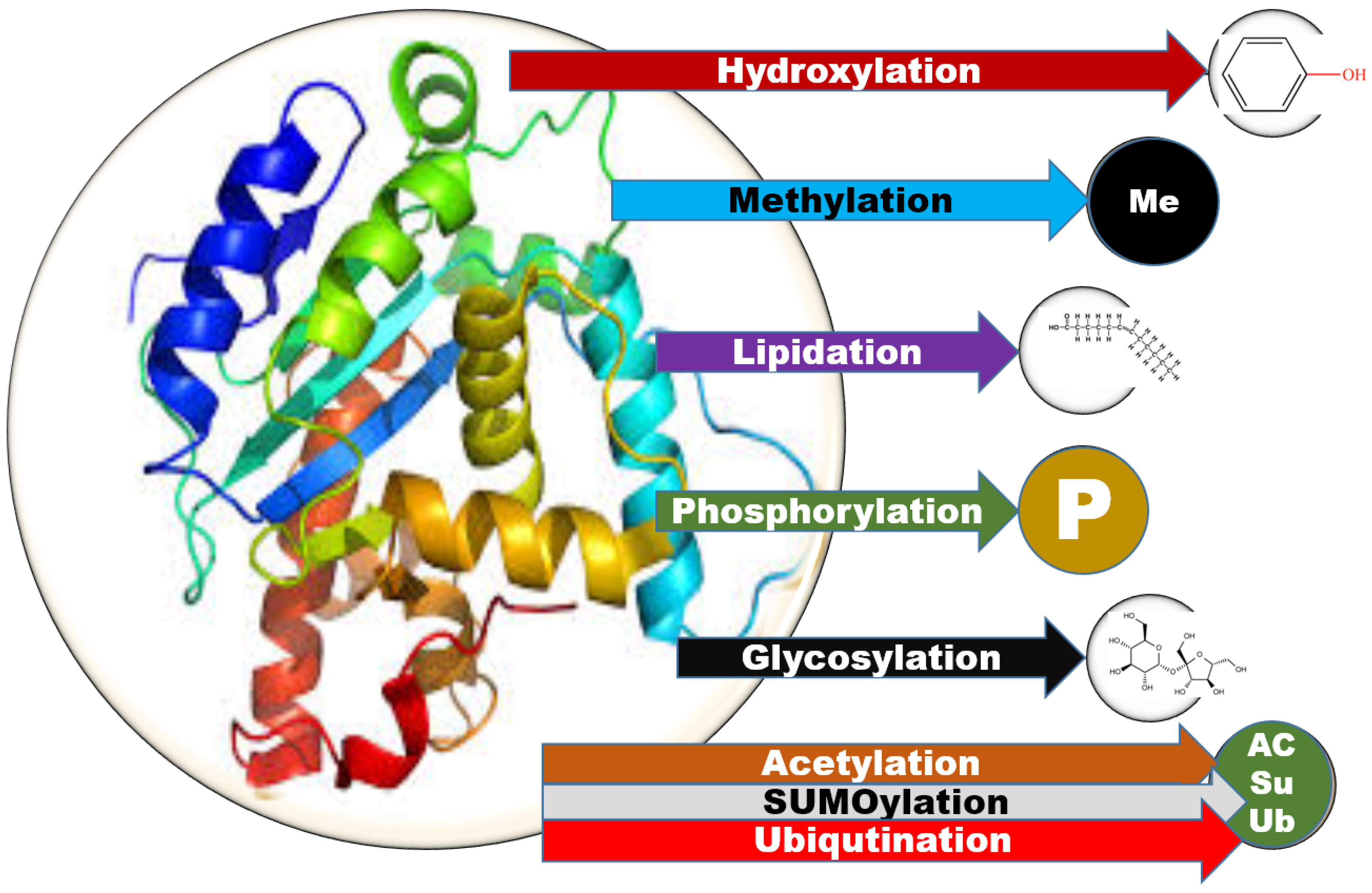
1. Introduction
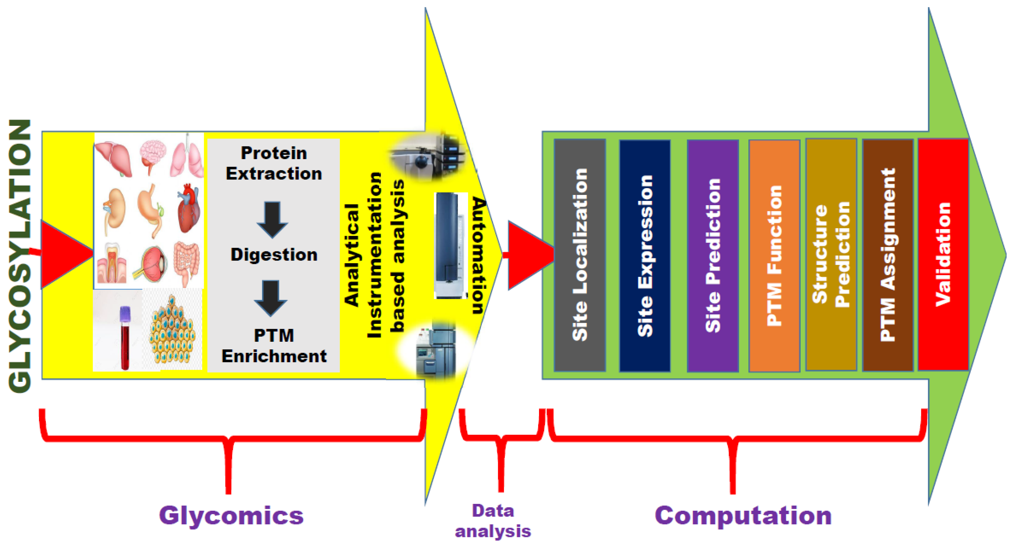
2. Snapshot of Bioinformatic Resources for Glycosylation
3. Scarce List of Bioinformatic Resources for Glycosylation in Cancers
4. Future Perspective
5. Conclusions
Author Contributions
Funding
Conflicts of Interest
References
- Ramamurthy, B.; Hook, P.; Larsson, L. An overview of carbohydrate-protein interactions with specific reference to myosin and ageing. Acta Physiol. Scand. 1999, 167, 327–329. [Google Scholar] [CrossRef]
- Mazola, Y.; Chinea, G.; Musacchio, A. Glycosylation and Bioinformatics: Current status for glycosylation prediction tools. Biotechnol. Appl. 2011, 28, 6–12. [Google Scholar]
- Mitra, N.; Sinha, S.; Ramya, T.N.; Surolia, A. N-linked oligosaccharides as outfitters for glycoprotein folding, form and function. Trends Biochem. Sci. 2006, 31, 156–163. [Google Scholar] [CrossRef] [PubMed]
- Mbonye, U.R.; Yuan, C.; Harris, C.E.; Sidhu, R.S.; Song, I.; Arakawa, T.; Smith, W.L. Two distinct pathways for cyclooxygenase-2 protein degradation. J. Biol. Chem. 2008, 283, 8611–8623. [Google Scholar] [CrossRef]
- Specks, U.; Fass, D.N.; Finkielman, J.D.; Hummel, A.M.; Viss, M.A.; Litwiller, R.D.; McDonald, C.J. Functional significance of Asn-linked glycosylation of proteinase 3 for enzymatic activity, processing, targeting, and recognition by anti-neutrophil cytoplasmic antibodies. J. Biochem. 2007, 141, 101–112. [Google Scholar] [CrossRef] [PubMed]
- Sola, R.J.; Griebenow, K. Effects of glycosylation on the stability of protein pharmaceuticals. J. Pharm. Sci. 2009, 98, 1223–1245. [Google Scholar] [CrossRef] [PubMed]
- Troy, F.A., 2nd. Polysialylation: From bacteria to brains. Glycobiology 1992, 2, 5–23. [Google Scholar] [CrossRef]
- Vagin, O.; Kraut, J.A.; Sachs, G. Role of N-glycosylation in trafficking of apical membrane proteins in epithelia. Am. J. Physiol. Renal Physiol. 2009, 296, F459–F469. [Google Scholar] [CrossRef]
- Couldrey, C.; Green, J.E. Metastases: The glycan connection. Breast Cancer Res. 2000, 2, 321–323. [Google Scholar] [CrossRef]
- Corthay, A.; Backlund, J.; Holmdahl, R. Role of glycopeptide-specific T cells in collagen-induced arthritis: An example how post-translational modification of proteins may be involved in autoimmune disease. Ann. Med. 2001, 33, 456–465. [Google Scholar] [CrossRef]
- Freeze, H.H. Update and perspectives on congenital disorders of glycosylation. Glycobiology 2001, 11, 129R–143R. [Google Scholar] [CrossRef] [PubMed]
- Varki, A.; Cummings, R.D.; Esko, J.D.; Freeze, H.H.; Hart, G.W.; Etzler, M.E. Essentials of Glycobiology, 2nd ed.; Cold Spring Harbor Laboratory Press: New York, NY, USA, 2008. [Google Scholar]
- Zaia, J. Mass spectrometry and the emerging field of glycomics. Chem. Biol. 2008, 15, 881–892. [Google Scholar] [CrossRef] [PubMed]
- Chen, Y.Z.; Tang, Y.R.; Sheng, Z.Y.; Zhang, Z. Prediction of mucin-type O-glycosylation sites in mammalian proteins using the composition of k-spaced amino acid pairs. BMC Bioinform. 2008, 9, 101. [Google Scholar] [CrossRef] [PubMed]
- Kianmehr, A.; Mohammadi, H.S.; Shokrgozar, M.A.; Omidinia, E. In silico design and analysis of a new hyperglycosylated analog of erythropoietin to improve drug efficacy. Adv. Biomed. Res. 2015, 4, 142. [Google Scholar] [CrossRef]
- Munkley, J.; Elliott, D.J. Hallmarks of glycosylation in cancer. Oncotarget 2016, 7, 35478–35489. [Google Scholar] [CrossRef]
- Azevedo, R.; Peixoto, A.; Gaiteiro, C.; Fernandes, E.; Neves, M.; Lima, L.; Santos, L.L.; Ferreira, J.A. Over forty years of bladder cancer glycobiology: Where do glycans stand facing precision oncology? Oncotarget 2017, 8, 91734–91764. [Google Scholar] [CrossRef]
- Christiansen, M.N.; Chik, J.; Lee, L.; Anugraham, M.; Abrahams, J.L.; Packer, N.H. Cell surface protein glycosylation in cancer. Proteomics 2014, 14, 525–546. [Google Scholar] [CrossRef]
- Palmigiano, A.; Barone, R.; Sturiale, L.; Sanfilippo, C.; Bua, R.O.; Romeo, D.A.; Messina, A.; Capuana, M.L.; Maci, T.; Le Pira, F.; et al. CSF N-glycoproteomics for early diagnosis in Alzheimer’s disease. J. Proteom. 2016, 131, 29–37. [Google Scholar] [CrossRef]
- Lauc, G.; Pezer, M.; Rudan, I.; Campbell, H. Mechanisms of disease: The human N-glycome. Biochim. Biophys. Acta 2016, 1860, 1574–1582. [Google Scholar] [CrossRef]
- Brockhausen, I.; Yang, J.M.; Burchell, J.; Whitehouse, C.; Taylor-Papadimitriou, J. Mechanisms underlying aberrant glycosylation of MUC1 mucin in breast cancer cells. Eur. J. Biochem. 1995, 233, 607–617. [Google Scholar] [CrossRef]
- Gils, A.; Pedersen, K.E.; Skottrup, P.; Christensen, A.; Naessens, D.; Deinum, J.; Enghild, J.J.; Declerck, P.J.; Andreasen, P.A. Biochemical importance of glycosylation of plasminogen activator inhibitor-1. Thromb. Haemost. 2003, 90, 206–217. [Google Scholar] [CrossRef] [PubMed]
- Lempiainen, A.; Hotakainen, K.; Blomqvist, C.; Alfthan, H.; Stenman, U.H. Hyperglycosylated human chorionic gonadotrOpin. in serum of testicular cancer patients. Clin. Chem. 2012, 58, 1123–1129. [Google Scholar] [CrossRef] [PubMed]
- Sato, Y.; Nakata, K.; Kato, Y.; Shima, M.; Ishii, N.; Koji, T.; Taketa, K.; Endo, Y.; Nagataki, S. Early recognition of hepatocellular carcinoma based on altered profiles of alpha-fetoprotein. N. Engl. J. Med. 1993, 328, 1802–1806. [Google Scholar] [CrossRef] [PubMed]
- Gadroy, P.; Stridsberg, M.; Capon, C.; Michalski, J.C.; Strub, J.M.; Van Dorsselaer, A.; Aunis, D.; Metz-Boutigue, M.H. Phosphorylation and O-glycosylation sites of human chromogranin A (CGA79-439) from urine of patients with carcinoid tumors. J. Biol. Chem. 1998, 273, 34087–34097. [Google Scholar] [CrossRef]
- Jankovic, M.M.; Milutinovic, B.S. Glycoforms of CA125 antigen as a possible cancer marker. Cancer Biomark. 2008, 4, 35–42. [Google Scholar] [CrossRef]
- Hua, L.; Liu, Y.; Zhen, S.; Wan, D.; Cao, J.; Gao, X. Expression and biochemical characterization of recombinant human epididymis protein 4. Protein Expr. Purif. 2014, 102, 52–62. [Google Scholar] [CrossRef]
- Aoki-Kinoshita, K.F. An introduction to bioinformatics for glycomics research. PLoS Comput. Biol. 2008, 4, e1000075. [Google Scholar] [CrossRef]
- von der Lieth, C.W.; Bohne-Lang, A.; Lohmann, K.K.; Frank, M. Bioinformatics for glycomics: Status, methods, requirements and perspectives. Brief. Bioinform. 2004, 5, 164–178. [Google Scholar] [CrossRef]
- Frank, M.; Schloissnig, S. Bioinformatics and molecular modeling in glycobiology. Cell Mol. Life Sci. 2010, 67, 2749–2772. [Google Scholar] [CrossRef]
- Yu, U.; Lee, S.H.; Kim, Y.J.; Kim, S. Bioinformatics in the post-genome era. J. Biochem. Mol. Biol. 2004, 37, 75–82. [Google Scholar] [CrossRef]
- Von der Lieth, C.W.; Lutteke, T.; Frank, M. Bioinformatics for Glycobiology and Glycomics: An Introduction, 1st ed.; John Wiley & Sons Ltd.: Hoboken, NJ, USA, 2009. [Google Scholar]
- Abrahams, J.L.; Taherzadeh, G.; Jarvas, G.; Guttman, A.; Zhou, Y.; Campbell, M.P. Recent advances in glycoinformatic platforms for glycomics and glycoproteomics. Curr. Opin. Struct. Biol. 2020, 62, 56–69. [Google Scholar] [CrossRef] [PubMed]
- Li, X.; Xu, Z.; Hong, X.; Zhang, Y.; Zou, X. Databases and Bioinformatic Tools for Glycobiology and Glycoproteomics. Int. J. Mol. Sci. 2020, 21, 6727. [Google Scholar] [CrossRef] [PubMed]
- Abdurakhmonov, I.Y. Bioinformatics: Basics, Development, and Future, Bioinformatics—Updated Features and Applications; Abdurakhmonov, I.Y., Ed.; IntechOpen: Rijeka, Croatia, 2016. [Google Scholar]
- Zhang, S.-Y.; Liu, S.-L. Bioinformatics, 2nd ed.; Academic Press: New York, NY, USA, 2013; pp. 338–340. [Google Scholar] [CrossRef]
- Kersey, P.; Apweiler, R. Linking publication, gene and protein data. Nat. Cell Biol. 2006, 8, 1183–1189. [Google Scholar] [CrossRef] [PubMed]
- Mulder, N.J.; Kersey, P.; Pruess, M.; Apweiler, R. In silico characterization of proteins: UniProt, InterPro and Integr8. Mol. Biotechnol. 2008, 38, 165–177. [Google Scholar] [CrossRef]
- Lutteke, T. Web resources for the glycoscientist. Chembiochem 2008, 9, 2155–2160. [Google Scholar] [CrossRef]
- Mahal, L.K. Glycomics: Towards bioinformatic approaches to understanding glycosylation. Anti Cancer Agents Med. Chem. 2008, 8, 37–51. [Google Scholar] [CrossRef]
- Mamitsuka, H. Informatic innovations in glycobiology: Relevance to drug discovery. Drug Discov. Today 2008, 13, 118–123. [Google Scholar] [CrossRef]
- Ranzinger, R.; Herget, S.; Lutteke, T.; Frank, M. Handbook of Glycomics; Cummings, R.D., Pierce, J.M., Eds.; Elsevier: Amsterdam, The Netherlands, 2009. [Google Scholar]
- von der Lieth, C.W. Comprehensive Glycoscience—From Chemistry to Systems Biology; Elsevier: Oxford, UK, 2007; Volume 2. [Google Scholar]
- von der Lieth, C.W.; Lutteke, T.; Frank, M. The role of informatics in glycobiology research with special emphasis on automatic interpretation of MS spectra. Biochim. Biophys. Acta 2006, 1760, 568–577. [Google Scholar] [CrossRef]
- Aoki-Kinoshita, K.F.; Kanehisa, M. Bioinformatics approaches in glycomics and drug discovery. Curr. Opin. Mol. Ther. 2006, 8, 514–520. [Google Scholar]
- Perez, S.; Mulloy, B. Prospects for glycoinformatics. Curr. Opin. Struct. Biol. 2005, 15, 517–524. [Google Scholar] [CrossRef]
- Marchal, I.; Golfier, G.; Dugas, O.; Majed, M. Bioinformatics in glycobiology. Biochimie 2003, 85, 75–81. [Google Scholar] [CrossRef]
- Walsh, I.; O’Flaherty, R.; Rudd, P.M. Bioinformatics applications to aid high-throughput glycan profiling. Perspect. Sci. 2017, 11, 31–39. [Google Scholar] [CrossRef]
- Brooksbank, C.; Camon, E.; Harris, M.A.; Magrane, M.; Martin, M.J.; Mulder, N.; O’Donovan, C.; Parkinson, H.; Tuli, M.A.; Apweiler, R.; et al. The European Bioinformatics Institute’s data resources. Nucleic Acids Res. 2003, 31, 43–50. [Google Scholar] [CrossRef] [PubMed]
- Wheeler, D.L.; Barrett, T.; Benson, D.A.; Bryant, S.H.; Canese, K.; Chetvernin, V.; Church, D.M.; Dicuccio, M.; Edgar, R.; Federhen, S.; et al. Database resources of the National Center for Biotechnology Information. Nucleic Acids Res. 2008, 36, D13–D21. [Google Scholar] [CrossRef] [PubMed]
- Whitfield, E.J.; Pruess, M.; Apweiler, R. Bioinformatics database infrastructure for biotechnology research. J. Biotechnol. 2006, 124, 629–639. [Google Scholar] [CrossRef] [PubMed]
- Doubet, S.; Bock, K.; Smith, D.; Darvill, A.; Albersheim, P. The Complex Carbohydrate Structure Database. Trends Biochem. Sci. 1989, 14, 475–477. [Google Scholar] [CrossRef]
- Doubet, S.; Albersheim, P. CarbBank. Glycobiology 1992, 2, 505. [Google Scholar] [CrossRef]
- Lutteke, T.; Bohne-Lang, A.; Loss, A.; Goetz, T.; Frank, M.; von der Lieth, C.W. GLYCOSCIENCES.de: An Internet portal to support glycomics and glycobiology research. Glycobiology 2006, 16, 71R–81R. [Google Scholar] [CrossRef]
- Hashimoto, K.; Goto, S.; Kawano, S.; Aoki-Kinoshita, K.F.; Ueda, N.; Hamajima, M.; Kawasaki, T.; Kanehisa, M. KEGG as a glycome informatics resource. Glycobiology 2006, 16, 63R–70R. [Google Scholar] [CrossRef]
- Raman, R.; Venkataraman, M.; Ramakrishnan, S.; Lang, W.; Raguram, S.; Sasisekharan, R. Advancing glycomics: Implementation strategies at the consortium for functional glycomics. Glycobiology 2006, 16, 82R–90R. [Google Scholar] [CrossRef]
- Toukach, F.V.; Knirel, Y.A. New database of bacterial carbohydrate structures. Glycoconj. J. 2005, 22, 216–217. [Google Scholar]
- Campbell, M.P.; Royle, L.; Radcliffe, C.M.; Dwek, R.A.; Rudd, P.M. GlycoBase and autoGU: Tools for HPLC-based glycan analysis. Bioinformatics 2008, 24, 1214–1216. [Google Scholar] [CrossRef] [PubMed]
- Cooper, C.A.; Joshi, H.J.; Harrison, M.J.; Wilkins, M.R.; Packer, N.H. GlycoSuiteDB: A curated relational database of glycoprotein glycan structures and their biological sources. 2003 update. Nucleic Acids Res. 2003, 31, 511–513. [Google Scholar] [CrossRef] [PubMed]
- Toukach, P.; Joshi, H.J.; Ranzinger, R.; Knirel, Y.; von der Lieth, C.W. Sharing of worldwide distributed carbohydrate-related digital resources: Online connection of the Bacterial Carbohydrate Structure DataBase and GLYCOSCIENCES.de. Nucleic Acids Res. 2007, 35, D280–D286. [Google Scholar] [CrossRef]
- Herget, S.; Ranzinger, R.; Maass, K.; Lieth, C.W. GlycoCT-a unifying sequence format for carbohydrates. Carbohydr. Res. 2008, 343, 2162–2171. [Google Scholar] [CrossRef]
- Packer, N.H.; von der Lieth, C.W.; Aoki-Kinoshita, K.F.; Lebrilla, C.B.; Paulson, J.C.; Raman, R.; Rudd, P.; Sasisekharan, R.; Taniguchi, N.; York, W.S. Frontiers in glycomics: Bioinformatics and biomarkers in disease. An NIH white paper prepared from discussions by the focus groups at a workshop on the NIH campus, Bethesda MD (September 11-13, 2006). Proteomics 2008, 8, 8–20. [Google Scholar] [CrossRef]
- Ranzinger, R.; Herget, S.; Wetter, T.; von der Lieth, C.W. GlycomeDB—Integration of open-access carbohydrate structure databases. BMC Bioinform. 2008, 9, 384. [Google Scholar] [CrossRef]
- Ranzinger, R.; Frank, M.; von der Lieth, C.W.; Herget, S. Glycome-DB.org: A portal for querying across the digital world of carbohydrate sequences. Glycobiology 2009, 19, 1563–1567. [Google Scholar] [CrossRef]
- Li, F.; Li, C.; Revote, J.; Zhang, Y.; Webb, G.I.; Li, J.; Song, J.; Lithgow, T. GlycoMine(struct): A new bioinformatics tool for highly accurate mapping of the human N-linked and O-linked glycoproteomes by incorporating structural features. Sci. Rep. 2016, 6, 34595. [Google Scholar] [CrossRef]
- Gupta, R.; Jung, E.; Brunak, S. Prediction of N-glycosylation sites in human proteins. 2004; in preparation. [Google Scholar]
- Steentoft, C.; Vakhrushev, S.Y.; Joshi, H.J.; Kong, Y.; Vester-Christensen, M.B.; Schjoldager, K.T.; Lavrsen, K.; Dabelsteen, S.; Pedersen, N.B.; Marcos-Silva, L.; et al. Precision mapping of the human O-GalNAc glycoproteome through SimpleCell technology. EMBO J. 2013, 32, 1478–1488. [Google Scholar] [CrossRef]
- Gupta, R.; Brunak, S. Prediction of Glycosylation across the Human Proteome and the Correlation to Protein Function. In Proceedings of the Pacific Symposium on Biocomputing, Lihue, Hawaii, 3–7 January 2002; pp. 310–322. [Google Scholar]
- Gupta, R.; Birch, H.; Rapacki, K.; Brunak, S.; Hansen, J.E. O-GLYCBASE version 4.0: A revised database of O-glycosylated proteins. Nucleic Acids Res. 1999, 27, 370–372. [Google Scholar] [CrossRef]
- Julenius, K. NetCGlyc 1.0: Prediction of mammalian C-mannosylation sites. Glycobiology 2007, 17, 868–876. [Google Scholar] [CrossRef]
- Li, F.; Li, C.; Wang, M.; Webb, G.I.; Zhang, Y.; Whisstock, J.C.; Song, J. GlycoMine: A machine learning-based approach for predicting N-, C- and O-linked glycosylation in the human proteome. Bioinformatics 2015, 31, 1411–1419. [Google Scholar] [CrossRef] [PubMed]
- Hamby, S.E.; Hirst, J.D. Prediction of glycosylation sites using random forests. BMC Bioinform. 2008, 9, 500. [Google Scholar] [CrossRef] [PubMed]
- Fankhauser, N.; Maser, P. Identification of GPI anchor attachment signals by a Kohonen self-organizing map. Bioinformatics 2005, 21, 1846–1852. [Google Scholar] [CrossRef] [PubMed]
- Pierleoni, A.; Martelli, P.L.; Casadio, R. PredGPI: A GPI-anchor predictor. BMC Bioinform. 2008, 9, 392. [Google Scholar] [CrossRef] [PubMed]
- Eisenhaber, B.; Bork, P.; Yuan, Y.; Loffler, G.; Eisenhaber, F. Automated annotation of GPI anchor sites: Case study C. elegans. Trends Biochem. Sci. 2000, 25, 340–341. [Google Scholar] [CrossRef]
- Lohmann, K.K.; von der Lieth, C.W. GlycoFragment and GlycoSearchMS: Web tools to support the interpretation of mass spectra of complex carbohydrates. Nucleic Acids Res. 2004, 32, W261–W266. [Google Scholar] [CrossRef]
- Lutteke, T.; Frank, M.; von der Lieth, C.W. Carbohydrate Structure Suite (CSS): Analysis of carbohydrate 3D structures derived from the PDB. Nucleic Acids Res. 2005, 33, D242–D246. [Google Scholar] [CrossRef]
- Krambeck, F.J.; Betenbaugh, M.J. A mathematical model of N-linked glycosylation. Biotechnol. Bioeng. 2005, 92, 711–728. [Google Scholar] [CrossRef]
- Umana, P.; Bailey, J.E. A mathematical model of N-linked glycoform biosynthesis. Biotechnol. Bioeng. 1997, 55, 890–908. [Google Scholar] [CrossRef]
- Lohmann, K.K.; von der Lieth, C.W. GLYCO-FRAGMENT: A web tool to support the interpretation of mass spectra of complex carbohydrates. Proteomics 2003, 3, 2028–2035. [Google Scholar] [CrossRef] [PubMed]
- Goldberg, D.; Sutton-Smith, M.; Paulson, J.; Dell, A. Automatic annotation of matrix-assisted laser desorption/ionization N-glycan spectra. Proteomics 2005, 5, 865–875. [Google Scholar] [CrossRef] [PubMed]
- Tang, H.; Mechref, Y.; Novotny, M.V. Automated interpretation of MS/MS spectra of oligosaccharides. Bioinformatics 2005, 21 (Suppl. S1), i431–i439. [Google Scholar] [CrossRef]
- Irungu, J.; Go, E.P.; Dalpathado, D.S.; Desaire, H. Simplification of mass spectral analysis of acidic glycopeptides using GlycoPep ID. Anal. Chem. 2007, 79, 3065–3074. [Google Scholar] [CrossRef]
- Cooper, C.A.; Gasteiger, E.; Packer, N.H. GlycoMod—A software tool for determining glycosylation compositions from mass spectrometric data. Proteomics 2001, 1, 340–349. [Google Scholar] [CrossRef]
- Bohm, M.; Bohne-Lang, A.; Frank, M.; Loss, A.; Rojas-Macias, M.A.; Lutteke, T. Glycosciences.DB: An annotated data collection linking glycomics and proteomics data (2018 update). Nucleic Acids Res. 2019, 47, D1195–D1201. [Google Scholar] [CrossRef]
- Rojas-Macias, M.A.; Mariethoz, J.; Andersson, P.; Jin, C.; Venkatakrishnan, V.; Aoki, N.P.; Shinmachi, D.; Ashwood, C.; Madunic, K.; Zhang, T.; et al. Towards a standardized bioinformatics infrastructure for N- and O-glycomics. Nat. Commun. 2019, 10, 3275. [Google Scholar] [CrossRef]
- Loss, A.; Bunsmann, P.; Bohne, A.; Loss, A.; Schwarzer, E.; Lang, E.; von der Lieth, C.W. SWEET-DB: An attempt to create annotated data collections for carbohydrates. Nucleic Acids Res. 2002, 30, 405–408. [Google Scholar] [CrossRef]
- Cooper, C.A.; Harrison, M.J.; Wilkins, M.R.; Packer, N.H. GlycoSuiteDB: A new curated relational database of glycoprotein glycan structures and their biological sources. Nucleic Acids Res. 2001, 29, 332–335. [Google Scholar] [CrossRef]
- Campbell, M.P.; Peterson, R.; Mariethoz, J.; Gasteiger, E.; Akune, Y.; Aoki-Kinoshita, K.F.; Lisacek, F.; Packer, N.H. UniCarbKB: Building a knowledge platform for glycoproteomics. Nucleic Acids Res. 2014, 42, D215–D221. [Google Scholar] [CrossRef] [PubMed]
- Alocci, D.; Mariethoz, J.; Gastaldello, A.; Gasteiger, E.; Karlsson, N.G.; Kolarich, D.; Packer, N.H.; Lisacek, F. GlyConnect: Glycoproteomics Goes Visual, Interactive, and Analytical. J. Proteome Res. 2019, 18, 664–677. [Google Scholar] [CrossRef] [PubMed]
- Kanehisa, M. KEGG GLYCAN; Aoki-Kinoshita, K.F., Ed.; Springer: Berlin/Heidelberg, Germany, 2017. [Google Scholar]
- Mariethoz, J.; Alocci, D.; Gastaldello, A.; Horlacher, O.; Gasteiger, E.; Rojas-Macias, M.; Karlsson, N.G.; Packer, N.H.; Lisacek, F. Glycomics@ExPASy: Bridging the Gap. Mol. Cell. Proteom. 2018, 17, 2164–2176. [Google Scholar] [CrossRef] [PubMed]
- Tiemeyer, M.; Aoki, K.; Paulson, J.; Cummings, R.D.; York, W.S.; Karlsson, N.G.; Lisacek, F.; Packer, N.H.; Campbell, M.P.; Aoki, N.P.; et al. GlyTouCan: An accessible glycan structure repository. Glycobiology 2017, 27, 915–919. [Google Scholar] [CrossRef]
- Hayes, C.A.; Karlsson, N.G.; Struwe, W.B.; Lisacek, F.; Rudd, P.M.; Packer, N.H.; Campbell, M.P. UniCarb-DB: A database resource for glycomic discovery. Bioinformatics 2011, 27, 1343–1344. [Google Scholar] [CrossRef] [PubMed]
- Campbell, M.P.; Nguyen-Khuong, T.; Hayes, C.A.; Flowers, S.A.; Alagesan, K.; Kolarich, D.; Packer, N.H.; Karlsson, N.G. Validation of the curation pipeline of UniCarb-DB: Building a global glycan reference MS/MS repository. Biochim. Biophys. Acta 2014, 1844, 108–116. [Google Scholar] [CrossRef]
- Remoroza, C.A.; Mak, T.D.; De Leoz, M.L.A.; Mirokhin, Y.A.; Stein, S.E. Creating a Mass Spectral Reference Library for Oligosaccharides in Human Milk. Anal. Chem. 2018, 90, 8977–8988. [Google Scholar] [CrossRef]
- Joshi, H.J.; Harrison, M.J.; Schulz, B.L.; Cooper, C.A.; Packer, N.H.; Karlsson, N.G. Development of a mass fingerprinting tool for automated interpretation of oligosaccharide fragmentation data. Proteomics 2004, 4, 1650–1664. [Google Scholar] [CrossRef]
- Apte, A.; Meitei, N.S. Bioinformatics in glycomics: Glycan characterization with mass spectrometric data using SimGlycan. Methods Mol. Biol. 2010, 600, 269–281. [Google Scholar] [CrossRef]
- Maxwell, E.; Tan, Y.; Tan, Y.; Hu, H.; Benson, G.; Aizikov, K.; Conley, S.; Staples, G.O.; Slysz, G.W.; Smith, R.D.; et al. GlycReSoft: A software package for automated recognition of glycans from LC/MS data. PLoS ONE 2012, 7, e45474. [Google Scholar] [CrossRef]
- Ashline, D.J.; Hanneman, A.J.; Zhang, H.; Reinhold, V.N. Structural documentation of glycan epitopes: Sequential mass spectrometry and spectral matching. J. Am. Soc. Mass Spectrom. 2014, 25, 444–453. [Google Scholar] [CrossRef] [PubMed][Green Version]
- Sun, W.; Lajoie, G.A.; Ma, B.; Zhang, K. Bioinformatics Research and Applications. In Proceedings of the 7th International Symposium (ISBRA 2011), Changsha, China, 27–29 May 2011. [Google Scholar]
- Hong, P.; Sun, H.; Sha, L.; Pu, Y.; Khatri, K.; Yu, X.; Tang, Y.; Lin, C. GlycoDeNovo—An Efficient Algorithm for Accurate de novo Glycan Topology Reconstruction from Tandem Mass Spectra. J. Am. Soc. Mass Spectrom. 2017, 28, 2288–2301. [Google Scholar] [CrossRef] [PubMed]
- Horlacher, O.; Jin, C.; Alocci, D.; Mariethoz, J.; Muller, M.; Karlsson, N.G.; Lisacek, F. Glycoforest 1.0. Anal. Chem. 2017, 89, 10932–10940. [Google Scholar] [CrossRef] [PubMed]
- Weatherly, D.B.; Arpinar, F.S.; Porterfield, M.; Tiemeyer, M.; York, W.S.; Ranzinger, R. GRITS Toolbox-a freely available software for processing, annotating and archiving glycomics mass spectrometry data. Glycobiology 2019, 29, 452–460. [Google Scholar] [CrossRef] [PubMed]
- Jansen, B.C.; Reiding, K.R.; Bondt, A.; Hipgrave Ederveen, A.L.; Palmblad, M.; Falck, D.; Wuhrer, M. MassyTools: A High-Throughput Targeted Data Processing Tool for Relative Quantitation and Quality Control Developed for Glycomic and Glycoproteomic MALDI-MS. J. Proteome Res. 2015, 14, 5088–5098. [Google Scholar] [CrossRef] [PubMed]
- Ranzinger, R.; Herget, S.; von der Lieth, C.W.; Frank, M. GlycomeDB--a unified database for carbohydrate structures. Nucleic Acids Res. 2011, 39, D373–D376. [Google Scholar] [CrossRef]
- Konishi, Y.; Aoki-Kinoshita, K.F. The GlycomeAtlas tool for visualizing and querying glycome data. Bioinformatics 2012, 28, 2849–2850. [Google Scholar] [CrossRef]
- Krambeck, F.J.; Bennun, S.V.; Narang, S.; Choi, S.; Yarema, K.J.; Betenbaugh, M.J. A mathematical model to derive N-glycan structures and cellular enzyme activities from mass spectrometric data. Glycobiology 2009, 19, 1163–1175. [Google Scholar] [CrossRef]
- Liu, G.; Marathe, D.D.; Matta, K.L.; Neelamegham, S. Systems-level modeling of cellular glycosylation reaction networks: O-linked glycan formation on natural selectin ligands. Bioinformatics 2008, 24, 2740–2747. [Google Scholar] [CrossRef]
- Hucka, M.; Finney, A.; Sauro, H.M.; Bolouri, H.; Doyle, J.C.; Kitano, H.; Arkin, A.P.; Bornstein, B.J.; Bray, D.; Cornish-Bowden, A.; et al. The systems biology markup language (SBML): A medium for representation and exchange of biochemical network models. Bioinformatics 2003, 19, 524–531. [Google Scholar] [CrossRef]
- Liu, G.; Puri, A.; Neelamegham, S. Glycosylation Network Analysis Toolbox: A MATLAB-based environment for systems glycobiology. Bioinformatics 2013, 29, 404–406. [Google Scholar] [CrossRef] [PubMed]
- Bohne, A.; Lang, E.; von der Lieth, C.W. SWEET—WWW-based rapid 3D construction of oligo- and polysaccharides. Bioinformatics 1999, 15, 767–768. [Google Scholar] [CrossRef] [PubMed]
- Bohne-Lang, A.; von der Lieth, C.W. GlyProt: In silico glycosylation of proteins. Nucleic Acids Res. 2005, 33, W214–W219. [Google Scholar] [CrossRef] [PubMed]
- Lutteke, T.; von der Lieth, C.W. pdb-care (PDB carbohydrate residue check): A program to support annotation of complex carbohydrate structures in PDB files. BMC Bioinform. 2004, 5, 69. [Google Scholar] [CrossRef] [PubMed]
- Perez, S.; Sarkar, A.; Rivet, A.; Breton, C.; Imberty, A. Glyco3D: A portal for structural glycosciences. Methods Mol. Biol. 2015, 1273, 241–258. [Google Scholar] [CrossRef] [PubMed]
- Ceroni, A.; Maass, K.; Geyer, H.; Geyer, R.; Dell, A.; Haslam, S.M. GlycoWorkbench: A tool for the computer-assisted annotation of mass spectra of glycans. J. Proteome Res. 2008, 7, 1650–1659. [Google Scholar] [CrossRef]
- Everest-Dass, A.V.; Kolarich, D.; Campbell, M.P.; Packer, N.H. Tandem mass spectra of glycan substructures enable the multistage mass spectrometric identification of determinants on oligosaccharides. Rapid Commun. Mass Spectrom. 2013, 27, 931–939. [Google Scholar] [CrossRef]
- Chuang, G.Y.; Boyington, J.C.; Joyce, M.G.; Zhu, J.; Nabel, G.J.; Kwong, P.D.; Georgiev, I. Computational prediction of N-linked glycosylation incorporating structural properties and patterns. Bioinformatics 2012, 28, 2249–2255. [Google Scholar] [CrossRef]
- Zhang, H.; Loriaux, P.; Eng, J.; Campbell, D.; Keller, A.; Moss, P.; Bonneau, R.; Zhang, N.; Zhou, Y.; Wollscheid, B.; et al. UniPep—A database for human N-linked glycosites: A resource for biomarker discovery. Genome Biol. 2006, 7, R73. [Google Scholar] [CrossRef]
- Chauhan, J.S.; Rao, A.; Raghava, G.P. In silico platform for prediction of N-, O- and C-glycosites in eukaryotic protein sequences. PLoS ONE 2013, 8, e67008. [Google Scholar] [CrossRef]
- Taherzadeh, G.; Dehzangi, A.; Golchin, M.; Zhou, Y.; Campbell, M.P. SPRINT-Gly: Predicting N- and O-linked glycosylation sites of human and mouse proteins by using sequence and predicted structural properties. Bioinformatics 2019, 35, 4140–4146. [Google Scholar] [CrossRef]
- Go, E.P.; Rebecchi, K.R.; Dalpathado, D.S.; Bandu, M.L.; Zhang, Y.; Desaire, H. GlycoPep DB: A tool for glycopeptide analysis using a “Smart Search”. Anal. Chem. 2007, 79, 1708–1713. [Google Scholar] [CrossRef]
- Carrot-Zhang, J.; Chambwe, N.; Damrauer, J.S.; Knijnenburg, T.A.; Robertson, A.G.; Yau, C.; Zhou, W.; Berger, A.C.; Huang, K.L.; Newberg, J.Y.; et al. Comprehensive Analysis of Genetic Ancestry and Its Molecular Correlates in Cancer. Cancer Cell 2020, 37, 639–654.e636. [Google Scholar] [CrossRef]
- Johansen, M.B.; Kiemer, L.; Brunak, S. Analysis and prediction of mammalian protein glycation. Glycobiology 2006, 16, 844–853. [Google Scholar] [CrossRef] [PubMed]
- Gupta, R.; Jung, E.; Gooley, A.A.; Williams, K.L.; Brunak, S.; Hansen, J. Scanning the available Dictyostelium discoideum proteome for O-linked GlcNAc glycosylation sites using neural networks. Glycobiology 1999, 9, 1009–1022. [Google Scholar] [CrossRef] [PubMed]
- Seu, C.S.; Chen, Y. Identification of SUMO-binding motifs by NMR. Methods Mol. Biol. 2009, 497, 121–138. [Google Scholar] [CrossRef] [PubMed]
- Lutteke, T.; Frank, M.; von der Lieth, C.W. Data mining the protein data bank: Automatic detection and assignment of carbohydrate structures. Carbohydr. Res. 2004, 339, 1015–1020. [Google Scholar] [CrossRef]
- Bohne-Lang, A.; Lang, E.; Forster, T.; von der Lieth, C.W. LINUCS: Linear notation for unique description of carbohydrate sequences. Carbohydr Res. 2001, 336, 1–11. [Google Scholar] [CrossRef]
- Farriol-Mathis, N.; Garavelli, J.S.; Boeckmann, B.; Duvaud, S.; Gasteiger, E.; Gateau, A.; Veuthey, A.L.; Bairoch, A. Annotation of post-translational modifications in the Swiss-Prot knowledge base. Proteomics 2004, 4, 1537–1550. [Google Scholar] [CrossRef]
- York, W.S.; Mazumder, R.; Ranzinger, R.; Edwards, N.; Kahsay, R.; Aoki-Kinoshita, K.F.; Campbell, M.P.; Cummings, R.D.; Feizi, T.; Martin, M.; et al. GlyGen: Computational and Informatics Resources for Glycoscience. Glycobiology 2020, 30, 72–73. [Google Scholar] [CrossRef]
- Hirabayashi, J.; Tateno, H.; Shikanai, T.; Aoki-Kinoshita, K.F.; Narimatsu, H. The Lectin Frontier Database (LfDB), and data generation based on frontal affinity chromatography. Molecules 2015, 20, 951–973. [Google Scholar] [CrossRef] [PubMed]
- Maes, E.; Bonachera, F.; Strecker, G.; Guerardel, Y. SOACS index: An easy NMR-based query for glycan retrieval. Carbohydr Res. 2009, 344, 322–330. [Google Scholar] [CrossRef] [PubMed]
- Lisacek, F.; Mariethoz, J.; Alocci, D.; Rudd, P.M.; Abrahams, J.L.; Campbell, M.P.; Packer, N.H.; Stahle, J.; Widmalm, G.; Mullen, E.; et al. Databases and Associated Tools for Glycomics and Glycoproteomics. Methods Mol. Biol. 2017, 1503, 235–264. [Google Scholar] [CrossRef] [PubMed]
- Gasteiger, E.; Gattiker, A.; Hoogland, C.; Ivanyi, I.; Appel, R.D.; Bairoch, A. ExPASy: The proteomics server for in-depth protein knowledge and analysis. Nucleic Acids Res. 2003, 31, 3784–3788. [Google Scholar] [CrossRef]
- Lutteke, T.; von der Lieth, C.W. MonoSaccharideDB: A reference resource to unify the notation of carbohydrate residues. Glycobiology 2005, 15, 1209–1210. [Google Scholar]
- Yamada, I.; Shiota, M.; Shinmachi, D.; Ono, T.; Tsuchiya, S.; Hosoda, M.; Fujita, A.; Aoki, N.P.; Watanabe, Y.; Fujita, N.; et al. The GlyCosmos Portal: A unified and comprehensive web resource for the glycosciences. Nat. Methods 2020, 17, 649–650. [Google Scholar] [CrossRef]
- Li, H.; Xing, X.; Ding, G.; Li, Q.; Wang, C.; Xie, L.; Zeng, R.; Li, Y. SysPTM: A systematic resource for proteomic research on post-translational modifications. Mol. Cell. Proteom. 2009, 8, 1839–1849. [Google Scholar] [CrossRef]
- Anugraham, M.; Jacob, F.; Nixdorf, S.; Everest-Dass, A.V.; Heinzelmann-Schwarz, V.; Packer, N.H. Specific glycosylation of membrane proteins in epithelial ovarian cancer cell lines: Glycan structures reflect gene expression and DNA methylation status. Mol. Cell. Proteom. 2014, 13, 2213–2232. [Google Scholar] [CrossRef]
- Munkley, J. The Role of Sialyl-Tn in Cancer. Int. J. Mol. Sci. 2016, 17, 275. [Google Scholar] [CrossRef]
- Tuccillo, F.M.; de Laurentiis, A.; Palmieri, C.; Fiume, G.; Bonelli, P.; Borrelli, A.; Tassone, P.; Scala, I.; Buonaguro, F.M.; Quinto, I.; et al. Aberrant glycosylation as biomarker for cancer: Focus on CD43. BioMed Res. Int. 2014, 2014, 742831. [Google Scholar] [CrossRef]
- Yin, B.W.; Dnistrian, A.; Lloyd, K.O. Ovarian cancer antigen CA125 is encoded by the MUC16 mucin gene. Int. J. Cancer 2002, 98, 737–740. [Google Scholar] [CrossRef] [PubMed]
- O’Brien, T.J.; Beard, J.B.; Underwood, L.J.; Dennis, R.A.; Santin, A.D.; York, L. The CA 125 gene: An extracellular superstructure dominated by repeat sequences. Tumour Biol. 2001, 22, 348–366. [Google Scholar] [CrossRef]
- Nustad, K.; Bast, R.C., Jr.; Brien, T.J.; Nilsson, O.; Seguin, P.; Suresh, M.R.; Saga, T.; Nozawa, S.; Bormer, O.P.; de Bruijn, H.W.; et al. Specificity and affinity of 26 monoclonal antibodies against the CA 125 antigen: First report from the ISOBM TD-1 workshop. International Society for Oncodevelopmental Biology and Medicine. Tumour Biol. 1996, 17, 196–219. [Google Scholar] [CrossRef] [PubMed]
- Yin, B.W.; Lloyd, K.O. Molecular cloning of the CA125 ovarian cancer antigen: Identification as a new mucin, MUC16. J. Biol. Chem. 2001, 276, 27371–27375. [Google Scholar] [CrossRef] [PubMed]
- Zurawski, V.R., Jr.; Orjaseter, H.; Andersen, A.; Jellum, E. Elevated serum CA 125 levels prior to diagnosis of ovarian neoplasia: Relevance for early detection of ovarian cancer. Int. J. Cancer 1988, 42, 677–680. [Google Scholar] [CrossRef] [PubMed]
- Bast, R.C., Jr.; Klug, T.L.; St John, E.; Jenison, E.; Niloff, J.M.; Lazarus, H.; Berkowitz, R.S.; Leavitt, T.; Griffiths, C.T.; Parker, L.; et al. A radioimmunoassay using a monoclonal antibody to monitor the course of epithelial ovarian cancer. N. Engl. J. Med. 1983, 309, 883–887. [Google Scholar] [CrossRef] [PubMed]
- Bast, R.C., Jr.; Badgwell, D.; Lu, Z.; Marquez, R.; Rosen, D.; Liu, J.; Baggerly, K.A.; Atkinson, E.N.; Skates, S.; Zhang, Z.; et al. New tumor markers: CA125 and beyond. Int. J. Gynecol. Cancer 2005, 15 (Suppl. S3), 274–281. [Google Scholar] [CrossRef]
- Gostout, B.S.; Brewer, M.A. Guidelines for referral of the patient with an adnexal mass. Clin. Obstet. Gynecol. 2006, 49, 448–458. [Google Scholar] [CrossRef]
- Pauler, D.K.; Menon, U.; McIntosh, M.; Symecko, H.L.; Skates, S.J.; Jacobs, I.J. Factors influencing serum CA125II levels in healthy postmenopausal women. Cancer Epidemiol. Biomark. Prev. 2001, 10, 489–493. [Google Scholar]
- Lauro, S.; Trasatti, L.; Bordin, F.; Lanzetta, G.; Bria, E.; Gelibter, A.; Reale, M.G.; Vecchione, A. Comparison of CEA, MCA, CA 15-3 and CA 27-29 in follow-up and monitoring therapeutic response in breast cancer patients. Anticancer Res. 1999, 19, 3511–3515. [Google Scholar]
- Uehara, M.; Kinoshita, T.; Hojo, T.; Akashi-Tanaka, S.; Iwamoto, E.; Fukutomi, T. Long-term prognostic study of carcinoembryonic antigen (CEA) and carbohydrate antigen 15-3 (CA 15-3) in breast cancer. Int. J. Clin. Oncol. 2008, 13, 447–451. [Google Scholar] [CrossRef]
- Kumpulainen, E.J.; Keskikuru, R.J.; Johansson, R.T. Serum tumor marker CA 15.3 and stage are the two most powerful predictors of survival in primary breast cancer. Breast Cancer Res. Treat. 2002, 76, 95–102. [Google Scholar] [CrossRef] [PubMed]
- Ebeling, F.G.; Stieber, P.; Untch, M.; Nagel, D.; Konecny, G.E.; Schmitt, U.M.; Fateh-Moghadam, A.; Seidel, D. Serum CEA and CA 15-3 as prognostic factors in primary breast cancer. Br. J. Cancer 2002, 86, 1217–1222. [Google Scholar] [CrossRef] [PubMed]
- Harris, L.; Fritsche, H.; Mennel, R.; Norton, L.; Ravdin, P.; Taube, S.; Somerfield, M.R.; Hayes, D.F.; Bast, R.C., Jr.; American Society of Clinical, O. American Society of Clinical Oncology 2007 update of recommendations for the use of tumor markers in breast cancer. J. Clin. Oncol. 2007, 25, 5287–5312. [Google Scholar] [CrossRef] [PubMed]
- Locker, G.Y.; Hamilton, S.; Harris, J.; Jessup, J.M.; Kemeny, N.; Macdonald, J.S.; Somerfield, M.R.; Hayes, D.F.; Bast, R.C., Jr.; Asco. ASCO 2006 update of recommendations for the use of tumor markers in gastrointestinal cancer. J. Clin. Oncol. 2006, 24, 5313–5327. [Google Scholar] [CrossRef]
- Safi, F.; Schlosser, W.; Kolb, G.; Beger, H.G. Diagnostic value of CA 19-9 in patients with pancreatic cancer and nonspecific gastrointestinal symptoms. J. Gastrointest. Surg. 1997, 1, 106–112. [Google Scholar] [CrossRef]
- Ychou, M.; Duffour, J.; Kramar, A.; Gourgou, S.; Grenier, J. Clinical significance and prognostic value of CA72-4 compared with CEA and CA19-9 in patients with gastric cancer. Dis. Markers 2000, 16, 105–110. [Google Scholar] [CrossRef]
- Marrelli, D.; Pinto, E.; De Stefano, A.; de Manzoni, G.; Farnetani, M.; Garosi, L.; Roviello, F. Preoperative positivity of serum tumor markers is a strong predictor of hematogenous recurrence of gastric cancer. J. Surg. Oncol. 2001, 78, 253–258. [Google Scholar] [CrossRef]
- Duraker, N.; Celik, A.N. The prognostic significance of preoperative serum CA 19-9 in patients with resectable gastric carcinoma: Comparison with CEA. J. Surg. Oncol. 2001, 76, 266–271. [Google Scholar] [CrossRef]
- Reiter, W.; Stieber, P.; Reuter, C.; Nagel, D.; Cramer, C.; Pahl, H.; Fateh-Moghadam, A. Prognostic value of preoperative serum levels of CEA, CA 19-9 and CA 72-4 in gastric carcinoma. Anticancer Res. 1997, 17, 2903–2906. [Google Scholar]
- Kirwan, A.; Utratna, M.; O’Dwyer, M.E.; Joshi, L.; Kilcoyne, M. Glycosylation-Based Serum Biomarkers for Cancer Diagnostics and Prognostics. BioMed Res. Int. 2015, 2015, 490531. [Google Scholar] [CrossRef] [PubMed]
- Kailemia, M.J.; Park, D.; Lebrilla, C.B. Glycans and glycoproteins as specific biomarkers for cancer. Anal. Bioanal. Chem. 2017, 409, 395–410. [Google Scholar] [CrossRef] [PubMed]
- Reis, C.A.; Osorio, H.; Silva, L.; Gomes, C.; David, L. Alterations in glycosylation as biomarkers for cancer detection. J. Clin. Pathol. 2010, 63, 322–329. [Google Scholar] [CrossRef] [PubMed]
- Peixoto, A.; Relvas-Santos, M.; Azevedo, R.; Santos, L.L.; Ferreira, J.A. Protein Glycosylation and Tumor Microenvironment Alterations Driving Cancer Hallmarks. Front. Oncol. 2019, 9, 380. [Google Scholar] [CrossRef]
- Wang, M.; Zhu, J.; Lubman, D.M.; Gao, C. Aberrant glycosylation and cancer biomarker discovery: A promising and thorny journey. Clin. Chem. Lab. Med. 2019, 57, 407–416. [Google Scholar] [CrossRef]
- Paulson, J.C.; Colley, K.J. Glycosyltransferases. Structure, localization, and control of cell type-specific glycosylation. J. Biol. Chem. 1989, 264, 17615–17618. [Google Scholar]
- Nakano, M.; Saldanha, R.; Gobel, A.; Kavallaris, M.; Packer, N.H. Identification of glycan structure alterations on cell membrane proteins in desoxyepothilone B resistant leukemia cells. Mol. Cell. Proteom. 2011, 10. [Google Scholar] [CrossRef]
- Alley, W.R.; Vasseur, J.A.; Goetz, J.A.; Syoboda, M.; Mann, B.F.; Matei, D.E.; Menning, N.; Hussein, A.; Mechref, Y.; Novotny, M.V. N-linked Glycan Structures and Their Expressions Change in the Blood Sera of Ovarian Cancer Patients. J. Proteome Res. 2012, 11, 2282–2300. [Google Scholar] [CrossRef]
- Shetty, V.; Hafner, J.; Shah, P.; Nickens, Z.; Philip, R. Investigation of ovarian cancer associated sialylation changes in N-linked glycopeptides by quantitative proteomics. Clin. Proteom. 2012, 9, 10. [Google Scholar] [CrossRef]
- Colombo, N.; Peiretti, M.; Parma, G.; Lapresa, M.; Mancari, R.; Carinelli, S.; Sessa, C.; Castiglione, M.; Group, E.G.W. Newly diagnosed and relapsed epithelial ovarian carcinoma: ESMO Clinical Practice Guidelines for diagnosis, treatment and follow-up. Ann. Oncol. 2010, 21 (Suppl. S5), v23–v30. [Google Scholar] [CrossRef]
- Schiess, R.; Wollscheid, B.; Aebersold, R. Targeted proteomic strategy for clinical biomarker discovery. Mol. Oncol. 2009, 3, 33–44. [Google Scholar] [CrossRef] [PubMed]
- Dube, D.H.; Bertozzi, C.R. Glycans in cancer and inflammation--potential for therapeutics and diagnostics. Nat. Rev. Drug Discov. 2005, 4, 477–488. [Google Scholar] [CrossRef] [PubMed]
- Lauc, G.; Huffman, J.E.; Pucic, M.; Zgaga, L.; Adamczyk, B.; Muzinic, A.; Novokmet, M.; Polasek, O.; Gornik, O.; Kristic, J.; et al. Loci associated with N-glycosylation of human immunoglobulin G show pleiotropy with autoimmune diseases and haematological cancers. PLoS Genet. 2013, 9, e1003225. [Google Scholar] [CrossRef] [PubMed]
- Munkley, J. Glycosylation is a global target for androgen control in prostate cancer cells. Endocr. Relat. Cancer 2017, 24, R49–R64. [Google Scholar] [CrossRef] [PubMed]
- Saldova, R.; Shehni, A.A.; Haakensen, V.D.; Steinfeld, I.; Hilliard, M.; Kifer, I.; Helland, A.; Yakhini, Z.; Borresen-Dale, A.L.; Rudd, P.M. Association of N-Glycosylation with Breast Carcinoma and Systemic Features Using High-Resolution Quantitative UPLC. J. Proteome Res. 2014, 13, 2314–2327. [Google Scholar] [CrossRef]
- Arnold, J.N.; Saldova, R.; Galligan, M.C.; Murphy, T.B.; Mimura-Kimura, Y.; Telford, J.E.; Godwin, A.K.; Rudd, P.M. Novel Glycan Biomarkers for the Detection of Lung Cancer. J. Proteome Res. 2011, 10, 1755–1764. [Google Scholar] [CrossRef]
- Albrecht, S.; Unwin, L.; Muniyappa, M.; Rudd, P.M. Glycosylation as a marker for inflammatory arthritis. Cancer Biomark. 2014, 14, 17–28. [Google Scholar] [CrossRef]
- Kaur, N.; Mathur, A.; Aggarwal, A.; Gupta, S.; Rashmi, T. Bioinformatic Analysis of Aberrant Glycosylation in Triple Negative Breast Cancer. In Proceedings of the 6th World Congress on Breast Cancer & Therapy, San Francisco, CA, USA, 16–18 October 2017. [Google Scholar]
- Gotz, L.; Abrahams, J.L.; Mariethoz, J.; Rudd, P.M.; Karlsson, N.G.; Packer, N.H.; Campbell, M.P.; Lisacek, F. GlycoDigest: A tool for the targeted use of exoglycosidase digestions in glycan structure determination. Bioinformatics 2014, 30, 3131–3133. [Google Scholar] [CrossRef]
- Zhang, D.; Xie, Q.; Wang, Q.; Wang, Y.; Miao, J.; Li, L.; Zhang, T.; Cao, X.; Li, Y. Mass spectrometry analysis reveals aberrant N-glycans in colorectal cancer tissues. Glycobiology 2019, 29, 372–384. [Google Scholar] [CrossRef]
- Lee, S.B.; Bose, S.; Ahn, S.H.; Son, B.H.; Ko, B.S.; Kim, H.J.; Chung, I.Y.; Kim, J.; Lee, W.; Ko, M.S.; et al. Breast cancer diagnosis by analysis of serum N-glycans using MALDI-TOF mass spectroscopy. PLoS ONE 2020, 15, e0231004. [Google Scholar] [CrossRef]
- Oriol, R.; Mollicone, R.; Cailleau, A.; Balanzino, L.; Breton, C. Divergent evolution of fucosyltransferase genes from vertebrates, invertebrates, and bacteria. Glycobiology 1999, 9, 323–334. [Google Scholar] [CrossRef] [PubMed]
- Goletz, S.; Danielczyk, A.; Stahn, R.; Karsten, U.; Stoeckl, L.; Loeffler, A.; Hillemann, A.; Baumeister, H. GlycoOptimization for Fully Human and Largely Improved Biopharmaceutical Antibodies and Proteins. In Proceedings of the 1st Beilstein Glyco-Bioinformatics Symposium—Bits ‘n’ Bytes of Sugars, Potsdam, Germany, 4–8 October 2009; pp. 87–100. [Google Scholar]
- Nadeem, T.; Khan, M.A.; Ijaz, B.; Ahmed, N.; Rahman, Z.U.; Latif, M.S.; Ali, Q.; Rana, M.A. Glycosylation of Recombinant Anticancer Therapeutics in Different Expression Systems with Emerging Technologies. Cancer Res. 2018, 78, 2787–2798. [Google Scholar] [CrossRef] [PubMed]


| Bioinformatics Resources | URL | Application | Reference |
|---|---|---|---|
| Glycosylation Structure Related Resources | |||
| UniCarb KB | http://unicarbkb.org/ | Repository for glycan structures of glycoproteins | [64] |
| GlycoMod | http://web.expasy.org/glycomod/ | Software tool for N-linked and O-linked glycan structures prediction | [84] |
| GlycosuiteDB | http://www.glycosuite.com | Relational database of glycoprotein glycan structures and their biological sources | [88] |
| GlycoDeNovo | https://www.cs.brandeis.edu/~hong/Research/GlycoDeNovo/GlycoDeNovo.htm | Algorithm for Accurate de novo Glycan Topology Reconstruction from Tandem Mass Spectras | [102] |
| Glycoforest | https://glycoforest.expasy.org/ | Partial de-novo algorithm for sequencing glycan structures based on MS/MS spectra | [103] |
| Sweet-II | http://www.glycosciences.de/modeling/sweet2/doc/index.php | Tool to construct 3D models of saccharides from their sequences using standard nomenclature | [112] |
| GlyProt | http://www.glycosciences.de/modeling/glyprot/php/main.php | Tool to connect N-glycans in silico to a given 3D protein structure. | [113] |
| PDB CArbohydrate REsidue check (pdb-care) | http://www.glycosciences.de/tools/pdb-care/ | Identify and assign carbohydrate structures using atom types and their 3D atom coordinates from PDB-files | [114] |
| Glyco3D | http://glyco3d.cermav.cnrs.fr/home.php | 3D structures of monosaccharides, disaccharides, oligosaccharides, polysaccharides, glycosyltransferases, lectins, monoclonal antibodies against carbohydrates, and glycosaminoglycan-binding proteins | [115] |
| LiGraph | http://www.glycosciences.de/tools/LiGraph/ | Convert a sugar graph to ASCII IUPAC sugar nomenclature or as a graph | [85] |
| KEGG Glycan database | https://www.genome.jp/kegg/glycan/ | Database for glycan structures | [55] |
| GlyTouCan | https://glytoucan.org/ | Repository for glycan structures. | [93] |
| Resources for Analytical Glycosylation | |||
| UniCarb-DB | https://unicarb-db.expasy.org/ | Repository for glycomic MS data | [89] |
| GlycanBuilder, GlycoWorkBench | http://www.eurocarbdb.org/applications/structure-tools | Tool to annotate Mass Spectra of Glycans | [116] |
| Multiglycan | https://bio.tools/multiglycan | Information provider for glycan profile information from LC-MS Spectra. | [117] |
| GlycoFragment and Distance Mapping | www.dkfz.de/spec/projekte/fragments/ | Web tool to interpret mass spectra of complex carbohydrates | [76] |
| Prediction of Glycosylation | |||
| NGlycPred | https://bioinformatics.niaid.nih.gov/nglycpred/ | Server to predict N-linked Glycosylation sites | [118] |
| UNIPEP | http://www.unipep.org/ | Human and mouse N-glycosylated proteins and their N-glycosylation sites for biomarker discovery | [119] |
| GlycoEP | https://bio.tools/GlycoEP | prediction of N-linked, O-linked and C-linked glycosites in eukaryotic glycoproteins | [120] |
| GlySeq | http://www.glycosciences.de/tools/glyseq/ | Analyze the sequences around glycosylation sites. | [77] |
| SPRINT-Gly | https://sparks-lab.org/server/sprint-gly/ | predictingN-andO-linked glycosylation sites | [121] |
| DictyOGlyc | http://www.cbs.dtu.dk/services/DictyOGlyc/ | Prediction of O-glycosylation sites in Dictyostelium discoideum proteins | [73] |
| GlycoMinestruct | https://glycomine.erc.monash.edu/Lab/GlycoMine_Struct/index.jsp | Highly accurate mapping of the human N-linked and O-linked glycoproteomes | [65] |
| GlycoForm | http://www.boxer.tcd.ie/gf/ | Mathematical model software for N-linked glycosylation | [78] |
| Glycopep | http://hexose.chem.ku.edu/predictiontable.php | N-linked glycosylation based on target protein and CID spectra analysis | [122] |
| Consortium for Functional Glycomics (CFC) | http://www.functionalglycomics.org/static/consortium/consortium.shtml | Glycomics resources of glycans and glycan-binding protein | [63] |
| NetNGlyc | http://www.cbs.dtu.dk/services/NetNGlyc/ | N-Glycosylation site predictor | [123] |
| NetOGlyc | http://www.cbs.dtu.dk/services/NetOGlyc/ | O-Glycosylation sites predictor | [75] |
| GPP | https://comp.chem.nottingham.ac.uk/glyco/ | N- and O-Glycosylation site predictor | [73] |
| Big-PI | https://mendel.imp.ac.at/gpi/gpi_server.html | GPI anchors predictor | [124] |
| GPI-SOM | http://gpi.unibe.ch/ | GPI anchors predictor | [125] |
| PredGPI | http://gpcr.biocomp.unibo.it/predgpi/ | GPI anchors predictor | [85] |
| NetCGlyc | http://www.cbs.dtu.dk/services/NetCGlyc/ | C-mannosylation sites prediction | [123] |
| Carbohydrate Knowledgebases | |||
| GlycoCT | www.eurocarbdb.org | Tool to convert unifying sequence format for carbohydrates | [61] |
| SWEET-DB | http://www.pdg.cnb.uam.es/cursos/Leon2002/pages/software/DatabasesListNAR2002/summary/300.html | Repository for annotated carbohydrates | [87] |
| SUgar MOtif search (sumo) | http://www.glycosciences.de/tools/sumo/ | Tool to search sugar motif regions from carbohydrate structures | [126] |
| Bacterial Carbohydrate Structure Database (BCSDB, 6789) | http://csdb.glycoscience.ru/bacterial/ | Repository for prokaryotic carbohydrate structures, taxonomy, bibliography, NMR data, etc. | [57] |
| pdb2linucs | http://www.glycosciences.de/tools/pdb2linucs/ | Extract carbohydrate information from pdb-files and display it using the LINUCS-Code | [77] |
| CCSD | https://cordis.europa.eu/project/id/BIOT0184 | Information system of carbohydrate science | [52] |
| Carbohydrate Ramachandran Plot (CARP) | http://www.glycosciences.de/tools/carp/ | Analyze carbohydrate data from PDB files using the pdb2linucs algorithm | [127] |
| LINUCS | http://www.glycosciences.de/tools/linucs/ | LInear Notation for Unique description of Carbohydrate Sequences | [128] |
| Miscellaneous Resources for Glycosylation | |||
| RESID | https://proteininformationresource.org/resid/togm.shtml | Table of Glycosylation Modifications | [129] |
| GlyGen | https://www.glygen.org/ | Informatics Resources for Glycoscience | [130] |
| LfDB | https://acgg.asia/lfdb2/ | Lectin Frontier DataBase | [131] |
| CFG | http://www.functionalglycomics.org/ | Comprehensive resource for functional glycomics research | [56] |
| GlycoStore | https://www.glycostore.org | A curated database of information on glycan retention properties with chromatographic, electrophoretic and mass-spectrometry composition data. | [132] |
| Galaxy, LipidBank | https://jcggdb.jp/database_en.html | Consortium for Glycobiology and Glycobiotechnology database | [133] |
| Glycosylation Network Analysis Toolbox (GNAT) | http://gnatmatlab.sourceforge.net/ | MATLAB-based environment for systems glycobiology | [111] |
| Glycomics@ExPASy | https://www.expasy.org/search/glycomics | Expasy resources for glycomic data. | [134] |
| MonosaccharideDB | http://www.monosaccharidedb.org/ | comprehensive resource of monosaccharides | [135] |
| GlycReSoft | http://www.bumc.bu.edu/msr/glycresoft/ | Software for Glycomics and Glycoproteomics | [99] |
| CarbBank | https://www.genome.jp/dbget-bin/www_bfind?carbbank | Database management program and the project system of CCSD | [53] |
| GlyCosmos | https://glycosmos.org/ | Comprehensive web resource for the glycosciences | [136] |
| SysPTM | http://lifecenter.sgst.cn/SysPTM/ | Resource for post-translational modification | [137] |
| Bioinformatics Resources | Cancer Type | URL | Application | Reference |
|---|---|---|---|---|
| RNA sequencing analysis of genes | prostate cancer cell lines and patients | A composite sequencing server | RNA sequencing analysis of identified a set of 700 androgen-regulated genes. | [174] |
| Gene ontology (GO) | prostate cancer cell lines and patients | Gene ontology (GO) is general functional annotation server | identified 72 terms with significant gene enrichment (p < 0.05) and defined glycosylation as an androgen-regulated process in prostate cancer cells. | [174] |
| GlycoBase (inactive now) | Breast cancer cells | (https://glycobase.nibrt.ie): | database of experimentally determined glycan structures originally developed from the EurocarbDB project | [58] |
| GlycoDigest | Breast cancer cells | (http://www.glycodigest.org): | a tool that simulates exoglycosidase digestion based on controlled rules acquired from expert knowledge and experimental evidence available in GlycoBase | [179] |
| GlycoMarker | Breast cancer cells | https://glycobase.nibrt.ie/glycomarker | Web application/server for Biomarker discovery, identifies markers in LC profiles | [48] |
| using KEGG, DAVID and Ingenuity databases, uniprot database | Breast cancer cells | KEGG, DAVID and Ingenuity databases, uniprot database these are very general functional analysis and sequence databases | Significant change in the expression profiling of glycosylation patterns of various proteins associated with Triple negative breast cancer was identified. Differential aberrant glycosylated proteins in breast cancer cells with respect to non-neoplastic cells | [178] |
| genome-wide association study (GWAS) | haematological cancers such as acute lymphoblastic leukaemia, Hodgkin lymphoma, and multiple myeloma | GWAS is a general study for genome wide citation | identify new loci that control glycosylation of a single plasma protein using GWAS | [178] |
| YinOYang | Breast cancer cells | http://www.cbs.dtu.dk/services/YinOYang/ | O-Glycosylation sites predictor | [125] |
| ANCOVA/MANCOVA statistics | Lung Cancer cells | https://www.statisticssolutions.com/multivariate-analysis-of-covariance-mancova/ | Statistical analysis | [176] |
| Student’s t test, orthogonal partial least squares discriminant analysis and receiver operating characteristic curve | colorectal cancer tissues (CRC) in Chinese patients | Statistical tools | Statistical analysis of MS data from Linear ion trap quadrupole-electrospray ionization mass spectrometry, on CRC tissues | [180] |
| Perseus™ (Max Planck Institute of Biochemistry, Berlin, Germany) | Breast cancer | https://pubmed.ncbi.nlm.nih.gov/29344888/ | Perseus™ 1.5.2.6 was used for hierarchical clustering, principal component analysis (PCA), and plotting for visualization and statistics | [181] |
Publisher’s Note: MDPI stays neutral with regard to jurisdictional claims in published maps and institutional affiliations. |
© 2020 by the authors. Licensee MDPI, Basel, Switzerland. This article is an open access article distributed under the terms and conditions of the Creative Commons Attribution (CC BY) license (http://creativecommons.org/licenses/by/4.0/).
Share and Cite
Muthu, M.; Chun, S.; Gopal, J.; Anthonydhason, V.; Haga, S.W.; Jacintha Prameela Devadoss, A.; Oh, J.-W. Insights into Bioinformatic Applications for Glycosylation: Instigating an Awakening towards Applying Glycoinformatic Resources for Cancer Diagnosis and Therapy. Int. J. Mol. Sci. 2020, 21, 9336. https://doi.org/10.3390/ijms21249336
Muthu M, Chun S, Gopal J, Anthonydhason V, Haga SW, Jacintha Prameela Devadoss A, Oh J-W. Insights into Bioinformatic Applications for Glycosylation: Instigating an Awakening towards Applying Glycoinformatic Resources for Cancer Diagnosis and Therapy. International Journal of Molecular Sciences. 2020; 21(24):9336. https://doi.org/10.3390/ijms21249336
Chicago/Turabian StyleMuthu, Manikandan, Sechul Chun, Judy Gopal, Vimala Anthonydhason, Steve W. Haga, Anna Jacintha Prameela Devadoss, and Jae-Wook Oh. 2020. "Insights into Bioinformatic Applications for Glycosylation: Instigating an Awakening towards Applying Glycoinformatic Resources for Cancer Diagnosis and Therapy" International Journal of Molecular Sciences 21, no. 24: 9336. https://doi.org/10.3390/ijms21249336
APA StyleMuthu, M., Chun, S., Gopal, J., Anthonydhason, V., Haga, S. W., Jacintha Prameela Devadoss, A., & Oh, J.-W. (2020). Insights into Bioinformatic Applications for Glycosylation: Instigating an Awakening towards Applying Glycoinformatic Resources for Cancer Diagnosis and Therapy. International Journal of Molecular Sciences, 21(24), 9336. https://doi.org/10.3390/ijms21249336