Review

Regulation of GABAA Receptor Subunit Expression in Substance Use Disorders

by
Jeffrey S. Barker
and
Rochelle M. Hines
*
Department of Psychology, University of Nevada, Las Vegas, Las Vegas, NV 89154, USA
*
Author to whom correspondence should be addressed.
Int. J. Mol. Sci. 2020, 21(12), 4445; https://doi.org/10.3390/ijms21124445
Submission received: 29 May 2020 / Revised: 17 June 2020 / Accepted: 19 June 2020 / Published: 22 June 2020
(This article belongs to the Section Molecular Neurobiology)

Abstract

:
The modulation of neuronal cell firing is mediated by the release of the neurotransmitter GABA (γ-aminobuytric acid), which binds to two major families of receptors. The ionotropic GABAA receptors (GABAARs) are composed of five distinct subunits that vary in expression by brain region and cell type. The action of GABA on GABAARs is modulated by a variety of clinically and pharmacologically important drugs such as benzodiazepines and alcohol. Exposure to and abuse of these substances disrupts homeostasis and induces plasticity in GABAergic neurotransmission, often via the regulation of receptor expression. Here, we review the regulation of GABAAR subunit expression in adaptive and pathological plasticity, with a focus on substance use. We examine the factors influencing the expression of GABAAR subunit genes including the regulation of the 5′ and 3′ untranslated regions, variations in DNA methylation, immediate early genes and transcription factors that regulate subunit expression, translational and post-translational modifications, and other forms of receptor regulation beyond expression. Advancing our understanding of the factors regulating GABAAR subunit expression during adaptive plasticity, as well as during substance use and withdrawal will provide insight into the role of GABAergic signaling in substance use disorders, and contribute to the development of novel targeted therapies.

Graphical Abstract

1. Introduction

Neuronal firing patterns in the brain are powerfully modulated by the inhibitory neurotransmitter γ-aminobutyric acid (GABA). GABAA receptors (GABAARs) are ligand-gated ion channels that mediate the majority of fast inhibitory neurotransmission in the brain, and GABAAR dysfunction is tied to many neurological and psychiatric illnesses, such as anxiety, epilepsy, and the development of substance use disorder. GABAARs are heteropentamers consisting of five distinct subunits that vary in their expression by brain region, cell type, and subcellular domain, as well as in their function. There are at least 19 receptor subunits, grouped by homologous amino acid sequences into subclasses: α1–6, β1–3, and γ1–3, δ, ε, π, θ, and ρ1–3 [1]. While the combination and function of receptor subunits varies, most are comprised of two α-, two β-, and one γ- [2,3]. GABAAR subunit expression is also temporally regulated, with the expression of GABAAR subunit α2, α3, α5, and β3 mRNA predominating during early development, which are superseded by α1, α4, β2 and δ subunit mRNAs in the adult brain [4]. These changes coincide with the switch in the reversal potential for chloride, transitioning GABA from being depolarizing to hyperpolarizing [4,5].
α subunits in particular are important determinants of receptor localization and function. GABAARs composed of α1β2γ2 are the most widely expressed in the adult brain, with α1 being the most highly expressed subunit [2,4,6]. While α1, β1-3, and γ2 immunoreactivities are found throughout the brain, α2 subunit containing receptors are enriched in the cerebellum and forebrain including the hippocampus, α3 containing receptors are cortically enriched, α4 containing receptors are enriched in the striatum and thalamus, and α5 containing receptors are enriched in the olfactory bulbs and hippocampus, while α6 immunoreactivity is relatively restricted to cerebellar granule cells and the cochlear nucleus [7,8].
α subunit expression also varies by subcellular localization, with α1–3 being predominantly enriched at synaptic sites, while α4–6 are often localized extrasynaptically [9]. Synaptic GABAARs are also targeted to multiple synapse subtypes where interneuron presynaptic compartments contact the dendrites, the soma, or the axon initial segment. The clustering of specific GABAAR subtypes to synaptic and extrasynaptic subcellular domains is thought to be regulated by subunit-specific interactions with scaffolding proteins including gephyrin, collybistin, and dystrophin [10,11,12].
Differences in agonist affinity, gating and pharmacological properties have been repeatedly shown by altering subunits of recombinant GABAARs [13,14,15,16]. Subunit composition also affects the potency of GABA, as α2- and α3-containing receptors show low potency, α1-, α4-, and α5-containing receptors show intermediate potency, and α6-containing receptors show high potency [17]. GABAARs are the site of action for many clinically relevant drugs, most of which act as positive allosteric modulators of the receptors and alter the phasic inhibition by prolonging the decay of inhibitory post-synaptic currents (IPSCs), which in turn can prevent neural firing in response to concurrent excitatory stimuli [13,18]. The kinetics of IPSCs at the postsynaptic GABAAR is determined by biophysical properties such as the subunit composition and how they cluster at the cell membrane [18]. The specific α subunit is a determining factor in receptor function and setting the kinetics of IPSCs [13,18,19,20].
GABAergic signaling is controlled at the cellular level by changes in neurotransmitter synthesis, vesicular storage, neurotransmitter release and re-uptake, and postsynaptic receptor clustering [1]. GABAergic signaling at the cellular level is often modulated by changes in the expression of GABAARs. GABAAR gene expression is temporally regulated throughout development and the life span, as well as in response to experience, substance use, and as a result of a number of neuropathologies [4,21,22]. The expression of genes encoding the GABAAR subunits can be altered at multiple levels including transcription initiation, alternative splicing, mRNA stability, translation, post-translational modifications, intracellular tracking and protein degradation [1] (Figure 1). In the present review, we will discuss what is known about the regulation of GABAAR expression across the levels in both normal and pathological states, with a focus on substance use and withdrawal.

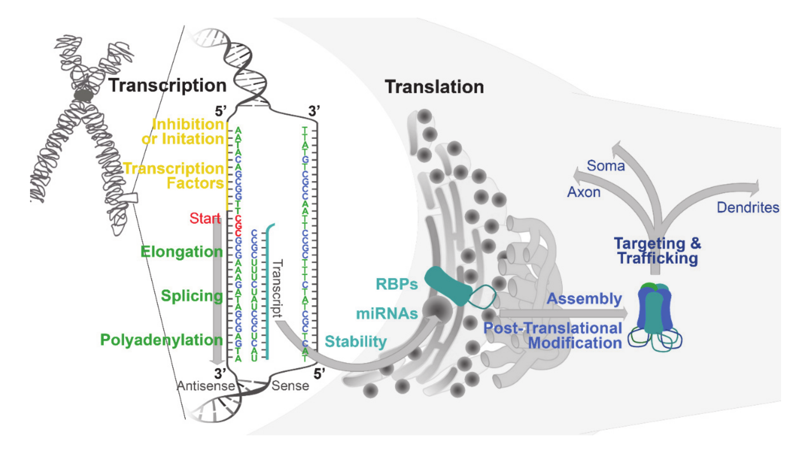
2. Basal Mechanisms Regulating GABAA Receptor Expression: Transcription, Translation, and Beyond
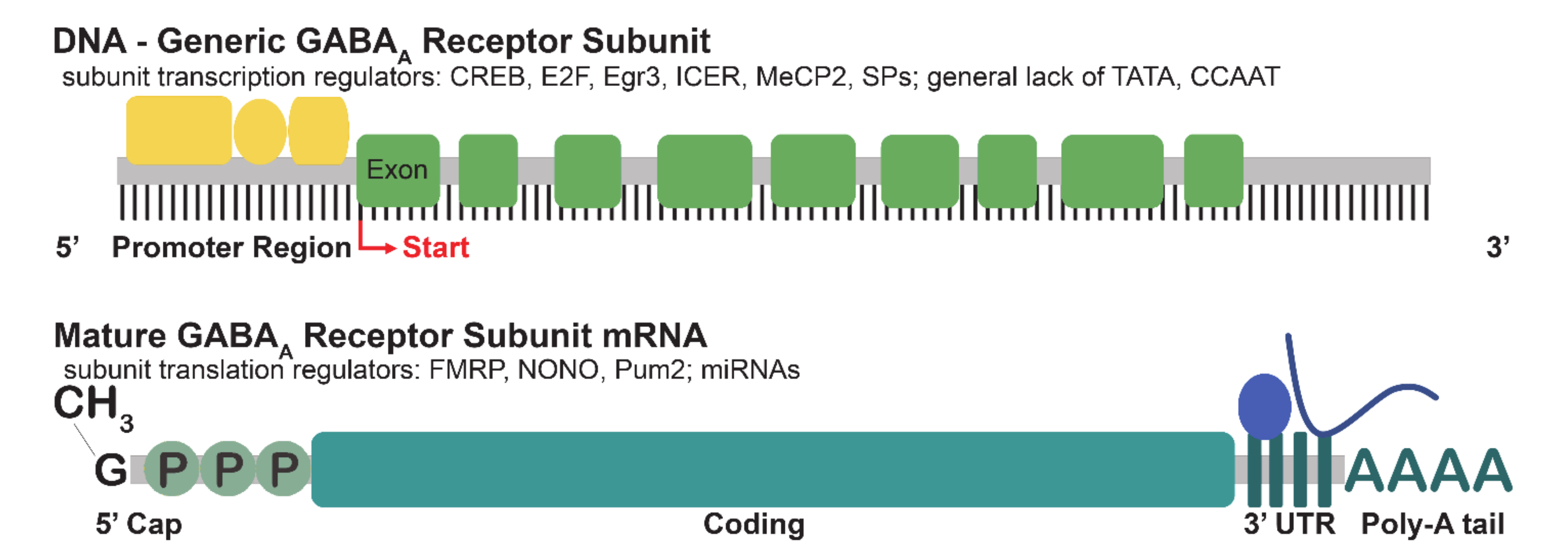
In order to understand the dynamic regulation of GABAAR expression in substance use, it will be important to examine the mechanisms that regulate subunit expression under basal physiological states. The examination of the factors that regulate transcription, translation and beyond reveals numerous levels of regulation, allowing for the precise tuning of GABAAR expression. In the following section, we summarize what is known about the transcription factors, epigenetic regulators, and RNA-binding proteins modulating GABAAR subunit expression.
The evolution of GABAARs and their subunits is highly accessible using a phylogenetic tree and the direct examination of gene organization on chromosomes [23]. Many of the subunit genes are organized in β-α-α-γ and β-α-γ clusters on different chromosomes, thought to have evolved from a single ancestral β-α-γ cluster [1,23]. In total, four clusters of genes coding for GABAAR subunits have been found in humans, and this organization is thought to be possibly involved in coordinated gene regulation [1,24]. The genes encoding β1, α2, α4, and γ1 (GABRB1; GABRA2; GABRA4; GABRG1) are all clustered on chromosome 4p14-q12 in humans [25,26]. The β1, α2, and α4 subunits are all enriched in the hippocampus of the adult rat, which may indicate that cluster organization may be necessary for maintaining region-specific expression [1,6]. Chromosome 5q32.1-q35 contains the cluster coding for the most common GABAAR subunits β2-α1-α6-γ2 [27] (GABRB2; GABRA1; GABRA6; GABRG2), again suggesting that the clustering of subunit genes plays a role in the regulation of expression, and that common factors may regulate the expression of multiple subunit genes.

2.1. Transcription

A primary control on mRNA levels and gene expression is transcription initiation, which requires the orchestration of multiple transcription factors and other DNA-binding proteins that recognize discrete motifs surrounding the gene, often in untranslated regions (UTRs) [28]. In active chromatin, conserved DNA motifs flanking the 5′ end of genes interact with a diverse set of DNA-binding proteins (Figure 1) that change in response to diverse physiological stimuli. Using genetic approaches in yeast and Drosophila, as well as biochemical assays in mammalian cells, large families of sequence-specific activators and accessory factors have been identified that help form the RNA Pol II complex necessary for the initiation of transcription [28]. Despite many advances in our global understanding of transcription control, very little is known about the detailed and elaborate regulation of specific individual genes [28], including the genes encoding GABAAR subunits.
In silico analysis and experimentation have been use to examine the promoters of GABAAR subunit genes, including the prediction of core elements, possible proximal transcription factors, as well as transcription start sites (Figure 2) [29,30]. The analysis of alignments from multiple species demonstrated a highly conserved 5′ sequence flanking GABRA1, including a site for the binding of specificity proteins (Sps), as well as a site for the binding of cAMP response element-binding protein (CREB) [29]. Sps are zinc-finger transcription factors known to regulate the expression of many genes. The neuron-specific transcription factor Sp4 has been shown to regulate the expression of GABRA1, as well as excitatory neurotransmitter receptor genes [31]. CREB and inducible cAMP early repressor (ICER) have been experimentally confirmed as transcriptional regulators of the α1 subunit [32]. CREB is a stimulus-induced transcription factor that has been implicated in mechanisms of plasticity [32,33]. CREB has a well-established role in learning and memory [34], and has also been implicated in the response to substance use [35,36].
The GABRA2 gene has been found to have multiple start sites, three promoter regions, and generate six mRNA isoforms arising from alternative splicing of exons 1A, 1B, and 1C [37]. The exons 1A, 1B, and 1C showed between 61–78% homology to that found in the cloned human DNA [37,38]. Alternative promoters are associated with developmental and tissue-specific gene regulation, which may explain the six α2 isoforms, despite no detected difference in the α2 protein product [37]. Nucleotide sequence analysis of these 5′-flanking regions of GABRA2 showed that all three of the alternative promoters are located at guanine–phosphate–cytosine dinucleotide (GpC)-rich regions and that two of the three lacked the typical TATA and CCAAT sequences. In silico comparison of rat and human GABRA2 confirms a conserved Sp1 site in the 5′ region flanking GABRA2 [29], which has been experimentally confirmed to be regulated by the neuron-specific transcription factor Sp4 [31].
The GABRA3 gene is localized to chromosome X, and in a cluster with the GABRB4 gene [39]. In mice and humans, cDNA showed a similar intron–exon structure between GABRA3 and other GABAAR subunit genes, with a highly unique promoter region containing GA repeats in the core promoter [39]. Multiple repeats vary by species, appear to be random, and generally have minor effects on transcription [39,40]. The GABRA3 gene appears to have several start sites, yet there is no evidence for more than one promoter [29]. Examination of the GABRA4 promoter has identified two Sp binding sites that bind Sp3 and Sp4, and are critical for the promoter activity in vivo [41]. The GABRA4 promoter also contains an early growth response protein (Egr) site that is highly conserved across species [42], and Egr3 has been shown to regulate the change in α4 subunit expression following a seizure [42]. Egr family members, especially Egr1, have been shown to be induced in the striatum following substance use [43,44].
Other GABAAR subunits have alternative promoters that help to ensure a proper temporal and spatial regulation of gene expression. The GABRA5 gene contains at least three different exons in humans that are homologous to those in rats, suggesting an evolutionary importance of this promoter [24]. The sequences of the alternative promoters of α5 are not homologous to those of other GABAAR subunit promoters, however they do share in common a lack of the canonical TATA and CCAAT boxes, and display cytosine–phosphate–guanine dinucleotide (CpG)-rich sequences [24]. The GABRA5 promoter contains putative sites for Sp binding, and an activator protein 2 (AP2) motif [29]. GABAARs containing the α6 subunit show a highly regulated expression restricted to the cerebellum, and the examination of the 5′-UTR points to how this regulation is achieved. A region which enhances GABRA6 gene expression exclusively in cerebellar granule cells has been found to contain a conserved nuclear factor 1 (NF-1) -binding site [45,46]. Chromatin immunoprecipitation demonstrated that NF-1 binds to the GABRA6 promoter, and NF1 deletion dramatically reduces the expression of α6 in cerebellar granule cells [45].
In addition to the 5′UTR regulation of transcription, GABAAR subunit genes are also regulated by influences on the gene body. For example, the 5′UTR as well as the GABRB3 gene contain binding sites for methyl CpG-binding protein 2 (MeCP2). MeCP2 is a reader of methylation that recognizes and binds 5-methylcytosine. MeCP2 binding is predominantly thought to lead to the repression of transcription through the slowing of polymerase [47], but has also been suggested to be an activator [48]. Loss of function mutations in the MECP2 gene lead to the neurodevelopmental disorder Rett syndrome [49,50]. The examination of post mortem tissue from patients with Rett syndrome, and the neurodevelopmental disorder Angelman syndrome, have shown a reduction in β3 subunit expression [50,51]. This finding is supported in MeCP2-null mice where the β3 subunit is downregulated in the cerebellum. In addition, it has been shown that the α1 subunit is downregulated in the frontal cortex, while α2 and α4 are reduced in the ventrolateral medulla of the MeCP2-null model [51,52]. In an in vitro study, MeCP2 expression activated the expression of the GABAAR α1 subunit [53]. The regulation of multiple GABAAR subunit expression patterns in tissue from Rett patients as well as models suggest that MeCP2 may be a global transcriptional regulator of GABAAR subunit genes [53]. Epigenetic regulation induced via the phosphorylation of MeCP2 has recently been implicated in the gene expression changes that follow exposure to substances [54].
Globally, multiple GABAAR α subunit genes are flanked by 5′-UTRs that are GpC or CpG-rich and lack the canonical TATA box (Figure 2). α subunit genes often have multiple promoters, start sites, and contain consensus sequences for more than one transcription factor, suggesting a high degree of regulation. Genes for β- and γ-subunits have shown similar features, with some (β1, β3, γ1) lacking the canonical TATA box and being relatively well conserved across species. The elucidation of transcription controls on GABAAR subunit expression provides a basis for the generation of mechanistic hypotheses regarding the spatial and temporal regulation of GABAAR subunit expression, yet further work is required for the validation of some of the transcription factor consensus sites, and to demonstrate a functional impact on expression. Further investigation may also examine the temporal regulation across development and the relationship to various stimuli. In addition, although many of the above described factors such as CREB, Egr family members, and MeCP2 have been implicated in the response to substance use, studies have not yet examined how these factors may regulate GABAAR expression in the maladaptive plasticity that contributes to the development of substance use disorder.

2.2. Translation

Following the production of mRNA via transcription, the translation of mRNA into a protein product provides further regulation of gene expression. The translation of mRNAs is a multistep process influenced by a variety of proteins, as well as by features such as the length and structural stability of the 3′-UTR, the amount of mRNA binding sites, and adenylate–uridylate-rich (AU) elements at the 3′-UTR [55,56]. mRNA is assembled into ribonucleoprotein particles consisting of mRNAs and a variety of RNA-binding proteins (RBPs), and the release of mRNA from their cognate RBPs allows for the translation to commence [57,58]. Recent studies have vastly expanded the number of known RBPs by identifying non-canonical RNA-binding domains [59]. The transcriptome-wide identification of RBP mRNA has revealed that most RBP binding occurs in the 3′ UTR of transcripts [60]. In comparison with the rest of the mouse 3′-UTRome, GABAAR subunits have a significant increase in the length of their 3′-UTR [55]. An increase in 3′-UTR length is associated with decreased translational activity in HEK cells and human neurons [55,61,62] suggesting a high degree of control over GABAAR subunit expression. The length of the 3′-UTR is also extended during development, which indicates an increased translational regulation in mature neurons as compared to developing ones. mRNA for GABAAR subunit proteins, scaffold proteins, and transport proteins have been identified as targets for RBPs (Figure 2) [55].
The RBP Pumilio2 (Pum2) is a posttranscriptional regulator that binds an eight-nucleotide consensus sequence in the 3′-UTR of its target mRNA [63]. In a Pum2 knockdown model with characteristics of epilepsy, GABRA2 mRNA was upregulated two-fold [63]. Prior studies have shown that Pum2 recruits a deadenylase complex that promotes RNA decay, which suggests that mRNA for the α2 subunit may become more stable in the absence of Pum2, leading to increased expression [63]. The non-POU domain-containing octamer-binding protein (NONO) is a DNA- and RNA-binding protein from the Drosophila behavior/human splicing (DBHS) family of proteins. Mutations in the gene encoding NONO have been linked to intellectual disability in humans [64]. NONO is a member of a neuronal RNA transport complex [57] and is localized to synapses in an activity dependent manner [65]. NONO has been shown to regulate a large number of transcripts, one third of which are synaptic proteins [64]. The effect of NONO on synaptic protein expression is regulated by its RNA-binding domain [64]. A reduction in mRNA and α2 subunit protein was detected in the hippocampus of a mouse model with NONO gene disruption [64]. These findings suggest that NONO may be an important promoter of α2 subunit mRNA stability and translation. Although no studies have examined Pum2 and NONO in substance use, other RBPs have been implicated in the development of substance use disorders [66], suggesting that the regulation of translation may be an interesting target to explore further.
Fragile X mental retardation protein (FMRP) is a ubiquitously expressed KH-containing RBP that associates with polyribosomes [67]. FMRP has been shown to inhibit the translation of mRNA by directly binding to the ribosome and precludes the binding of tRNA and the translation elongation factors on the ribosome [68]. FMRP has also been suggested to play a role in the trafficking and localization of mRNA to synaptic sites, as well as in synaptic protein synthesis [69]. Some estimates suggest that FMRP targets around 5% of nervous system-enriched genes. A CCG trinucleotide repeat expansion of the 5′-UTR of the fragile X mental retardation 1 (FMR1) gene causes the silencing of FRMP, leading to the neurodevelopmental disorder fragile X syndrome [70]. Multiple studies in FMRP knockout mice show a downregulation of α1, β2 and δ subunit expression [71], as well as a reduction in mRNA for the α1,3-5, β1-3, γ1,2 and δ subunits with regional specificity [72,73,74]. Together, these results suggest that FMRP may be responsible for regulating the trafficking and/or translation of select subunits of GABAARs [55,67], although the mechanism by which FMRP regulates subunit expression remains unclear. Recent data have also linked FMRP to substance use, with studies indicating that FMRP acts as a negative regulator of the plasticity induced by exposure to substances [54].
MicroRNAs (miRNAs) are small, non-coding RNAs that bind with 3′UTRs of mRNA and influence gene expression [75,76]. miRNAs exert control over translation via base pairing with complementary sequences within mRNAs, and act to silence gene expression via reducing translation efficiency, destabilizing mRNA, or causing the cleavage of mRNA. In silico analysis predicted binding sites for known miRNAs on the 3′UTR of mRNAs for multiple GABAAR subunits [77]. These studies predicted at least six binding sites for miRNAs in α1 mRNA, the most abundant number of sites of any of the α subunits [77]. In these studies, no binding sites were predicted for α2 or 3, and only miRanda predicted sites in α4 and 5 [77]. There were multiple miRNA binding sites predicted in mRNA for β1-3 subunits [77]. The γ2 subunit was predicted to have two sites [77]. In general, the most commonly expressed subunits had the most miRNA binding sites predicted, and those subunits with a more restricted expression had fewer sites, possibly due to shorter 3′UTRs [77]. Experimentally, the mRNA encoding the α1 subunit has been shown to be regulated by miR-181a, and increasing levels of miR-181a downregulate α1 subunit expression [78]. The expression of miR-186, miR-24, or miR-375 has been shown to downregulate α4 expression [79]. miR-203 has been shown to reduce luciferase activity-reporting 3′UTR activity in mRNA encoding the α5 subunit [80]. Evidence is accumulating for miRNAs as regulators of GABAAR subunit expression, although a number of details remain to be resolved. Evidence has also accumulated for the support of a role of miRNA regulation of gene expression in substance use disorders [81,82,83], which we will discuss in more detail below.

3. Experience-Dependent Plasticity Regulates GABAA Receptor Subunit Expression

The ability to continuously adapt to external and internal stimuli is a unique property of the nervous system. The brain is known to adapt to physiological as well as pathological contexts by regulating synaptic connectivity and signaling as a primary response mechanism. Several lines of evidence have demonstrated overlap between the mechanisms regulating plasticity downstream of learning and memory, with the plasticity that is involved in responding to substance use and the development of substance use disorders.
It is well established that synapses are dynamically regulated by neuronal activity [84,85,86,87]. While many studies have examined experience-dependent excitatory synapse plasticity, less is known about the molecular mechanisms that regulate inhibitory synapses [88,89]. Sensory experience controls multiple steps in the development and refinement of the mammalian brain, many of which stem from the release of glutamate at excitatory synapses and ultimately result in changes in the strength and number of synapses [84,86,88]. The number and strength of inhibitory synapses is in turn influenced by sensory input, as well as the level of excitatory activity, in order to maintain homeostasis [88,90,91,92]. Dynamic regulation of GABAergic inhibition is likely essential to maintaining homeostasis while allowing the network to adapt [93].
CREB is well established to be central in the mechanisms of neuronal plasticity. CREB is a transcription factor that binds to sequences within DNA known as cAMP response elements (CRE), which are found in the 5′UTR of a large number of genes. The production of cAMP or increased intracellular Ca2+ activates kinases, that in turn phosphorylate CREB and activate it. Phosphorylated CREB binds the CRE and interacts with the coactivator CREB-binding protein (CBP), which leads to the recruitment of histone acetyltransferases to the promoter. As mentioned above, multiple GABAAR subunit genes have consensus sites for CREB binding in their promoter regions. Other downstream targets of CREB activation include immediate early genes (IEGs) that act as a gateway to further genetic regulation. IEG transcription factors such as c-Fos, c-Myc, and c-Jun are widely known as ubiquitous regulators of cell growth and differentiation. Given that the nervous system is continually regulated by stimuli and experience, it is not surprising that a number of IEG transcription factors have been identified and characterized in neurons. Activity-regulated cytoskeleton-associated protein (Arc) and Egr-1, among other IEG transcription factors, have been shown to reflect changes in neuronal activity caused by sensory experience, and their upregulation has been used as a proxy to determine neuronal activity [94,95].
The IEG brain-derived neurotrophic factor (BDNF) is a downstream target of CREB, and is established as a key regulator of GABAAR subunit expression. BDNF drives the CREB regulation of α1 subunit transcription by activating the Janus kinase (JAK)/signal transducers and activators of transcription (STAT) pathway. The JAK/STAT pathway regulates the expression of BDNF, which ultimately represses GABRA1 transcription via CREB/ICER binding to the 5′UTR [32]. BDNF also modulates expression of the α4 subunit through activating the protein kinase c (PKC)/mitogen-activated protein kinase (MAPK) pathway and modulating levels of Egr3. In addition to α1 and 4 subunits, BDNF is also implicated in modulating the expression of α2, β2, β3, and γ2 subunits [96]. BDNF is a well-known contributor to neurodevelopment, as well as to plasticity induced by learning and memory [97], and substance use [98,99].
More recently, the IEG transcription factor neuronal PAS domain protein 4 (NPAS4) has gained attention as an important regulator of inhibitory synaptic connectivity downstream of neuronal activity. NPAS4 is expressed exclusively in response to membrane depolarization, making it explicitly experience-dependent [88,89]. In hippocampal pyramidal neurons, NPAS4 regulates inhibitory synapse number through biasing inhibitory synaptic input to the cell body, away from the apical dendrites. This effect on subcellular targeting has been shown to be mediated by the NPAS4 target gene BDNF [100]. Sensory enrichment increases NPAS4 levels in CA1 of the mouse hippocampus, and recruits synapses made by inhibitory cholecystokinin (CCK) basket cells, but not parvalbumin basket cells [100]. Increasing the number of contacts from CCK basket cells presumably results in the increased expression of GABAARs, that are enriched at these sites on the pyramidal cell soma, but the mechanisms of this subunit regulation remain to be elucidated.

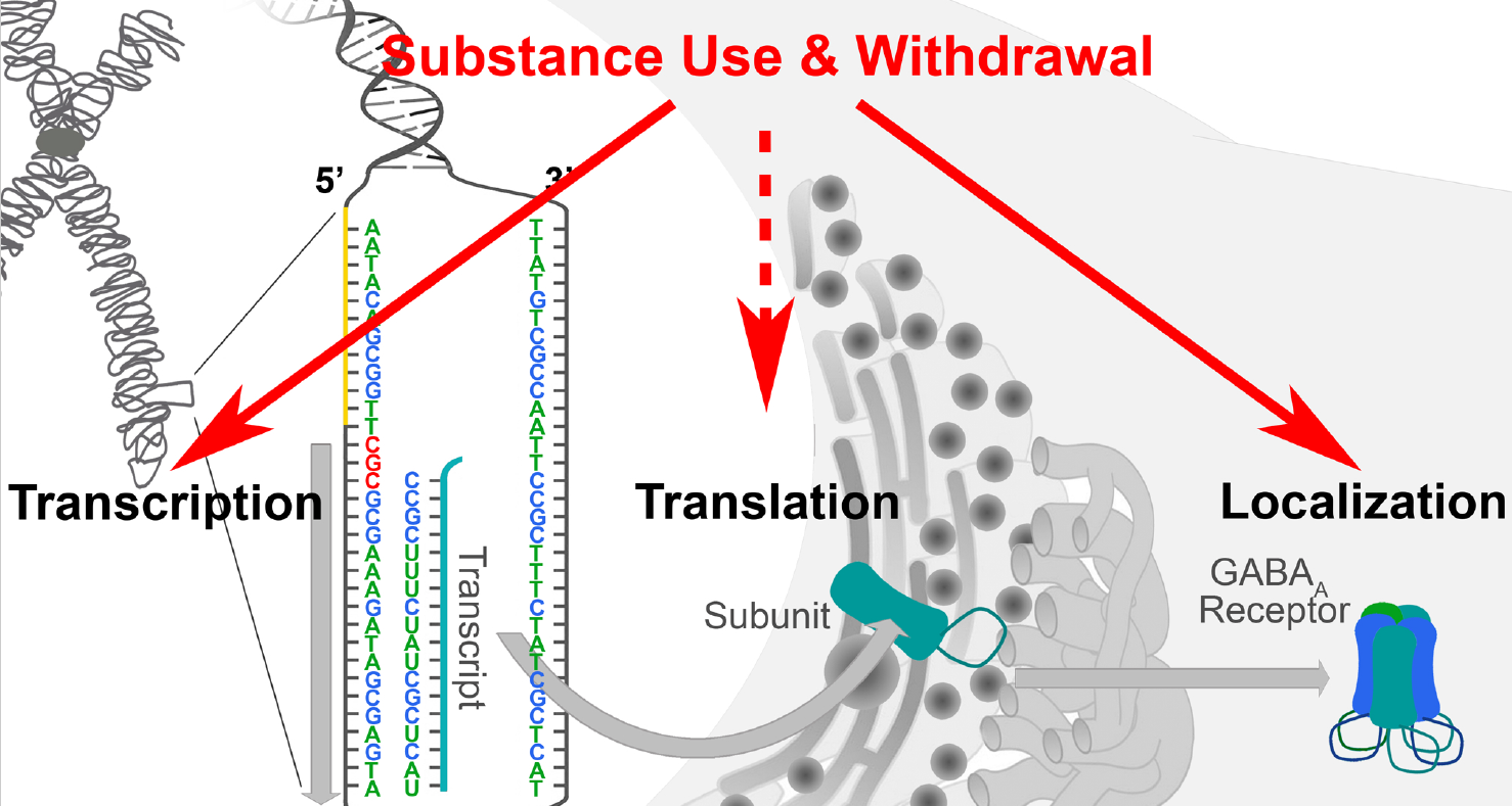
4. Substance Use Regulates GABAA Receptor Subunit Expression

Persistent substance use induces pathological plasticity in the brain’s reward system and converts the normal reinforcement of homeostatic behaviors into compulsive drug seeking. Broadly, substance use disorder is defined as an inability to control the use of a substance or drug, either illegal or legal, despite harmful consequences. Substance use disorders are diagnosed in the fifth edition of the Diagnostic and Statistical Manual of Mental Disorders (DSM-5) by 12 criteria that include hazardous use, neglecting major roles to use, tolerance, withdrawal, and cravings, amongst others [101]. Meeting more than two criterion constitutes a substance use disorder, with the severity increasing as more criteria are met. Tolerance refers to the adaptive response of the body and the nervous system to repeated substance use, where reduced responsiveness is seen for successive equal doses. Withdrawal occurs upon the abrupt removal of the substance, and involves key motivational elements such as chronic irritability, emotional pain, malaise, dysphoria, stress, and loss of motivation for natural rewards [102]. Repeated use of commonly abused substances causes positive reinforcement through dopamine (DA) signaling in the mesolimbic pathway, primarily from the ventral tegmental area (VTA) to the medial shell of the nucleus accumbens (NAc) [103,104]. This pathway is implicated in the reinforcing effects of abused substances, as well as natural reinforcers such as food [103,105]. Repeated substance use potentiates excitatory afferents onto DA neurons in the VTA, and can cause synaptic plasticity in the NAc [106]. Circuit adaptations also induce the aversive response to the abrupt termination of long-term drug use through stress mediators such as corticotropin-releasing factor and noradrenaline in the extended amygdala, as well as adaptations by the NAc [103,105]. Transcriptional and post-transcriptional gene regulation are thought to play a large role in the changes in brain function related to developing and maintaining substance use disorders [55,107].
Despite a lack of direct action on DAergic signaling, many of the drugs that positively modulate GABAARs are prone to abuse. The action of GABA on GABAARs is well known to be modulated by a variety of pharmacologically and clinically important drugs including benzodiazepines (BZs), barbiturates, steroids, and anesthetics [2]. Generally, these drugs act as positive allosteric modulators (PAMs) at unique sites on the GABAAR, resulting in alterations to the effects of GABA binding [108]. BZs typically act as PAMs of GABAARs by binding to a specific BZ site in the extracellular domain between α and γ subunits [108]. GABAARs are also the targets of general anesthetics such as propofol, etomidate, and halothane, with different anesthetics possessing distinct subunit selectivity and binding sites [108,109]. Alcohol is also thought to act upon GABAARs, stemming from the sensitivity of the GABA inhibitory Cl- current to alcohol use at low doses (≤30 mm), although some controversy exists about the precise mechanism of the modulation [108,110,111,112]. Many studies have examined the action of GABAAR PAMs, yet many questions remain about how the positive modulation of GABAergic signaling leads to the activation of the DAergic reward pathway, and the precise role for GABAergic signaling in the development and maintenance of substance use disorders.

4.1. Benzodiazepine Use

GABAARs are the primary binding sites for BZs, which are commonly used to treat disorders such as anxiety, insomnia, and epilepsy [113]. GABAARs composed of α (1,2,3, or 5), along with β and γ subunits, are considered BZ-sensitive receptors [114]. BZs modulate the Cl conductance of GABAARs by increasing the frequency at which the ion channel opens [115]. BZs are highly prescribed but are associated with a number of side effects, and also display a rapid development of tolerance, produce dependence and withdrawal symptoms, and are commonly abused substances. The development of tolerance, dependence, and withdrawal have been associated with changes in GABAAR expression, binding, and function (summarized in Table 1).
With BZs, the tolerance to anxiolytic effects has been shown 48 hours after even a single dose of lorazepam in mice [127]. The mechanisms underlying the development of tolerance are still not completely known, but altered GABAAR subunit gene expression downstream of repeated BZ use has been demonstrated. GABAAR subunit expression measured by quantitative real-time PCR during the chronic administration of BZs in vivo demonstrated a 50% decrease in the levels of cortical α1 subunit mRNA after 14 or 28 days of administration of the full PAM lorazepam [116]. In contrast, fewer lorazepam administration days (1, 2, 4, 7, and 10) did not modify α1 subunit mRNA [116]. The γ2 subunit mRNA shows a similar time-course and level of reduction in response to chronic lorazepam treatment [116].
Epigenetic mechanisms may be involved in the downregulation of GABAAR subtypes that lead to the development of tolerance, particularly to full PAMs that have been well established to elicit these changes, through DNA methylation, histone acetylation and methylation, chromatin modifications and other lesser understood mechanisms [117]. Epigenetic regulation of gene expression is implicated in psychiatric disorders, including substance use disorders, as well as in nervous system adaptations to substance use such as tolerance, dependence, and withdrawal [128]. The partial PAM imidazenil does not produce tolerance for the anti-convulsant effects of BZs with chronic use and does not lead to a decreased expression of mRNA for the α1 subunit seen with full PAMs [117]. The lack of effect on α1 subunit expression with partial PAMs may be due to differences in histone acetylation at the promoter region upstream of GABRA1 [117]. The full PAM diazepam showed decreased acetylated histone H3 at the promoter, and increased MeCP2 occupancy in the promoter region [117]. The decrease in H3 acetylation was due to an increase in the expression of histone deacetylase (HDAC) enzymes HDAC1 and HDAC2 [117]. Class I HDACs are often found in a complex together with MeCP2 that acts as a transcriptional repressor at the promoter of a gene, suggesting a possible mechanism of α1 subunit downregulation when given a chronic administration of full PAMs at the BZ site [117,129,130]. The assessment of total acetylated histone H3 protein verified that the chronic use of diazepam was not reducing the overall H3 protein levels in the cortex, and the reduction was specific to that bound to the promoter for GABRA1 [117].
Beyond subunit expression, multiple studies have shown evidence for the uncoupling of GABA and BZ binding sites wherein allosteric enhancement is reduced without changes in binding affinity [113,131,132,133]. When the full PAM diazepam or partial PAM Ro 16 6028 are administered, rat neurons in vitro showed a 50% reduction in receptor sensitivity when administered GABA [134]. Early experiments used embryonic chick neuronal cultures to show a similar rapid development of tolerance to the BZ flurazepam via uncoupling of about 34% in 18h [131]. An experiment in which rat cultured cortical neurons were administered diazepam for 48 h and measured for potentiation via a binding assay showed the uncoupling of the sites [135]. Furthermore, uncoupling was prevented by use of the BZ site antagonist flumazenil, or picrotoxin, a GABAAR channel blocker, or nifedipine, an inhibitor of L-type voltage gated Ca2+ channels, all pointing towards a mechanism that involves the binding of diazepam to a specific site and the subsequent activation of a GABAAR.
The surface level of postsynaptic GABAARs is related to the strength of synaptic inhibition and is modulated by regulated trafficking steps to and from the plasma membrane. Receptor removal, degradation, insertion and diffusion have all been shown to be dynamically regulated [136]. Jacob et al. looked at GABAAR surface levels after treatment with the BZ flurazepam, and demonstrated a dramatic decrease in α2 subunit-containing GABAAR surface expression along with total levels of the subunit [113]. BZ treatment did not alter the insertion and endocytosis rates of α2-containing GABAARs, but did promote degradation, which was reversed by blocking lysosomal degradation [113]. This loss in total GABAAR levels may begin the series of adaptations that contribute to tolerance, as degradation occurs over the first 24 h of treatment.

4.2. Alcohol Use

Alcohol use produces anxiolytic, anticonvulsant, sedative-hypnotic, cognitive-impairing, and motor coordination-impairing properties, similar to other drugs that act on the GABAergic system. The mechanisms behind the effects of alcohol include direct and indirect effects on GABAARs, as well as modulation of GABA release and the synthesis, and availability of endogenous neuroactive steroids [120], as well as actions on other neurotransmitter systems. Research on the exposure to alcohol has shown that there has to be epigenetic mechanisms involved in alcohol use and the development of alcohol use disorder [137,138]. In search of a reliable bio-marker for chronic alcohol use, an epigenome-wide association study was performed looking at the differing methylation of CpG sites in relation to alcohol consumption levels across 13 different cohorts [139]. They found 144 CpGs to be associated with alcohol consumption, and that epigenetic changes in GABAAR and GABABR subunit genes were significantly associated with the expression level of a number of genes that are involved in immune function [139]. Of the CpGs most significantly tied to alcohol use, cg04781796 is located on a CpG island intronic to the GABAAR δ subunit (GABRD), and cg09577455 is located on a CpG island intronic to the GABABR subunit 1 (GABBR1), and both are implicated in immune function [139]. Similar to BZs, the modulation of histone acetylation caused by a decrease in HDAC activity is seen with alcohol use, and is thought to take part in the anxiolytic effects of alcohol consumption [119]. The CREB target-genes BDNF, Arc, and neuropeptide Y are all increased in the amygdala with acute alcohol exposure [36], and may initiate cascades that result in the regulated expression of GABAAR subunits.
Long-term alcohol administration has been shown to cause differential changes in the expression of GABAAR subunit mRNA and protein levels in various brain regions (summarized in Table 1) [120]. mRNA for the α2 subunit is down-regulated in the central amygdala of human alcoholics [140]. RT-PCR studies in humans have shown increased α1 expression in the frontal cortex of alcoholics, as well as total α subunit concentration compared to controls [120,121,141]. In contrast, rodent studies have shown a decrease in α1 expression and an increase in α4 expression with chronic alcohol exposure [120,122,126]. In a chronic intermittent ethanol (CIE) rat model, as well under acute alcohol doses, α2-containing GABAARs are upregulated in the hippocampus, basolateral amygdala, and NAc, all of which are implicated in regulating addiction [126]. Chronic exposure to alcohol in cynomolgus macaques reduces mRNA for α2 and α3 subunits, and increases α1 subunit mRNA in the basolateral amygdala [142]. Chronic alcohol exposure also affects GABAAR subunit mRNA expression in different cortical areas of cynomolgus macaques [143]. mRNA for α2, α4, β1, β3, γ1 and γ3 subunits are significantly reduced in the orbitofrontal cortex, while β1, β2, γ1 and δ subunit mRNAs are reduced in the dorsolateral prefrontal cortex [143].
Rodent mutant models lacking specific GABAAR subunits show changes in physiological and behavioral responses to alcohol consumption [144,145,146,147,148,149]. α1-null mutant mice show a decrease in alcohol consumption and an increased aversion to alcohol [144]. α1 and β2 null mutants experience changes in the sedative effects of alcohol and display shorter periods of loss of righting reflex following consumption [145]. α1 and β2 knockout mice also show increased locomotor stimulant effects from alcohol exposure [144,146,147]. A knockdown for α2 also displays a reduction in binge-like drinking [149]. Using viral-mediated mRNA interference to reduce α4 subunit expression in the NAc shell has been shown to reduce alcohol consumption at low-to-moderate doses, indicating that the α4 subunit may be involved in developing a preference for alcohol [150]. Mice lacking the δ subunit exhibit less of a preference for voluntary alcohol consumption [148]. δ subunit knockout mice also have reduced hyperexcitability from withdrawal after chronic alcohol exposure [148]. Studies in animal models and human alcohol use disorder populations, combined with genetic association in families with multiple alcoholics, identify the regulation of GABAAR α subunit expression as a contributor to alcohol use disorder [151,152].
miRNAs and other non-coding RNAs are emerging as targets in substance use disorders stemming from their role in experience-dependent synaptic plasticity [76,137]. miRNA analysis in frontal cortex tissue from human subjects with alcohol use disorder revealed the upregulation of about 35 miRNAs compared to the controls [153]. Target prediction and classification suggested that mediators of synaptic plasticity are major targets of the detected miRNA alterations [153]. Similarly, in animal models of chronic alcohol exposure, alterations in the expression of over 100 mRNAs and ~30 miRNAs were detected with some regional specificity [154]. Gene ontology and pathway analysis suggested that major targets were involved in synaptic plasticity among other cell signaling pathways [154]. Although GABAAR subunits have not been identified as major targets of miRNA alterations in chronic alcohol exposure, related pathways and known regulators such as BDNF have been identified across multiple studies.
In addition to the regulation of expression levels, chronic alcohol exposure has shown a reduction in α1 subunit-containing GABAAR surface expression in both the cerebral cortex and the hippocampus [155]. An experiment in cultured rat neurons from the cerebral cortex showed a dose-dependent (50 mm) reduction in surface α1 subunit-containing GABAARs after 4h of acute ethanol exposure as measured by biotinylation and Western blotting [156]. The 4 h exposure also reduced the GABAergic response to the subunit selective PAM zolpidem, further showing a functional impact of reduced surface expression [156]. The mechanism behind the internalization of GABAARs has been linked to alterations in the expression and localization of protein kinase C (PKC) [156]. Phosphorylation is a well-known regulator of GABAAR function under various physiological and pathological conditions [157,158,159], including alcohol use disorder [160]. GABAARs have a consensus site for protein kinases PKA and PKC, located on the β subunit whose phosphorylation can lead to changes in GABA binding, conductance, and possibly internalization [161,162]. A PKC activator mimics the effects of ethanol exposure on GABAergic signaling, and produces a similar reduction in surface level receptors [156]. The internalization of α1 subunit-containing GABAARs was prevented by combining the PKC inhibitor calphostin C with ethanol exposure [156]. Immunoprecipitation in conjunction with ethanol administration showed increased association between α1 subunit and PKCγ, but not PKCβ [156]. In addition, mice lacking the isoform PKCϵ show an increased behavioral response to ethanol through reduced γ2 subunit phosphorylation [163].

4.3. Use of Other Highly Abused Substances

While stimulant drugs do not directly bind to GABAARs, the use of stimulants including nicotine [164,165], cocaine [166,167,168], and amphetamines [169] has been associated with changes in GABAAR subunit expression. With similarity to families with alcohol use, several single nucleotide polymorphisms in GABRA4 and GABRA2 have been linked with an increased likelihood for nicotine dependence [164]. The α2 subunit is thought to lead to nicotine dependence by the activation of Toll-like receptor 4 in VTA neurons, leading to the activation of CREB and the upregulation of corticotropin-releasing factor and tyrosine hydroxylase, which play a role in the sensitization to and reinforcing effects of nicotine [165]. In samples from the hippocampus of human cocaine addicts, GABRG2 is down-regulated along with the gene encoding the GABAAR-associated protein gephyrin (GPHN) [168]. GABRG2 and GPHN are both up-regulated in the hippocampus of alcohol-preferring rats, which also show increased nicotine self-administration and cocaine-seeking behaviors [168]. Other studies have shown that the deletion of the GABRA2 gene abolishes the ability of cocaine to facilitate conditioned reinforcement [166]. In animal models, α2 subunit levels are decreased in the NAc shell with sensitization to cocaine, but not the NAc core [167]. Sensitization to methamphetamine in rats leads to decreased α2 expression in the NAc core and shell, and increased expression in the caudate nucleus [170]. Methamphetamine-sensitized rats also show the upregulation of mRNA for α3 and β1 subunits in the prefrontal cortex [169].
In addition to stimulant use, there are indications that opiate exposure may also induce alterations in GABAAR subunit expression [171]. A microarray study of gene families suggests a mix of the induction and repression of specific subunits in the NAc during a 14-day morphine exposure paradigm [172]. Overall, α and β subunit expression was somewhat repressed, while γ, δ, and ε subunit expression was enhanced, particularly around 8 days of morphine exposure [172].

4.4. Withdrawal

As we have described above, GABAAR expression and function have been shown to change in response to substance use, and contribute to tolerance and the development of substance use disorder. Withdrawal from an abused substance has also been shown to modify the expression and function of GABAAR subunits [123]. To observe the effects of withdrawal, hippocampal neurons in culture were given 5 days of continuous exposure to ethanol followed by a non-ethanol medium for 3–24 h, and mRNA levels, neuronal morphology, and the functional and pharmacological responses of GABAARs were examined. Ethanol treatment alone induced decreases in mRNAs for α1, α3, α4, and α5, as well as two variants of the γ2 subunit, while not causing a change in α2 mRNA levels [123]. During withdrawal, however, α2, α3, and α4 all significantly increased, peaking at around 3 h after alcohol removal, while α1 and γ2 subunits had returned to baseline by 9–12 h after ethanol removal [123]. To observe the pharmacological responses during withdrawal, cultured neurons were incubated in media containing compounds shown to reduce withdrawal symptoms in human subjects with alcohol use disorder and alcohol-dependent laboratory animals, including diazepam, gamma-hydroxybutyrate, or the GABABR agonist baclofen. Both diazepam and gamma hydroxybutyrate mitigated the withdrawal-induced increase in α2, α3, and α4 subunits [123]. The BZ diazepam, a full PAM, also shows increased α4 mRNA and total protein upon 6 h of withdrawal, coupled with decreases in α1 and γ2 subunits [118]. Interestingly, the partial PAM imidazenil that does not cause robust changes in subunit expression during use, still causes a comparable increase in α4 expression and decreased α1 and γ2 expressions during withdrawal [118]. In addition to withdrawal from alcohol and BZs, alterations in the expression of GABAAR subunits have been noted in opiate withdrawal. In morphine-tolerant rats, withdrawal induced an upregulation of ε subunit mRNA in the locus coeruleus [173], while a microarray study suggested a relatively broad downregulation of GABAAR subunit genes in the NAc [172]. Between 4 and 18 days of abstinence, there was a repression of the α, β, γ, δ, and ε subunits assayed [172]. These results suggest that changes in GABAAR subunit expression may be a common feature of withdrawal plasticity.
Withdrawal from alcohol has also been shown to lead to the decreased phosphorylation of CREB, decreased histone H3 and H4 acetylation, and decreases in BDNF, Arc, and neuropeptide Y in the amygdala, an area of the brain implicated in the anxiety induced by withdrawal [36]. These changes are the opposite of what occurs during acute exposure, and are thought to be caused by increased HDAC activity upon withdrawal [36]. Inhibiting HDAC activity in rats reduces alcohol withdrawal-induced hyperalgesia, suggesting that epigenetic modifications stemming from withdrawal can also alter pain processing [174]. Protracted abstinence from chronic alcohol exposure in rats has also revealed alterations in miRNA levels in the frontal cortex [175]. In this study, over 40 rat miRNAs were found to be altered in the frontal cortex in the protracted abstinence phase, along with alteration in approximately 165 mRNAs [175]. Using miRNA–mRNA expression pairing revealed 33 miRNAs putatively targeting 88 mRNAs, many of which were involved in the regulation of synaptic signaling, including BDNF [175].
Taken together, these data clearly support an alteration in GABAAR expression as a downstream effect of substance use, as well as in response to withdrawal from an abused substance. In addition, changes in GABAAR subunit expression are not only seen with exposure to substances that directly act on the GABAergic system, but also with exposure to other highly abused substances like stimulants and opiates. The changes in GABAAR subunit expression vary by subunit, substance under study, experimental paradigm, time point, and brain region, adding much complexity to the picture. Changes in specific brain regions such as the NAc and VTA are associated with drug reward, while changes in cortical areas such as the prefrontal cortex, may be related to drug seeking and choice behaviors. The mechanisms that lead up to the changes in GABAAR expression are diverse, yet some common factors emerge—there is a compelling overlap with the mechanisms and factors that underly plasticity in response to learning and memory (CREB and BDNF), as well as with factors that relate to neurodevelopmental plasticity (MeCP2 and FMRP). Regardless of the complexity, changes in GABAAR expression appear to be a consistent hallmark of the maladaptive plasticity that occurs with repeated exposure to substances of abuse as well as withdrawal, warranting further detailed study. Future studies may endeavor to link changes in specific subunits, across multiple brain areas, in association with specific timepoints following exposure. It will also be important to examine specific facets of substance use disorder including reward and reinforcement, motivational and hedonic influences, as well as contributions from stress and anxiety, which are known contributors to the complex picture of substance use.

5. Conclusions

GABAARs are highly specialized receptors comprised of a variety of subunits that vary in expression based on their location and function [2,3]. The expression of specific subunits occurs in an adaptive and plastic manner, responding to stimuli ranging from sensory experience to substance use and neurodegeneration. Different classes of sunstances such as BZs and alcohol act upon and modulate the expression of GABAARs by altering the expression with regional and subunit selectivity, as well as altering receptor surface expression [113,116,117,120,122,126,155]. Transcriptional regulation of mRNA is the most prevalent means of changes in expression, but the factors controlling the transcription of individual GABAAR subunit genes are not completely known. Due to their clustered position on their respective chromosomes, GABAAR subunits are thought to have evolved from a single gene, and most GABAAR subunit genes are flanked with 5′-UTRs that are CpG-rich and lack the canonical TATA box [1,23,24]. CREB and BDNF are thought to be important and interrelated regulators of the expression of multiple GABAAR subunits, via multiple signaling pathways [96,176]. Both of these proteins are well established as key regulators of learning and memory [34,97], as well as in the plasticity associated with substance use [35,36,98,99].
Post-transcriptional regulation also contributes to the expression of GABAAR subunits. The 3′UTRrome of the genes coding for α subunits is longer than the general 3′UTRome in mice, leading to the availability of many binding sites for RBPs and miRNAs to exert the translational control of expression [1]. A small number of RBPs have been identified to regulate the translation of GABAAR subunit mRNAs into protein, including Pum2 [63,177], NONO [64,65] and FMRP [55,67,68,69,70,71,72,73,74]. Multiple binding sites for miRNAs have been identified on the 3′UTR of GABAAR subunit genes. miRNAs are implicated in experience-dependent plasticity at the 3′UTR, and are emerging as a candidate in the development of substance use disorders by changing the expression of the genes involved in dependence and withdrawal [75,76,77,137,138].
BZs and alcohol act upon GABAARs, and both cause changes in the expression of subunits, as well as epigenetic modifications such as increased histone acetylation through the inhibition of HDACs [36,117,138,174]. The most consistent changes identified across both BZ and alcohol use in both human subjects and animal models include alterations in the expression of α1, 2, 4, and γ2 subunits [113,116,117,118,120,121,122,123,126,136,141,149,151,155,156]. While changes in the expression and regulation of multiple subunits has been identified in alcohol use disorder and animal models, the α2 subunit has also been genetically linked to alcoholism [108,149,151,152]. The α2 subunit is known to mediate the anxiolytic effects of alcohol, and is also upregulated in the amygdala during withdrawal states characterized by increased anxiety [118,123,174]. The use of other commonly abused substances such as stimulants also modifies the expression of GABAAR subunits, again with the α2 subunit identified by genetic, human postmortem and animal model studies [164,165,166,167,168,169,170,171,172]. While RBP binding sites have been identified for the α2 subunit, specific miRNAs and RBPs that may be affecting expression require further examination, particularly in the context of substance use. Identifying the factors upstream of expression regulation could possibly lead to better, more specific therapies for substance use disorder targeting the 3′UTR, as well as a further understanding of the factors regulating gene expression during acute and chronic exposure to drugs, and subsequent withdrawal.

Author Contributions

J.S.B. and R.M.H. co-wrote the manuscript. R.M.H. prepared the figures, J.S.B. prepared the table. All authors have read and agreed to the published version of the manuscript.

Funding

The publication fees for this article were supported by the UNLV University Libraries Open Article Fund.

Conflicts of Interest

The authors declare no conflict of interest.

References

  1. Steiger, J.L.; Russek, S.J. GABAA receptors: Building the bridge between subunit mRNAs, their promoters, and cognate transcription factors. Pharmacol. Ther. 2004, 101, 259–281. [Google Scholar] [CrossRef] [PubMed]
  2. Sieghart, W.; Fuchs, K.; Tretter, V.; Ebert, V.; Jechlinger, M.; Höger, H.; Adamiker, D. Structure and subunit composition of GABAA receptors. Neurochem. Int. 1999, 34, 379–385. [Google Scholar] [CrossRef]
  3. Backus, K.H.; Arigoni, M.; Drescher, U.; Scheurer, L.; Malherbe, P.; Möhler, H.; Benson, J.A. Stoichiometry of a recombinant GABAA receptor deduced from mutation-induced rectification. Neuroreport 1993, 5, 285–288. [Google Scholar] [CrossRef]
  4. Laurie, D.J.; Wisden, W.; Seeburg, P.H. The distribution of thirteen GABAA receptor subunit mRNAs in the rat brain. III. Embryonic and postnatal development. J. Neurosci. 1992, 12, 4151–4172. [Google Scholar] [CrossRef]
  5. Ganguly, K.; Schinder, A.F.; Wong, S.T.; Poo, M. GABA Itself Promotes the Developmental Switch of Neuronal GABAergic Responses from Excitation to Inhibition. Cell 2001, 105, 521–532. [Google Scholar] [CrossRef] [Green Version]
  6. Wisden, W.; Laurie, D.J.; Monyer, H.; Seeburg, P.H. The Distribution of 13 GABA, Receptor Subunit mRNAs in the Rat Brain. I. Telencephalon, Diencephalon, Mesencephalon. J. Neurosci. 1992, 12, 1040–1062. [Google Scholar] [CrossRef] [Green Version]
  7. Pirker, S.; Schwarzer, C.; Wieselthaler, A.; Sieghart, W.; Sperk, G. GABAA receptors: Immunocytochemical distribution of 13 subunits in the adult rat brain. Neuroscience 2000, 101, 815–850. [Google Scholar] [CrossRef]
  8. Hörtnagl, H.; Tasan, R.O.; Wieselthaler, A.; Kirchmair, E.; Sieghart, W.; Sperk, G. Patterns of mRNA and protein expression for 12 GABAA receptor subunits in the mouse brain. Neuroscience 2013, 236, 345–372. [Google Scholar] [CrossRef] [PubMed] [Green Version]
  9. Wu, X.; Wu, Z.; Ning, G.; Guo, Y.; Ali, R.; Macdonald, R.L.; De Blas, A.L.; Luscher, B.; Chen, G. GABAA receptor alpha subunits play a direct role in synaptic versus extrasynaptic targeting. J. Biol. Chem. 2012, 287, 27417–27430. [Google Scholar] [CrossRef] [Green Version]
  10. Körber, C.; Richter, A.; Kaiser, M.; Schlicksupp, A.; Mükusch, S.; Kuner, T.; Kirsch, J.; Kuhse, J. Effects of distinct collybistin isoforms on the formation of GABAergic synapses in hippocampal neurons. Mol. Cell. Neurosci. 2012, 50, 250–259. [Google Scholar] [CrossRef] [PubMed]
  11. Panzanelli, P.; Gunn, B.G.; Schlatter, M.C.; Benke, D.; Tyagarajan, S.K.; Scheiffele, P.; Belelli, D.; Lambert, J.J.; Rudolph, U.; Fritschy, J.-M. Distinct mechanisms regulate GABAA receptor and gephyrin clustering at perisomatic and axo-axonic synapses on CA1 pyramidal cells. J. Physiol. 2011, 589, 4959–4980. [Google Scholar] [CrossRef] [Green Version]
  12. Kneussel, M.; Brandstätter, J.H.; Gasnier, B.; Feng, G.; Sanes, J.R.; Betz, H. Gephyrin-independent clustering of postsynaptic GABA(A) receptor subtypes. Mol. Cell. Neurosci. 2001, 17, 973–982. [Google Scholar] [CrossRef] [Green Version]
  13. Eyre, M.D.; Renzi, M.; Farrant, M.; Nusser, Z. Setting the Time Course of Inhibitory Synaptic Currents by Mixing Multiple GABAA Receptor Subunit Isoforms. J. Neurosci. 2012, 32, 5853–5867. [Google Scholar] [CrossRef] [PubMed] [Green Version]
  14. Bright, D.P.; Renzi, M.; Bartram, J.; McGee, T.P.; MacKenzie, G.; Hosie, A.M.; Farrant, M.; Brickley, S.G. Profound desensitization by ambient GABA limits activation of δ-containing GABAA receptors during spillover. Version 2. J. Neurosci. 2011, 31, 753–763. [Google Scholar] [CrossRef] [PubMed]
  15. Picton, A.J.; Fisher, J.L. Effect of the alpha subunit subtype on the macroscopic kinetic properties of recombinant GABA(A) receptors. Brain Res. 2007, 1165, 40–49. [Google Scholar] [CrossRef] [Green Version]
  16. Bianchi, M.T.; Haas, K.F.; Macdonald, R.L. α1 and α6 subunits specify distinct desensitization, deactivation and neurosteroid modulation of GABAA receptors containing the δ subunit. Neuropharmacology 2002, 43, 492–502. [Google Scholar] [CrossRef]
  17. Mortensen, M.; Patel, B.; Smart, T.G. GABA Potency at GABAA Receptors Found in Synaptic and Extrasynaptic Zones. Front. Cell. Neurosci. 2012, 6, 1–10. [Google Scholar] [CrossRef] [PubMed] [Green Version]
  18. Schofield, C.M.; Huguenard, J.R. GABA Affinity Shapes IPSCs in Thalamic Nuclei. J. Neurosci. 2007, 27, 7954–7962. [Google Scholar] [CrossRef] [PubMed]
  19. Keramidas, A.; Harrison, N.L. The activation mechanism of α1β2γ2S and α3β3γ2S GABAA receptors. J. Gen. Physiol. 2010, 135, 59–75. [Google Scholar] [CrossRef]
  20. Dixon, C.; Sah, P.; Lynch, J.W.; Keramidas, A. GABAA Receptor α and γ Subunits Shape Synaptic Currents via Different Mechanisms. J. Biol. Chem. 2014, 289, 5399–5411. [Google Scholar] [CrossRef] [Green Version]
  21. Mizukami, K.; Grayson, D.R.; Ikonomovic, M.D.; Sheffield, R.; Armstrong, D.M. GABAA receptor β2 and β3 subunits mRNA in the hippocampal formation of aged human brain with Alzheimer-related neuropathology. Mol. Brain Res. 1998, 56, 268–272. [Google Scholar] [CrossRef]
  22. Ali, N.J.; Olsen, R.W. Chronic benzodiazepine treatment of cells expressing recombinant GABAA receptors uncouples allosteric binding: Studies on possible mechanisms. J. Neurochem. 2001, 79, 1100–1108. [Google Scholar] [CrossRef] [PubMed]
  23. Russek, S.J. Evolution of GABAA receptor diversity in the human genome. Gene 1999, 227, 213–222. [Google Scholar] [CrossRef]
  24. Brooks-Kayal, A.R.; Shumate, M.D.; Jin, H.; Lin, D.D.; Rikhter, T.Y.; Holloway, K.L.; Coulter, D.A. Human Neuronal γ-Aminobutyric AcidA Receptors: Coordinated Subunit mRNA Expression and Functional Correlates in Individual Dentate Granule Cells. J. Neurosci. 1999, 19, 8312–8318. [Google Scholar] [CrossRef] [PubMed] [Green Version]
  25. Buckle, V.J.; Fujita, N.; Ryder-Cook, A.S.; Derry, J.M.; Barnard, P.J.; Lebo, R.V.; Schofield, P.R.; Seeburg, P.H.; Bateson, A.N.; Darlison, M.G.; et al. Chromosomal localization of GABAA receptor subunit genes: Relationship to human genetic disease. Neuron 1989, 3, 647–654. [Google Scholar] [CrossRef]
  26. McLean, P.J.; Farb, D.H.; Russek, S.J. Mapping of the alpha 4 subunit gene (GABRA4) to human chromosome 4 defines an alpha 2-alpha 4-beta 1-gamma 1 gene cluster: Further evidence that modern GABAA receptor gene clusters are derived from an ancestral cluster. Genomics 1995, 26, 580–586. [Google Scholar] [CrossRef]
  27. Wilcox, A.S.; Warrington, J.A.; Gardiner, K.; Berger, R.; Whiting, P.; Altherr, M.R.; Wasmuth, J.J.; Patterson, D.; Sikela, J.M. Human chromosomal localization of genes encoding the gamma 1 and gamma 2 subunits of the gamma-aminobutyric acid receptor indicates that members of this gene family are often clustered in the genome. Proc. Natl. Acad. Sci. USA 1992, 89, 5857–5861. [Google Scholar] [CrossRef] [Green Version]
  28. Lemon, B.; Tjian, R. Orchestrated response: A symphony of transcription factors for gene control. Genes Dev. 2000, 14, 2551–2569. [Google Scholar] [CrossRef] [Green Version]
  29. Joyce, C.J. In silico comparative genomic analysis of GABAA receptor transcriptional regulation. BMC Genomics 2007, 8, 203. [Google Scholar] [CrossRef] [Green Version]
  30. Bateson, A.N.; Ultsch, A.; Darlison, M.G. Isolation and sequence analysis of the chicken GABAA receptor α1-subunit gene promoter. Gene 1995, 153, 243–247. [Google Scholar] [CrossRef]
  31. Nair, B.; Johar, K.; Priya, A.; Wong-Riley, M.T.T. Specificity protein 4 (Sp4) transcriptionally regulates inhibitory GABAergic receptors in neurons. Biochim. Biophys. Acta 2016, 1863, 1–9. [Google Scholar] [CrossRef] [PubMed] [Green Version]
  32. Brooks-Kayal, A.R.; Russek, S.J. Regulation of GABAA Receptor Gene Expression and Epilepsy. In Jasper’s Basic Mechanisms of the Epilepsies; Noebels, J.L., Avoli, M., Rogawski, M.A., Olsen, R.W., Delgado-Escueta, A.V., Eds.; National Center for Biotechnology Information: Bethesda, MD, USA, 2012. [Google Scholar]
  33. Lonze, B.E.; Ginty, D.D. Function and regulation of CREB family transcription factors in the nervous system. Neuron 2002, 35, 605–623. [Google Scholar] [CrossRef] [Green Version]
  34. Kandel, E.R. The molecular biology of memory: cAMP, PKA, CRE, CREB-1, CREB-2, and CPEB. Mol. Brain 2012, 5, 14. [Google Scholar] [CrossRef] [PubMed] [Green Version]
  35. Carlezon, W.A., Jr.; Duman, R.S.; Nestler, E.J. The many faces of CREB. Trends Neurosci. 2005, 28, 436–445. [Google Scholar] [CrossRef] [PubMed]
  36. Pandey, S.C.; Kyzar, E.J.; Zhang, H. Epigenetic Basis of the Dark Side of Alcohol Addiction. Neuropharmacology 2017, 122, 74–84. [Google Scholar] [CrossRef]
  37. Fuchs, K.; Celepirovic, N. The 5′-flanking region of the rat GABAA receptor α2-subunit gene (Gabra2). J. Neurochem. 2002, 82, 1512–1523. [Google Scholar] [CrossRef]
  38. Hadingham, K.L.; Wingrove, P.; Le Bourdelles, B.; Palmer, K.J.; I Ragan, C.; Whiting, P.J. Cloning of cDNA sequences encoding human alpha 2 and alpha 3 gamma-aminobutyric acidA receptor subunits and characterization of the benzodiazepine pharmacology of recombinant alpha 1-, alpha 2-, alpha 3-, and alpha 5-containing human gamma-aminobutyric acidA receptors. Mol. Pharmacol. 1993, 43, 970–975. [Google Scholar]
  39. Mu, W.; Burt, D.R. The mouse GABAA receptor α3 subunit gene and promoter. Mol. Brain Res. 1999, 73, 172–180. [Google Scholar] [CrossRef]
  40. De Groen, P.C.; Eggen, B.J.; Gispen, W.H.; Schotman, P.; Schrama, L.H. Cloning and promoter analysis of the human B-50/GAP-43 gene. J. Mol. Neurosci. 1995, 6, 109–119. [Google Scholar] [CrossRef]
  41. Ma, L.; Song, L.; Radoi, G.E.; Harrison, N.L. Transcriptional regulation of the mouse gene encoding the alpha-4 subunit of the GABAA receptor. J. Biol. Chem. 2004, 279, 40451–40461. [Google Scholar] [CrossRef] [Green Version]
  42. Roberts, D.S.; Raol, Y.H.; Bandyopadhyay, S.; Lund, I.V.; Budreck, E.C.; Passini, M.J.; Wolfe, J.H.; Brooks-Kayal, A.R.; Russek, S.J. Egr3 stimulation of GABRA4 promoter activity as a mechanism for seizure-induced up-regulation of GABA(A) receptor alpha4 subunit expression. Proc. Natl. Acad. Sci. USA 2005, 102, 11894–11899. [Google Scholar] [CrossRef] [PubMed] [Green Version]
  43. Bhat, R.V.; Cole, A.J.; Baraban, J.M. Role of monoamine systems in activation of zif268 by cocaine. J. Psychiatry Neurosci. 1992, 17, 94–102. [Google Scholar] [PubMed]
  44. Blackwood, C.A.; McCoy, M.T.; Ladenheim, B.; Cadet, J.L. Escalated Oxycodone Self-Administration and Punishment: Differential Expression of Opioid Receptors and Immediate Early Genes in the Rat Dorsal Striatum and Prefrontal Cortex. Front. Neurosci. 2019, 13, 1392. [Google Scholar] [CrossRef] [Green Version]
  45. Wang, W.; Stock, R.E.; Gronostajski, R.M.; Wong, Y.W.; Schachner, M.; Kilpatrick, D.L. A role for nuclear factor I in the intrinsic control of cerebellar granule neuron gene expression. J. Biol. Chem. 2004, 279, 53491–53497. [Google Scholar] [CrossRef] [PubMed] [Green Version]
  46. McLean, P.J.; Shpektor, D.; Bandyopadhyay, S.; Russek, S.J.; Farb, D.H. A minimal promoter for the GABA(A) receptor alpha6-subunit gene controls tissue specificity. J. Neurochem. 2000, 74, 1858–1869. [Google Scholar] [CrossRef] [PubMed]
  47. Cholewa-Waclaw, J.; Shah, R.; Webb, S.; Chhatbar, K.; Ramsahoye, B.; Pusch, O.; Yu, M.; Greulich, P.; Waclaw, B.; Bird, A. Quantitative modelling predicts the impact of DNA methylation on RNA polymerase II traffic. Proc. Natl. Acad. Sci. USA 2019, 116, 14995–15000. [Google Scholar] [CrossRef] [Green Version]
  48. Chahrour, M.; Jung, S.Y.; Shaw, C.A.; Zhou, X.; Wong, S.T.C.; Qin, J.; Zoghbi, H.Y. MeCP2, a Key Contributor to Neurological Disease, Activates and Represses Transcription. Science 2008, 320, 1224–1229. [Google Scholar] [CrossRef] [Green Version]
  49. Kozinetz, C.A.; Skender, M.L.; Macnaughton, N.; Almes, M.J.; Schultz, R.J.; Percy, A.K.; Glaze, D.G. Epidemiology of Rett syndrome: A population-based registry. Pediatrics 1993, 91, 445–450. [Google Scholar]
  50. Zhang, Z.-W.; Zak, J.D.; Liu, H. MeCP2 Is Required for Normal Development of GABAergic Circuits in the Thalamus. J. Neurophysiol. 2010, 103, 2470–2481. [Google Scholar] [CrossRef] [Green Version]
  51. Samaco, R.C.; Hogart, A.; LaSalle, J.M. Epigenetic overlap in autism-spectrum neurodevelopmental disorders: MECP2 deficiency causes reduced expression of UBE3A and GABRB3. Hum. Mol. Genet. 2005, 14, 483–492. [Google Scholar] [CrossRef] [Green Version]
  52. Medrihan, L.; Tantalaki, E.; Aramuni, G.; Sargsyan, V.; Dudanova, I.; Missler, M.; Zhang, W. Early defects of GABAergic synapses in the brain stem of a MeCP2 mouse model of Rett syndrome. J. Neurophysiol. 2008, 99, 112–121. [Google Scholar] [CrossRef] [PubMed] [Green Version]
  53. Oyarzabal, A.; Xiol, C.; Castells, A.A.; Grau, C.; O’Callaghan, M.; Fernández, G.; Alcántara, S.; Pineda, M.; Armstrong, J.; Altafaj, X.; et al. Comprehensive Analysis of GABAA-A1R Developmental Alterations in Rett Syndrome: Setting the Focus for Therapeutic Targets in the Time Frame of the Disease. Int. J. Mol. Sci. 2020, 21, 518. [Google Scholar] [CrossRef] [PubMed] [Green Version]
  54. Rothwell, P.E. Autism Spectrum Disorders and Drug Addiction: Common Pathways, Common Molecules, Distinct Disorders? Front Neurosci. 2016, 10, 20. [Google Scholar] [CrossRef] [Green Version]
  55. Schieweck, R.; Kiebler, M.A. Posttranscriptional Gene Regulation of the GABA Receptor to Control Neuronal Inhibition. Front. Mol. Neurosci. 2019, 12, 152. [Google Scholar] [CrossRef]
  56. Jackson, R.J.; Hellen, C.U.T.; Pestova, T.V. The Mechanism of Eukaryotic Translation Initiation and Principles of its Regulation. Nat. Rev. Mol. Cell Biol. 2010, 11, 113–127. [Google Scholar] [CrossRef] [PubMed] [Green Version]
  57. Kanai, Y.; Dohmae, N.; Hirokawa, N. Kinesin Transports RNA: Isolation and Characterization of an RNA-Transporting Granule. Neuron 2004, 43, 513–525. [Google Scholar] [CrossRef] [PubMed] [Green Version]
  58. Fritzsche, R.; Karra, D.; Bennett, K.L.; Ang, F.Y.; Heraud-Farlow, J.E.; Tolino, M.; Doyle, M.; Bauer, K.E.; Thomas, S.; Planyavsky, M.; et al. Interactome of Two Diverse RNA Granules Links mRNA Localization to Translational Repression in Neurons. Cell Rep. 2013, 5, 1749–1762. [Google Scholar] [CrossRef]
  59. Hentze, M.W.; Castello, A.; Schwarzl, T.; Preiss, T. A brave new world of RNA-binding proteins. Nat. Rev. Mol. Cell Biol. 2018, 19, 327–341. [Google Scholar] [CrossRef]
  60. Andreassi, C.; Riccio, A. To localize or not to localize: mRNA fate is in 3′UTR ends. Trends Cell Biol. 2009, 19, 465–474. [Google Scholar] [CrossRef]
  61. Floor, S.N.; Doudna, J.A. Tunable protein synthesis by transcript isoforms in human cells. Elife 2016, 5, e10921. [Google Scholar] [CrossRef]
  62. Blair, J.D.; Hockemeyer, D.; Doudna, J.A.; Bateup, H.S.; Floor, S.N. Widespread translational remodeling during human neuronal differentiation. Cell Rep. 2017, 21, 2005–2016. [Google Scholar] [CrossRef] [PubMed] [Green Version]
  63. Follwaczny, P.; Schieweck, R.; Riedemann, T.; Demleitner, A.; Straub, T.; Klemm, A.H.; Bilban, M.; Sutor, B.; Popper, B.; Kiebler, M. Pumilio2-deficient mice show a predisposition for epilepsy. Dis. Model. Mech. 2017, 10, 1333–1342. [Google Scholar] [CrossRef] [PubMed] [Green Version]
  64. Mircsof, D.; The DDD Study; Langouet, M.; Rio, M.; Moutton, S.; Siquier-Pernet, K.; Bole-Feysot, C.; Cagnard, N.; Nitschké, P.; Gaspar, L.; et al. Mutations in NONO lead to syndromic intellectual disability and inhibitory synaptic defects. Nat. Neurosci. 2015, 18, 1731–1736. [Google Scholar] [CrossRef] [PubMed] [Green Version]
  65. Zhang, G.; Neubert, T.A.; Jordan, B.A. RNA Binding Proteins Accumulate at the Postsynaptic Density with Synaptic Activity. J. Neurosci. 2012, 32, 599–609. [Google Scholar] [CrossRef] [Green Version]
  66. Bryant, C.D.; Yazdani, N. RNA-binding proteins, neural development and the addictions. Genes Brain Behav. 2016, 15, 169–186. [Google Scholar] [CrossRef] [Green Version]
  67. Gantois, I.; Vandesompele, J.; Speleman, F.; Reyniers, E.; D’Hooge, R.; Severijnen, L.-A.; Willemsen, R.; Tassone, F.; Kooy, R.F. Expression profiling suggests underexpression of the GABAA receptor subunit δ in the fragile X knockout mouse model. Neurobiol. Dis. 2006, 21, 346–357. [Google Scholar] [CrossRef]
  68. Chen, E.; Sharma, M.R.; Shi, X.; Agrawal, R.K.; Joseph, S. Fragile X Mental Retardation Protein Regulates Translation by Binding Directly to the Ribosome. Mol. Cell 2014, 54, 407–417. [Google Scholar] [CrossRef] [Green Version]
  69. Antar, L.N.; Dictenberg, J.B.; Plociniak, M.; Afroz, R.; Bassell, G.J. Localization of FMRP-associated mRNA granules and requirement of microtubules for activity-dependent trafficking in hippocampal neurons. Genes Brain Behav. 2005, 4, 350–359. [Google Scholar] [CrossRef]
  70. Liu, B.; Li, Y.; Stackpole, E.E.; Novak, A.; Gao, Y.; Zhao, Y.; Zhao, X.; Richter, J.D. Regulatory discrimination of mRNAs by FMRP controls mouse adult neural stem cell differentiation. Proc. Natl. Acad. Sci. USA 2018, 115, E11397–E11405. [Google Scholar] [CrossRef] [Green Version]
  71. Adusei, D.C.; Pacey, L.K.K.; Chen, D.; Hampson, D.R. Early developmental alterations in GABAergic protein expression in fragile X knockout mice. Neuropharmacology 2010, 59, 167–171. [Google Scholar] [CrossRef] [Green Version]
  72. D’Hulst, C.; De Geest, N.; Reeve, S.P.; Van Dam, D.; De Deyn, P.P.; Hassan, B.A.; Kooy, R.F. Decreased expression of the GABAA receptor in fragile X syndrome. Brain Res. 2006, 1121, 238–245. [Google Scholar] [CrossRef]
  73. Hong, A.; Zhang, A.; Ke, Y.; El Idrissi, A.; Shen, C.-H. Downregulation of GABA(A) β subunits is transcriptionally controlled by Fmr1p. J. Mol. Neurosci. 2012, 46, 272–275. [Google Scholar] [CrossRef] [PubMed]
  74. Curia, G.; Papouin, T.; Séguéla, P.; Avoli, M. Downregulation of tonic GABAergic inhibition in a mouse model of fragile X syndrome. Cereb. Cortex 2009, 19, 1515–1520. [Google Scholar] [CrossRef] [PubMed] [Green Version]
  75. Bushati, N.; Cohen, S.M. microRNA Functions. Annu. Rev. Cell Dev. Biol. 2007, 23, 175–205. [Google Scholar] [CrossRef] [PubMed]
  76. Sartor, G.C.; St. Laurent, G.; Wahlestedt, C. The Emerging Role of Non-Coding RNAs in Drug Addiction. Front. Genet. 2012, 3, 106. [Google Scholar] [CrossRef] [Green Version]
  77. Zhao, C.; Huang, C.; Weng, T.; Xiao, X.; Ma, H.; Liu, L. Computational prediction of MicroRNAs targeting GABA receptors and experimental verification of miR-181, miR-216 and miR-203 targets in GABA-A receptor. BMC Res. Notes 2012, 5, 91. [Google Scholar] [CrossRef] [Green Version]
  78. Sengupta, J.; Pochiraju, S.; Pochiraju, S.; Kannampalli, P.; Bruckert, M.; Addya, S.; Yadav, P.; Miranda, A.; Shaker, R.; Banerjee, B. MicroRNA-mediated GABAAα-1 receptor subunit downregulation in adult spinal cord following neonatal cystitis-induced chronic visceral pain in rats. Pain 2013, 154, 59–70. [Google Scholar] [CrossRef] [Green Version]
  79. Bekdash, R.A.; Harrison, N.L. Downregulation of Gabra4 expression during alcohol withdrawal is mediated by specific microRNAs in cultured mouse cortical neurons. Brain Behav. 2015, 5, e00355. [Google Scholar] [CrossRef] [Green Version]
  80. Janeczek, P.; Colson, N.; Dodd, P.R.; Lewohl, J.M. Sex Differences in the Expression of the α5 Subunit of the GABAA Receptor in Alcoholics with and without Cirrhosis of the Liver. Alcohol. Clin. Exp. Res. 2020, 44, 423–434. [Google Scholar] [CrossRef]
  81. Bali, P.; Kenny, P.J. MicroRNAs and Drug Addiction. Front. Genet. 2013, 4, 335–344. [Google Scholar] [CrossRef] [Green Version]
  82. Li, M.D.; van der Vaart, A.D. MicroRNAs in addiction: Adaptation’s middlemen? Mol. Psychiatry 2011, 16, 1159–1168. [Google Scholar] [CrossRef] [PubMed]
  83. Smith, A.C.W.; Kenny, P.J. MicroRNAs regulate synaptic plasticity underlying drug addiction. Genes Brain Behav. 2018, 17, e12424. [Google Scholar] [CrossRef] [PubMed] [Green Version]
  84. Zito, K.; Svoboda, K. Activity-Dependent Synaptogenesis in the Adult Mammalian Cortex. Neuron 2002, 35, 1015–1017. [Google Scholar] [CrossRef] [Green Version]
  85. Wong, R.O.L.; Ghosh, A. Activity-dependent regulation of dendritic growth and patterning. Nat. Rev. Neurosci. 2002, 3, 803–812. [Google Scholar] [CrossRef] [PubMed]
  86. Spitzer, N.C. Electrical activity in early neuronal development. Nature 2006, 444, 707–712. [Google Scholar] [CrossRef]
  87. Katz, L.C.; Shatz, C.J. Synaptic Activity and the Construction of Cortical Circuits. Science 1996, 274, 1133–1138. [Google Scholar] [CrossRef] [PubMed] [Green Version]
  88. Lin, Y.; Bloodgood, B.L.; Hauser, J.L.; Lapan, A.D.; Koon, A.C.; Kim, T.-K.; Hu, L.S.; Malik, A.N.; Greenberg, M.E. Activity-dependent regulation of inhibitory synapse development by Npas4. Nature 2008, 455, 1198–1204. [Google Scholar] [CrossRef]
  89. Hartzell, A.L.; Martyniuk, K.M.; Brigidi, G.S.; Heinz, D.A.; Djaja, N.A.; Payne, A.; Bloodgood, B.L. NPAS4 recruits CCK basket cell synapses and enhances cannabinoid-sensitive inhibition in the mouse hippocampus. eLife 2018, 7, e35927. [Google Scholar] [CrossRef] [PubMed]
  90. Benevento, L.A.; Bakkum, B.W.; Cohen, R.S. gamma-Aminobutyric acid and somatostatin immunoreactivity in the visual cortex of normal and dark-reared rats. Brain Res. 1995, 689, 172–182. [Google Scholar] [CrossRef]
  91. Hensch, T.K. Critical period plasticity in local cortical circuits. Nat. Rev. Neurosci. 2005, 6, 877–888. [Google Scholar] [CrossRef]
  92. Marty, S.; Wehrlé, R.; Sotelo, C. Neuronal Activity and Brain-Derived Neurotrophic Factor Regulate the Density of Inhibitory Synapses in Organotypic Slice Cultures of Postnatal Hippocampus. J. Neurosci. 2000, 20, 8087–8095. [Google Scholar] [CrossRef] [PubMed] [Green Version]
  93. Turrigiano, G.G.; Nelson, S.B. Homeostatic plasticity in the developing nervous system. Nat. Rev. Neurosci. 2004, 5, 97–107. [Google Scholar] [CrossRef] [PubMed]
  94. Renier, N.; Adams, E.L.; Kirst, C.; Wu, Z.; Azevedo, R.; Kohl, J.; Autry, A.E.; Kadiri, L.; Venkataraju, K.U.; Zhou, Y.; et al. Mapping of Brain Activity by Automated Volume Analysis of Immediate Early Genes. Cell 2016, 165, 1789–1802. [Google Scholar] [CrossRef] [PubMed] [Green Version]
  95. Bullitt, E. Expression of c-fos-like protein as a marker for neuronal activity following noxious stimulation in the rat. J. Comp. Neurol. 1990, 296, 517–530. [Google Scholar] [CrossRef]
  96. Roberts, D.S.; Hu, Y.; Lund, I.V.; Brooks-Kayal, A.R.; Russek, S.J. Brain-derived Neurotrophic Factor (BDNF)-induced Synthesis of Early Growth Response Factor 3 (Egr3) Controls the Levels of Type A GABA Receptorα4 Subunits in Hippocampal Neurons. J. Biol. Chem. 2006, 281, 29431–29435. [Google Scholar] [CrossRef] [PubMed] [Green Version]
  97. Cunha, C.; Brambilla, R.; Thomas, K.L. A Simple Role for BDNF in Learning and Memory? Front. Mol. Neurosci. 2010, 3, 1. [Google Scholar] [CrossRef] [Green Version]
  98. Logrip, M.L.; Barak, S.; Warnault, V.; Ron, D. Corticostriatal BDNF and alcohol addiction. Brain Res. 2015, 1628, 60–67. [Google Scholar] [CrossRef] [Green Version]
  99. Koskela, M.; Bäck, S.; Voikar, V.; Richie, C.T.; Domanskyi, A.; Harvey, B.K.; Airavaara, M. Update of neurotrophic factors in neurobiology of addiction and future directions. Neurobiol. Dis. 2017, 97, 189–200. [Google Scholar] [CrossRef] [Green Version]
  100. Bloodgood, B.L.; Sharma, N.; Browne, H.A.; Trepman, A.Z.; Greenberg, M.E. The activity-dependent transcription factor NPAS4 regulates domain-specific inhibition. Nature 2013, 503, 121–125. [Google Scholar] [CrossRef]
  101. Hasin, D.S.; O’Brien, C.P.; Auriacombe, M.; Borges, G.; Bucholz, K.; Budney, A.; Compton, W.M.; Crowley, T.; Ling, W.; Petry, N.M.; et al. DSM-5 Criteria for Substance Use Disorders: Recommendations and Rationale. Am. J. Psychiatry 2013, 170, 834–851. [Google Scholar] [CrossRef] [Green Version]
  102. Koob, G.F.; Volkow, N.D. Neurobiology of addiction: A neurocircuitry analysis. Lancet Psychiatry 2016, 3, 760–773. [Google Scholar] [CrossRef]
  103. Lüscher, C.; Robbins, T.W.; Everitt, B.J. The transition to compulsion in addiction. Nat. Rev. Neurosci. 2020, 21, 247–263. [Google Scholar] [CrossRef]
  104. Sanchis-Segura, C.; Spanagel, R. Behavioural assessment of drug reinforcement and addictive features in rodents: An overview. Addict Biol. 2006, 11, 2–38. [Google Scholar] [CrossRef] [PubMed]
  105. Koob, G.F.; Volkow, N.D. Neurocircuitry of Addiction. Neuropsychopharmacol 2010, 35, 217–238. [Google Scholar] [CrossRef] [Green Version]
  106. Mameli, M.; Halbout, B.; Creton, C.; Engblom, D.; Parkitna, J.R.; Spanagel, R.; Lüscher, C. Cocaine-evoked synaptic plasticity: Persistence in the VTA triggers adaptations in the NAc. Nat. Neurosci. 2009, 12, 1036–1041. [Google Scholar] [CrossRef]
  107. Bali, P.; Kenny, P.J. Transcriptional mechanisms of drug addiction. Dialogues Clin. Neurosci. 2019, 21, 379–387. [Google Scholar] [PubMed] [Green Version]
  108. Olsen, R.W. GABAA Receptor: Positive and Negative Allosteric Modulators. Neuropharmacology 2018, 136, 10–22. [Google Scholar] [CrossRef]
  109. Forman, S.A.; Miller, K.W. Mapping General Anesthetic Sites in Heteromeric Gamma-Aminobutyric Acid Type A Receptors Reveals a Potential For Targeting Receptor Subtypes. Anesth. Analg. 2016, 123, 1263–1273. [Google Scholar] [CrossRef] [Green Version]
  110. Centanni, S.W.; Burnett, E.J.; Trantham-Davidson, H.; Chandler, L.J. Loss of δ-GABAA receptor-mediated tonic currents in the adult prelimbic cortex following adolescent alcohol exposure. Addict Biol. 2017, 22, 616–628. [Google Scholar] [CrossRef] [Green Version]
  111. Wei, W.; Faria, L.C.; Mody, I. Low ethanol concentrations selectively augment the tonic inhibition mediated by delta subunit-containing GABAA receptors in hippocampal neurons. J. Neurosci. 2004, 24, 8379–8382. [Google Scholar] [CrossRef] [Green Version]
  112. Herman, M.; Roberto, M. Cell type-specific tonic GABA signaling in the rat central amygdala is selectively altered by acute and chronic ethanol. Addict. Biol. 2016, 21, 72–86. [Google Scholar] [CrossRef] [PubMed] [Green Version]
  113. Jacob, T.C.; Michels, G.; Silayeva, L.; Haydon, J.; Succol, F.; Moss, S.J. Benzodiazepine treatment induces subtype-specific changes in GABAA receptor trafficking and decreases synaptic inhibition. Proc. Natl. Acad. Sci. USA 2012, 109, 18595–18600. [Google Scholar] [CrossRef] [PubMed] [Green Version]
  114. Rudolph, U.; Möhler, H. Analysis of GABAA receptor function and dissection of the pharmacology of benzodiazepines and general anesthetics through mouse genetics. Annu. Rev. Pharmacol. Toxicol. 2004, 44, 475–498. [Google Scholar] [CrossRef] [PubMed]
  115. Poncer, J.-C.; Dürr, R.; Gähwiler, B.H.; Thompson, S.M. Modulation of Synaptic GABAA Receptor Function by Benzodiazepines in Area CA3 of Rat Hippocampal Slice Cultures. Neuropharmacology 1996, 35, 1169–1179. [Google Scholar] [CrossRef]
  116. Kang, I.; Miller, L.G. Decreased GABAA receptor subunit mRNA concentrations following chronic lorazepam administration. Br. J. Pharmacol. 1991, 103, 1285–1287. [Google Scholar] [CrossRef] [Green Version]
  117. Auta, J.; Gatta, E.; Davis, J.M.; Pandey, S.C.; Guidotti, A. Potential role for histone deacetylation in chronic diazepam-induced downregulation of α1-GABAA receptor subunit expression. Pharmacol. Res. Perspect. 2018, 6, e00416. [Google Scholar] [CrossRef]
  118. Follesa, P.; Cagetti, E.; Mancuso, L.; Biggio, F.; Manca, A.; Maciocco, E.; Massa, F.; Desole, M.S.; Carta, M.; Busonero, F.; et al. Increase in expression of the GABA(A) receptor alpha(4) subunit gene induced by withdrawal of, but not by long-term treatment with, benzodiazepine full or partial agonists. Brain Res. Mol. Brain Res. 2001, 92, 138–148. [Google Scholar] [CrossRef]
  119. Pandey, S.C.; Ugale, R.; Zhang, H.; Tang, L.; Prakash, A. Brain Chromatin Remodeling: A Novel Mechanism of Alcoholism. J. Neurosci. 2008, 28, 3729–3737. [Google Scholar] [CrossRef]
  120. Kumar, S.; Porcu, P.; Werner, D.F.; Matthews, U.B.; Diaz-Granados, J.L.; Helfand, R.S.; Morrow, A.L. The role of GABAA receptors in the acute and chronic effects of ethanol: A decade of progress. Psychopharmacology 2009, 205, 529. [Google Scholar] [CrossRef] [Green Version]
  121. Lewohl, J.M.; Crane, D.I.; Dodd, P.R. Expression of the alpha 1, alpha 2 and alpha 3 isoforms of the GABAA receptor in human alcoholic brain. Brain Res. 1997, 751, 102–112. [Google Scholar] [CrossRef]
  122. Matthews, D.B.; Devaud, L.L.; Fritschy, J.M.; Sieghart, W.; Morrow, A.L. Differential regulation of GABA(A) receptor gene expression by ethanol in the rat hippocampus versus cerebral cortex. J. Neurochem. 1998, 70, 1160–1166. [Google Scholar] [CrossRef] [PubMed]
  123. Sanna, E.; Mosallino, M.C.; Busonero, F.; Talani, G.; Tranquilli, S.; Mameli, M.; Spiga, S.; Follesa, P.; Biggio, G. Changes in GABAA Receptor Gene Expression Associated with Selective Alterations in Receptor Function and Pharmacology after Ethanol Withdrawal. J. Neurosci. 2003, 23, 11711–11724. [Google Scholar] [CrossRef] [PubMed] [Green Version]
  124. Gutiérrez, M.L.; Ferreri, M.C.; Gravielle, M.C. GABA-induced uncoupling of GABA/benzodiazepine site interactions is mediated by increased GABAA receptor internalization and associated with a change in subunit composition. Neuroscience 2014, 257, 119–129. [Google Scholar] [CrossRef] [PubMed] [Green Version]
  125. Prasad, A.; Reynolds, J.N. Uncoupling of GABA-benzodiazepine receptors in chick cerebral cortical neurons requires co-activation of both receptor sites. Brain Res. 1992, 591, 327–331. [Google Scholar] [CrossRef]
  126. Lindemeyer, A.K.; Shen, Y.; Yazdani, F.; Shao, X.M.; Spigelman, I.; Davies, D.L.; Olsen, R.W.; Liang, J. α2 Subunit—Containing GABAA Receptor Subtypes Are Upregulated and Contribute to Alcohol-Induced Functional Plasticity in the Rat Hippocampus. Mol. Pharmacol. 2017, 92, 101–112. [Google Scholar] [CrossRef]
  127. File, S.E.; Wilks, L.J.; Mabbutt, P.S. Withdrawal, tolerance and sensitization after a single dose of lorazepam. Pharmacol. Biochem. Behav. 1988, 31, 937–940. [Google Scholar] [CrossRef]
  128. Berkel, T.D.M.; Pandey, S.C. Emerging Role of Epigenetic Mechanisms in Alcohol Addiction. Alcohol. Clin. Exp. Res. 2017, 41, 666–680. [Google Scholar] [CrossRef] [Green Version]
  129. Nott, A.; Cheng, J.; Gao, F.; Lin, Y.-T.; Gjoneska, E.; Ko, T.; Minhas, P.; Zamudio, A.V.; Meng, J.; Zhang, F.; et al. Histone deacetylase 3 associates with MeCP2 to regulate FOXO and social behavior. Nat. Neurosci. 2016, 19, 1497–1505. [Google Scholar] [CrossRef]
  130. Tesone-Coelho, C.; Varela, P.; Escosteguy-Neto, J.C.; Cavarsan, C.; Mello, L.E.; Santos-Junior, J.G. Effects of ethanol on hippocampal neurogenesis depend on the conditioned appetitive response. Addict. Biol. 2013, 18, 774–785. [Google Scholar] [CrossRef]
  131. Roca, D.J.; Rozenberg, I.; Farrant, M.; Farb, D.H. Chronic agonist exposure induces down-regulation and allosteric uncoupling of the gamma-aminobutyric acid/benzodiazepine receptor complex. Mol. Pharmacol. 1990, 37, 37–43. [Google Scholar]
  132. Holt, R.A.; Bateson, A.N.; Martin, I.L. Decreased GABA Enhancement of Benzodiazepine Binding after a Single Dose of Diazepam. J. Neurochem. 1999, 72, 2219–2222. [Google Scholar] [CrossRef] [PubMed] [Green Version]
  133. Wong, G.; Lyon, T.; Skolnick, P. Chronic exposure to benzodiazepine receptor ligands uncouples the gamma-aminobutyric acid type A receptor in WSS-1 cells. Mol. Pharmacol. 1994, 46, 1056–1062. [Google Scholar] [PubMed]
  134. Hernandez, T.D.; Heninger, C.; Wilson, M.A.; Gallager, D.W. Relationship of agonist efficacy to changes in GABA sensitivity and anticonvulsant tolerance following chronic benzodiazepine ligand exposure. Eur. J. Pharmacol. 1989, 170, 145–155. [Google Scholar] [CrossRef]
  135. Foitzick, M.F.; Medina, N.B.; Iglesias García, L.C.; Gravielle, M.C. Benzodiazepine exposure induces transcriptional down-regulation of GABAA receptor α1 subunit gene via L-type voltage-gated calcium channel activation in rat cerebrocortical neurons. Neurosci. Lett. 2020, 721, 134801. [Google Scholar] [CrossRef]
  136. Jacob, T.C.; Moss, S.J.; Jurd, R. GABAA receptor trafficking and its role in the dynamic modulation of neuronal inhibition. Nat. Rev. Neurosci. 2008, 9, 331–343. [Google Scholar] [CrossRef] [Green Version]
  137. Feng, J.; Nestler, E.J. Epigenetic Mechanisms of Drug Addiction. Curr. Opin. Neurobiol. 2013, 23, 521–528. [Google Scholar] [CrossRef] [Green Version]
  138. Starkman, B.G.; Sakharkar, A.J.; Pandey, S.C. Epigenetics—Beyond the Genome in Alcoholism. Alcohol. Res. 2012, 34, 293–305. [Google Scholar]
  139. Liu, C.; Marioni, R.E.; Hedman, Å.K.; Pfeiffer, L.; Tsai, P.-C.; Reynolds, L.M.; Just, A.C.; Duan, Q.; Boer, C.G.; Tanaka, T.; et al. A DNA methylation biomarker of alcohol consumption. Mol. Psychiatry 2018, 23, 422–433. [Google Scholar] [CrossRef]
  140. Jin, Z.; Bhandage, A.K.; Bazov, I.; Kononenko, O.; Bakalkin, G.; Korpi, E.R.; Birnir, B. Expression of specific ionotropic glutamate and GABA-A receptor subunits is decreased in central amygdala of alcoholics. Front. Cell. Neurosci. 2014, 8, 288. [Google Scholar] [CrossRef]
  141. Mitsuyama, H.; Little, K.Y.; Sieghart, W.; Devaud, L.L.; Morrow, A.L. GABA(A) receptor alpha1, alpha4, and beta3 subunit mRNA and protein expression in the frontal cortex of human alcoholics. Alcohol. Clin. Exp. Res. 1998, 22, 815–822. [Google Scholar]
  142. Floyd, D.W.; Friedman, D.P.; Daunais, J.B.; Pierre, P.J.; Grant, K.A.; McCool, B.A. Long-Term Ethanol Self-Administration by Cynomolgus Macaques Alters the Pharmacology and Expression of GABAA Receptors in Basolateral Amygdala. J. Pharmacol. Exp. Ther. 2004, 311, 1071–1079. [Google Scholar] [CrossRef] [PubMed] [Green Version]
  143. Hemby, S.E.; O’Connor, J.A.; Acosta, G.; Floyd, D.; Anderson, N.; McCool, B.A.; Friedman, D.; Grant, K.A. Ethanol-Induced Regulation of GABAA Subunit mRNAs in Prefrontal Fields of Cynomolgus Monkeys. Alcohol. Clin. Exp. Res. 2006, 30, 1978–1985. [Google Scholar] [CrossRef] [PubMed]
  144. Blednov, Y.A.; Walker, D.; Alva, H.; Creech, K.; Findlay, G.; Harris, R.A. GABAA Receptor α1 and β2 Subunit Null Mutant Mice: Behavioral Responses to Ethanol. J. Pharmacol. Exp. Ther. 2003, 305, 854–863. [Google Scholar] [CrossRef] [PubMed]
  145. Blednov, Y.A.; Jung, S.; Alva, H.; Wallace, D.; Rosahl, T.; Whiting, P.-J.; Harris, R.A. Deletion of the α1 or β2 Subunit of GABAAReceptors Reduces Actions of Alcohol and Other Drugs. J. Pharmacol. Exp. Ther. 2003, 304, 30–36. [Google Scholar] [CrossRef] [PubMed] [Green Version]
  146. Boehm, S.L.; Ponomarev, I.; Jennings, A.W.; Whiting, P.J.; Rosahl, T.W.; Garrett, E.M.; Blednov, Y.A.; Harris, R.A. γ-Aminobutyric acid A receptor subunit mutant mice: New perspectives on alcohol actions. Biochem. Pharmacol. 2004, 68, 1581–1602. [Google Scholar] [CrossRef]
  147. June, H.L.; Foster, K.L.; Eiler, W.J.A.; Goergen, J.; Cook, J.B.; Johnson, N.; Mensah-Zoe, B.; Simmons, J.O.; Yin, W.; Cook, J.M.; et al. Dopamine and Benzodiazepine-Dependent Mechanisms Regulate the EtOH-Enhanced Locomotor Stimulation in the GABA A α 1 Subunit Null Mutant Mice. Neuropsychopharmacology 2007, 32, 137–152. [Google Scholar] [CrossRef] [PubMed] [Green Version]
  148. Mihalek, R.M.; Bowers, B.J.; Wehner, J.M.; Kralic, J.E.; Vandoren, M.J.; Morrow, A.L.; Homanics, G.E. GABAA-Receptor δ Subunit Knockout Mice Have Multiple Defects in Behavioral Responses to Ethanol. Alcohol. Clin. Exp. Res. 2001, 25, 1708–1718. [Google Scholar]
  149. Liu, J.; Yang, A.R.; Kelly, T.; Puche, A.; Esoga, C.; Elnabawi, A.; Merchenthaler, I.; Sieghart, W.; June, H.L.; Aurelian, L. Binge alcohol drinking is associated with GABAA α2-regulated Toll-like receptor 4 (TLR4) expression in the central amygdala. Proc. Natl. Acad. Sci. USA 2011, 108, 4465–4470. [Google Scholar] [CrossRef] [Green Version]
  150. Rewal, M.; Jurd, R.; Gill, T.M.; He, D.; Ron, D.; Janak, P.H. α4-Containing GABAA Receptors in the Nucleus Accumbens Mediate Moderate Intake of Alcohol. J. Neurosci. 2009, 29, 543–549. [Google Scholar] [CrossRef]
  151. Edenberg, H.J.; Dick, D.M.; Xuei, X.; Tian, H.; Almasy, L.; Bauer, L.O.; Crowe, R.R.; Goate, A.; Hesselbrock, V.; Jones, K.; et al. Variations in GABRA2, encoding the alpha 2 subunit of the GABA(A) receptor, are associated with alcohol dependence and with brain oscillations. Am. J. Hum. Genet. 2004, 74, 705–714. [Google Scholar] [CrossRef] [Green Version]
  152. Bierut, L.J.; Agrawal, A.; Bucholz, K.K.; Doheny, K.F.; Laurie, C.; Pugh, E.; Fisher, S.; Fox, L.; Howells, W.; Bertelsen, S.; et al. A genome-wide association study of alcohol dependence. Proc. Natl. Acad. Sci. USA 2010, 107, 5082–5087. [Google Scholar] [CrossRef] [PubMed] [Green Version]
  153. Lewohl, J.M.; Nunez, Y.O.; Dodd, P.R.; Tiwari, G.R.; Harris, R.A.; Mayfield, R.D. Up-Regulation of MicroRNAs in Brain of Human Alcoholics. Alcohol. Clin. Exp. Res. 2011, 35, 1928–1937. [Google Scholar] [CrossRef] [PubMed] [Green Version]
  154. Sinirlioglu, Z.A.; Coskunpinar, E.; Akbas, F. miRNA and mRNA expression profiling in rat brain following alcohol dependence and withdrawal. Cell. Mol. Biol. 2017, 63, 49–56. [Google Scholar] [CrossRef] [PubMed]
  155. Kumar, S.; Kralic, J.E.; O’Buckley, T.K.; Grobin, A.C.; Morrow, A.L. Chronic ethanol consumption enhances internalization of α1 subunit-containing GABAA receptors in cerebral cortex. J. Neurochem. 2003, 86, 700–708. [Google Scholar] [CrossRef]
  156. Kumar, S.; Suryanarayanan, A.; Boyd, K.N.; Comerford, C.E.; Lai, M.A.; Ren, Q.; Morrow, A.L. Ethanol Reduces GABAA α1 Subunit Receptor Surface Expression by a Protein Kinase Cγ-Dependent Mechanism in Cultured Cerebral Cortical Neurons. Mol. Pharmacol. 2010, 77, 793–803. [Google Scholar] [CrossRef] [Green Version]
  157. Moss, S.J.; Doherty, C.A.; Huganir, R.L. Identification of the cAMP-dependent protein kinase and protein kinase C phosphorylation sites within the major intracellular domains of the beta 1, gamma 2S, and gamma 2L subunits of the gamma-aminobutyric acid type A receptor. J. Biol. Chem. 1992, 267, 14470–14476. [Google Scholar]
  158. McDonald, B.J.; Moss, S.J. Differential phosphorylation of intracellular domains of gamma-aminobutyric acid type A receptor subunits by calcium/calmodulin type 2-dependent protein kinase and cGMP-dependent protein kinase. J. Biol. Chem. 1994, 269, 18111–18117. [Google Scholar]
  159. Parakala, M.L.; Zhang, Y.; Modgil, A.; Chadchankar, J.; Vien, T.N.; Ackley, M.A.; Doherty, J.J.; Davies, P.A.; Moss, S.J. Metabotropic, but not allosteric, effects of neurosteroids on GABAergic inhibition depend on the phosphorylation of GABAA receptors. J. Biol. Chem. 2019, 294, 12220–12230. [Google Scholar] [CrossRef]
  160. Kumar, S.; Lane, B.M.; Morrow, A.L. Differential Effects of Systemic Ethanol Administration on Protein Kinase Cϵ, γ, and β Isoform Expression, Membrane Translocation, and Target Phosphorylation: Reversal by Chronic Ethanol Exposure. J. Pharmacol. Exp. Ther. 2006, 319, 1366–1375. [Google Scholar] [CrossRef]
  161. Mcdonald, B.J.; Moss, S.J. Conserved phosphorylation of the intracellular domains of GABAA receptorβ2 and β3 subunits by cAMP-dependent protein kinase, cGMP-dependent protein kinase, protein kinase C and Ca2+/calmodulin type II-dependent protein kinase. Neuropharmacology 1997, 36, 1377–1385. [Google Scholar] [CrossRef]
  162. Oh, S.; Jang, C.-G.; Ma, T.; Ho, I.K. Activation of protein kinase C by phorbol dibutyrate modulates GABAA receptor binding in rat brain slices. Brain Res. 1999, 850, 158–165. [Google Scholar] [CrossRef]
  163. Qi, Z.-H.; Song, M.; Wallace, M.; Wang, D.; Newton, P.; McMahon, T.; Chou, W.-H.; Zhang, C.; Shokat, K.; Messing, R.O. Protein Kinase Cϵ Regulates γ-Aminobutyrate Type A Receptor Sensitivity to Ethanol and Benzodiazepines through Phosphorylation of γ2 Subunits. J. Biol. Chem. 2007, 282, 33052–33063. [Google Scholar] [CrossRef] [PubMed] [Green Version]
  164. Agrawal, A.; Pergadia, M.L.; Saccone, S.F.; Hinrichs, A.L.; Lessov-Schlaggar, C.N.; Saccone, N.L.; Neuman, R.J.; Breslau, N.; Johnson, E.O.; Hatsukami, R.; et al. Gamma-aminobutyric acid receptor genes and nicotine dependence: Evidence for association from a case–control study. Addiction 2008, 103, 1027–1038. [Google Scholar] [CrossRef] [PubMed]
  165. Balan, I.; Warnock, K.T.; Puche, A.C.; Gondre-Lewis, M.C.; June, H.; Aurelian, L. The GABAA Receptor α2 Subunit Activates a Neuronal TLR4 Signal in the Ventral Tegmental Area that Regulates Alcohol and Nicotine Abuse. Brain Sci. 2018, 8, 72. [Google Scholar] [CrossRef] [PubMed] [Green Version]
  166. Dixon, C.I.; Morris, H.V.; Breen, G.; Desrivières, S.; Jugurnauth, S.; Steiner, R.C.; Vallada, H.; Guindalini, C.; Laranjeira, R.; Messas, G.; et al. Cocaine effects on mouse incentive-learning and human addiction are linked to α2 subunit-containing GABAA receptors. Proc. Natl. Acad. Sci. USA 2010, 107, 2289–2294. [Google Scholar] [CrossRef] [Green Version]
  167. Chen, Q.; Lee, T.H.; Wetsel, W.C.; Sun, Q.; Liu, Y.; Davidson, C.; Xiong, X.; Ellinwood, E.H.; Zhang, X. Reversal of cocaine sensitization-associated changes in GAD67 and GABAA receptor α2 subunit expression, and PKC ζ activity. Biochem. Biophys. Res. Commun. 2007, 356, 733–738. [Google Scholar] [CrossRef] [Green Version]
  168. Enoch, M.-A.; Zhou, Z.; Kimura, M.; Mash, D.C.; Yuan, Q.; Goldman, D. GABAergic Gene Expression in Postmortem Hippocampus from Alcoholics and Cocaine Addicts; Corresponding Findings in Alcohol-Naïve P and NP Rats. PLoS ONE 2012, 7, e29369. [Google Scholar] [CrossRef] [Green Version]
  169. Wearne, T.A.; Parker, L.M.; Franklin, J.L.; Goodchild, A.K.; Cornish, J.L. GABAergic mRNA expression is upregulated in the prefrontal cortex of rats sensitized to methamphetamine. Behav. Brain Res. 2016, 297, 224–230. [Google Scholar] [CrossRef]
  170. Zhang, X.; Lee, T.H.; Xiong, X.; Chen, Q.; Davidson, C.; Wetsel, W.C.; Ellinwood, E.H. Methamphetamine induces long-term changes in GABAA receptor α2 subunit and GAD67 expression. Biochem. Biophys. Res. Commun. 2006, 351, 300–305. [Google Scholar] [CrossRef]
  171. Ammon-Treiber, S.; Höllt, V. Morphine-induced Changes of Gene Expression in the Brain. Addict. Biol. 2005, 10, 81–89. [Google Scholar] [CrossRef]
  172. Spijker, S.; Houtzager, S.W.J.; De Gunst, M.C.M.; De Boer, W.P.H.; Schoffelmeer, A.N.M.; Smit, A.B. Morphine exposure and abstinence define specific stages of gene expression in the rat nucleus accumbens. FASEB J. 2004, 18, 848–850. [Google Scholar] [CrossRef] [PubMed]
  173. Heikkilä, A.T.; Echenko, O.; Uusi-Oukari, M.; Sinkkonen, S.T.; Korpi, E.R. Morphine withdrawal increases expression of GABA(A) receptor epsilon subunit mRNA in locus coeruleus neurons. Neuroreport 2001, 12, 2981–2985. [Google Scholar] [CrossRef] [PubMed]
  174. Pradhan, A.A.; Tipton, A.F.; Zhang, H.; Akbari, A.; Pandey, S.C. Effect of Histone Deacetylase Inhibitor on Ethanol Withdrawal-Induced Hyperalgesia in Rats. Int. J. Neuropsychopharmacol. 2019, 22, 523–527. [Google Scholar] [CrossRef] [PubMed] [Green Version]
  175. Tapocik, J.D.; Solomon, M.; Flanigan, M.E.; Meinhardt, M.; Barbier, E.; Schank, J.R.; Schwandt, M.; Sommer, W.H.; Heilig, M. Coordinated dysregulation of mRNAs and microRNAs in the rat medial prefrontal cortex following a history of alcohol dependence. Pharm. J. 2013, 13, 286–296. [Google Scholar] [CrossRef] [PubMed] [Green Version]
  176. Vithlani, M.; Hines, R.M.; Zhong, P.; Terunuma, M.; Hines, D.J.; Revilla-Sanchez, R.; Jurd, R.; Haydon, P.; Rios, M.; Brandon, N.; et al. The ability of BDNF to modify neurogenesis and depressive-like behaviors is dependent upon phosphorylation of tyrosine residues 365/367 in the GABA(A)-receptor γ2 subunit. J. Neurosci. 2013, 33, 15567–15577. [Google Scholar] [CrossRef] [PubMed] [Green Version]
  177. Staley, K. Molecular mechanisms of epilepsy. Nat. Neurosci. 2015, 18, 367–372. [Google Scholar] [CrossRef] [Green Version]
Figure 1. Levels of regulation impacting GABAAR expression and function. Expression of a GABAAR subunit begins with the initiation of transcription, which is controlled by a multiprotein complex that regulates the 5′UTR flanking genes encoding individual subunits. Transcription factors and other proteins that regulate transcription (yellow) bind to consensus sites in the 5′UTR and either promote or repress initiation. Many GABAAR subunit 5′UTRs contain consensus sites for more than one transcription factor, as well as multiple transcription start sites (red). Additional phases of splicing and polyadenylation (green) occur in the nucleus prior to the shuttling of the transcript to the rough endoplasmic reticulum for translation at ribosomes (grey circles). Translation of the subunit transcripts has been shown to be regulated by RNA-binding proteins (RBPs; turquoise), as well as microRNAs (miRNAs; turquoise). Once translated, the subunits are assembled (blue) together in quaternary structure to form a functional heteropentameric GABAAR. GABAARs are also subject to regulation via post-translational modifications (blue) such as phosphorylation and palmitoylation, as well as regulated targeting and trafficking to become preferentially localized to specific subcellular domains.
Ijms 21 04445 g001
Figure 2. Overview of the major regulators of transcription and translation influencing the expression of GABAAR subunits. Generally, the 5′ UTR of GABAAR subunit genes contains multiple consensus sites for transcription factors (yellow), and in many cases, multiple transcription start sites (red). The majority of subunits lack canonical TATA and CCAAT sequences in their 5′UTRs. A number of regulators have been found to influence the transcription of multiple subunits including CREB and MeCP2. Maturation of the mRNA involves the addition of a 5′ cap as well as a poly-A tail adjacent to the 3′UTR. Mature GABAAR transcripts are transported out of the nucleus to ribosomes on the rough endoplasmic reticulum, where translation is influenced by RNA-binding proteins (blue oval) such as FMRP, NONO, and Pum2, as well as by microRNAs (miRNAs; blue line).
Ijms 21 04445 g002
Table 1. Summary of the regulation of GABAA receptor subunits with benzodiazepine (BZ) and alcohol use, and withdrawal.
Benzodiazepine (BZ) UseAlcohol Use
Transcription
  • ↓ GABAAR α1 and γ2 subunit mRNA with chronic exposure [116]
  • ↓ acetylated histone HC at α1 subunit promoter and ↑ HDAC 1 and 2 [117]
  • α4, ↓ α1, ↓ γ2 mRNAs upon withdrawal [118]
  • ↑ increase BDNF (known regulator of subunit transcription) [119]
  • α1 mRNA in frontal cortex of human alcoholics [120,121]
  • α1 and ↑ α4 mRNA chronic exposure [122]
  • α2, α3, and α4 subunit mRNAs upon withdrawal [123]
Post-Translational Modification
  • ↑ GABAAR uncoupling at BZ site [124,125]
  • β subunit and γ2 subunit phosphorylation [120]
Total Expression
  • α4 ↓ α1, and ↓ γ2 total protein upon withdrawal [118]
  • α2 expression and ↓ α1 and ↓ γ2 with acute and chronic doses [126]
Surface Expression
  • ↑ GABAAR internalization [113]
  • ↓ GABAAR α2 surface expression with 24 h exposure [113]
  • ↑ GABAAR internalization [120]

