Article

A Small Aromatic Compound Has Antifungal Properties and Potential Anti-Inflammatory Effects against Intestinal Inflammation

1
Institut National de la Santé et de la Recherche Médicale, U995/Team2, F-59000 Lille, France
2
Lille Inflammation Research International Center, University Lille, U995-LIRIC, F-59000 Lille, France
3
Service de Parasitologie Mycologie, Pôle de Biologie Pathologie Génétique, CHU Lille, F-59000 Lille, France
4
Laboratoire de Chimie Durable et Santé, Ecole des Hautes Etudes d’Ingénieur (HEI), Yncréa Hauts-de-France, 13 Rue de Toul, F-59046 Lille, France
5
Faculty of Chemistry, Al. I. Cuza’ University of Iasi, B-dul Carol 1 nr. 11, 700506 Iasi, Romania
*
Author to whom correspondence should be addressed.
Int. J. Mol. Sci. 2019, 20(2), 321; https://doi.org/10.3390/ijms20020321
Submission received: 7 December 2018 / Revised: 4 January 2019 / Accepted: 4 January 2019 / Published: 14 January 2019

Abstract

:
Resistance of the opportunistic pathogen Candida albicans to antifungal drugs has increased significantly in recent years. After screening 55 potential antifungal compounds from a chemical library, 2,3-dihydroxy-4-methoxybenzaldehyde (DHMB) was identified as having potential antifungal activity. The properties of DHMB were then assessed in vitro and in vivo against C. albicans overgrowth and intestinal inflammation. Substitution on the aromatic ring of DHMB led to a strong decrease in its biological activity against C. albicans. The MIC of DHMB was highly effective at eliminating C. albicans when compared to that of caspofungin or fluconazole. Additionally, DHMB was also effective against clinically isolated fluconazole- or caspofungin-resistant C. albicans strains. DHMB was administered to animals at high doses. This compound was not cytotoxic and was well-tolerated. In experimental dextran sodium sulphate (DSS)-induced colitis in mice, DHMB reduced the clinical and histological score of inflammation and promoted the elimination of C. albicans from the gut. This finding was supported by a decrease in aerobic bacteria while anaerobic bacteria populations were re-established in mice treated with DHMB. DHMB is a small organic molecule with antifungal properties and anti-inflammatory activity by exerting protective effects on intestinal epithelial cells.

Graphical Abstract

1. Introduction

Inflammatory bowel diseases (IBDs), comprising Crohn’s disease (CD) and ulcerative colitis, have a multifactorial aetiology with complex interactions between genetic susceptibility, the immune system, environment and the microbiota [1]. The biodiversity of the gut microbiota is frequently decreased in IBD patients, in particular a reduction in Firmicutes and an increase in Proteobacteria are often observed [2]. Escherichia coli is part of the Proteobacteria phylum and is increased greatly while Lactobacillus species belonging to the Firmicutes phylum are reduced in IBD patients [3,4]. Other observations supporting a role for the gut microbiota in IBD include an abundance of fungi in the inflamed mucosa and faeces [5,6].
Colonisation with Candida albicans, an opportunistic human fungal pathogen, is consistently increased in CD patients [7]. In addition, abundance of this fungus is correlated with serological markers of CD, namely anti-Saccharomyces cerevisiae antibodies (ASCA) [5,8]. Recently, in an experimental murine model of dextran sodium sulphate (DSS)-induced colitis, the population of aerobic bacteria, in particular E. coli and Enterococcus faecalis, increased, whereas the population of anaerobic bacteria such as Lactobacillus johnsonii and Bifidobacterium species decreased. These microbiota modifications were associated with overgrowth of fungi suggesting that dysbiosis could play a crucial role in IBD [9].
Different anti-inflammatory compounds including 5-aminosalicylic acid (5-ASA) or anti-TNFα antibodies have been used to treat IBD patients, but no compounds have a dual effect of antifungal and anti-inflammatory activity [10]. Some of the anti-inflammatory drugs are not effective at treating active CD or preventing disease relapse; they only treat the symptoms and are sometimes only effective for a few years [11]. In parallel, an increase in antifungal resistance is considered a major problem to clinicians and leads to high morbidity and mortality rates in patients with invasive mycoses [12]. The increase of new fungal species that are resistant to different classes of available antifungal drugs constitutes an urgent need for developing new antifungal compounds [13].
In the current study, we assessed the in vitro and in vivo antifungal properties of a novel compound, 2,3-dihydroxy-4-methoxybenzaldehyde (DHMB), against C. albicans colonisation and intestinal inflammation.

2. Results

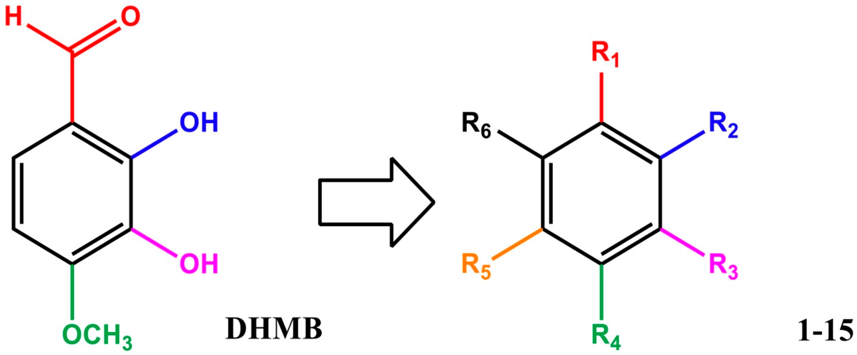
2.1. Structure and Characterisation of DHMB

A series of 55 compounds from the private chemical library of the laboratory was screened for antifungal and antibacterial activities, revealing that DHMB was a highly effective molecule against C. albicans. The DHMB MIC, at which C. albicans growth was fully inhibited, was 8 µg/mL. From a chemical point of view, DHMB is a small organic molecule (M.W. = 168.15 g/mol) bearing a highly reactive aromatic aldehyde moiety and two free phenol groups (Figure 1). We then evaluated some derivatives of DHMB for their antifungal activity; the structure and activities of these derivatives are summarised in Table 1. The biological activity of these compounds against C. albicans was evaluated at 4 × MIC (32 µg/mL). The aldehyde group in position 1 was modified with little or no change in positions 2–5 on the aromatic moiety (Table 1, entries 1–6). Deletion of the aldehyde substituent led to a total loss of activity (Compound 1, Table 1, entry 2). Compounds 2 and 3, in which the carbonyl group in position 1 is still present but included in a phenylethanone moiety, exhibited weak inhibition at 32 µg/mL (13% and 27%, respectively). Moreover, as exemplified by compounds 4 and 5, modification of the phenolic hydroxyl groups in positions 2 and 3 led to a loss of activity (Table 1, entries 5–6). Rigidification of the system and introduction of a lactone ring (compound 6) significantly decreased the biological activity (Table 1, entry 7).
From these data, the aldehyde group in position 1 seems to be essential for activity against C. albicans. The importance of the catechol and methoxy groups on the aromatic ring was also studied. Unexpectedly, replacement of the 4-methoxy substituent by a hydroxyl one led to a complete loss of activity (Table 1, entry 8). This shows that this para-methoxy group plays an essential role in the biological activity, possibly due to its hydrophobicity compared with the hydroxyl moiety. Modification of position 6 was then undertaken whilst retaining the 2,3-dihydroxy-4-methoxy- sequence on the aromatic ring. A carboxylic acid was introduced in that position (3,4-dihydroxy-5-methoxybenzoic acid, compound 8), but no activity against C. albicans was detected, and modifications of the hydrophilicity/hydrophobicity of benzoic acid derivatives did not result in a gain in activity (compounds 9 and 10, Table 1). Passing from benzoic acid to benzylic acid did not improve the activity (compare compounds 9 and 11, Table 1, entries 10 and 12). A similar conclusion can be drawn on replacement of the carboxylic acid by an ethoxy substituent at position 6 (compound 12). In the same way, replacing the carboxylic acid by an aldehyde, an acetate or a hydrogen group did not improve the activity (comparison of compound 10 with compounds 13–15).

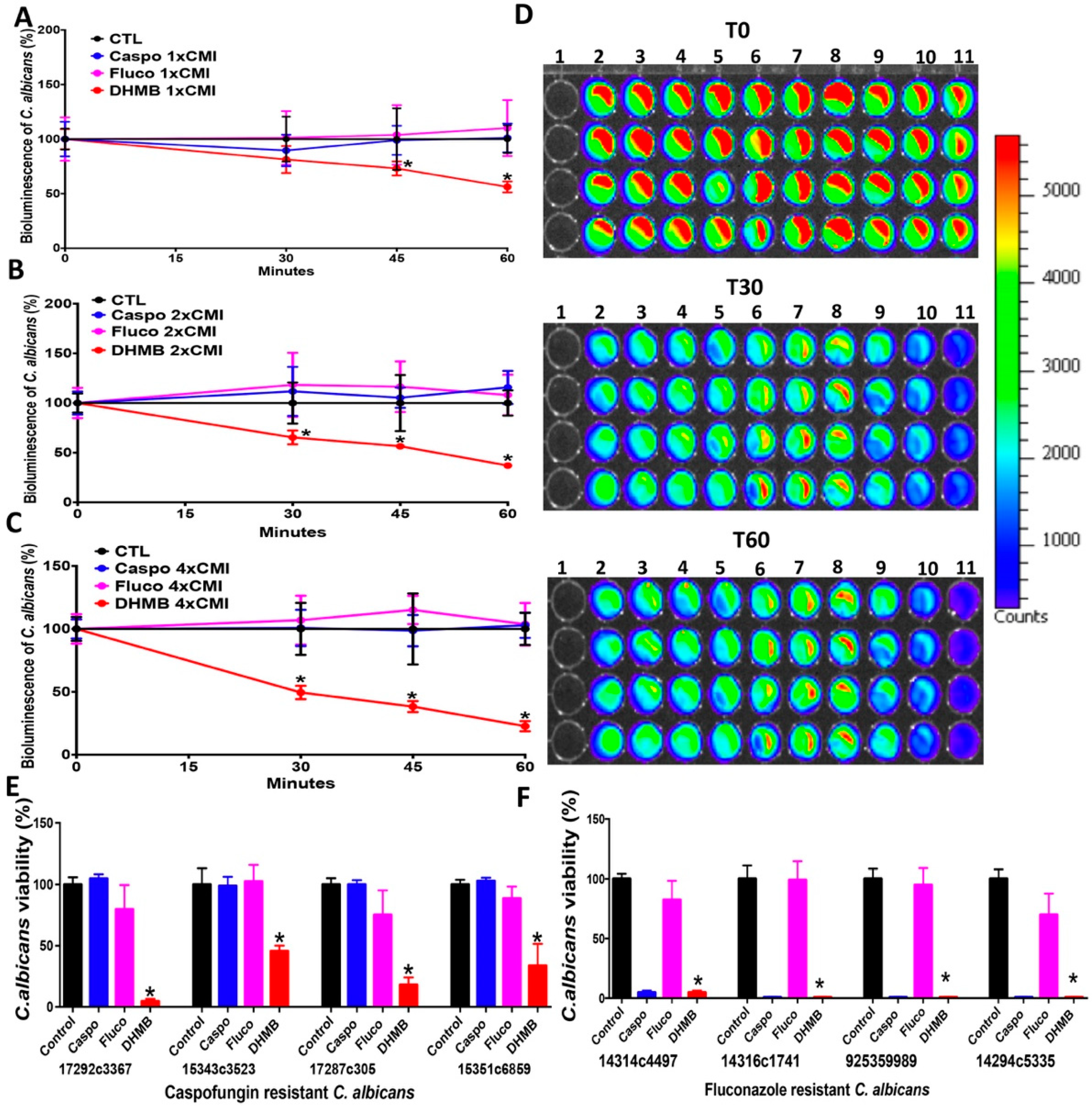
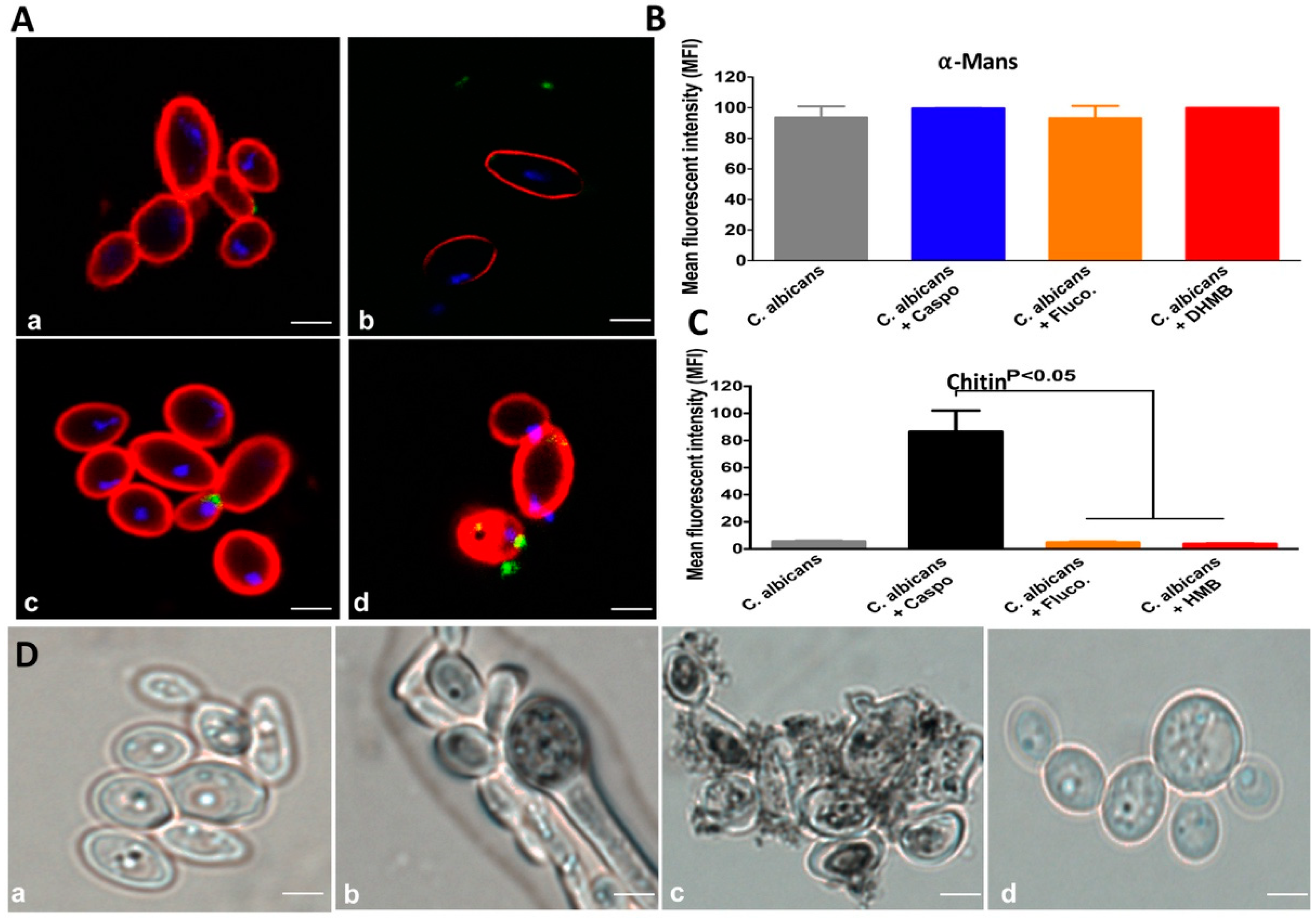
2.2. In Vitro Antifungal Activity of DHMB against C. albicans

The antifungal activity of DHMB against C. albicans was compared with that of caspofungin and fluconazole. The viability of C. albicans cells was tracked in real time using a fungal bioluminescent strain challenged with either DHMB, caspofungin or fluconazole at their MICs (8 µg/mL), 2 × MICs (16 µg/mL) or 4 × MICs (32 µg/mL) utilising different exposure times: 0, 15, 30 and 60 min (Figure 2). DHMB led to a significant reduction in bioluminescence as well as in the number of fungal colonies indicating rapid fungicidal activity after incubation with C. albicans. This fungicidal activity of DHMB was similar to that of caspofungin while fluconazole exhibited a fungistatic effect. The antifungal activity of DHMB was assessed on drug-resistant C. albicans clinical isolates (Figure 2E,F). The viability of drug-resistant C. albicans clinical isolates was significantly reduced after treatment with DHMB at 10 × MIC in terms of viable colony counts using fungal culture media (Figure 3). To assess whether DHMB has an impact on modulation of the glycan cell wall, flow cytometry analysis and confocal microscopic observations were performed. Confocal microscopy revealed that, in contrast to fluconazole and DHMB, caspofungin challenge for 1 h produced a damaged polysaccharide cell wall; in particular, the β-glucan and chitin layers were mainly affected by this antifungal treatment (Figure 3A). Similarly, flow cytometry analysis showed that treatment with either fluconazole or DHMB did not affect the fungal cell wall when compared to untreated C. albicans while C. albicans challenged with caspofungin exhibited a significant increase in chitin levels when compared to unchallenged C. albicans. No changes in mannan levels were observed in C. albicans challenged with either fluconazole, caspofungin or DHMB (Figure 3B,C). To determine whether DHMB has an impact on the melanisation, which is involved in the virulence of C. albicans, in particular resistance to antifungal agents and protection against oxidative stresses, microscopic observations were realised. A remarkable reduction in the melanin production was observed in C. albicans treated with DHMB when compared to that treated with caspofungin (Figure 3D).

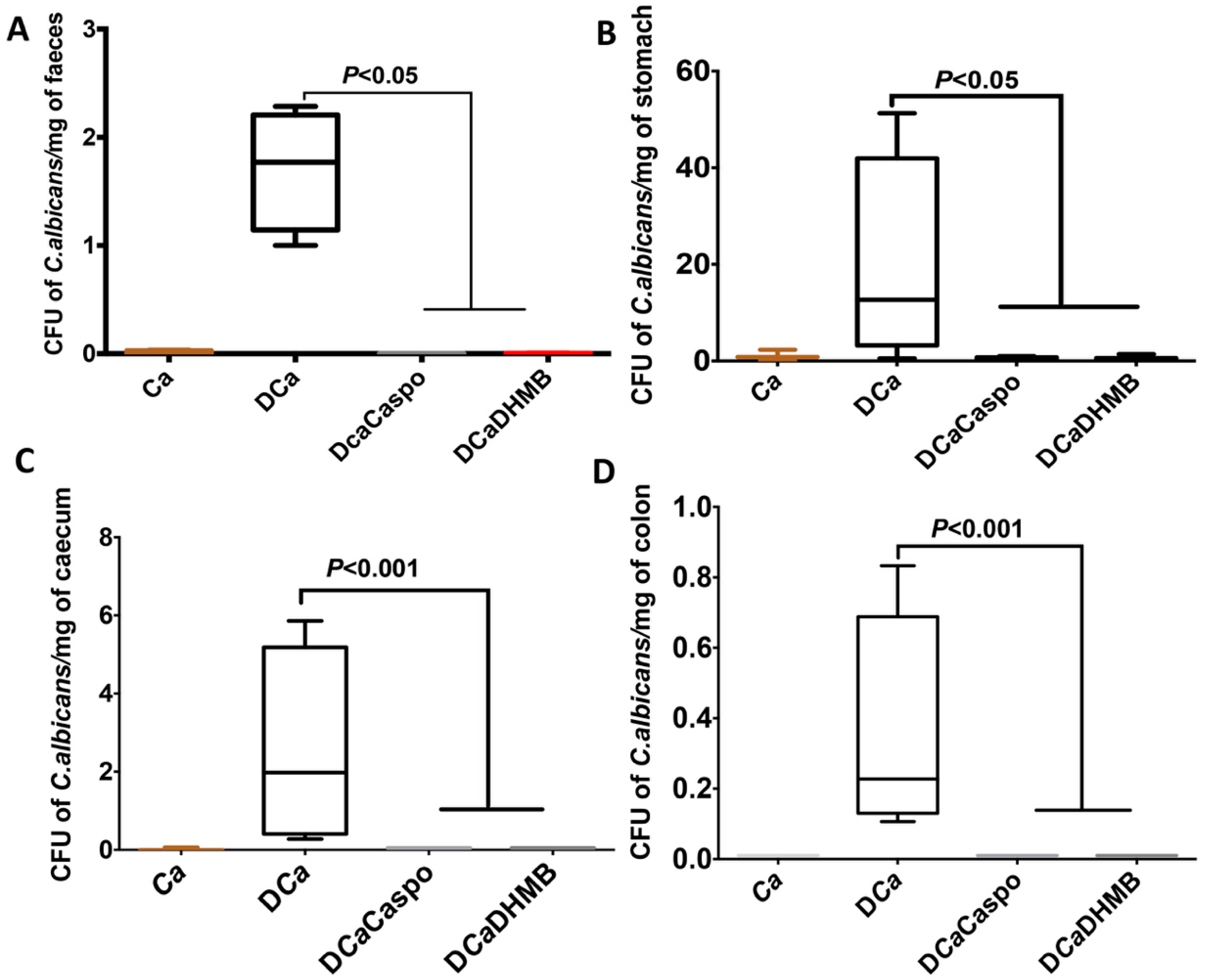
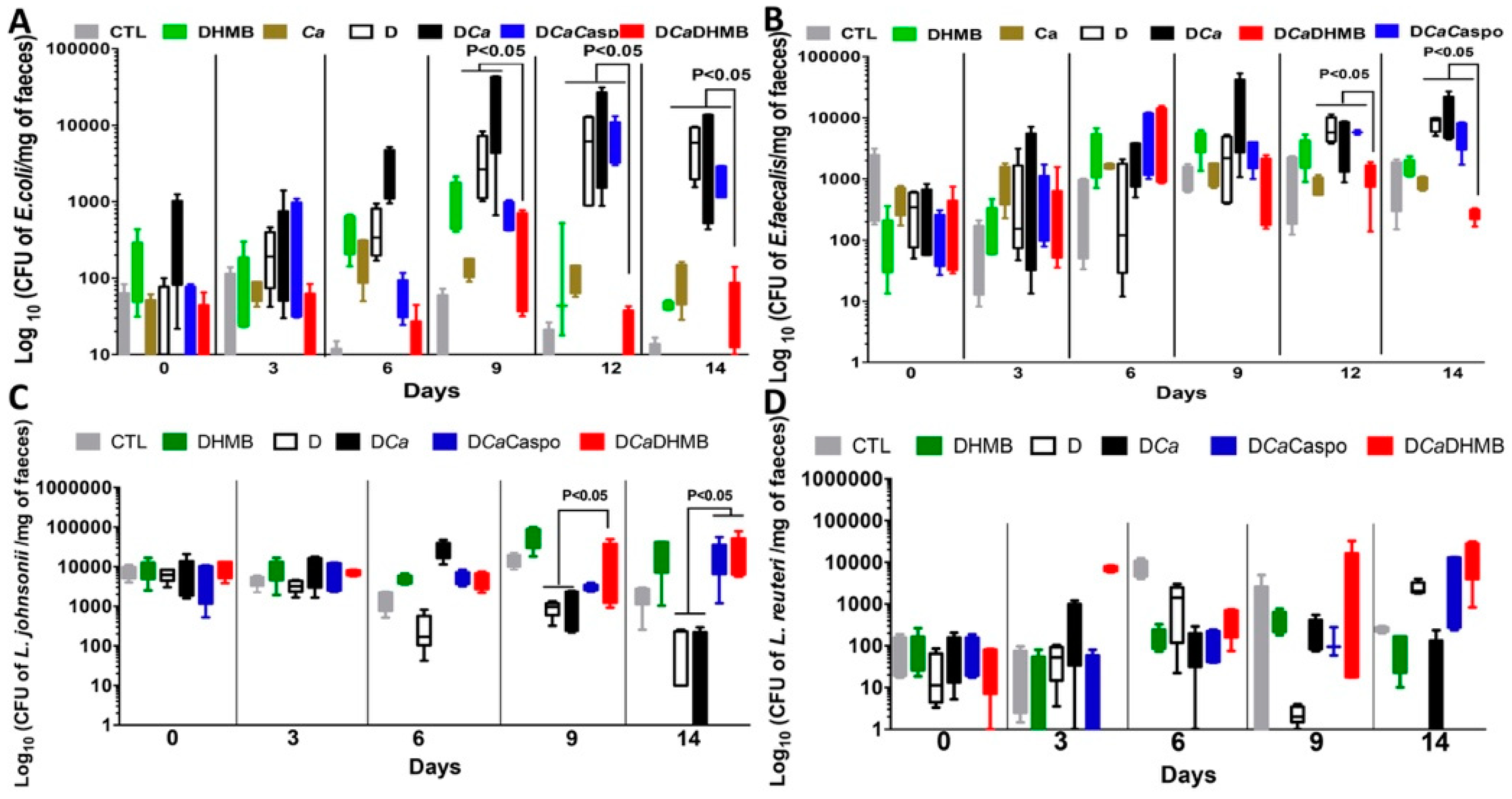
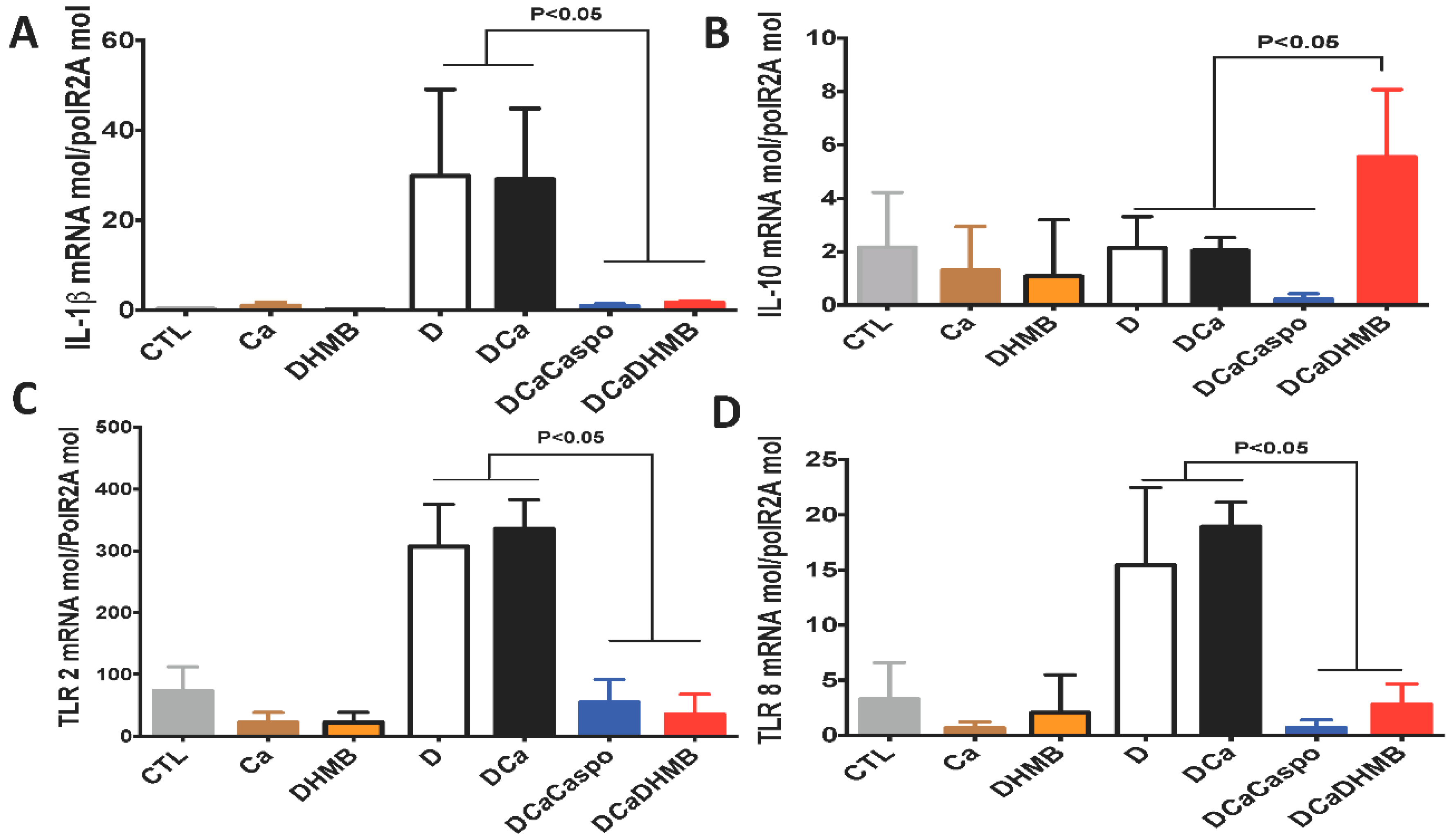
2.3. Effect of DHMB on C. albicans Colonisation and Intestinal Inflammation in DSS-Induced Colitis Model

To assess whether DHMB can be tolerated in mice, a high dose of DHMB (100 mg/kg) was injected intraperitoneally in mice. No mortality was observed following DHMB challenge (data not shown). Body weight remained stable and no clinical signs were observed over two weeks.
The efficacy of DHMB at eliminating C. albicans was assessed in a DSS-induced colitis model. After C. albicans challenge, DHMB was administered for five days (10 mg/kg) to assess the therapeutic properties of this compound. As the DHMB compound has fungicidal activity against C. albicans, its effectiveness was compared to that of caspofungin in terms of C. albicans elimination from the gut. No sign of inflammation was observed in mice receiving water (CTL), C. albicans only (Ca) or DHMB (Figure 4). Following DSS treatment alone (D) or C. albicans challenge with DSS treatment (CaD), mice showed a gradual reduction in body mass over two weeks, whereas CaD mice receiving DHMB showed less severe weight loss than those untreated or treated with caspofungin (CaDCaspo) (Figure 4A). The clinical score for inflammation, including diarrhoea and rectal bleeding, decreased significantly in CaD mice treated with DHMB while caspofungin treatment did not significantly decrease these clinical symptoms (Figure 4B). Histological analysis of colon samples showed that DHMB treatment significantly reduced disease severity in CaD mice when compared to those treated with caspofungin (Figure 4C).
In contrast to CaD or CaDCaspo, colon sections from the CaDDHMB group showed low levels of leukocyte infiltrates, oedema and cryptic abscesses, and the surface epithelia structures remained intact (Figure 5).
The number of C. albicans colony-forming units (CFUs) and microbiota changes were assessed in freshly collected stool samples from each tagged mouse. A high number of C. albicans CFUs was recovered from stools from all groups on Day 1 (Figure 6). In the absence of DSS, C. albicans was dramatically decreased in mice. DSS treatment promoted a significant increase in C. albicans CFUs starting on Day 12 while DHMB administration favoured C. albicans elimination in DSS treated mice. The number of C. albicans CFUs was assessed in the stomach, caecum and colon (Figure 6). Treatment with either caspofungin or DHMB promoted C. albicans elimination from the mouse gut. For the gut microbiota, high numbers of E. coli and E. faecalis populations were observed following DSS treatment. Overgrowth of these two populations occurred regardless of C. albicans colonisation (Figure 7). DHMB treatment significantly maintained low levels of E. coli and E. faecalis populations while caspofungin treatment failed to reduce them. In terms of anaerobic bacteria, the number of L. johnsonii colonies was significantly reduced in both DSS and C. albicans mice, while DHMB treatment significantly restored L. johnsonii population in colitic mice challenged with C. albicans. In contrast, L. reuteri population showed unpredictable changes (Figure 7). The expression levels of pro-inflammatory cytokine IL-1β and anti-inflammatory cytokine IL-10 were assessed in the colons (Figure 8A,B). Expression of IL-1β was significantly lower in the colons of CaD mice treated with DHMB than in CaD or DSS mice (Figure 8A,B). In contrast, expression of IL-10 was significantly higher in colons of CaD mice challenged with DHMB when compared to that of CaD or DSS mice. Additionally, the expression levels of TLR-4 and TLR-8 were determined. TLR-4 and TLR-8 expression increased significantly in response to both C. albicans overgrowth and colitis while treatment with DHMB decreased the expression of these receptors in the colons (Figure 8A,B).

3. Discussion

The resistance of opportunistic yeast pathogens to antifungal drugs has increased significantly over the past decade and this led us to screen 55 molecules from our chemical library for antifungal activity. DHMB was found to be a promising molecule in terms of MIC value and solubility. In addition, DHMB was not cytotoxic in vitro against a human embryonic kidney cell line (HEK293) at high concentrations (data not shown).
DHMB is often used as a key intermediate in the synthesis of different natural compounds such as the antibacterial agents (±)-isoperbergin and perbergin [14], the antineoplastic agent combretastatin A-1 [15], the allergy-inducing benzofuranoquinone acamelin [16], natural coumarins such as fraxetin and fraxidin [17], or the photoreactive compound pimpinellin [18], but the role of this molecule alone in inflammation and fungal elimination has not yet been studied. An hydroxy-deleted related compound, 2-hydroxy-4-methoxybenzaldehyde (HMB), the principal component of root bark essential oil of Periploca sepium Bunge, which is a woody climbing vine [19], exhibits some interesting biological activities. The dried root bark of this plant is traditionally used in the treatment of autoimmune diseases, in particular rheumatoid arthritis [20]. It also appears to have insecticidal activity against several insect species [21].
In the present study, DHMB was highly effective against clinically isolated fluconazole- or caspofungin-resistant C. albicans strains indicating that this compound could potentially be used to fight drug-resistant fungi. Some antifungal drugs can induce changes in the cell wall structure after C. albicans challenge. In contrast to caspofungin, which induces important cell wall changes in terms of chitin levels, DHMB did not induce any important changes in the cell wall. DHMB was also involved in the inhibition of the melanin production. It has been shown that melanisation is a virulence factor in C. albicans while inhibition of melanin biosynthesis by anti-melanin antibodies leads to a decrease in fungal virulence [22,23].
In experimental animals, we observed that the DHMB compound that we administered to animals at high doses was not cytotoxic and was well-tolerated. The animals were still alive and healthy two weeks after the end of the experiment. Furthermore, the efficiency of DHMB was assessed in vivo in the DSS colitis model. This compound reduced the clinical and histological scores for inflammation and promoted elimination of C. albicans from the gut. This finding is supported by a decrease in aerobic bacteria while anaerobic bacterial populations were re-established in mice treated with DHMB indicating that DHMB was not only able to reduce C. albicans colonisation but also, unexpectedly, to diminish intestinal inflammation. It has been shown that neutrophils and macrophages are recruited to the damaged colonic mucosa during DSS-induced colitis [24]. IL-1β expression, which is predominantly produced by these leukocytes, reduced in mice treated with DHMB. Recently, we observed after colitis development and C. glabrata challenge a high production of IL-6 and IL-1β, which are highly important in the recruitment of neutrophils and macrophages into the inflammatory site [9].
It has been shown that melanin concentrate hormone is highly expressed in the colonic mucosa of colitic mice and in patients with IBD suggesting that inhibition of melanin expression by DHMB can potentially reduce the pathogenesis of intestinal inflammation [25].
In conclusion, DHMB is a small organic molecule and any modification or substitution on the aromatic ring led to a strong decrease in the antifungal activity of the molecule, indicating that each group on the ring plays a specific and crucial role in its activity. This compound had antifungal properties against C. albicans, was safe and well-tolerated in different culture cell types and animals, and was effective at eliminating drug-resistant fungi, as well as had an anti-tyrosinase effect through the inhibition of the melanisation process in C. albicans. In DSS induced colitis model, DHMB had antifungal properties through the elimination of C. albicans from the gut and anti-inflammatory activity by re-establishing the anaerobic bacteria. This finding was also supported by a decrease of pro-inflammatory cytokine IL-1β in mice treaded with DHMB. Therefore, this aromatic small molecule holds great potential as an antifungal agent and anti-inflammatory properties.

4. Materials and Methods

4.1. C. albicans Strains and Growth Conditions

The C. albicans strains used in the current study are shown in Table 2. Fungal culture was carried out in YPD medium (yeast extract 1%, peptone 1%, dextrose 1%), on a rotary shaker for 18 h at 37 °C. The fungal culture obtained was then centrifuged at 2500 rpm for 5 min and washed twice in PBS (phosphate buffered saline).

4.2. Antifungal Compounds

The molecule 2,3-dihydroxy-4-methoxybenzaldehyde (DHMB) was synthesised and provided by Hautes Etudes d’Ingénieur (HEI), Lille, France. DHMB was used at a final concentration of 100 µg/mL, diluted in PBS during the various in vitro experiments. Commercially available caspofungin (Merck, Semoy, France) and fluconazole (Fresenius, Sèvres, France) were used as positive controls.

4.3. Fungal Viability Assays

Minimum inhibitory concentrations (MICs) were measured using the broth microdilution method following Clinical Laboratory Standards Institute (CLSI) procedures. After drug inoculation, the plates were incubated at 37 °C for 24 h and MICs were measured as the lowest concentration of drug below growth levels [27]. For antifungal assays on C. albicans ATCC 90028, this screening was carried out by CO-ADD (Community for Antimicrobial Drug Discovery) and the University of Queensland (Santa Lucia, Australia). C. albicans ATCC 90028 was cultured for 3 days on YPD agar at 30 °C. A yeast suspension of 1 × 10⁶ to 5 × 10⁶ CFU/mL was prepared and the suspension was subsequently diluted and added to each well of the compound-containing plates giving a final cell density of fungi of 2.5 × 10³ CFU/mL in a total volume of 50 μL. All plates were covered and incubated at 35 °C for 36 h without shaking. Growth inhibition of C. albicans ATCC 90028 was determined by measuring absorbance at 630 nm (OD630), after the addition of resazurin (0.001% final concentration) and incubation at 35 °C for 2 h. The absorbance was measured using a Biotek Multiflo Synergy HTX plate reader. The percentage growth inhibition was calculated for each well, using the negative control (media only) and positive control (fungi without inhibitor) on the same plate. The MIC was determined as the lowest concentration at which growth was fully inhibited, defined by an inhibition of ≥80% for C. albicans (total inhibition in the case of DHMB at 8 µg/mL concentration).
For the viability assays, the bioluminescent C. albicans strain was suspended in PBS at a volume of 106 cells/well (96-well black plates, Chimney well). DHMB, caspofungin or fluconazole were then added at their final MIC concentrations (Table 1). Coelenterazine was then added to the wells at a concentration of 2 μM. Bioluminescence kinetics were then measured (at 0, 30 and 60 min) and analysed with a FLUOstar Omega Fluorometer (BMG Labtech, Champigny sur Marne, France). The positive control consisted of C. albicans strain alone. The kinetics were also measured with the Xenogen device. For fungal CFU/mL counting, serial dilutions were performed by incubation of 105 C. albicans cells for 1 h with antifungal at the MIC concentration and an aliquot of 100 µL of each dilution was plated on Sabouraud dextrose agar and incubated for 48 h.
For the melanin production, C. albicans yeast cells at 2 × 10⁶ cells/mL were incubated in a minimal medium (15 mM glucose, 10 mM MgSO4, 29.4 mM KH2PO4, 13 mM glycine, 3 mM vitamin B1, pH 5.5) with or without 5 mM l-3,4-dihydroxyphenylalanine (DOPA, Sigma, St. Quentin Fallavier, France) at 37 °C in the dark. After 3 days, C. albicans cells grown with DOPA were treated with either DHMB (8 µg/mL) or caspofungin (0.0128 µg/mL). After one day of antifungal incubation, the production of melanin was examined using a Zeiss Axioplan microscope (Axioplan2, Jena, Germany).

4.4. Flow Cytometry and Confocal Microscopy

C. albicans was suspended in PBS at a volume of 106 cells/well. DHMB, caspofungin or fluconazole were then added to each well at a final concentration of 100 µM. The expression of chitin or α-Mans was assessed using wheat germ agglutinin-fluorescein isothiocyanate and concanavalin A-rhodamine. The obtained data were analysed using Kaluza software. In parallel, a negative control without addition of lectin marker was prepared under the same conditions as those described previously for the samples. To examine the C. albicans cell wall by confocal microscopy after antifungal treatment, C. albicans cells were stained with wheat germ agglutinin-fluorescein isothiocyanate, concanavalin A-rhodamine and DAPI. Specific slides (well 6.7 mm; Thermo Scientific, Montigny le Bretonne, France) were used in this experiment and the coverslips were examined by confocal microscopy (Zeiss LSM710, Jena, Germany).

4.5. Animals

The animals used were wild female C57BL/6 mice aged 3–4 months, certified free from infection and purchased from Janvier Laboratories, Le Genest-Saint-Isle, France. The mice were housed at the pet store of the Faculty of Medicine, Lille, France. The temperature of the room was maintained at 21 °C and the mice had free access to water and food with exposure to light 12 h/day. Water bottles and food were daily examined in each cage before the evaluation of the clinical score for each tagged mouse. The studies were carried out in accordance with the decrees relating to the ethics of animal experimentation (Decree 86/609/EC, 8/2/2016).

4.6. C. albicans Challenge and Induction of Colitis

Each mouse was administered with 300 µL of PBS containing 107 live C. albicans cells by oral gavage on Day 1. For antifungal treatment, mice were injected intraperitoneally for 5 days with 300 µL containing 10 mg/kg/day of DHMB or caspofungin. The DSS model is a reflection of how intestinal inflammation can promote C. albicans overgrowth in patients with CD. Mice received 2% DSS (M.W. 36−50 kDa; MP Biomedicals, LLC, Eschwege, Germany) in drinking water from Day 1 to Day 14 to promote colitis development and C. albicans intestinal colonisation [28]. The presence of C. albicans in the gut was assessed daily by measuring the number of CFUs in stools (approximately 0.1 g/sample). Stools were collected every 2 days from each tagged mouse. Faecal samples were suspended in 1 mL PBS, homogenised with a glass tissue homogeniser and plated onto Candi-Select medium (Bio-Rad Laboratories, Marnes la Coquette, France) [29]. The presence of C. albicans was assessed in the stomach, caecum and colon. The tissues were cut longitudinally and washed several times in PBS to avoid surface contamination from organisms present in the lumen. Serial dilutions of gut homogenates were prepared. The results were noted as C. albicans CFU/mg of tissue.
For the isolation of aerobic bacteria, the faecal samples were plated onto MacConkey agar (Sigma-Aldrich, Saint Louis, MO, USA) and bile esculin azide agar (BEA, HongKong, China; Sigma-Aldrich, Saint Louis, MO, USA) plates. Serial dilutions of these samples were performed. For the isolation of anaerobic bacteria, Columbia agar (Sigma-Aldrich, Saint Louis, MO, USA) were used. MALDI-TOF MS (Microflex-Bruker Daltonics) was used to identify the bacteria [9].

4.7. Determination of Clinical and Histological Scores of Inflammation

The clinical score was evaluated daily in each tagged mouse based on the presence of blood in the stools, rectal bleeding and stool consistency [5]. These three clinical markers of inflammation were recorded to give a score between 0 (healthy mouse) and 12 (maximum inflammation of the colon reflecting the severity of the inflammation).
Colon samples were fixed in 4% paraformaldehyde-acid and embedded in paraffin for histological analysis. Histological scores were assessed by two independent, blinded investigators who observed two sections per mouse at magnifications of X10 and X100. The histological score was analysed according to two sub-scores: (i) a score for the presence of inflammatory cells; and (ii) a score for epithelial damage. The histological score ranged from 0 (no changes) to 6 (extensive cell infiltration and tissue damage) [5].

4.8. Real-Time mRNA Quantification of Pro-Inflammatory Cytokines and Innate Immune Receptors

RNA extraction was performed with a NucleoSpin RNA® kit (Macherey-Nagel, Hoerdt, France). RNA was quantified by spectrophotometry (Nanodrop; Nyxor Biotech, Paris, France). cDNA synthesis was performed according to the High Capacity DNA Reverse Transcription (RT) protocol, using Master Mix (Applied Biosystems, Foster, CA, USA). Fast SYBR green (Applied Biosystems) was used to amplify the cDNA by PCR using the one-step system (Applied Biosystems). SYBR green dye intensity was assessed using one-step software. All results were normalised to the reference gene, POLR2A.

5. Statistical Analysis

All data are presented as the mean ± standard deviation (SD) of individual experimental groups. Statistical analyses were performed using the Mann–Whitney U test to compare pairs of groups. Differences were considered significant when the p value was as follows: p < 0.05; p < 0.01; p < 0.001. Prism 4.0 from GraphPad and XLSTAT were used for the statistical analyses.

Author Contributions

C.B., M.B., R.C., K.L., and S.J. performed the experiments. C.B., M.B., R.C., and S.J. analysed the data. C.B., M.B., R.C., K.L., A.G., B.S. and S.J. interpreted the results of the experiments. M.B., A.G., and B.S. contributed reagents/materials/analysis tools. S.J. designed the experiments and drafted the manuscript.

Funding

This work was partially funded by Agence Nationale de la Recherche (ANR) in the setting the project “InnateFun”, promotional reference ANR-16-IFEC-0003-05, in the “Infect-ERA” program.

Acknowledgments

The authors thank Nadine François and Antonino Bongiovanni for their excellent technical assistance. The authors would like to thank the Community for Antimicrobial Drug Discovery (CO-ADD), funded by the Welcome Trust (UK), and the University of Queensland (Australia) for performing the DHMB antimicrobial screening.

Conflicts of Interest

The authors declare no conflict of interest.

References

  1. Baumgart, D.C.; Sandborn, W.J. Inflammatory bowel disease: Clinical aspects and established and evolving therapies. Lancet 2007, 369, 1641–1657. [Google Scholar] [CrossRef]
  2. Oyri, S.F.; Muzes, G.; Sipos, F. Dysbiotic gut microbiome: A key element of Crohn’s disease. Comp. Immunol. Microbiol. Infect. Dis. 2015, 43, 36–49. [Google Scholar] [CrossRef] [PubMed]
  3. Gevers, D.; Kugathasan, S.; Denson, L.A.; Vazquez-Baeza, Y.; Van Treuren, W.; Ren, B.; Schwager, E.; Knights, D.; Song, S.J.; Yassour, M.; et al. The treatment-naive microbiome in new-onset Crohn’s disease. Cell Host Microbe 2014, 15, 382–392. [Google Scholar] [CrossRef] [PubMed]
  4. Darfeuille-Michaud, A.; Neut, C.; Barnich, N.; Lederman, E.; Di Martino, P.; Desreumaux, P.; Gambiez, L.; Joly, B.; Cortot, A.; Colombel, J.F. Presence of adherent Escherichia coli strains in ileal mucosa of patients with Crohn’s disease. Gastroenterology 1998, 115, 1405–1413. [Google Scholar] [CrossRef]
  5. Jawhara, S.; Thuru, X.; Standaert-Vitse, A.; Jouault, T.; Mordon, S.; Sendid, B.; Desreumaux, P.; Poulain, D. Colonization of mice by Candida albicans is promoted by chemically induced colitis and augments inflammatory responses through galectin-3. J. Infect. Dis. 2008, 197, 972–980. [Google Scholar] [CrossRef] [PubMed]
  6. Jawhara, S.; Poulain, D. Saccharomyces boulardii decreases inflammation and intestinal colonization by Candida albicans in a mouse model of chemically-induced colitis. Med. Mycol. 2007, 45, 691–700. [Google Scholar] [CrossRef] [PubMed] [Green Version]
  7. Standaert-Vitse, A.; Sendid, B.; Joossens, M.; Francois, N.; Vandewalle-El Khoury, P.; Branche, J.; Van Kruiningen, H.; Jouault, T.; Rutgeerts, P.; Gower-Rousseau, C.; et al. Candida albicans colonization and ASCA in familial Crohn’s disease. Am. J. Gastroenterol. 2009, 104, 1745–1753. [Google Scholar] [CrossRef]
  8. Poulain, D.; Sendid, B.; Standaert-Vitse, A.; Fradin, C.; Jouault, T.; Jawhara, S.; Colombel, J.F. Yeasts: Neglected pathogens. Dig. Dis. 2009, 27 (Suppl. 1), 104–110. [Google Scholar] [CrossRef]
  9. Charlet, R.; Pruvost, Y.; Tumba, G.; Istel, F.; Poulain, D.; Kuchler, K.; Sendid, B.; Jawhara, S. Remodeling of the Candida glabrata cell wall in the gastrointestinal tract affects the gut microbiota and the immune response. Sci. Rep. 2018, 8, 3316. [Google Scholar] [CrossRef]
  10. Ardizzone, S.; Cassinotti, A.; Manes, G.; Porro, G.B. Immunomodulators for all patients with inflammatory bowel disease? Ther. Adv. Gastroenterol. 2010, 3, 31–42. [Google Scholar] [CrossRef]
  11. Campieri, M. New steroids and new salicylates in inflammatory bowel disease: A critical appraisal. Gut 2002, 50 (Suppl. 3), III43–III46. [Google Scholar] [CrossRef] [PubMed]
  12. Wiederhold, N.P. Antifungal resistance: Current trends and future strategies to combat. Infect. Drug Resist. 2017, 10, 249–259. [Google Scholar] [CrossRef] [PubMed]
  13. Ostrosky-Zeichner, L.; Casadevall, A.; Galgiani, J.N.; Odds, F.C.; Rex, J.H. An insight into the antifungal pipeline: Selected new molecules and beyond. Nat. Rev. Drug Discov. 2010, 9, 719–727. [Google Scholar] [CrossRef] [PubMed]
  14. Almabruk, K.H.; Chang, J.H.; Mahmud, T. Total Synthesis of (+/−)-Isoperbergins and Correction of the Chemical Structure of Perbergin. J. Nat. Prod. 2016, 79, 2391–2396. [Google Scholar] [CrossRef] [PubMed]
  15. Pettit, G.R.; Singh, S.B.; Niven, M.L.; Hamel, E.; Schmidt, J.M. Isolation, structure, and synthesis of combretastatins A-1 and B-1, potent new inhibitors of microtubule assembly, derived from Combretum caffrum. J. Nat. Prod. 1987, 50, 119–131. [Google Scholar] [CrossRef]
  16. McKittrick, B.A.; Stevenson, R. A simple synthesis of the natural benzofuranoquinone, acamelin. J. Chem. Soc. Perkin Trans. 1 1983, 10, 2423–2424. [Google Scholar] [CrossRef]
  17. Spath, E.; Dobrovolny, E. Natural coumarins. XLII. Synthesis of fraxetin, fraxidin and isofraxidin. Berichte der Deutschen Chemischen Gesellschaft 1938, 71B, 1831–1836. [Google Scholar] [CrossRef]
  18. Cervi, A.; Aillard, P.; Hazeri, N.; Petit, L.; Chai, C.L.; Willis, A.C.; Banwell, M.G. Total syntheses of the coumarin-containing natural products pimpinellin and fraxetin using Au(I)-catalyzed intramolecular hydroarylation (IMHA) chemistry. J. Org. Chem. 2013, 78, 9876–9882. [Google Scholar] [CrossRef]
  19. Wang, L.; Yin, Z.Q.; Zhang, L.H.; Ye, W.C.; Zhang, X.Q.; Shen, W.B.; Zhao, S.X. Chemical constituents from root barks of Periploca sepium. China J. Chin. Mater. Med. 2007, 32, 1300–1302. [Google Scholar]
  20. Tokiwa, T.; Harada, K.; Matsumura, T.; Tukiyama, T. Oriental medicinal herb, Periploca sepium, extract inhibits growth and IL-6 production of human synovial fibroblast-like cells. Biol. Pharm. Bull. 2004, 27, 1691–1693. [Google Scholar] [CrossRef]
  21. Chu, S.S.; Jiang, G.H.; Liu, W.L.; Liu, Z.L. Insecticidal activity of the root bark essential oil of Periploca sepium Bunge and its main component. Nat. Prod. Res. 2012, 26, 926–932. [Google Scholar] [CrossRef] [PubMed]
  22. Morris-Jones, R.; Gomez, B.L.; Diez, S.; Uran, M.; Morris-Jones, S.D.; Casadevall, A.; Nosanchuk, J.D.; Hamilton, A.J. Synthesis of melanin pigment by Candida albicans in vitro and during infection. Infect. Immun. 2005, 73, 6147–6150. [Google Scholar] [CrossRef] [PubMed]
  23. Eisenman, H.C.; Duong, R.; Chan, H.; Tsue, R.; McClelland, E.E. Reduced virulence of melanized Cryptococcus neoformans in Galleria mellonella. Virulence 2014, 5, 611–618. [Google Scholar] [CrossRef] [PubMed] [Green Version]
  24. Qualls, J.E.; Kaplan, A.M.; van Rooijen, N.; Cohen, D.A. Suppression of experimental colitis by intestinal mononuclear phagocytes. J. Leukoc. Biol. 2006, 80, 802–815. [Google Scholar] [CrossRef] [Green Version]
  25. Kokkotou, E.; Moss, A.C.; Torres, D.; Karagiannides, I.; Cheifetz, A.; Liu, S.; O’Brien, M.; Maratos-Flier, E.; Pothoulakis, C. Melanin-concentrating hormone as a mediator of intestinal inflammation. Proc. Natl. Acad. Sci. USA 2008, 105, 10613–10618. [Google Scholar] [CrossRef] [Green Version]
  26. Doyle, T.C.; Nawotka, K.A.; Kawahara, C.B.; Francis, K.P.; Contag, P.R. Visualizing fungal infections in living mice using bioluminescent pathogenic Candida albicans strains transformed with the firefly luciferase gene. Microb. Pathog. 2006, 40, 82–90. [Google Scholar] [CrossRef] [PubMed]
  27. Bustamante, B.; Martins, M.A.; Bonfietti, L.X.; Szeszs, M.W.; Jacobs, J.; Garcia, C.; Melhem, M.S. Species distribution and antifungal susceptibility profile of Candida isolates from bloodstream infections in Lima, Peru. J. Med. Microbiol. 2014, 63 Pt 6, 855–860. [Google Scholar] [CrossRef]
  28. Choteau, L.; Parny, M.; Francois, N.; Bertin, B.; Fumery, M.; Dubuquoy, L.; Takahashi, K.; Colombel, J.F.; Jouault, T.; Poulain, D.; et al. Role of mannose-binding lectin in intestinal homeostasis and fungal elimination. Mucosal Immunol. 2016, 9, 767–776. [Google Scholar] [CrossRef]
  29. Sendid, B.; Francois, N.; Standaert, A.; Dehecq, E.; Zerimech, F.; Camus, D.; Poulain, D. Prospective evaluation of the new chromogenic medium CandiSelect 4 for differentiation and presumptive identification of the major pathogenic Candida species. J. Med. Microbiol. 2007, 56 Pt 4, 495–499. [Google Scholar] [CrossRef] [Green Version]
Figure 1. Chemical structure of DHMB and related investigated compounds.
Ijms 20 00321 g001
Figure 2. Impact of DHMB on C. albicans viability. (AC) Bioluminescent C. albicans strain was treated with either DHMB, flucanoazole or caspofungin at their MICs, 2 × MICs or 4 × MICs and monitored at 0, 15, 30 and 60 min. Control corresponds to C. albicans alone without antifungal treatment; (D) Visualisation of DHMB effect on bioluminescent C. albicans strain in real time. Bioluminescent C. albicans strain was treated with either DHMB, fluconazole or caspofungin at their MICs, 2 × MICs or 4 × MICs and monitored at 0, 15, 30 and 60 min. Line 1, PBS without yeasts; Line 2, C. albicans alone; Lines 3–5, C. albicans treated with caspofungin at 1 × MIC, 2 × MIC and 4 × MIC, respectively; Lines 6–8, C. albicans challenged with fluconazole at 1 × MIC, 2 × MIC and 4 × MIC, respectively; Lines 9–11, C. albicans + DHMB at 1 x MIC, 2 × MIC and 4 × MIC, respectively. * p < 0.0001; (E,F) Effect of DHMB on drug-resistant C. albicans clinical isolates. Control represents C. albicans clinical isolate. Caspo (caspofungin), Fluco (fluconazole) or DHMB (2,3-dihydroxy-4-methoxybenzaldehyde) were added to C. albicans strains at 10 × MIC.
Ijms 20 00321 g002
Figure 3. Effect of DHMB on modulation of chitin and α-mannan expression. (A) Confocal microscopic observation after antifungal challenge. C. albicans was treated with PBS (a) caspofungin (b), fluconazole (c) or DHMB (d) at a final concentration of 100 µM for 1 h and the fungal cell wall was analysed by confocal microscopy and flow cytometry; (B) Percentage of viable C. albicans cells labelled with concanavalin A. α-Mannans were labelled with concanavalin A-rhodamine; (C) Percentage of viable C. albicans cells labelled with WGA-IFTC. Chitin was labelled with wheat germ agglutinin-fluorescein isothiocyanate (WGA-IFTC); (D) Microscopic observation of the melanin pigments in C. albicans after DHMB treatment. (a) C. albicans cells grown without DOPA; (b) C. albicans grown with 1 mM DOPA; (c) C. albicans cells grown with DOPA and treated with caspofungin; (d), C. albicans cells were incubated with DOPA and challenged with DHMB. Bars 10 nm.
Ijms 20 00321 g003
Figure 4. Determination of inflammatory parameters after DHMB treatment in a DSS-induced colitis model. (A) Body weight loss during colitis development. CTL refers to the control group receiving water. Ca corresponds to mice receiving C. albicans alone. DHMB refers to mice treated with DHMB only. D corresponds to mice receiving DSS only. DCa corresponds to mice receiving DSS and challenged with C. albicans. DCaCaspo and DCaDHMB refers to mice receiving DSS and challenged with C. albicans and treated with either caspofungin or DHMB (* p < 0.05); (B) Clinical score for inflammation; (C) Histological score for inflammation. Data are the mean ± SD of 10 mice per group from two independent experiments.
Ijms 20 00321 g004
Figure 5. Analysis of histological colon sections from DSS-induced colitis: (ac) colon sections from mice receiving water (control), C. albicans alone and DHMB, respectively; (d) colon sections from mice receiving DSS; (e) colon sections from mice receiving C. albicans and DSS; and (f) colon sections from CaD mice treated with DHMB; (gi) colon sections from either DSS mice or CaD show tissue destruction, an important inflammatory cell infiltrate and oedema in the mucosa and submucosa of colon wall structures (*). Scale bars represent 50 µm (af) and 10 µm (gi).
Ijms 20 00321 g005
Figure 6. Effect of DHMB on C. albicans elimination from the gut. (AD) Number of C. albicans colonies recovered from the stools, stomach, cecum and colon. Data are the mean ± SD of 10 mice per group from two independent experiments (p < 0.001).
Ijms 20 00321 g006
Figure 7. Determination of viable faecal aerobic and anaerobic bacteria in mice with colitis treated with DHMB. For all experiments, stool bacteria were collected from mice on Day 0 before C. albicans challenge and DSS treatment. (AD) Number of E. coli, E. faecalis, L. johnsonii and L. reuteri colonies recovered from stools. Data are the mean ± SD of 10 mice per group from two independent experiments.
Ijms 20 00321 g007
Figure 8. Cytokine and receptor expression after DHMB treatment. (AD) Relative expression levels of IL-1b, IL-10, TLR-2 and TLR-8 mRNA in mouse colons. Data are the mean ± SD of 10 mice per group from two independent experiments.
Ijms 20 00321 g008
Table 1. Chemical modifications of DHMB and their impact on activity.
EntryCompoundR1R2R3R4R5R6% Inhibition at 32 µg/mL a
1DHMBCHOOHOHOCH3HH100
21HOHOHOCH3HH3
32 Ijms 20 00321 i001OHOCH3OCH3HH13
43 Ijms 20 00321 i002OHOHOCH3HH27
54OHOCOCH2ClOCH3HH6
65OCOCH2ClOCOCH2ClOCH3HH4
76 Ijms 20 00321 i003H2
87CHOOHOHOHHH4
98HOHOHOCH3HCOOH2
109HOCH3OCH3OCH3HCOOH2
1110HOCH3OHOCH3HCOOH0
1211HOCH3OCH3OCH3HCH2-COOH1
1312HOCH3OCH3OCH3HCH2-OH2
1413HOCH3OHOCH3HCHO1
1514HOCH3OHOCH3HCOCH34
1615HOCH3OHOCH3HH0
a Screening carried out on C. albicans ATCC 90028.
Table 2. Antifungal activity of DHMB vs. caspofungin and fluconazole.
StrainsDescriptionMIC Caspofungin (µg/mL)MIC Fluconazole (µg/mL)MIC DHMB (µg/mL)Ref.
C. albicans ATCC 90028Wild-type0.030.58This study
Bioluminescent C. albicansC. albicans strain CAI4 (ura3::imm434/ura3::imm434)0.030.58[26]
C. albicans 14314c4497Anal, fluconazole resistant0.0612880This study
C. albicans 14316c1746Bronchoalveolar lavage, fluconazole resistant0.0312880This study
C. albicans 92535989Tracheal secretion, fluconazole resistant0.066480This study
C. albicans 14294c5335Stools, fluconazole resistant0.06580This study
C. albicans 17292c3367Venous catheter, caspofungin resistant80.580This study
C. albicans 15343c3523Blood, caspofungin resistant20.580This study
C. albicans 17287c305Blood, caspofungin resistant80.580This study
C. albicans 15351c6859Venous catheter, caspofungin resistant4180This study

