Article

Natural Tyrosinase Inhibitors from Lycopodium japonicum

1
School of Chemistry and Chemical Engineering, Central South University of Forestry and Technology, 498 South Shaoshan Road, Changsha 410004, China
2
School of Forestry, Central South University of Forestry and Technology, 498 South Shaoshan Road, Changsha 410004, China
*
Author to whom correspondence should be addressed.
Molecules 2025, 30(19), 4024; https://doi.org/10.3390/molecules30194024
Submission received: 15 July 2025 / Revised: 4 September 2025 / Accepted: 8 September 2025 / Published: 9 October 2025
(This article belongs to the Special Issue Terpenes and Their Derivatives: From Nature to Medical Applications)

Abstract

Natural tyrosinase inhibitors are an important group of compounds with cosmetical and medicinal applications. With the aim of finding new types of natural tyrosinase inhibitors from ornamental and medicinal plants, Lycopodium japonicum was selected and studied. As a result, fifteen structurally diverse secondary metabolites 115 were isolated and identified. Their chemical structures were identified by analysis of their spectral data and compared with those reported in the literature. In the tyrosinase inhibitory bioassay, five phytochemicals, 4, 12, 13, 14, and 15, exhibited significant inhibitory effects, with half-maximal inhibitory concentration (IC50) values ranging from 1.46 to 6.82 mM. Additionally, molecular docking studies disclosed that Lys376, Lys379, Gln307, and other amino acid residues played key roles in the potential binding interactions between the active compounds and the tyrosinase. These findings suggest that the species L. japonicum is a warehouse of natural tyrosinase inhibitors.

1. Introduction

Tyrosinase is a multifunctional enzyme, which is responsible for the oxidation of phenolic compounds [1]. For humans, this enzyme plays a key physiological role because its dysregulated expression can lead to various skin disorders, such as melanoma, freckles, chloasma, and vitiligo [2,3]. Due to its crucial role in melanogenesis, tyrosinase inhibitors are regarded as an important group of compounds with promising cosmetic and medicinal values, such as skin tone enhancement and depigmentation [1,4].
Lycopodium japonicum belongs to the family Lycopodiaceae [5], which is distributed across Japan, China, India, and also Southeast Asia [6]. For a long time, this plant has been used as a medicinal material for the treatment of arthritic pain, quadriplegia, dysmenorrhea, contusions, and other health problems [6]. Nowadays, it is also used as an ornamental plant, due to its evergreen and beautiful branches and leaves. Previous phytochemical investigations of this plant disclosed that Lycopodium alkaloids [7,8,9,10] and serratane triterpenes [11,12,13,14] are two major groups of chemical constituents from L. japonicum. In addition, lignans [15], sterols [16], and other compounds are minor groups in the chemical composition of L. japonicum. These compounds exhibit an array of biological activities, including neuroprotective [7], anti-renal fibrosis [8], acetylcholinesterase inhibitory [17,18], cytotoxic [12], anti-inflammatory [19,20], anti-HIV-1 [18], and α-glucosidase inhibitory [17,21] activities. However, the tyrosinase inhibitory effect of the plant has not been investigated yet.
In our continuing efforts to search for bioactive substances derived from ornamental and medicinal plants for cosmetic products [22,23], the plant L. japonicum was studied. This work is intended to explore the non-alkaloidal constituents of the titular species with tyrosinase inhibitory activity in vitro and in silico.

2. Results

2.1. Structure Elucidation of the Isolates

A phytochemical investigation of the aerial parts of L. japonicum led to the isolation of fifteen structurally diverse compounds 115 (Figure 1). Their chemical structures were determined based on an analysis of nuclear magnetic resonance (NMR) data and a comparison with those reported in the literature (Table 1).
The 1H and 13C NMR spectra of chemical constituent 1 showed the signals attributable to one α,β-unsaturated ketone [δH 5.71 (1H, t, J = 2.5 Hz, H-15); δC 201.4 (C-16), 163.7 (C-14), 128.8 (C-15)], two oxygenated methines [δH 3.41 (1H, t, J = 2.9 Hz, H-3), 3.35 (1H, t, J = 2.9 Hz, H-21); δC 76.9 (C-3), 76.1 (C-21)], and seven methyls [δH 0.95 (3H, s, H-23), 0.87 (3H, s, H-24), 0.83 (3H, s, H-25), 0.80 (3H, s, H-26), 0.84 (3H, s, H-28), 1.12 (3H, s, H-29), 1.25 (3H, s, H-30); δC 28.5 (C-23), 22.3 (C-24), 15.8 (C-25), 20.1 (C-26), 15.0 (C-28), 21.6 (C-29), 28.0 (C-30)]. Considering the features revealed by the 1H and 13C NMR spectra, this compound likely belonged to the family of serratane triterpenes [5]. It was found that the overall 1H and 13C NMR spectral data of 1 displayed a high degree of resemblance to those of the serratane triterpene 16-oxo-3α-hydroxyserrat-14-en-21β-ol [16] (Table S1). Consequently, the structure of compound 1 was depicted as shown in Figure 1.
In the 1H and 13C NMR spectra of phytochemical 2, the signals attributable to one double bond [δH 5.32 (1H, br s, H-15); δC 138.7 (C-14), 122.2 (C-15)], two oxygenated methines [δH 3.45 (1H, t, J = 2.9 Hz, H-21), 3.18 (1H, dd, J = 11.7, 4.6 Hz, H-3); δC 79.0 (C-3), 76.4 (C-21)], and seven methyls [δH 0.80 (3H, s, H-23), 0.88 (3H, s, H-24), 0.77 (3H, s, H-25), 0.84 (3H, s, H-26), 0.69 (3H, s, H-28), 0.93 (3H, s, H-29), 0.97 (3H, s, H-30); δC 28.3 (C-23), 15.6 (C-24), 15.9 (C-25), 19.9 (C-26), 13.4 (C-28), 21.9 (C-29), 27.9 (C-30)] were observed. These characteristic 1H and 13C NMR spectral data suggested that this compound was a serratane-type triterpene. The subsequent literature survey revealed the overall 1H and 13C NMR spectral data of 2 resembled those of 21-epi-serratenediol [24] (Table S2). Therefore, the structure of compound 2 was determined as displayed in Figure 1.
The 1H and 13C NMR spectral data of compound 3 indicated the presence of one double bond [δH 5.38 (1H, br s, H-15); δC 138.5 (C-14), 122.1 (C-15)], one carbonyl [δC 217.2 (C-21)], one oxygenated methine [δH 3.19 (1H, dd, J = 11.7, 4.6 Hz, H-3); δC 79.0 (C-3)], and seven methyls [δH 0.97 (3H, s, H-23), 0.77 (3H, s, H-24), 0.80 (3H, s, H-25), 0.83 (3H, s, H-26), 0.92 (3H, s, H-28), 1.04 (3H, s, H-29), 1.08 (3H, s, H-30); δC 28.3 (C-23), 15.6 (C-24), 15.9 (C-25), 19.9 (C-26), 13.1 (C-28), 24.6 (C-29), 21.7 (C-30)]. The 1H and 13C NMR spectra of compound 3 exhibited close similarity to those of 2, except for the appearance of a carbonyl group in 3 instead of one oxygenated methine group in 2. Accordingly, compound 3 was identified as 3β-hydroxy-14-serraten-21-one as depicted in Figure 1, whose data matched well with those recorded in the literature [25] (Table S3).
Interestingly, the 1H and 13C NMR spectral data of compound 4 exhibited close similarity to those of 2, including one double bond [δH 5.33 (1H, br s, H-15); δC 138.3 (C-14), 122.3 (C-15)], two oxygenated methines [δH 3.23 (1H, dd, J = 11.6, 4.2 Hz, H-21), 3.19 (1H, dd, J = 11.7, 4.6 Hz, H-3); δC 79.0 (C-3), 79.3 (C-21)], and seven methyls [δH 0.80 (3H, s, H-23), 0.83 (6H, s, H-24, H-26), 0.77 (3H, s, H-25), 0.67 (3H, s, H-28), 0.96 (3H, s, H-29), 0.97 (3H, s, H-30); δC 15.6 (C-23), 28.3 (C-24), 15.9 (C-25), 19.9 (C-26), 13.6 (C-28), 14.8 (C-29), 27.7 (C-30)]. Their difference was in the 1H and 13C NMR data of the oxygenated methine at C-3, revealing the different configurations of the hydroxy group at C-3. Thus, 4 was identified as serratenediol (Figure 1), which was confirmed by the almost superposable 1H and 13C NMR data reported in the literature [24,26] (Table S4).
Inspection of the 1H and 13C NMR spectra of compound 5 revealed there was one double bond [δH 5.51 (1H, br s, H-15); δC 139.8 (C-14), 123.3 (C-15)], two oxygenated methines [δH 3.70 (1H, br s, H-21), 3.36 (1H, dd, J = 12.2, 4.6 Hz, H-3); δC 78.4 (C-3), 75.6 (C-21)], and six methyls [δH 1.58 (3H, s, H-23), 0.8 (3H, s, H-25), 0.88 (3H, s, H-26), 0.85 (3H, s, H-28), 0.97 (3H, s, H-29), 1.18 (3H, s, H-30); δC 24.6 (C-23), 14.1 (C-25), 20.1 (C-26), 14.2 (C-28), 22.5 (C-29), 29.1 (C-30)] in the chemical structure of 5. As revealed by the 1H and 13C NMR data, 5 exhibited close structural similarity with 2. However, the remarkably different 1H and 13C NMR data of one ester functionality [δH 3.64 (3H, s, -OCH3); δC 178.4 (C-24)] in 5 were observed, which indicated that the methyl group in 2 was replaced by the methyl ester group in 5. Based on the above-mentioned analysis along with an extensive literature survey [27,28] (Table S5), compound 5 was identified as methyl lycernuate A, as displayed in Figure 1.
The 1H and 13C NMR spectra of chemical constituent 6 showed the signals attributable to two double bonds [δH 4.83 (2H, s, H-26a, H-27a), 4.56 (2H, s, H-26b, H-27b); δC 148.6 (C-8, C-14), 106.9 (C-26, C-27)], two oxygenated methines [δH 3.25 (2H, d, J = 11.7 Hz, H-3, H-21); δC 79.1 (C-3, C-21)], and six methyls [δH 0.99 (6H, s, H-23, H-30), 0.76 (6H, s, H-24, H-29), 0.64 (6H, s, H-25, H-28); δC 28.5 (C-23, C-29), 15.5 (C-24, C-30), 14.7 (C-25, C-28)]. These characteristic 1H and 13C NMR data indicated that this compound was a serratane-type triterpene [5]. A further literature survey revealed that the overall 1H and 13C NMR data of 6 displayed a high degree of resemblance to those of α-onocerin [29] (Table S6).
The 1H and 13C NMR spectra of 7 exhibited close similarity to those of 6, including one double bond [δH 4.90 (2H, s, H-27); δC 147.5 (C-14), 108.0 (C-27)], two oxygenated methines [δH 3.33 (1H, d, J = 11.6 Hz, H-3), 3.23 (1H, d, J = 11.6 Hz, H-21); δC 78.8 (C-3), 79.1 (C-21)], and six singlet methyls [δH 0.69 (3H, s, H-23), 0.81 (3H, s, H-24), 1.09 (3H, s, H-25), 0.99 (3H, s, H-28), 0.63 (3H, s, H-29), 0.76 (3H, s, H-30); δC 27.7 (C-23), 15.5 (C-24), 14.6 (C-25), 14.9 (C-28), 28.5 (C-29), 15.5 (C-30)]. Their difference was in the appearance of a carbonyl [δC 212.1 (C-8)] in 7 instead of the one double bond [δH 4.83 (1H, s, H-26a), 4.56 (1H, s, H-26b); δC 148.6 (C-8), 106.9 (C-26)] in 6. Accordingly, compound 7 was identified as 26-nor-8-oxo-α-onocerin, whose data matched well with those recorded in the literature [30] (Table S7).
In the 1H and 13C NMR spectra of compound 8, one double bond [δH 5.35 (1H, m); δC 140.9 (s), 121.9 (d)], one oxygenated methine [δH 3.52 (1H, m); δC 72.0 (d)], and six methyls [δH 1.01 (3H, s), 0.92 (3H, d, J = 6.6 Hz), 0.84 (3H, t, J = 7.7 Hz), 0.83 (3H, d, J = 6.7 Hz), 0.81 (3H, d, J = 6.8 Hz), 0.68 (3H, s); δC 12.0 (q), 19.2 (q), 18.9 (q), 19.5 (q), 20.0 (q), 12.1 (q)] were recognized. It was found that the 1H and 13C NMR spectral data of 8 were identical to those of the β-stiosterol [23] (Table S8). Consequently, the structure of chemical composition 8 was assigned as displayed in Figure 1.
In the 1H and 13C NMR spectra of compound 9, one α,β-conjugated carbonyl [δH 7.61 (1H, d, J = 15.9 Hz, H-1′), 6.29 (1H, d, J = 15.9 Hz, H-2′); δC 144.8 (C-1′), 115.8 (C-2′), 167.4 (C-3′)], one tri-substituted benzene [δH 7.03 (1H, d, J = 2.0 Hz, H-2), 6.92 (1H, d, J = 8.2 Hz, H-5), 7.07 (1H, dd, J = 8.2, 1.9 Hz, H-6); δC 127.2 (C-1), 109.4 (C-2), 148.0 (C-3), 146.9 (C-4), 114.8 (C-5), 123.2 (C-6)], one methoxyl [δH 3.93 (3H, s, -OCH3); δC 56.1 (-OCH3)], and one ethoxyl [δH 4.26 (2H, q, J = 7.1 Hz, H-4′), 1.33 (3H, t, J =7.1 Hz, H-5′); δC 60.5 (C-4′), 14.5 (C-5′)] were observed. These NMR data revealed that the chemical structure of compound 9 was trans-ethyl ferulate, which was confirmed by the well-matched data in the literature [31] (Table S9).
The 1H and 13C NMR spectra of compound 10 showed the signals attributed to one α,β-conjugated carbonyl [δH 6.79 (1H, d, J = 12.9 Hz, H-1′), 5.81 (1H, d, J = 12.9 Hz, H-2′); δC 143.8 (C-1′), 117.0 (C-2′), 166.7 (C-3′)], one tri-substituted benzene [δH 7.77 (1H, d, J = 2.0 Hz, H-2), 6.88 (1H, d, J = 8.2 Hz, H-5), 7.11 (1H, dd, J = 8.3, 2.0 Hz, H-6); δC 127.4 (C-1), 112.9 (C-2), 146.0 (C-3), 147.2 (C-4), 113.9 (C-5), 125.8 (C-6)], one methoxyl [δH 3.93 (3H, s, -OCH3); δC 56.1 (-OCH3)], and one ethoxyl [δH 4.19 (2H, q, J = 7.1 Hz, H-4′), 1.29 (3H, t, J = 7.2 Hz, H-5′); δC 60.3 (C-4′), 14.4 (C-5′)]. These data indicated the close structural similarity between 10 and 9. However, the smaller coupling constant 12.0 Hz between H-1′ and H-2′ suggested the cis-geometry of the double bond Δ1′. Thus, 10 was identified as cis-ethyl ferulate, which was supported by the comparison of the data reported in the literature [31,32] (Table S10).
As shown in the 1H and 13C NMR spectra of compound 11, one carbonyl carbon [δC 166.6 (C-1′)], one tri-substituted benzene [δH 7.55 (1H, d, J = 1.9 Hz, H-2), 6.93 (1H, d, J = 8.3 Hz, H-5), 7.64 (1H, dd, J = 8.3, 1.9 Hz, H-6); δC 124.2 (C-1), 111.8 (C-2), 146.3 (C-3), 150.1 (C-4), 114.1 (C-5), 122.8 (C-6)], one methoxyl [δH 3.95 (3H, s, -OCH3); δC 56.2 (-OCH3)], and one ethoxyl [δH 4.35 (2H, q, J =7.1 Hz, H-2′), 1.38 (3H, t, J = 7.1 Hz, H-3′); δC 60.9 (C-2′), 14.5 (C-3′)] were recognized. It was found that the 1H and 13C NMR spectral data of 11 were identical to those of ethyl 4-hydroxy-3-methoxybenzoate [33] (Table S11), which are displayed in Figure 1.
In the 1H and 13C NMR spectra of compound 12, the signals attributed to one tri-substituted benzene [δH 7.31 (1H, d, J = 2.4 Hz, H-3), 7.08 (1H, dd, J = 8.2, 2.4 Hz, H-5), 6.60 (1H, d, J = 8.2 Hz, H-6); (δC 151.9 (C-1), 143.1 (C-2), 116.1 (C-3), 135.3 (C-4), 123.7 (C-5), 124.2 (C-6)], and six methyls [δH 1.30 (9H, s, H-8, H-9, H-10), 1.43 (9H, s, H-12, H-13, H-14); δC 31.8 (C-8, C-9, C-10), 29.8 (C-12, C-13, C-14)] were observed. It was revealed that the 1H and 13C NMR spectral data of 12 resembled those of the 2,4-di-tert-butylphenol [34] (Table S12). As a result, the structure of chemical composition 12 was determined as shown in Figure 1.
In the 1H and 13C NMR spectra of compound 13, one α,β-conjugated carbonyl [δH 7.61 (1H, d, J = 15.9 Hz, H-1′), 6.33 (1H, d, J = 15.9 Hz, H-2′); δC 146.3 (C-1′), 115.6 (C-2′), 169.3 (C-3′)], and one para-disubstituted benzene [δH 6.82 (2H, d, J = 8.6, H-2, H-6), 7.48 (2H, d, J = 8.6, H-3, H-5); δC 161.3 (C-1), 116.8 (C-2), 131.1 (C-3), 127.2 (C-4), 131.1 (C-5), 116.8 (C-6)] were observed. It was found that the 1H and 13C NMR spectral data of 13 were identical to those of the ginkwanghol A [35] (Table S13). Consequently, the structure of chemical composition 13 was assigned as displayed in Figure 1.
As shown in the 1H and 13C NMR spectra of compound 14, the presence of one aldehyde [δH 9.62 (1H, s, -CHO); δC 177.9 (C-1)], one di-substituted furan ring [δH 6.52 (1H, d, J = 3.6 Hz, H-3), 7.21 (1H, d, J = 3.5 Hz, H-4); δC 158.9 (C-2), 122.1 (C-3), 111.1 (C-4), 152.7 (C-5)], one oxymethylene [δH 4.53 (2H, s, H-6); δC 66.8 (C-6)], and one ethoxyl [δH 3.59 (2H, q, J = 7.0 Hz, H-7), 1.24 (3H, t, J = 7.0 Hz, H-8); δC 64.9 (C-7), 15.2 (C-8)] was recognized. The literature survey revealed that the overall 1H and 13C NMR spectra data of 14 were identical to those reported for 5-ethoxymethyl furfural [36] (Table S14).
In the 1H and 13C NMR spectra of compound 15, the signals attributed to one aldehyde carbon [δH 9.60 (1H, s, -CHO); δC 177.8 (C-1)], one di-substituted furan ring [δH 6.52 (1H, d, J = 3.5 Hz, H-3), 7.21 (1H, d, J = 3.5 Hz, H-4); δC 160.6 (C-2), 122.3 (C-3), 110.1 (C-4), 152.6 (C-5)], and one hydroxymethyl [δH 4.72 (2H, s, H-6); δC 57.8 (C-6)] were observed. It was revealed that the overall 1H and 13C NMR spectra data of 15 resembled those of 5-hydroxymethyl furfural [37] (Table S15). Consequently, the structure of compound 15 was assigned as shown in Figure 1.
Among these isolated compounds, 1, 2, 4, 6, 7, and 8 had been reported from the species L. japonicum [12,13,16,24,38,39,40] in these earlier investigations, respectively. In other words, the other nine compounds were first found in the species L. japonicum.

2.2. Tyrosinase Inhibitory Bioassays

The tyrosinase inhibitory effects of substances 69 were evaluated in our previous studies [23,41]. In this study, all of the other isolates were subjected to tyrosinase inhibitory bioassay. As a result (Table 2), compounds 4, 12, 13, 14, and 15 exhibited different levels of tyrosinase inhibition, with IC50 values differing from 1.5 mM to 6.8 mM.

2.3. Molecular Docking Studies

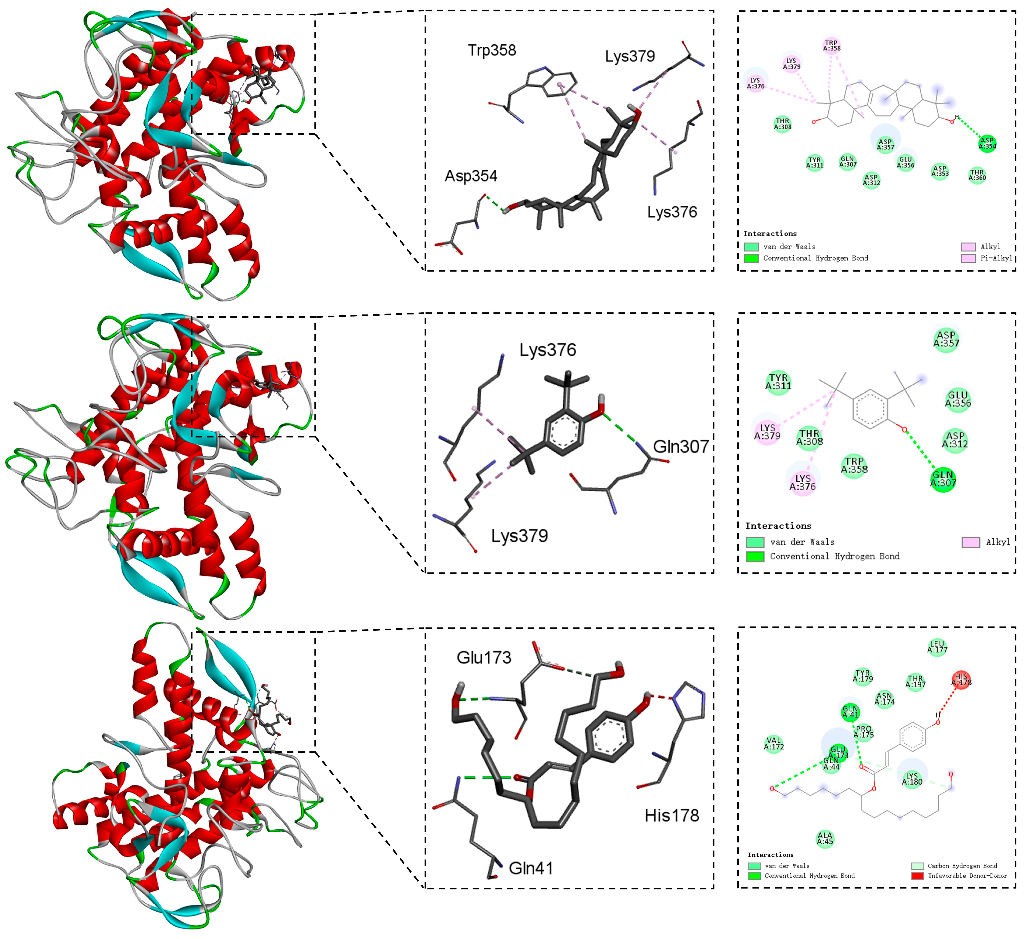
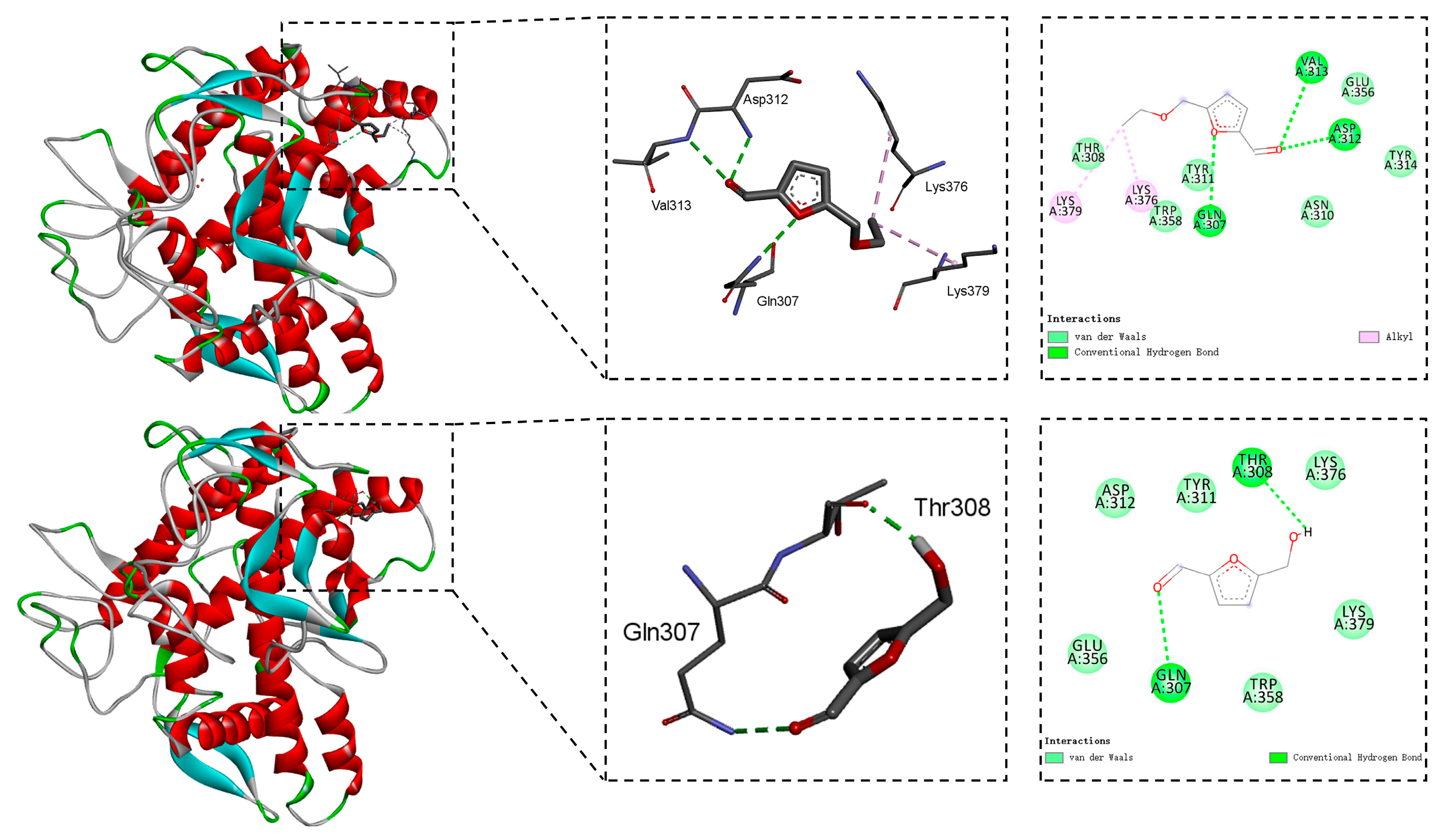
The binding mechanisms between the five components, 4, 12, 13, 14, and 15, and the mushroom tyrosinase were deciphered through molecular docking calculations [42]. Figure 2 and Figure 3 represent the 2D and 3D interactive plots of these constituents with tyrosinase in the docked complexes.
As for compound 4, its hydroxyl group formed one hydrogen bond with the amino acid Asp354. Additionally, two alkyl interactions with Lys376, and Lys379 and one alkyl stacking interaction with Trp358 were observed (Figure 2). Figure 2 also illustrates that the hydrogen bond with amino acid Gln307 and alkyl stacking interactions with Lys376 and Lys379 were engaged in the binding mode between constituent 12 and the mushroom tyrosinase. Figure 2 also demonstrates the different types of interactions in the complexes of compound 13 and the mushroom tyrosinase. One hydrogen bond was formed between the carbonyl at C-3′ of 13 and amino acid residue Gln41, while another hydrogen bond was established by the hydroxyl groups at C-10′ and Glu173. Furthermore, the hydroxyl groups at C-19′ of substance 13 engaged in a carbon hydrogen bond with Lys180.
As shown in Figure 3, the hydrogen bonds and alkyl interactions played key roles in the binding mode between component 14 and the mushroom tyrosinase. The amino acids Lys376 and Lys379 were involved in the alkyl interactions [43]. Furthermore, substance 14 induced hydrogen bonds with Gln307, Val313, and Asp312. Figure 3 also disclosed that compound 15 formed two hydrogen bonds with the amino acids Gln307 and Thr308.

3. Materials and Methods

3.1. General Experimental Procedures

NMR spectra were measured on Bruker DRX-400 and 600 spectrometers (Bruker Biospin AG, Fällanden, Germany). Commercial silica gel (200–300 and 300–400 mesh, Qingdao Haiyang Chemical Group Co., Ltd., Qingdao, China) and Sephadex LH-20 gel (Amersham Biosciences, Amersham, UK) were used for column chromatography, and precoated silica gel plates (GF-254, Yan Tai Zi Fu Chemical Group Co., Yantai, China) were used for analytical TLC. All solvents used for column chromatography were of analytical grade (Shanghai Chemical Reagents Co., Ltd., Shanghai, China).

3.2. Plant Material

The aboveground parts of the plant L. japonicum were collected from Baiyunxian Mountain in the Hunan Province of China in January 2023. The authentication of the plant sample was performed by Dr. L. Wu, one author of this manuscript. A specimen (No. P2023HN2) for certification was kept in the Laboratory of Natural Products Chemistry, Central South University of Forestry and Technology (Changsha, China).

3.3. Extraction and Isolation

The aerial parts of the plant L. japonicum were air-dried (dried weight 2.2 kg). They were extracted with 90% ethanol using ultrasonication at room temperature (2 h × 3). A dark residue was obtained after the removal of the solvent from the combined ethanol extracts by a rotavapor. Then, the suspension of the residue in H2O was successively partitioned with the solvents petroleum ether (P) and ethyl acetate (E) to yield two corresponding P and E extracts, respectively.
The E extract was first chromatographed over silica gel (P/E 20:1→0:1) to give seven fractions, Fr. A–G. Six subfractions, Fr. C1–C6, were obtained from Fr. C by silica gel column chromatography (P/E 20:1→1:1). Fr. C2 was further divided into three subfractions (Fr. C2A–C2C) by chromatography over silica gel (P/E 20:1→5:1). Purification of Fr. C2A by chromatography over silica gel (P/E 20:1) yielded compound 12 (9.5 mg). Similarly, purification of Fr. C3 afforded component 9 (15.3 mg) using silica gel column chromatography (P/E 10:1→3:1). Separation of Fr. C4 by silica gel (dichromethane (D)/methanol (M) 200:1→50:1) yielded five subfractions, Fr. C4A–C4F. Purification of Fr. C4B by silica gel (P/E 7:1→3:1) resulted in three subfractions, Fr. C4B1–C4B3. Compound 14 (5.9 mg) was obtained from Fr. C4B2 by chromatography over Sephadex LH-20 (P/D/M 2:1:1). Compound 8 (38.3 mg) was obtained from Fr. C4B3 by silica gel column chromatography (P/E 7:1). Three subfractions, Fr. C4C1–C4C3, were revealed by silica gel column chromatography of Fr. C4C (D/M 200:1→100:1). Compound 11 (4.1 mg) was obtained from Fr. C4C2 by chromatography over Sephadex LH-20 (P/D/M 2:1:1). Separation of Fr. C4D by silica gel (P/E 5:1→3:1) gave four subfractions, Fr. C4D1–C4D4. Compound 1 (2.2 mg) was isolated from Fr. C4D3 by silica gel column chromatography (P/E 4:1). Fr. C5 was divided into six subfractions, Fr. C5A–C5F by chromatography over silica gel (P/E 5:1→3:1). Compound 2 (14.3 mg) was obtained from Fr. C5C by silica gel column chromatography (D/M 100:1). Fr. C5E (35.2 mg) was successively processed using chromatography over silica gel (D/M 100:1→30:1) and Sephadex LH-20 (P/D/M 2:1:1) to produce compound 3 (7.9 mg).
Fr. F was divided into five subfractions, Fr. F1–F5, by silica gel column chromatography (D/M 100:1→5:1). Compound 6 (573.9 mg) was isolated from Fr. F1 by chromatography over silica gel (P/E 3:1). Fr. F2 was divided into three subfractions, Fr. F2A–F2C by silica gel column chromatography (P/E 3:1→1:1). Compound 10 (8.7 mg) was obtained from Fr. F2B by chromatography over silica gel (D/M 100:1). Purification of F2C by silica gel column chromatography (D/M 80:1) produced compound 4 (13.1 mg). Separation of Fr. F3 by silica gel column chromatography (D/M 50:1→20:1) yielded five subfractions, Fr. F3A–F3E. Compound 7 (4.8 mg) was obtained from Fr. F3B by chromatography over silica gel (P/E 7:3). Compound 5 (7.4 mg) was isolated from Fr. F3D by silica gel column chromatography (P/E 2:1). Fr. F4 (176.4 mg) was purified by silica gel column chromatography (P/E 2:1→1:1) to give four subfractions, Fr. F4A–F4D. Purification of Fr. F4B by chromatography over Sephadex LH-20 (P/D/M 2:1:1) resulted in three subfractions, Fr. F4B1–F4B3. Compound 15 (12.4 mg) was obtained from Fr. F4B3 by silica gel column chromatography (D/M 20:1). Fr. F5 (428.2 mg) was divided into six subfractions, Fr. F5A–F5F by chromatography over silica gel (P/E 2:1→0:1) Separation of Fr. F5D by chromatography over Sephadex LH-20 CC (P/D/M 2:1:1) yielded two subfractions, Fr. F5D1–F5D2. Compound 13 (10.6 mg) was isolated from Fr. F5D2 using silica gel column chromatography (P/E 1:1).
Figure 4 summarizes the aforementioned key isolation and purification experimental procedures of components 115.

3.4. Characteristic 1H and 13C NMR Spectral Data of Isolates

Compound 1: 1H-NMR (600 MHz, CDCl3) δ: 5.71 (1H, t, J = 2.5 Hz, H-15), 3.41 (1H, t, J = 2.9 Hz, H-3), 3.35 (1H, t, J = 2.9 Hz, H-21), 1.25 (3H, s, H-30), 1.12 (3H, s, H-29), 0.95 (3H, s, H-23), 0.87 (3H, s, H-24), 0.84 (3H, s, H-28), 0.83 (3H, s, H-25), 0.80 (3H, s, H-26); 13C-NMR (150 MHz, CDCl3) δ: 33.3 (C-1), 26.6 (C-2), 76.9 (C-3), 38.4 (C-4), 49.4 (C-5), 18.8 (C-6), 45.0 (C-7), 38.2 (C-8), 62.6 (C-9), 37.8 (C-10), 24.7 (C-11), 25.6 (C-12), 59.0 (C-13), 163.7 (C-14), 128.8 (C-15), 201.4 (C-16), 58.8 (C-17), 44.4 (C-18), 31.6 (C-19), 25.2 (C-20), 76.1 (C-21), 36.9 (C-22), 28.5 (C-23), 22.3 (C-24), 15.8 (C-25), 20.1 (C-26), 56.0 (C-27), 15.0 (C-28), 21.6 (C-29), 28.0 (C-30).
Compound 2: 1H-NMR (600 MHz, CDCl3) δ: 5.32 (1H, br s, H-15), 3.45 (1H, t, J = 2.9 Hz, H-21), 3.18 (1H, dd, J = 11.7, 4.6 Hz, H-3), 0.97 (3H, s, H-30), 0.93 (3H, s, H-29), 0.88 (3H, s, H-24), 0.84 (3H, s, H-26), 0.80 (3H, s, H-23), 0.77 (3H, s, H-25), 0.69 (3H, s, H-28); 13C-NMR (150 MHz, CDCl3) δ: 38.8 (C-1), 27.3 (C-2), 79.0 (C-3), 38.3 (C-4), 55.9 (C-5), 19.1 (C-6), 45.3 (C-7), 37.6 (C-8), 63.1 (C-9), 39.1 (C-10), 25.4 (C-11), 27.7 (C-12), 57.0 (C-13), 138.7 (C-14), 122.2 (C-15), 24.2 (C-16), 43.5 (C-17), 36.1 (C-18), 31.4 (C-19), 25.6 (C-20), 76.4 (C-21), 37.3 (C-22), 28.3 (C-23), 15.6 (C-24), 15.9 (C-25), 19.9 (C-26), 56.4 (C-27), 13.4 (C-28), 21.9 (C-29), 27.9 (C-30).
Compound 3: 1H-NMR (600 MHz, CDCl3) δ: 5.38 (1H, br s, H-15), 3.19 (1H, dd, J = 11.7, 4.6 Hz, H-3), 1.08 (3H, s, H-30), 1.04 (3H, s, H-29), 0.97 (3H, s, H-23), 0.92 (3H, s, H-28), 0.83 (3H, s, H-26), 0.80 (3H, s, H-25), 0.77 (3H, s, H-24); 13C-NMR (150 MHz, CDCl3) δ: 38.7 (C-1), 27.7 (C-2), 79.0 (C-3), 38.3 (C-4), 51.3 (C-5), 19.0 (C-6), 45.3 (C-7), 37.2 (C-8), 62.9 (C-9), 39.1 (C-10), 25.6 (C-11), 27.3 (C-12), 56.6 (C-13), 138.5 (C-14), 122.1 (C-15), 24.7 (C-16), 55.9 (C-17), 36.3 (C-18), 34.9 (C-19), 38.5 (C-20), 217.2 (C-21), 47.8 (C-22), 28.3 (C-23), 15.6 (C-24), 15.9 (C-25), 19.9 (C-26), 56.0 (C-27), 13.1 (C-28), 24.6 (C-29), 21.7 (C-30).
Compound 4: 1H-NMR (600 MHz, CDCl3) δ: 5.33 (1H, br s, H-15), 3.23 (1H, dd, J = 11.6, 4.2 Hz, H-21), 3.19 (1H, dd, J = 11.7, 4.6 Hz, H-3), 0.97 (3H, s, H-30), 0.96 (3H, s, H-29), 0.83 (6H, s, H-24, H-26), 0.80 (3H, s, H-23), 0.77 (3H, s, H-25), 0.67 (3H, s, H-28); 13C-NMR (150 MHz, CDCl3) δ: 38.7 (C-1), 27.8 (C-2), 79.0 (C-3), 39.0 (C-4), 55.8 (C-5), 19.0 (C-6), 45.3 (C-7), 37.2 (C-8), 63.0 (C-9), 38.3 (C-10), 25.4 (C-11), 27.4 (C-12), 57.3 (C-13), 138.3 (C-14), 122.3 (C-15), 24.2 (C-16), 49.6 (C-17), 36.3 (C-18), 37.3 (C-19), 27.7 (C-20), 79.3 (C-21), 39.1 (C-22), 15.6 (C-23), 28.3 (C-24), 15.9 (C-25), 19.9 (C-26), 56.2 (C-27), 13.6 (C-28), 14.8 (C-29), 27.7 (C-30).
Compound 5: 1H-NMR (600 MHz, C5D5N) δ: 5.51 (1H, br s, H-15), 3.70 (1H, br s, H-21), 3.64 (3H, s, -OCH3), 3.36 (1H, dd, J = 12.2, 4.6 Hz, H-3), 1.58 (3H, s, H-23), 1.18 (3H, s, H-30), 0.97 (3H, s, H-29), 0.88 (3H, s, H-26), 0.85 (3H, s, H-28), 0.80 (3H, s, H-25); 13C-NMR (150 MHz, C5D5N) δ: 39.8 (C-1), 29.3 (C-2), 78.4 (C-3), 50.2 (C-4), 57.0 (C-5), 21.4 (C-6), 45.7 (C-7), 37.6 (C-8), 62.9 (C-9), 39.0 (C-10), 27.0 (C-11), 28.1 (C-12), 57.7 (C-13), 139.8 (C-14), 123.3 (C-15), 25.0 (C-16), 44.2 (C-17), 36.8 (C-18), 32.2 (C-19), 26.0 (C-20), 75.6 (C-21), 38.4 (C-22), 24.6 (C-23), 178.4 (C-24), 14.1 (C-25), 20.1 (C-26), 57.2 (C-27), 14.2 (C-28), 22.5 (C-29), 29.1 (C-30), 51.4 (-OCH3).
Compound 6: 1H-NMR (600 MHz, CDCl3) δ: 4.83 (2H, s, H-26a, H-27a), 4.56 (2H, s, H-26b, H-27b), 3.25 (2H, dd, J = 11.7, 4.2 Hz, H-3, H-21), 0.99 (6H, s, H-23, H-30), 0.76 (6H, s, H-24, H-29), 0.64 (6H, s, H-25, H-28); 13C-NMR (150 MHz, CDCl3) δ: 37.2 (C-1, C-19), 28.1 (C-2, C-20), 79.1 (C-3, C-21), 39.4 (C-4, C-22), 54.8 (C-5, C-17), 24.2 (C-6, C-16), 38.4 (C-7, C-15), 148.6 (C-8, C-14), 57.7 (C-9, C-13), 39.3 (C-10, C-18), 22.8 (C-11, C-12), 28.5 (C-23, C-29), 15.5 (C-24, C-30), 14.7 (C-25, C-28), 106.9 (C-26, C-27).
Compound 7: 1H-NMR (400 MHz, CDCl3) δ: 4.91 (1H, s, H-27a), 4.89 (1H, s, H-27b), 3.34 (1H, dd, J = 11.7, 4.0 Hz, H-3), 3.23 (1H, dd, J = 11.8, 4.2 Hz, H-21), 1.09 (3H, s, H-25), 0.99 (3H, s, H-28), 0.81 (3H, s, H-24), 0.76 (3H, s, H-30), 0.69 (3H, s, H-23), 0.63 (3H, s, H-29); 13C-NMR (100 MHz, CDCl3) δ: 37.1 (C-1), 28.5 (C-2), 78.8 (C-3), 39.3 (C-4), 54.8 (C-5), 27.7 (C-6), 42.5 (C-7), 212.1 (C-8), 64.8 (C-9), 42.4 (C-10), 21.2 (C-11), 23.7 (C-12), 57.4 (C-13), 147.5 (C-14), 37.3 (C-15), 23.7 (C-16), 53.7 (C-17), 38.4 (C-18), 37.1 (C-19), 28.1 (C-20), 79.1 (C-21), 39.4 (C-22), 27.7 (C-23), 15.5 (C-24), 14.6 (C-25), 108.0 (C-27), 14.9 (C-28), 28.5 (C-29), 15.5 (C-30).
Compound 8: 1H-NMR (600 MHz, CDCl3) δ: 5.35 (1H, m, H-6), 3.52 (1H, m, H-3), 1.01 (3H, s, H-19), 0.92 (3H, d, J = 6.6 Hz, H-21), 0.84 (3H, t, J = 7.7 Hz, H-29), 0.83 (3H, d, J = 6.7 Hz, H-26), 0.81 (3H, d, J = 6.8 Hz, H-27), 0.68 (3H, s, H-18); 13C-NMR (150 MHz, CDCl3) δ: 37.4 (C-1), 32.1 (C-2), 72.0 (C-3), 42.4 (C-4), 140.9 (C-5), 121.9 (C-6), 32.1 (C-7), 31.8 (C-8), 50.3 (C-9), 36.7 (C-10), 21.2 (C-11), 39.9 (C-12), 42.5 (C-13), 56.9 (C-14), 24.5 (C-15), 28.4 (C-16), 56.2 (C-17), 12.0 (C-18), 19.2 (C-19), 36.3 (C-20), 18.9 (C-21), 34.1 (C-22), 26.2 (C-23), 46.0 (C-24), 29.3 (C-25), 19.5 (C-26), 20.0 (C-27), 23.2 (C-28), 12.1 (C-29).
Compound 9: 1H-NMR (600 MHz, CDCl3) δ: 7.61 (1H, d, J = 15.9 Hz, H-1′), 7.07 (1H, dd, J = 8.2, 2.0 Hz, H-6), 7.03 (1H, d, J = 1.9 Hz, H-2), 6.92 (1H, d, J = 8.2 Hz, H-5), 6.29 (1H, d, J = 15.9 Hz, H-2′), 5.84 (1H, br s, 4-OH), 4.26 (2H, q, J = 7.1 Hz, H-4′), 3.93 (3H, s, -OCH3), 1.33 (3H, t, J =7.1 Hz, H-5′); 13C-NMR δ: (150 MHz, CDCl3) δ: 127.2 (C-1), 109.4 (C-2), 148.0 (C-3), 146.9 (C-4), 114.8 (C-5), 123.2 (C-6), 144.8 (C-1′), 115.8 (C-2′), 167.4 (C-3′), 60.5 (C-4′), 14.5 (C-5′), 56.1 (-OCH3).
Compound 10: 1H-NMR (600 MHz, CDCl3) δ: 7.77 (1H, d, J = 2.0 Hz, H-2), 7.11 (1H, dd, J = 8.3, 2.0 Hz, H-6), 6.88 (1H, d, J = 8.2 Hz, H-5), 6.79 (1H, d, J = 12.9 Hz, H-1′), 5.82 (1H, br s, -OH), 5.81 (1H, d, J = 12.9 Hz, H-2′), 4.19 (2H, q, J = 7.1 Hz, H-4′), 3.93 (3H, s, -OCH3), 1.29 (3H, t, J = 7.2 Hz, H-5′); 13C-NMR (150 MHz, CDCl3) δ: 127.4 (C-1), 112.9 (C-2), 146.0 (C-3), 147.2 (C-4), 113.9 (C-5), 125.8 (C-6), 143.8 (C-1′), 117.0 (C-2′), 166.7 (C-3′), 60.3 (C-4′), 14.4 (C-5′), 56.1 (-OCH3).
Compound 11: 1H-NMR (600 MHz, CDCl3) δ: 7.64 (1H, dd, J =8.3, 1.9 Hz, H-6), 7.55 (1H, d, J =1.9 Hz, H-2), 6.93 (1H, d, J =8.3 Hz, H-5), 4.35 (2H, q, J =7.1 Hz, H-2′), 3.95 (3H, s, -OCH3), 1.38 (3H, t, J = 7.1 Hz, H-3′); 13C-NMR (150 MHz, CDCl3) δ: 124.2 (C-1), 111.8 (C-2), 146.3 (C-3), 150.1 (C-4), 114.1 (C-5), 122.8 (C-6), 166.6 (C-1′), 60.9 (C-2′), 14.5 (C-3′), 56.2 (-OCH3).
Compound 12: 1H-NMR (600 MHz, CDCl3) δ: 7.31 (1H, d, J = 2.4 Hz, H-3), 7.08 (1H, dd, J = 8.2, 2.4 Hz, H-5), 6.60 (1H, d, J = 8.2 Hz, H-6), 1.43 (9H, s, H-12, H-13, H-14), 1.30 (9H, s, H-8, H-9, H-10); 13C-NMR (150 MHz, CDCl3) δ: 151.9 (C-1), 143.1 (C-2), 116.1 (C-3), 135.3 (C-4), 123.7 (C-5), 124.2 (C-6), 34.4 (C-7), 31.8 (C-8, C-9, C-10), 34.9 (C-11), 29.8 (C-12, C-13, C-14).
Compound 13: 1H-NMR (600 MHz, CD3OD) δ: 7.61 (1H, d, J =15.9 Hz, H-1′), 7.48 (2H, d, J = 8.6 Hz, H-3, H-5), 6.82 (2H, d, J = 8.6 Hz, H-2, H-6), 6.33 (1H, d, J = 15.9 Hz, H-2′), 5.01 (1H, m, H-4′), 3.55 (2H, t, J = 6.6 Hz, H-10′), 3.54 (2H, t, J = 6.6 Hz, H-19′), 1.63 (4H, m, H-5′, H-11′), 1.53 (4H, m, H-9′, H-18′); 13C-NMR (150 MHz, CD3OD) δ: 161.3 (C-1), 116.8 (C-2, C-6), 131.1 (C-3, C-5), 127.2 (C-4), 146.3 (C-1′), 115.6 (C-2′), 169.3 (C-3′), 75.4 (C-4′), 35.4 (C-5′, C-11′), 26.5 (C-6′), 30.7 (C-7′), 26.9 (C-8′), 33.7 (C-9′), 63.0 (C-10′), 26.4 (C-12′), 30.6 (C-13′, C-14′, C-15′), 30.5 (C-16′), 26.8 (C-17′), 33.5 (C-18′), 62.9 (C-19′).
Compound 14: 1H-NMR (600 MHz, CDCl3) δ: 9.62 (1H, s, -CHO), 7.21 (1H, d, J = 3.5 Hz, H-4), 6.52 (1H, d, J = 3.6 Hz, H-3), 4.53 (2H, s, H-6), 3.59 (2H, q, J = 7.0 Hz, H-7), 1.24 (3H, t, J = 7.0 Hz, H-8); 13C-NMR (150 MHz, CDCl3) δ: 177.9 (C-1), 158.9 (C-2), 122.1 (C-3), 111.1 (C-4), 152.7 (C-5), 66.8 (C-6), 64.9 (C-7), 15.2 (C-8).
Compound 15: 1H-NMR (600 MHz, CDCl3) δ: 9.60 (1H, s, -CHO), 7.21 (1H, d, J = 3.5 Hz, H-4), 6.52 (1H, d, J = 3.5 Hz, H-3), 4.72 (2H, s, H-6); 13C-NMR (150 MHz, CDCl3) δ: 177.8 (C-1), 160.6 (C-2), 122.3 (C-3), 110.1 (C-4), 152.6 (C-5), 57.8 (C-6).

3.5. In Vitro Tyrosinase Inhibitory Bioassay

The tyrosinase inhibition bioassay was conducted with minor modifications based on the previously described method [44]. In the wells of a 96-well plate, there was phosphate buffer (pH 6.8, 50 μL), tyrosinase (100 U/mL, 25 μL), sample solutions (25 μL), and L-tyrosine (100 μL). These mixtures were incubated at 37 °C for 0.5 h. Notably, 475 nm was selected for the measurement of absorbance of each well. The positive control was Kojic acid. The equation for the calculation of the inhibition rate was as follows: inhibition rate (%) = [1 − (A1 − A2)/(A3 − A0)] × 100, where A1 represents the absorbance of the sample solution, A2 represents the absorbance of the sample solution control, A3 represents the absorbance of blank, and A0 represents the absorbance of the blank control. The test of each concentration was performed three times parallelly. The fitting curve of inhibition rates vs. different concentrations of the test compound derived a half-maximal inhibitory concentration (IC50).

3.6. Molecular Docking Experiments

The software AutoDock Vina (v4.2) was employed for the molecular docking studies. The crystal structure of the mushroom tyrosinase with PDB No. 2Y9X was obtained from the RCSB Protein Data Bank. Removal of the ligand and water molecules from the crystal structure was conducted by the software Pymol (v2.4). The software AutoDock Vina (v4.2) detected the active site of the crystal structure and set the coordinates to (−7.40 −23.55 −32.51) and the grid box size at 70.36 Å × 70.36 Å × 70.36 Å. The software Discovery Studio Visualizer (v19.1) was used for the analysis.

4. Conclusions

In summary, a detailed chemical investigation of the ornamental and medicinal plant L. japonicum led to the isolation and identification of an array of components with various structural characteristics, including seven serratane-type triterpenes 17, one steroid 8, five benzene derivatives 913, and two furan derivatives 14 and 15. Among them, compounds 3, 5 and 915 were reported from the plant L. japonicum for the first time. All the isolates except for the previously investigated 69 were assessed for tyrosinase inhibitory activity. As a result, compounds 4, 12, 13, 14, and 15 exhibited significant inhibition against tyrosinase (IC50 values ranging from 1.46 mM to 6.82 mM), which were close in value to the positive control Kojic acid (IC50 = 0.17 mM). This work not only enriched the chemical warehouse of the species L. japonicum but also supplemented the foundation for the future cosmetical and medicinal utilization of this species.

Supplementary Materials

The following supporting information can be downloaded at https://www.mdpi.com/article/10.3390/molecules30194024/s1, Tables S1–S15: The 1H and 13C NMR data of compound 115 and comparison with the data in the literature.

Author Contributions

Conceptualization, L.-F.L.; methodology, Z.-Y.G., Q.-B.Y. and M.Z.; software, Z.-Y.G., Q.-B.Y. and X.-Y.Y.; validation, Z.-Y.G. and L.W.; formal analysis, Z.-Y.G., Q.-B.Y., X.-Y.Y., L.W. and M.Z.; investigation, Z.-Y.G. and Y.-Q.W.; resources, L.W.; data curation, Z.-Y.G.; writing—original draft preparation, Z.-Y.G.; writing—review and editing, L.-F.L.; visualization, Z.-Y.G. and Y.-Q.W.; supervision, L.-F.L.; project administration, L.-F.L.; funding acquisition, L.-F.L. All authors have read and agreed to the published version of the manuscript.

Funding

This research was funded by the Human Provincial Natural Science Foundation of China (No. 2025JJ50098 and 2025JJ50122), the Changsha Municipal Natural Science Foundation (No. kq2402254), the National Natural Science Foundation of China (No. 41876194).

Institutional Review Board Statement

Not applicable.

Informed Consent Statement

Not applicable.

Data Availability Statement

Data is contained within the article.

Conflicts of Interest

The authors declare no conflicts of interest.

References

  1. Chang, T.-S. An updated review of tyrosinase inhibitors. Int. J. Mol. Sci. 2009, 10, 2440–2475. [Google Scholar] [CrossRef]
  2. Arrowitz, C.; Schoelermann, A.M.; Mann, T.; Jiang, L.I.; Weber, T.; Kolbe, L. Effective tyrosinase inhibition by Thiamidol results in significant improvement of mild to moderate melasma. J. Investig. Dermatol. 2019, 139, 1691–1698. [Google Scholar] [CrossRef]
  3. Godoy-Gallardo, M.; Labay, C.; Hosta-Rigau, L. Tyrosinase-loaded multicompartment microreactor toward melanoma depletion. ACS Appl. Mater. Interfaces 2019, 11, 5862–5876. [Google Scholar] [CrossRef]
  4. Zolghadri, S.; Bahrami, A.; Hassan Khan, M.T.; Munoz-Munoz, J.; Garcia-Molina, F.; Garcia-Canovas, F.; Saboury, A.A. A comprehensive review on tyrosinase inhibitors. J. Enzyme Inhib. Med. Chem. 2019, 34, 279–309. [Google Scholar] [CrossRef] [PubMed]
  5. Boonya-udtayan, S.; Thasana, N.; Jarussophon, N.; Ruchirawat, S. Serratene triterpenoids and their biological activities from Lycopodiaceae plants. Fitoterapia 2019, 136, 104181. [Google Scholar] [CrossRef] [PubMed]
  6. Chen, Y.; Yang, Q.; Zhang, Y. Lycopodium japonicum: A comprehensive review on its phytochemicals and biological activities. Arab. J. Chem. 2020, 13, 5438–5450. [Google Scholar] [CrossRef]
  7. Xie, Z.; Liu, Y.; Luo, Q.; Wang, Q.; Min, Y.-B.; Liu, L.; Zheng, D.-K. New Lycopodium alkaloids with neuroprotective activities from Lycopodium japonicum Thunb. Nat. Prod. Res. 2025, 39, 718–724. [Google Scholar] [CrossRef]
  8. Xia, D.; Wang, Z.-H.; Jiang, J.-M.; Yang, X.-W.; Gao, Y.; Xu, Y.-Y.; Chang, L.-Y.; Zhu, D.; Zhao, B.-J.; Zhu, X.-L.; et al. Lycojapomines A–E: Lycopodium alkaloids with anti-renal fibrosis potential from Lycopodium japonicum. Org. Lett. 2022, 24, 4684–4688. [Google Scholar] [CrossRef] [PubMed]
  9. Zhu, Y.; Dong, L.-B.; Zhang, Z.-J.; Fan, M.; Zhu, Q.-F.; Qi, Y.-Y.; Liu, Y.-C.; Peng, L.-Y.; Wu, X.-D.; Zhao, Q.-S. Three new Lycopodium alkaloids from Lycopodium japonicum. J. Asian Nat. Prod. Res. 2019, 21, 17–24. [Google Scholar] [CrossRef]
  10. Wang, X.-J.; Zhang, G.-J.; Zhuang, P.-Y.; Zhang, Y.; Yu, S.-S.; Bao, X.-Q.; Zhang, D.; Yuan, Y.-H.; Chen, N.-H.; Ma, S.-G.; et al. Lycojaponicumins A–C, three alkaloids with an unprecedented skeleton from Lycopodium japonicum. Org. Lett. 2012, 14, 2614–2617. [Google Scholar] [CrossRef]
  11. Sun, Z.-H.; Li, W.; Tang, G.-H.; Yin, S. A new serratene triterpenoid from Lycopodium japonicum. J. Asian Nat. Prod. Res. 2017, 19, 299–303. [Google Scholar] [CrossRef] [PubMed]
  12. Zhang, Y.; Yi, P.; Chen, Y.; Mei, Z.-n.; Hu, X.; Yang, G.-Z. Lycojaponicuminol A–F: Cytotoxic serratene triterpenoids from Lycopodium japonicum. Fitoterapia 2014, 96, 95–102. [Google Scholar] [CrossRef] [PubMed]
  13. Wang, X.; Yu, D.; Yu, S. Two new triterpenoids from Lycopodium japonicum Thunb. Chin. J. Chem. 2014, 32, 1007–1010. [Google Scholar] [CrossRef]
  14. Li, X.-L.; Zhao, Y.; Cheng, X.; Tu, L.; Peng, L.-Y.; Xu, G.; Zhao, Q.-S. Japonicumins A–D: Four new compounds from Lycopodium japonicum. Helv. Chim. Acta 2006, 89, 1467–1473. [Google Scholar] [CrossRef]
  15. Ren, Q.; Zou, Z.; Liu, Y.; Chen, X.; Xu, K.; Tan, G. Two new lignans from Lycopodium japonicum Thunb. Rec. Nat. Prod. 2019, 13, 468–474. [Google Scholar] [CrossRef]
  16. Li, M.-J.; Liu, J.; Zhang, Y.-B.; Chen, N.-H.; Wang, G.-C.; Li, Y.-L. Chemical constituents from whole herb of Lycopodium japonicum. Chin. Tradit. Herb. Drugs 2015, 46, 33–37. [Google Scholar] [CrossRef]
  17. Wang, X.; Wang, F.; Wu, J.; Chen, S.-Q.; Jiang, C.-S.; Yang, S.-P.; Wang, C.; Cai, Y.-S. Japonisine A, a fawcettimine-type Lycopodium alkaloid with an unusual skeleton from Lycopodium japonicum Thunb. Fitoterapia 2022, 156, 105069. [Google Scholar] [CrossRef]
  18. He, J.; Chen, X.-Q.; Li, M.-M.; Zhao, Y.; Xu, G.; Cheng, X.; Peng, L.-Y.; Xie, M.-J.; Zheng, Y.-T.; Wang, Y.-P.; et al. Lycojapodine A, a novel alkaloid from Lycopodium japonicum. Org. Lett. 2009, 11, 1397–1400. [Google Scholar] [CrossRef]
  19. Wang, X.-J.; Li, L.; Si, Y.-K.; Yu, S.-S.; Ma, S.-G.; Bao, X.-Q.; Zhang, D.; Qu, J.; Liu, Y.-B.; Li, Y. Nine new lycopodine-type alkaloids from Lycopodium japonicum Thunb. Tetrahedron 2013, 69, 6234–6240. [Google Scholar] [CrossRef]
  20. Wang, X.-J.; Liu, Y.-B.; Li, L.; Yu, S.-S.; Lv, H.-N.; Ma, S.-G.; Bao, X.-Q.; Zhang, D.; Qu, J.; Li, Y. Lycojaponicumins D and E: Two new alkaloids from Lycopodium japonicum. Org. Lett. 2012, 14, 5688–5691. [Google Scholar] [CrossRef] [PubMed]
  21. Yang, Q.; Zhu, Y.; Zhan, R.; Chen, Y.-g. A new fawcettimine-related alkaloid from Lycopodium japonicum. Chem. Nat. Compd. 2018, 54, 729–731. [Google Scholar] [CrossRef]
  22. Bu, Q.; Yang, Q.-B.; Ge, Z.-Y.; Shi, Z.-X.; Liang, L.-F. Phytochemicals and health-promoting evaluations of Ophiorrhiza puffii, a Chinese endemic plant. Nat. Prod. Res. 2025, in press. [Google Scholar] [CrossRef] [PubMed]
  23. Bu, Q.; Ge, Z.-Y.; Liang, L.-F. Chemical and biological investigation on the potential ornamental plant Ophiorrhiza chinensis. Agronomy 2024, 14, 1872. [Google Scholar] [CrossRef]
  24. Yang, G.-X.; Zang, Y.; Hu, C.-L.; Xiong, J.; Hu, J.-F. Serratene triterpenoids in Lycopodium japonicum from Hubei province. Chin. Tradit. Herb. Drugs 2014, 45, 3524–3527. [Google Scholar] [CrossRef]
  25. Sa, N.H.; Tam, N.T.; Quan, T.D.; Tinh, B.X.; Thien, D.D.; Sung, T.V.; Adorisio, S.; Delfino, D.V. Chemical constituents and their biological activity of Pinus dalatensis. Part 1. Terpenoids from the leaves. Vietnam J. Chem. 2017, 55, 509–513. [Google Scholar] [CrossRef]
  26. Zheng, K.-K.; Zhao, Y.-Y.; Yuan, P.-L.; Wu, Y.-C.; Zhang, L.-Q.; Guo, F.-J.; Li, Y.-M. Chemical constituents of non-alkaloids from Huperzia serrata. Chin. Tradit. Herb. Drugs 2016, 47, 15–20. [Google Scholar] [CrossRef]
  27. Tsuda, Y.; Isobe, K.; Sano, T. Triterpenoid chemistry. IX. Lycopodium triterpenoid. (6). The structures of three new tetra-ols, lycocryptol, 21-epilycocryptol, and diepilycocryptol, and two new acids, lycernuic acid-A and -B. Chem. Pharm. Bull. 1975, 23, 264–271. [Google Scholar] [CrossRef]
  28. Zhang, Z.; ElSohly, H.N.; Jacob, M.R.; Pasco, D.S.; Walker, L.A.; Clark, A.M. Natural products inhibiting Candida albicans secreted aspartic proteases from Lycopodium cernuum. J. Nat. Prod. 2002, 65, 979–985. [Google Scholar] [CrossRef]
  29. Zhu, M.; Wang, W.-Y.; Chen, W.-J.; Wei, J.-H.; Liang, C.-Y.; Feng, X.; Lu, C.-S. Chemical constituents and antioxidant activity in vitro of Zhuang medicine Dalbergia rimosa. J. Chin. Med. Mater. 2024, 47, 1147–1152. [Google Scholar] [CrossRef]
  30. Teng, X.-F.; Liu, Y.-L.; Yang, F.; Wang, H.-J.; He, L. Chemical constituents from Lycopodium fargesii and their anti-osteoporotic activity. Nat. Prod. Res. Dev. 2022, 34, 226–231. [Google Scholar] [CrossRef]
  31. Pal, P.P.; Begum, S.A.; Basha, A.S.; Araya, H.; Fujimoto, Y. A new lignan (polonilignan) and inhibitors of nitric oxide production from Penicillium polonicum, an endophytic fungi of Piper nigrum. Chem. Biodivers. 2023, 20, e202200840. [Google Scholar] [CrossRef] [PubMed]
  32. Cao, X.-X.; Sun, J.-Y.; Liu, C.; Zhang, J.-S.; Zhang, H. Antiradical aromatic constituents from Pleurotus eryngii. Rec. Nat. Prod. 2021, 15, 169–174. [Google Scholar] [CrossRef]
  33. Li, Y.; Zhou, B.-P.; Zhang, W.-J.; Yang, G.-L.; Zhang, C.-L.; Cao, Z.-Y. Chemical constituents from aerial parts of Ribes mandshuricum. Chin. Tradit. Herb. Drugs 2018, 49, 772–779. [Google Scholar] [CrossRef]
  34. Cho, J.-Y.; Kim, M.-S.; Lee, Y.G.; Jeong, H.Y.; Lee, H.J.; Ham, K.-S.; Moon, J.-H. A phenyl lipid alkaloid and flavone C-diglucosides from Spergularia marina. Food Sci. Biotechnol. 2016, 25, 63–69. [Google Scholar] [CrossRef]
  35. Lee, K.H.; Kim, J.K.; Yu, J.S.; Jeong, S.Y.; Choi, J.H.; Kim, J.-C.; Ko, Y.-J.; Kim, S.-H.; Kim, K.H. Ginkwanghols A and B, osteogenic coumaric acid-aliphatic alcohol hybrids from the leaves of Ginkgo biloba. Arch. Pharm. Res. 2021, 44, 514–524. [Google Scholar] [CrossRef]
  36. Bredihhin, A.; Mäeorg, U.; Vares, L. Evaluation of carbohydrates and lignocellulosic biomass from different wood species as raw material for the synthesis of 5-bromomethyfurfural. Carbohydr. Res. 2013, 375, 63–67. [Google Scholar] [CrossRef]
  37. Miyazawa, M.; Anzai, J.; Fujioka, J.; Isikawa, Y. Insecticidal compounds against Drosophila melanogaster from Cornus officinalis Sieb. Et Zucc. Nat. Prod. Res. 2003, 17, 337–339. [Google Scholar] [CrossRef]
  38. Yang, C. On the nomenclature revision of the Chinese drug shisong and its resources. Zhongyao Tongbao 1981, 6, 12–15. [Google Scholar]
  39. Yan, J.; Zhang, X.-M.; Li, Z.-R.; Zhou, L.; Chen, J.-C.; Sun, L.-R.; Qiu, M.-H. Three new triterpenoids from Lycopodium japonicum Thunb. Helv. Chim. Acta 2005, 88, 240–244. [Google Scholar] [CrossRef]
  40. Teng, C.; He, Y.; Feng, J.; Ba, X.; Li, D. Chemical composition of Lycopodium japonicum. Chin. Tradit. Herb. Drugs 2010, 41, 1960–1963. [Google Scholar]
  41. Zhu, J.-Y.; Ge, Z.-Y.; Yang, Q.-B.; Jiang, C.-F.; Wu, L.; Jiang, X.-Y.; Liang, L.-F. Bioassay-guided isolation of chemical constituents from Lycopodiastrum casuarinoides and targeted evaluation of their potential efficacy in cosmetics. Cosmetics 2025, 12, 174. [Google Scholar] [CrossRef]
  42. Ashraf, Z.; Rafiq, M.; Nadeem, H.; Hassan, M.; Afzal, S.; Waseem, M.; Afzal, K.; Latip, J. Carvacrol derivatives as mushroom tyrosinase inhibitors; synthesis, kinetics mechanism and molecular docking studies. PLoS ONE 2017, 12, e0178069. [Google Scholar] [CrossRef] [PubMed]
  43. Ismaya, W.T.; Rozeboom, H.J.; Weijn, A.; Mes, J.J.; Fusetti, F.; Wichers, H.J.; Dijkstra, B.W. Crystal structure of Agaricus bisporus mushroom tyrosinase: Identity of the tetramer subunits and interaction with tropolone. Biochemistry 2011, 50, 5477–5486. [Google Scholar] [CrossRef] [PubMed]
  44. Souza, P.M.; Elias, S.T.; Simeoni, L.A.; de Paula, J.E.; Gomes, S.M.; Guerra, E.N.S.; Fonseca, Y.M.; Silva, E.C.; Silveira, D.; Magalhães, P.O. Plants from Brazilian Cerrado with potent tyrosinase inhibitory activity. PLoS ONE 2012, 7, e48589. [Google Scholar] [CrossRef]
Figure 1. The chemical structures of the isolates 115 from L. japonicum.
Molecules 30 04024 g001
Figure 2. The binding modes between compounds 4, 12, and 13 and the mushroom tyrosinase (PDB: 2Y9X).
Molecules 30 04024 g002
Figure 3. The binding modes between compounds 14 and 15 and the mushroom tyrosinase (PDB: 2Y9X).
Molecules 30 04024 g003
Figure 4. The summary of key experimental procedures of compounds 115.
Molecules 30 04024 g004
Table 1. The identification of compounds 115 from L. japonicum.
CompoundsNamesReferences
116-oxo-3α-hydroxyserrat-14-en-21β-ol[16]
221-epi-serratenediol[24]
33β-hydroxy-14-serraten-21-one[25]
4serratenediol[24,26]
5methyl lycernuate A[27,28]
6α-onocerin[29]
726-nor-8-oxo-α-onocerin[30]
8β-stiosterol[23]
9trans-ethyl ferulate[31]
10cis-ethyl ferulate[31,32]
11ethyl 4-hydroxy-3-methoxybenzoate[33]
122,4-di-tert-butylphenol[34]
13ginkwanghol A[35]
145-ethoxymethylfurfural[36]
155-hydroxymethyl furfural[37]
Table 2. The tyrosinase inhibitory results of compounds 4, 12, 13, 14, and 15 and the positive control Kojic acid.
CompoundsIC50 (mM)
42.16 ± 0.03
123.28 ± 0.03
131.46 ± 0.03
145.92 ± 0.06
156.82 ± 0.09
Kojic acid 10.17 ± 0.01
1 Positive control.
Disclaimer/Publisher’s Note: The statements, opinions and data contained in all publications are solely those of the individual author(s) and contributor(s) and not of MDPI and/or the editor(s). MDPI and/or the editor(s) disclaim responsibility for any injury to people or property resulting from any ideas, methods, instructions or products referred to in the content.