Review

The Role of Lactoferrin in Combating Candida spp. Infections Through Regulation of Oxidative Stress, Immune Response, and Nutritional Support in Women and Newborns

by
Anna Długosz
1,*,
Joanna Wróblewska
2,*,
Paweł Kołaczyk
1 and
Weronika Wróblewska
3
1
Department of Food Industry Technology and Engineering, Faculty of Chemical Technology and Engineering, Bydgoszcz University of Science and Technology, 3 Seminaryjna St., 85-326 Bydgoszcz, Poland
2
Department of Medical Biology and Biochemistry, Faculty of Medicine, Ludwik Rydygier Collegium Medicum in Bydgoszcz, Nicolaus Copernicus University in Toruń, 24 Karłowicza St., 85-092 Bydgoszcz, Poland
3
Student Research Club of Medical Biology and Biochemistry, Department of Medical Biology and Biochemistry, Faculty of Medicine, Ludwik Rydygier Collegium Medicum in Bydgoszcz, Nicolaus Copernicus University in Toruń, 24 Karłowicza St., 85-092 Bydgoszcz, Poland
*
Authors to whom correspondence should be addressed.
Molecules 2025, 30(11), 2416; https://doi.org/10.3390/molecules30112416
Submission received: 28 April 2025 / Revised: 26 May 2025 / Accepted: 29 May 2025 / Published: 31 May 2025

Abstract

:
Lactoferrin (LF) is a natural glycoprotein with strong antimicrobial, immunomodulatory, and nutritional potential and is widely present in milk and mucosal secretions. This paper aims to review the current knowledge on the application of lactoferrin and its bioactive peptides in the context of fungal infections caused by Candida spp., focusing on newborns and pregnant women as high-risk groups. The multifaceted mechanisms of LF action are discussed, including iron chelation, destabilization of fungal cell membranes, and modulation of the immune response. Additionally, data demonstrating the effectiveness of LF in the prevention and supportive treatment of Candida spp. infections are presented.

1. Introduction

Fungal infections caused by yeasts of the genus Candida, particularly Candida albicans, represent a significant health concern in both women and newborns [1,2]. While C. albicans remains the most common etiological agent of these infections, an increasing prevalence of non-albicans species such as Candida glabrata, Candida krusei, Candida parapsilosis, and Candida tropicalis is being observed, especially in patients with recurrent infections or poor response to antifungal therapy. These species are often more resistant to azole treatments and tend to present with milder clinical symptoms [3].
The yeast form of Candida spp. is a natural component of the lower female genital tract. However, under favorable conditions such as disruption of the microbiota, elevated estrogen levels, or increased vaginal pH, it may convert into the hyphal form and cause symptomatic infection [3,4]. The most common clinical manifestation of fungal infection in women is vulvovaginal candidiasis. It is estimated that up to 75% of women experience it at least once in their lifetime, and in 5 to 10% of cases, it becomes recurrent [1]. Pregnancy is a period of exceptionally high risk for such infections. Hormonal and immunological changes associated with pregnancy facilitate vaginal colonization by Candida yeasts [3,5]. Lower genital tract infections during pregnancy are linked to unfavorable outcomes such as miscarriage, premature rupture of membranes, and preterm birth [6]. Vaginal candidiasis is most frequently diagnosed in the first trimester of pregnancy and much less commonly in the third trimester, which may suggest increased susceptibility to fungal colonization in early pregnancy [7]. However, this finding is based on cross-sectional data, and it remains unclear whether the observed decrease later in pregnancy reflects actual clearance, reduced detection, or underdiagnosis. Information on how Candida spp. colonization changes over time during pregnancy is limited. One study that sampled women at multiple time points found relatively stable colonization rates throughout pregnancy [8]. Infections caused by Candida spp. can also be asymptomatic, meaning many cases remain undiagnosed [7]. The defense of vaginal tissues against Candida spp. invasion involves the cooperation of innate and adaptive immune mechanisms. These include the production of cytokines (such as interleukin 17 (IL-17) and interleukin 23 (IL-23)), the presence of antibodies, the activity of antimicrobial peptides (AMP), and the presence of probiotic bacteria from the Lactobacillus genus [4].
Although direct invasion of placental tissues by yeasts has not been confirmed, research indicates that Candida spp. vaginal infection during pregnancy may disrupt proper placental development, leading to pregnancy complications and neonatal colonization [6,7]. Colonization of the gastrointestinal tract by Candida spp., especially in extremely premature infants, is a significant risk factor for the development of invasive candidiasis [9]. Epidemiological data confirm that early-onset neonatal invasive candidiasis, occurring within the first week of life, primarily affects infants with very low birth weight (<1000 g) and is often caused by C. albicans infection [10]. Mortality associated with neonatal invasive candidiasis can reach up to 75% [2]. C. albicans naturally colonizes healthy individuals’ gastrointestinal tract, mucous membranes, and skin. However, disturbances in immune or microenvironmental balance can lead to a transition of Candida spp. from a commensal to a pathogenic form, resulting in superficial infections and life-threatening systemic candidiasis [11]. According to studies, Candida spp. is present in the oral cavities of infants and newborns and shows an increased tendency to overgrow in children with immune or microbiota imbalances [11]. One of the most common superficial candidiasis manifestations in infants is oral thrush. The lesions affect the mucous membranes of the throat, gums, cheeks, lips, tongue, and lip corners and appear as white plaques resembling curdled milk [12]. Data from the literature report that the prevalence of pseudomembranous candidiasis ranges from 4% to 15% [13]. Pathogen transmission within the mother–infant–mother loop may occur in breastfeeding women, whereby C. albicans detected in the infant’s oral cavity can be transmitted to the mother’s nipples [14].
In recent years, increasing attention has been paid to lactoferrin (LF), a natural antimicrobial protein found in milk, mucus, and other bodily secretions. LF is attributed with numerous functions, including microbistatic and microbicidal activity against a range of pathogens, modulation of the immune response (through both anti-inflammatory and pro-inflammatory mechanisms), support for hematopoiesis, wound healing, bone metabolism, and potential anticancer and antioxidant effects [15]. In newborns, especially those who are breastfed, lactoferrin in maternal milk is an antimicrobial agent. It provides nutritional and immunological support that protects against colonization by pathogenic yeasts. LF exhibits broad antifungal activity against several Candida species, including C. albicans, C. glabrata, and C. tropicalis [16].
This paper aims to discuss the role of LF in proper nutrition and the prevention supportive treatment of infections caused by Candida spp., both in women of reproductive age and newborns. In the context of increasing reports of yeast resistance to conventional antifungal therapy, the use of lactoferrin (both human lactoferrin (hLf) and bovine lactoferrin (bLf)) may represent a promising strategy to support treatment and reduce the recurrence of infections within the urogenital and gastrointestinal tracts in both pregnant women and infants.

2. Biological Activity of Lactoferrin and Its Mechanisms of Action in the Human Body

LF is a multifunctional, non-heme iron-binding glycoprotein belonging to the transferrin family and is considered an AMP [17,18]. One of its most important functions is its involvement in iron metabolism [15]. LF exists in an iron-saturated form (holo-LF) and an iron-free form (apo-LF). This protein binds iron with high affinity in a reversible manner, stabilizing the molecule and making it less susceptible to enzymatic degradation and thermal denaturation. Unlike transferrin, LF retains iron even in acidic environments such as the intestine and supports transport through a specific receptor in enterocytes. After endocytosis, iron is released by reducing Fe3+ to Fe2+ [18]. Notably, Fe2+ is a more reactive form that can generate reactive oxygen species (ROS). Therefore, the iron-chelating activity of LF reduces ROS formation by maintaining iron in a less reactive state, thereby preventing oxidative stress. As such, LF plays an essential role in iron metabolism and in protecting cells from oxidative damage [18]. By binding free iron, LF limits its participation in the Fenton reaction, leading to the formation of highly harmful hydroxyl radicals. LF binds iron stably in neutral pH environments typical of most tissues, preventing its involvement in redox reactions. Through this ability, LF protects cells from iron-induced damage and supports the maintenance of cellular function, particularly under conditions of disturbed iron homeostasis [18].
LF releases previously bound iron in the acidic environment of phagolysosomes (e.g., in neutrophils and macrophages), where the pH drops to 3–4. The resulting Fe2+ ions can generate ROS, which possess vigorous cytotoxic activity against pathogens. Thus, beyond its protective role for host cells, LF enhances the microbicidal potential of phagocytes, supporting the immune response [15]. Additionally, LF acts as a potent antioxidant not only by directly scavenging ROS but also by enhancing the activity of endogenous antioxidant enzymes such as superoxide dismutase (SOD) and glutathione peroxidase. These effects reduce lipid peroxidation products and hydrogen peroxide concentrations in the blood. A general increase in plasma antioxidant capacity has also been observed, underscoring LF’s systemic protective role and its potential in preventing chronic diseases associated with oxidative stress [18].
LF is synthesized and secreted by both neutrophils and glandular epithelial cells. Its presence has been demonstrated in the nasal mucosa, bronchi, skin (particularly in sweat glands), and various other bodily secretions, highlighting its essential role in local and mucosal immunity [19,20,21]. In women with a regular menstrual cycle, lactoferrin levels in cervicovaginal secretions fluctuate according to estrogen concentration, increasing during the proliferative (follicular) phase and decreasing during the secretory (luteal) phase. Additionally, lactoferrin is present in the amniotic fluid and the thick cervical mucus plug during pregnancy, suggesting its involvement in protecting the early fetal environment [22]. Table 1 presents lactoferrin concentrations in selected human body fluids.
LF released from neutrophil granules enters the bloodstream and sites of inflammation, which actively contributes to regulating immune responses [18,23]. In a healthy individual, approximately 5 g of neutrophil-derived LF is produced daily, with about 0.5 g released during neutrophil degranulation [15]. LF acts on various immune cells by binding to their surface receptors and modulating intracellular signaling pathways, particularly those involving Toll-like receptors (TLRs), which can either activate or suppress inflammatory responses depending on the immunological context [24]. In macrophages, it engages both TLR4-related and alternative signaling routes. For example, the expression of the CD40 receptor depends on TLR4, whereas the production of IL-6 does not, indicating distinct mechanisms of action [25]. LF also stimulates NK cells, enhancing their cytotoxic function and aiding in the elimination of infected cells [5]. It primarily supports innate immunity by targeting key effector cells such as neutrophils, macrophages, NK cells, and dendritic cells, which are essential for initiating adaptive responses [23,25] Moreover, LF functions as an alarmin by signaling danger and promoting the recruitment and activation of antigen-presenting cells like dendritic cells, thereby indirectly enhancing T-cell activation and the development of adaptive immunity [5,26].
Due to the increased demand for iron during pregnancy, its deficiency may lead to anemia and perinatal complications. Therefore, LF is considered a safe and effective alternative to traditional iron supplementation and may help avoid the need for higher iron doses during pregnancy. It supports iron absorption and regulation, improves hematological parameters, and is better tolerated than conventional iron preparations [5]. Combined oral and intravaginal administration of LF may serve a preventive function in preterm birth, partly by lowering interleukin-6 (IL-6) and prostaglandin levels in cervicovaginal secretions [5,27].
LF, as an antimicrobial protein, influences the composition of the genital tract microbiota through both direct and indirect prebiotic activity. It directly promotes the growth of probiotic strains such as Lactobacillus spp. and Bifidobacterium spp. and indirectly limits the growth of pathogenic microorganisms. Importantly, it acts selectively by eliminating harmful microbes without disturbing the balance of the natural probiotic microbiota [5,22].
Table 1. Lactoferrin concentration in selected human body fluids.
Body Fluid LF ConcentrationRef.
Vaginal mucusBefore menstruation3.8–11.4 µg/mg[17]
After menstruation62.9–218 µg/mg[17]
During contraception use19.8 µg/mg[17]
Amniotic fluidEarly pregnancy1–2 μg/ml[22]
From the 32nd week of pregnancy until delivery5–15 μg/ml[22]
Cervical mucus plug 10–1000 µg/ml[22]
Human colostrum 6–7 g/L[5,28]
Mature human milk 1–3 g/L[5,28]
Saliva 10–100 µg/mL[22]
Blood 10−3–200 µg/mL[17]
Lactoferrin (LF).

3. The Importance of Lactoferrin in Nutrition

Human milk is widely regarded as the most optimal form of infant nutrition, supporting their healthy development [29]. It contains varying concentrations of LF [28]. This protein may be higher in the milk of mothers who deliver preterm than those who give birth at term [28]. The protective potential of lactoferrin may be maintained or even enhanced during later stages of breastfeeding through maternal milk delivery [30]. Breastfed infants have a lower incidence of infections, necrotizing enterocolitis (NEC), and sudden infant death syndrome, which contributes to reduced neonatal mortality. In later life, breastfeeding is associated with a significantly lower risk of developing various chronic diseases, such as overweight and obesity, type I and II diabetes, asthma (which is hypothesized to be reduced, particularly in early childhood), lymphoma, Hodgkin’s disease, acute lymphoblastic leukemia, and elevated cholesterol levels [31,32,33]. Breastfeeding provides benefits not only for the infant but also for the mother. Shortly after delivery, it helps shorten the duration of postpartum bleeding, accelerates uterine involution, supports lactational amenorrhea as a natural form of contraception, facilitates a return to pre-pregnancy body weight, and may alleviate symptoms of postpartum depression [31,34,35,36,37]. Moreover, studies indicate that lactation has long-term positive effects on maternal health. It is believed to reduce the risk of breast and ovarian cancers and osteoporosis and also lower the incidence of hip fractures, hypertension, diabetes, and lipid disorders [31,38,39,40]. Both breastfeeding and formula feeding have their advantages and limitations. The infant-feeding method should result from an informed decision made by the parents in consultation with healthcare professionals, considering the child’s health, family lifestyle, cultural context, and available resources [41]. It is also worth noting that after breastfeeding ends, the availability of lactoferrin in the daily diet decreases sharply. Cow’s milk contains only 0.1–0.3 mg/mL of LF, and thermally processed products such as UHT milk are almost entirely devoid of this protein due to its denaturation and loss of iron-binding capacity [5,15]. Pasteurization of milk at 62.5 °C for 30 min can reduce LF levels by up to 88%, significantly diminishing its anti-infective and antioxidant potential [42]. In response to the low lactoferrin content in cow’s milk, infant formula manufacturers enrich their products with bLf and recombinant human lactoferrin (rhLf) [43]. Since bLf shares 70–74% amino acid sequence identity with hLf, it is widely used as an ingredient in infant formulas and food products for special medical purposes [23,28,44]. Although the level of bLf in commercially available infant formulas is significantly lower than in natural human milk [28], clinical studies have shown that bLf supplementation can improve hematological parameters, support immune system maturation, and reduce the risk of infections and sepsis in preterm infants [45,46]. King et al. [45] investigated the effects of infant formula enriched with LF on healthy infants born at 34 weeks of gestation and enrolled in a pediatric clinic at 4 weeks of age. The infants were randomly assigned to one of two groups: the intervention group, consisting of 26 infants, received formula containing 850 mg/L of LF, while the control group, also consisting of 26 infants, received standard cow’s milk-based formula containing 102 mg/L of LF. Supplementation was administered over a 12-month period. Infants fed with LF-enriched formula showed a significantly higher hematocrit level at 9 months of age, along with a trend toward higher hemoglobin levels. In a prospective, randomized trial, Akin et al. [46] demonstrated that daily supplementation with bLf (200 mg/day) in very-low-birth-weight preterm infants significantly reduced the incidence of hospital-acquired sepsis and led to an increase in the level of T-regulatory cells, which are essential for immune balance and intestinal protection against inflammation. Numerous studies have confirmed that bLf supplementation in preterm infants decreases the incidence of late-onset sepsis and NEC, improves survival rates, and does not cause adverse effects [47]. A systematic review and meta-analysis published in 2018 identified nine randomized controlled trials involving a total of 1834 preterm infants. The pooled analysis demonstrated that bLf supplementation significantly reduced the incidence of NEC and late-onset sepsis (LOS) and lowered the risk of hospital-acquired infection and infection-related mortality in preterm infants without evident adverse effects [48]. It is worth noting, however, that more recent studies have yielded mixed results. A study published in 2019—a large, multicenter, double-blind, randomized controlled trial (the ELFIN trial)—found no significant effect of bLf supplementation on the incidence of late-onset infections or other complications, such as NEC or mortality, in very preterm infants. The authors concluded that these findings do not support the routine use of bLf in this population [49]. Similarly, a 2022 meta-analysis reported that enteral lactoferrin supplementation was associated with a significant reduction in LOS but did not significantly affect the incidence of NEC or all-cause mortality [50]. These discrepancies suggest that while earlier studies demonstrated promising results regarding bLf supplementation, more recent research presents a more nuanced picture. Factors such as study design, population differences, and variations in supplementation protocols may contribute to these differing outcomes. Therefore, ongoing research and updated meta-analyses are essential to fully understand the efficacy and safety of bLf supplementation in preterm infants. Infants fed formulas enriched with lactoferrin experience fewer respiratory tract infections than those receiving standard formula milk [43]. However, the optimal dose of bLf for effective prevention and therapeutic support has not yet been established [28]. In a study conducted by Gao et al. [51], it was demonstrated that human milk supplemented with a bovine colostrum-based fortifier (bCF) exhibited more potent antimicrobial activity against pathogens commonly associated with neonatal sepsis, such as Escherichia coli, Staphylococcus epidermidis, and Enterococcus faecalis. Although this study did not assess the antifungal effects of lactoferrin, the observed outcomes suggest the general immunoprotective potential of bovine colostrum. Notably, the antimicrobial effect persisted even after bCF was supplemented with iron, suggesting that lactoferrin’s activity is not solely dependent on iron chelation but also involves synergy with other bioactive components of colostrum, such as osteopontin and growth factors. The authors suggest that such enrichment of milk may modulate gut microbiota composition and enhance the intestinal barrier, thereby reducing the risk of systemic infections and NEC in preterm infants [51].
The U.S. Food and Drug Administration and the European Food Safety Authority have recognized lactoferrin as safe for use as a food additive and dietary supplement. Daily lactoferrin intake in doses ranging from 100 mg to 4.5 g has shown no signs of toxicity [5]. According to the literature, hLf can be administered orally and parenterally (intravenously, subcutaneously, topically to the skin or wounds, and for body cavity rinses). In contrast, bLf, due to its non-human origin, is administered only orally or enterally [5].
In response to the limited availability of hLf after the breastfeeding period ends, the market for dietary supplements and food products enriched with this protein is expanding. Globally, products such as infant formulas, juices, yogurts, mineral waters, and even chewing gums with added lactoferrin are available, along with hygiene products and dermocosmetics [15].
LF present in fermented dairy products is one of the key dietary components supporting the development of probiotic microbiota. In addition to probiotic bacteria, these products provide prebiotics such as lactose, oligosaccharides, α-lactalbumin, lactoperoxidase, and lysozyme [5]. Lactoferrin exhibits prebiotic activity toward the intestinal microbiota and indirectly supports the beneficial composition of the vaginal microbiota. Oral administration of lactoferrin may further modulate immune responses within the gastrointestinal and reproductive tracts, which can offer long-term benefits in preventing recurrent infections of the urogenital tract [22]. Although lactoferrin may undergo partial enzymatic degradation in the adult gastrointestinal tract, studies indicate that its activity is locally preserved by generating immunoregulatory bioactive peptides [22]. These peptides, such as lactoferricin and lactoferrampin, retain or even enhance the antimicrobial activity of lactoferrin despite lacking iron-binding capacity, and they are resistant to further enzymatic degradation. Lactoferricin is naturally generated from LF during gastric digestion following oral administration [52].
A characteristic feature of the neonatal period is the immaturity of the gastrointestinal system. This is manifested by higher gastric pH, reduced digestive enzyme activity, and increased gut–blood barrier permeability [15,43]. Additionally, in breastfed newborns, protein digestion is further limited due to the presence of protease inhibitors in colostrum. As a result, a significant portion of lactoferrin ingested with milk remains in the intestine and may enter the bloodstream [15]. LF, especially in its iron-saturated form, shows high resistance to proteolytic enzymes in the stomach, pancreas, and small intestine [15]. Studies have demonstrated that both hLf and bLf can be detected in the feces of infants in intact or partially digested form, confirming their stability in the neonatal gastrointestinal tract and potential for systemic activity [52]. Figure 1 shows the mechanisms of action of LF and the risk factors leading to invasive neonatal candidiasis.

4. Antifungal Functions of Lactoferrin: From Molecular Mechanisms to the Modulation of the Host Immune Response

LF exhibits antifungal properties whose mechanism, similar to its antibacterial action, is associated with the protein’s positively charged N-terminal polypeptide chain [53]. This region, particularly rich in arginine and tryptophan residues, interacts electrostatically with negatively charged components of the fungal membrane and cell wall (e.g., glucans or mannans), leading to lipid structure destabilization, increased permeability, cytoplasmic leakage, and, ultimately, cell death [17,54]. Additionally, lactoferrin can induce fungal cell apoptosis through chromatin condensation, DNA fragmentation, and the accumulation of ROS [55].
Biofilm formation is one of the primary virulence factors of microorganisms and plays a significant role in the persistence and treatment resistance of infections. In C. albicans, this process begins with an early phase during which yeast cells adhere to a surface and form initial microcolonies [56]. The synthetic peptide hLF1-11, derived from the N-terminal domain of human LF, has been shown to interfere with this phase by inhibiting cell adhesion and disrupting the initial steps of biofilm development. During this early stage, hLF1-11 effectively suppresses biofilm formation in a dose-dependent manner at concentrations ranging from 44 to 88 mg/L [57]. As the biofilm develops into the intermediate phase, yeast cells begin producing extracellular polymeric substances and organizing into a bilayer structure composed of yeast forms, germ tubes, and early hyphae [56]. hLF1-11 maintains some level of activity during this phase, particularly by interfering with the yeast-to-hyphae transition, which is regulated by the Ras1-cAMP-Efg1 signaling pathway. However, its inhibitory effect becomes weaker at this point, requiring higher concentrations to achieve a comparable response [57]. The synthetic peptides LfcinB17–30 and Lfampin (Lfampin265–284) as well as full-length LF may exert antifungal effects through more complex mechanisms. These peptides are transported into yeast cells via an energy-dependent process, activating mitochondria to synthesize and secrete ATP. The released ATP then interacts with surface receptors, leading to pore formation in the membrane and cell death through a mechanism resembling mitochondrial apoptosis [52]. As the biofilm matures, it forms a dense, three-dimensional structure composed of yeast and filamentous cells embedded in a protective extracellular matrix. This matrix acts as both a physical and biochemical barrier, significantly reducing susceptibility to antifungal agents [56]. At this stage, hLF1-11 exhibits minimal or no antifungal activity even at the highest tested concentration of 176 mg/L, indicating that its efficacy is limited primarily to the early phases of biofilm development [57].
The vaginal environment in healthy women is characterized by an acidic pH, typically ranging from 3.8 to 4.5, which serves as a protective factor against pathogen colonization [58]. Unlike in the phagolysosomal environment of phagocytes, where acidic pH and the presence of reductases promote Fe2+ release from LF and ROS generation [15], in the less highly acidic vaginal environment, lactoferrin retains its ability to bind Fe3+, providing protection [3,55]. Vaginal acidity (<4.5) also inhibits the transition of C. albicans to the hyphal (filamentous) form, thereby limiting its invasive potential and inflammation [3]. In such an environment, the cell wall of Candida spp. undergoes pH-dependent remodeling. This process leads to the exposure of immunogenic components, such as β-glucans and chitin, which are masked under neutral pH conditions. Their exposure significantly enhances the fungus’s recognition by immune cells, especially phagocytes, resulting in a stronger host inflammatory response and reducing its ability to evade immune defenses [59]. One of the key antifungal mechanisms of LF is its unique ability to sequester iron in the infection environment [17,18,60]. In vitro study has shown that the iron-free form of lactoferrin significantly reduces the viability of C. albicans cells depending on time, temperature, and pH. This effect was most pronounced at neutral pH and 37 °C, with significant fungicidal activity observed after 25 min of exposure to apo-LF. In contrast, this antifungal activity is lost when LF is saturated with iron, underscoring that iron deprivation is a key mechanism limiting the growth of yeast-like fungi. When LF is saturated with iron, its fungicidal properties are abolished, confirming that iron removal from the environment is a crucial factor limiting the growth of yeast-like fungi [60]. Iron deficiency also limits the adhesion of Candida spp. cells and the formation of biofilm, a structure that protects the fungus from environmental factors and the immune system [18,55]. Furthermore, apo-LF has effectively reduced biofilm density and its metabolic activity [61].
The host’s immune mechanisms can further support the antimicrobial activity of LF. During phagocytosis, one of the key defenses against C. albicans is the production of ROS. Yeast-like fungi have developed survival strategies in the presence of ROS by activating an antioxidant response based on enzymes such as catalase 1, thioredoxin peroxidase, and SOD1 and SOD5, with their expression increasing even before phagocytosis, indicating an early defense reaction. However, once inside the phagocyte, the combination of oxidative and cationic stress (e.g., K+ influx into the phagosome) leads to “stress pathway interference”. As a result, the fungus loses the ability to activate protective genes, becoming more susceptible to phagocyte destruction due to inhibiting key transcription factors like Cap1 and kinase Hog1. In response to ROS, the fungus may induce a filamentous form that facilitates escape from phagosomes and survival within the host [62].
LF and other salivary proteins, such as lysozyme, mucins, and defensins, play an essential role as a humoral factor in the oropharyngeal cavity, limiting Candida spp. colonization of mucosal surfaces [63]. Its presence supports innate immunity, partly by activating phagocytic mechanisms carried out by neutrophils and macrophages. Although effective phagocytosis usually requires opsonization of Candida spp. cells, macrophages can also capture non-opsonized blastoconidia via mannose receptors [63]. Once bound, neutrophils and macrophages eliminate yeast-like fungi by generating ROS and nitric oxide, and this process is regulated by cytokines such as granulocyte–macrophage colony-stimulating factor (GM-CSF), interferons, and prostaglandins [63]. Importantly, LF can act synergistically with other host defense proteins in mucosal secretions, such as lactoperoxidase (LPO), forming the LF–LPO system. Together, they exert a more substantial antimicrobial, including antifungal, effect than either component alone [64]. LF and its peptides support the immune response by inducing the expression of interleukin-1 beta, IL-6, interleukin-12, interleukin-17, transforming growth factor beta, tumor necrosis factor-alpha, as well as enzymes such as inducible nitric oxide synthase and myeloperoxidase. Therefore, exogenous lactoferrin may exert a protective effect in oral candidiasis by reducing inflammation and the number of Candida spp. cells on mucosal surfaces [55].

5. Lactoferrin in Clinical Practice and the Treatment of Fungal Infections

In the experimental study by Andersson et al. [29], human milk and purified LF were tested against C. albicans and Rhodotorula rubra, revealing a fungistatic effect. This effect was reversed by the addition of iron, indicating that apo-LF deprives fungi of this essential nutrient. However, in iron-rich environments, LF may even serve as a nutrient source for Candida spp. In contrast, Zarzosa-Moreno et al. [52] summarized evidence from multiple studies indicating that apo-LF can exert direct microbicidal effects, including disruption of fungal cell membranes and induction of apoptosis-like processes in pathogenic yeasts, provided that adequate concentrations and conditions are met. These findings emphasize the dual role of LF in fungal infections, as its activity depends heavily on iron saturation status, environmental conditions, and dosage. Species-specific susceptibility of Candida spp. strains to LF has been demonstrated in in vitro studies. Among the tested species, C. tropicalis, C. krusei, and C. albicans were more susceptible, whereas C. glabrata exhibited the highest resistance. C. guilliermondii and C. parapsilosis displayed intermediate levels of sensitivity [65]. In the study by Samaranayake et al. [66], which used apo-LF, no significant intragroup differences were observed in the susceptibility of C. krusei or C. albicans isolates. Differences in susceptibility among Candida spp. may be related to variations in cell wall structure and iron acquisition mechanisms, which have been extensively described in the review literature on LF’s activity against various microorganisms [23,52]. Although not yet demonstrated in Candida spp., some pathogens are known to utilize holo-LF as an iron source, suggesting a theoretical risk that similar mechanisms could emerge, potentially limiting the therapeutic efficacy of lactoferrin-based strategies [52].
In the study by Giunta et al. [67], administration of 200 mg/day of rhLF to pregnant women led to the normalization of vaginal microbiota and a reduction in IL-6 levels in cervicovaginal fluid, confirming its anti-inflammatory properties and its role in supporting mucosal immunity. In a murine model of vulvovaginal candidiasis, a genetically modified probiotic strain of Lactobacillus casei producing bLF demonstrated significant prophylactic and therapeutic effects. Mice treated with this strain showed inhibited hyphal growth of C. albicans and a notable reduction in pro-inflammatory cytokines, including IL-17 and interleukin 23 [4]. Additional evidence of the prophylactic efficacy of LF comes from a randomized clinical trial conducted in Italian neonatal intensive care units. In very-low-birth-weight preterm infants, daily oral administration of 100 mg of bLF, alone or in combination with the probiotic Lactobacillus rhamnosus GG, significantly reduced the incidence of invasive fungal infections compared to the placebo group. Notably, bLF did not affect the rate of fungal colonization itself but effectively prevented the progression from colonization to systemic infection, highlighting its potential as a natural immunomodulatory agent that enhances the intestinal barrier in neonates [68].
Itraconazole (ITZ), voriconazole (VRZ), and fluconazole (FLC) are azole antifungal agents widely used in the treatment of vaginal infections caused by Candida spp., especially in recurrent vulvovaginal candidiasis [69,70,71]. Although they share a common mechanism of action, the efficacy of individual triazoles varies significantly between Candida spp., as demonstrated by susceptibility testing in clinical isolates. In cases of azole resistance, alternative topical antifungal agents such as nystatin (NYS), boric acid, flucytosine (5-FC), and amphotericin B (AMB) are occasionally used, primarily in more severe or treatment-resistant infections [69]. In pregnant women, therapeutic recommendations are significantly modified. Due to the potential teratogenicity, oral azoles are not recommended during pregnancy, especially in the first trimester. Their use has been associated with an increased risk of congenital malformations, including skeletal and cardiac abnormalities, as well as spontaneous miscarriages [70]. Clotrimazole (CTZ) is considered the first-line drug for the treatment of vulvovaginal candidiasis during pregnancy [71]. During lactation, the availability of data regarding antifungal drug safety is limited. However, according to current recommendations, topical azoles such as CTZ and miconazole are regarded as safe. In contrast, systemic antifungals such as oral triazoles should be cautiously approached and considered only when the potential benefits to the mother outweigh the possible risks to the infant. In clinical practice, topical formulations are preferred, as they minimize the risk of drug exposure to the breastfeeding infant [72]. In neonates, particularly preterm infants and those with very low birth weight, invasive Candida spp. infections pose a serious clinical threat. In neonates, first-line treatment includes amphotericin B deoxycholate and FLC, with the latter recommended only for patients who have not previously received prophylactic therapy with this drug. 5-FC is primarily used in cases involving the central nervous system due to its good penetration into the cerebrospinal fluid [73]. Caspofungin (CAS), a member of the echinocandin class, is increasingly utilized in neonatal intensive care units as salvage therapy or in cases involving resistant Candida spp. strains due to its broad-spectrum antifungal activity and favorable safety profile [73]. According to national pediatric practice, mild oral candidiasis in infants is usually treated with topical antifungal therapy. In Poland, NYS is most commonly administered; if symptoms persist or if the infection does not subside, FLC is administered. In addition, great emphasis is placed on hygiene measures: pacifiers and bottles should be regularly boiled or sterilized, and in breastfed infants, simultaneous local treatment of the mother’s nipples is necessary to prevent re-infection. This comprehensive approach improves the effectiveness of treatment and reduces relapses [12,73]. As a dietary supplement combined with antifungal therapy, LF supports the immune system and limits the overgrowth of opportunistic microorganisms such as Candida spp. Lactoferrin-derived peptides represent an auspicious adjunctive approach in antifungal treatment. In a study by Danesi et al. [74], synthetic peptides derived from the N-terminal region of LF demonstrated more potent activity against C. albicans than the full-length LF molecule. These peptides exhibited direct fungicidal activity and synergistic effects when combined with other antimicrobial agents such as AMB, GM-CSF, lysozyme, or secretory leukocyte protease inhibitor, resulting in enhanced antifungal efficacy. Structural and biochemical studies on the chimeric peptide LFchimera, composed of fragments from lactoferricin and lactoferrampin, demonstrated that its unique structure, formed by linking both peptides via a lysine residue, significantly enhances its ability to destabilize microbial membranes. LFchimera exhibits more vigorous antibacterial and antimicrobial activity than the individual peptides or their mixture. This enhanced activity is believed to result from increased electrostatic interactions with microbial membranes and a structure rich in α-helical content, which may also contribute to its potential antifungal properties [75]. Table 2 presents data on the synergistic effects of various forms of LF combined with antifungal agents against Candida spp. strains. Additionally, it was demonstrated that LF in combination with AMB can inhibit the adhesion of C. albicans and C. glabrata to abiotic surfaces, thereby suppressing biofilm formation and reducing the metabolic activity of cells within mature biofilms [16].

6. Conclusions

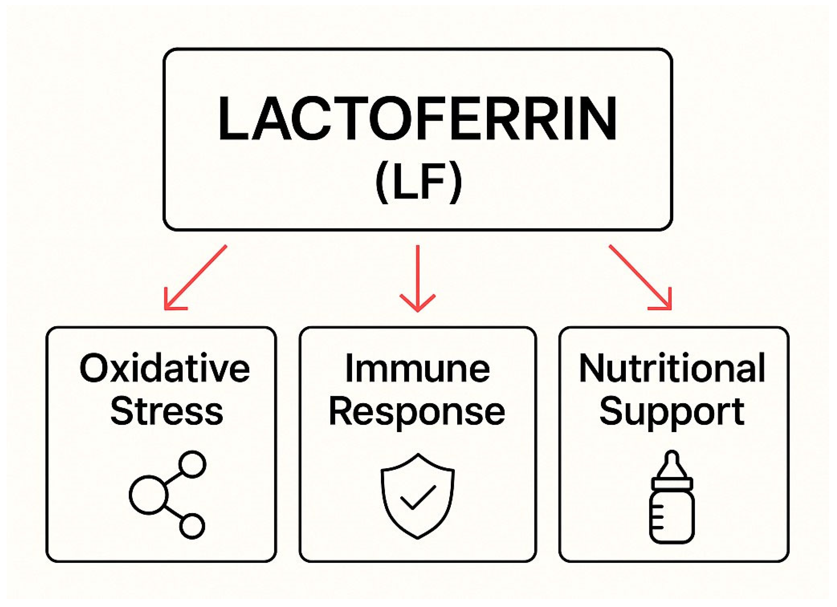
LF exhibits multifaceted activity supporting the body in combating Candida spp. infections, which is particularly important in pregnant women and newborns. Iron chelation and the reduction in Fenton reactions decrease oxidative stress, protecting host tissues and supporting phagocytic mechanisms. It modulates the immune response by activating immune cells (neutrophils, macrophages, and T and B lymphocytes) and influencing cytokine production and antigen presentation. LF also acts as a nutritional factor. It delivers bioavailable iron and supports the development of a healthy microbiota, especially in breastfed infants. Its effectiveness in reducing colonization and preventing the progression of infection has been confirmed in clinical studies, and its synergistic action with antifungal drugs makes it a valuable adjunct to therapy. Due to its safety profile, LF holds great potential in preventing and treating fungal infections in high-risk populations. Together, these multifaceted actions illustrate the complexity of lactoferrin’s antifungal mechanisms and highlight its role in host defense strategies, as summarized in Figure 2.

Author Contributions

Conceptualization J.W.; writing—original draft preparation, A.D., J.W., P.K. and W.W.; writing—review and editing, A.D., J.W., P.K. and W.W.; visualization A.D. and P.K.; supervision, A.D. All authors have read and agreed to the published version of the manuscript.

Funding

This research received no external funding.

Institutional Review Board Statement

Not applicable.

Informed Consent Statement

Not applicable.

Conflicts of Interest

The authors declare no conflicts of interest.

Abbreviations

The following abbreviations are used in this manuscript:
5-FCFlucytosine
AMBAmphotericin B
AMPAntimicrobial peptide
Apo-LFIron-free form of lactoferrin
bCFBovine colostrum fortifier
bLfBovine lactoferrin
CASCaspofungin
CTZClotrimazole
ELFIN trialEnteral lactoferrin in neonates trial
FLCFluconazole
GM-CSFGranulocyte–macrophage colony-stimulating factor
hLfHuman lactoferrin
Holo-LFIron-saturated form of lactoferrin
IL-6Interleukin 6
ITZItraconazole
LFLactoferrin
LOSLate-onset sepsis
LPOLactoperoxidase
NECNecrotizing enterocolitis
NYSNystatin
rhLFRecombinant human lactoferrin
ROSReactive oxygen species
SODSuperoxide dismutases
TLRToll-like receptors
VRZVoriconazole

References

  1. Benedict, K.; Singleton, A.L.; Jackson, B.R.; Molinari, N.A.M. Survey of Incidence, Lifetime Prevalence, and Treatment of Self-Reported Vulvovaginal Candidiasis, United States, 2020. BMC Women’s Health 2022, 22, 147. [Google Scholar] [CrossRef] [PubMed]
  2. Cook, A.; Ferreras-Antolin, L.; Adhisivam, B.; Ballot, D.; Berkley, J.A.; Bernaschi, P.; Carvalheiro, C.G.; Chaikittisuk, N.; Chen, Y.; Chibabhai, V.; et al. Neonatal Invasive Candidiasis in Low-and Middle-Income Countries: Data from the NeoOBS Study. Med. Mycol. 2023, 61, myad010. [Google Scholar] [CrossRef] [PubMed]
  3. Willems, H.M.E.; Ahmed, S.S.; Liu, J.; Xu, Z.; Peters, B.M. Vulvovaginal Candidiasis: A Current Understanding and Burning Questions. J. Fungi 2020, 6, 27. [Google Scholar] [CrossRef] [PubMed]
  4. Liao, H.; Liu, S.; Wang, H.; Su, H.; Liu, Z. Enhanced Antifungal Activity of Bovine Lactoferrin-Producing Probiotic Lactobacillus casei in the Murine Model of Vulvovaginal Candidiasis. BMC Microbiol. 2019, 19, 7. [Google Scholar] [CrossRef]
  5. Gawel, P.; Krolak-Olejnik, B. Lactoferrin Supplementation during Pregnancy—A Review of the Literature and Current Recommendations. Ginekol. Pol. 2023, 94, 570–580. [Google Scholar] [CrossRef]
  6. Dong, Z.; Fan, C.; Hou, W.; Rui, C.; Wang, X.; Fan, Y.; Zhao, L.; Wang, Q.; Wang, Z.; Zeng, X.; et al. Vaginal Exposure to Candida albicans During Early Gestation Results in Adverse Pregnancy Outcomes via Inhibiting Placental Development. Front. Microbiol. 2022, 12, 816161. [Google Scholar] [CrossRef]
  7. Maftei, N.-M.; Arbune, M.; Georgescu, C.V.; Elisei, A.M.; Iancu, A.V.; Tatu, A.L. Vulvovaginal Candidiasis in Pregnancy—Between Sensitivity and Resistance to Antimycotics. J. Xenobiotics 2023, 13, 312–322. [Google Scholar] [CrossRef]
  8. Payne, M.S.; Ireland, D.J.; Watts, R.; Nathan, E.A.; Furfaro, L.L.; Kemp, M.W.; Keelan, J.A.; Newnham, J.P. Ureaplasma parvum Genotype, Combined Vaginal Colonisation with Candida albicans, and Spontaneous Preterm Birth in an Australian Cohort of Pregnant Women. BMC Pregnancy Childbirth 2016, 16, 312. [Google Scholar] [CrossRef]
  9. Hemedez, C.; Trail-Burns, E.; Mao, Q.; Chu, S.; Shaw, S.K.; Bliss, J.M.; De Paepe, M.E. Pathology of Neonatal Non-Albicans Candidiasis: Autopsy Study and Literature Review. Pediatr. Dev. Pathol. 2019, 22, 98–105. [Google Scholar] [CrossRef]
  10. Barton, M.; Shen, A.; O’Brien, K.; Robinson, J.L.; Davies, H.D.; Simpson, K.; Asztalos, E.; Langley, J.; Le Saux, N.; Sauve, R.; et al. Early-Onset Invasive Candidiasis in Extremely Low Birth Weight Infants: Perinatal Acquisition Predicts Poor Outcome. Clin. Infect. Dis. 2017, 64, 921–927. [Google Scholar] [CrossRef]
  11. Talapko, J.; Juzbašić, M.; Matijević, T.; Pustijanac, E.; Bekić, S.; Kotris, I.; Škrlec, I. Candida albicans—The Virulence Factors and Clinical Manifestations of Infection. J. Fungi 2021, 7, 79. [Google Scholar] [CrossRef] [PubMed]
  12. Neuman-Łaniece, M. Management of Oral Candidiasis in Children—The General Practitioner’s Approach. Terapia 2016, 3, 65–67. [Google Scholar]
  13. Vainionpää, A.; Tuomi, J.; Kantola, S.; Anttonen, V. Neonatal Thrush of Newborns: Oral Candidiasis? Clin. Exp. Dent. Res. 2019, 5, 580–582. [Google Scholar] [CrossRef]
  14. Plachouri, K.-M.; Mulita, F.; Oikonomou, C.; Papadopoulou, M.; Akrida, I.; Vryzaki, E.; Verras, G.-I.; Georgiou, S. Nipple Candidiasis and Painful Lactation: An Updated Overview. Adv. Dermatol. Allergol. 2022, 39, 651–655. [Google Scholar] [CrossRef]
  15. Artym, J. Lactoferrin—A Sensor and Regulator of Iron Absorption. Postępy Biol. Komórki 2015, 2, 283–308. [Google Scholar]
  16. Fernandes, K.E.; Weeks, K.; Carter, D.A. Lactoferrin Is Broadly Active against Yeasts and Highly Synergistic with Amphotericin B. Antimicrob. Agents Chemother. 2020, 64, 10–1128. [Google Scholar] [CrossRef]
  17. De Felipe, L.O.; da Silva Júnior, W.F.; de Araújo, K.C.; Fabrino, D.L. Lactoferrin, Chitosan and Melaleuca alternifolia—Natural Products That Show Promise in Candidiasis Treatment. Braz. J. Microbiol. 2018, 49, 212–219. [Google Scholar] [CrossRef]
  18. Rascón-Cruz, Q.; Siqueiros-Cendón, T.S.; Siañez-Estrada, L.I.; Villaseñor-Rivera, C.M.; Ángel-Lerma, L.E.; Olivas-Espino, J.A.; León-Flores, D.B.; Espinoza-Sánchez, E.A.; Arévalo-Gallegos, S.; Iglesias-Figueroa, B.F. Antioxidant Potential of Lactoferrin and Its Protective Effect on Health: An Overview. Int. J. Mol. Sci. 2024, 26, 125. [Google Scholar] [CrossRef]
  19. Alexander, D.B.; Iigo, M.; Yamauchi, K.; Suzui, M.; Tsuda, H. Lactoferrin: An Alternative View of Its Role in Human Biological Fluids. Biochem. Cell Biol. 2012, 90, 279–306. [Google Scholar] [CrossRef] [PubMed]
  20. Park, J.-H.; Park, G.-T.; Cho, I.H.; Sim, S.-M.; Yang, J.-M.; Lee, D.-Y. An Antimicrobial Protein, Lactoferrin Exists in the Sweat: Proteomic Analysis of Sweat. Exp. Dermatol. 2011, 20, 369–371. [Google Scholar] [CrossRef] [PubMed]
  21. Ward, P.P.; Paz, E.; Conneely, O.M. Multifunctional Roles of Lactoferrin: A Critical Overview. Cell. Mol. Life Sci. 2005, 62, 2540–2548. [Google Scholar] [CrossRef] [PubMed]
  22. Artym, J.; Zimecki, M. Antimicrobial and Prebiotic Activity of Lactoferrin in the Female Reproductive Tract: A Comprehensive Review. Biomedicines 2021, 9, 1940. [Google Scholar] [CrossRef] [PubMed]
  23. Ohradanova-Repic, A.; Praženicová, R.; Gebetsberger, L.; Moskalets, T.; Skrabana, R.; Cehlar, O.; Tajti, G.; Stockinger, H.; Leksa, V. Time to Kill and Time to Heal: The Multifaceted Role of Lactoferrin and Lactoferricin in Host Defense. Pharmaceutics 2023, 15, 1056. [Google Scholar] [CrossRef] [PubMed]
  24. Rascón-Cruz, Q.; Espinoza-Sánchez, E.A.; Siqueiros-Cendón, T.S.; Nakamura-Bencomo, S.I.; Arévalo-Gallegos, S.; Iglesias-Figueroa, B.F. Lactoferrin: A Glycoprotein Involved in Immunomodulation, Anticancer, and Antimicrobial Processes. Molecules 2021, 26, 205. [Google Scholar] [CrossRef]
  25. Curran, C.S.; Demick, K.P.; Mansfield, J.M. Lactoferrin Activates Macrophages via TLR4-Dependent and -Independent Signaling Pathways. Cell. Immunol. 2006, 242, 23–30. [Google Scholar] [CrossRef]
  26. de la Rosa, G.; Yang, D.; Tewary, P.; Varadhachary, A.; Oppenheim, J.J. Lactoferrin Acts as an Alarmin to Promote the Recruitment and Activation of APCs and Antigen-Specific Immune Responses. J. Immunol. 2008, 180, 6868–6876. [Google Scholar] [CrossRef]
  27. Otsuki, K.; Nishi, T.; Kondo, T.; Okubo, K. Review, Role of Lactoferrin in Preventing Preterm Delivery. BioMetals 2023, 36, 521–530. [Google Scholar] [CrossRef]
  28. Chen, K.; Jin, S.; Chen, H.; Cao, Y.; Dong, X.; Li, H.; Zhou, Z.; Liu, C. Dose Effect of Bovine Lactoferrin Fortification on Diarrhea and Respiratory Tract Infections in Weaned Infants with Anemia: A Randomized, Controlled Trial. Nutrition 2021, 90, 111288. [Google Scholar] [CrossRef]
  29. Andersson, Y.; Lindquist, S.; Lagerqvist, C.; Hernell, O. Lactoferrin Is Responsible for the Fungistatic Effect of Human Milk. Early Hum. Dev. 2000, 59, 95–105. [Google Scholar] [CrossRef]
  30. Perrin, M.T.; Fogleman, A.D.; Newburg, D.S.; Allen, J.C. A Longitudinal Study of Human Milk Composition in the Second Year Postpartum: Implications for Human Milk Banking. Matern. Child Nutr. 2017, 13, e12239. [Google Scholar] [CrossRef]
  31. Dombrowska-Pali, A.; Wiktorczyk-Kapischke, N.; Chrustek, A.; Olszewska-Słonina, D.; Gospodarek-Komkowska, E.; Socha, M.W. Human Milk Microbiome—A Review of Scientific Reports. Nutrients 2024, 16, 1420. [Google Scholar] [CrossRef] [PubMed]
  32. Miliku, K.; Azad, M. Breastfeeding and the Developmental Origins of Asthma: Current Evidence, Possible Mechanisms, and Future Research Priorities. Nutrients 2018, 10, 995. [Google Scholar] [CrossRef] [PubMed]
  33. Amitay, E.L.; Raz, G.D.; Keinan-Boker, L. Breastfeeding, Other Early Life Exposures and Childhood Leukemia and Lymphoma. Nutr. Cancer 2016, 68, 968–977. [Google Scholar] [CrossRef] [PubMed]
  34. Baker, J.L.; Gamborg, M.; Heitmann, B.L.; Lissner, L.; Sørensen, T.I.A.; Rasmussen, K.M. Breastfeeding Reduces Postpartum Weight Retention. Am. J. Clin. Nutr. 2008, 88, 1543–1551. [Google Scholar] [CrossRef]
  35. Calik-Ksepka, A.; Stradczuk, M.; Czarnecka, K.; Grymowicz, M.; Smolarczyk, R. Lactational Amenorrhea: Neuroendocrine Pathways Controlling Fertility and Bone Turnover. Int. J. Mol. Sci. 2022, 23, 1633. [Google Scholar] [CrossRef]
  36. Nagel, E.M.; Howland, M.A.; Pando, C.; Stang, J.; Mason, S.M.; Fields, D.A.; Demerath, E.W. Maternal Psychological Distress and Lactation and Breastfeeding Outcomes: A Narrative Review. Clin. Ther. 2022, 44, 215–227. [Google Scholar] [CrossRef]
  37. Amelia, R.; Masrul, M.; Sriyanti, R. The Effect of Breastfeeding on The Uterine Involution Post Partum Mothers. World J. Res. Rev. 2019, 8, 1–3. [Google Scholar] [CrossRef]
  38. Ip, S.; Chung, M.; Raman, G.; Trikalinos, T.A.; Lau, J. A Summary of the Agency for Healthcare Research and Quality’s Evidence Report on Breastfeeding in Developed Countries. Breastfeed. Med. 2009, 4, S-17–S-30. [Google Scholar] [CrossRef]
  39. Bjørnerem, Å.; Ahmed, L.A.; Jørgensen, L.; Størmer, J.; Joakimsen, R.M. Breastfeeding Protects against Hip Fracture in Postmenopausal Women: The Tromsø Study. J. Bone Miner. Res. 2011, 26, 2843–2850. [Google Scholar] [CrossRef]
  40. Stuebe, A. Associations Among Lactation, Maternal Carbohydrate Metabolism, and Cardiovascular Health. Clin. Obstet. Gynecol. 2015, 58, 827–839. [Google Scholar] [CrossRef]
  41. Zinaliyeva, A.N.; Tulegenova, G.A.; Zhelisbayeva, K.R.; Akhmetzhanova, M.B. The Role of Breastfeeding and Formula Feeding in Infant Health and Development: A Mini Review. J. Environ. Treat. Tech. 2024, 12, 30–38. [Google Scholar] [CrossRef]
  42. Telang, S. Lactoferrin: A Critical Player in Neonatal Host Defense. Nutrients 2018, 10, 1228. [Google Scholar] [CrossRef]
  43. Bialic, K.; Bialic, A.; Bakalarczyk, R.; Majewski, P.; Bednarz, L.; Szala-Czerwonka, K.; Woś, N.; Rejmer, A.; Rojek, K.; Wijas, K. Exploring the Potential of Lactoferrin in Neonatology and Obstetrics—Promising Advancements for Maternal and Infant Health. J. Educ. Health Sport 2023, 34, 34–50. [Google Scholar] [CrossRef]
  44. Baker, E.N.; Baker, H.M. A Structural Framework for Understanding the Multifunctional Character of Lactoferrin. Biochimie 2009, 91, 3–10. [Google Scholar] [CrossRef]
  45. King, J.C.; Cummings, G.E.; Guo, N.; Trivedi, L.; Readmond, B.X.; Keane, V.; Feigelman, S.; de Waard, R. A Double-Blind, Placebo-Controlled, Pilot Study of Bovine Lactoferrin Supplementation in Bottle-fed Infants. J. Pediatr. Gastroenterol. Nutr. 2007, 44, 245–251. [Google Scholar] [CrossRef]
  46. Akin, I.; Atasay, B.; Dogu, F.; Okulu, E.; Arsan, S.; Karatas, H.; Ikinciogullari, A.; Turmen, T. Oral Lactoferrin to Prevent Nosocomial Sepsis and Necrotizing Enterocolitis of Premature Neonates and Effect on T-Regulatory Cells. Am. J. Perinatol. 2014, 31, 1111–1120. [Google Scholar] [CrossRef] [PubMed]
  47. Manzoni, P. Clinical Benefits of Lactoferrin for Infants and Children. J. Pediatr. 2016, 173, S43–S52. [Google Scholar] [CrossRef]
  48. He, Y.; Cao, L.; Yu, J. Prophylactic Lactoferrin for Preventing Late-Onset Sepsis and Necrotizing Enterocolitis in Preterm Infants. Medicine 2018, 97, e11976. [Google Scholar] [CrossRef]
  49. Griffiths, J.; Jenkins, P.; Vargova, M.; Bowler, U.; Juszczak, E.; King, A.; Linsell, L.; Murray, D.; Partlett, C.; Patel, M.; et al. Enteral Lactoferrin Supplementation for Very Preterm Infants: A Randomised Placebo-Controlled Trial. Lancet 2019, 393, 423–433. [Google Scholar] [CrossRef]
  50. Supranoto, Y.T.N.; Tanuwijaya, D.C.D. Meta-Analysis of Enteral Lactoferrin Supplementation for Reducing the Risk of Preterm Infants Sepsis. Smart Med. J. 2022, 5, 78. [Google Scholar] [CrossRef]
  51. Gao, X.; Li, Y.; Olin, A.B.; Nguyen, D.N. Fortification with Bovine Colostrum Enhances Antibacterial Activity of Human Milk. J. Parenter. Enter. Nutr. 2021, 45, 1417–1424. [Google Scholar] [CrossRef]
  52. Zarzosa-Moreno, D.; Avalos-Gómez, C.; Ramírez-Texcalco, L.S.; Torres-López, E.; Ramírez-Mondragón, R.; Hernández-Ramírez, J.O.; Serrano-Luna, J.; de la Garza, M. Lactoferrin and Its Derived Peptides: An Alternative for Combating Virulence Mechanisms Developed by Pathogens. Molecules 2020, 25, 5763. [Google Scholar] [CrossRef]
  53. Gajda-Morszewski, P.; Śpiewak, K. Lactoferrin—A Multipotential Protein. Sci. J. Dr. Stud. Assoc. Jagiellonian Univ. Nat. Sci. 2015, 10, 177–188. [Google Scholar]
  54. Vogel, H.J.; Schibli, D.J.; Jing, W.; Lohmeier-Vogel, E.M.; Epand, R.F.; Epand, R.M. Towards a Structure-Function Analysis of Bovine Lactoferricin and Related Tryptophan- and Arginine-Containing Peptides. Biochem. Cell Biol. 2002, 80, 49–63. [Google Scholar] [CrossRef]
  55. Krupińska, A.M.; Bogucki, Z. Lactoferrin as a Potential Therapeutic for the Treatment of Candida-Associated Denture Stomatitis. J. Oral Biosci. 2024, 66, 308–313. [Google Scholar] [CrossRef] [PubMed]
  56. Cavalheiro, M.; Teixeira, M.C. Candida Biofilms: Threats, Challenges, and Promising Strategies. Front. Med. 2018, 5, 28. [Google Scholar] [CrossRef] [PubMed]
  57. Morici, P.; Fais, R.; Rizzato, C.; Tavanti, A.; Lupetti, A. Inhibition of Candida albicans Biofilm Formation by the Synthetic Lactoferricin Derived Peptide HLF1-11. PLoS ONE 2016, 11, e0167470. [Google Scholar] [CrossRef] [PubMed]
  58. Valenti, P.; Rosa, L.; Capobianco, D.; Lepanto, M.S.; Schiavi, E.; Cutone, A.; Paesano, R.; Mastromarino, P. Role of Lactobacilli and Lactoferrin in the Mucosal Cervicovaginal Defense. Front. Immunol. 2018, 9, 376. [Google Scholar] [CrossRef]
  59. Sherrington, S.L.; Sorsby, E.; Mahtey, N.; Kumwenda, P.; Lenardon, M.D.; Brown, I.; Ballou, E.R.; MacCallum, D.M.; Hall, R.A. Adaptation of Candida Albicans to Environmental PH Induces Cell Wall Remodelling and Enhances Innate Immune Recognition. PLoS Pathog. 2017, 13, e1006403. [Google Scholar] [CrossRef]
  60. Soukka, T. Fungicidal Effect of Human Lactoferrin against Candida albicans. FEMS Microbiol. Lett. 1992, 90, 223–228. [Google Scholar] [CrossRef]
  61. Fais, R.; Rizzato, C.; Franconi, I.; Tavanti, A.; Lupetti, A. Synergistic Activity of the Human Lactoferricin-Derived Peptide HLF1-11 in Combination with Caspofungin against Candida Species. Microbiol. Spectr. 2022, 10, e01240-22. [Google Scholar] [CrossRef] [PubMed]
  62. Dantas, A.; Day, A.; Ikeh, M.; Kos, I.; Achan, B.; Quinn, J. Oxidative Stress Responses in the Human Fungal Pathogen, Candida albicans. Biomolecules 2015, 5, 142–165. [Google Scholar] [CrossRef] [PubMed]
  63. Tanida, T.; Rao, F.; Hamada, T.; Ueta, E.; Osaki, T. Lactoferrin Peptide Increases the Survival of Candida albicans—Inoculated Mice by Upregulating Neutrophil and Macrophage Functions, Especially in Combination with Amphotericin B and Granulocyte-Macrophage Colony-Stimulating Factor. Infect. Immun. 2001, 69, 3883–3890. [Google Scholar] [CrossRef]
  64. Nakano, M.; Suzuki, M.; Wakabayashi, H.; Hayama, K.; Yamauchi, K.; Abe, F.; Abe, S. Synergistic Anti-Candida Activities of Lactoferrin and the Lactoperoxidase System. Drug Discov. Ther. 2019, 13, 28–33. [Google Scholar] [CrossRef] [PubMed]
  65. Xu, Y.Y.; Samaramayake, Y.H.; Samaramayake, L.P.; Nikawa, H. In Vitro Susceptibility of Candida Species to Lactoferrin. Med. Mycol. 1999, 37, 35–41. [Google Scholar] [CrossRef]
  66. Samaranayake, Y.H.; Samaranayake, L.P.; Wu, P.C.; So, M. The Antifungal Effect of Lactoferrin and Lysozyme on Candida krusei and Candida albicans. APMIS 1997, 105, 875–883. [Google Scholar] [CrossRef]
  67. Cianci, A. Influence of Lactoferrin in Preventing Preterm Delivery: A Pilot Study. Mol. Med. Rep. 2011, 5, 162–166. [Google Scholar] [CrossRef]
  68. Manzoni, P.; Stolfi, I.; Messner, H.; Cattani, S.; Laforgia, N.; Romeo, M.G.; Bollani, L.; Rinaldi, M.; Gallo, E.; Quercia, M.; et al. Bovine Lactoferrin Prevents Invasive Fungal Infections in Very Low Birth Weight Infants: A Randomized Controlled Trial. Pediatrics 2012, 129, 116–123. [Google Scholar] [CrossRef]
  69. Li, L.; Zhang, X.; Li, Q.; Zhong, W.; Zou, H. The Increasing Trend of Triazole-Resistant Candida from Vulvovaginal Candidiasis. Infect. Drug Resist. 2024, 17, 4301–4310. [Google Scholar] [CrossRef]
  70. Patel, M.A.; Aliporewala, V.M.; Patel, D.A. Common Antifungal Drugs in Pregnancy: Risks and Precautions. J. Obstet. Gynecol. India 2021, 71, 577–582. [Google Scholar] [CrossRef]
  71. Farr, A.; Effendy, I.; Frey Tirri, B.; Hof, H.; Mayser, P.; Petricevic, L.; Ruhnke, M.; Schaller, M.; Schaefer, A.P.A.; Sustr, V.; et al. Guideline: Vulvovaginal Candidosis (AWMF 015/072, Level S2k). Mycoses 2021, 64, 583–602. [Google Scholar] [CrossRef] [PubMed]
  72. Yaghi, M.; McMullan, P.; Truong, T.M.; Rothe, M.; Murase, J.; Grant-Kels, J.M. Safety of Dermatologic Medications in Pregnancy and Lactation: An Update—Part II: Lactation. J. Am. Acad. Dermatol. 2024, 91, 651–668. [Google Scholar] [CrossRef]
  73. Dermitzaki, N.; Balomenou, F.; Gialamprinou, D.; Giapros, V.; Rallis, D.; Baltogianni, M. Perspectives on the Use of Echinocandins in the Neonatal Intensive Care Unit. Antibiotics 2024, 13, 1209. [Google Scholar] [CrossRef] [PubMed]
  74. Danesi, R.; Senesi, S.; Wout, J.W.V.; van Dissel, J.T.; Lupetti, A.; Nibbering, P.H. Antimicrobial Peptides: Therapeutic Potential for the Treatment of Candida Infections. Expert Opin. Investig. Drugs 2002, 11, 309–318. [Google Scholar] [CrossRef]
  75. Haney, E.F.; Nazmi, K.; Bolscher, J.G.M.; Vogel, H.J. Structural and Biophysical Characterization of an Antimicrobial Peptide Chimera Comprised of Lactoferricin and Lactoferrampin. Biochim. Biophys. Acta-Biomembr. 2012, 1818, 762–775. [Google Scholar] [CrossRef] [PubMed]
  76. Wakabayashi, H.; Abe, S.; Okutomi, T.; Tansho, S.; Kawase, K.; Yamaguchi, H. Cooperative Anti-Candida Effects of Lactoferrin or Its Peptides in Combination with Azole Antifungal Agents. Microbiol. Immunol. 1996, 40, 821–825. [Google Scholar] [CrossRef]
  77. Kuipers, M.E.; de Vries, H.G.; Eikelboom, M.C.; Meijer, D.K.F.; Swart, P.J. Synergistic Fungistatic Effects of Lactoferrin in Combination with Antifungal Drugs against Clinical Candida Isolates. Antimicrob. Agents Chemother. 1999, 43, 2635–2641. [Google Scholar] [CrossRef]
  78. Lupetti, A.; Paulusma-Annema, A.; Welling, M.M.; Dogterom-Ballering, H.; Brouwer, C.P.J.M.; Senesi, S.; van Dissel, J.T.; Nibbering, P.H. Synergistic Activity of the N-Terminal Peptide of Human Lactoferrin and Fluconazole against Candida Species. Antimicrob. Agents Chemother. 2003, 47, 262–267. [Google Scholar] [CrossRef]
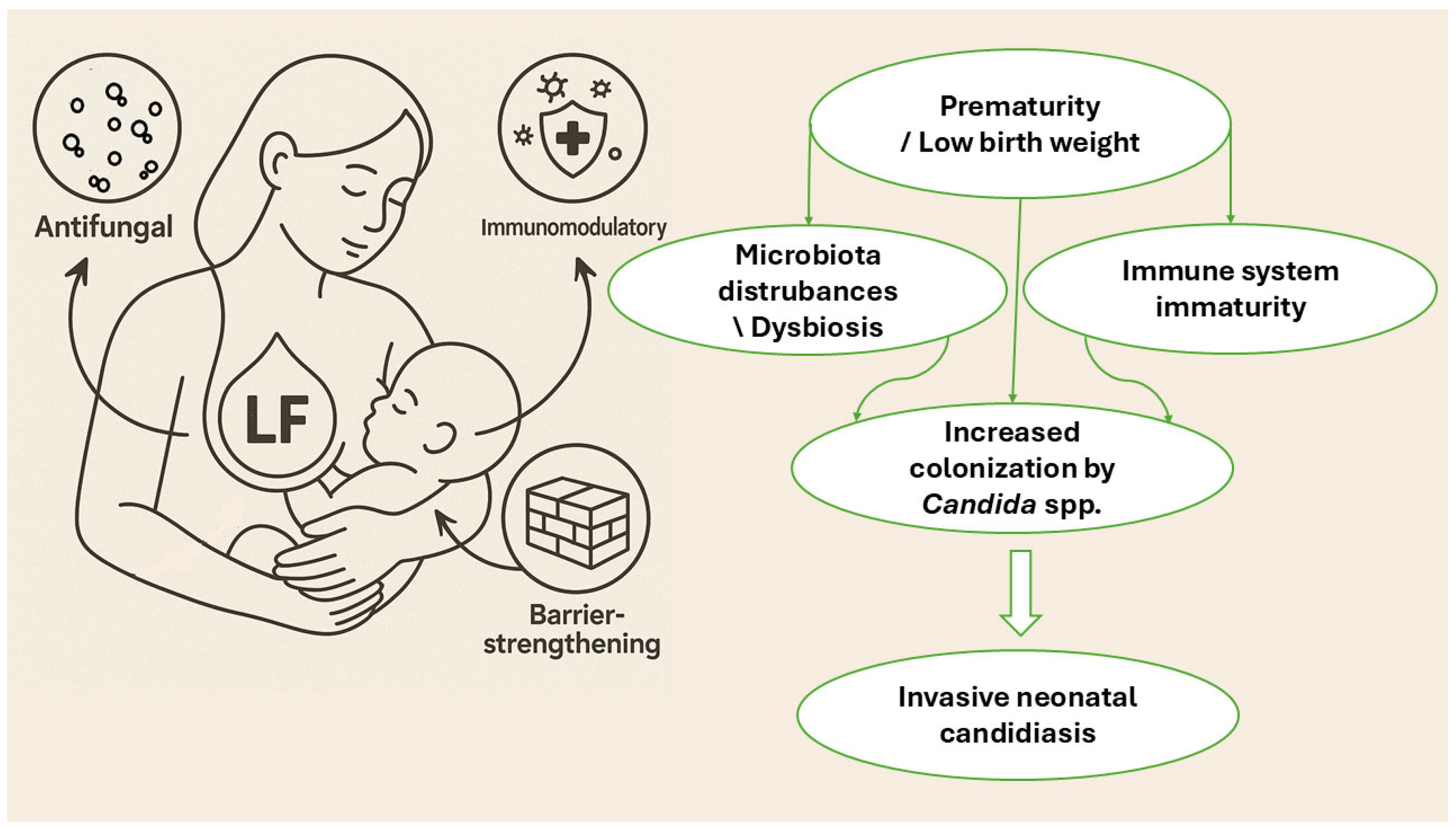
Figure 1. Risk factors for Candida spp. infection in newborns.
Molecules 30 02416 g001
Figure 2. The role of lactoferrin in combating Candida spp. infections—a summary of mechanisms of action.
Molecules 30 02416 g002
Table 2. Clinical and experimental applications of lactoferrin in fungal infections.
CompoundForm and OriginTarget PathogensSynergistic EffectPlanktonic and Biofilm FormsRef.
LFIntact bLf (whole protein)C. albicans (including azole-sensitive and -resistant strains)Synergistic with CTZ; also effective with FLC and ITZPlanktonic only[76]
LF-BIsolated peptide from the N-terminal region of LFC. albicans (including azole-sensitive and -resistant strains)Synergistic with CTZPlanktonic only[76]
LFhydEnzymatically digested lactoferrin (mixture of peptides)C. albicans (including azole-sensitive and -resistant strains)Synergy with azolePlanktonic only[76]
LFDairy-derived bLf (partially digested, low iron saturation)C. albicans and C. glabrataStrong synergy with AMBReduces hyphal growth; prevents biofilm formation; less effective against mature biofilm[16]
hLF1-11Synthetic peptide from human LF (N-terminal region)C. albicans, C. glabrata, C. tropicalis, and C. parapsilosisStrong synergy with CASActive against planktonic cells; prevents biofilm formation; reduces metabolic activity of mature biofilm (fungistatic)[61]
LFApo-lactoferrinC. albicans, C. glabrata, and C. tropicalisSynergistic with FLC; synergistic with 5-FCPlanktonic only[77]
LFNative bLfC. albicans, C. glabrata, and C. tropicalisSynergistic with FLCPlanktonic only[77]
hLF(1-11)Synthetic N-terminal peptide from hLFC. albicans (FLC-sensitive and FLC- resistant), C. glabrata, C. krusei, C. tropicalis, and C. parapsilosisSynergistic with FLCPlanktonic only[78]
Amphotericin B (AMB); bovine lactoferrin (bLf); caspofungin (CAS); clotrimazole (CTZ); fluconazole (FLC); human lactoferrin (hLf); itraconazole (ITZ); lactoferrin (LF); lactoferrin-derived peptide from bovine LF (LF-B); pepsin hydrolysate of lactoferrin (LFhyd); 5-fluorocytosine (5-FC).
Disclaimer/Publisher’s Note: The statements, opinions and data contained in all publications are solely those of the individual author(s) and contributor(s) and not of MDPI and/or the editor(s). MDPI and/or the editor(s) disclaim responsibility for any injury to people or property resulting from any ideas, methods, instructions or products referred to in the content.