Article

Biomedical Applications of Scutellaria edelbergii Rech. f.: In Vitro and In Vivo Approach

1
Department of Botany, Abdul Wali Khan University Mardan, Mardan 23200, Pakistan
2
Natural and Medical Sciences Research Center, University of Nizwa, P.O. Box 33, Birkat Al Mauz, Nizwa 616, Oman
3
Department of Chemistry, University of Malakand, Chakdara 18800, Pakistan
4
Department of Microbiology, Abdul Wali Khan University Mardan, Mardan 23200, Pakistan
5
Department of Biochemistry, University of Malakand, Chakdara 18800, Pakistan
6
Department of Botany, Hazara University Mansehra, Mansehra 21310, Pakistan
7
Department of Clinical Laboratory Sciences, Turabah University College, Taif University, P.O. Box 11099, Taif 21944, Saudi Arabia
8
Department of Pharmacology and Therapeutics, Faculty of Veterinary Medicine, Damanhour University, Damanhour 22511, Egypt
*
Authors to whom correspondence should be addressed.
Molecules 2021, 26(12), 3740; https://doi.org/10.3390/molecules26123740
Submission received: 26 April 2021 / Revised: 12 June 2021 / Accepted: 17 June 2021 / Published: 19 June 2021
(This article belongs to the Special Issue Food Biophysics and Biochemistry)

Abstract

:
In the current study, in vitro antimicrobial and antioxidant activities and in vivo anti-inflammatory and analgesic activities of Scutellaria edelbergii Rech. f. (crude extract and subfractions, i.e., n-hexane, ethyl acetate (EtOAc), chloroform, n-butanol (n-BuOH) and aqueous) were explored. Initially, extraction and fractionation of the selected medicinal plant were carried out, followed by phytochemical qualitative tests, which were mostly positive for all the extracts. EtOAc fraction possessed a significant amount of phenolic (79.2 ± 0.30 mg GAE/g) and flavonoid (84.0 ± 0.39 mg QE/g) content. The EtOAc fraction of S. edelbergii exhibited appreciable antibacterial activity against Gram-negative (Escherichia coli and Klebsiella pneumoniae) strains and significant zones of inhibition were observed against Gram-positive bacterial strains (Bacillus subtilis and Staphylococcus aureus). However, it was found inactive against Candida Albicans and Fusarium oxysporum fungal strains. The chloroform fraction was the most effective with an IC50 value of 172 and 74 µg/mL against DPPH (1,1-Diphenyl-2-picryl-hydrazyl) and ABTS assays, in comparison with standard ascorbic acid 59 and 63 µg/mL, respectively. Moreover, the EtOAc fraction displayed significant in vivo anti-inflammatory activity (54%) using carrageenan-induced assay and significant (55%) in vivo analgesic activity using acetic acid-induced writing assay. In addition, nine known compounds, ursolic acid (UA), ovaul (OV), oleanolic acid (OA), β-sitosterol (BS), micromeric acid (MA), taraxasterol acetate (TA), 5,3′,4′-trihydroxy-7-methoxy flavone (FL-1), 5,7,4′-trihydroxy-6,3′-dimiethoxyflavone (FL-2) and 7-methoxy catechin (FL-3), were isolated from methanolic extract of S. edelbergii. These constituents have never been obtained from this source. The structures of all the isolated constituents were elucidated by spectroscopic means. In conclusion, the EtOAc fraction and all other fractions of S. edelbergii, in general, displayed a significant role as antibacterial, free radical scavenger, anti-inflammatory and analgesic agents which may be due to the presence of these constituents and other flavonoids.

1. Introduction

More than 10% of the 32,000 reported species of higher plants are of medicinal importance worldwide [1,2,3]. In developing countries, synthetic pharmaceutical drugs are expensive and not affordable for an average person; therefore, the plants are the primary source of phytomedicines for inflammation, pain and microbial resistance [4]. Herbal medicines are important to enhance clinical research and quality control along with treatment and prevention of disease [5]. The demands for herbal medicines are frequently increasing and can be met by screening them in vitro biological and in vivo pharmacological features. The investigation of ethnomedicinal plants is required in developing nations due to costly allopathic medicines [5,6]. Therefore, it is essential to determine the significance of plants and enhance plant medication knowledge using modern approaches as they are less toxic and more effective for humanity [7].
One of the important human health issues is the drug resistance developed by microbes against the available antimicrobial drugs and it has been reported that within next 30 years, existing drugs will be totally inactive against them [8,9,10]. Consequently, there is a need to investigate plants for new therapeutic agents as plant-derived drugs are usually associated with fewer side effects in comparison to drugs prepared through synthetic routes [9].
Oxidative stress is another health issue prevalent these days. Several oxygen-based free radicals are usually formed during normal physiological and biochemical processes taking place in human bodies that are immediately detoxified by the human defense system immediately. However, in the past few decades, human food habits have changed drastically [10] and dependence on synthetic ingredients in foods has increased, which has given rise to the overproduction of free radicals in human bodies. Several chronic disorders, such as atherosclerosis, inflammations, cancer, diabetes, pain, aging and other degenerative complications in humans, are due to oxidative stress [4]. Plants and plant products (fruits, vegetables, medicinal herbs) contain various phenolic compounds that can scavenge free radicals as they have aromatic rings which can stabilize the free radicals formed inside body and in vitro [11]. The intake of natural antioxidants reduces the chances of cancer, cardiovascular abnormalities, dermatological infections, diabetes and other acute or chronic infections linked with aging [12,13].
The research on antioxidants from plant origins has also revealed that several antioxidants are effective in treating health complication associated with oxidative stress. Biological potentials such as anti-inflammatory, analgesic, anti-cancer, antipyretic and antibacterial activities of some antioxidants from plant origin are well documented [12]. Regardless of the latest developments in pain therapies, scientists still want to explore safe, effective and potent analgesic medication from plant origin as they are associated with fewer side effects, as mentioned earlier [14,15].
Scutellaria is a genus of the family Lamiaceae, also termed as skullcap and mostly found in temperate regions [16]. Generally, the genus is a rich source of scutellarin, baicalin, alkaloids, saponins, tannins and glycosides, having significant in vivo pharmacological effects in anti-inflammatory, analgesic, antibacterial, antifungal and antioxidant potentials [17,18,19]. Scutellaria edelbergii dried leaves powder is used as a blood purifier and an antimicrobial, anti-inflammatory and analgesic agent. Traditionally, its leaves are also used as green tea (kava) as it has medicinal importance. The extracts of S. baicalensis and its major chemical constituents have been reported to possess anti-viral, anti-tumor, anti-bacterial, antioxidant, anti-inflammatory, hepatoprotective and neuroprotective activities [17,20]. In addition, 126 molecules including flavonoids, flavones, isoflavones and six polysaccharides have been isolated from S. baicalensis [20]. The dried herb of S. indica is a crude drug (known as han xin cao in China) that has been used as anti-analgesic, antidotic and hemostatic for the treatment of hemoptysis, hematemesis and other diseases [18]. S. edelbergii have not been studied before for the mentioned pharmacological properties.
To attract the attention of researchers, the ongoing investigations aimed to provide information about the effectiveness of selected plants in the number of health complications and revive the significance of natural products in several health complications in general. S. edelbergii was subjected to extraction and fractionation followed by phytochemical investigation using different techniques. The extracts were also evaluated for antibacterial, antifungal and antioxidant potential in vitro; also, in vivo pharmacological activities such as analgesic and anti-inflammatory were also carried out.

2. Results and Discussion

Based on information collected from local inhabitants of Kalam, District Swat, Pakistan, the selected plant crude extract and subfractions (n-hexane, EtOAc, chloroform, n-BuOH, aqueous) were investigated for their phytochemical, in vitro antibacterial, antifungal and antioxidant potentials while in vivo pharmacological (anti-inflammatory and analgesic) activities were determined in Swiss albino mice using standard protocols. As S. edelbergii has not been investigated for the mentioned phytochemical, in vitro biological and in vivo pharmacological potentials, this study was conducted to promote the folkloric uses of the selected plant and provide scientific validation of S. edelbergii in terms of its phytochemical composition and observed biological potentials. In modern methods of isolating drugs from plants, ethnopharmacological information was collected from local practitioners and communities, then phytochemical analysis was performed and finally pharmacological studies were carried out to confirm the claimed medicinal potentials. The in vitro and in vivo investigation are basic approaches that expose the therapeutic significance of a given plant [19,21,22,23,24].

2.1. Qualitative Phytochemical Analysis of Extracts

Phytochemical analysis was performed to determine the chemical groups in the crude extract and their sub-fractions. All the extracts have shown the presence of flavonoids, phenols, alkaloids, saponins, carbohydrates and glycosides. The qualitative phytochemical tests were carried out to confirm the representative groups of phytochemicals in the tested extracts. Most of the fractions were positive for the applied tests (Table 1). Our results are in complete agreement with the reported data of S. havanensis [25].

2.2. Phenolic and Flavonoids Contents

The quantitative estimation of phenolic and flavonoid contents in crude extract and subfractions are presented in Table 2. The highest numbers of phenolic compounds were present in EtOAc fraction, crude extract and n-BuOH fractions. Comparable amounts of phenolic compounds were also found in the chloroform fraction. As phenolic compounds are polar, its high amount was therefore present in polar solvents. Nearly the same trend was observed for flavonoid contents; however, their amount was high as compared to phenolic contents. Similarly, the highest number of flavonoids was present in the EtOAc fraction, crude extract, n-BuOH and chloroform fractions. Both the phenolic and flavonoid contents were compared with standards and statistically analyzed at p ≤ 0.01 (Table 2). The EtOAc fraction was the richest fraction amongst the tested extracts. About 79.2 ± 0.30 mg GAE/g of total phenolic contents and 84.0 ± 0.39 mg QE/g total flavonoid contents were estimated in it. Our findings are in complete agreement with the study of Mousavi et al. [6], where Scutellaria platystegia was investigated for the mentioned contents. Higher phenolic and flavonoid contents were reported in S. platystegia, S. barbata and S. orientalis, belonging to the same genus [26,27].

2.3. Antibacterial Activity

The antibacterial potential of different extracts was tested against two Gram-negative (E. coli and K. pneumonia) and two Gram-positive (S. aureus and B. subtilis) bacterial strains along with standards and negative control (Table 3). Among the tested extracts, the EtOAc (SEEA) fraction exhibited the highest inhibitory potential for Gram-negative bacteria. At 50 µL and 100 µL, SEEA produced a zone of inhibition (ZOI) of 14.5 ± 0.04 and 17.9 ± 0.03 mm against E. coli and 14.3 ± 0.02 and 17.3± 0.02 mm against the K. pneumonia, respectively. Comparatively, K. pneumoniae was highly susceptible to S. edelbergii extracts. Moreover, SEEA was also found most effective against S. aureus and B. subtilis with the zone of inhibition of 15.03 ± 0.01 and 19.07 ± 0.02 mm and 4.97 ± 0.01 and 18.87 ± 0.04 mm at 50 µL and 100 µL, respectively. Moreover, all the fractions were found active in the antibacterial assay. The results are summarized in Table 3.
Appreciable inhibition potentials were observed for all fractions against tested bacterial strains. The EtOAc fraction was found to be the most active fraction with 19.07 ± 0.02 and 17.09 ± 0.01 mm zones of inhibitions against S. aureus and B. subtilis, respectively, at high doses only. The chloroform fraction exhibited zones of inhibition of 17.03 ± 0.03 and 17.09 ± 0.01 mm against S. aureus and B. subtilis, respectively, at higher doses only. The Gram-negative bacteria E. coli was inhibited up to 17.9 ± 0.03 mm and K. pneumonia to an extent of 17.3 ± 0.02 mm zones of inhibition by the EtOAc fraction. Our results agree with the reported data published by Ozkul et al. [28] and Yilmaz et al. [29], who tested different fractions of Scutellaria species against selected bacterial and fungal strains. Mostly, Gram-negative strains exhibited high drug resistance in comparison to the Gram-positive bacterial strains, as they do not have high contents of peptidoglycan in their cell walls [30]. The selected plant was found inactive against the fungal strains used, which negate the published findings [28].

2.4. Antifungal Activities

The prepared extracts were further tested against C. Albicans and F. oxysporum, but no activity was observed against these fungal strains.

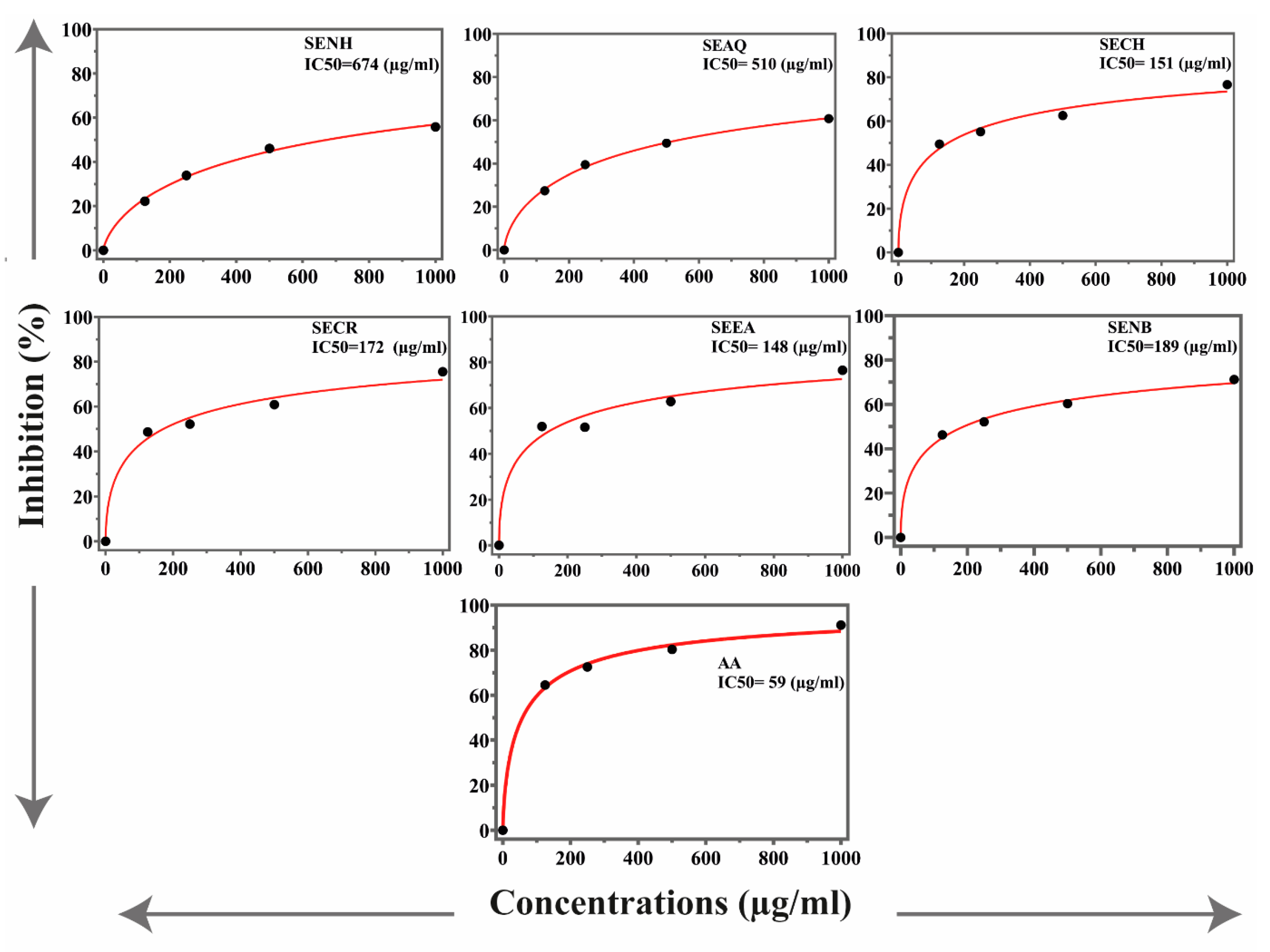
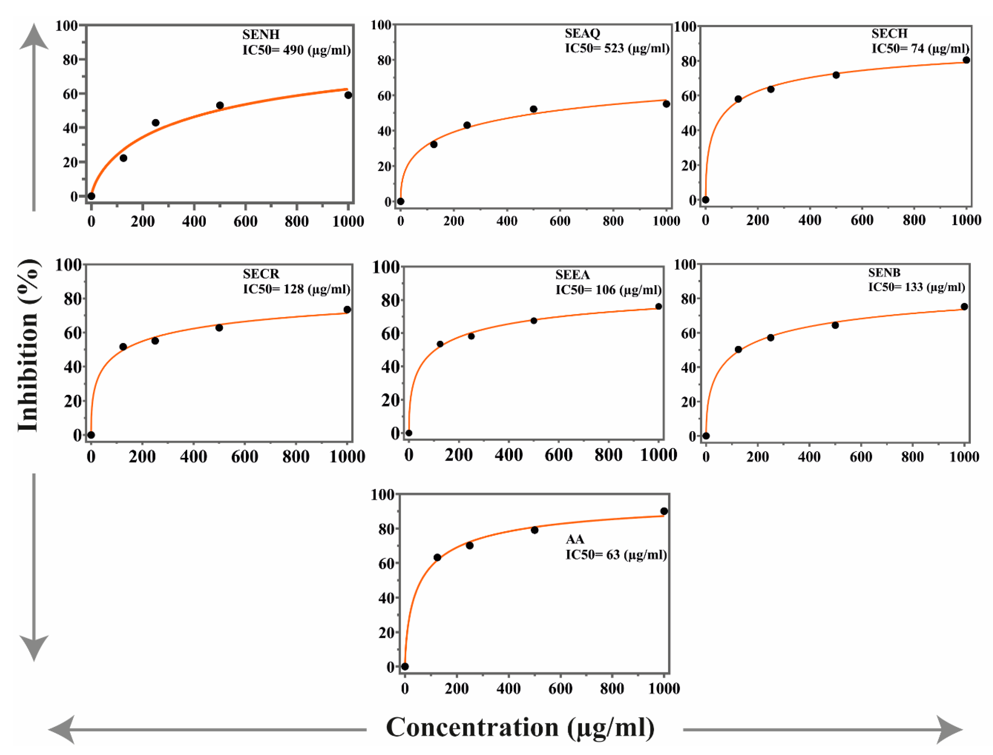
2.5. Antioxidant Activities

The antioxidant potential of the S. edelbergii extract and fraction was determined through synthetic free radical DPPH and ABTS assays. The results are presented in Figure 1 and Figure 2. The chloroform fraction exhibited the highest antioxidant activities, with IC50 values of 151 and 74 µg/mL in DPPH and ABTS, respectively. However, the EtOAc fraction, crude extract and n-BuOH fraction showed antioxidant activities with value IC50 = 148, 172 and 189 µg/mL in DPPH assay, respectively. Similarly, fractions of chloroform, EtOAc, crude extract and n-BuOH fraction demonstrated good activities with IC50 values of 106, 128, 133 and 190 µg/mL in ABTS assay, respectively. Meanwhile, aqueous and n-hexane fractions were found least active in both DPPH and ABTS assays. Ascorbic acid was used as a standard that possessed IC50 59 and 63 µg/mL DPPH and ABTS assays, respectively.
S. edelbergii was also tested for in vitro antioxidant potentials using different concentrations of the extracts (1000, 500, 250 and 125 µg/mL) using DPPH and ABTS assays and appreciable potential was exhibited in comparison to ascorbic acid used as standard (IC50 = 25 µg/mL). Among the investigated fractions against DPPH and ABTS, chloroform fraction with IC50 value of 175 and 155 µg/mL was the most potent, followed by EtOAc with IC50 values of 205 and 160 µg/mL. Our results match with reported data of Vergun et al. [31], who studied Scutellaria baicalensis in the same fractions and concentrations against DPPH radical. Our study is also in agreement with the study of Grzegorczyk-Karolak et al. [32], Delazar et al. [33] and Zengin et al., [34] who have performed the same tests for S. altissima and S. pinnatifida, S. Orientalis L. and S. salviifolia. Plants enriched with total phenols and flavonoids are the key source to neutralize the reactive oxygen species; thus, our findings are like the literature available for S. altissima and S. galericulata [28].

2.6. Anti-Inflammatory Activities

The anti-inflammatory activity of S. edelbergii crude extract and subfractions are presented in Table 4. Among the tested extracts, the EtOAc fraction was highly potent (54% inhibition) as compared to other extracts. Crude and n-BuOH fraction also exhibited 50% and 47% inhibition, respectively. However, the least activity was obtained for the aqueous fraction (35% inhibition). Diclofenac sodium was used as a standard that produced 74% inhibition of inflammation caused by carrageenan in the tested experimental animals.
The in vivo anti-inflammatory potential of extracts was examined in Swiss albino mice, using the carrageenan-induced assay. The EtOAc fraction was found to be the most active fraction with 47% and 54% inhibition at a concentration of 50 and 100 mg/kg body weight doses followed by chloroform fraction with 42% and 50% inhibition for low and high doses, respectively. The standard diclofenac sodium exhibited 74% inhibition. Our findings agree with the finding of Lee et al. [35], Kim et al. [36] and Liu et al. [26], who investigated S. baicalensis and S. barbata using similar approach and fractions.

2.7. Analgesic Activity

The analgesic activity of S. edelbergii extract and sub-fractions in Swiss albino mice are presented in Table 5. The EtOAc fraction exhibited the highest analgesic activity with 55% inhibition, followed by chloroform and n-BuOH fractions with 48% and 42% inhibition, respectively. However, the aqueous fraction demonstrated the least activity. Aspirin was used as a standard which exhibited 68% inhibition of writhes caused by acetic acid.
The analgesic potential of S. edelbergii in the form of crude extract and subfraction was determined in Swiss albino mice using different concentrations to cure writhes induced by acetic acid. Aspirin was used as a standard. EtOAc fraction was found to be the most active fraction that exhibited 37% and 55% inhibition at doses of 50 and 100 mg/kg body weight followed by chloroform fraction which brought about 29% and 48% inhibition at doses of 50 and 100 mg/kg body weight, respectively. Our data are in complete agreement with the reported data of Lee et al. [35] and Yimam et al. [37], who have studied S. baicalensis using the same assay and fractions.

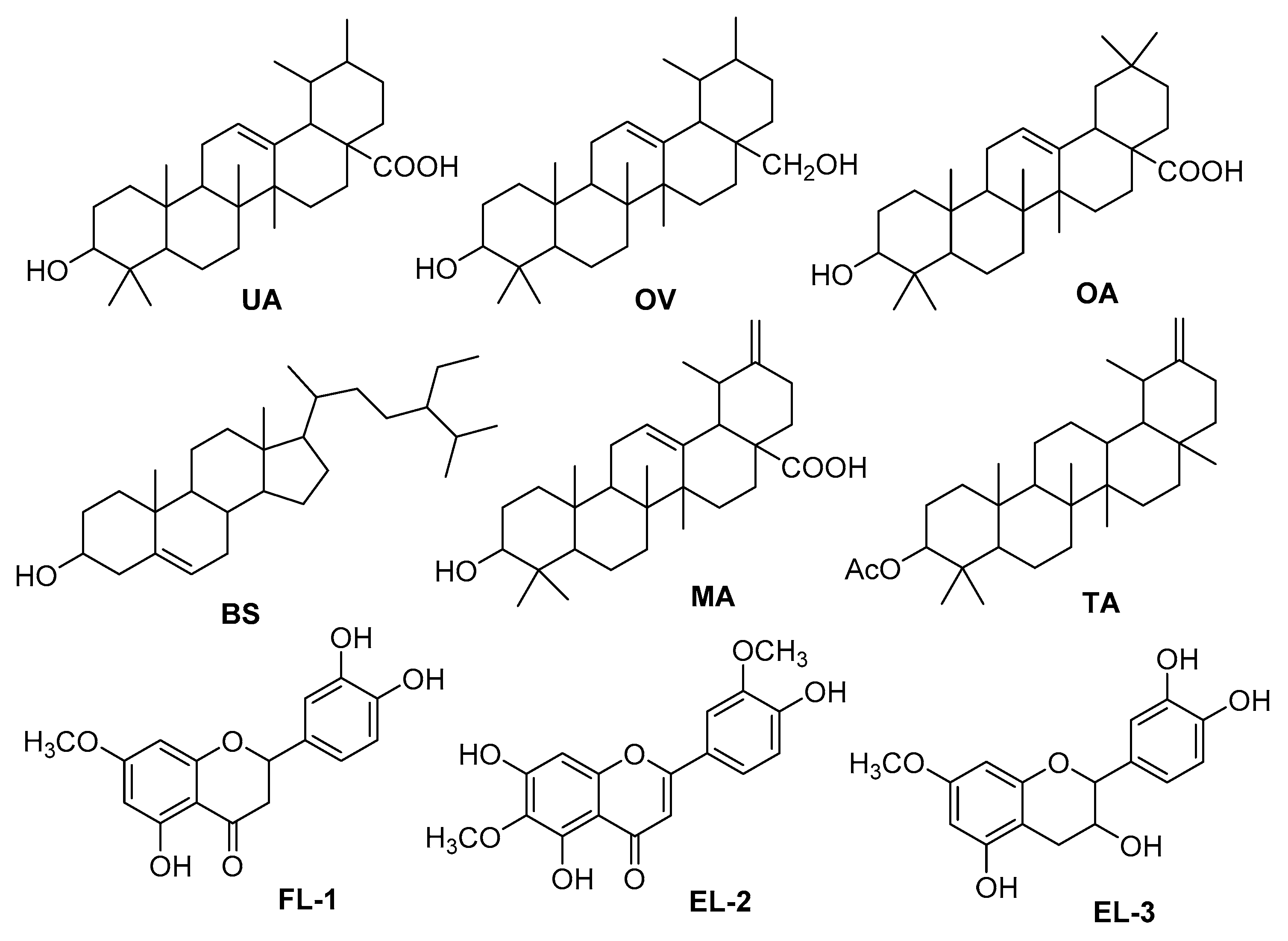
2.8. Isolation of Known Compounds

Six triterpenoids including ursolic acid (UA), ovaul (OV), oleanolic acid (OA), β-sitosterol (BS), micromeric acid (MA), taraxasterol acetate (TA) and three flavonoids viz., 5,3′,4′-trihydroxy-7-methoxy flavone (FL-1), 5,7,4′-trihydroxy-6,3′-dimiethoxyflavone (FL-2) and 7-methoxy catechin (FL-3) were isolated from the methanolic extract of S. edelbergii (Figure 3). Their structures were confirmed by combined spectroscopic techniques including 1D (1H and 13C) and 2D (HMBC, HSQC, COSY) nuclear magnetic resonance (NMR) and high-resolution electrospray ionization-mass spectrometry (ESI-HRMS) and comparison of the spectral data of known compounds with those reported in literature.
Compounds UA and OA and epimer MA have already been reported from the roots and leaves of S. strigillosa of the same genus [38,39], while compound BS was isolated from the root of S. baicalensis [18] and S. columnae [40] of the same genus. Several flavonoids, flavanones, flavones and their glycosides were reported from the different species of the same genus [18,38,41]. Altinier et al. [42] reported UA, OA and MA as the main anti-inflammatory principles of the leaves of Rosmainrus officinalis. Compounds MA, TA and OV were not only isolated, for the first time, from the S. edelbergii but also from the genus as well, which was previously reported from different plant species [42,43,44,45,46]. The anti-inflammatory and analgesic agent of EtOAc may be due to the presence of these triterpenoid acids in the fraction.
Flavonoids, especially flavanones and flavan-3-ols, are an important class of natural products belonging to a class of plant secondary metabolites having a polyphenolic structure, widely found in fruits, plants, marine species, vegetables and certain beverages [47,48]. They are associated with a broad spectrum of health-promoting effects concerned with a variety of nutraceutical, pharmaceutical, medicinal and cosmetic applications [49]. This is because of their antioxidative, anti-inflammatory, anti-mutagenic and anti-carcinogenic properties coupled with their capacity to modulate key cellular enzyme functions [47]. The antioxidant activity (DPPH and ABTS) of the chloroform fraction could be possible due to the presence of these flavonoids.
The plant and its derived products are varying under environmental gradients such as water, temperature and light [50]. Mainly, the genus is a rich basis of flavonoid contents which are mainly influenced by the optimum level of the environmental gradient for the genus Scutellaria; thus, our finding agrees with the reported data of Chen et al. [51], who studied S. baicalensis. As mostly the species of genus Scutellaria found with their habitats on the mountain tail with colder dry weather, similarly, the understudy sample was collected from mountain tail enriched with flavonoids and phenols. Thus, our results are matching with the findings of Grzegorczyk-Karolak et al. [32], who investigated S. altissima and S. galericulata for mentioned studies. It is revealed from S. altissima having high contents of phenols and flavonoids observed for S. edelbergii content [52]. Moreover, the bioactive compound scutellarin is present in the Scutellaria beside its habitats and environment, detected by Matsa et al. [53].

3. Material and Methods

3.1. General Instrumentation

The 1H- and 13C NMR spectra were measured on 600 MHz and 150 MHz NMR spectrometer (BRUKER, Zürich, Switzerland) using the solvent peaks (CDCl3, δH: 7.26; δC: 77.0), (CD3OD, δH: 4.87; δC: 48.5) as internal references. Data were determined in the following order: chemical shift (δ) in ppm; multiplicities are indicated s = singlet, d = doublet, t = triplet, dd = doublet of doublet, m = multiplet; coupling constants (J) are in hertz (Hz). High-resolution electrospray ionization mass spectrometry (HR-ESI-MS) spectra were recorded on Agilent 6530 LC Q-TOF (country of origin USA/EU, made in Singapore). Column chromatography was carried out by using silica gel of the selected particle size of 100–200 mesh. For TLC, pre-coated aluminum sheets (TLC silica gel 60 F254, Merck, Darmstadt, Germany) were used. Infrared (IR) spectra were recorded on a Bruker ATR-Tensor 37 spectrophotometer, Bruker (Ettlingen, Baden-Württemberg, Germany) with wave numbers (ν) in cm−1. Visualization was accomplished with UV light (254 and 366 nm) or I2 stain and by spraying with the ceric sulfate (Ce(SO4)2) reagent.

3.2. Plant Collection and Identification

The whole plant material of S. edelbergii was collected (April–June 2019) from Kalam, District Swat, Khyber Pakhtunkhwa, Pakistan, and identified by the plant taxonomist Prof. Mehboob Rahman, Matta College Swat, KPK, Pakistan. The plant specimen (AWKUM/Herb/2234) was placed in the herbarium of Abdul Wali Khan University, Mardan.
S. edelbergii is a perennial herb, spreading with a thick woody rootstock. Stems are very slender, procumbent or weakly ascending, round-quadrangular, leafy and much-branched. Leaves are thick-textured, triangular or narrow ovate and acute. Flowers subtended by elliptic to broad elliptic bracts which are entire, cuneate to acute, Calyx with a small often purple scutellum, enlarging in fruit to 3 mm with a 2.5 mm high scutellum. Petals are yellow or blue violet, with the darker lower lip spreading erect or erect, densely glandular pilose. Flowering is produced in April to July found between 1660–2200 m. Furthermore, it grows in moist, loamy soil, moist temperate regions and spread in different areas of northwest Pakistan, mainly influenced by the tail-end of monsoon conditions.

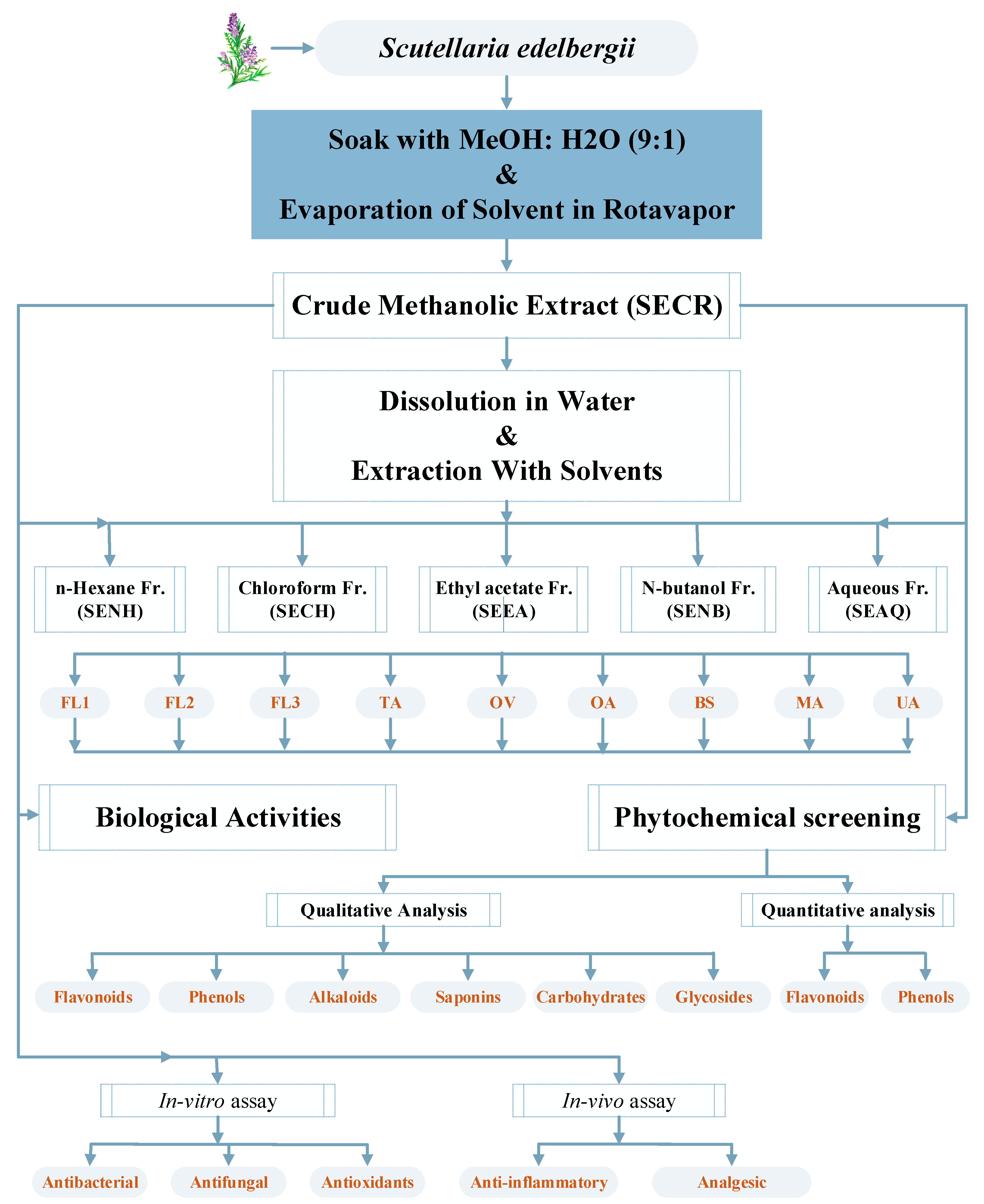
3.3. Extraction and Fractionation

The whole air-dried plant material of S. edelbergii was washed with tap water to remove soil/silicate particles and dried at room temperature for three weeks under shade to avoid loss of volatile components. The dried plant material was crushed into very fine powder through an electric grinder. The powders were kept in the refrigerator at 4 °C until further use. To prepare the crude extract, 2 kg of fine powder were soaked in commercial-grade methanol (6 L). The mixture was periodically shaken and then filtered through a muslin cloth after 21 days. The residue left was again immersed in 10% water/methanol for additional 21 days and filtered again. The filtrates from both the steps were combined and subjected to evaporation in a rotary evaporator at 40 °C. The semi-solid mass obtained was then placed in the open air till dryness; later, 600 g of crude extract were obtained and stored in airtight containers for further use. To prepare different fractions, 500 gm of crude extract were immersed in 1 L distilled water, which was then subjected to solvent-solvent extraction in a separating funnel from less to high polar solvents (n-hexane < chloroform < ethyl acetate < n-butanol) (Figure 4). The obtained fractions were evaporated in a rotary evaporator at 40 °C that yielded the dry mass of 21, 19, 20 and 18 g of n-hexane, chloroform, EtOAc and n-BuOH, respectively. In comparison, the aqueous extract was obtained in more quantity (35 g).
The obtained EtOAc fraction was further subjected over the silica gel (70–230 mesh; Merck) column chromatography (CC) using n-hexane, n-hexane/ethyl acetate and ethyl acetate/MeOH with 10% increasing polarity to afford twelve fractions (SE1–SE12). After taking TLC, four factions (SE3–SE6; 10–20% n-hexane/ethyl acetate) were combined and subjected to further small CC using n-hexane/ethyl acetate with increasing polarity (1:9, 2:8, 4:6, 6:4) to afford four sub-fractions (SSE1–SSE4). SSE2 (10–20% n-hexane/ethyl acetate) was further subjected to CC and preparative TLC to afford known compounds BS (18.5 mg), UA (22.0 mg), OV (5.0 mg), MA (7.0 mg) and OA (6.5 mg). Similarly, flavonoids FL-1 (4.5 mg), FL-3 (6.0 mg) and FL-2 (5.0 mg) were isolated from the chloroform fraction of S. edelbergii using solvent system of 40–60% n-hexane/ethyl acetate (Figure 4). All the isolated compounds are known (isolated first time from the source) and characterized by detail spectroscopic techniques. The detail NMR data, physical state and mass spectrometry are given below (show in Supplementary Materials).
(3β)-3-hydroxy-urs-12-en-28-oic acid (UA): Colorless crystals; 1H-NMR (600 MHz, Chloroform-d, δ = ppm): 5.23 (1H, br. s), 3.20 (1H, t, 10.8, 6.6 Hz), 2.17 (1H, d, 10.8 Hz), 1.29 (3H, s), 1.11 (3H, s), 0.99 (3H, s), 0.90 (3H, s), 0.87 (3H, s), 0.86 (3H, s), 0.69 (3H, s), 13C-NMR (150 MHz, Chloroform-d, δ = ppm): 181.2, 137.9, 125.9, 79.0, 55.2, 52.7, 47.9, 47.6,42.0, 39.5, 39.1, 38.8, 38.7, 38.6, 37.0, 36.7, 33.0, 30.6, 29.7, 28.1, 28.0, 27.2, 24.2, 23.6, 23.3, 21.2, 18.3, 17.1, 17.0, 15.6, 15.5; ESI-HRMS (negative): m/z 455.3531 [M − H]+ [50,54].
Ovaul (OV): 1H-NMR (600 MHz, Chloroform-d): White powders; 5.11 (1H, t, J = 3.6, H-12), 3.51 (1H, d, J = 10.8, H-28a), 3.20 (1H, dd, J = 11.4, 4.8, H-3α), 3.19 (1H, d, J = 10.8, H-28b), 1.11, 1.00, 0.95, 0.93, 0.79 (each 3H, s), 0.94, 0.81 (each 3H, d, J = 6.0).13C NMR (150 MHz, Chloroform-d, δ = ppm): 138.6, 125.0, 79.0, 69.9, 55.1, 54.0, 47.6, 42.0, 40.0, 39.4, 39.3, 38.7, 38.0, 36.8, 35.1, 32.8, 30.6, 28.1, 27.2, 25.9, 23.4, 23.3, 23.3, 21.3, 18.3, 17.3, 16.7, 15.6, 15.6; ESI-HRMS (positive): m/z 443.3311 [M + H]+ [50].
3β-hydroxy-olea-12-en-28-oic acid (OA): White crystals; 1H-NMR (600 MHz, Chloroform-d): 5.28 (1H, br. s), 3.23 (1H, s, dd, 11.4, 3.6 Hz), 2.83 (1H, dd, 13.8, 3.6 Hz), 1.13 (3H, s), 0.97 (3H, s), 0.94 (3H, 0.94 (3H, s), 0.88 (3H, s), 0.85–0.75 (6H, s); 13C NMR (150 MHz, Chloroform-d, δ = ppm): 182.9, 143.6, 122.6, 79.0, 55.2, 47.6, 46.5, 45.9, 41.6, 41.0, 39.3, 38.7, 38.4, 37.1, 33.8, 33.1, 32.6, 32.4, 30.7, 28.1, 27.7, 27.2, 25.9, 23.6, 23.4, 22.9, 18.3, 17.1, 15.5, 15.3; ESI-HRMS (negative): m/z 455.3516 [M − H]+ [54].
β-Sitosterol (BS): White powder; 1H-NMR (600 MHz, Chloroform-d): 5.33 (1H, br. s), 3.51 (1H, br. t), 1.03 (S, 3H), 0.93 (3H, d, J = 6.5 Hz), 0.84 (3H, t, J = 7.2 Hz), 0.83 (3H, d, J = 6.4 Hz), 0.81 (3H, d, J = 6.4 Hz), 0.68 (3H, s); 13C NMR (150 MHz, Chloroform-d, δ = ppm): 140.8, 121.7, 71.8, 56.8, 56.1, 50.1, 45.9, 42.3, 39.8, 37.3, 36.5, 36.1, 34.0, 31.9, 31.8, 29.7, 29.2, 28.2, 26.1, 24.3, 23.1, 21.1, 19.8, 19.4, 19.0, 18.8, 12.0, 11.8; ESI-HRMS (positive): m/z 437.3417 [M + Na]+ [55].
Micromeric acid (MA): Colorless powder; 1H NMR (600 MHz, Pyr-d, δ = ppm): 5.52 (td, J = 6.2, 1.0 Hz, 1H), 4.83 (dd, J = 13.7, 1.1 Hz, 1H), 4.78 (dd, J = 13.7, 1.1 Hz, 1H), 3.49 (td, J = 7.1, 5.0 Hz, 1H), 2.79 (pd, J = 7.4, 6.8, 1.3 Hz, 1H), 2.67–2.65 (m, 2H), 2.35–2.29(m, 2H), 2.15–2.09 (m, 3H), 1.99–1.77 (m, 4H), 1.61–1.36 (m, 4H), 1.34–1.22 (m, 2H), 1.25–1.16 (m, 2H), 1.20–1.11 (m, 2H), 1.14 (s, 3H), 1.12 (s, 2H), 1.00–0.95 (s, 9H), 0.92 (s, 3H), 0.86 (s, 3H); 13C-NMR (150 MHz, Pyr-d, δ = ppm): 180.4, 154.3, 139.8, 126.2, 105.6, 78.6, 56.4, 54.1, 48.6, 48.5, 43.0, 40.5, 39.6, 39.9, 39.6, 38.02, 37.86, 34.15, 31.64, 29.38, 29.2, 28.7, 24.2, 24.2, 21.9, 19.3, 18.0, 18.0, 17.1, 16.2; ESI-HRMS (positive): m/z 455.3563 [M + H]+ [46,47].
Taraxasterol acetate (TA): White powder; 1H NMR (600 MHz, Chloroform-d, δ = ppm) 4.59 (dt, J = 10.1, 2.4 Hz, 2H), 4.46 (dd, J = 10.9, 5.6 Hz, 1H), 2.11–2.00 (m, 1H), 2.02 (s, 3H), 1.74–1.45 (m, 4H), 1.36 (s, 2H), 1.43–1.29 (m, 3H), 1.30–1.17 (m, 2H), 1.15–1.03 (m, 1H), 1.00 (t, J = 3.4 Hz, 5H), 0.98–0.90 (m, 3H), 0.91 (s, 3H), 0.87–0.80 (m, 9H), 0.82 (s, 3H); 13C NMR (150 MHz, Chloroform-d, δ = ppm) 171.01, 154.64, 107.10, 80.98, 55.45, 55.44, 50.40, 48.65, 42.03, 40.92, 39.38, 39.16, 38.86, 38.44, 38.29, 37.80, 37.05, 34.53, 33.99, 27.94, 26.64, 26.15, 25.61, 25.48, 23.69, 21.46, 21.32, 19.48, 18.18, 16.49, 16.33, 15.89, 14.72; ESI-HRMS (positive): m/z 491.3731 [M + Na]+ [48,49].
5,3′,4′-trihydroxy-7-methoxy flavone (FL-1): Yellow powder; 1H-NMR (600 MHz, DMSO-d6) δ 7.53 (1H, br. s, H-2′), 7.48 (lH, d, J = 8.6, H-6′), 6.87 (1H, d, J = 8.4, H-5′), 5.98 (2H, d, J = 2.0, Hz, H-6 and H-8), 5.22 (lH, dd, J = 12.2, 3.2 Hz, H-2), 3.82 (3H, s, OMe), 2.90 (2H, m, H-3); 13C-NMR (150 MHz, DMSO-d, δ = ppm): 195.8, 167.0, 166.6, 164.2, 149.1, 143.9, 129.4, 123.0, 115.7, 114.6, 113.5, 96.7, 95.6, 79.0, 51.7, 43.2; ESI-HRMS (positive): m/z 289.0706 [M − CH2 + H]+ [56,57,58,59].
5,7,4′-trihydroxy-6,3′-dimiethoxyflavone (FL-2); Yellow powder; 1H-NMR (600 MHz, CH3OH-d, δ = ppm): 7.43 (1H, dd, J = 8.4, 1.8 Hz, H-6′), 7.39 (1H, d, J = 1.8 Hz, H-2′), 6.90 (1H, d, J = 8.4 Hz, H-5′), 6.79 (1H, s, H-8), 6.60 (1H, s, H-3), 3.97 (3H, s, OCH3), 3.82 (3H, s, OCH3); 13C-NMR (150 MHz, CH3OH-d, δ = ppm): 184.2, 166.8, 160.6, 154.9, 151.3, 149.1, 147.1, 133.2, 123.5, 120.4, 116.7, 114.1, 106.5, 103.8, 92.2, 61.0, 56.9; ESI-MS (positive): m/z 352.94 [M + Na]+ [60,61,62].
7-methoxy catechin (FL-3): Yellow powder, brown gummy solid; 1H-NMR (600 MHz, DMSO-d, δ = ppm): 6.70 (d, 1H, J = 11.2 Hz, H-5′), 6.59 (dd, 1H, J = 10.8 & 3.2 Hz, H-6′), 6.59 (d, 1H, J = 3.2 Hz, H-2′), 5.86 (s, 1H, H-8), 5.69 (1H, s, H-6), 4.47 (d, 1H, J = 10.4 Hz, H-2), 3.71 (d, J = 6.8, 10.4 & 11.6 Hz, H-3), 3.16 (s, OCH3, H-11), 2.54 (dd, 1H, J = 20.8 and 6.8 Hz, H-4α), 2.50 (1H, m, H-4β); 13C-NMR (150 MHz, DMSO-d, δ = ppm): 156.3, 156.1, 155.2, 144.8, 130.5, 118.4, 115.1, 114.4, 99.1, 95.2, 93.9, 80.8, 66.2, 48.6, 27.6; ESI-HRMS (positive): m/z 291.0729 [M − CH2 + H] + [63,64,65].

3.4. Preliminary Phytochemical Analysis

Crude extract and their fractions were subjected to preliminary phytochemical tests to confirm the presence of flavonoids, phenols, alkaloids, saponins, glycosides and carbohydrates using reported methods [66,67,68]. The crude extract along with different fractions (1 g) were dissolved in the 5 mL DMSO (99.99%, Fischer Scientific, Loughborough, UK) to make a stock solution, and DMSO was further used for re-solubilization (Figure 4).

3.4.1. Flavonoids

A few drops from the stock solution of crude extract and different fractions of S. edelbergii were mixed with diluted in 5% NaOH solution, followed by the addition of a few drops of hydrochloric acid (HCl). The presence of flavonoids was detected through color changes from yellow to colorless.

3.4.2. Phenols

A few drops from the stock solution of crude extract and different fractions were added to the FeCl3 solution, followed by vigorous shaking. The appearance of a bluish-green color confirms the presence of phenols.

3.4.3. Alkaloids

Qualitative detection of alkaloids was performed by mixing 0.5 mL of stock solution of crude extract and fractions with 2% H2SO4 solution, followed by heating for 3 min. Then, some drops of Dragendorff’s reagent were added to the mixture and allowed to cool. The appearance of an orange-red color precipitate revealed the presence of alkaloids.

3.4.4. Saponins

From the stock solution of S. edelbergii extracts and fractions, a few drops were mixed with 0.4 mL of distilled water, followed by shaking. After 5 min, the mixture was boiled and resulted in the formation of small bubbles (froth), which confirmed the presence of saponins.

3.4.5. Carbohydrates

The presence of carbohydrates was determined by mixing 3 mL of extract solution with 2 mL of research-grade Benedict’s reagent. The mixture was then placed in a hot water bath for 3 min. The appearance of a reddish-brown precipitate revealed the presence of carbohydrates.

3.4.6. Glycosides

A few drops of HCl were mixed with small quantities of extract solutions that caused hydrolysis of glycosides, which were then neutralized through the addition of (how much amount) NaOH solution. After neutralization, about 0.5 mL from Fehling’s A and B solutions were added to each extract mixture. Glycoside’s confirmation was revealed by the appearance of a red color precipitate.

3.4.7. Flavonoids and Phenolic Contents

The selected plant was investigated for quantitative estimation of the total phenolic and flavonoids contents in crude extract and subfraction using standard procedure [69].

3.4.8. Estimation of Total Phenolic Contents

The phenolic content in S. edelbergii extracts and subfractions was determined using the Folin–Ciocalteu reagent. The total phenolic content was measured as milligrams of gallic acid equivalent (mg GAE/g) per gram of dry sample mass. Different extracts in distilled methanol (5 mg in 5 mL) were mixed with 10 mL distilled water and placed undisturbed for 5 min. About 1 mL from each mixture was taken in a test tube, and its volume was raised to 10 mL by adding distilled water. Then, 1 mL of Folin-Ciocalteu reagent was added to each test tube and incubated for 6 min. After incubation, 10 mL of 7% sodium carbonate solution were added. The final volume of the reaction mixture was raised to 26 mL through the addition of distilled water and incubated for 90 min at room temperature. The absorbance was noted at 760 nm using a UV-visible spectrophotometer.

3.4.9. Quantification of Total Flavonoid Contents

The total flavonoid content was represented as a quercetin equivalent (mg QE/g) of the dry sample mass of the extract. About 9 mL of the distal water was mixed with 1 mL of each extract stock solution, along with 1 mL of 5% NaNO2 in a test tube, which was then incubated for 6 min. Then, 2 mL of 10% AlCl3 was added to each test tube, and the reaction mixture was then placed undisturbed for 5 min. Finally, 2 mL of 1 M sodium hydroxide was added to each test tube, and the absorbance of the resultant mixture was noted at 510 nm via a UV-visible spectrophotometer.

3.5. In Vitro Biological Activities

In vitro antibacterial and antifungal activities were carried out on E. coli, K. pneumoniae, B. Subtilis, S. aureus, C. albican and F. oxysporum strains. The fungal and bacterial strains were obtained from the laboratory of Microbiology, verified by Dr. Hazir Rahman, Chairman, Department of Microbiology, AWKUM, Mardan.

3.5.1. Antibacterial Activities

The crude extract and different fractions (1 g) were dissolved in the 5 mL DMSO to make a stock solution and DMSO was further used for re-solubilization for antibacterial activity. The antimicrobial potential of the crude extract and subfractions of S. edelbergii was examined through agar well diffusion assay [70]. Twenty-eight grams (28 g) of nutrient agar were added to 1000 mL of distilled water and shaken vigorously until its complete dissolution. The nutrient agar media, loop, corn borer and Petri dishes were autoclaved at 121 °C for 15 min. In an aseptic condition of laminar flow hood, 20 mL agar were poured to each Petri plate and allowed to solidify. Bacterial strains (E. coli, K. pneumonia, S. aureus, B. subtilis) were inoculated through a wire loop using a safety kit using concentration of bacterial cell density of 1.5 × 10⁸ CFU/mL. Four wells of 3 mm size were made on a cork borer in each plate with the same distances. The selected plant extract at concentrations of C1 = 50 µL and C2 = 100 µL were poured into the wells. The same method was used for positive control. Levofloxacin and Erythromycin were used as a positive control for Gram-positive and Gram-negative strains, respectively. Dimethyl sulfoxide (DMSO) was used as a negative control. The Petri plates were placed in the incubator at 37 °C for 24 h. Three biological replicates with 2 different volumes were used. Later, plates were then taken out to measure the zone of inhibition around each hole. The results were taken in triplicate.

3.5.2. Antifungal Activity

For antifungal assay, the crude extract and different fractions (1 g) were dissolved in the 5 mL DMSO to make a stock solution and DMSO was further used for re-solubilization. Antifungal activities were performed on potato dextrose agar media [70]. Thirty-nine grams (39 g) of potato dextrose agar media were added to 1 L distilled water and sterilized in an autoclave at 121 °C. The media plates were incubated for 24 h in an incubator to avoid contamination of unwanted strains. Then, four holes were created in media under laminar flow and holes were poured with extract solution. The plates inoculated with fungal strains from inoculum having concentration 108–109 CFU/mL and left for 3 days in the incubator at 25 °C. The zone of inhibition of standard and test extracts were measured in mm.

3.5.3. Antioxidant Activity (DPPH and ABTS)

To evaluate the antioxidant potential of crude extract and sub-fractions, 2,2-diphenyl-1-picrylhydrazyl (DPPH) and (2,2′-azino-bis (3-ethylbenzothiazoline-6-sulfonic acid) (ABTS) [71] were used. In the DPPH assay, 3 mg of DDPH was dissolved in 100 mL distilled methanol and kept in the dark for 30 min for the formation of free radicals in the solutions. Different dilutions of each extract (1000, 500, 250, 125 µg/mL) were also prepared. Then, 2 mL of each dilution were added with 2 mL DPPH stock solution and incubated for 20 min in the dark. After incubation, the absorbance of each sample was measured at 517 nm by UV/Vis spectrophotometer. Similar dilutions of ascorbic acid were also prepared and incubated with 2 mL of DPPH stock solution for the aforementioned duration to determine its antioxidant potential. The following formula was used to determine the antioxidant potentials of the samples:
%   S c a v e n g i n g   a c t i v i t y = A B A × 100
where A is the control absorbance and B is the standard absorbance
The antioxidant potential of these extracts was also determined by the ABTS method. A total of 383 mg of ABTS and 66.2 mg of Potassium persulfate (K2S2O8) were dissolved individually in 100 mL methanol and mixed. Later, 2 mL of the mixture were incubated with 2 mL of extract dilutions for 25 min. The absorbance was noted at 746 nm by UV spectrophotometer. (The free radical scavenging potential of the extracts was calculated by Equation (1)).

3.6. In Vivo Pharmacological Activities

For in vivo anti-inflammatory and analgesic activities, approval was taken from the ethical committee (No: AWKUM/Bot/2019/1720, Date: 29 January 2019) of Abdul Wali Khan University Mardan, Mardan (AWKUM) (provided in hard to the publisher).

3.6.1. Experimental Animals

Healthy Swiss albino mice (approved by the ethical committee (No: AWKUM/Bot/2019/1720, Date: 29 January 2019), Botany Department, AWKUM) of either sex was placed in rubber cages in germ-free conditions for 45 days at around 20 °C and fed with rodent pellet food and water.

3.6.2. Anti-Inflammatory Activities

Twenty (20) Swiss albino mice with a bodyweight of 24–30 g were used in this study. The mice were brought from the Veterinary Center Peshawar and kept in the animal house of AWKUM. The animals were given nutrient pellets and water. ARRIVE guidelines were followed while selecting sample size, experimental design and inclusion and exclusion techniques for statistically acceptable/satisfactory results [72].
The animals were divided into four groups as per the given details (five mice in each group):
Group A: 1 mL carrageenan was injected to induce paw edema in mice.
Group B: 1 mL carrageenan solution and Diclofenac sodium (as a standard drug, 50 mg/kg body weight) were injected.
Group C: 1 mL carrageenan was injected with 50 mg/kg body weight of each extract.
Group D: 1 mL carrageenan was injected with 100 mg/kg body weight of each extract.
The diameter of the carrageenan injected paw edema was measured after 1, 2 and 3 h. The average % inhibition was calculated in comparison with the standard by the following formula:
%   i n h i b i t i o n = A B A × 100
where A is the induced paw diameter and B is the tested drug (in case of analgesic activities, A is writhes induced by acetic acid).

3.6.3. Analgesic Activities

The analgesic activity was also determined in Swiss albino mice. The animals were divided into four groups (5 mice each) as per the given details:
Group A: 1 mL acetic acid solution was injected into this group to cause writhing.
Group B: 1 mL acetic acid and 1 mL aspirin (standard drug) were injected.
Group C: 1 mL acetic acid and 50 mg/kg body weight of each extract were injected.
Group D: 1 mL acetic acid and 100 mg/kg body weight of each extract were injected.
After injection, the rate of writhing was noted for 10 min, and % inhibition was noted by comparing it with the standard by Equation (2).

3.7. Statistical Analysis

All the data were taken in triplicate and analyzed through one-way ANOVA (analysis of variance) followed by Bonferroni’s test to determine significance level [p = (≤0.05) and p = (≤0.01)] of all the obtained results. A nonlinear regression graph was constructed between % inhibition and concentration, and the IC50 was measured with the GraphPad Prism 9 program for windows (GraphPad-Software, San Diego, California, 2020) using Equation (1).
Y = 100 / 1 + ( ^ H i l l S l o p e )
where 1 = the concentration of the inhibitors used, Y = the inhibitor’s reaction and HillSlope indicates the steepness of the curves.

4. Conclusions

The present analysis revealed that S. edelbergii is a rich source of secondary metabolites that could be effectively used as a remedy for several health complications. The EtOAc fraction exhibited the highest phenolic and flavonoid contents. Significant in vitro antibacterial activity was observed, especially against Gram-positive bacterial strains in comparison to the Gram-negative strain used in the study. The tested extracts were found inactive against the selected fungal strains. The chloroform fraction was the most effective scavenger of the DPPH and ABTS free radicals. The EtOAc fraction was found to act as an anti-inflammatory and analgesic agent in Swiss albino mice. Furthermore, six known triterpenoids including UA, OV, OA, BS, MA and TA as well as three flavonoids viz., FL-1, FL-2 and FL-3 were isolated, for the first time, from the methanolic extract of S. edelbergii. Based on the results of our investigation, the selected plant could be used as an antibacterial, analgesic, anti-inflammatory and antioxidant agent due to the presence of triterpenoids and flavonoids. However, further studies are needed to determine more responsible compounds of the observed biological and pharmacological activities.

Supplementary Materials

The following are available online, Figure S1: 1H-NMR of compound UA, Figure S2: 13C-NMR of compound UA, Figure S3: ESI-HRMS of UA, Figure S4: 1H-NMR of compound OV, Figure S5: 13C-NMR of compound OV, Figure S6: ESI-HRMS of OV, Figure S7: 1H-NMR of compound OA, Figure S8: 13C-NMR of compound OA, Figure S9: ESI-HRMS of OA, Figure S10: 1H-NMR of compound BS, Figure S11: 13C-NMR of compound BS, Figure S12: ESI-HRMS of BS, Figure S13: 1H-NMR of compound MA, Figure S14: 13C-NMR of compound MA, Figure S15: ESI-HRMS of MA, Figure S16: 1H-NMR of compound TA, Figure S17: 13C-NMR of compound TA, Figure S18: ESI-HRMS of TA, Figure S19: 1H-NMR of compound FL-1, Figure S20: 13C-NMR of compound FL-1, Figure S21: ESI-HRMS of FL-1, Figure S22: 1H-NMR of compound FL-2, Figure S23: 13C-NMR of compound FL-2, Figure S24: ESI-HRMS of FL-2, Figure S25: 1H-NMR of compound FL-3, Figure S26: 13C-NMR of compound FL-3, and Figure S27: ESI-HRMS of FL-3.

Author Contributions

M.S., M.A., H.R., M.Z. and S.M. designed and performed all the experiments including antioxidant, analgesic and anti-inflammatory activities. M.A., M.A.N. and G.E.-S.B. studied antimicrobial activities. M.S. and N.U.R. isolated and characterized pure compounds through chromatographic and spectroscopic techniques. N.U.R., S.A.H. and A.K. wrote the original draft of the manuscript. A.A.-H. and W.M. supervised the project and assisted in reviewing and editing the manuscript. All authors have read and agreed to the published version of the manuscript.

Funding

The research was supported by the Oman Research Council (TRC) funded through the project (BFP/RGP/CBS/18/011).

Institutional Review Board Statement

The study was conducted according to the guidelines of the Declaration of Helsinki, and approved by the ethical committee (No: AWKUM/Bot/2019/1720, Date: 29 January 2019) of Abdul Wali Khan University Mardan, Mardan (AWKUM).

Informed Consent Statement

Not applicable.

Data Availability Statement

The data are available to the researchers upon request.

Acknowledgments

The authors are grateful to the University of Nizwa, Nizwa, Oman, for the generous support of this project. The authors would also like to thank the Taif University Researchers Supporting Project number (TURSP-2020/71), Taif University, Taif, Saudi Arabia.

Conflicts of Interest

The authors declare that they have no conflict of interest concerning the submission of this manuscript.

Sample Availability

Samples of the compounds are not available from the authors.

References

  1. Khan, I.; Zahoor, M.; Zeb, A.; Ul Bari, W. In Vitro Antioxidant, Antidiabetic, and Anticholinesterase, and In Vivo Toxicological, Hypoglycemic, and Antilipidemic Potentials of Ziziphus oxyphylla. Lat. Am. J. Pharm. 2020, 39, 7–21. [Google Scholar]
  2. Khan, H.; Saeed, M.; Mehmood, M.H.; Rehman, N.U.; Muhammad, N.; Haq, I.U.; Ashraf, N.; El-Tahir, K.E.H.; Gilani, A.-H. Studies on tracheorelaxant and anti-inflammatory activities of rhizomes of Polygonatum verticillatum. BMC Complement. Altern. Med. 2013, 13, 197. [Google Scholar] [CrossRef] [PubMed] [Green Version]
  3. Zahoor, M.; Khan, I.; Zeb, A.; Sahibzada, M.U.; Naz, S.; Bari, W.U.; Kamran, A.W. Pharmacological evaluation and in-silico modeling study of compounds isolated from Ziziphus oxyphylla. Heliyon 2021, 7, e06367. [Google Scholar] [CrossRef] [PubMed]
  4. Zahoor, M.; Bari, W.U.; Zeb, A.; Khan, I. Toxicological, anticholinesterase, antilipidemic, antidiabetic and antioxidant potentials of Grewia optiva Drummond ex Burret extracts. J. Basic Clin. Physiol. Pharmacol. 2020, 31. [Google Scholar] [CrossRef] [PubMed]
  5. Nazir, N.; Zahoor, M.; Nisar, M.; Karim, N.; Latif, A.; Ahmad, S.; Uddin, Z. Evaluation of neuroprotective and anti-amnesic effects of Elaeagnus umbellata Thunb. On scopolamine-induced memory impairment in mice. BMC Complement. Med. Ther. 2020, 20, 143. [Google Scholar] [CrossRef] [PubMed]
  6. Mousavi, S.N.M.; Delazar, A.; Nazemiyeh, H.; Khodaie, L. Biological Activity and Phytochemical Study of Scutellaria platystegia. Iran. J. Pharm. Res. 2015, 14, 215–223. [Google Scholar]
  7. Iqbal, N.; Hussain, S.; Ahmed, Z.; Yang, F.; Wang, X.; Liu, W.; Yong, T.; Du, J.; Shu, K.; Yang, W.; et al. Comparative analysis of maize–soybean strip intercropping systems: A review. Plant Prod. Sci. 2019, 22, 131–142. [Google Scholar] [CrossRef] [Green Version]
  8. Herridge, W.P.; Shibu, P.; O’Shea, J.; Brook, T.C.; Hoyles, L. Bacteriophages of Klebsiella spp., their diversity and potential therapeutic uses. J. Med. Microbiol. 2020, 69, 176–194. [Google Scholar] [PubMed]
  9. Ikram, M.; Beshbishy, A.M.; Kifayatullah, M.; Olukanni, A.; Zahoor, M.; Naeem, M.; Amin, M.; Shah, M.; Abdelaziz, A.S.; Ullah, R.; et al. Chemotherapeutic potential of carthamus oxycantha root extract as antidiarrheal and in vitro antibacterial activities. Antibiotics 2020, 9, 226. [Google Scholar] [CrossRef]
  10. Kim, M.; Jo, Y.; Hwang, Y.J.; Hong, H.W.; Hong, S.S.; Park, K.; Myung, H. Phage-antibiotic synergy via delayed lysis. Appl. Environ. Microbiol. 2018, 84, e02085-18. [Google Scholar] [CrossRef] [Green Version]
  11. Zahoor, M.; Ahmed, M.; Naz, S.; Ayaz, M. Cytotoxic, Antibacterial and antioxidant activities of extracts of the bark of Melia azedarach (China Berry). Nat. Prod. Res. 2015, 29, 1170–1172. [Google Scholar] [CrossRef]
  12. Nazir, N.; Jan, M.R.; Ali, A.; Asif, M.; Idrees, M.; Nisar, M.; Zahoor, M.; Abd El-Salam, N.M. Prevalence of hepatitis-C virus genotypes and potential transmission risks in Malakand Khyber Pakhtunkhwa, Pakistan. Virol. J. 2017, 14, 160. [Google Scholar] [CrossRef] [PubMed] [Green Version]
  13. Bari, W.U.; Zahoor, M.; Zeb, A.; Khan, I.; Nazir, Y.; Khan, A.; Rehman, N.U.; Ullah, R.; Shahat, A.A.; Mahmood, H.M. Anticholinesterase, Antioxidant potentials, and molecular docking studies of isolated bioactive compounds from Grewia optiva. Int. J. Food Prop. 2019, 22, 1386–1396. [Google Scholar] [CrossRef] [Green Version]
  14. Hassan, G.S.; Abdel Rahman, D.E.; Abdelmajeed, E.A.; Refaey, R.H.; Alaraby Salem, M.; Nissan, Y.M. New pyrazole derivatives: Synthesis, Anti-inflammatory activity, cyclooxygenase inhibition assay and evaluation of mPGES. Eur. J. Med. Chem. 2019, 171, 332–342. [Google Scholar] [CrossRef] [PubMed]
  15. Bardakci, H.; Türköz Acar, E.; Kırmızıbekmez, H. Simultaneous quantification of six phenylethanoid glycosides in some Turkish Scutellaria species by a new HPLC-DAD method. Nat. Prod. Res. 2019, 33, 2116–2119. [Google Scholar] [CrossRef]
  16. Swati Paul, D.S. Analgesic Activity of Methanol Extract of Plumbago indica (L.) by Acetic Acid-Induced Writhing Method. Asian J. Pharm. Tech. 2012, 2, 74–76. [Google Scholar]
  17. Miyaichi, Y.; Imoto, Y.; Tomimori, T.; Lin, C.C. Studies on the constituents of Scutellaria species. IX. On the flavonoid constituents of the root of Scutellaria indica L. Chem. Pharm. Bull. 1987, 35, 3720–3725. [Google Scholar] [CrossRef] [Green Version]
  18. Gupta, D.; Dubey, J.; Kumar, M. Phytochemical analysis and antimicrobial activity of some medicinal plants against selected common human pathogenic microorganisms. Asian Pac. J. Trop. Dis. 2016, 6, 15–20. [Google Scholar] [CrossRef]
  19. Zeng, J.; Ma, R.-J.; Wang, L.; Zhang, S.-N.; Song, H.-Z.; Yang, Y.; Tan, Q.-G. Chemical constituents from the leaves of Melia azedarach. Nat. Prod. Res. 2019, 33, 2860–2863. [Google Scholar] [CrossRef]
  20. Dai, S.J.; Tao, J.Y.; Liu, K.; Jiang, Y.T.; Shen, L. neo-Clerodane diterpenoids from Scutellaria barbata with cytotoxic activities. Phytochemistry 2006, 67, 1326–1330. [Google Scholar] [CrossRef]
  21. Wang, Z.L.; Wang, S.; Kuang, Y.; Hu, Z.M.; Qiao, X.; Ye, M. A comprehensive review on phytochemistry, Pharmacology, and flavonoid biosynthesis of Scutellaria baicalensis. Pharm. Biol. 2018, 56, 465–484. [Google Scholar] [CrossRef] [Green Version]
  22. Kowalczyk, E.; Krzesiński, P.; Kura, M.; Niedworok, J.; Blaszczyk, J. Pharmacological effects of flavonoids from Scutellaria baicalensis. Prz. Lek. 2006, 63, 95–96. [Google Scholar]
  23. Zahoor, M.; Shah, A.; Gul, S.; Amin, S. HPLC-UV analysis of antioxidants in Citrus sinensis stem and root extracts. J. Chem. Soc. Pak. 2018, 40, 595–601. [Google Scholar]
  24. Zahoor, M. Biological and phytochemical evaluation of Cotoneaster microphyllus, Ficus auriculata and Calotropis procera. Lat. Am. J. Pharm. 2019, 38, 945–953. [Google Scholar]
  25. Marrero Delange, D.; Morales Rico, C.; Canavaciolo, C.V.L.; Oliver, E.S.; Pérez, R.C.; Rodríguez-Leyes, E.; Vicente Murillo, R. Phytochemical screening of Scutellaria havanensis Jacq. Rev. Cuba. Plantas Med. 2012, 17, 402–407. [Google Scholar]
  26. Liu, H.-L.; Kao, T.-H.; Shiau, C.-Y.; Chen, B.-H. Functional components in Scutellaria barbata D. Don with anti-inflammatory activity on RAW 264.7 cells. J. Food Drug Anal. 2018, 26, 31–40. [Google Scholar] [CrossRef] [PubMed] [Green Version]
  27. Gharari, Z.; Aghajanzadeh, M.; Sharafi, A. Scutellaria orientalis subsp. Bornmuelleri: Phytochemical composition and biological activities. Nat. Prod. Res. 2021, 1–6. [Google Scholar] [CrossRef]
  28. Ozkul, C.; Renda, G.; Ekizoglu, M.; EZER, N.; Arıtuluk, Z. Antimicrobial activity of three Scutellaria L. species from Turkey. J. Res. Pharm. 2019, 23, 552–558. [Google Scholar] [CrossRef] [Green Version]
  29. Yilmaz, G.; Şimşek, D.; Altanlar, N.; Çiçek, M. Comparison of the antimicrobial activity of some Scutellaria orientalis L. taxa growing in Turkey. Int. J. Second. Metab. 2020, 192–199. [Google Scholar] [CrossRef]
  30. Shi, G.-X.; Shao, J.; Wang, T.-M.; Wang, C.Z. New advance in studies on antimicrobal activity of Scutellaria baicalensis and its effective ingredients. Zhongguo Zhong Yao Za Zhi 2014, 39, 3713–3718. [Google Scholar]
  31. Vergun, O.; Svydenko, L.; Grygorieva, O.; Shymanska, O.; Rakhmetov, D.; Brindza, J.; Ivanišová, E. Antioxidant capacity of plant raw material of Scutellaria baicalensis Georgi. Potravin. Slovak J. Food Sci. 2019, 1, 614–621. [Google Scholar] [CrossRef] [Green Version]
  32. Grzegorczyk-Karolak, I.; Kuźma, Ł.; Wysokińska, H. Study on the chemical composition and antioxidant activity of extracts from shoot culture and regenerated plants of Scutellaria altissima L. Acta Physiol. Plant. 2015, 37, 1736. [Google Scholar] [CrossRef] [Green Version]
  33. Delazar, A.; Nazemiyeh, H.; Afshar, F.H.; Barghi, N.; Esnaashari, S.; Asgharian, P. Chemical compositions and biological activities of Scutellaria pinnatifida A. Hamilt aerial parts. Res. Pharm. Sci. 2017, 12, 187–195. [Google Scholar] [PubMed] [Green Version]
  34. Zengin, G.; Llorent-Martínez, E.; García, L.; de Cordova, M.L.; Aktumsek, A.; Uysal, S.; Rengasamy, K.; Aumeeruddy, M.; Bahadori, M.B.; Mahomoodally, F. Chemical profile, Antioxidant, and enzyme inhibitory properties of two Scutellaria species: S. orientalis L. and S. salviifolia Benth. J. Pharm. Pharmacol. 2019, 71, 270–280. [Google Scholar] [CrossRef]
  35. Lee, J.K.; Song, Y.K.; Lim, H.H. Analgesic and anti-Inflammatory effect of Scutellaria baicalensis. J. Korean Med. 2007, 28, 124–135. [Google Scholar]
  36. Kim, E.H.; Shim, B.; Kang, S.; Jeong, G.; Lee, J.; Yu, Y.-B.; Chun, M. Anti-inflammatory effects of Scutellaria baicalensis extract via suppression of immune modulators and MAP kinase signaling molecules. J. Ethnopharmacol. 2009, 126, 320–331. [Google Scholar] [CrossRef]
  37. Yimam, M.; Brownell, L.; Hodges, M.; Jia, Q. Analgesic effects of a standardized bioflavonoid composition from Scutellaria baicalensis and Acacia catechu. J. Diet. Suppl. 2012, 9, 155–165. [Google Scholar] [CrossRef] [Green Version]
  38. Miyaichi, Y.; Ishii, K.; Kuno, T.; Tomimori, T. Studies on the Constituents of Scutellaria Species XX: Constituents of roots of Scutellaria strigillosa Hemsley. Nat. Med. 1999, 53, 237–241. [Google Scholar]
  39. Miyaichi, Y.; Morimoto, T.; Yaguchi, K.; Kizu, H. Studies on the constituents of Scutellaria species (XXI): Constituents of the leaves of Scutellaria strigillosa Hemsley. J. Nat. Med. 2006, 60, 157–158. [Google Scholar] [CrossRef]
  40. Stojakowska, A.; Kisiel, W. Secondary metabolites from a callus culture of Scutellaria columnae. Fitoterapia 1999, 70, 324–325. [Google Scholar] [CrossRef]
  41. Shang, X.; He, X.; He, X.; Li, M.; Zhang, R.; Fan, P.; Zhang, Q.; Jia, Z. The genus Scutellaria an ethnopharmacological and phytochemical review. J. Ethnopharmacol. 2010, 128, 279–313. [Google Scholar] [CrossRef]
  42. Altinier, G.; Sosa, S.; Aquino, R.P.; Mencherini, T.; Loggia, R.D.; Tubaro, A. Characterization of topical antiinflammatory compounds in Rosmarinus officinalis L. J. Agric. Food Chem. 2007, 55, 1718–1723. [Google Scholar] [CrossRef]
  43. Maria, F.; Kabouche, Z.; Fujimoto, Y.; Araya, H. Two new triterpenes and other compounds from Mentha aquatica (Lamiaceae). Nat. Prod. Commun. 2017, 12, 483–486. [Google Scholar] [CrossRef] [Green Version]
  44. Babaamer, Z.; Sekhri, L.; Al-Jaber, H.; Al-Qudah, M.; Abu Zarga, M. Extraction and identification of triterpenoids from Pergularia Tomentosa L. Ann. Sci. Technol. 2013, 5, 1–5. [Google Scholar] [CrossRef]
  45. Silva, L.; Strapasson, R.; Riva, D.; Salvador, M.; Stefanello, M.É. Triterpenes from the flowers of Gochnatia polymorpha subsp. floccosa. Rev. Bras. Farmacogn. 2011, 21, 556–559. [Google Scholar] [CrossRef] [Green Version]
  46. Bano, Z.; Begum, S.; Ali, S.S.; Kiran, Z.; Siddiqui, B.S.; Ahmed, A.; Khawaja, S.; Fatima, F.; Jabeen, A. Phytochemicals from Carissa carandas with potent cytotoxic and anti-inflammatory activities. Nat. Prod. Res. 2021, 1–6. [Google Scholar] [CrossRef] [PubMed]
  47. Panche, A.N.; Diwan, A.D.; Chandra, S.R. Flavonoids: An overview. J. Nutr. Sci. 2016, 5, e47. [Google Scholar] [CrossRef] [PubMed] [Green Version]
  48. Hussain, J.; Rehman, N.U.; Khan, A.L.; Ali, L.; Kim, J.-S.; Zakarova, A.; Al-Harrasi, A.; Shinwari, Z.K. Phytochemical and biological assessment of medicinally important plant Ochradenus arabicus. Pak. J. Bot. 2014, 46, 2027–2034. [Google Scholar]
  49. Havsteen, B. The biochemistry and medical significance of the flavonoids. Pharmacol. Ther. 2002, 96, 67–202. [Google Scholar] [CrossRef]
  50. Georgieva, Y.; Katsarova, M.; Stoyanov, P.; Mladenov, R.; Denev, P.; Teneva, D.; Plotnikov, E.; Bozov, P.; Dimitrova, S. Metabolite profile and antioxidant activity of some species of Genus Scutellaria growing in Bulgaria. Plants 2021, 10, 45. [Google Scholar] [CrossRef]
  51. Chen, S.; Yuan, Y.; Luo, Y.; Huang, L.; Chen, P.; Li, X. Effects of light on flavonoids accumulation and related gene expression in suspension cultures of Scutellaria baicalensis. Zhongguo Zhong Yao Za. Zhi. 2010, 35, 682–685. [Google Scholar]
  52. Vaidya, B.N.; Brearley, T.A.; Joshee, N. Antioxidant capacity of fresh and dry leaf extracts of sixteen Scutellaria species. J. Med. Act. Plants 2014, 2, 42–49. [Google Scholar]
  53. Matsa, M.; Bardakci, H.; Gousiadou, C.; Kirmizibekmez, H.; Skaltsa, H. Secondary metabolites from Scutellaria albida L. ssp. velenovskyi (Rech. f.) Greuter & Burdet. Biochem. Syst. Ecol. 2019, 83, 71–76. [Google Scholar]
  54. Marzouk, A. Hepatoprotective Triterpenes from Hairy Root Cultures of Ocimum basilicum L. Z. Naturforschung C 2009, 64, 201–209. [Google Scholar] [CrossRef]
  55. Ododo, M.M.; Choudhury, M.K.; Dekebo, A.H. Structure elucidation of β-sitosterol with antibacterial activity from the root bark of Malva parviflora. Springerplus 2016, 5, 1210. [Google Scholar] [CrossRef] [Green Version]
  56. Vasconcelos, J.M.J.; Silva, A.M.S.; Cavaleiro, J.A.S. Chromones and flavanones from artemisia campestris subsp. maritima. Phytochemistry 1998, 49, 1421–1424. [Google Scholar] [CrossRef]
  57. Chuang, T.-H.; Chan, H.-H.; Wu, T.-S.; Li, C.-F. Chemical constituents and biological studies of the leaves of Grevillea robusta. Molecules 2011, 16, 9331–9339. [Google Scholar] [CrossRef] [PubMed] [Green Version]
  58. Mossa, J.S.; Sattar, E.A.; Abou-Shoer, M.; Galal, A.M. Free Flavonoids from Rhus retinorrhoea Steud, ex Olive. Int. J. Pharmacogn. 1996, 34, 198–201. [Google Scholar] [CrossRef]
  59. Wang, Y.; Qing, W.-X.; Li, L.-X.; Zhao, D.-B.; Liu, X.-H. Isolation and identification of 5,3’,4’-Trihydroxy-7-methoxyflavanone from Artemisia sphaerocephala Kraschen. Jiegou Huaxue 2014, 33, 199–203. [Google Scholar]
  60. Medjroubi, K.; Mezhoud, S.; Benayache, F.; Seguin, E.; Tillequin, F. Flavonoids of the aerial part of Centaurea pullata. Chem. Nat. Compd. 2005, 41, 226–227. [Google Scholar] [CrossRef]
  61. Sadhu, S.K.; Hirata, K.; Li, X.; Ohtsuki, T.; Koyano, T.; Preeprame, S.; Kowithayakorn, T.; Ishibashi, M. Flavonoids and sesquiterpenoids, constituents from Eupatorium capillifolium, found in a screening study guided by cell growth inhibitory activity. J. Nat. Med. 2006, 60, 325–328. [Google Scholar] [CrossRef]
  62. Martínez, V.; Barberá, O.; Sánchez-Parareda, J.; Alberto Marco, J. Phenolic and acetylenic metabolites from Artemisia assoana. Phytochemistry 1987, 26, 2619–2624. [Google Scholar] [CrossRef]
  63. Ara, A.; Saleh-E-In, M.M.; Ahmad, M.; Hashem, M.A.; Hasan, C.M. Isolation and Characterization of Compounds from the Methanolic Bark Extract of Adenanthera pavonina L. Anal. Chem. Lett. 2020, 10, 49–59. [Google Scholar] [CrossRef]
  64. Bilia, A.R.; Morelli, I.; Hamburger, M.; Hostetmann, K. Flavans and A-type proanthocyanidins from Prunus prostrata. Phytochemistry 1996, 43, 887–892. [Google Scholar] [CrossRef]
  65. Kang, J.; Price, W.E.; Ashton, J.; Tapsell, L.C.; Johnson, S. Identification and characterization of phenolic compounds in hydromethanolic extracts of sorghum wholegrains by LC-ESI-MSn. Food Chem. 2016, 211, 215–226. [Google Scholar] [CrossRef] [PubMed] [Green Version]
  66. Gul, R.; Jan, S.U.; Faridullah, S.; Sherani, S.; Jahan, N. Preliminary Phytochemical screening, quantitative analysis of alkaloids, and antioxidant activity of crude plant extracts from Ephedra intermedia indigenous to Balochistan. Sci. World J. 2017, 2017, 5873648. [Google Scholar] [CrossRef] [Green Version]
  67. Auwal, M.S.; Saka, S.; Mairiga, I.; Sanda, K.A.; Shuaibu, A.; Ibrahim Dirar, A. Preliminary phytochemical and elemental analysis of aqueous and fractionated pod extracts of Acacia nilotica (Thorn mimosa). Vet. Res. Forum. 2014, 5, 95–100. [Google Scholar]
  68. Mohamed amine, G.; Telli, A.; Hiba, G.; Aminata, K. Phytochemical screening, antifungal, and antioxidant activities of three medicinal plants from Algerian steppe and Sahara (preliminary screening studies). SN Appl. Sci. 2019, 1, 1–13. [Google Scholar] [CrossRef] [Green Version]
  69. Saleem, A.; Saleem, M.; Akhtar, M.; Sharif, A.; Javaid, Z.; Sohail, K. In vitro and in vivo anti-arthritic evaluation of Polystichum braunii to validate its folkloric claim. Pak. J. Pharm. Sci. 2019, 32, 1167–1173. [Google Scholar]
  70. Cazella, L.N.; Glamoclija, J.; Soković, M.; Gonçalves, J.E.; Linde, G.A.; Colauto, N.B.; Gazim, Z.C. Antimicrobial activity of essential oil of Baccharis dracunculifolia DC (Asteraceae) aerial parts at flowering period. Front. Plant Sci. 2019, 10, 27. [Google Scholar] [CrossRef] [Green Version]
  71. Kedare, S.B.; Singh, R.P. Genesis and development of DPPH method of antioxidant assay. J. Food Sci. Technol. 2011, 48, 412–422. [Google Scholar] [CrossRef] [PubMed] [Green Version]
  72. Du Sert, N.P.; Ahluwalia, A.; Alam, S.; Avey, M.T.; Baker, M.; Browne, W.J.; Clark, A.; Cuthill, I.C.; Dirnagl, U.; Emerson, M.; et al. Reporting animal research: Explanation and elaboration for the ARRIVE guidelines 2.0. PLoS Biol. 2020, 18, e3000411. [Google Scholar] [CrossRef]
Figure 1. The antioxidant activities of S. edelbergii extracts. SE, S. edelbergii; NH, n-hexane; AQ, aqueous; CH, chloroform; NB, n-BuOH; EA, EtOAc; CR, crude; AA, ascorbic acid. Data were taken as mean ± SEM and n = 3 with p ≤ 0.01.
Molecules 26 03740 g001
Figure 2. ABTS activities of S. edelbergii extracts. SE, S. edelbergii; NH, n-hexane; AQ, aqueous; CH, chloroform; NB, n-BuOH; EA, EtOAc; CR, crude; AA, ascorbic acid. Data were taken as mean ± SEM and n = 3 with p ≤ 0.01.
Molecules 26 03740 g002
Figure 3. Compounds isolated from S. edelbergii.
Molecules 26 03740 g003
Figure 4. Schematic representation of the current project.
Molecules 26 03740 g004
Table 1. Phytochemical composition of S. edelbergii extracts.
FractionsFlavonoidsPhenolsAlkaloidsSaponinsCarbohydratesGlycosides
SENH-+----
SEAQ++++++
SECH+++---
SENB++++++
SEEA++++++
SECR++++++
SE, S. edelbergii; NH, n-hexane; AQ, aqueous; CH, chloroform; NB, n-BuOH; EA, EtOAc; CR, crude. The + indicates the presence of phytochemical group.
Table 2. Total phenols and flavonoids estimation of S. edelbergii extracts.
FractionsTotal Phenolic Contents
(mg GAE/g) Dry Fractions
Mean ± SEM
Total Flavonoid Contents
(mg QE/mg) of Dry Fractions
Mean ± SEM
SENH36.5 ± 0.11 **41.0 ± 0.29 **
SEAQ49.3 ± 0.11 **52.5 ± 0.37 **
SECH58.1 ± 0.14 **64.0 ± 0.37 **
SENB64.5 ± 0.17 **75.4 ± 0.28 **
SEEA79.2 ± 0.30 **84.0 ± 0.39 **
SECR69.9 ± 0.13 **80.5 ± 0.42 **
SE, S. edelbergii; NH, n-hexane; AQ, aqueous; CH, chloroform; NB, n-BuOH; EA, EtOAc; CR, crude; TPC, total phenolic contents; TFC, total flavonoid contents; QE, quercetin equivalent; GAE, gallic acid equivalent; n, 3. Data are expressed as mean ± SEM (standard error mean), p ≤ 0.01 ** Mean ± SEM.
Table 3. Antibacterial activities of different extracts of S. edelbergii.
Antibacterial Activities of Extracts and Fractions against Gram-Negative Bacterial Strains
FractionsEscherichia coliKlebsiella pneumonia
C1C2C1C2
SECR12.3 ± 0.02 *16.3 ± 0.03 *13.6 ± 0.04 *16.6 ± 0.05 *
SEEA14.5 ± 0.04 *17.9 ± 0.03 *14.3 ± 0.02 *17.3 ± 0.02 *
SECH13.6 ± 0.01 *16.3 ± 0.02 *13.9 ± 0.05 *16.6 ± 0.01 *
SENB13.3 ± 0.03 *16.6 ± 0.04 *12.6 ± 0.03 *16.3 ± 0.04 *
SENH12.6 ± 0.02 *15.6 ± 0.01 *12.3 ± 0.04 *15.3 ± 0.01 *
SEAQ12.3 ± 0.01 *15.3 ± 0.01 *11.9 ± 0.01 *14.3 ± 0.02 *
Levofloxacin17.6 ± 0.0324.3 ± 0.0216.3 ± 0.0421.3 ± 0.04
DMSO----
Antibacterial activities of extract and fractions against Gram-positive bacterial strains
FractionsStaphylococcus aureus Bacillus subtilis
C1C2C1C2
SECR14.03 ± 0.03 *18.03 ± 0.01 *13.07 ± 0.03 *18.21 ± 0.03 *
SEEA15.03 ± 0.01 *19.07 ± 0.02 *14.97 ± 0.01 *18.87 ± 0.04 *
SECH14.57 ± 0.03 *17.03 ± 0.03 *14.07 ± 0.03 *17.09 ± 0.01 *
SENB13.87 ± 0.02 *16.71 ± 0.02 *14.07 ± 0.04 *16.27 ± 0.03 *
SENH13.03 ± 0.04 *16.03 ± 0.01 *12.03 ± 0.01 *15.81 ± 0.01 *
SEAQ12.03 ± 0.01 *15.11 ± 0.04 *11.87 ± 0.02 *14.51 ± 0.01 *
Erythromycin17.03 ± 0.0322.09 ± 0.0316.90 ± 0.0522.03 ± 0.04
DMSO----
SE, S. edelbergii; NH, n-hexane; AQ, aqueous, CH, chloroform; NB, n-BuOH; EA, EtOAc; CR, crude; C, concentration; C1, 50 µL; C2, 100 µL. p ≤ 0.05 *; n, 3, compared with positive control (Levofloxacin and Erythromycin) and negative control (dimethyl sulfoxide (DMSO)). Mean ± SEM, n = 3 (data were taken as mean ± SEM). The positive control is Erythromycin and Levofloxacin; the negative control is DMSO.
Table 4. Anti-inflammatory potentials of S. edelbergii extracts.
Paw Volume Changes (Mean ± SEM)
TreatmentDose (mg/kg)1 h 2 h3 h Average Reading % Inhibition
Carrageenan1 mL1.07 ± 0.031.31 ± 0.011.63 ± 0.021.33 ± 0.02
Diclofenac500.51 ± 0.020.35 ± 0.050.19 ± 0.030.35 ± 0.0574
SECR500.91 ± 0.070.82 ± 0.010.69 ± 0.050.80 ± 0.04 *39
1000.79 ± 0.040.66 ± 0.020.53 ± 0.060.66 ± 0.04 *50
SEEA500.82 ± 0.060.73 ± 0.030.60 ± 0.010.71 ± 0.03 *47
1000.74 ± 0.030.61 ± 0.020.49 ± 0.030.61 ± 0.02 *54
SECH500.88 ± 0.050.79 ± 0.020.65 ± 0.040.77 ± 0.03 *42
1000.79 ± 0.020.68 ± 0.040.56 ± 0.010.67 ± 0.02 *50
SENB500.89 ± 0.030.78 ± 0.050.70 ± 0.020.79 ± 0.03 *41
1000.81 ± 0.040.70 ± 0.020.62 ± 0.010.71 ± 0.02 *47
SENH500.93 ± 0.010.83 ± 0.060.74 ± 0.030.83 ± 0.03 *38
1000.86 ± 0.030.77 ± 0.030.68 ± 0.020.77 ± 0.03 *42
SEAQ500.95 ± 0.020.87 ± 0.020.79 ± 0.030.87 ± 0.02 *35
1000.88 ± 0.030.81 ± 0.020.75 ± 0.010.81 ± 0.02 *39
SE, S. edelbergii; NH, n-hexane; AQ, aqueous; CH, chloroform; NB, n-BuOH; EA, EtOAc; CR, crude. Diclofenac sodium is the positive control. n = 3 with p ≤ 0.05 *; data were taken as mean ± SEM.
Table 5. Analgesic activities of S. edelbergii fractions.
TreatmentDose
(mg/kg)
No of Writhes
Mean ± SEM
% Reduction in Writhing
Acetic acid1 mL29.6 ± 0.02
Aspirin1 mL9.3 ± 0.0268
SECR5021.4± 0.07 **28
10017.3 ± 0.04 **42
SEEA5018.6 ± 0.06 **37
10013.3 ± 0.03 **55
SECH5021.2 ± 0.05 **29
10015.3 ± 0.02 **48
SENB5021.3 ± 0.03 **28
10017.3 ± 0.04 **42
SENH5020.3 ± 0.01 **31
10017.3 ± 0.03 **41
SEAQ5022.3 ± 0.02 **25
10017.6 ± 0.03 **40
SE, S. edelbergii; NH, n-hexane; AQ, aqueous; CH, chloroform; NB, n-BuOH; EA, EtOAc; CR, crude. Data were taken as mean ± SEM. Aspirin is the positive control; n = 3 with p ≤ 0.01 **.
Publisher’s Note: MDPI stays neutral with regard to jurisdictional claims in published maps and institutional affiliations.

