
Comparative Chemical Profiling and Monacolins Quantification in Red Yeast Rice Dietary Supplements by 1H-NMR and UHPLC-DAD-MS

 ,
,
Abstract
:
1. Introduction
2. Results and Discussion
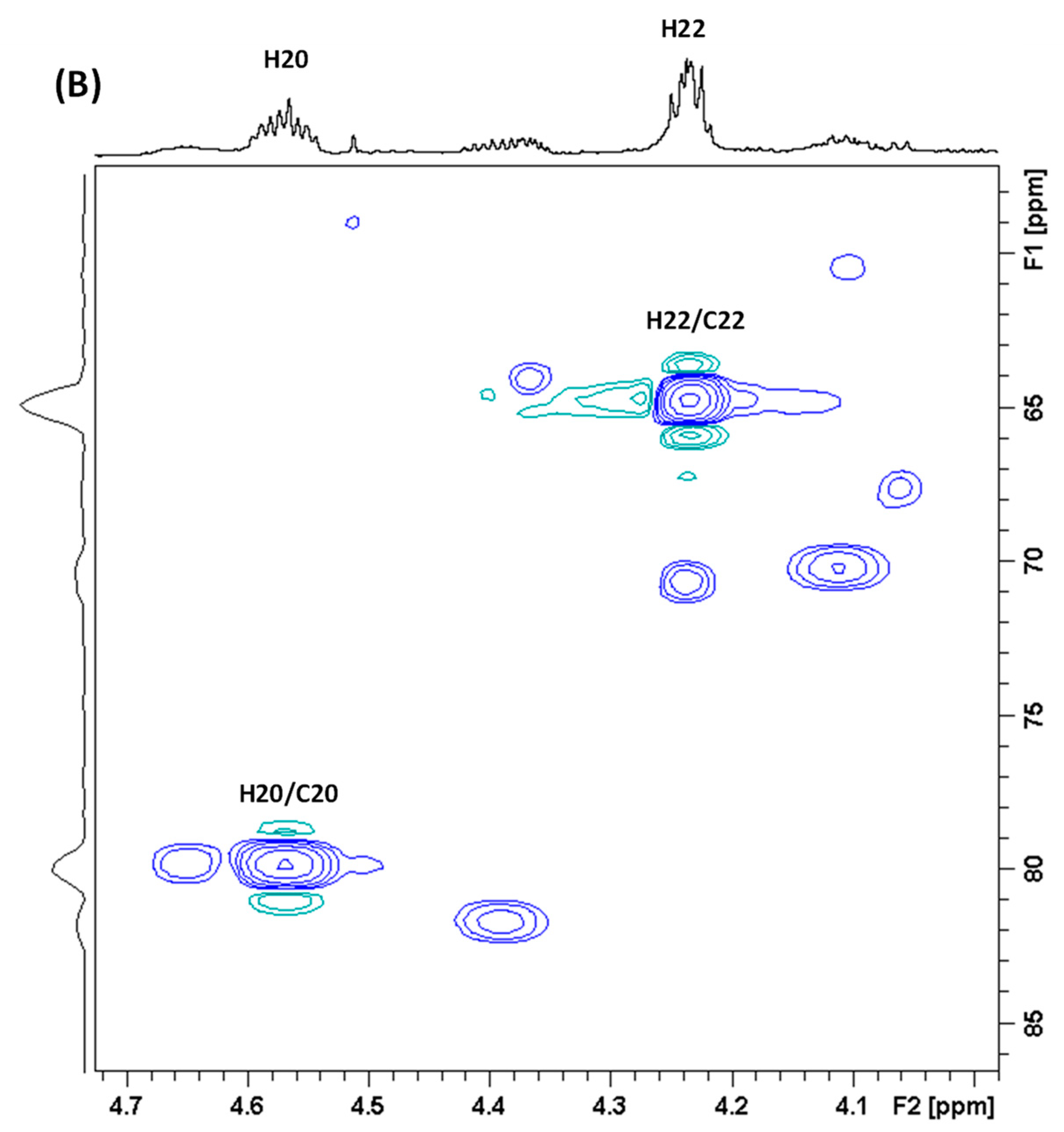
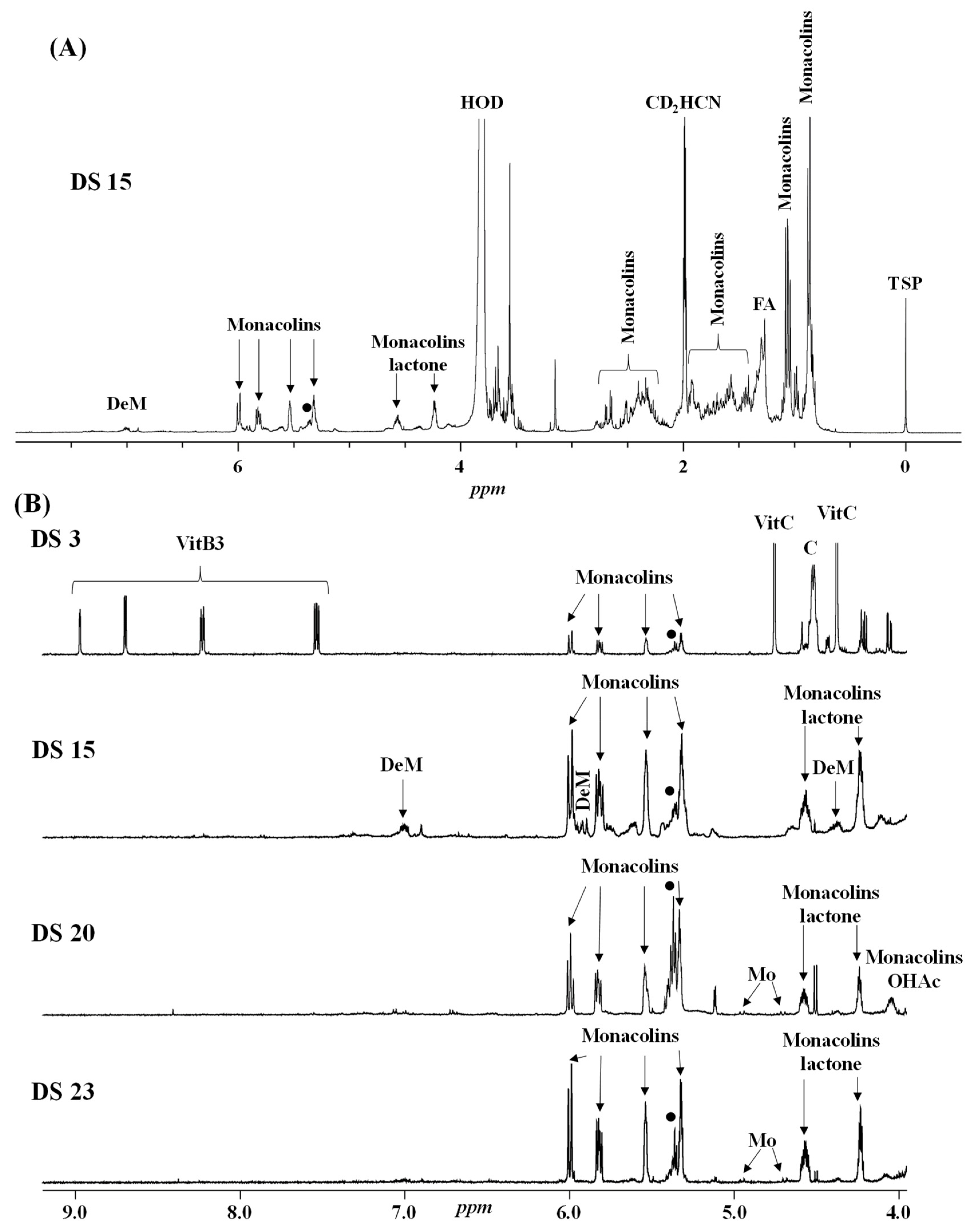
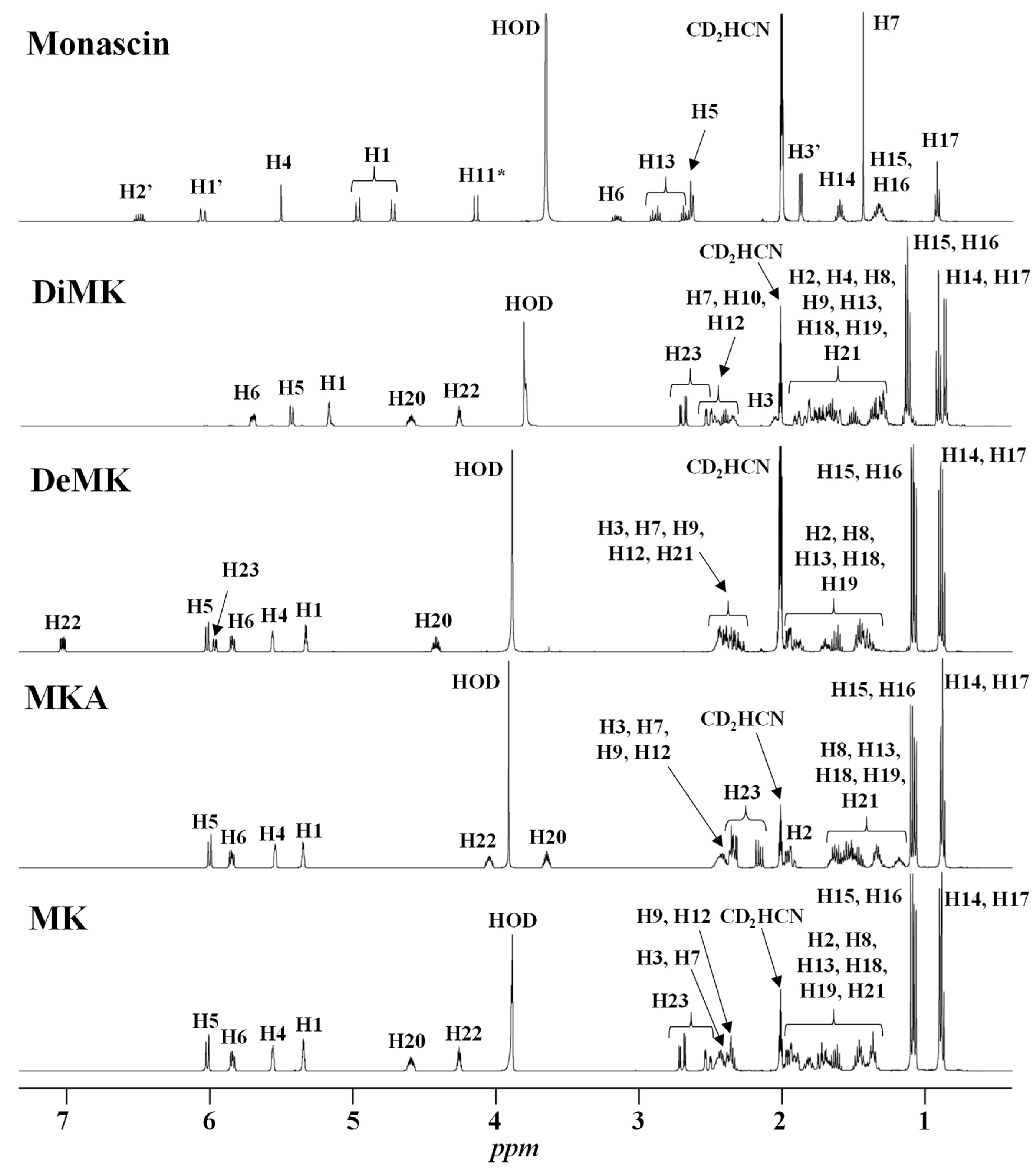
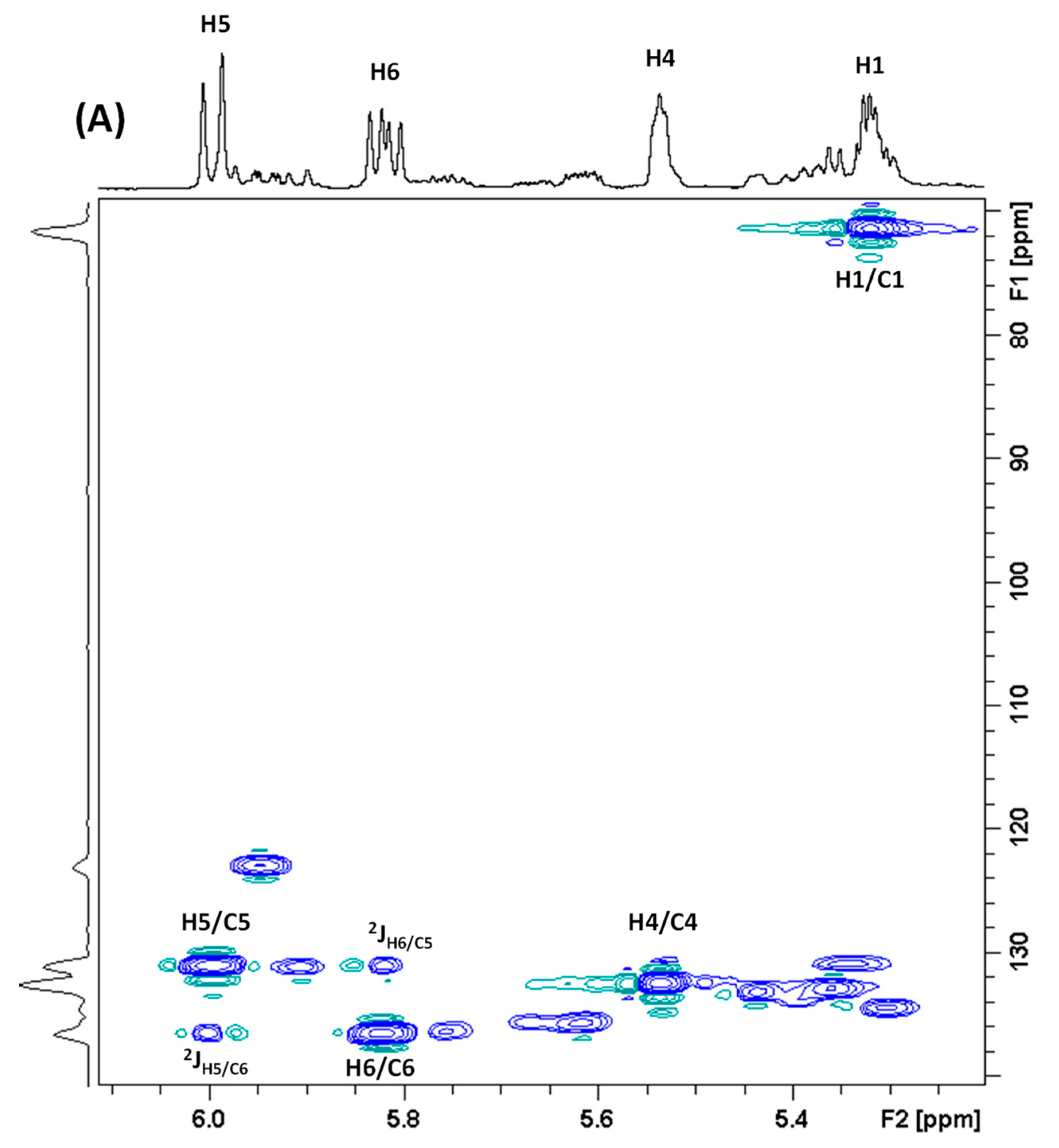
2.1. 1H-NMR Analysis
2.1.1. Qualitative 1H-NMR Analysis

| Compound | Structure | 1H-NMR 1 |
|---|---|---|
| δ (ppm) (Multiplicity 2, J (Hz), Number of Protons, Attribution) | ||
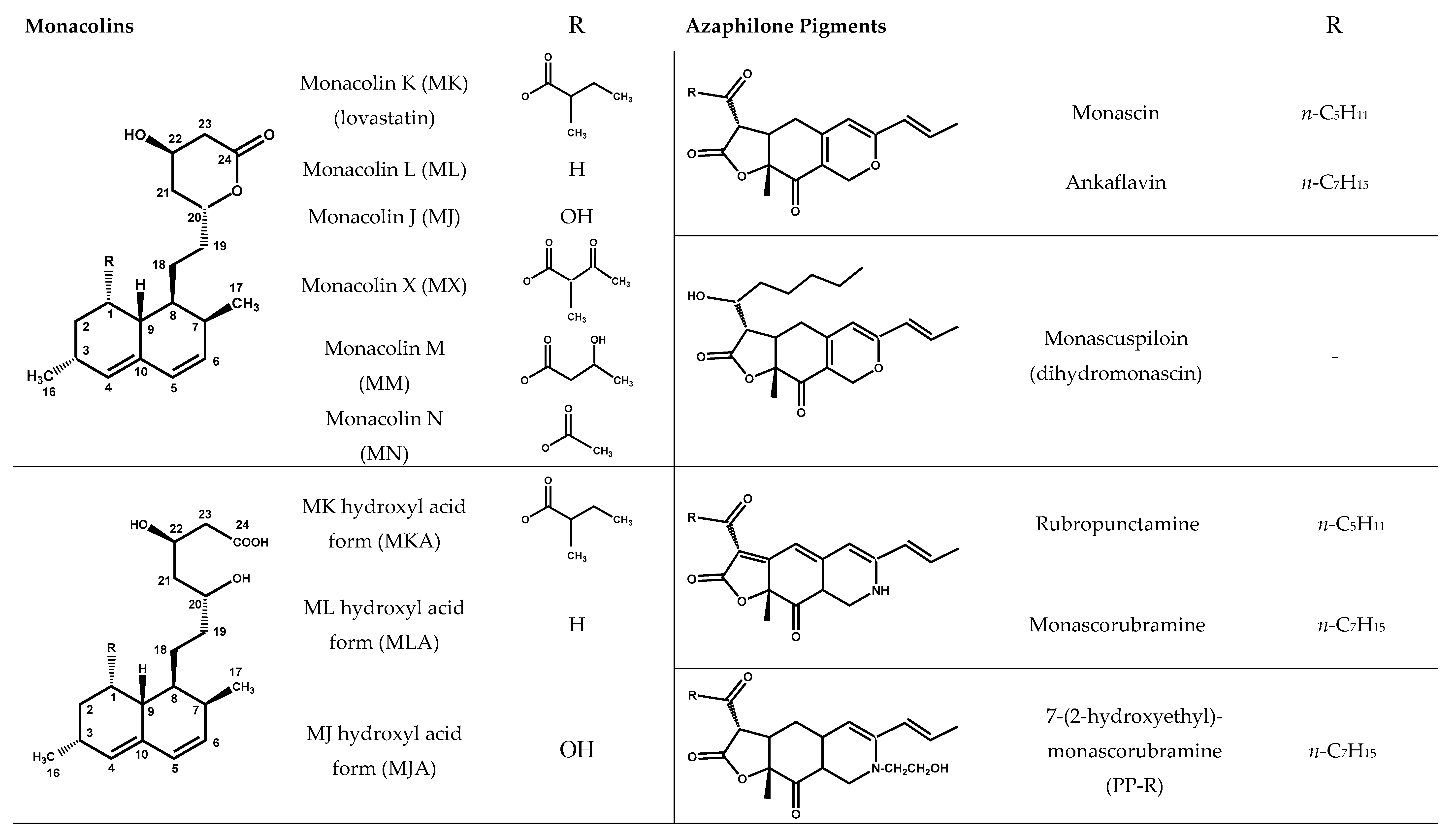
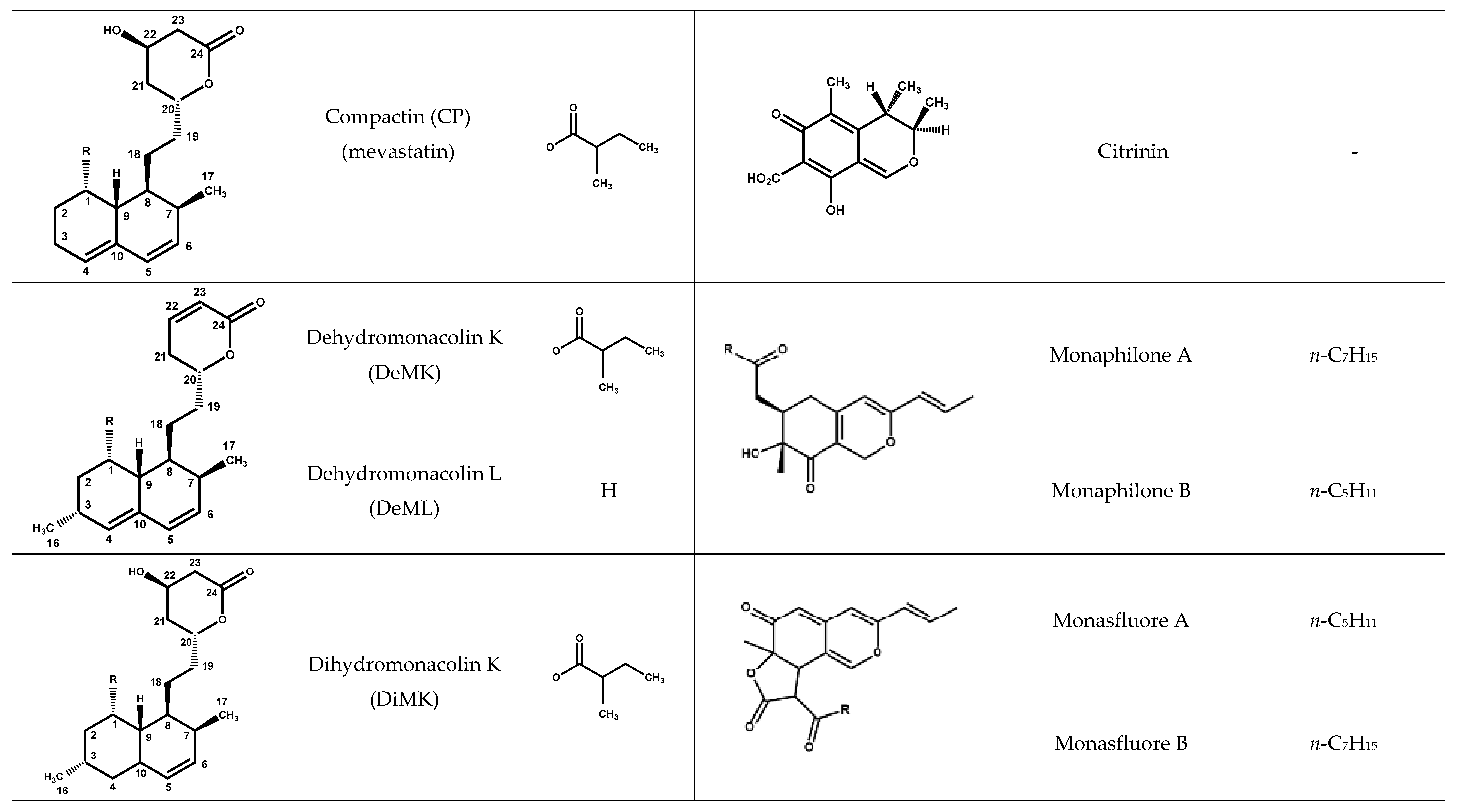
| Monacolin K lactone form (MK) | 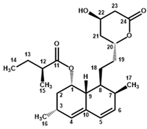 | 6.01 (d, J = 9.6, 1H, H-5), 5.84 (dd, J = 6.1, 9.6, 1H, H-6), 5.56 (app t, J = 2.8, 1H, H-4), 5.35 (q, J = 3.2, 1H, H-1), 4.59 (m, 1H, H-20), 4.25 (app quint, J = 3.9, 1H, H-22), 2.69 (Ad, J = 4.9, 17.6, 1H, H-23), 2.51 (Bdd, J = 1.7, 3.8, 17.6, 1H, H-23), 2.45 (m, 1H, H-3), 2.42 (m, 1H, H-7), 2.37 (m, 1H, H-9), 2.35 (m, 1H, H-12), 1.96 (m, 2H, H-2), 1.90 and 1.71 (two m, 2H, H-21), 1.81 and 1.37 (two m, 2H, H-19), 1.69 (m, 1H, H-8), 1.62 (app qd, J = 7.4, 13.6, 1H, H-13), 1.46 (m, 2H, H-13 and H-18), 1.36 (m, 1H, H-18), 1.08 (d, J = 6.9, 3H, H-15), 1.06 (d, J = 7.4, 3H, H-16), 0.89 (d, J = 6.9, 3H, H-17), 0.88 (t, J = 7.5, 3H, H-14) |
| Monacolin K hydroxyl acid form (MKA) | 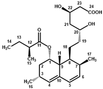 | 5.99 (d, J = 9.6, 1H, H-5), 5.83 (dd, J = 6.1, 9.6, 1H, H-6), 5.53 (app t, J = 2.8, 1H, H-4), 5.33 (q, J = 3.2, 1H, H-1), 4.05 (m, 1H, H-22), 3.63 (app hept, J = 4.1, 1H, H-20), 2.42 (m, 1H, H-3), 2.39 (m, 1H, H-7), 2.35 (m, 1H, H-9), 2.33 (m, 2H, H-12 and H-23), 2.16 (dd, J = 8.7, 15.2, 1H, H-23), 1.93 (m, 2H, H-2), 1.63 (m, 1H, H-8), 1.59 and 1.45 (two m, 2H, H-13), 1.57 and 1.51 (two m, 2H, H-21), 1.53 and 1.15 (two m, 2H, H-19), 1.32 (m, 2H, H-18), 1.08 (d, J = 6.9, 3H, H-15), 1.05 (d, J = 7.4, 3H, H-16), 0.87 (d, J = 6.9, 3H, H-17), 0.86 (t, J = 7.4, 3H, H-14) |
| Compactin (CP) = Mevastatin | 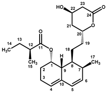 | 6.00 (d, J = 9.7, 1H, H-5), 5.79 (dd, J = 6.0, 9.7, 1H, H-6), 5.57 (m, 1H, H-4), 5.30 (m, 1H, H-1), 4.60 (m, 1H, H-20), 4.25 (app quint, J = 3.9, 1H, H-22), 2.69 (Ad, J = 4.8, 17.6, 1H, H-23), 2.51 (Bdd, J = 1.7, 3.6, 17.6, 1H, H-23), 2.42 (m, 2H, H-7 and H-9), 2.38 (m, 1H, H-12), 2.15 (m, 2H, H-3), 2.08 (m, 1H, H-2), 1.90 (m, 1H, H-21), 1.81 and 1.37 (two m, 2H, H-19), 1.72 (m, 2H, H-2 and H-21), 1.68 (m, 1H, H-8), 1.62 (app qd, J = 7.6, 13.6, 1H, H-13), 1.48 and 1.39 (two m, 2H, H-18), 1.46 (m, 1H, H-13), 1.11 (d, J = 7.0, 3H, H-15), 0.90 (d, J = 7.0, 3H, H-17), 0.89 (t, J = 7.5, 3H, H-14) |
| Dehydromonacolin K (DeMK) |  | 7.03 (ddd, J = 2.5, 6.0, 9.8, 1H, H-22), 6.02 (d, J = 9.6, 1H, H-5), 5.97 (ddd, J = 1.0, 2.7, 9.8, 1H, H-23), 5.84 (dd, J = 6.1, 9.6, 1H, H-6), 5.56 (app t, J = 2.8, 1H, H-4), 5.33 (q, J = 3.2, 1H, H-1), 4.42 (m, 1H, H-20), 2.44 (m, 1H, H-3), 2.43 (m, 1H, H-7), 2.42 and 2.30 (two m, 2H, H-21), 2.36 (m, 1H, H-9), 2.35 (m, 1H, H-12), 1.96 (m, 2H, H-2), 1.90 and 1.41 (two m, 2H, H-19), 1.70 (m, 1H, H-8), 1.62 (app qd, J = 7.5, 13.8, 1H, H-13), 1.46 (m, 1H, H-13), 1.45 and 1.39 (two m, 2H, H-18), 1.09 (d, J = 6.9, 3H, H-15), 1.07 (d, J = 7.4, 3H, H-16), 0.89 (d, J = 6.9, 3H, H-17), 0.88 (t, J = 7.5, 3H, H-14) |
| Dihydromonacolin K (DiMK) | 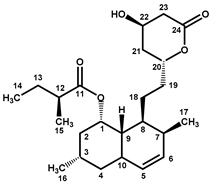 | 5.69 (ddd, J = 2.7, 5.0, 9.8, 1H, H-6), 5.42 (d, J = 9.8, 1H, H-5), 5.15 (q, J = 2.7, 1H, H-1), 4.58 (m, 1H, H-20), 4.25 (app quint, J = 3.8, 1H, H-22), 2.68 (Ad, J = 4.9, 17.7, 1H, H-23), 2.50 (Bdd, J = 1.7, 3.7, 17.7, 1H, H-23), 2.46 (m, 1H, H-10), 2.39 (m, 1H, H-12), 2.34 (m, 1H, H-7), 2.05 (m, 1H, H-3), 1.88 and 1.72 (two m, 2H, H-21), 1.81 and 1.37 (two m, 2H, H-19), 1.80 (m, 2H, H-2), 1.66 (m, 1H, H-8), 1.64 (m, 1H, H-13), 1.61 and 1.35 (two m, 2H, H-4), 1.49 (app qd, J = 7.4, 13.6, 1H, H-13), 1.35 and 1.30 (two m, 2H, H-18), 1.12 (d, J = 7.0, 3H, H-15), 1.11 (d, J = 7.6, 3H, H-16), 0.90 (t, J = 7.5, 3H, H-14), 0.86 (d, J = 7.0, 3H, H-17) |
| Monascin 3 | 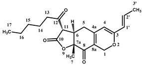 | 6.50 (qd, J = 7.0, 15.5, 1H, H-2′), 6.05 (qd, J = 1.7, 15.5, 1H, H-1′), 5.51 (s, 1H, H-4), 4.97 (At, J = 0.9, 12.7, 1H, H-1), 4.72 (Bt, J = 1.4, 12.7, 1H, H-1), 4.14 (d, J = 13.3, 1H, H-11), 3.15 (m, 1H, H-6), 2.88 (At, J = 7.3, 18.2, 1H, H-13), 2.67 (Bt, J = 7.3, 18.2, 1H, H-13), 2.63 (app br d, J = 7.7, 2H, H-5), 1.87 (dd, J = 1.7, 7.0, 3H, H-3′), 1.59 (quint, J = 7.3, 2H, H-14), 1.43 (s, 3H, H-7), 1.32 (m, 4H, H-15, H-16), 0.91 (t, J = 7.2, 3H, H-17) |
| Citrinin 4 | 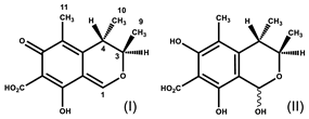 | 8.45 (br s, H-1 (I)), 5.94 (s, H-1 (II major), 5.90 (br s, H-1 (II minor)), 4.92 (br signal, H-3 (I)), 4.09 (quint, J = 6.6, H-3 (II major and minor)), 3.15 (br signal, H-4 (I)), 2.76 (br signal, H-4 (II minor)), 2.67 (quint, J = 6.8, H-4 (II major)), 2.03 (s, H-11 *), 2.01 (s, H-11 *), 2.00 (s, H-11 *), 1.30 (d, J = 6.7, H-9 *), 1.27 (d, J = 6.4, H-9 *), 1.20 (d, J = 6.9, H-10 *), 1.17 (very br signal, H-10 *) |
| Piperine | 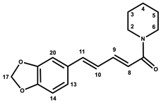 | 7.28 (ddd, J = 2.7, 7.3, 14.7, 1H, H-9), 7.11 (d, J = 1.6, 1H, H-20), 6.99 (dd, J = 1.7, 8.1, 1H, H-13), 6.87 (d, J = 8.0, 1H, H-14), 6.83 (m, 2H, H-10, H-11), 6.60 (d, J = 14.7, 1H, H-8), 6.00 (s, 2H, H-17), 3.55 (m, 4H, H-2, H-6), 1.65 (m, 2H, H-4), 1.56 (m, 4H, H-3, H-5) |
| Carnitine | 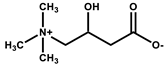 | 4.56 (br q, J = 7.1, 1H, CH-OH), 3.39 (m, 2H, CH2-N+), 3.17 (s, 9H, (CH3)3-N+), 2.58 (m, 2H, CH2-COO−) |
| Chlorogenic acid | 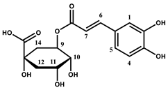 | 7.61 (d, J = 15.9, 1H, H-6), 7.16 (d, J = 2.0, 1H, H-1), 7.06 (dd, J = 2.0, 8.2, 1H, H-5), 6.88 (d, J = 8.2, 1H, H-4), 6.34 (d, J = 15.9, 1H, H-7), 5.28 (ddd, J = 4.6, 9.4, 10.5, 1H, H-9), 4.18 (app q, J = 3.5, 1H, H-10), 3.75 (dd, J = 3.2, 9.4, 1H, H-11), 2.26–1.97 (m, 4H, H-12, H-14) |
| Saturated fatty acids | 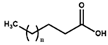 | 2.20 (t, J = 7.5, 2H, CH2-COOH), 1.59 (m, 2H, CH2-CH2-COOH), 1.30 (m, 2nH, (CH2)n), 0.90 (t, J = 6.3, 3H, CH3) |
| α/β Glucose | 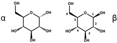 | 5.14 (d, J = 3.7, 1H, H-1α), 4.52 (d, J = 7.9, 1H, H-1β), 3.85-3.60 (m, 7H, H-3α, H-4α/β, H-6α/β), 3.45–3.28 (m, 4H, H-2α, H-3β, H-5α/β), 3.15 (t, J = 8.5, 1H, H-2β) |
| Glycerol |  | 3.66 (tt, J = 4.6, 6.3, 1H, CH), 3.55 (Ad, J = 4.6, 11.5, 2H, CH2), 3.48 (Bd, J = 6.2, 11.5, CH2) |
| Isopropyl alcohol |  | 3.93 (hept, J = 6.1, 1H, CH), 1.14 (d, J = 6.1, 6H, CH3) |
| Linoleic acid 5 | 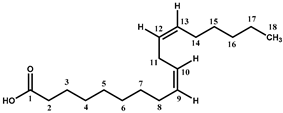 | 5.36 (m, 4H, H-9, H-10, H-12, H-13), 2.78 (t, J = 6.7, 2H, H-11), 2.27 (t, J = 7.5, 2H, H-2), 2.06 (m, 4H, H-8, H-14), 1.56 (m, 2H, H-3), 1.40-1.27 (m, 14H, H-4, H-5, H-6, H-7, H-15, H-16, H-17), 0.89 (t, J = 6.8, 3H, H-18) |
| Sorbitol | 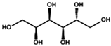 | 3.82–3.54 (m, 8H) |
| Vitamin B3 (niacinamide form) | 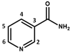 | 8.99 (d, J = 2.3, 1H, H-2), 8.72 (dd, J = 4.9, 1.6, 1H, H-6), 8.25 (td, J = 8.0, 1.9, 1H, H-4), 7.56 (dd, J = 8.0, 4.9, 1H, H-5) |
| Vitamin C | 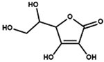 | 4.76 (d, X part of an ABMX system, JXM = 2.0, 1H, CH-O-), 3.94 (td, M part of an ABMX system, JMX = 1.9, JMA = JMB = 6.6, 1H, CH-OH), 3.66 (AB part of an ABMX system, JAB = 11.3, JAM = JBM = 6.6, 2H, CH2-OH) |
2.1.2. Quantitative 1H-NMR Analysis
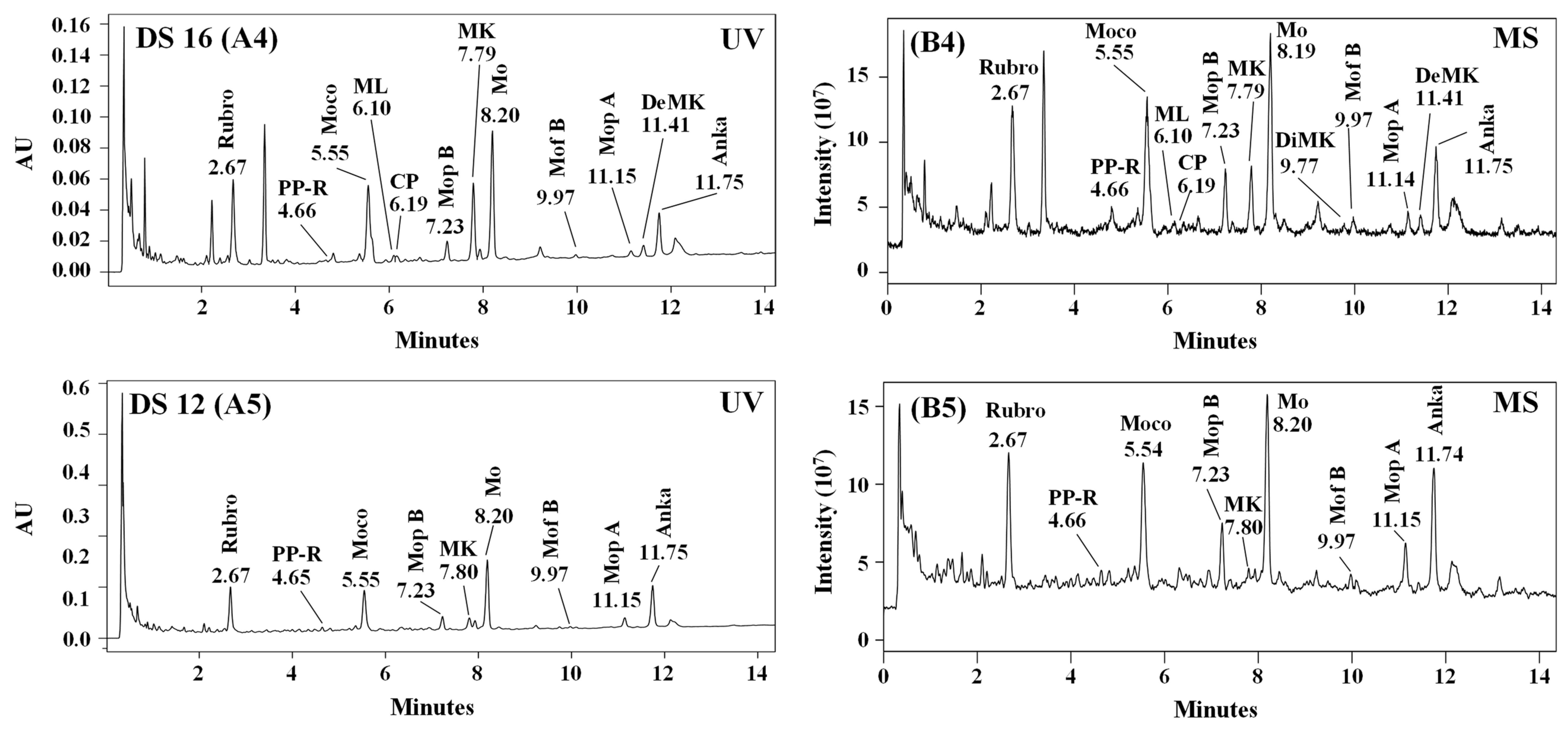
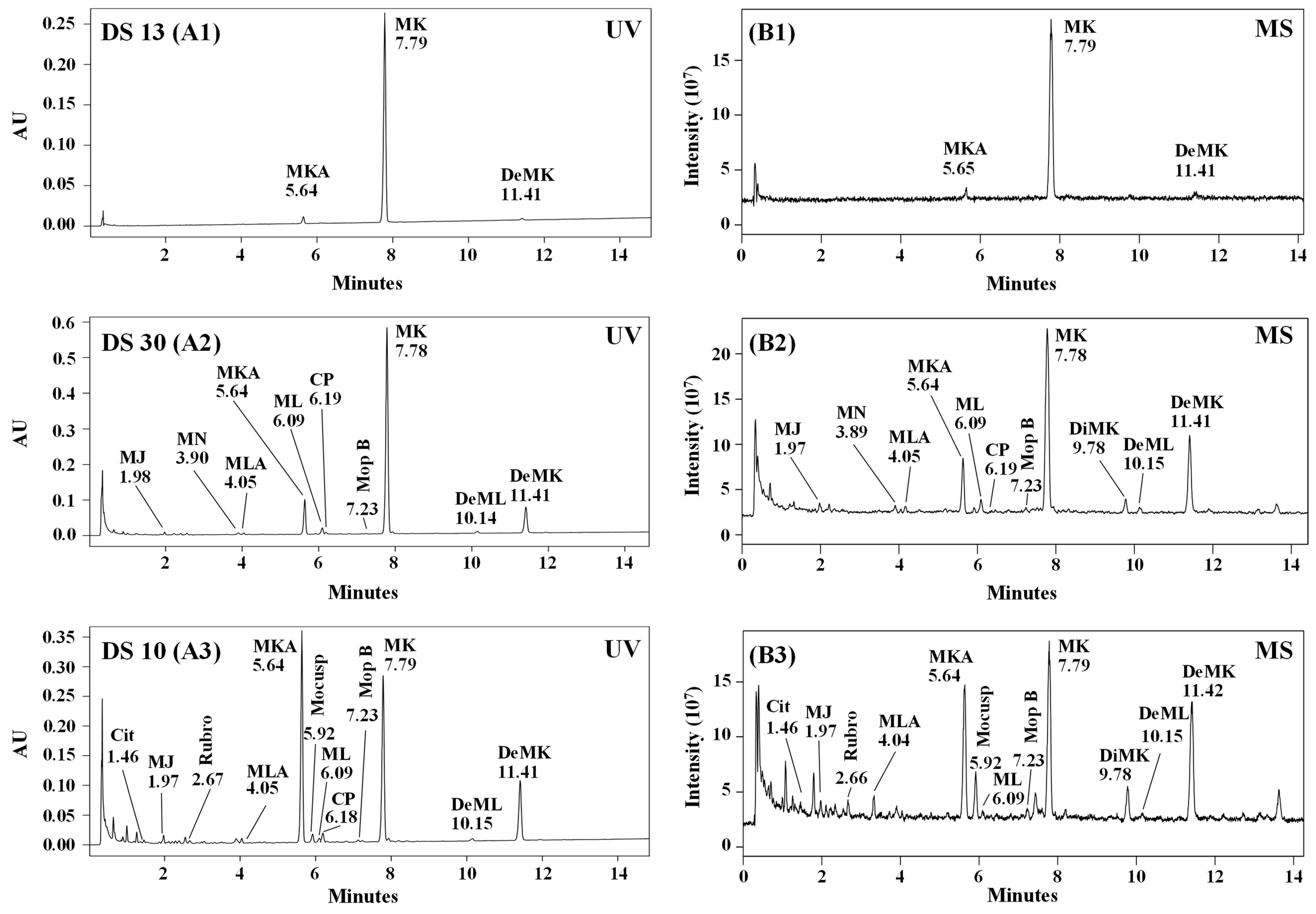
2.2. Chromatographic Analysis
2.2.1. Identification of Monacolins and Pigments
2.2.2. Quantitative Analysis of Monacolins
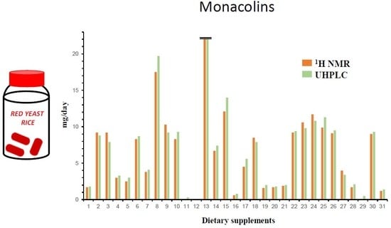
2.3. Comparison of 1H-NMR and UHPLC Results
2.4. Quality Control Issues
2.4.1. About Monacolin Labelling of RYR DS
2.4.2. About the Variability of Monacolins Consumed Per Day in RYR DS
3. Materials and Methods
3.1. Red Yeast Rice Dietary Supplements
3.2. Chemicals and Reagents
3.3. Choice of Extraction Solvent and Preparation of Samples for Analyses
3.4. 1H-NMR Analysis
3.4.1. Recording Conditions
3.4.2. Quantification
3.5. UHPLC-DAD-MS Analysis
3.5.1. Instrumentation
3.5.2. Validation Procedure
3.6. High-Resolution Mass Spectrometry (HR-MS) Experiments
3.7. Statistical Analysis
4. Conclusions
Supplementary Materials
Author Contributions
Funding
Acknowledgments
Conflicts of Interest
References
- Ma, J.; Li, Y.; Ye, Q.; Li, J.; Hua, Y.; Ju, D.; Zhang, D.; Cooper, R.; Chang, M. Constituents of red yeast rice, a traditional Chinese food and medicine. J. Agric. Food Chem. 2000, 48, 5220–5225. [Google Scholar] [CrossRef]
- Wang, T.-H.; Lin, T.-F. Monascus rice products. Adv. Food Nutr. Res. 2007, 53, 123–159. [Google Scholar]
- Li, Y.; Jiang, L.; Jia, Z.; Xin, W.; Yang, S.; Yang, Q.; Wang, L. A meta-analysis of red yeast rice: An effective and relatively safe alternative approach for dyslipidemia. PLoS ONE 2014, 9, e98611. [Google Scholar] [CrossRef]
- Gerards, M.C.; Terlou, R.J.; Yu, H.; Koks, C.H.W.; Gerdes, V.E.A. Traditional Chinese lipid-lowering agent red yeast rice results in significant LDL reduction but safety is uncertain—A systematic review and meta-analysis. Atherosclerosis 2015, 240, 415–423. [Google Scholar] [CrossRef] [PubMed] [Green Version]
- Chen, C.-H.; Yang, J.-C.; Uang, Y.-S.; Lin, C.-J. Improved dissolution rate and oral bioavailability of lovastatin in red yeast rice products. Int. J. Pharm. 2013, 444, 18–24. [Google Scholar] [CrossRef] [PubMed]
- ANSES, OPINION of the French Agency for Food, Environmental and Occupational Health & Safety on the Risks Associated with the Presence of “Red Yeast Rice” in Food Supplements. 2014. Available online: https://www.anses.fr/fr/system/files/NUT2012sa0228EN.pdf (accessed on 9 September 2019).
- Mazzanti, G.; Moro, P.A.; Raschi, E.; Da Cas, R.; Menniti-Ippolito, F. Adverse reactions to dietary supplements containing red yeast rice: Assessment of cases from the Italian surveillance system. Br. J. Clin. Pharmacol. 2017, 83, 894–908. [Google Scholar] [CrossRef] [PubMed]
- EFSA Panel on Food Additives and Nutrient Sources added to Food (ANS). Scientific opinion on the safety of monacolins in red yeast rice. EFSA J. 2018, 16, 5368. [Google Scholar] [CrossRef] [Green Version]
- Red Yeast Rice—An Update Overview of the Reported ADRs. Netherlands Pharmacovigilance Centre Lareb. Available online: https://www.lareb.nl/media/3204/signals_2019_rode-gistrijst_nvwa-update.pdf (accessed on 9 January 2020).
- Li, Y.-G.; Zhang, F.; Wang, Z.-T.; Hu, Z.-B. Identification and chemical profiling of monacolins in red yeast rice using high-performance liquid chromatography with photodiode array detector and mass spectrometry. J. Pharm. Biomed. Anal. 2004, 35, 1101–1112. [Google Scholar] [CrossRef]
- Wu, C.-L.; Kuo, Y.-H.; Lee, C.-L.; Hsu, Y.-W.; Pan, T.-M. Synchronous high-performance liquid chromatography with a photodiode array detector and mass spectrometry for the determination of citrinin, monascin, ankaflavin, and the lactone and acid forms of monacolin K in red mold rice. J. AOAC Int. 2011, 94, 179–190. [Google Scholar]
- Mornar, A.; Sertic, M.; Nigovic, B. Development of a rapid LC/DAD/FLD/MSn method for the simultaneous determination of monacolins and citrinin in red fermented rice products. J. Agric. Food Chem. 2013, 61, 1072–1080. [Google Scholar] [CrossRef]
- Avula, B.; Cohen, P.A.; Wang, Y.H.; Sagi, S.; Feng, W.; Wang, M.; Zweigenbaum, J.; Shuangcheng, M.; Khan, I.A. Chemical profiling and quantification of monacolins and citrinin in red yeast rice commercial raw materials and dietary supplements using liquid chromatography-accurate QToF mass spectrometry: Chemometrics application. J. Pharm. Biomed. Anal. 2014, 100, 243–253. [Google Scholar] [CrossRef] [PubMed]
- Song, F.; El-Demerdash, A.; Lee, S.-J.S.H.; Smith, R.E. Fast screening of lovastatin in red yeast rice products by flow injection tandem mass spectrometry. J. Pharm. Biomed. Anal. 2012, 57, 76–81. [Google Scholar] [CrossRef] [PubMed]
- Svoboda, P.; Sander, D.; Plachká, K.; Nováková, L. Development of matrix effect-free MISPE-UHPLC-MS/MS method for determination of lovastatin in Pu-erh tea, oyster mushroom, and red yeast rice. J. Pharm. Biomed. Anal. 2017, 140, 367–376. [Google Scholar] [CrossRef] [PubMed]
- Malet-Martino, M.; Holzgrabe, U. NMR techniques in biomedical and pharmaceutical analysis. J. Pharm. Biomed. Anal. 2011, 55, 1–15. [Google Scholar] [CrossRef] [PubMed]
- Simmler, C.; Napolitano, J.G.; McAlpine, J.B.; Chen, S.-N.; Pauli, G.F. Universal quantitative NMR analysis of complex natural samples. Curr. Opin. Biotechnol. 2014, 25, 51–59. [Google Scholar] [CrossRef]
- Lachenmeier, D.W.; Monakhova, Y.B.; Kuballa, T.; Löbell-Behrends, S.; Maixner, S.; Kohl-Himmelseher, M.; Waldner, A.; Steffen, C. NMR evaluation of total statin content and HMGCoA reductase inhibition in red yeast rice (Monascus spp.) food supplements. Chin. Med. 2012, 7, 8. [Google Scholar] [CrossRef] [Green Version]
- Zhu, L.; Yau, L.-F.; Lu, J.-G.; Zhu, G.-Y.; Wang, J.-R.; Han, Q.-B.; Hsiao, W.-L.; Jiang, Z.-H. Cytotoxic dehydromonacolins from red yeast rice. J. Agric. Food Chem. 2012, 60, 934–939. [Google Scholar] [CrossRef]
- Albers-Schönberg, G.; Joshua, H.; Lopez, M.B.; Hensens, O.D.; Springer, J.P.; Chen, J.; Ostrove, S.; Hoffman, C.H.; Alberts, A.W.; Patchett, A.A. Dihydromevinolin, a potent hypocholesterolemic metabolite produced by Aspergillus terreus. J. Antibiot. 1981, 34, 507–512. [Google Scholar] [CrossRef]
- Haruyama, H.; Kuwano, H.; Kinoshita, T.; Terahara, A.; Nishigaki, T.; Tamura, C. Structure elucidation of the bioactive metabolites of ML-236B (mevastatin) isolated from dog urine. Chem. Pharm. Bull. (Tokyo) 1986, 34, 1459–1467. [Google Scholar] [CrossRef] [Green Version]
- Barber, J.; Cornford, J.L.; Howard, T.D.; Sharplese, D. The structure of citrinin in vivo. J. Chem. Soc. Perkin Trans. 1987, 1, 2743–2744. [Google Scholar] [CrossRef]
- Lankhorst, P.P.; Poot, M.M.; De Lange, M.P.A. Quantitative determination of lovastatin and dihydrolovastatin by means of 1H-NMR spectroscopy. Pharmacopeial. Forum 1996, 22, 2414–2422. [Google Scholar]
- Simpson, J. Lovastatin in chloroform-D. In NMR Case Studies. Data Analysis of Complicated Molecules, 1st ed.; Elsevier: Cambridge, MA, USA, 2017; pp. 94–104. [Google Scholar]
- Su, N.-W.; Lin, Y.-L.; Lee, M.-H.; Ho, C.-Y. Ankaflavin from Monascus-fermented red rice exhibits selective cytotoxic effect and induces cell death on Hep G2 cells. J. Agric. Food Chem. 2005, 53, 1949–1954. [Google Scholar] [CrossRef] [PubMed]
- Li, M.-N.; Li, C.-R.; Gao, W.; Li, P.; Yang, H. Highly sensitive strategy for identification of trace chemicals in complex matrix: Application to analysis of monacolin analogues in monascus-fermented rice product. Anal. Chim. Acta 2017, 982, 156–167. [Google Scholar] [CrossRef]
- Endo, A. Monacolin K, a new hypocholesterolemic agent produced by a Monascus species. J. Antibiot. 1979, 32, 852–854. [Google Scholar] [CrossRef] [Green Version]
- Endo, A.; Hasumi, K.; Negishi, S. Monacolins J and L, new inhibitors of cholesterol biosynthesis produced by Monascus ruber. J. Antibiot. 1985, 38, 420–422. [Google Scholar] [CrossRef]
- Mapari, S.A.; Hansen, M.E.; Meyer, A.S.; Thrane, U. Computerized screening for novel producers of Monascus-like food pigments in Penicillium species. J. Agric. Food Chem. 2008, 56, 9981–9989. [Google Scholar] [CrossRef] [PubMed]
- Zheng, Y.; Xin, Y.; Guo, Y. Study on the fingerprint profile of Monascus products with HPLC–FD, PAD and MS. Food Chem. 2009, 113, 705–711. [Google Scholar] [CrossRef]
- Hsu, Y.-W.; Hsu, L.-C.; Liang, Y.-H.; Kuo, Y.-H.; Pan, T.-M. Monaphilones A-C, three new antiproliferative azaphilone derivatives from Monascus purpureus NTU 568. J. Agric. Food Chem. 2010, 58, 8211–8216. [Google Scholar] [CrossRef]
- Chiu, H.-W.; Chen, M.-H.; Fang, W.-H.; Hung, C.-M.; Chen, Y.-L.; Wu, M.-D.; Yuan, G.-F.; Wu, M.-J.; Wang, Y.-J. Preventive effects of monascus on androgen-related diseases: Androgenetic alopecia, benign prostatic hyperplasia, and prostate cancer. J. Agric. Food Chem. 2013, 61, 4379–4386. [Google Scholar] [CrossRef]
- Ogihara, J.; Kato, J.; Oishi, K.; Fujimoto, Y. PP-R, 7-(2-hydroxyethyl)-monascorubramine, a red pigment produced in the mycelia of Penicillium sp. AZ. J. Biosci. Bioeng. 2001, 91, 44–47. [Google Scholar] [CrossRef]
- Mapari, S.A.S.; Meyer, A.S.; Thrane, U. Colorimetric characterization for comparative analysis of fungal pigments and natural food colorants. J. Agric. Food Chem. 2006, 54, 7027–7035. [Google Scholar] [CrossRef] [PubMed]
- Woo, P.C.Y.; Lam, C.-W.; Tam, E.W.T.; Lee, K.-C.; Yung, K.K.Y.; Leung, C.K.F.; Sze, K.-H.; Lau, S.K.P.; Yuen, K.-Y. The biosynthetic pathway for a thousand-year-old natural food colorant and citrinin in Penicillium marneffei. Sci. Rep. 2014, 4, 6728. [Google Scholar] [CrossRef] [PubMed]
- Bijinu, B.; Suh, J.-W.; Park, S.-H.; Kwon, H.-J. Delineating Monascus azaphilone pigment biosynthesis: Oxidoreductive modifications determine the ring cyclization pattern in azaphilone biosynthesis. RSC Adv. 2014, 4, 59405–59408. [Google Scholar] [CrossRef]
- Li, X.-M.; Shen, X.-H.; Duan, Z.-W.; Guo, S.-R. A new monacolin analogue from Xuezhikang capsule. Acta Pharm. Sin. 2011, 46, 564–567. [Google Scholar]
- EFSA Panel on Dietetic Products, Nutrition and Allergies (NDA). Scientific Opinion on the substantiation of health claims related to monacolin K from red yeast rice and maintenance of normal blood LDL-cholesterol concentrations (ID 1648, 1700) pursuant to Article 13(1) of Regulation (EC) No 1924/2006. EFSA J. 2011, 9, 2304. [Google Scholar] [CrossRef] [Green Version]
- Gordon, R.Y.; Cooperman, T.; Obermeyer, W.; Becker, D.J. Marked variability of monacolin levels in commercial red yeast rice products: Buyer beware! Arch. Intern. Med. 2010, 170, 1722–1727. [Google Scholar] [CrossRef] [PubMed] [Green Version]
- Heber, D.; Yip, I.; Ashley, J.M.; Elashoff, D.A.; Elashoff, R.M.; Go, V.L. Cholesterol-lowering effects of a proprietary Chinese red-yeast-rice dietary supplement. Am. J. Clin. Nutr. 1999, 69, 231–236. [Google Scholar] [CrossRef]
- Becker, D.J.; Gordon, R.Y.; Morris, P.B.; Yorko, J.; Gordon, Y.J.; Li, M.; Iqbal, N. Simvastatin vs therapeutic lifestyle changes and supplements: Randomized primary prevention trial. Mayo Clin. Proc. 2008, 86, 758–764. [Google Scholar] [CrossRef] [Green Version]
- Becker, D.J.; Gordon, R.Y.; Halbert, S.C.; French, B.; Morris, P.B.; Rader, D.J. Red yeast rice for dyslipidemia in statin-intolerant patients: A randomized trial. Ann. Intern. Med. 2009, 150, 830–839. [Google Scholar] [CrossRef]
- Maesschalck, J.; Gruyters, A.M.; Corthout, J. Levure de riz rouge. Que choisir et quel conseil donner? J. Pharm. Belg. 2018, 4, 6–17. [Google Scholar]
- Endo, A. Compactin (ML-236B) and related compounds as potential cholesterol-lowering agents that inhibit HMG-CoA reductase. J. Med. Chem. 1985, 28, 401–405. [Google Scholar] [CrossRef] [PubMed]
- Phainuphong, P.; Rukachaisirikul, V.; Saithong, S.; Phongpaichit, S.; Bowornwiriyapan, K.; Muanprasat, C.; Srimaroeng, C.; Duangjai, A.; Sakayaroj, J. Lovastatin analogues from the soil-derived fungus Aspergillus sclerotiorum PSU-RSPG178. J. Nat. Prod. 2016, 79, 1500–1507. [Google Scholar] [CrossRef] [PubMed]
- Heinz, T.; Schuchardt, J.P.; Möller, K.; Hadji, P.; Hahn, A. Low daily dose of 3 mg monacolin K from RYR reduces the concentration of LDL-C in a randomized, placebo-controlled intervention. Nutr. Res. 2016, 36, 1162–1170. [Google Scholar] [CrossRef] [PubMed] [Green Version]
- Li, Y.-G.; Liu, H.; Wang, Z.-T. A validated stability-indicating HPLC with photodiode array detector (PDA) method for the stress tests of Monascus purpureus-fermented rice, red yeast rice. J. Pharm. Biomed. Anal. 2005, 39, 82–90. [Google Scholar] [CrossRef] [PubMed]
- Theunis, M.; Naessens, T.; Verhoeven, V.; Hermans, N.; Apers, S. Development and validation of a robust high-performance liquid chromatographic method for the analysis of monacolins in red yeast rice. Food Chem. 2017, 234, 33–37. [Google Scholar] [CrossRef]
Sample Availability: Samples of the formulations are available from the authors. |


| Number | Formulation Name (Origin 1) | Batch Number Expiration Date | Form | RYR Extract Content Per Capsule or Tablet on the Label (mg) | |
|---|---|---|---|---|---|
| 1 | Arkopharma (HFS) | C02194A | 04/2016 | Capsule | 175 |
| 2 | Arterin (I) | 1983-1 | 07/2016 | Tablet | 670 |
| 3 | B.concept (I) | 13DN8 | 09/2016 | Capsule | 186 |
| 4 | Belle & Bio (I) | B524A | 06/2015 | Capsule | 250 |
| 5 | Blue bonnet (I) | 31201702 | 01/2017 | Capsule | 600 |
| 6 | Boutique nature (I) | 32443A | 03/2016 | Capsule | 222.3 |
| 7 | Doctor’s best (I) | ML847 | 01/2017 | Tablet | 1200 |
| 8 | Ephyto (I) | B1204059CZ | 01/2015 | Tablet | 600 |
| 9 | Fushi (I) | EPAN110412 | 08/2015 | Capsule | 600 |
| 10 | Hanoju (I) | 20130612 | 06/2016 | Capsule | 450 |
| 11 | Health Ace (I) | 106012W9 | 03/2015 | Capsule | 650 |
| 12 | Health Spark (I) | 1060012/A1 | 10/2014 | Capsule | 650 |
| 13 | Liposterol (HFS) | 03023 | 05/2016 | Tablet | 600 |
| 14 | MRM (I) | 130736 | 08/2016 | Capsule | 600 |
| 15 | Nat et form (I) | 14387J | 10/2016 | Capsule | 600 |
| 16 | Natrol (I) | 2055200 | 10/2015 | Tablet | 400 |
| 17 | Nature algues (I) | G0112112602 PF01G | 11/2016 | Capsule | 250 |
| 18 | Nature’s plus (I) | 1249540 | 09/2016 | Tablet | 600 |
| 19 | Nature’s way (I) | 20012472 | 05/2016 | Capsule | 600 |
| 20 | Naturland (HFS) | C02196A | 04/2016 | Capsule | 175 |
| 21 | Now (I) | 16117750521 | 06/2015 | Capsule | 600 |
| 22 | Nutrisanté (I) | 961PAA | 05/2016 | Capsule | 222.2 |
| 23 | Pharma nature (HFS) | 1009.211 | 12/2014 | Capsule | 300 |
| 24 | Phytalessence (HFS) | 10134 | 10/2016 | Tablet | 600 |
| 25 | Phytoreponse (I) | B1210229CZ | 10/2015 | Tablet | 600 |
| 26 | Rizocol (I) | 9545 | 06/2016 | Tablet | 335 |
| 27 | Santé verte (HFS) | L066X2 | 03/2016 | Tablet | 600 |
| 28 | Solaray (I) | 171306 | 12/2016 | Capsule | 600 |
| 29 | Solgar (I) | 747369-02 | 07/2016 | Capsule | 600 |
| 30 | Tradition soleil levant (I) | 89853 | 07/2016 | Capsule | 208 |
| 31 | Vit’All + (HFS) | A7248-11 | 11/2013 | Tablet | 600 |
| DS Number | Signal at 5.84 ppm 2 H6 | Signal at 5.56 ppm 2 H4 | Mean Intensity of Signals H6 and H4 | Signal at 5.33 ppm 2 H1 | Signal at 4.60 ppm 2 H20 | Signal at 4.25 ppm 2 H22 | Compounds Identified Other than Monacolins 3,4 |
|---|---|---|---|---|---|---|---|
| 1 | 1.67 | 1.70 | 1.685 | 3.31 | 1.40 | 1.34 | SFA *, UFA, glycerol, glucose, monascin |
| 2 | 9.14 | 9.18 | 9.16 | 9.26 | 7.10 | SFA *, UFA, glycerol, glucose | |
| 3 | 3.07 | 3.07 | 3.07 | 2.39 | SFA *, UFA, carnitine *, vitamin B3 *, vitamin C * | ||
| 4 | 1.02 | 0.95 | 0.985 | 1.64 | 0.71 | SFA, UFA, glucose | |
| 5 | 2.47 | 2.53 | 2.50 | 11.3 | 2.15 | SFA *, UFA, glycerol, glucose, monascin | |
| 6 | 2.92 | 2.63 | 2.775 | 3.27 | 2.19 | SFA *, UFA, glucose, monascin | |
| 7 | 3.76 | 3.73 | 3.745 | SFA *, UFA, glucose, piperine * | |||
| 8 | 8.68 | 8.79 | 8.735 | 8.65 | 6.89 | 7.73 | SFA, UFA, glycerol, monascin |
| 9 | 5.14 | 5.21 | 5.125 | 8.03 | 4.55 | 4.55 | SFA *, UFA, glycerol, glucose, monascin |
| 10 | 2.10 | 2.04 | 2.07 | 8.67 | 1.22 | SFA, UFA, glycerol, glucose, monascin | |
| 11 | SFA, UFA, glycerol, glucose, monascin | ||||||
| 12 | SFA, UFA, glycerol, glucose, monascin | ||||||
| 13 | 23.9 | 23.9 | 23.9 | 21.9 | 23.9 | 23.2 | SFA *, UFA, glycerol, glucose |
| 14 | 3.45 | 3.28 | 3.365 | 10.8 | 2.26 | SFA, UFA, glycerol, glucose, monascin | |
| 15 | 11.9 | 12.3 | 12.1 | 9.85 | SFA *, UFA, glycerol, monascin | ||
| 16 | 0.31 | SFA *, UFA, glycerol, glucose, monascin, piperine * | |||||
| 17 | 1.47 | 1.51 | 1.49 | 4.06 | 1.28 | SFA, UFA, glycerol, monascin | |
| 18 | 8.84 | 8.19 | 8.515 | 6.68 | 7.04 | SFA *, UFA, glycerol, glucose, monascin | |
| 19 | 0.36 | 0.44 | 0.40 | SFA *, UFA, glycerol, glucose, monascin | |||
| 20 | 1.75 | 1.77 | 1.76 | 3.51 | 1.27 | 1.49 | SFA *, UFA, glycerol, glucose, monascin |
| 21 | 0.48 | 0.47 | 0.475 | SFA *, UFA, glycerol | |||
| 22 | 3.09 | 3.02 | 3.055 | 2.26 | SFA *, UFA, glycerol, glucose, monascin | ||
| 23 | 10.6 | 10.7 | 10.65 | 10.0 | 10.0 | 9.77 | SFA *, UFA, glycerol, glucose, monascin |
| 24 | 11.5 | 11.8 | 11.65 | 9.38 | 5.97 | SFA *, UFA, glucose, sorbitol * | |
| 25 | 10.0 | 9.72 | 9.87 | 5.97 | 7.26 | SFA, UFA, glycerol, monascin | |
| 26 | 4.51 | 4.54 | 4.525 | 3.88 | SFA *, UFA, glycerol, glucose, sorbitol | ||
| 27 | 4.13 | 3.84 | 3.985 | 2.97 | 2.90 | SFA, UFA, glycerol, glucose, monascin, chlorogenic acid * | |
| 28 | 1.64 | 1.75 | 1.695 | 1.05 | 1.31 | SFA *, UFA, glycerol, glucose | |
| 29 | SFA *, UFA, glycerol, glucose | ||||||
| 30 | 2.99 | 3.04 | 3.015 | 2.61 | 2.43 | SFA, UFA | |
| 31 | 1.21 | 1.28 | 1.245 | 2.16 | SFA *, UFA, glycerol, glucose, monascin, isopropanol |
| Compound Name | Retention Time (min) | UV-Vis λmax (nm) | Formula (Monoisotopic Mass) | Calculated Mass (m/z) | Measured Mass (m/z) | Mass Error (mDa) | Relative Mass Error (ppm) | Major ions of [M + H]+ MS/MS Fragmentation Unless Otherwise Indicated (m/z) | |
|---|---|---|---|---|---|---|---|---|---|
| Monacolin J hydroxyl acid (MJA) | 0.90 | 230, 239, 244 | C19H30O5 (338.2172) | [M − H]− | 337.2015 | 337.2010 | −0.5 | −1.5 | Not recorded |
| Citrinin | 1.45 | 243, 326 1 | C13H14O5 (250.0841) | [M + H]+ | 251.0919 | 251.0922 | +0.3 | +1.2 | Not recorded |
| Monacolin J (MJ) | 1.98 | 230, 239, 246 | C19H28O4 (320.1988) | [M + H]+ [M + Na]+ [M − H]− | 321.2066 343.1885 319.1909 | 321.2067 343.1881 319.1910 | +0.1 −0.4 +0.1 | +0.3 −1.2 +0.3 | 303.1945, 285.1847, 267.1711, 205.1604, 199.1451, 159.1162 |
| Rubropunctamine | 2.70 | 252, 303, 415, 525 | C21H23NO4 (353.1627) | [M + H]+ [M + Na]+ [M − H]− | 354.1705 376.1525 352.1549 | 354.1710 376.1521 352.1551 | +0.5 −0.4 +0.2 | +1.4 −1.1 +0.6 | 337.1308, 294.0762, 267.0523, 256.0866, 239.0580 |
| Monacolin N (MN) | 3.89 | 230, 239, 247 | C21H30O5 (362.2093) | [M + H]+ [M + Na]+ | 363.2171 385.1991 | 363.2159 385.1988 | −1.2 −0.3 | −3.3 −0.8 | 345.2052, 285.1865, 267.1750, 243.1711, 199.1490, 173.1335, 159.1173, 143.0856 |
| Monacolin L hydroxyl acid (MLA) | 4.05 | 230, 239, 247 | C19H30O4 (322.2144) | [M + H]+ [M + Na]+ [M − H]− | 323.2222 345.2042 321.2066 | 323.2220 345.2037 321.2072 | −0.2 −0.5 +0.6 | −0.6 −1.5 +1.9 | 305.2126, 287.1986, 269.1901, 225.1640, 203.1800, 159.1172 |
| Monacolin X (MX) | 4.38 | 230, 239, 246 | C24H34O6 (418.2355) | [M + H]+ [M + Na]+ | 419.2434 441.2253 | 419.2434 441.2250 | 0 −0.3 | 0 −0.7 | 303.1961, 285.1855, 267.1749, 243.1751, 225.1643, 199.1489, 173.1331, 159.1173, 143.0712 |
| 7-(2-hydroxyethyl)-monascorubramine (PP-R) | 4.66 | 251, 302, 423, 530 | C25H31NO5 (425.2202) | [M + H]+ [M + Na]+ | 426.2280 448.2100 | 426.2265 448.2092 | −1.5 −0.8 | −3.5 −1.8 | Not recorded |
| Monascorubramine | 5.55 | 251, 307, 412, 530 | C23H27NO4 (381.1940) | [M + H]+ [M + Na]+ [M − H]− | 382.2018 404.1838 380.1862 | 382.2018 404.1842 380.1867 | 0 +0.4 +0.5 | 0 +1.0 +1.3 | 365.1624, 294.0765, 267.0528, 250.0870, 239.0582 |
| Monacolin K hydroxyl acid (MKA) | 5.64 | 229, 238, 246 | C24H38O6 (422.2669) | [M + H]+ [M + Na]+ [M − H]− [M – H + CO]− | 423.2747 445.2566 421.2590 449.2539 | ND2 445.2561 421.2589 449.2540 | −0.5 −0.1 +0.1 | −1.1 −0.2 +0.2 | MS/MS [M − H]− 319.1902, 101.0602, 85.0286 |
| Monascuspiloin (dihydromonascin) | 5.92 | 233, 291, 390 | C21H28O5 (360.1937) | [M + H]+ [M + Na]+ | 361.2015 383.1834 | 361.2009 383.1836 | −0.6 +0.2 | −1.7 +0.5 | 345.2045, 300.2883, 261.1133, 215.1081, 187.1134 |
| Monacolin L (ML) | 6.09 | 230, 238, 246 | C19H28O3 (304.2039) | [M + H]+ [M + Na]+ | 305.2117 327.1936 | 305.2116 327.1935 | −0.1 −0.1 | −0.3 −0.3 | 287.2004, 269.1902, 251.1179, 225.1649, 203.1797, 201.1643, 199.1488, 173.1333, 159.1174, 145.1015 |
| Compactin (CP) (mevastatin) | 6.18 | 230, 238, 246 | C23H34O5 (390.2406) | [M + H]+ [M + Na]+ | 391.2484 413.2304 | 391.2488 413.2305 | +0.4 +0.1 | +1.0 +0.2 | 289.1793, 271.1693, 253.1588, 229.1587, 211.1481, 185.1324, 159.1168 |
| Monasfluore A | 6.28 | 379 3 | C21H24O5 (356.1624) | [M + H]+ [M + Na]+ | 357.1702 379.1521 | 357.1701 379.1522 | −0.1 +0.1 | −0.3 +0.3 | Not recorded |
| Monaphilone B | 7.23 | 227, 285, 386 | C20H28O4 (332.1988) | [M + H]+ [M + Na]+ | 333.2066 355.1885 | 333.2062 355.1880 | −0.4 −0.5 | −1.2 −1.4 | 287.2012, 217.1234, 201.0911, 189.1385, 173.0970 |
| Monacolin K (MK) (lovastatin) | 7.79 | 229, 238, 246 | C24H36O5 (404.2563) | [M + H]+ [M + Na]+ [2M + Na]+ | 405.2641 427.2460 831.5023 | 405.2642 427.2461 831.5026 | +0.1 +0.1 +0.3 | +0.3 +0.3 +0.4 | 303.1995, 285.1855, 267.1753, 243.1751, 225.1652, 199.1490, 173.1328, 159.1176, 143.0854 |
| Monascin | 8.19 | 232, 291, 392 | C21H26O5 (358.1781) | [M + H]+ [M + Na]+ | 359.1859 381.1678 | 359.1854 381.1684 | −0.5 +0.6 | −1.4 +1.6 | 343.2274, 315.2317, 261.1112, 215.1073, 187.1121 |
| Dihydromonacolin K (DiMK) | 9.78 | ND 4 | C24H38O5 (406.2719) | [M + H]+ [M + Na]+ | 407.2798 429.2617 | 407.2796 429.2618 | −0.2 +0.1 | −0.5 +0.2 | 305.2115, 287.2015, 269.1899, 227.1797, 203.1802 |
| Monasfluore B | 9.97 | 378 3 | C23H28O5 (384.1937) | [M + H]+ [M + Na]+ | 385.2015 407.1835 | ND 2 407.1839 | +0.4 | +1.0 | Not recorded |
| Dehydromonacolin L (DeML) | 10.15 | 230, 238, 246 | C19H26O2 (286.1933) | [M + H]+ [M + Na]+ | 287.2011 309.1830 | 287.2007 ND 2 | −0.4 | −1.4 | 269.1896, 225.1636, 201.1645, 199.1483, 173.1329, 159.1171, 145.1013 |
| Monaphilone A | 11.15 | 230, 289, 389 | C22H32O4 (360.2301) | [M + H]+ [M + Na]+ | 361.2379 383.2198 | 361.2380 ND 2 | +0.1 | +0.3 | 287.2020, 217.1322, 189.1390, 173.0952 |
| Dehydromonacolin K (DeMK) | 11.42 | 230, 238, 246 | C24H34O4 (386.2457) | [M + H]+ [M + Na]+ [2M + Na]+ | 387.2535 409.2355 795.4812 | 387.2532 409.2356 795.4821 | −0.3 +0.1 +0.9 | −0.8 +0.3 +1.1 | 345.2037, 285.1851, 267.1747, 249.1625, 199.1484, 173.1325, 159.1167, 143.0858 |
| Ankaflavin | 11.75 | 232, 290, 391 | C23H30O5 (386.2093) | [M + H]+ [M + Na]+ | 387.2172 409.1991 | 387.2173 409.1994 | +0.1 +0.3 | +0.3 +0.7 | 359.2238, 315.2325, 261.1117, 215.1076, 187.1125 |
| DS | Monacolins 1 | Total M 2 | MK+MKA (%) | MK/MKA | %DiMK/TotalM | Azaphilones 3 | |||||||||||
|---|---|---|---|---|---|---|---|---|---|---|---|---|---|---|---|---|---|
| MJA | MJ | MN | MLA | MX | MKA | ML | CP | MK | DiMK | DeML | DeMK | ||||||
| 1 | - | - | - | - | - | 0.49 | - | - | 1.33 | - | - | 0.02 | 1.84 | 1.82 (98.9) | 2.7 | - | Mocusp, Mop B, Mo |
| 2 | - | - | - | 0.04 | - | 0.82 | 0.20 | 0.03 | 6.77 | 0.21 | 0.05 | 0.68 | 8.80 | 7.59 (86.3) | 8.3 | 2.4 | |
| 3 | - | - | - | - | - | 0.23 | 0.04 | 0.01 | 2.16 | 0.04 | 0.01 | 0.13 | 2.62 | 2.39 (91.2) | 9.4 | 1.5 | |
| 4 | - | 0.01 | 0.02 | 0.01 | - | 0.10 | 0.04 | 0.03 | 0.48 | 0.11 | 0.02 | 0.29 | 1.11 | 0.58 (52.3) | 4.8 | 9.9 | Mocusp |
| 5 | - | 0.01 | 0.02 | 0.02 | - | 0.50 | 0.05 | 0.04 | 1.80 | 0.20 | 0.02 | 0.38 | 3.04 | 2.30 (75.7) | 3.6 | 6.6 | Rubro, Moco, Mocusp, Mop B, Mo, Mof B, Mop A, Anka |
| 6 | - | - | - | 0.03 | - | 0.31 | 0.08 | 0.03 | 2.05 | 0.11 | 0.03 | 0.27 | 2.91 | 2.36 (81.1) | 6.6 | 3.8 | Mop B |
| 7 | - | 0.06 | - | 0.06 | - | 0.37 | 0.19 | 0.08 | 2.22 | 0.20 | 0.06 | 0.85 | 4.09 | 2.59 (63.3) | 6.0 | 4.9 | |
| 8 | - | 0.02 | 0.09 | 0.12 | - | 2.13 | 0.18 | 0.28 | 6.26 | 0.59 | - | 0.16 | 9.83 | 8.39 (85.4) | 2.9 | 6.0 | Cit, Rubro, Mocusp, Mof A, Mop B, Mo |
| 9 | - | - | - | - | - | 0.13 | - | - | 4.17 | - | - | 0.28 | 4.58 | 4.30 (93.9) | 32.1 | - | Mo, Anka |
| 10 | - | 0.02 | - | 0.01 | - | 0.96 | 0.02 | 0.04 | 0.85 | 0.10 | 0.01 | 0.31 | 2.32 | 1.81 (78.0) | 0.9 | 4.3 | Cit, Rubro, Mocusp, Mop B |
| 11 | - | - | - | - | - | - | - | - | 0.07 | - | - | - | 0.07 | 0.07 (100) | - | - | Rubro, Moco, Mop B, Mo, Mop A, Anka |
| 12 | - | - | - | - | - | - | - | - | 0.08 | - | - | - | 0.08 | 0.08 (100) | - | - | Rubro, PP-R, Moco, Mop B, Mo, Mof B, Mop A, Anka |
| 13 | - | - | - | - | - | 0.62 | - | - | 23.18 | - | - | 0.04 | 23.84 | 23.80 (99.8) | 37.4 | - | |
| 14 | 0.03 | 0.05 | 0.07 | 0.04 | - | 0.27 | 0.18 | 0.08 | 2.19 | 0.23 | 0.06 | 0.56 | 3.76 | 2.46 (65.4) | 8.1 | 6.1 | Mocusp, Mop B, Mo |
| 15 | - | - | - | 0.19 | - | 1.33 | 0.86 | 0.58 | 8.27 | 1.16 | 0.16 | 1.44 | 13.99 | 9.60 (68.6) | 6.2 | 8.3 | Rubro, Mocusp, Mof A, Mop B, Mo |
| 16 | - | - | - | - | - | - | 0.02 | 0.02 | 0.33 | 0.01 | - | 0.03 | 0.41 | 0.33 (80.5) | - | 2.4 | Rubro, PP-R, Moco, Mop B, Mo, Mof B, Mop A, Anka |
| 17 | - | - | - | 0.03 | - | 0.16 | 0.12 | 0.16 | 1.09 | 0.13 | 0.02 | 0.15 | 1.86 | 1.25 (67.2) | 6.8 | 7.0 | Mocusp, Mof A, Mop B, Mo |
| 18 | - | - | - | - | - | 0.98 | - | - | 6.44 | 0.13 | - | 0.38 | 7.93 | 7.42 (93.6) | 6.6 | 1.6 | Mocusp, Mop B, Mo |
| 19 | - | 0.01 | - | 0.01 | - | 0.05 | 0.03 | 0.01 | 0.25 | 0.02 | 0.01 | 0.11 | 0.50 | 0.30 (60.0) | 5.0 | 4.0 | |
| 20 | - | - | - | - | - | 0.48 | - | - | 1.29 | - | - | 0.02 | 1.79 | 1.77 (98.9) | 2.7 | - | Mocusp, Mop B, Mo |
| 21 | - | 0.01 | 0.01 | 0.01 | - | 0.07 | 0.02 | - | 0.24 | 0.02 | 0.01 | 0.12 | 0.51 | 0.31 (60.8) | 3.4 | 3.9 | |
| 22 | - | - | - | 0.02 | - | 0.68 | 0.03 | 0.08 | 1.77 | 0.17 | 0.01 | 0.37 | 3.13 | 2.45 (78.3) | 2.6 | 5.4 | Rubro, Mocusp, Mop B, Mo |
| 23 | - | - | - | 0.04 | 0.01 | 0.68 | 0.05 | 0.10 | 8.47 | 0.17 | 0.03 | 0.23 | 9.78 | 9.15 (93.6) | 12.5 | 1.7 | Rubro, Mocusp, Mop B, Mo |
| 24 | - | - | - | - | - | 5.50 | - | - | 5.21 | - | - | 0.07 | 10.78 | 10.71 (99.4) | 0.95 | - | |
| 25 | - | 0.09 | 0.10 | 0.11 | 0.04 | 3.56 | 0.14 | 0.29 | 5.69 | 0.67 | 0.03 | 0.56 | 11.28 | 9.25 (82.0) | 1.6 | 5.9 | Rubro, Mocusp, Mof A, Mop B, Mo |
| 26 | - | - | - | 0.035 | - | 0.39 | 0.10 | 0.04 | 3.52 | 0.15 | 0.035 | 0.48 | 4.75 | 3.91 (82.3) | 9.0 | 3.2 | Mocusp |
| 27 | - | - | - | - | - | 0.61 | - | - | 2.68 | - | - | 0.09 | 3.38 | 3.29 (97.3) | 4.4 | - | Mop B, Mo |
| 28 | - | 0.03 | 0.04 | 0.04 | - | 0.41 | 0.07 | 0.05 | 0.98 | 0.15 | 0.02 | 0.32 | 2.11 | 1.39 (65.9) | 2.4 | 7.1 | Mop B |
| 29 | - | - | - | - | - | 0.01 | - | - | 0.08 | - | - | 0.04 | 0.13 | 0.09 (69.2) | 8.0 | - | |
| 30 | - | 0.02 | 0.03 | 0.02 | - | 0.37 | 0.05 | 0.02 | 2.19 | 0.07 | 0.02 | 0.30 | 3.09 | 2.56 (82.8) | 5.9 | 2.3 | Mop B |
| 31 | - | - | - | - | - | 0.31 | 0.04 | 0.03 | 0.85 | 0.07 | 0.01 | 0.12 | 1.43 | 1.16 (81.1) | 2.7 | 4.9 | Rubro, Mop B, Mo, Anka |
| N° | Amount of Monacolin(s) Claimed Per Pill (mg) 1 | % Measured/Claimed Per Pill | Serving(s) Per Day 4 | MK Amount (mg/day) 5 | MK + MKA Amount (mg/day) 5 | Total Monacolins (mg/day) 5 | ||||
|---|---|---|---|---|---|---|---|---|---|---|
| MK 2 | MK + MKA 2 | Total Monacolins 3 | UHPLC 3 | NMR 3 | ||||||
| UHPLC | NMR | |||||||||
| 1 | MK 2.6 | 51 | 70 | 71 | 65 | 1 | 1.3 | 1.8 | 1.8 | 1.7 |
| 2 | MK + MKA 10.05 | 68 | 76 | 88 | 91 | 1 | 6.8 | 7.6 | 8.8 | 9.2 |
| 3 | MK 2.8 | 77 | 85 | 94 | 110 | 3 | 6.5 | 7.2 | 7.9 | 9.2 |
| 4 | M. purpureus 1.0 6 | 48 | 58 | 109 | 99 | 3 | 1.4 | 1.7 | 3.3 | 3.0 |
| 5 | 1 | 1.8 | 2.3 | 3.0 | 2.5 | |||||
| 6 | MK 3.33 | 62 | 71 | 87 | 83 | 3 | 6.2 | 7.1 | 8.7 | 8.3 |
| 7 | 1 | 2.2 | 2.6 | 4.1 | 3.8 | |||||
| 8 | 2 | 12.5 | 16.8 | 19.7 | 17.5 | |||||
| 9 | 2 | 8.3 | 8.6 | 9.2 | 10.3 | |||||
| 10 | MK 9 | 9 | 20 | 26 | 23 | 4 | 3.4 | 7.2 | 9.3 | 8.3 |
| 11 | MK 2.6 | 3 | 3 | 3 | 4 | 0.3 | 0.3 | 0.3 | ||
| 12 | MK 2.6 | 3 | 3 | 3 | 1 | 0.08 | 0.08 | 0.08 | ||
| 13 | MK 24.0 | 97 | 99 | 99 | 100 | 2 | 46.2 | 47.6 | 47.7 | 47.8 |
| 14 | 2 | 4.4 | 4.9 | 7.5 | 6.7 | |||||
| 15 | MK 9.6 | 86 | 100 | 146 | 126 | 1 | 8.3 | 9.6 | 14.0 | 12.1 |
| 16 | 2 | 0.7 | 0.7 | 0.8 | 0.6 | |||||
| 17 | 2–3 | 2.2–3.3 | 2.5–3.8 | 3.7–5.6 | 3.0–4.5 | |||||
| 18 | 1 | 6.4 | 7.4 | 7.9 | 8.5 | |||||
| 19 | 2–4 | 0.5–1.0 | 0.6–1.2 | 1.0–2.0 | 0.8–1.6 | |||||
| 20 | MK 2.6 | 50 | 68 | 69 | 68 | 1 | 1.8 | 1.8 | 1.8 | 1.7 |
| 21 | 2–4 | 0.5–1.0 | 0.6–1.2 | 1.0–2.0 | 0.95–1.9 | |||||
| 22 | MK 2.33 | 76 | 105 | 134 | 131 | 3 | 5.3 | 7.4 | 9.4 | 9.2 |
| 23 | MK 9 | 94 | 102 | 109 | 118 | 1 | 8.5 | 9.2 | 9.8 | 10.6 |
| 24 | MK 10.0 | 52 | 107 | 108 | 117 | 1 | 5.2 | 10.7 | 10.8 | 11.7 |
| 25 | Monacolin 9.6 | 59 | 96 | 118 | 103 | 1 | 5.7 | 9.3 | 11.3 | 9.9 |
| 26 | MK 5.0 | 70 | 78 | 95 | 91 | 1–2 | 3.5–7.0 | 3.9–7.8 | 4.8–9.5 | 4.5–9.1 |
| 27 | MK 2.6 | 103 | 126 | 130 | 153 | 1 | 2.7 | 3.3 | 3.4 | 4.0 |
| 28 | 1 | 1.0 | 1.4 | 2.1 | 1.7 | |||||
| 29 | 2–4 | 0.2–0.3 | 0.2–0.4 | 0.25–0.5 | ||||||
| 30 | Monacolin 3.3 | 66 | 77 | 93 | 91 | 3 | 6.6 | 7.7 | 9.3 | 9.0 |
| 31 | 1 | 0.9 | 1.2 | 1.4 | 1.2 | |||||
© 2020 by the authors. Licensee MDPI, Basel, Switzerland. This article is an open access article distributed under the terms and conditions of the Creative Commons Attribution (CC BY) license (http://creativecommons.org/licenses/by/4.0/).
Share and Cite
Hachem, R.; Assemat, G.; Balayssac, S.; Martins-Froment, N.; Gilard, V.; Martino, R.; Malet-Martino, M. Comparative Chemical Profiling and Monacolins Quantification in Red Yeast Rice Dietary Supplements by 1H-NMR and UHPLC-DAD-MS. Molecules 2020, 25, 317. https://doi.org/10.3390/molecules25020317
Hachem R, Assemat G, Balayssac S, Martins-Froment N, Gilard V, Martino R, Malet-Martino M. Comparative Chemical Profiling and Monacolins Quantification in Red Yeast Rice Dietary Supplements by 1H-NMR and UHPLC-DAD-MS. Molecules. 2020; 25(2):317. https://doi.org/10.3390/molecules25020317
Chicago/Turabian StyleHachem, Rabab, Gaëtan Assemat, Stéphane Balayssac, Nathalie Martins-Froment, Véronique Gilard, Robert Martino, and Myriam Malet-Martino. 2020. "Comparative Chemical Profiling and Monacolins Quantification in Red Yeast Rice Dietary Supplements by 1H-NMR and UHPLC-DAD-MS" Molecules 25, no. 2: 317. https://doi.org/10.3390/molecules25020317
APA StyleHachem, R., Assemat, G., Balayssac, S., Martins-Froment, N., Gilard, V., Martino, R., & Malet-Martino, M. (2020). Comparative Chemical Profiling and Monacolins Quantification in Red Yeast Rice Dietary Supplements by 1H-NMR and UHPLC-DAD-MS. Molecules, 25(2), 317. https://doi.org/10.3390/molecules25020317