Article

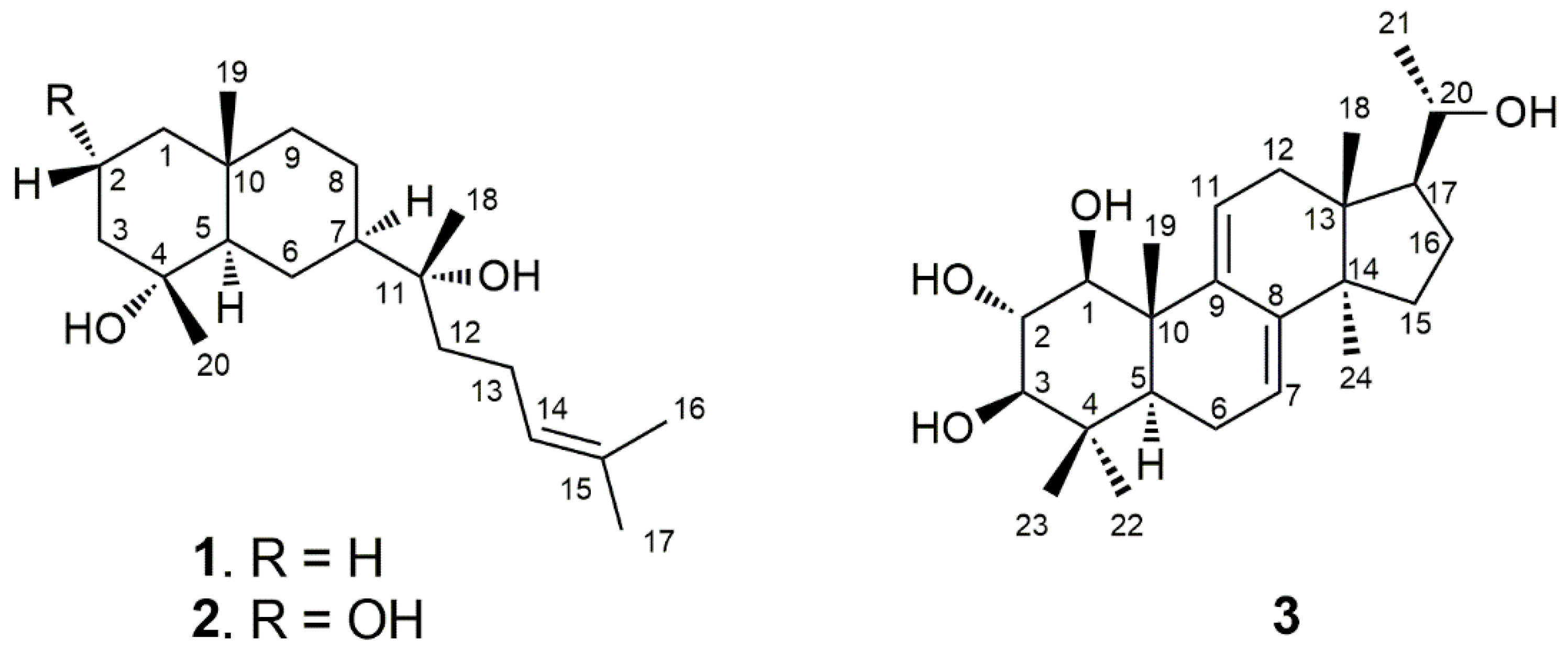
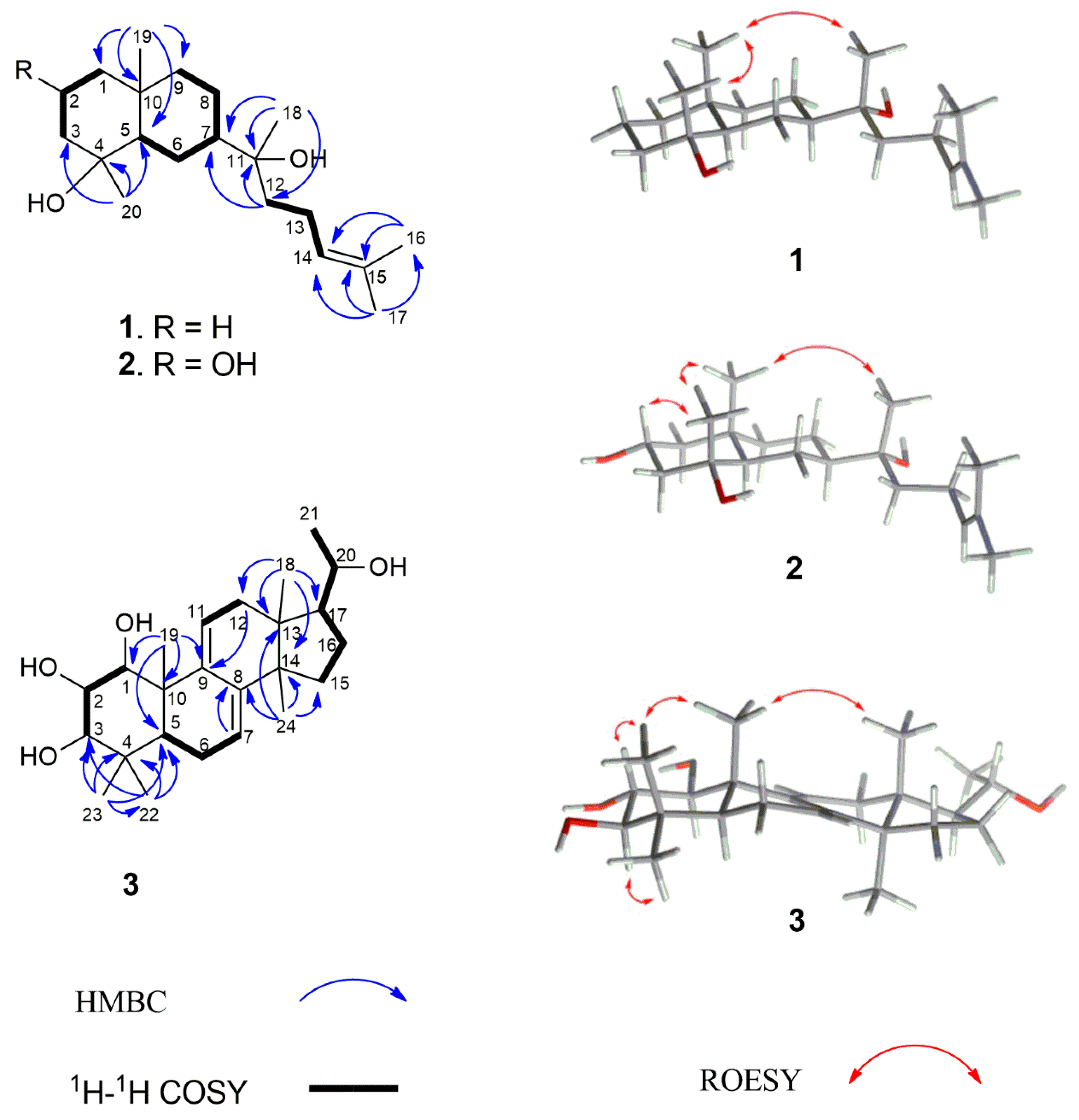
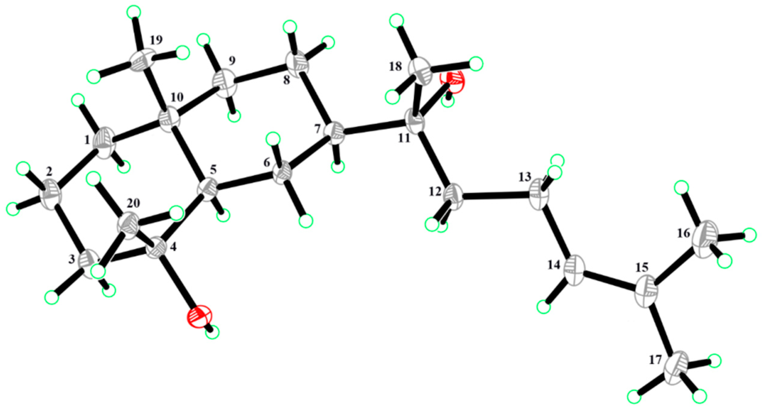
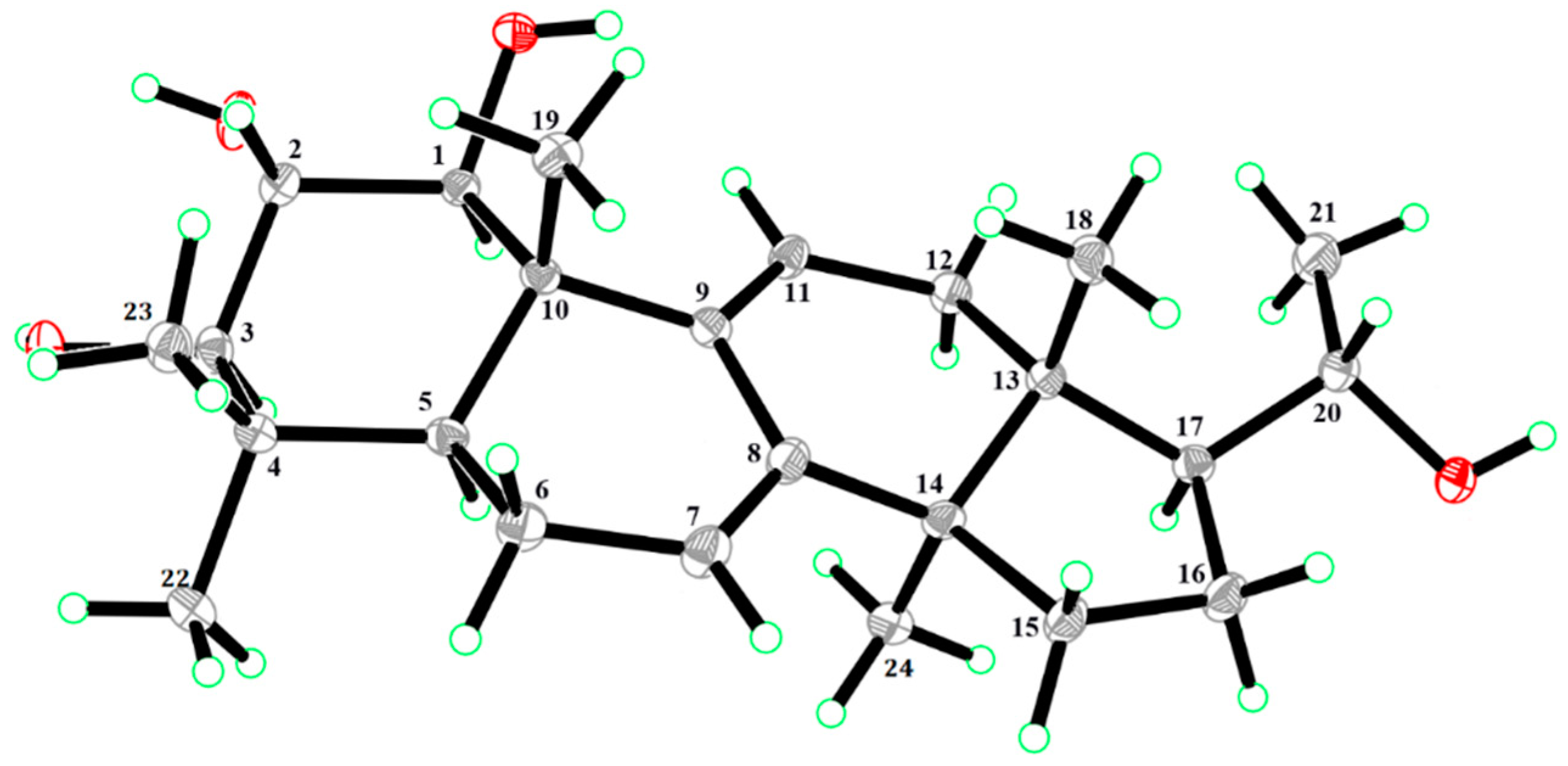
Prenyleudesmanes and A Hexanorlanostane from the Roots of Lonicera macranthoides

1
Jiangsu Key Laboratory for the Research and Utilization of Plant Resources, The Jiangsu Provincial Platform for Conservation and Utilization of Agricultural Germplasm, Institute of Botany, Jiangsu Province and Chinese Academy of Sciences, Nanjing 210000, China
2
Max Planck Institute for Chemical Ecology, D-07745 Jena, Germany
3
Naval Compound Community Health Care Station, Beijing 100853, China
*
Authors to whom correspondence should be addressed.
These two authors contributed equally to this work.
Molecules 2019, 24(23), 4276; https://doi.org/10.3390/molecules24234276
Submission received: 22 October 2019 / Revised: 16 November 2019 / Accepted: 21 November 2019 / Published: 23 November 2019
(This article belongs to the Special Issue Terpenes and Terpene Derivatives)

Abstract

:
Three previously undescribed compounds, two prenyleudesmanes (1 and 2), and one hexanorlanostane (3), were isolated from the roots of Lonicera macranthoides. Their structures were established based on 1D and 2D nuclear magnetic resonance (NMR) spectra and high-resolution electrospray ionization mass spectral (HR-ESI-MS) data. The absolute configurations of 1 and 3 were determined by X-ray diffraction. To the best of our knowledge, this is the first time that the absolute configuration of a prenyleudesmane with a trans-decalin system and a hexanorlanostane have been unambiguously confirmed by single-crystal X-ray diffraction with Cu Kα radiation. Thecompounds were tested for their antiproliferative activity on the cancer cell lines (HepG2 and HeLa). The compounds 13 exhibited moderate inhibitory effects against two human cancer cell lines.

1. Introduction

Lonicera macranthoides Hand.-Mazz., a plant of the genus Lonicera in the family Caprifoliaceae, is mainly distributed in the southwest of China [1]. The dried flower buds of L. macranthoides are commonly used as a raw material in traditional Chinese medicine for treating fever, inflammation, and infectious diseases [2]. Earlier phytochemical studies on the plant have shown the presence of various triterpenoid saponins (e.g., hederagenin saponins, oleanolic acid saponins, 18-oleanene saponins, and lupane saponins) [3,4,5,6], flavonoids [7], phenolic acids [8,9], and iridoids [8,9] in aerial parts and flowers of the plant. Because of our studies of L. macranthoides, we became interested in the diterpenes of this species. Recently, we reported the first known occurrence of diterpenes (e.g., labdane, aphidicolane, and syn-pimarane) in the roots of L. macranthoides [10,11,12]. To explore further unknown diterpenes, we reinvestigated the roots of L. macranthoides. Here, we report on the isolation and characterization of two new diterpenes, lonimacranthoidin C (1) and lonimacranthoidin D (2), and a novel hexanorlanostane, lonimacranthoidin E (3). Compounds 13 were screened for antiproliferative activity against two human cancer cell lines, HepG2 and HeLa.

2. Results and Discussion

An ethanolic extract of dried roots of L. macranthoides was suspended in water and partitioned sequentially between petroleum ether and ethyl acetate (EtOAc). The EtOAc fraction was subjected to repeated separation by column chromatography (CC) over silica gel and Sephadex LH-20. Selected fractions were further purified by preparative HPLC to yield three pure compounds, including the two prenyleudesmanes (1, 2) and a hexanorlanostane (3), as seen in Figure 1. The structure elucidation was carried out by high-resolution mass spectrometry (HRMS), nuclear magnetic resonance (NMR) spectroscopy (1H NMR, 13C NMR, 1H-1H homonuclear chemical shift correlation spectroscopy (COSY), 1H-13C heteronuclear single quantum coherence (HSQC), 1H-13C heteronuclear multiple bond correlation (HMBC), and 1H-1H rotating frame Overhauser effect spectroscopy (ROESY)), and single-crystal X-ray diffraction analysis.
Compound 1 was obtained as colorless crystals. The molecular formula of C20H36O2Na was determined by the pseudomolecular ion peak at m/z 331.2607 [M + Na]+ (calculated: 331.2608) in positive HR-ESI-MS, corresponding to three unsaturations. The UV spectrum showed the absorption maximum at λmax 201 nm. The compound showed a positive optical rotation of [α ] D 25 +26.4 (c 0.100 in methanol). The 1H NMR spectrum of 1 displayed signals for one olefinic proton at δH 5.14 (1H, dd, J = 7.0/7.0, H-14), five methyl singlets at δH 0.86 (3H, s, H3-19), δH 1.11 (3H, s, H3-20), δH 1.16 (3H, s, H3-18), δH 1.63 (3H, s, H3-17), and δH 1.69 (3H, s, H3-16), and overlapping aliphatic methylene and/or methine signals (δH 1.05–2.04). The assignment of the latter could be accomplished by a series of selective total correlation spectroscopy (SELTOCSY) experiments (see Supporting Information). The 13C NMR and distortionless enhancement by polarization transfer (DEPT) NMR spectra of 1 showed the presence of 20 carbon resonances, including two signals of olefinic carbons at δC 124.6 (C-14) and δC 131.6 (C-15), five signals for methyl groups at δC 17.6 (C-16), δC 18.7 (C-19), δC 22.6 (C-20), δC 24.1 (C-18), and δC 25.7 (C-17), eight methylene signals at δC 41.0 (C-1), δC 20.1 (C-2), δC 43.5 (C-3), δC 21.4 (C-6), δC 21.8 (C-8), δC 44.6 (C-9), δC 39.7 (C-12), and δC 22.3 (C-13), two methine signals at δC 55.0 (C-5) and δC 48.3 (C-7), one signal for a quaternary carbon at δC 34.6 (C-10), and two signals of oxygenated tertiary carbons at δC 72.3 (C-4) and δC 74.5 (C-11). The interpretation of NMR spectra and the degree of unsaturation deduced from HRMS data suggested that compound 1 was a bicyclic diterpene possessing a trisubstituted double bond and two hydroxyl groups (OH-4 and OH-11). Analysis of the 1H-1H COSY and HSQC spectra of 1 provided three partial structures shown by bold lines in Figure 2. The interpretation of the HMBC spectrum of 1 showed correlations from H3-17 to C-14, C-15, and C-16, and from H3-16 to C-14, C-15, and C-17; these enabled the localization of the double bond at C-14. This spin system is further characterized by a coupling of H-12 to H-14. HMBC correlations from H-12 to C-11, and from H3-18 to C-11 and C-12, eventually resulted in the definition of a side chain of eight carbons, including a trisubstituted double bond (Δ14). A further HMBC correlation from H-12 to C-7 and from H3-18 to C-7 indicated the linkage to C-7 of the decalin ring system. The two hydroxylated positions at C-4 and C-11, respectively, could be confirmed by the HMBC correlations from H3-20 to C-3, C-4, and C-5, and from H3-18 to C-11 and C-12, as seen in Figure 2. In summary, the NMR data analysis, as seen in Table 1, revealed a prenyleudesmane skeleton similar to dysokusone A [13], and the structure of 1 was determined as shown in Figure 1. The relative configuration of 1 was partially established by analyzing its ROESY correlations. Nuclear Overhauser effect (NOE) correlations between H3-18, H3-19, and H3-20 suggested a cofacial arrangement. Finally, crystals of compound 1 were obtained and subjected to X-ray diffraction analysis, as seen in Figure 3. The absolute configuration of 1 was determined as (4R,5R,7R,10R)-4,10-dimethyl-7-(11R-hydroxy-11,15-dimethyl-14-ene-11-yl)-trans-decalin-4-ol by Cu X-ray crystallography (Flack parameter = −0.05 (11), Figure 3) [14,15] and named as lonimacranthoidin C (1). Ours was the first successful single-crystal X-ray analysis of a prenyleudesmane with a trans-decalin scaffold.
Compound 2 was obtained as milky oil with the molecular formula C20H36O3 as determined by HR-ESI-MS (m/z 347.2551 [M + Na]+, calculated: m/z 347.2557 for [C20H36O3 + Na]+). Similar to 1, compound 2 showed three unsaturations. The UV spectrum of 2 showed an absorption at λmax 201 nm and a positive optical rotation of [α ] D 25 + 10.0 (c 0.100 in methanol) was determined. The assignment of all proton and carbon chemical shifts of 2 was achieved by analyzing the 1D and 2D NMR spectra, as seen in Table 1. Similar to 1, the structure of 2 was also elucidated as a prenyleudesmane-type diterpene. Unlike the chemical shifts of position 2 in 1 (δC 20.1 and δH 1.56/1.58 (H-2β/H-2α, respectively)), the corresponding structural elements in 2 were an oxygenated methylene (δC 68.2) and a hydroxymethine H 4.28, H-2). Thus, 2 was determined as the C-2 hydroxylated derivative of 1, as shown in Figure 1. The stereochemistry at C-2 was established by the occurrence of NOE correlations between H3-19, H3-20 H3-18, and H-2 which indicated a cofacial orientation. Hence, the absolute configuration for C-2 was assigned, based on the X-ray determined configuration of 1, as R-configured. Due to the occurrence of similar chemical shifts for C-4, C-5, C-7, C-10, and C-11, as seen in Table 1, similar NOESY correlations, as seen in Figure 2, similar values for the optical rotation, and the above defined configuration of C2, compound 2 was determined as (2R,4R,5R,7R,10R)-4,10-dimethyl-7-(11R-hydroxy-11,15-dimethyl-14-ene-11-yl)-trans-decalin-2,4-diol, and named lonimacranthoidin D (2).
The molecular formula of compound 3 was assigned as C24H38O4 by its positive HR-ESI-MS data (m/z 413.2667, [M + Na]+; calculated: 413.2662), which indicated six unsaturations in the molecule. Compound 3 was obtained as colorless crystals with an UV spectrum having an absorption maximum at λmax 243 nm. The compound showed a positive optical rotation of [α ] D 25 + 12.6 (c 0.100 in methanol). The 1H NMR spectrum of 3 showed resonances of two olefinic protons at δH 6.31 (1H, d, J = 6.0 Hz, H-11) and δH 5.49 (1H, d, J = 5.0 Hz, H-7), six methyl resonances at δH 0.57 (3H, s, H3-18), δH 1.06 (3H, s, H3-19), δH 1.21 (3H, d, J = 6.2 Hz, H3-21), δH 0.90 (3H, s, H3-22), δH 1.02 (3H, s, H3-23), and δH 0.91 (3H, s, H3-24), four hydroxymethines at δH 3.57 (1H, d, J = 10.0 Hz, H-1), δH 3.48 (1H, dd, J = 10.0/10.0 Hz, H-2), δH 3.04 (1H, d, J = 10.0 Hz, H-3), and δH 3.62 (1H, dd, J = 6.2/9.0 Hz, H-20), and further overlapping aliphatic methylenes and/or methines in the range δH 1.10 to δH 2.30, which were assigned by means of SELTOCSY experiments (see Supporting Information). The 13C NMR and DEPT spectra of 3 showed the presence of four olefinic carbons at δC 119.1 (C-7), δC 144.0 (C-8), δC 142.1 (C-9), and δC 119.1 (C-11). The remaining four MS-predicted unsaturations were assigned to a tetracyclic ring system. In addition, six methyl groups at δC 15.4 (C-18), δC 15.9 (C-19), δC 22.4 (C-21), δC 15.9 (C-22), δC 27.6 (C-23), and δC 24.9 (C-24), four methylenes at δC 22.4 (C-6), δC 36.4 (C-12), δC 31.1 (C-15), and δC 25.8 (C-16), two methines at δC 47.1 (C-5) and δC 53.0 (C-17), four oxygenated methines at δC 78.4 (C-1), δC 72.9 (C-2), δC 78.7 (C-3), and δC 70.4 (C-20), and four quaternary carbons at δC 38.1 (C-4), δC 43.2 (C-10), δC 42.0 (C-13), and δC 49.9 (C-14) were observed. Thus, 3 was assigned as a hexanortriterpene derivative with two trisubstituted double bonds (Δ7 and Δ9) and four hydroxyl groups (OH-1, OH-2, OH-3, and OH-20). Interpretation of the 1H-1H COSY data resulted in the identification of four spin systems: H-1/H-2/H-3, H-5/H-6/H-7, H-11/H-12, and H-15/H-16/H-17/H-20/H-21. The HMBC correlations from H3-18 to C-12, C-13, C-14, and C-17; from H3-19 to C-1, C-5, C-9, and C-10; from H3-22 to C-3, C-4, C-5, and C-23; from H3-23 to C-3, C-4, C-5, and C-22; and from H3-24 to C-8, C-13, C-14, and C-15 allowed for the positioning of two methyl groups at C-4 and four methyl groups at C-10, C-13, C-14, and C-20, respectively. These data indicated that compound 3 was an unusual hexanorlanostane that had been described earlier as aglycon, from the saponins of the sea cucumber Cucumaria koraiensis [16,17]. The full assignment of all positions in the molecule was accomplished by the interpretation of the HSQC and HMBC data, as seen in Table 1, suggesting that 3 was 1,2,3,20-tetrahydroxy-hexanorlanostan-7, 9(11)-diene, as shown in Figure 1. In the ROESY spectrum of 3, correlations between H3-18/H3-19/H3-23/H-2 and H3-22/H-3 indicated that H3-18, H3-19, H3-23, and H-2 were on one side of the molecular plane, while H3-22 and H-3 were located on the opposite side. Fortunately, crystals of compound 3 could be obtained and were subjected to single-crystal X-ray diffraction analysis, as seen in Figure 4. Based on our results, the absolute configuration of 3 was determined as (1S,2S,3R,5S,10S,13R,14R,17S,20S)-1,2,3,20-tetrahydroxy-hexanorlanostan-7, 9(11)-diene (3) by Cu X-ray crystallography (Flack parameter = 0.20 (6), Figure 4) and named lonimacranthoidin E.
Compounds 13 were furthermore tested for their antiproliferative effect on the Human Hepatocellular Carcinoma Cell lines (HepG2), Human Cervical Carcinoma Cell line (HeLa), and the Human Aortic Smooth Muscle Cell line (HASMC). The results, as seen in Table 2, demonstrated that 13 showed moderate antiproliferative activities (IC50 12.5 ± 0.9 to 64.9 ± 3.5 μM) against the two tumor cell lines. No significant effect against HASMC was observed.
Prenyleudesmanes are a rare class of diterpenes that were originally isolated from marine algae [18,19] and marine mollusks [20,21,22,23,24]. Prenyleudesmanes were also found in fungi [25] and plants of the genus Dysoxylum [13,26,27,28]. Our report on the isolation and full structure elucidation of lonimacranthoidin C (1) and lonimacranthoidin D (2) from L. macranthoides therefore suggests another source for prenyleudesmanes in nature. Interestingly, the hexanorlanostane 3 was initially found in sea creatures [16,17]. Lonimacranthoidin E (3) is the first example of a hexanorlanostane isolated from a terrestrial plant.

3. Materials and Methods

3.1. General Experimental Procedures

Thin-layer chromatography was carried out on silica gel 60 GF254 (Merck) plates. Preparative HPLC (LC-20AR, Shimadzu, Kyoto, Japan) was conducted on a Shim-pack GIS C18 column (5 μm, 250 × 20 mm, Shimadzu). Column chromatography (CC) was performed on silica gel (200–300 mesh) and Sephadex LH-20. LC-HRMS spectra were obtained from an Agilent 1260 UPLC-DAD-6530 ESI Q-TOF MS (Agilent Technologies GmbH, Waldbronn, Germany). Optical rotation values were measured using a Jasco P-1020 polarimeter. NMR data were obtained using a Bruker Avance 500 MHz or Bruker Avance 300 MHz spectrometers (Bruker Biospin GmbH, Karlsruhe, Germany). Tetramethylsilane was used as an internal standard. The X-ray structures were solved by direct methods (SHELXL-97). The X-ray crystallographic data were collected on a Bruker SMART APEX-II CCD diffractometer using graphite monochromatic Cu Kα radiation.

3.2. Plant Material

The roots of L. macranthoides were collected from Longhui in the Hunan province of China in July 2015. The plants were taxonomically identified by Professor Changqi Yuan (Institute of Botany, Jiangsu province and Chinese Academy of Sciences). A voucher specimen (No. 20150701) has been deposited in the herbarium of the Institute of Botany, Jiangsu province, and Chinese Academy of Sciences.

3.3. Extraction and Isolation

The dried roots (4.0 kg) of L. macranthoides were milled and repeatedly extracted with 95% EtOH for 2 h under reflux (80 °C). After evaporation in vacuo, the crude extract (472.6 g) was resuspended in H2O and partitioned with petroleum ether and ethyl acetate (EtOAc), in succession. The EtOAc fraction (94 g) was subjected to column chromatography (silica gel, CH2Cl2-MeOH 100:0–0:100) to produce six fractions (F1-F6) on the basis of TLC analysis. F2 (21 g) was purified by column chromatography on Sephadex LH-20 (CH2Cl2-MeOH, 1:1), followed by preparative HPLC using MeOH-H2O (65:35, v/v, flow rate 1.0 mL/min) as an eluent to obtain the compounds 1 (11.0 mg) and 2 (6.0 mg). F5 (11 g) was purified by preparative HPLC with MeOH–H2O (55:45, v/v, flow rate 3.0 mL/min) as an eluent to obtain compound 3 (13.0 mg).

3.4. Compound Characterization

Lonimacranthoidin C (1): colorless crystals (MeOH); [α ] D 25 + 26.4 (c 0.100 in MeOH); UV (MeOH) λmax 201 nm; mp 101–103 °C; HR-ESI-MS m/z 331.2607 [M + Na]+ (calculated for C20H36O2: 331.2608); 1H NMR (500 MHz, CDCl3) and 13C NMR (125 MHz, CDCl3) spectroscopic data, see Table 1. Lonimacranthoidin C (1) was recrystallized in methanol/ethyl acetate (3:1). A single-crystal X-ray diffraction analysis using Cu Kα radiation (1.54178 Å) was carried out to confirm the structure. M = 308.49, monoclinic, P21 21 21, a = 11.5498 (17) Å, b = 12.2435 (14) Å, c = 27.246 (3) Å, α = γ = β = 90.00°, V = 3852.8 (9) Å3, Z = 8, Dc = 1.064 mg mm−3, T = 153 (2) K, F (000) = 1376.0. The crystallographic data centre has assigned the code Cambridge Crystallographic Data Centre (CCDC) 1,941,729 for the crystal structure of lonimacranthoidin C (1). The CCDC contains the supplementary crystallographic data for this paper. These data can be obtained free of charge via http://www.ccdc.cam.ac.uk/conts/retrieving.html.
Lonimacranthoidin D (2): milky oil (MeOH); [α ] D 25 + 10.0 (c 0.100 in MeOH); UV (MeOH) λmax 201 nm; HR-ESI-MS m/z 347.2551 [M + Na]+ (calculated for C20H36O3, 347.2557); 1H NMR (500 MHz, CDCl3) and 13C NMR (125 MHz, CDCl3) spectroscopic data are listed in Table 1.
Lonimacranthoidin E (3): colorless crystals (MeOH); [α ] D 25 + 12.6 (c 0.100 in MeOH); UV (MeOH) λmax 243 nm; mp 244–246 °C; HR-ESI-MS m/z 413.2667 [M + Na]+ (calculated for C24H38O4: 413.2662); 1H NMR (300 MHz, in CDCl3:CD3OD = 1:1) and 13C NMR (75 MHz, in CDCl3:CD3OD = 1:1), for NMR spectroscopic data, see Table 1. Lonimacranthoidin E (3) was re-crystallized in methanol/ethyl acetate (1:1). A single-crystal X-ray diffraction analysis using Cu Kα radiation (1.54178 Å) was carried out to confirm the structure. M = 390.54, monoclinic, P21 21 21, a = 6.0714 (4) Å, b = 13.2797 (8) Å, c = 28.6232 (17) Å, α = γ = β = 90.00°, V = 2307.87 (2) Å3, Z = 4, Dc = 1.124 mg mm−3, T = 153 (2) K, F (000) = 856. The crystallographic data centre has assigned the code CCDC 1,941,730 for the crystal structure of lonimacranthoidin E (3) The CCDC contains the supplementary crystallographic data for this paper. These data can be obtained free of charge via http://www.ccdc.cam.ac.uk/conts/retrieving.html.

3.5. Biological Assay

HepG2, HeLa, and HASMC cell lines were cultured in Dulbecco’s modified Eagle’s medium (Gibco, Grand Island, NY, USA) supplemented with 10% fetal bovine serum (Gibco), 100 μg/mL penicillin, and 100 μg/mL streptomycin. The cells were cultivated in a humidified atmosphere of 5% CO2 at 37 °C. Antiproliferative assays of the compounds 13 against the above-mentioned three cell lines were evaluated using the 3-(4,5-dimethylthiazol-2-yl)-2,5-diphenyltetrazoliumbromide (MTT) assay, carried out according to protocols [29] described previously.

Supplementary Materials

1H and 13C NMR, 13C-DEPT, 1H-13C HSQC, 1H-13C HMBC, 1H-1H COSY, 1H-1H ROESY, SELTOCSY, UV, and HR-ESI-MS data of compounds 13 are available in the Supporting Information.

Author Contributions

Funding acquisition, X.F. and Y.C.; Investigation, H.L., W.L., B.B., Y.S. and C.P.; Methodology, Y.C.; Project administration, Y.C.; Supervision, X.F. and Y.C.; Writing—original draft, H.L.; Writing—review & editing, C.P. and Y.C.

Acknowledgments

This research was supported financially by the National Natural Science Foundation of China (31770383, 31970375) and Research Project of 333 High-Level Talents Cultivation of Jiangsu Province (BRA2016463). We thank Emily Wheeler for editorial assistance.

Conflicts of Interest

The authors declare no conflict of interest.

References

  1. FOC (Flora of China) Flora of China Website. 1988. Available online: http://frps.iplant.cn/frps/Lonicera%20macranthoides (accessed on 15 October 2019).
  2. Chinese Pharmacopeia Commission. Pharmacopeia of the People’s Republic of China; China Medical Science Press: Beijing, China, 2015; Volume 1, p. 30. [Google Scholar]
  3. Chen, Y.; Zhao, Y.; Wang, M.; Wang, Q.; Shan, Y.; Guan, F.; Feng, X. A new lupane-type triterpenoid saponin from Lonicera macranthoides. Chem. Nat. Compd. 2014, 49, 1087–1090. [Google Scholar] [CrossRef]
  4. Chen, Y.; Feng, X.; Jia, X.; Wang, M.; Liang, J.; Dong, Y. Triterpene glycosides from Lonicera. Isolation and structural determination of seven glycosides from flower buds of Lonicera macranthoides. Chem. Nat. Compd. 2008, 44, 39–43. [Google Scholar] [CrossRef]
  5. Chen, Y.; Feng, X.; Wang, M.; Zhao, Y.Y.; Dong, Y.F. Triterpene glycosides from Lonicera. II. Isolation and structural determination of glycosides from flower buds of Lonicera macranthoides. Chem. Nat. Compd. 2009, 45, 514–518. [Google Scholar]
  6. Chen, Y.; Shan, Y.; Zhao, Y.Y.; Wang, Q.Z.; Wang, M.; Feng, X.; Liang, J.Y. Two new triterpenoid saponins from Lonicera macranthoides. Chin. Chem. Lett. 2012, 3, 325–328. [Google Scholar] [CrossRef]
  7. Sun, M.; Feng, X.; Yin, M.; Chen, Y.; Zhao, X.; Dong, Y. A biflavonoid from stems and leaves of Lonicera macranthoides. Chem. Nat. Compd. 2012, 48, 231–233. [Google Scholar] [CrossRef]
  8. Sun, M.; Feng, X.; Lin, X.H.; Yin, M.; Zhao, X.Z.; Chen, Y.; Shan, Y. Studies on the chemical constituents from stems and leaves of Lonicera macranthoides. J. Chin. Med. Mater. 2011, 34, 218–220. [Google Scholar]
  9. Liu, J.; Zhang, J.; Wang, F.; Chen, X.F. Chemical constituents from the buds of Lonicera macranthoides in Sichuan, China. Biochem. Syst. Ecol. 2014, 54, 68–70. [Google Scholar] [CrossRef]
  10. Liu, W.J.; Chen, Y.; Ma, X.; Zhao, Y.Y.; Feng, X. Study on chemical constituents from roots of Lonicera macranthoides. Chin. Med. Mat. 2014, 37, 2207–2209. [Google Scholar]
  11. Bai, B.; Chen, Y.; Liu, W.J.; Yin, M.; Wang, M.; Feng, X. Chemical constituents of petroleum ether fraction of Lonicera macranthoides roots. J. Chin. Med. Mater. 2015, 38, 518–520. [Google Scholar]
  12. Lyu, H.; Liu, W.J.; Xu, S.; Shan, Y.; Feng, X.; Chen, Y. Two 9, 10-syn-pimarane diterpenes from the roots of Lonicera macranthoides. Phytochem. Lett. 2018, 25, 175–179. [Google Scholar] [CrossRef]
  13. Fujioka, T.; Yamamoto, M.; Kashiwada, Y.; Fujii, H.; Mihashi, K.; Ikeshiro, Y.; Chen, I.S.; Lee, K.H. Novel cytotoxic diterpenes from the stem of Dysoxylum kuskusense. Bioorg. Med. Chem. Lett. 1998, 8, 3479–3482. [Google Scholar] [CrossRef]
  14. Flack, H.D. On enantiomorph-polarity estimation. Acta. Cryst. A Found. Crystallogr. 1983, 39, 876–881. [Google Scholar] [CrossRef]
  15. Flack, H.D.; Bernardinelli, G. Reporting and evaluating absolute-structure and absolute-configuration determinations. J. Appl. Crystallogr. 2000, 33, 1143–1148. [Google Scholar] [CrossRef]
  16. Avilov, S.A.; Kalinovsky, A.I.; Kalinin, V.I.; Stonik, V.A.; Riguera, R.; Jiménez CKoreoside, A. A new nonholostane triterpene glycoside from the sea cucumber Cucumaria koraiensis. J. Nat. Prod. 1997, 60, 808–810. [Google Scholar] [CrossRef] [PubMed]
  17. Kalinin, V.I.; Silchenko, A.S.; Avilov, S.A.; Stonik, V.A. Non-holostane aglycones of sea cucumber triterpene glycosides. Structure, biosynthesis, evolution. Steroids 2018, 147, 42–51. [Google Scholar] [CrossRef] [PubMed]
  18. Sun, H.H.; Waraszkiewicz, S.M.; Erickson, K.L.; Finer, J.; Clardy, J. Dictyoxepin and dictyolene, two new diterpenes from the marine alga Dictyota acutiloba (Phaeophyta). J. Am. Chem. Soc. 1977, 99, 3516–3517. [Google Scholar] [CrossRef]
  19. Takahashi, Y.; Suzuki, M.; Abe, T.; Masuda, M. Anhydroaplysiadiol from Laurencia japonensis. Phytochemistry 1998, 48, 987–990. [Google Scholar] [CrossRef]
  20. Ojika, M.; Yoshida, Y.; Okumura, M.; Ieda, S.; Yamada, K. Aplysiadiol, A new brominated diterpene from the marine mollusc Aplysia kurodai. J. Nat. Prod. 1990, 53, 1619–1622. [Google Scholar] [CrossRef]
  21. Matthée, G.F.; König, G.M.; Wright, A.D. Three new diterpenes from the marine soft coral Lobophytum crassum. J. Nat. Prod. 1998, 61, 237–240. [Google Scholar] [CrossRef]
  22. Cheng, S.Y.; Chuang, C.T.; Wang, S.K.; Wen, Z.H.; Chiou, S.F.; Hsu, C.H.; Dai, C.F.; Duh, C.Y. Antiviral and anti-inflammatory diterpenoids from the soft coral Sinularia gyrosa. J. Nat. Prod. 2010, 73, 1184–1187. [Google Scholar] [CrossRef]
  23. Li, L.; Sheng, L.; Wang, C.Y.; Zhou, Y.B.; Huang, H.; Li, X.B.; Li, J.; Molllo, E.; Gavagnin, M.; Guo, Y.W. Diterpenes from the Hainan soft coral Lobophytum cristatum Tixier-Durivault. J. Nat. Prod. 2011, 74, 2089–2094. [Google Scholar] [CrossRef] [PubMed]
  24. Ye, F.; Zhu, Z.D.; Gu, Y.C.; Li, J.; Zhu, W.L.; Guo, Y.W. Further new diterpenoids as PTP1B inhibitors from the Xisha soft coral Sinularia polydactyla. Mar. Drugs 2018, 16, 103. [Google Scholar] [CrossRef] [PubMed]
  25. Liu, D.Z.; Liang, B.W.; Li, X.F.; Liu, Q. Induced production of new diterpenoids in the fungus Penicillium funiculosum. Nat. Prod. Commun. 2014, 9, 607–608. [Google Scholar] [CrossRef] [PubMed]
  26. Duh, C.Y.; Wang, S.K.; Chen, I.S. Cytotoxic prenyleudesmane diterpenes from the fruits of Dysoxylum kuskusense. J. Nat. Prod. 2000, 63, 1546–1547. [Google Scholar] [CrossRef]
  27. Gu, J.; Qian, S.Y.; Zhao, Y.L.; Cheng, G.G.; Hu, D.B.; Zhang, B.H.; Liu, Y.P.; Luo, X.D. Prenyleudesmanes, rare natural diterpenoids from Dysoxylum densiflorum. Tetrahedron 2014, 70, 1375–1382. [Google Scholar] [CrossRef]
  28. Zhang, P.; Lin, Y.; Wang, F.; Fang, D.; Zhang, G. Diterpenes from Dysoxylum lukii Merr. Phytochem. Lett. 2019, 29, 53–56. [Google Scholar] [CrossRef]
  29. Alley, M.C.; Scudiero, D.A.; Monks, A.; Hursey, M.L.; Czerwinski, M.J.; Fine, D.L.; Abbott, B.J.; Mayo, J.G.; Shoemaker, R.H.; Boyd, M.R. Feasibility of drug screening with panels of human tumor cell lines using a microculture tetrazolium assay. Cancer Res. 1998, 48, 589–601. [Google Scholar]
Sample Availability: Samples of the compounds 1 and 3 are available from the authors.
Figure 1. Chemical structures of the compounds 13.
Molecules 24 04276 g001
Figure 2. Key 2D NMR correlations of compounds 1, 2, and 3.
Molecules 24 04276 g002
Figure 3. X-ray Oak Ridge thermal-ellipsoid plot program (ORTEP) drawing of compound 1.
Molecules 24 04276 g003
Figure 4. X-ray ORTEP drawing of compound 3.
Molecules 24 04276 g004
Table 1. 1H and 13C NMR spectral data of compounds 1, 2, and 3.
Atom1 a, c2 a,c3 b,c
δCδHδCδHδCδH
41.01.07 ddd 12.5/12.5/6.0 46.71.34 dd 14.0/3.278.43.57 d 10.0
1.38 d 12.5 1.67 m
20.11.58 m 68.24.28 ddd 7.0/3.0/3.072.93.48 dd 10.0/10.0
1.56 m
43.51.35 ddd 12.5/12.5/5.5 48.51.66 m78.73.04 d 10.0
1.79 d 12.5 2.01 d 14.0
472.3 71.6 38.1
555.01.19 d 12.554.41.29 dd 12.5/2.047.11.16 dd 5.0/12.0
21.41.86 d 12.5 21.21.87 d 12.522.42.20 dd 12.0/17.0
1.03 ddd 12.5/12.5/12.5 1.14 ddd 12.5/12.5/12.5 2.15 dd 5.0/5.0/17.0
748.31.41 dddd 12.5/12.5/3.0/3.0 48.51.42 dddd 12.5/12.5/3.0/3.0119.55.49 d 5.0
21.81.59 d 12.5 21.21.56 d 12.5142.1
1.33 dddd 12.5/12.5/12.5/3.0 1.37 dddd 12.5/12.5/12.5/3.5
44.61.45 ddd 12.5/3.0/3.044.91.49 ddd 12.5/3.0/3.0144.0
1.15 ddd 12.5/12.5/3.0 1.12 m
1034.6 34.0 43.2
1174.5 74.6 119.16.31 d 6.0
12α39.71.53 dd 8.2/8.239.51.52 dd 8.1/8.136.42.18 d 17.0
12β 2.06 d 6.0/17.0
1322.32.06 m 22.32.04 m42.0
14124.65.14 dd 7.0/7.0124.55.13 dd 6.7/6.749.9
15α131.6 131.6 31.11.68 ddd 7.5/11.5/11.5
15β 1.46 dd 9.0/11.5
16α17.61.63 s17.81.62 s25.82.07 ddd 9.0/14.0/17.0
16β 1.60 dd 9.0/14.0
1725.71.69 s25.81.68 s53.01.78 dd 9.0/17.0
1824.11.16 s24.21.16 s15.40.57 s
1918.70.86 s20.31.14 s15.91.06 s
2022.61.11 s25.01.33 s70.43.62 dd 6.2/9.0
21 22.41.21 d 6.2
22 15.90.90 s
23 27.61.02 s
24 24.90.91 s
a Data were measured at 500 MHz for 1H and 125 MHz for 13C in CDCl3, δ in ppm, J in Hz.; b Data were measured at 300 MHz for 1H and 75 MHz for 13C in CDCl3:CD3OD = 1:1, δ in ppm, J in Hz.; c Overlapping signals were assigned by HSQC, HMBC, COSY, and SELTOCSY experiments.
Table 2. Antiproliferative activities of compounds 13 against two cancer cells and one normal cell line. a.
Cell Line123Etoposide
HepG236.3 ± 2.164.9 ± 3.546.0 ± 2.425.4 ± 1.7
HeLa13.8 ± 1.127.1 ± 1.412.5 ± 0.921.2 ± 1.3
HASMC>100>100>10063.7
a Results are expressed as IC50 values in μM.

