
pH-Responsive Polypeptide-Based Smart Nano-Carriers for Theranostic Applications


Abstract
1. Introduction
2. Types of pH-Responsive Polypeptides
2.1. Cationic Polypeptides
2.1.1. Poly(l-lysine)-Based Polypeptides
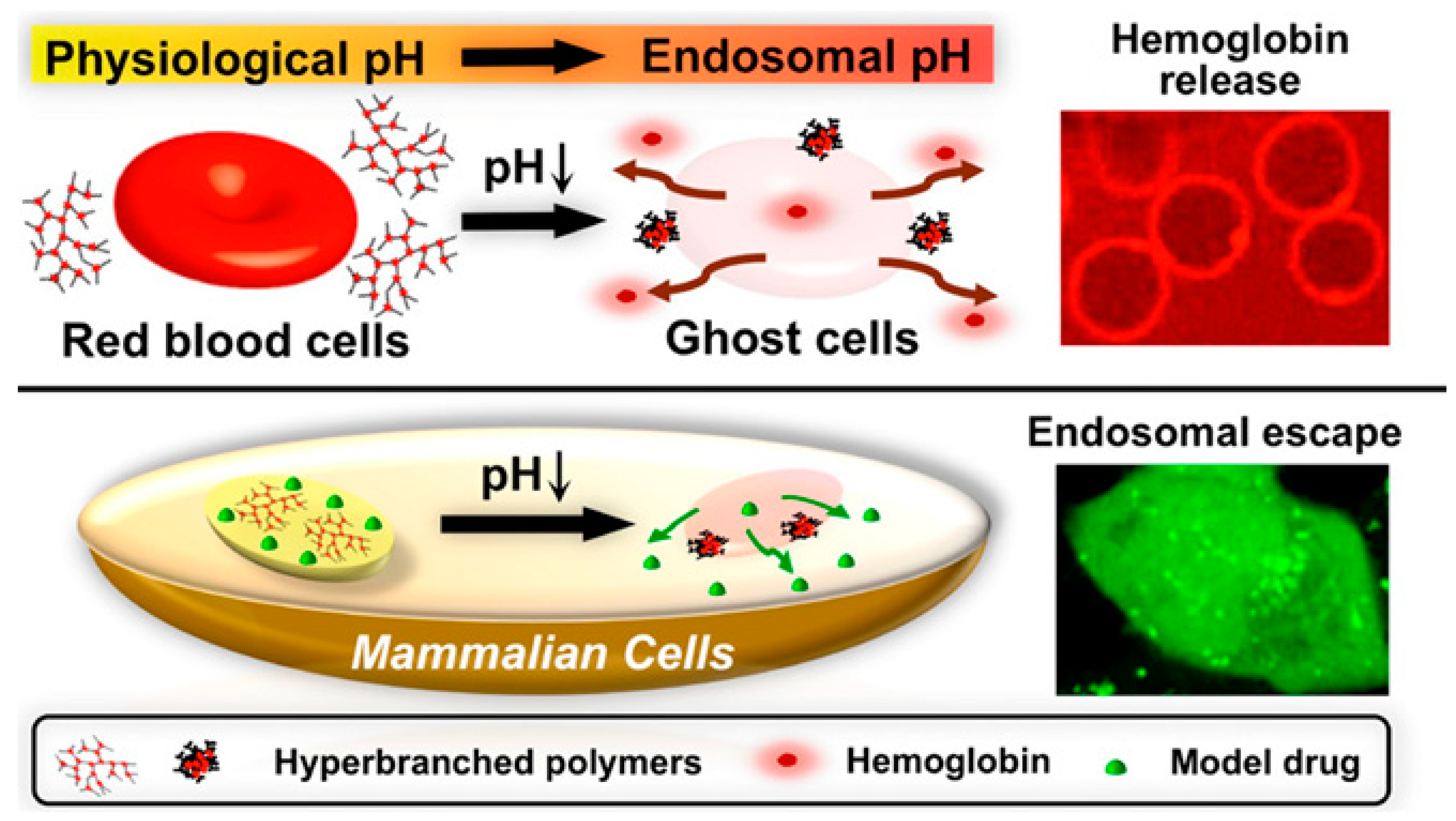
2.1.2. Poly(l-histidine)-Based Polypeptides
2.1.3. Polyarginine-Based Polypeptides
2.1.4. Polytyrosine-Based Polypeptides
2.1.5. Polyproline- and Polytryptophan-Based Polypeptides
2.2. Anionic Polypeptides
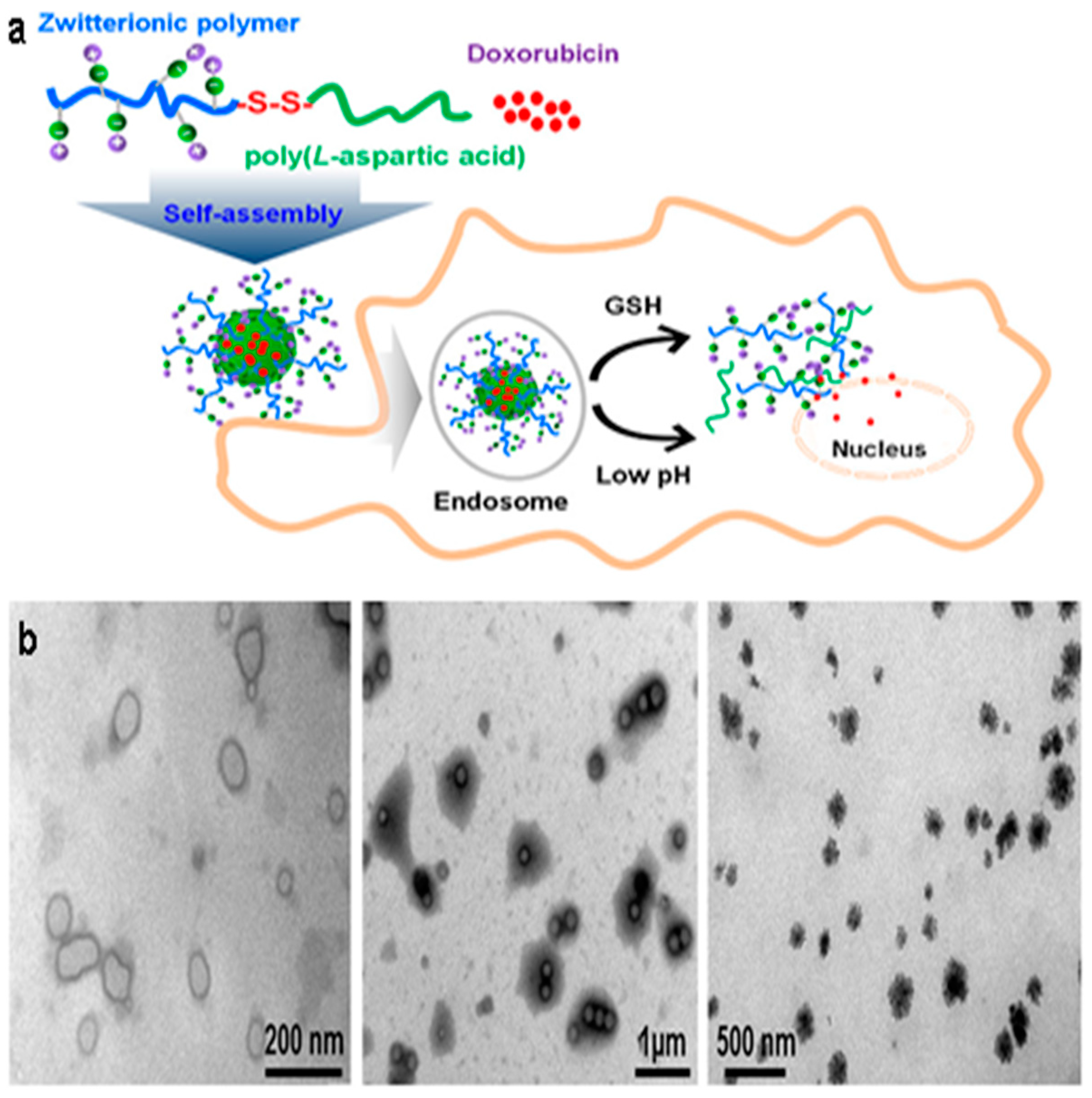
2.2.1. Poly(aspartic acid)-Based Polypeptides
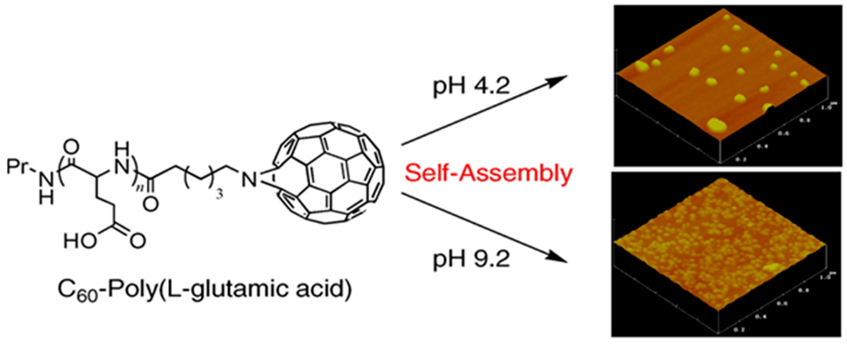
2.2.2. Poly(glutamic acid)-Based Polypeptides
3. pH-Responsive Drug Release in the Organ Level
4. pH-Responsive Drug Release in the Tissue Level
5. pH-Responsive Drug Release at the Cellular Level
6. Combination with Other Responsive Materials
7. Conclusions and Outlook
Author Contributions
Funding
Conflicts of Interest
Abbreviations
| AA | Amino acid |
| ANS | 8-Anilinonaphthalene-1-sulfonate |
| BSA | Bovine serum albumin |
| CD | Circular dichroism |
| Ce6 | Chlorin e6 |
| CELG | Chloroethyl-l-glutamate |
| CMSC | Carboxymethyl chitosan |
| DFS | Diclofenac Na |
| DOE | Design of experiments |
| Dox | Doxarubicin |
| DPPE | 1,2-Dipalmitoyl-sn-glycero-3-phosphoethanolamine |
| DTX | Docetaxel |
| FITC | Fluourescein isothiocyanate |
| 5FU | 5-Fluorouracil |
| FRET | Forster resonance energy transfer |
| HA | Hyaluronic acid |
| HEMA | 2-Hydroxyethyl methacrylate |
| HES | Hydroxyethyl starch |
| HPTS | 8-Hydroxypyrene-1,3,6-trisulfonic acid |
| IPN | Interpenetrating polymer network |
| KLA | Lysine-leucine-alanine |
| LGA | l-Glutamic acid |
| MBG | Mesoporous bioactive glass nanoparticles |
| MPC | Methacryloyloxyethyl phosphoryl-choline |
| OEI | Oligoethylenimine |
| NLC | Nanostructured lipid carriers |
| pAA | Poly(allylamine) |
| pArg | Polyarginine |
| pAsp | Polyaspartic acid |
| PBS | Phosphate buffer solution |
| PC | Phosphatidylcholine |
| PE | Phosphatidylethanolamine |
| PEG | Poly(ethylene glycol) |
| PEI | Polyethyleneimine |
| PHEA | Poly(N-2-hydroxyethyl-d,l-aspartamide) |
| PHLIP | pH low insertion peptide |
| PICs | Polyionic complexes |
| PL | Polylysine |
| PLA | Polylactide |
| PLGA | Poly(l-glutamic acid) |
| PLL | Poly(l-lysine) |
| pLeu | Polyleucine |
| pNIPAm | Poly(N-isopropylacrylamide) |
| pHis | Poly(l-histidine) |
| PPO | Poly(propylene oxide) |
| pPro | Poly(l-proline) |
| PSI | Polysuccinimide |
| PTD | Protein transduction domains |
| pTyr | Polytyrosine |
| RGD | Arginine-glycine-aspartic peptide |
| ROP | Ring opening polymerization |
| SPION | Superparamagnetic iron oxide |
| SPL | Succinylated ε-polylysine |
| SVAs | Supramolecular vesicular aggregates |
| SWNT | Single walled carbon nanotube |
| TAPP | 5,10,15,20-Tetrakis-(4-aminophenyl)-21H, 23H porphyrin |
References
- Torchilin, V.P. Micellar nanocarriers: Pharmaceutical perspectives. Pharm. Res. 2007, 24, 1–16. [Google Scholar] [CrossRef] [PubMed]
- Brannon-Peppas, L.; Blanchette, J.O. Nanoparticle and targeted systems for cancer therapy. Adv. Drug Deliv. Rev. 2004, 56, 1649–1659. [Google Scholar] [CrossRef] [PubMed]
- Brigger, I.; Dubernet, C.; Couvreur, P. Nanoparticles in cancer therapy and diagnosis. Adv. Drug Deliv. Rev. 2002, 54, 631–651. [Google Scholar] [CrossRef]
- Lipinski, C.A.; Lombardo, F.; Dominy, B.W.; Feeney, P.J. Experimental and computational approaches to estimate solubility and permeability in drug discovery and development settings. Adv. Drug Deliv. Rev. 2001, 46, 3–26. [Google Scholar] [CrossRef]
- Fernandez, A.M.; Van Derpoorten, K.; Dasnois, L.; Lebtahi, K.; Dubois, V.; Lobl, T.J.; Gangwar, S.; Oliyai, C.; Lewis, E.R.; Shochat, D.; et al. N-Succinyl-(beta-alanyl-l-leucyl-l-alanyl-l-leucyl)doxorubicin: An extracellularly tumor-activated prodrug devoid of intravenous acute toxicity. J. Med. Chem. 2001, 44, 3750–3753. [Google Scholar] [CrossRef] [PubMed]
- Muthu, M.S.; Rajesh, C.V.; Mishra, A.; Singh, S. Stimulus-responsive targeted nanomicelles for effective cancer therapy. Nanomedicine 2009, 4, 657–667. [Google Scholar] [CrossRef] [PubMed]
- Li, Y.; Dong, H.; Wang, K.; Shi, D.; Zhang, X.; Zhuo, R. Stimulus-responsive polymeric nanoparticles for biomedical applications. Sci. China Chem. 2010, 53, 447–457. [Google Scholar] [CrossRef]
- Badi, N. Non-linear PEG-based thermoresponsive polymer systems. Prog. Polym. Sci. 2017, 66, 54–79. [Google Scholar] [CrossRef]
- Yuba, E.; Yamaguchi, A.; Yoshizaki, Y.; Harada, A.; Kono, K. Bioactive polysaccharide-based pH-sensitive polymers for cytoplasmic delivery of antigen and activation of antigen-specific immunity. Biomaterials 2017, 120, 32–45. [Google Scholar] [CrossRef] [PubMed]
- Johnson, R.P.; Uthaman, S.; John, J.V.; Lee, H.R.; Lee, S.J.; Park, H.; Park, I.K.; Suh, H.; Kim, I. Poly(PEGA)-b-poly(l-lysine)-b-poly(l-histidine) Hybrid Vesicles for Tumoral pH-Triggered Intracellular Delivery of Doxorubicin Hydrochloride. ACS Appl. Mater. Interfaces 2015, 7, 21770–21779. [Google Scholar] [CrossRef]
- Gao, W.; Chan, J.M.; Farokhzad, O.C. pH-Responsive Nanoparticles for Drug Delivery. Mol. Pharm. 2010, 7, 1913–1920. [Google Scholar] [CrossRef] [PubMed]
- Volk, T.; Jähde, E.; Fortmeyer, H.P.; Glüsenkamp, K.H.; Rajewsky, M.F. pH in human tumour xenografts: Effect of intravenous administration of glucose. Br. J. Cancer 1993, 68, 492–500. [Google Scholar] [CrossRef] [PubMed]
- Singh, B.; Maharjan, S.; Jiang, T.; Kang, S.K.; Choi, Y.J.; Cho, C.S. Attuning hydroxypropyl methylcellulose phthalate to oral delivery vehicle for effective and selective delivery of protein vaccine in ileum. Biomaterials 2015, 59, 144–159. [Google Scholar] [CrossRef] [PubMed]
- Woraphatphadung, T.; Sajomsang, W.; Gonil, P.; Saesoo, S.; Opanasopit, P. Synthesis and characterization of pH-responsive N-naphthyl-N,O-succinyl chitosan micelles for oral meloxicam delivery. Carbohydr. Polym. 2015, 121, 99–106. [Google Scholar] [CrossRef] [PubMed]
- Knipe, J.M.; Chen, F.; Peppas, N.A. Enzymatic Biodegradation of Hydrogels for Protein Delivery Targeted to the Small Intestine. Biomacromolecules 2015, 16, 962–972. [Google Scholar] [CrossRef] [PubMed]
- Horev, B.; Klein, M.I.; Hwang, G.; Li, Y.; Kim, D.; Koo, H.; Benoit, D.S. pH-activated nanoparticles for controlled topical delivery of farnesol to disrupt oral biofilm virulence. ACS Nano 2015, 9, 2390–2404. [Google Scholar] [CrossRef] [PubMed]
- Gupta, K.M.; Barnes, S.R.; Tangaro, R.A.; Roberts, M.C.; Owen, D.H.; Katz, D.F.; Kiser, P.F. Temperature and pH sensitive hydrogels: An approach towards smart semen-triggered vaginal microbicidal vehicles. J. Pharm. Sci. 2007, 96, 670–681. [Google Scholar] [CrossRef]
- Priftis, D.; Leon, L.; Song, Z.; Perry, S.L.; Margossian, K.O.; Tropnikova, A.; Cheng, J.; Tirrell, M. Self-assembly of alpha-helical polypeptides driven by complex coacervation. Angew. Chem. Int. Ed. 2015, 54, 11128–11132. [Google Scholar] [CrossRef]
- Ma, H.; Fei, J.; Li, Q.; Li, J. Photo-induced reversible structural transition of cationic diphenylalanine peptide self-assembly. Small 2015, 11, 1787–1791. [Google Scholar] [CrossRef]
- Fuchs, S.M.; Raines, R.T. Pathway for polyarginine entry into mammalian cells. Biochemistry 2004, 43, 2438–2444. [Google Scholar] [CrossRef]
- Leifert, J.A.; Whitton, J.L. “Translocatory proteins” and “protein transduction domains”: A critical analysis of their biological effects and the underlying mechanisms. Mol. Ther. 2003, 8, 13–20. [Google Scholar] [CrossRef]
- Kosuge, M.; Takeuchi, T.; Nakase, I.; Jones, A.T.; Futaki, S. Cellular internalization and distribution of arginine-rich peptides as a function of extracellular peptide concentration, serum, and plasma membrane associated proteoglycans. Bioconjugate Chem. 2008, 19, 656–664. [Google Scholar] [CrossRef] [PubMed]
- Duchardt, F.; Fotin-Mleczek, M.; Schwarz, H.; Fischer, R.; Brock, R. A comprehensive model for the cellular uptake of cationic cell-penetrating peptides. Traffic 2007, 8, 848–866. [Google Scholar] [CrossRef] [PubMed]
- Jiao, C.Y.; Delaroche, D.; Burlina, F.; Alves, I.D.; Chassaing, G.; Sagan, S. Translocation and endocytosis for cell-penetrating peptide internalization. J. Biol. Chem. 2009, 284, 33957–33965. [Google Scholar] [CrossRef] [PubMed]
- Hirose, H.; Takeuchi, T.; Osakada, H.; Pujals, S.; Katayama, S.; Nakase, I.; Kobayashi, S.; Haraguchi, T.; Futaki, S. Transient focal membrane deformation induced by arginine-rich peptides leads to their direct penetration into cells. Mol. Ther. 2012, 20, 984–993. [Google Scholar] [CrossRef] [PubMed]
- Wadia, J.S.; Stan, R.V.; Dowdy, S.F. Transducible TAT-HA fusogenic peptide enhances escape of TAT-fusion proteins after lipid raft macropinocytosis. Nat. Med. 2004, 10, 310–315. [Google Scholar] [CrossRef]
- Erazo-Oliveras, A.; Najjar, K.; Dayani, L.; Wang, T.-Y.; Johnson, G.A.; Pellois, J.-P. Protein delivery into live cells by incubation with an endosomolytic agent. Nat. Meth. 2014, 11, 861. [Google Scholar] [CrossRef] [PubMed]
- Madani, F.; Lindberg, S.; Langel, U.; Futaki, S.; Graslund, A. Mechanisms of cellular uptake of cell-penetrating peptides. J. Biophys. 2011, 2011, 414729. [Google Scholar] [CrossRef]
- Erazo-Oliveras, A.; Muthukrishnan, N.; Baker, R.; Wang, T.Y.; Pellois, J.P. Improving the endosomal escape of cell-penetrating peptides and their cargos: Strategies and challenges. Pharmaceuticals 2012, 5, 1177–1209. [Google Scholar] [CrossRef]
- Lundberg, M.; Wikstrom, S.; Johansson, M. Cell surface adherence and endocytosis of protein transduction domains. Mol. Ther. 2003, 8, 143–150. [Google Scholar] [CrossRef]
- Richard, J.P.; Melikov, K.; Vives, E.; Ramos, C.; Verbeure, B.; Gait, M.J.; Chernomordik, L.V.; Lebleu, B. Cell-penetrating peptides. A reevaluation of the mechanism of cellular uptake. J. Biol. Chem. 2003, 278, 585–590. [Google Scholar] [CrossRef] [PubMed]
- Zeng, Y.; Zhou, Z.; Fan, M.; Gong, T.; Zhang, Z.; Sun, X. PEGylated Cationic Vectors Containing a Protease-Sensitive Peptide as a miRNA Delivery System for Treating Breast Cancer. Mol. Pharm. 2017, 14, 81–92. [Google Scholar] [CrossRef] [PubMed]
- Lazarus Geraldine, G.; Singh, M. Cationic modified gold nanoparticles show enhanced gene delivery in vitro. Nanotechnol. Rev. 2016, 5, 425–434. [Google Scholar]
- Avila, L.A.; Aps, L.; Ploscariu, N.; Sukthankar, P.; Guo, R.; Wilkinson, K.E.; Games, P.; Szoszkiewicz, R.; Alves, R.P.S.; Diniz, M.O.; et al. Gene delivery and immunomodulatory effects of plasmid DNA associated with Branched Amphiphilic Peptide Capsules. J. Control. Release 2016, 241, 15–24. [Google Scholar] [CrossRef] [PubMed]
- Chen, S.; Lei, Q.; Li, S.Y.; Qin, S.Y.; Jia, H.Z.; Cheng, Y.J.; Zhang, X.Z. Fabrication of dual responsive co-delivery system based on three-armed peptides for tumor therapy. Biomaterials 2016, 92, 25–35. [Google Scholar] [CrossRef] [PubMed]
- Chevalier, M.T.; Garcia, M.C.; Gonzalez, D.; Gomes-Filho, S.M.; Basseres, D.S.; Farina, H.; Alvarez, V.A. Preparation, characterization and in vitro evaluation of epsilon-polylysine-loaded polymer blend microparticles for potential pancreatic cancer therapy. J. Microencapsulation 2017, 34, 582–591. [Google Scholar] [CrossRef] [PubMed]
- Han, Y.; Duan, Q.; Li, Y.; Tian, J. In vitro and in vivo investigation of chitosan–polylysine polymeric nanoparticles for ovalbumin and CpG co-delivery. RSC Adv. 2017, 7, 39962–39969. [Google Scholar] [CrossRef]
- Xu, H.-L.; Xu, J.; Shen, B.-X.; Zhang, S.-S.; Jin, B.-H.; Zhu, Q.-Y.; ZhuGe, D.-L.; Wu, X.-Q.; Xiao, J.; Zhao, Y.-Z. Dual Regulations of Thermosensitive Heparin–Poloxamer Hydrogel Using ε-Polylysine: Bioadhesivity and Controlled KGF Release for Enhancing Wound Healing of Endometrial Injury. ACS Appl. Mater. Interfaces 2017, 9, 29580–29594. [Google Scholar] [CrossRef]
- Xu, T.; Chi, B.; Chu, M.; Zhang, Q.; Zhan, S.; Shi, R.; Xu, H.; Mao, C. Hemocompatible ɛ-polylysine-heparin microparticles: A platform for detecting triglycerides in whole blood. Biosens. Bioelectron. 2018, 99, 571–577. [Google Scholar] [CrossRef]
- Song, M.; Lopez-Pena, C.L.; McClements, D.J.; Decker, E.A.; Xiao, H. Safety evaluation and lipid-lowering effects of food-grade biopolymer complexes (epsilon-polylysine-pectin) in mice fed a high-fat diet. Food Funct. 2017, 8, 1822–1829. [Google Scholar] [CrossRef]
- Shirakashi, R.; Amano, Y.; Yamada, J. Measurement of the Water Relaxation Time of ε-Polylysine Aqueous Solutions. Int. J. Thermophys. 2017, 38, 75. [Google Scholar] [CrossRef]
- Shi, C.; He, Y.; Feng, X.; Fu, D. epsilon-Polylysine and next-generation dendrigraft poly-l-lysine: Chemistry, activity, and applications in biopharmaceuticals. J. Biomater. Sci. Polym. Ed. 2015, 26, 1343–1356. [Google Scholar] [CrossRef] [PubMed]
- Shukla, S.C.; Singh, A.; Pandey, A.K.; Mishra, A. Review on production and medical applications of ɛ-polylysine. Biochem. Eng. J. 2012, 65, 70–81. [Google Scholar] [CrossRef]
- Yue, Z.; Eccleston, M.E.; Slater, N.K.H. Modulation of the pH-responsive properties of poly(l-lysine iso-phthalamide) grafted with a poly(ethylene glycol) analogue. Biomaterials 2005, 26, 6357–6366. [Google Scholar] [CrossRef] [PubMed]
- Ho, V.H.B.; Slater, N.K.H.; Chen, R. pH-responsive endosomolytic pseudo-peptides for drug delivery to multicellular spheroids tumour models. Biomaterials 2011, 32, 2953–2958. [Google Scholar] [CrossRef] [PubMed]
- Nguyen, C.T.; Webb, R.I.; Lambert, L.K.; Strounina, E.; Lee, E.C.; Parat, M.O.; McGuckin, M.A.; Popat, A.; Cabot, P.J.; Ross, B.P. Bifunctional Succinylated epsilon-Polylysine-Coated Mesoporous Silica Nanoparticles for pH-Responsive and Intracellular Drug Delivery Targeting the Colon. ACS Appl. Mater. Interfaces 2017, 9, 9470–9483. [Google Scholar] [CrossRef]
- Wang, C.; Wang, G.; Wang, Z.; Zhang, X. A pH-Responsive Superamphiphile Based on Dynamic Covalent Bonds. Chem. Eur. J. 2011, 17, 3322–3325. [Google Scholar] [CrossRef]
- Amali, A.J.; Singh, S.; Rangaraj, N.; Patra, D.; Rana, R.K. Poly(l-Lysine)–pyranine-3 coacervate mediated nanoparticle-assembly: Fabrication of dynamic pH-responsive containers. Chem. Commun. 2012, 48, 856–858. [Google Scholar] [CrossRef]
- Naik, S.S.; Ray, J.G.; Savin, D.A. Temperature- and pH-responsive self-assembly of poly(propylene oxide)-b-poly(lysine) block copolymers in aqueous solution. Langmuir 2011, 27, 7231–7240. [Google Scholar] [CrossRef]
- Hong, Y.J.; Lee, M.-S.; Kim, J.-C. pH-dependent release of alginate beads coated with polylysine. J. Ind. Eng. Chem. 2011, 17, 410–414. [Google Scholar] [CrossRef]
- Yan, Y.; Li, J.; Zheng, J.; Pan, Y.; Wang, J.; He, X.; Zhang, L.; Liu, D. Poly(l-lysine)-based star-block copolymers as pH-responsive nanocarriers for anionic drugs. Colloids Surf. B 2012, 95, 137–143. [Google Scholar] [CrossRef] [PubMed]
- Gao, X.; Zhou, P.; Yang, R.; Yang, D.; Zhang, N. Protein-loaded comb-shape copolymer-based pH-responsive nanoparticles to improve the stability of proteins. J. Mater. Chem. B 2013, 1, 4992–5002. [Google Scholar] [CrossRef]
- Liu, B.; Gao, G.H.; Liu, P.; Yi, H.Q.; Wei, W.; Ge, Z.C.; Cai, L.T. A Tunable pH-Responsive Nanomaterials for Cancer Delivery. Adv. Mater. Res. 2013, 750-752, 1476–1479. [Google Scholar] [CrossRef]
- Matsumoto, K.; Kawamura, A.; Miyata, T. Structural Transition of pH-responsive Poly(l-lysine) Hydrogel Prepared via Chemical Crosslinking. Chem. Lett. 2015, 44, 1284–1286. [Google Scholar] [CrossRef]
- Chen, L.; Chen, T.; Fang, W.; Wen, Y.; Lin, S.; Lin, J.; Cai, C. Synthesis and pH-Responsive “Schizophrenic” Aggregation of a Linear-Dendron-Like Polyampholyte Based on Oppositely Charged Polypeptides. Biomacromolecules 2013, 14, 4320–4330. [Google Scholar] [CrossRef] [PubMed]
- Wei, Y.; Liao, R.; Mahmood, A.A.; Xu, H.; Zhou, Q. pH-responsive pHLIP (pH low insertion peptide) nanoclusters of superparamagnetic iron oxide nanoparticles as a tumor-selective MRI contrast agent. Acta Biomater. 2017, 55, 194–203. [Google Scholar] [CrossRef] [PubMed]
- Wang, D.; Chen, L. Temperature and pH-Responsive Single-Walled Carbon Nanotube Dispersions. Nano Lett. 2007, 7, 1480–1484. [Google Scholar] [CrossRef] [PubMed]
- Wang, D.; Chen, L. Temperature and pH-responsive “smart” carbon nanotube dispersions. Methods Mol. Biol. 2010, 625, 27–38. [Google Scholar] [PubMed]
- Liu, H.; Li, Y.; Yang, R.; Gao, X.; Ying, G. pH-Responsive Polyethylene Glycol Monomethyl Ether-ε-Polylysine-G-Poly (Lactic Acid)-Based Nanoparticles as Protein Delivery Systems. PLoS ONE 2016, 11, e0159296. [Google Scholar] [CrossRef]
- Meyer, M.; Philipp, A.; Oskuee, R.; Schmidt, C.; Wagner, E. Breathing Life into Polycations: Functionalization with pH-Responsive Endosomolytic Peptides and Polyethylene Glycol Enables siRNA Delivery. J. Am. Chem. Soc. 2008, 130, 3272–3273. [Google Scholar] [CrossRef]
- Wada, T.; Kano, A.; Shimada, N.; Maruyama, A. α-amino acid pendant polymers as endosomal pH-responsive gene carriers. Macromol. Res. 2012, 20, 302–308. [Google Scholar] [CrossRef]
- Tian, H.; Guo, Z.; Lin, L.; Jiao, Z.; Chen, J.; Gao, S.; Zhu, X.; Chen, X. pH-responsive zwitterionic copolypeptides as charge conversional shielding system for gene carriers. J. Control. Release 2014, 174, 117–125. [Google Scholar] [CrossRef] [PubMed]
- Zhang, Q.; Gao, B.; Muhammad, K.; Zhang, X.; Ren, X.-k.; Guo, J.; Xia, S.; Zhang, W.; Feng, Y. Multifunctional gene delivery systems with targeting ligand CAGW and charge reversal function for enhanced angiogenesis. J. Mater. Chem. B 2019, 7, 1906–1919. [Google Scholar] [CrossRef]
- Zhou, X.; Zhang, Q.; Chen, L.; Nie, W.; Wang, W.; Wang, H.; Mo, X.; He, C. Versatile Nanocarrier Based on Functionalized Mesoporous Silica Nanoparticles to Codeliver Osteogenic Gene and Drug for Enhanced Osteodifferentiation. ACS Biomater. Sci. Eng. 2019, 5, 710–723. [Google Scholar] [CrossRef]
- Guo, Z.; Zhao, K.; Liu, R.; Guo, X.; He, B.; Yan, J.; Ren, J. pH-sensitive polymeric micelles assembled by stereocomplexation between PLLA-b-PLys and PDLA-b-mPEG for drug delivery. J. Mater. Chem. B 2019, 7, 334–345. [Google Scholar] [CrossRef]
- Sun, X.; Wang, J.; Wang, Y.; Huang, C.; Yang, C.; Chen, M.; Chen, L.; Zhang, Q. Scaffold with Orientated Microtubule Structure Containing Polylysine-Heparin Sodium Nanoparticles for the Controlled Release of TGF-β1 in Cartilage Tissue Engineering. ACS Appl. Bio Mater. 2018, 1, 2030–2040. [Google Scholar] [CrossRef]
- Qu, J.; Peng, S.; Wang, R.; Yang, S.-t.; Zhou, Q.-h.; Lin, J. Stepwise pH-sensitive and biodegradable polypeptide hybrid micelles for enhanced cellular internalization and efficient nuclear drug delivery. Colloids Surf. B 2019, 181, 315–324. [Google Scholar] [CrossRef] [PubMed]
- Chen, Y.; Yu, L.; Zhang, B.; Feng, W.; Xu, M.; Gao, L.; Liu, N.; Wang, Q.; Huang, X.; Li, P.; et al. Design and Synthesis of Biocompatible, Hemocompatible, and Highly Selective Antimicrobial Cationic Peptidopolysaccharides via Click Chemistry. Biomacromolecules 2019, 20, 2230–2240. [Google Scholar] [CrossRef]
- Kojima, S.; Nagata, F.; Inagaki, M.; Kugimiya, S.; Kato, K. Enzyme immobilisation on poly-l-lysine-containing calcium phosphate particles for highly sensitive glucose detection. RSC Adv. 2019, 9, 10832–10841. [Google Scholar] [CrossRef]
- Wang, C.; Chen, L.; Wang, P.; Li, M.; Liu, D. A novel ultrasensitive electrochemiluminescence biosensor for glutathione detection based on poly-l-lysine as co-reactant and graphene-based poly(luminol/aniline) as nanoprobes. Biosens. Bioelectron. 2019, 133, 154–159. [Google Scholar] [CrossRef]
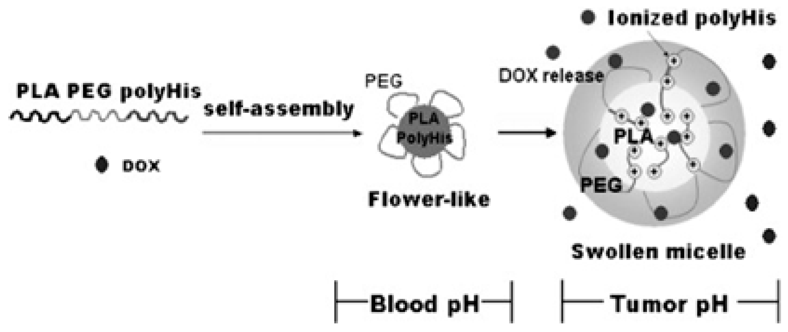
- Lee, E.S.; Oh, K.T.; Kim, D.; Youn, Y.S.; Bae, Y.H. Tumor pH-responsive flower-like micelles of poly(l-lactic acid)-b-poly(ethylene glycol)-b-poly(l-histidine). J. Control. Release 2007, 123, 19–26. [Google Scholar] [CrossRef] [PubMed]
- Hu, J.; Miura, S.; Na, K.; Bae, Y.H. pH-responsive and charge shielded cationic micelle of poly(l-histidine)-block-short branched PEI for acidic cancer treatment. J. Control. Release 2013, 172, 69–76. [Google Scholar] [CrossRef] [PubMed]
- Hong, W.; Chen, D.; Jia, L.; Gu, J.; Hu, H.; Zhao, X.; Qiao, M. Thermo- and pH-responsive copolymers based on PLGA-PEG-PLGA and poly(l-histidine): Synthesis and in vitro characterization of copolymer micelles. Acta Biomater. 2014, 10, 1259–1271. [Google Scholar] [CrossRef] [PubMed]
- Hwang, J.H.; Choi, C.W.; Kim, H.W.; Kim, D.H.; Kwak, T.W.; Lee, H.M.; Kim, C.H.; Chung, C.W.; Jeong, Y.I.; Kang, D.H. Dextran-b-poly(l-histidine) copolymer nanoparticles for ph-responsive drug delivery to tumor cells. Int. J. Nanomed. 2013, 8, 3197–3207. [Google Scholar]
- Qiu, L.; Li, Z.; Qiao, M.; Long, M.; Wang, M.; Zhang, X.; Tian, C.; Chen, D. Self-assembled pH-responsive hyaluronic acid–g-poly(l-histidine) copolymer micelles for targeted intracellular delivery of doxorubicin. Acta Biomater. 2014, 10, 2024–2035. [Google Scholar] [CrossRef] [PubMed]
- Bagherifam, S.; Skjeldal, F.M.; Griffiths, G.; Maelandsmo, G.M.; Engebraten, O.; Nystrom, B.; Hasirci, V.; Hasirci, N. pH-responsive nano carriers for doxorubicin delivery. Pharm. Res. 2015, 32, 1249–1263. [Google Scholar] [CrossRef]
- John, J.V.; Chung, C.-W.; Johnson, R.P.; Jeong, Y.-I.; Chung, K.-D.; Kang, D.H.; Suh, H.; Chen, H.; Kim, I. Dual Stimuli-Responsive Vesicular Nanospheres Fabricated by Lipopolymer Hybrids for Tumor-Targeted Photodynamic Therapy. Biomacromolecules 2016, 17, 20–31. [Google Scholar] [CrossRef] [PubMed]
- John, J.V.; Uthaman, S.; Augustine, R.; Manickavasagam Lekshmi, K.; Park, I.-K.; Kim, I. Biomimetic pH/redox dual stimuli-responsive zwitterionic polymer block poly(l-histidine) micelles for intracellular delivery of doxorubicin into tumor cells. J. Polym. Sci. Part A Polym. Chem. 2017, 55, 2061–2070. [Google Scholar] [CrossRef]
- John, J.V.; Uthaman, S.; Augustine, R.; Chen, H.; Park, I.-K.; Kim, I. pH/redox dual stimuli-responsive sheddable nanodaisies for efficient intracellular tumour-triggered drug delivery. J. Mater. Chem. B 2017, 5, 5027–5036. [Google Scholar] [CrossRef]
- Bilalis, P.; Varlas, S.; Kiafa, A.; Velentzas, A.; Stravopodis, D.; Iatrou, H. Preparation of hybrid triple-stimuli responsive nanogels based on poly(l-histidine). J. Polym. Sci. Part A Polym. Chem. 2016, 54, 1278–1288. [Google Scholar] [CrossRef]
- Kim, A.J.; Woodworth, G.F.; Boylan, N.J.; Suk, J.S.; Hanes, J. Highly compacted pH-responsive DNA nanoparticles mediate transgene silencing in experimental glioma. J. Mater. Chem. B 2014, 2, 8165–8173. [Google Scholar] [CrossRef] [PubMed]
- Lee, S.-J.; Jeong, Y.-I.L. Hybrid nanoparticles based on chlorin e6-conjugated hyaluronic acid/poly(l-histidine) copolymer for theranostic application to tumors. J. Mater. Chem. B 2018, 6, 2851–2859. [Google Scholar] [CrossRef]
- Thai, H.B.D.; Yu, J.K.; Park, Y.-J.; Ahn, D.-R. A dual-responsive pH-sensor and its potential as a universal probe for assays of pH-changing enzymes. Analyst 2015, 140, 2804–2809. [Google Scholar] [CrossRef] [PubMed]
- Li, Z.; Chen, Q.; Qi, Y.; Liu, Z.; Hao, T.; Sun, X.; Qiao, M.; Ma, X.; Xu, T.; Zhao, X.; et al. Rational Design of Multifunctional Polymeric Nanoparticles Based on Poly(l-histidine) and d-alpha-Vitamin E Succinate for Reversing Tumor Multidrug Resistance. Biomacromolecules 2018, 19, 2595–2609. [Google Scholar] [CrossRef] [PubMed]
- Hayashi, T.; Shinagawa, M.; Kawano, T.; Iwasaki, T. Drug delivery using polyhistidine peptide-modified liposomes that target endogenous lysosome. Biochem. Biophys. Res. Commun. 2018, 501, 648–653. [Google Scholar] [CrossRef] [PubMed]
- Tang, Q.; Zhao, D.; Yang, H.; Wang, L.; Zhang, X. A pH-responsive self-healing hydrogel based on multivalent coordination of Ni2+ with polyhistidine-terminated PEG and IDA-modified oligochitosan. J. Mater. Chem. B 2019, 7, 30–42. [Google Scholar] [CrossRef]
- Pan, J.; Lei, S.; Chang, L.; Wan, D. Smart pH-responsive nanoparticles in a model tumor microenvironment for enhanced cellular uptake. J. Mater. Sci. 2019, 54, 1692–1702. [Google Scholar] [CrossRef]
- Gao, Y.; Jia, L.; Wang, Q.; Hu, H.; Zhao, X.; Chen, D.; Qiao, M. pH/Redox Dual-Responsive Polyplex with Effective Endosomal Escape for Codelivery of siRNA and Doxorubicin against Drug-Resistant Cancer Cells. ACS Appl. Mater. Interfaces 2019, 11, 16296–16310. [Google Scholar] [CrossRef]
- Ohtake, K.; Natsume, H.; Ueda, H.; Morimoto, Y. Analysis of transient and reversible effects of poly-l-arginine on the in vivo nasal absorption of FITC-dextran in rats. J. Control. Release 2002, 82, 263–275. [Google Scholar] [CrossRef]
- De Koker, S.; De Geest, B.G.; Singh, S.K.; De Rycke, R.; Naessens, T.; Van Kooyk, Y.; Demeester, J.; De Smedt, S.C.; Grooten, J. Polyelectrolyte microcapsules as antigen delivery vehicles to dendritic cells: Uptake, processing, and cross-presentation of encapsulated antigens. Angew. Chem. Int. Ed. 2009, 48, 8485–8489. [Google Scholar] [CrossRef]
- Wedemeyer, H.; Schuller, E.; Schlaphoff, V.; Stauber, R.E.; Wiegand, J.; Schiefke, I.; Firbas, C.; Jilma, B.; Thursz, M.; Zeuzem, S.; et al. Therapeutic vaccine IC41 as late add-on to standard treatment in patients with chronic hepatitis C. Vaccine 2009, 27, 5142–5151. [Google Scholar] [CrossRef] [PubMed]
- Lozano, M.V.; Lollo, G.; Alonso-Nocelo, M.; Brea, J.; Vidal, A.; Torres, D.; Alonso, M.J. Polyarginine nanocapsules: A new platform for intracellular drug delivery. J. Nanopart. Res. 2013, 15, 1515. [Google Scholar] [CrossRef]
- Holowka, E.P.; Sun, V.Z.; Kamei, D.T.; Deming, T.J. Polyarginine segments in block copolypeptides drive both vesicular assembly and intracellular delivery. Nat. Mater. 2006, 6, 52–57. [Google Scholar] [CrossRef] [PubMed]
- Nemoto, E.; Ueda, H.; Akimoto, M.; Natsume, H.; Morimoto, Y. Ability of poly-l-arginine to enhance drug absorption into aqueous humor and vitreous body after instillation in rabbits. Biol. Pharm. Bull. 2007, 30, 1768–1772. [Google Scholar] [CrossRef] [PubMed]
- Rawat, A.; Yang, T.; Hussain, A.; Ahsan, F. Complexation of a Poly-l-Arginine with Low Molecular Weight Heparin Enhances Pulmonary Absorption of the Drug. Pharm. Res. 2008, 25, 936–948. [Google Scholar] [CrossRef] [PubMed]
- Heckl, S.; Sturzu, A.; Regenbogen, M.; Beck, A.; Feil, G.; Gharabaghi, A.; Echner, H. A Novel Polyarginine Containing Smac Peptide Conjugate that Mediates Cell Death in Tumor and Healthy Cells. Med. Chem. 2008, 4, 348–354. [Google Scholar] [CrossRef] [PubMed]
- Chen, A.Z.; Wang, S.B.; Weng, L.J.; Chen, M.Y. Preparation of Alginate/Poly(l-Arginine)-Chitosan Ternary Complex Microcapsules. J. Biomim. Biomater. Tissue Eng. 2009, 3, 25–35. [Google Scholar] [CrossRef]
- Park, K. Self-exploding microcapsules for pulsed drug delivery. J. Control. Release 2009, 135, 185. [Google Scholar] [CrossRef]
- Lehto, T.; Abes, R.; Oskolkov, N.; Suhorutšenko, J.; Copolovici, D.-M.; Mäger, I.; Viola, J.R.; Simonson, O.E.; Ezzat, K.; Guterstam, P.; et al. Delivery of nucleic acids with a stearylated (RxR)4 peptide using a non-covalent co-incubation strategy. J. Control. Release 2010, 141, 42–51. [Google Scholar] [CrossRef]
- Lemeshko, V.V. Potential-dependent membrane permeabilization and mitochondrial aggregation caused by anticancer polyarginine-KLA peptides. Arch. Biochem. Biophys. 2010, 493, 213–220. [Google Scholar] [CrossRef]
- Shah, P.P.; Desai, P.R.; Channer, D.; Singh, M. Enhanced skin permeation using polyarginine modified nanostructured lipid carriers. J. Control. Release 2012, 161, 735–745. [Google Scholar] [CrossRef] [PubMed]
- Desai, P.R.; Shah, P.P.; Hayden, P.; Singh, M. Investigation of Follicular and Non-follicular Pathways for Polyarginine and Oleic Acid-Modified Nanoparticles. Pharm. Res. 2013, 30, 1037–1049. [Google Scholar] [CrossRef] [PubMed]
- Oyarzun-Ampuero, F.A.; Goycoolea, F.M.; Torres, D.; Alonso, M.J. A new drug nanocarrier consisting of polyarginine and hyaluronic acid. Eur. J. Pharm. Biopharm. 2011, 79, 54–57. [Google Scholar] [CrossRef] [PubMed]
- Lollo, G.; Gonzalez-Paredes, A.; Garcia-Fuentes, M.; Calvo, P.; Torres, D.; Alonso, M.J. Polyarginine Nanocapsules as a Potential Oral Peptide Delivery Carrier. J. Pharm. Sci. 2017, 106, 611–618. [Google Scholar] [CrossRef] [PubMed]
- He, X.; Lin, M.; Guo, J.; Qu, Z.; Xu, F. Experimental and simulation studies of polyarginines across the membrane of giant unilamellar vesicles. RSC Adv. 2016, 6, 30454–30459. [Google Scholar] [CrossRef]
- Wang, T.Y.; Sun, Y.; Muthukrishnan, N.; Erazo-Oliveras, A.; Najjar, K.; Pellois, J.P. Membrane Oxidation Enables the Cytosolic Entry of Polyarginine Cell-penetrating Peptides. J. Biol. Chem. 2016, 291, 7902–7914. [Google Scholar] [CrossRef] [PubMed]
- Cui, Y.; Sui, J.; He, M.; Xu, Z.; Sun, Y.; Liang, J.; Fan, Y.; Zhang, X. Reduction-Degradable Polymeric Micelles Decorated with PArg for Improving Anticancer Drug Delivery Efficacy. ACS Appl. Mater. Interfaces 2016, 8, 2193–2203. [Google Scholar] [CrossRef] [PubMed]
- Niu, Z.; Tedesco, E.; Benetti, F.; Mabondzo, A.; Montagner, I.M.; Marigo, I.; Gonzalez-Touceda, D.; Tovar, S.; Dieguez, C.; Santander-Ortega, M.J.; et al. Rational design of polyarginine nanocapsules intended to help peptides overcoming intestinal barriers. J. Control. Release 2017, 263, 4–17. [Google Scholar] [CrossRef]
- Gonzalez-Paredes, A.; Torres, D.; Alonso, M.J. Polyarginine nanocapsules: A versatile nanocarrier with potential in transmucosal drug delivery. Int. J. Pharm. 2017, 529, 474–485. [Google Scholar] [CrossRef]
- Kim, H.-K.; Davaa, E.; Myung, C.-S.; Park, J.-S. Enhanced siRNA delivery using cationic liposomes with new polyarginine-conjugated PEG-lipid. Int. J. Pharm. 2010, 392, 141–147. [Google Scholar] [CrossRef]
- Zhao, Z.-X.; Gao, S.-Y.; Wang, J.-C.; Chen, C.-J.; Zhao, E.-Y.; Hou, W.-J.; Feng, Q.; Gao, L.-Y.; Liu, X.-Y.; Zhang, L.-R.; et al. Self-assembly nanomicelles based on cationic mPEG-PLA-b-Polyarginine(R15) triblock copolymer for siRNA delivery. Biomaterials 2012, 33, 6793–6807. [Google Scholar] [CrossRef] [PubMed]
- Zhang, T.; Xue, X.; He, D.; Hsieh, J.T. A prostate cancer-targeted polyarginine-disulfide linked PEI nanocarrier for delivery of microRNA. Cancer Lett. 2015, 365, 156–165. [Google Scholar] [CrossRef] [PubMed]
- Li, X.; Zhao, L.; Liang, Q.; Liang, Q.; Ye, J.; Komatsu, N.; Zhang, Q.; Gao, W.; Xu, M.; Chen, X. Cationic Polyarginine Conjugated Mesoporous Bioactive Glass Nanoparticles with Polyglycerol Coating for Efficient DNA Delivery. J. Biomed. Nanotechnol. 2017, 13, 280–289. [Google Scholar] [CrossRef] [PubMed]
- Marshall, J.; Wong, K.Y.; Rupasinghe, C.N.; Tiwari, R.; Zhao, X.; Berberoglu, E.D.; Sinkler, C.; Liu, J.; Lee, I.; Parang, K.; et al. Inhibition of N-Methyl-d-aspartate-induced Retinal Neuronal Death by Polyarginine Peptides Is Linked to the Attenuation of Stress-induced Hyperpolarization of the Inner Mitochondrial Membrane Potential. J. Biol. Chem. 2015, 290, 22030–22048. [Google Scholar] [CrossRef] [PubMed]
- Candan, G.; Michiue, H.; Ishikawa, S.; Fujimura, A.; Hayashi, K.; Uneda, A.; Mori, A.; Ohmori, I.; Nishiki, T.-i.; Matsui, H.; et al. Combining poly-arginine with the hydrophobic counter-anion 4-(1-pyrenyl)-butyric acid for protein transduction in transdermal delivery. Biomaterials 2012, 33, 6468–6475. [Google Scholar] [CrossRef]
- Movafegh, B.; Jalal, R.; Mohammadi, Z.; Aldaghi, S.A. Poly-l-arginine: Enhancing Cytotoxicity and Cellular Uptake of Doxorubicin and Necrotic Cell Death. Anticancer Agents Med. Chem. 2018, 18, 1448–1456. [Google Scholar] [CrossRef] [PubMed]
- Hu, J.; Lou, Y.; Wu, F. Improved Intracellular Delivery of Polyarginine Peptides with Cargoes. J. Phys. Chem. B 2019, 123, 2636–2644. [Google Scholar] [CrossRef]
- Tian, T.; Song, Y.; Li, K.; Sun, Y.; Wang, Q. Synthesis, Characterization, and Evaluation of Triptolide Cell-Penetrating Peptide Derivative for Transdermal Delivery of Triptolide. Mol. Pharm. 2018, 15, 560–570. [Google Scholar] [CrossRef]
- Rasmussen, D.D.; Ishizuka, B.; Quigley, M.E.; Yen, S.S. Effects of tyrosine and tryptophan ingestion on plasma catecholamine and 3,4-dihydroxyphenylacetic acid concentrations. J. Clin. Endocrinol. Metabol. 1983, 57, 760–763. [Google Scholar] [CrossRef]
- Reinstein, D.K.; Lehnert, H.; Wurtman, R.J. Dietary tyrosine suppresses the rise in plasma corticosterone following acute stress in rats. Life Sci. 1985, 37, 2157–2163. [Google Scholar] [CrossRef]
- Chadha, H.; Deluca, P.P. Effect of Polytyrosine on the Hydrophobicity of Hydroxyethyl Starch Microspheres. Pharm. Dev. Technol. 1998, 3, 597–606. [Google Scholar] [CrossRef] [PubMed]
- Gitsov, I.; Wang, L.; Vladimirov, N.; Simonyan, A.; Kiemle, D.J.; Schutz, A. “Green” Synthesis of Unnatural Poly(Amino Acid)s with Zwitterionic Character and pH-Responsive Solution Behavior, Mediated by Linear–Dendritic Laccase Complexes. Biomacromolecules 2014, 15, 4082–4095. [Google Scholar] [CrossRef] [PubMed]
- Lewandowski, A.T.; Small, D.A.; Chen, T.; Payne, G.F.; Bentley, W.E. Tyrosine-based “Activatable Pro-Tag”: Enzyme-catalyzed protein capture and release. Biotechnol. Bioeng. 2006, 93, 1207–1215. [Google Scholar] [CrossRef] [PubMed]
- Xue, S.; Gu, X.; Zhang, J.; Sun, H.; Deng, C.; Zhong, Z. Construction of Small-Sized, Robust, and Reduction-Responsive Polypeptide Micelles for High Loading and Targeted Delivery of Chemotherapeutics. Biomacromolecules 2018, 19, 3586–3593. [Google Scholar] [CrossRef] [PubMed]
- Gu, X.; Wei, Y.; Fan, Q.; Sun, H.; Cheng, R.; Zhong, Z.; Deng, C. cRGD-decorated biodegradable polytyrosine nanoparticles for robust encapsulation and targeted delivery of doxorubicin to colorectal cancer in vivo. J. Control. Release 2019, 301, 110–118. [Google Scholar] [CrossRef] [PubMed]
- Williamson, M.P. The structure and function of proline-rich regions in proteins. Biochem. J. 1994, 297, 249–260. [Google Scholar] [CrossRef]
- Deber, C.M.; Bovey, F.A.; Carver, J.P.; Blout, E.R. Nuclear magnetic resonance evidence for cis-peptide bonds in proline oligomers. J. Am. Chem. Soc. 1970, 92, 6191–6198. [Google Scholar] [CrossRef]
- Salinas, Y.; Castilla, A.M.; Resmini, M. An l-proline based thermoresponsive and pH-switchable nanogel as a drug delivery vehicle. Polym. Chem. 2018, 9, 2271–2280. [Google Scholar] [CrossRef]
- Lin, C.-H.; Wen, H.-C.; Chiang, C.-C.; Huang, J.-S.; Chen, Y.; Wang, S.-K. Polyproline Tri-Helix Macrocycles as Nanosized Scaffolds to Control Ligand Patterns for Selective Protein Oligomer Interactions. Small 2019, 15, 1900561. [Google Scholar] [CrossRef]
- Sugimoto, H.; Nakanishi, E.; Hanai, T.; Yasumura, T.; Inomata, K. Aggregate formation and release behaviour of hydrophobic drugs with graft copolypeptide-containing tryptophan. Polym. Int. 2004, 53, 972–983. [Google Scholar] [CrossRef]
- Nakanishi, E.; Yamuchi, F.; Hanai, T.; Sakamoto, Y. Controlled Release loading model drugs by pH-resoponsive polypeptide Aggregates. Toyota Kenkyu Hokuku 2001, 54, 61–67. [Google Scholar]
- Bajpai, A.K.; Shukla, S.K.; Bhanu, S.; Kankane, S. Responsive polymers in controlled drug delivery. Prog. Polym. Sci. 2008, 33, 1088–1118. [Google Scholar] [CrossRef]
- Mano, J.F. Stimuli-Responsive Polymeric Systems for Biomedical Applications. Adv. Eng. Mater. 2008, 10, 515–527. [Google Scholar] [CrossRef]
- Bawa, P.; Pillay, V.; Choonara, Y.E.; du Toit, L.C. Stimuli-responsive polymers and their applications in drug delivery. Biomed. Mater. 2009, 4, 022001. [Google Scholar] [CrossRef] [PubMed]
- Bazban-Shotorbani, S.; Hasani-Sadrabadi, M.M.; Karkhaneh, A.; Serpooshan, V.; Jacob, K.I.; Moshaverinia, A.; Mahmoudi, M. Revisiting structure-property relationship of pH-responsive polymers for drug delivery applications. J. Control. Release 2017, 253, 46–63. [Google Scholar] [CrossRef] [PubMed]
- Krogstad, D.V.; Wang, D.; Lin-Gibson, S. Polyaspartic Acid Concentration Controls the Rate of Calcium Phosphate Nanorod Formation in High Concentration Systems. Biomacromolecules 2017, 18, 3106–3113. [Google Scholar] [CrossRef] [PubMed]
- Shi, S.; Wu, Y.; Wang, Y.; Yu, J.; Xu, Y. Synthesis and characterization of a biodegradable polyaspartic acid/2-amino-2-methyl-1-propanol graft copolymer and evaluation of its scale and corrosion inhibition performance. RSC Adv. 2017, 7, 36714–36721. [Google Scholar] [CrossRef]
- Liu, T.; Wang, R.; Cao, H.; Lin, A. Polyaspartic acid alleviates heavy metal toxicity in zebrafish (Danio rerio). Chem. Ecol. 2017, 33, 684–693. [Google Scholar] [CrossRef]
- Sharma, S.; Dua, A.; Malik, A. Biocompatible stimuli responsive superabsorbent polymer for controlled release of GHK-Cu peptide for wound dressing application. J. Polym. Res. 2017, 24, 104. [Google Scholar] [CrossRef]
- Zohuriaan-Mehr, M.J.; Pourjavadi, A.; Salimi, H.; Kurdtabar, M. Protein- and homo poly(amino acid)-based hydrogels with super-swelling properties. Polym. Adv. Technol. 2009, 20, 655–671. [Google Scholar] [CrossRef]
- Prabaharan, M.; Grailer, J.J.; Pilla, S.; Steeber, D.A.; Gong, S. Amphiphilic multi-arm-block copolymer conjugated with doxorubicin via pH-sensitive hydrazone bond for tumor-targeted drug delivery. Biomaterials 2009, 30, 5757–5766. [Google Scholar] [CrossRef] [PubMed]
- Zhao, Y.; Kang, J.; Tan, T. Salt-, pH- and temperature-responsive semi-interpenetrating polymer network hydrogel based on poly(aspartic acid) and poly(acrylic acid). Polymer 2006, 47, 7702–7710. [Google Scholar] [CrossRef]
- Yang, J.; Fang, L.; Wang, F.; Tan, T. Preparation and characterization of a novel pH-, thermo-, and ionic strength-responsive hydrogels based on xanthan gum–poly(aspartic acid). J. Appl. Polym. Sci. 2007, 105, 539–546. [Google Scholar] [CrossRef]
- Moon, J.R.; Kim, J.-H. Biodegradable thermo- and pH-responsive hydrogels based on amphiphilic polyaspartamide derivatives containing N,N-Diisopropylamine pendants. Macromol. Res. 2008, 16, 489–491. [Google Scholar] [CrossRef]
- Moon, J.R.; Park, Y.H.; Kim, J.-H. Synthesis and characterization of novel thermo- and pH-responsive copolymers based on amphiphilic polyaspartamides. J. Appl. Polym. Sci. 2009, 111, 998–1004. [Google Scholar] [CrossRef]
- Liu, M.; Su, H.; Tan, T. Synthesis and properties of thermo- and pH-sensitive poly(N-isopropylacrylamide)/polyaspartic acid IPN hydrogels. Carbohyd. Polym. 2012, 87, 2425–2431. [Google Scholar] [CrossRef]
- Némethy, Á.; Solti, K.; Kiss, L.; Gyarmati, B.; Deli, M.A.; Csányi, E.; Szilágyi, A. pH- and temperature-responsive poly(aspartic acid)-l-poly(N-isopropylacrylamide) conetwork hydrogel. Eur. Polym. J. 2013, 49, 2392–2403. [Google Scholar] [CrossRef]
- Liu, C.; Chen, Y.; Chen, J. Synthesis and characteristics of pH-sensitive semi-interpenetrating polymer network hydrogels based on konjac glucomannan and poly(aspartic acid) for in vitro drug delivery. Carbohyd. Polym. 2010, 79, 500–506. [Google Scholar] [CrossRef]
- Huang, H.; Li, Y.; Sa, Z.; Sun, Y.; Wang, Y.; Wang, J. A smart drug delivery system from charge-conversion polymer-drug conjugate for enhancing tumor therapy and tunable drug release. Macromol. Biosci. 2014, 14, 485–490. [Google Scholar] [CrossRef]
- Budai-Szu Cs, M.; Horvat, G.; Gyarmati, B.; Szilagyi, B.A.; Szilagyi, A.; Csihi, T.; Berko, S.; Szabo-Revesz, P.; Mori, M.; Sandri, G.; et al. In vitro testing of thiolated poly(aspartic acid) from ophthalmic formulation aspects. Drug Dev. Ind. Pharm. 2016, 42, 1241–1246. [Google Scholar] [CrossRef]
- Carbone, E.J.; Rajpura, K.; Jiang, T.; Kan, H.-M.; Yu, X.; Lo, K.W.H. Osteotropic Nanoscale Drug Delivery System via a Single Aspartic Acid as the Bone-Targeting Moiety. J. Nanosci. Nanotechnol. 2017, 17, 1747–1752. [Google Scholar] [CrossRef]
- Licciardi, M.; Paolino, D.; Celia, C.; Giammona, G.; Cavallaro, G.; Fresta, M. Folate-targeted supramolecular vesicular aggregates based on polyaspartyl-hydrazide copolymers for the selective delivery of antitumoral drugs. Biomaterials 2010, 31, 7340–7354. [Google Scholar] [CrossRef] [PubMed]
- Mandracchia, D.; Denora, N.; Franco, M.; Pitarresi, G.; Giammona, G.; Trapani, G. New Biodegradable Hydrogels Based on Inulin and α,β-Polyaspartylhydrazide Designed for Colonic Drug Delivery: In Vitro Release of Glutathione and Oxytocin. J. Biomater. Sci. Polym. Ed. 2011, 22, 313–328. [Google Scholar] [CrossRef] [PubMed][Green Version]
- Ma-Ham, A.; Wu, H.; Wang, J.; Kang, X.; Zhang, Y.; Lin, Y. Apoferritin-based nanomedicine platform for drug delivery: Equilibrium binding study of daunomycin with DNA. J. Mater. Chem. 2011, 21, 8700–8708. [Google Scholar] [CrossRef]
- Liu, C.; Gan, X.; Chen, Y. A novel pH-sensitive hydrogels for potential colon-specific drug delivery: Characterization and in vitro release studies. Starch 2011, 63, 503–511. [Google Scholar] [CrossRef]
- Wang, X.; Wu, G.; He, T.; Wang, Y.; Wang, Y.; Fan, Y.; Gao, H.; Ma, J. Novel pH-sensitive zwitterionic poly(amino acid) derivatives for drug delivery. J. Control. Release 2011, 152 (Suppl. 1), e93–e94. [Google Scholar] [CrossRef]
- Rios-Doria, J.; Carie, A.; Costich, T.; Burke, B.; Skaff, H.; Panicucci, R.; Sill, K. A Versatile Polymer Micelle Drug Delivery System for Encapsulation and In Vivo Stabilization of Hydrophobic Anticancer Drugs. J. Drug Deliv. 2012, 2012, 8. [Google Scholar] [CrossRef] [PubMed]
- Han, S.; Liu, Y.; Nie, X.; Xu, Q.; Jiao, F.; Li, W.; Zhao, Y.; Wu, Y.; Chen, C. Efficient Delivery of Antitumor Drug to the Nuclei of Tumor Cells by Amphiphilic Biodegradable Poly(l-Aspartic Acid-co-Lactic Acid)/DPPE Co-Polymer Nanoparticles. Small 2012, 8, 1596–1606. [Google Scholar] [CrossRef]
- Wang, X.; Wu, G.; Lu, C.; Zhao, W.; Wang, Y.; Fan, Y.; Gao, H.; Ma, J. A novel delivery system of doxorubicin with high load and pH-responsive release from the nanoparticles of poly (alpha,beta-aspartic acid) derivative. Eur. J. Pharm. Sci. 2012, 47, 256–264. [Google Scholar] [CrossRef]
- Yoon, S.R.; Yang, H.M.; Park, C.W.; Lim, S.; Chung, B.H.; Kim, J.D. Charge-conversional poly(amino acid)s derivatives as a drug delivery carrier in response to the tumor environment. J. Biomed. Mater. Res. Part A 2012, 100, 2027–2033. [Google Scholar] [CrossRef]
- Hsu, S.-P.; Chu, I.M. Design of polyanionic nanocarriers based on modified poly (aspartic acid)s for oral administration: Synthesis and characterization. J. Polym. Res. 2012, 19, 9913. [Google Scholar] [CrossRef]
- Zhang, J.; Yuan, Z.-F.; Wang, Y.; Chen, W.-H.; Luo, G.-F.; Cheng, S.-X.; Zhuo, R.-X.; Zhang, X.-Z. Multifunctional Envelope-Type Mesoporous Silica Nanoparticles for Tumor-Triggered Targeting Drug Delivery. J. Am. Chem. Soc. 2013, 135, 5068–5073. [Google Scholar] [CrossRef] [PubMed]
- Tran, T.H.; Ramasamy, T.; Choi, J.Y.; Nguyen, H.T.; Pham, T.T.; Jeong, J.H.; Ku, S.K.; Choi, H.G.; Yong, C.S.; Kim, J.O. Tumor-targeting, pH-sensitive nanoparticles for docetaxel delivery to drug-resistant cancer cells. Int. J. Nanomed. 2015, 10, 5249–5262. [Google Scholar]
- Zhao, Y.; Su, H.; Fang, L.; Tan, T. Superabsorbent hydrogels from poly(aspartic acid) with salt-, temperature- and pH-responsiveness properties. Polymer 2005, 46, 5368–5376. [Google Scholar] [CrossRef]
- Gyarmati, B.; Vajna, B.; Nemethy, A.; Laszlo, K.; Szilagyi, A. Redox- and pH-responsive cysteamine-modified poly(aspartic acid) showing a reversible sol-gel transition. Macromol. Biosci. 2013, 13, 633–640. [Google Scholar] [CrossRef] [PubMed]
- Gyarmati, B.; Némethy, Á.; Szilágyi, A. Reversible response of poly(aspartic acid) hydrogels to external redox and pH stimuli. RSC Adv. 2014, 4, 8764–8771. [Google Scholar] [CrossRef]
- Quader, S.; Cabral, H.; Mochida, Y.; Ishii, T.; Liu, X.; Toh, K.; Kinoh, H.; Miura, Y.; Nishiyama, N.; Kataoka, K. Selective intracellular delivery of proteasome inhibitors through pH-sensitive polymeric micelles directed to efficient antitumor therapy. J. Control. Release 2014, 188, 67–77. [Google Scholar] [CrossRef]
- Lu, C.; Li, B.; Liu, N.; Wu, G.; Gao, H.; Ma, J. A hydrazone crosslinked zwitterionic polypeptide nanogel as a platform for controlled drug delivery. RSC Adv. 2014, 4, 50301–50311. [Google Scholar] [CrossRef]
- Hill, M.R.; MacKrell, E.J.; Forsthoefel, C.P.; Jensen, S.P.; Chen, M.; Moore, G.A.; He, Z.L.; Sumerlin, B.S. Biodegradable and pH-Responsive Nanoparticles Designed for Site-Specific Delivery in Agriculture. Biomacromolecules 2015, 16, 1276–1282. [Google Scholar] [CrossRef]
- Johnson, R.P.; Uthaman, S.; Augustine, R.; Zhang, Y.; Jin, H.; Choi, C.I.; Park, I.-K.; Kim, I. Glutathione and endosomal pH-responsive hybrid vesicles fabricated by zwitterionic polymer block poly(l-aspartic acid) as a smart anticancer delivery platform. React. Funct. Polym. 2017, 119, 47–56. [Google Scholar] [CrossRef]
- Cheng, H.; Li, Y.Y.; Zeng, X.; Sun, Y.X.; Zhang, X.Z.; Zhuo, R.X. Protamine sulfate/poly(l-aspartic acid) polyionic complexes self-assembled via electrostatic attractions for combined delivery of drug and gene. Biomaterials 2009, 30, 1246–1253. [Google Scholar] [CrossRef] [PubMed]
- Wang, T.; Xu, Q.; Wu, Y.; Zeng, A.; Li, M.; Gao, H. Quaternized chitosan (QCS)/poly (aspartic acid) nanoparticles as a protein drug-delivery system. Carbohyd. Res. 2009, 344, 908–914. [Google Scholar] [CrossRef] [PubMed]
- Hakeem, A.; Zahid, F.; Zhan, G.; Yi, P.; Yang, H.; Gan, L.; Yang, X. Polyaspartic acid-anchored mesoporous silica nanoparticles for pH-responsive doxorubicin release. Int. J. Nanomed. 2018, 13, 1029–1040. [Google Scholar] [CrossRef] [PubMed]
- Zhou, M.; Hou, T.; Li, J.; Yu, S.; Xu, Z.; Yin, M.; Wang, J.; Wang, X. Self-Propelled and Targeted Drug Delivery of Poly(aspartic acid)/Iron-Zinc Microrocket in the Stomach. ACS Nano 2019, 13, 1324–1332. [Google Scholar] [CrossRef] [PubMed]
- Debnath, K.; Jana, N.R.; Jana, N.R. Designed Polymer Micelle for Clearing Amyloid Protein Aggregates via Up-Regulated Autophagy. ACS Biomater. Sci. Eng. 2019, 5, 390–401. [Google Scholar] [CrossRef]
- Niwa, M.; Morikawa, M.; Higashi, N. Controllable Orientation of Helical Poly(l-glutamic acid) Rods through Macrodipole Interaction on Gold Surfaces and Vectorial Electron Transfer. Angew. Chem. Int. Ed. 2000, 39, 960–963. [Google Scholar] [CrossRef]
- Niwa, M.; Morikawa, M.; Higashi, N. Discrimination between N- and C-Termini of Polypeptides by a Two-Dimensional Array of Helical Poly(l-glutamic acid) Rods on Gold Surfaces. Langmuir 1999, 15, 5088–5092. [Google Scholar] [CrossRef]
- Niwa, M.; Morikawa, M.-a.; Nabeta, T.; Higashi, N. Preparation of Anthryl Group-Tagged Helical Poly(γ-benzyl l-glutamate) Self-Assembled Film on Gold Surface and Its Interaction with DNA. Macromolecules 2002, 35, 2769–2775. [Google Scholar] [CrossRef]
- Higashi, N.; Shosu, T.; Koga, T.; Niwa, M.; Tanigawa, T. pH-responsive, self-assembling nanoparticle from a fullerene-tagged poly(l-glutamic acid) and its superoxide dismutase mimetic property. J. Colloid Interface sci. 2006, 298, 118–123. [Google Scholar] [CrossRef]
- Zhao, C.; He, P.; Xiao, C.; Gao, X.; Zhuang, X.; Chen, X. Synthesis of temperature and pH-responsive crosslinked micelles from polypeptide-based graft copolymer. J. Colloid Interface Sci. 2011, 359, 436–442. [Google Scholar] [CrossRef]
- Yang, N.; Wang, Y.; Zhang, Q.; Chen, L.; Zhao, Y. γ-Polyglutamic acid mediated crosslinking PNIPAAm-based thermo/pH-responsive hydrogels for controlled drug release. Polym. Degrad. Stab. 2017, 144, 53–61. [Google Scholar] [CrossRef]
- Zhao, C.; He, P.; Xiao, C.; Gao, X.; Zhuang, X.; Chen, X. Photo-cross-linked biodegradable thermo- and pH-responsive hydrogels for controlled drug release. J.Appl. Polym. Sci. 2012, 123, 2923–2932. [Google Scholar] [CrossRef]
- Ding, J.; Xiao, C.; Yan, L.; Tang, Z.; Zhuang, X.; Chen, X.; Jing, X. pH and dual redox responsive nanogel based on poly(l-glutamic acid) as potential intracellular drug carrier. J. Control. Release 2011, 152 (Suppl. 1), e11–e13. [Google Scholar] [CrossRef]
- Yan, Y.; Liao, L.; Liu, D. Poly(l-glutamic acid)-based star-block copolymers as pH-responsive release systems. J. Control. Release 2011, 152 (Suppl. 1), e60–e61. [Google Scholar] [CrossRef]
- Ding, J.; Shi, F.; Li, D.; Chen, L.; Zhuang, X.; Chen, X. Enhanced endocytosis of acid-sensitive doxorubicin derivatives with intelligent nanogel for improved security and efficacy. Biomater. Sci. 2013, 1, 633–646. [Google Scholar] [CrossRef]
- Mildner, R.; Menzel, H. Hydrophobic Spacers Enhance the Helicity and Lectin Binding of Synthetic, pH-Responsive Glycopolypeptides. Biomacromolecules 2014, 15, 4528–4533. [Google Scholar] [CrossRef] [PubMed]
- Liu, X.; Su, S.; Wei, F.; Rong, X.; Yang, Z.; Liu, J.; Li, M.; Wu, Y. Construction of nanoparticles based on amphiphilic copolymers of poly(gamma-glutamic acid co-l-lactide)-1,2-dipalmitoyl-sn-glycero-3-phosphoethanolamine as a potential drug delivery carrier. J. Colloid Interface Sci. 2014, 413, 54–64. [Google Scholar] [CrossRef]
- Dai, X.-H.; Yang, W.-H.; Wu, C.; Jin, H.; Chang, D.-D.; Dai, Y.-R.; Pan, J.-M.; Yan, Y.-S. Synthesis and Characterization of Star-Shaped Porphyrin-cored Poly(Glutamic Acid) Conjugates as Highly Efficient Photosensitizers. J. Photopolym. Sci. Technol. 2016, 29, 823–829. [Google Scholar] [CrossRef][Green Version]
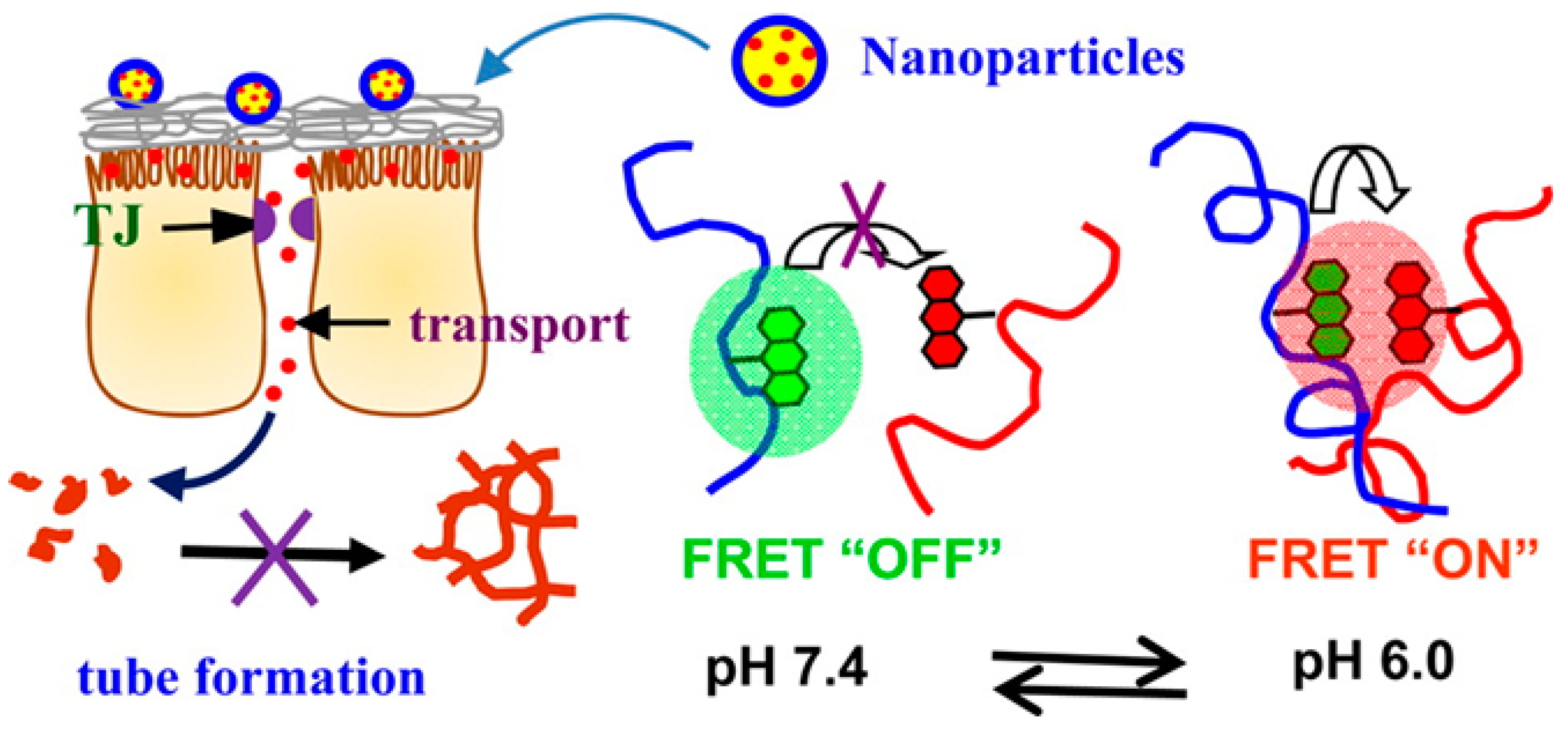
- Lu, K.Y.; Lin, C.W.; Hsu, C.H.; Ho, Y.C.; Chuang, E.Y.; Sung, H.W.; Mi, F.L. FRET-based dual-emission and pH-responsive nanocarriers for enhanced delivery of protein across intestinal epithelial cell barrier. ACS Appl. Mater. Interfaces 2014, 6, 18275–18289. [Google Scholar] [CrossRef]
- Urimi, D.; Agrawal, A.K.; Kushwah, V.; Jain, S. Polyglutamic Acid Functionalization of Chitosan Nanoparticles Enhances the Therapeutic Efficacy of Insulin Following Oral Administration. AAPS Pharm. Sci. Tech. 2019, 20, 131. [Google Scholar] [CrossRef]
- Štaka, I.; Cadete, A.; Surikutchi, B.T.; Abuzaid, H.; Bradshaw, T.D.; Alonso, M.J.; Marlow, M. A novel low molecular weight nanocomposite hydrogel formulation for intra-tumoural delivery of anti-cancer drugs. Int. J. Pharm. 2019, 565, 151–161. [Google Scholar] [CrossRef] [PubMed]
- Huang, M.; Zhang, S.F.; Lu, S.; Qi, T.; Yan, J.; Gao, C.; Liu, M.; Li, T.; Ji, Y. Synthesis of mesoporous silica/polyglutamic acid peptide dendrimer with dual targeting and its application in dissolving thrombus. J. Mater. Res. Part A 2019, 107, 1824–1831. [Google Scholar] [CrossRef] [PubMed]
- Liu, L.; Yao, W.; Rao, Y.; Lu, X.; Gao, J. pH-Responsive carriers for oral drug delivery: Challenges and opportunities of current platforms. Drug Deliv. 2017, 24, 569–581. [Google Scholar] [CrossRef] [PubMed]
- Lin, Y.H.; Sonaje, K.; Lin, K.M.; Juang, J.H.; Mi, F.L.; Yang, H.W.; Sung, H.W. Multi-ion-crosslinked nanoparticles with pH-responsive characteristics for oral delivery of protein drugs. J. Control. Release 2008, 132, 141–149. [Google Scholar] [CrossRef] [PubMed]
- Zheng, Y.; Yang, W.; Wang, C.; Hu, J.; Fu, S.; Dong, L.; Wu, L.; Shen, X. Nanoparticles based on the complex of chitosan and polyaspartic acid sodium salt: Preparation, characterization and the use for 5-fluorouracil delivery. Eur. J. Pharm. Biopharm. 2007, 67, 621–631. [Google Scholar] [CrossRef] [PubMed]
- Sonaje, K.; Lin, K.J.; Wey, S.P.; Lin, C.K.; Yeh, T.H.; Nguyen, H.N.; Hsu, C.W.; Yen, T.C.; Juang, J.H.; Sung, H.W. Biodistribution, pharmacodynamics and pharmacokinetics of insulin analogues in a rat model: Oral delivery using pH-responsive nanoparticles vs. subcutaneous injection. Biomaterials 2010, 31, 6849–6858. [Google Scholar] [CrossRef] [PubMed]
- Chang, C.H.; Lin, Y.H.; Yeh, C.L.; Chen, Y.C.; Chiou, S.F.; Hsu, Y.M.; Chen, Y.S.; Wang, C.C. Nanoparticles incorporated in pH-sensitive hydrogels as amoxicillin delivery for eradication of Helicobacter pylori. Biomacromolecules 2010, 11, 133–142. [Google Scholar] [CrossRef]
- Deng, L.; Dong, H.; Dong, A.; Zhang, J. A strategy for oral chemotherapy via dual pH-sensitive polyelectrolyte complex nanoparticles to achieve gastric survivability, intestinal permeability, hemodynamic stability and intracellular activity. Eur. J. Pharm. Biopharm. 2015, 97 (Pt A), 107–117. [Google Scholar] [CrossRef] [PubMed]
- Wang, Q.M.; Gao, Z.; Liu, S.; Fan, B.; Kang, L.; Huang, W.; Jin, M. Hybrid polymeric micelles based on bioactive polypeptides as pH-responsive delivery systems against melanoma. Biomaterials 2014, 35, 7008–7021. [Google Scholar] [CrossRef]
- Liu, N.; Li, B.; Gong, C.; Liu, Y.; Wang, Y.; Wu, G. A pH- and thermo-responsive poly(amino acid)-based drug delivery system. Colloids Surf. B 2015, 136, 562–569. [Google Scholar] [CrossRef]
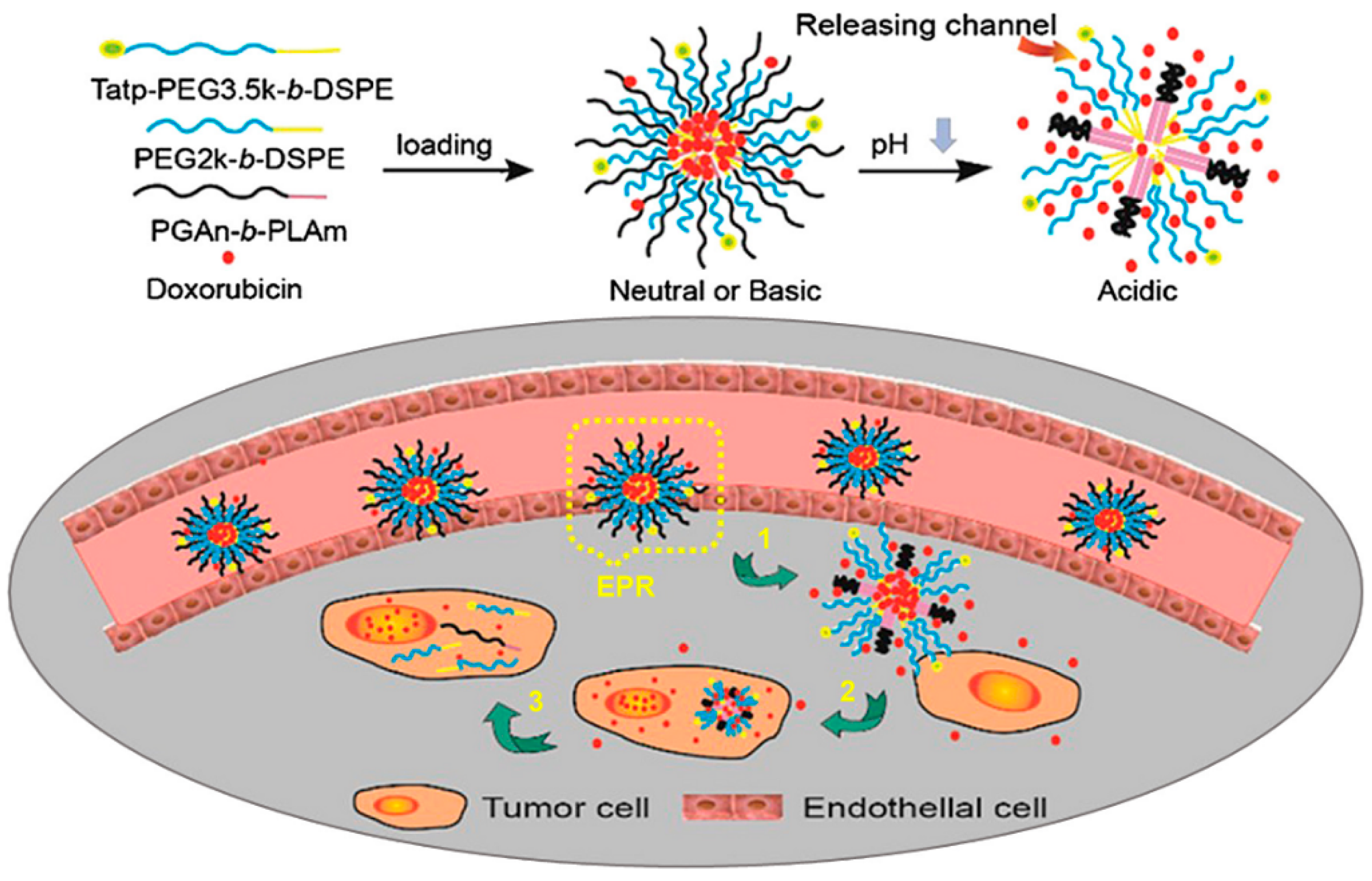
- Dong, Y.; Yang, J.; Liu, H.; Wang, T.; Tang, S.; Zhang, J.; Zhang, X. Site-Specific Drug-Releasing Polypeptide Nanocarriers Based on Dual-pH Response for Enhanced Therapeutic Efficacy against Drug-Resistant Tumors. Theranostics 2015, 5, 890–904. [Google Scholar] [CrossRef] [PubMed]
- Murphy, R.F.; Powers, S.; Cantor, C.R. Endosome pH measured in single cells by dual fluorescence flow cytometry: Rapid acidification of insulin to pH 6. J. Cell Biol. 1984, 98, 1757–1762. [Google Scholar] [CrossRef] [PubMed]
- Benjaminsen, R.V.; Mattebjerg, M.A.; Henriksen, J.R.; Moghimi, S.M.; Andresen, T.L. The possible “proton sponge “effect of polyethylenimine (PEI) does not include change in lysosomal pH. Mol. Ther. 2013, 21, 149–157. [Google Scholar] [CrossRef] [PubMed]
- Chiang, W.H.; Huang, W.C.; Shen, M.Y.; Wang, C.H.; Huang, Y.F.; Lin, S.C.; Chern, C.S.; Chiu, H.C. Dual-layered nanogel-coated hollow lipid/polypeptide conjugate assemblies for potential pH-triggered intracellular drug release. PLoS ONE 2014, 9, e92268. [Google Scholar] [CrossRef] [PubMed]
- Gao, Y.; Zhang, C.; Zhou, Y.; Li, J.; Zhao, L.; Li, Y.; Liu, Y.; Li, X. Endosomal pH-Responsive Polymer-Based Dual-Ligand-Modified Micellar Nanoparticles for Tumor Targeted Delivery and Facilitated Intracellular Release of Paclitaxel. Pharm. Res. 2015, 32, 2649–2662. [Google Scholar] [CrossRef] [PubMed]
- Chen, P.; Qiu, M.; Deng, C.; Meng, F.; Zhang, J.; Cheng, R.; Zhong, Z. pH-Responsive Chimaeric Pepsomes Based on Asymmetric Poly(ethylene glycol)-b-Poly(l-leucine)-b-Poly(l-glutamic acid) Triblock Copolymer for Efficient Loading and Active Intracellular Delivery of Doxorubicin Hydrochloride. Biomacromolecules 2015, 16, 1322–1330. [Google Scholar] [CrossRef] [PubMed]
- Sacchetti, F.; D’Arca, D.; Genovese, F.; Pacifico, S.; Maretti, E.; Hanuskova, M.; Iannuccelli, V.; Costi, M.P.; Leo, E. Conveying a newly designed hydrophilic anti-human thymidylate synthase peptide to cisplatin resistant cancer cells: Are pH-sensitive liposomes more effective than conventional ones? Drug Dev. Ind. Pharm. 2017, 43, 465–473. [Google Scholar] [CrossRef]
- Yang, Z.; Li, Y.; Gao, J.; Cao, Z.; Jiang, Q.; Liu, J. pH and redox dual-responsive multifunctional gene delivery with enhanced capability of transporting DNA into the nucleus. Colloids Surf. B 2017, 153, 111–122. [Google Scholar] [CrossRef]
- Wang, S.; Chen, R. pH-Responsive, Lysine-Based, Hyperbranched Polymers Mimicking Endosomolytic Cell-Penetrating Peptides for Efficient Intracellular Delivery. Chem. Mater. 2017, 29, 5806–5815. [Google Scholar] [CrossRef]
- Guo, X.; Cheng, Y.; Zhao, X.; Luo, Y.; Chen, J.; Yuan, W.-E. Advances in redox-responsive drug delivery systems of tumor microenvironment. J. Nanobiotech. 2018, 16, 74. [Google Scholar] [CrossRef]
- Peng, M.-Y.; Zheng, D.-W.; Wang, S.-B.; Cheng, S.-X.; Zhang, X.-Z. Multifunctional Nanosystem for Synergistic Tumor Therapy Delivered by Two-Dimensional MoS2. ACS Appl. Mater. Interfaces 2017, 9, 13965–13975. [Google Scholar] [CrossRef] [PubMed]
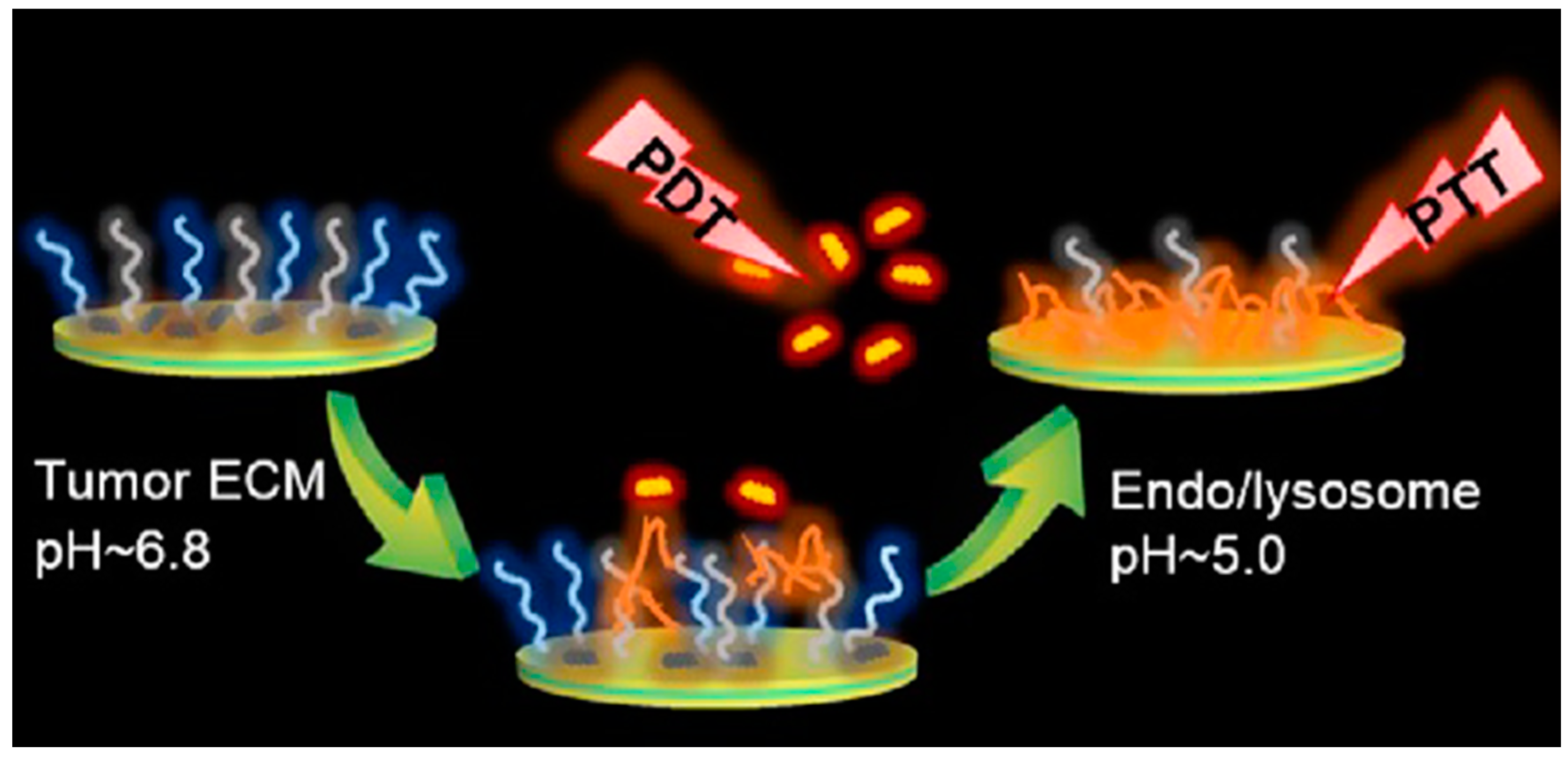
- Zhang, N.; Zhao, F.; Zou, Q.; Li, Y.; Ma, G.; Yan, X. Multitriggered Tumor-Responsive Drug Delivery Vehicles Based on Protein and Polypeptide Coassembly for Enhanced Photodynamic Tumor Ablation. Small 2016, 12, 5936–5943. [Google Scholar] [CrossRef] [PubMed]
- Yang, Y.; Xie, X.; Yang, Y.; Li, Z.; Yu, F.; Gong, W.; Li, Y.; Zhang, H.; Wang, Z.; Mei, X. Polymer Nanoparticles Modified with Photo- and pH-Dual-Responsive Polypeptides for Enhanced and Targeted Cancer Therapy. Mol. Pharm. 2016, 13, 1508–1519. [Google Scholar] [CrossRef] [PubMed]
- Hollingshead, S.; Lin, C.Y.; Liu, J.C. Designing Smart Materials with Recombinant Proteins. Macromol. Biosci. 2017, 17, 1600554. [Google Scholar] [CrossRef] [PubMed]
- Ma, G.; Lin, W.; Yuan, Z.; Wu, J.; Qian, H.; Xu, L.; Chen, S. Development of ionic strength/pH/enzyme triple-responsive zwitterionic hydrogel of the mixed l-glutamic acid and l-lysine polypeptide for site-specific drug delivery. J. Mater. Chem. B 2017, 5, 935–943. [Google Scholar] [CrossRef]
- Koga, T.; Mima, K.; Matsumoto, T.; Higashi, N. Amino Acid-derived Polymer with Changeable Enzyme Degradability based on pH-induced Structural Conversion from Polyester to Polypeptide. Chem. Lett. 2015, 44, 1720–1722. [Google Scholar] [CrossRef]
Sample Availability: Samples of the compounds are not available. |


| Polymers a | Micelle Types | Diameter | Ref. |
|---|---|---|---|
| PEG-b-PLL | Sphere | ~65 nm | [47] |
| PPO-b-PLL | Vesicle | ~80 nm | [49] |
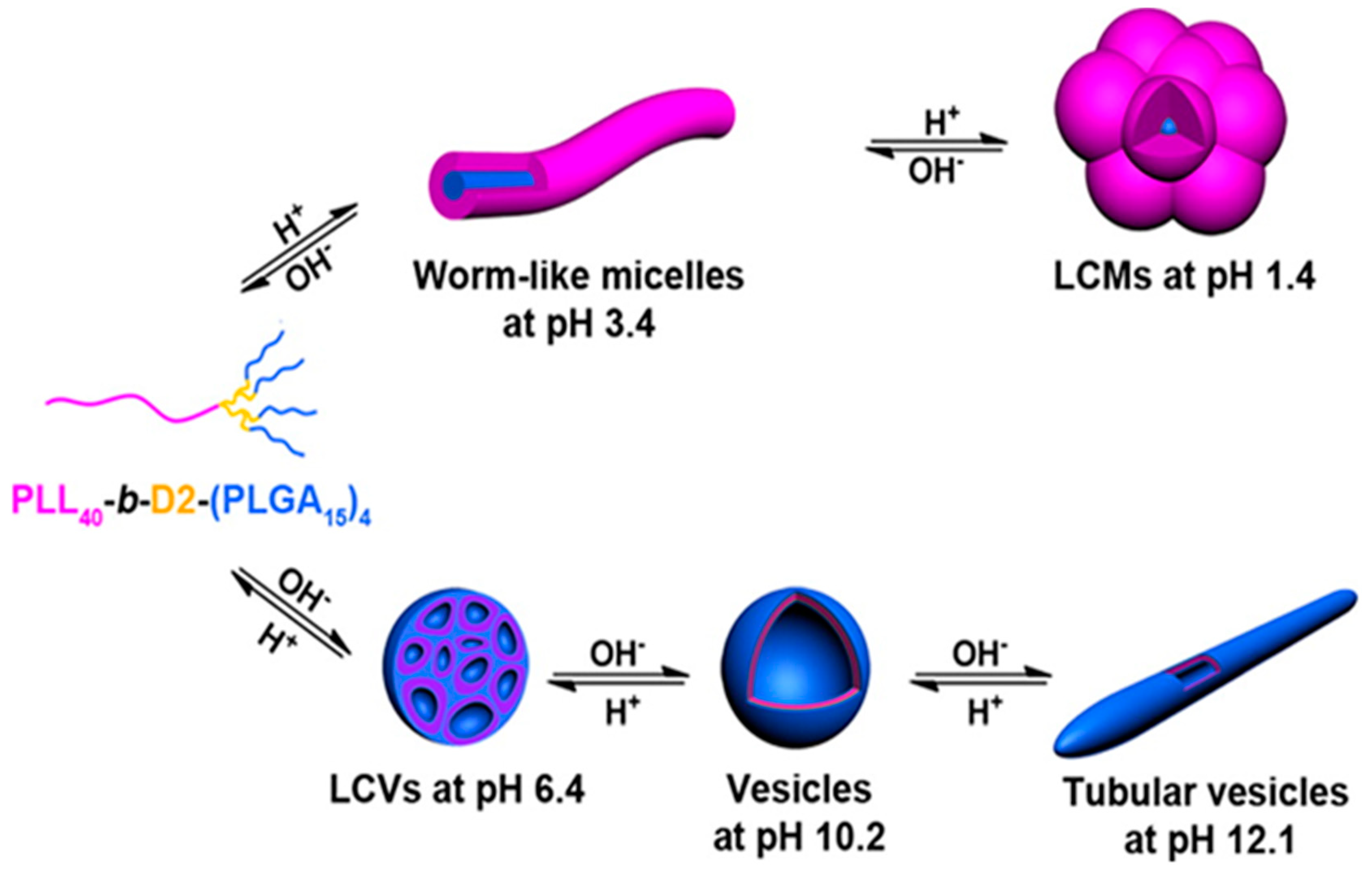
| (PLL)4-d2-b-d1-(PLGA)2 | Worm, vesicle, tubular vesicle, compound vesicle | 16 nm–2 µm | [55] |
| PEI-b-PLL-b-PLGA | Polyplex | 200 nm | [62] |
| pHis-b-PEI | Sphere | 34 nm | [72] |
| Polysebacic anhydride-pHis | Nanocapsule | 150–350 nm | [76] |
| p(MPC)-b-pHis | Nanodaisies | ~100 nm | [79] |
| PEG-b-pHis-b-PLL | Rod | ~325 × 13 nm | [81] |
| mPEG-b-PLA-pHis-S-S-OEI | Polyplex | 116–658 nm | [88] |
| Alginate/pArg | Microcapsule | 351 µm | [97] |
| HA-pArg | Sphere | 116–155 nm | [103] |
| PArg | Nanocapsule | <200 nm | [104] |
| Lipid-pArg-PEG | Liposome | 148–188 nm | [110] |
| mPEG-PLA-b-pArg | Micelleplex | 54 nm | [111] |
| PEG-b-pTyr-lipoic acid | Sphere | 45 nm | [124] |
| PEG-b-pTyr | Sphere | 70 nm | [125] |
| Type of AA | Payload | Target/pH | Polymer a | Ref. |
|---|---|---|---|---|
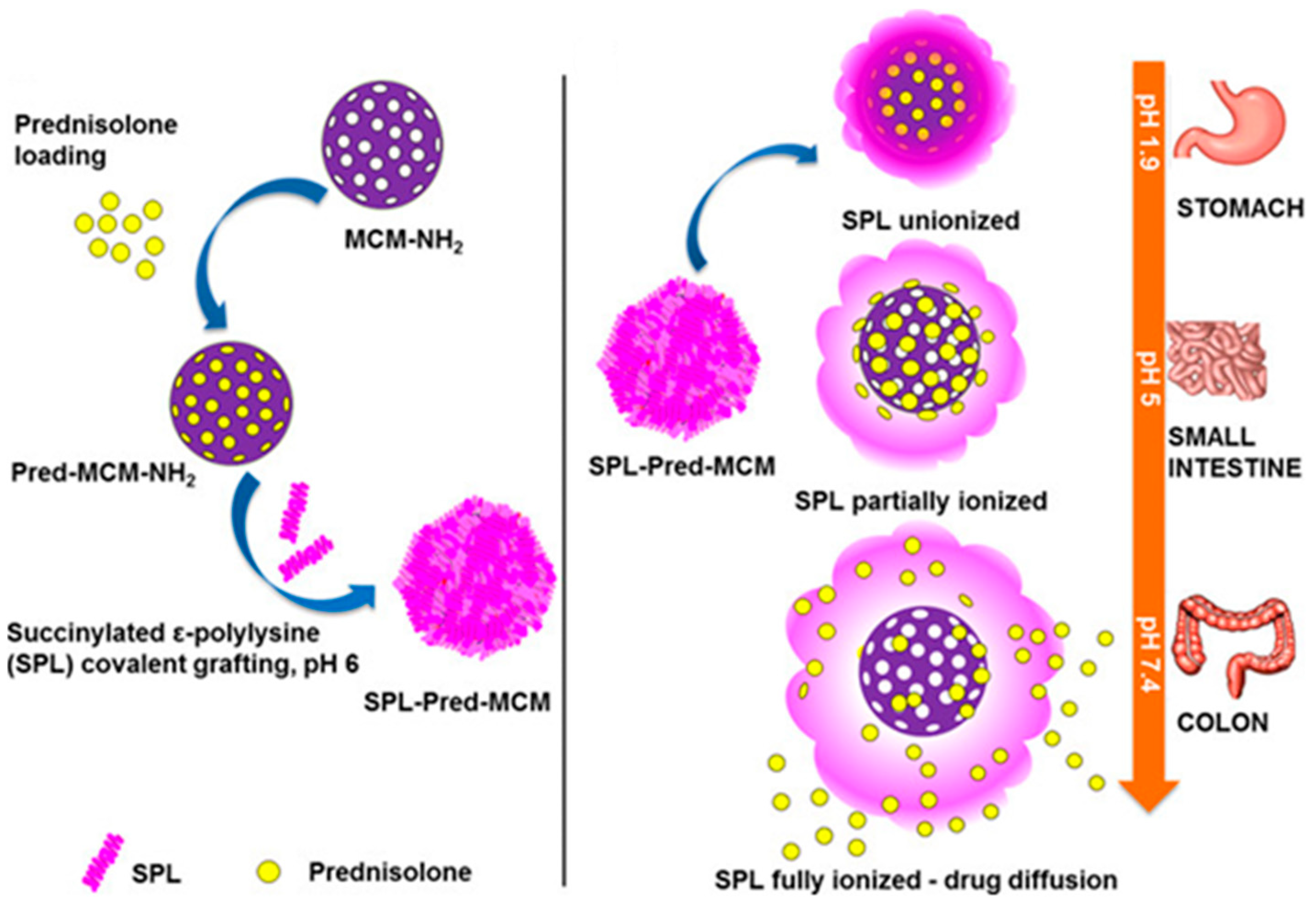
| Lys | Prednisolone | Colon | Succinylated ε-PLL | [46] |
| FITC-dextran | pH 3.5 | Alginate bead coated with PLL | [50] | |
| Diclofenac sodium | pH 10-11 or 2-3 | PEI-(PLL-b-PEG) | [51] | |
| Gene | pH 6.5 | PEI-PLL-PLGA | [62] | |
| His | Antitumor drug | pH 6.5 | PLGA-b-PEG-b-pHis | [71] |
| Dox | Tumor pH | Dextrin-b-pHis | [74] | |
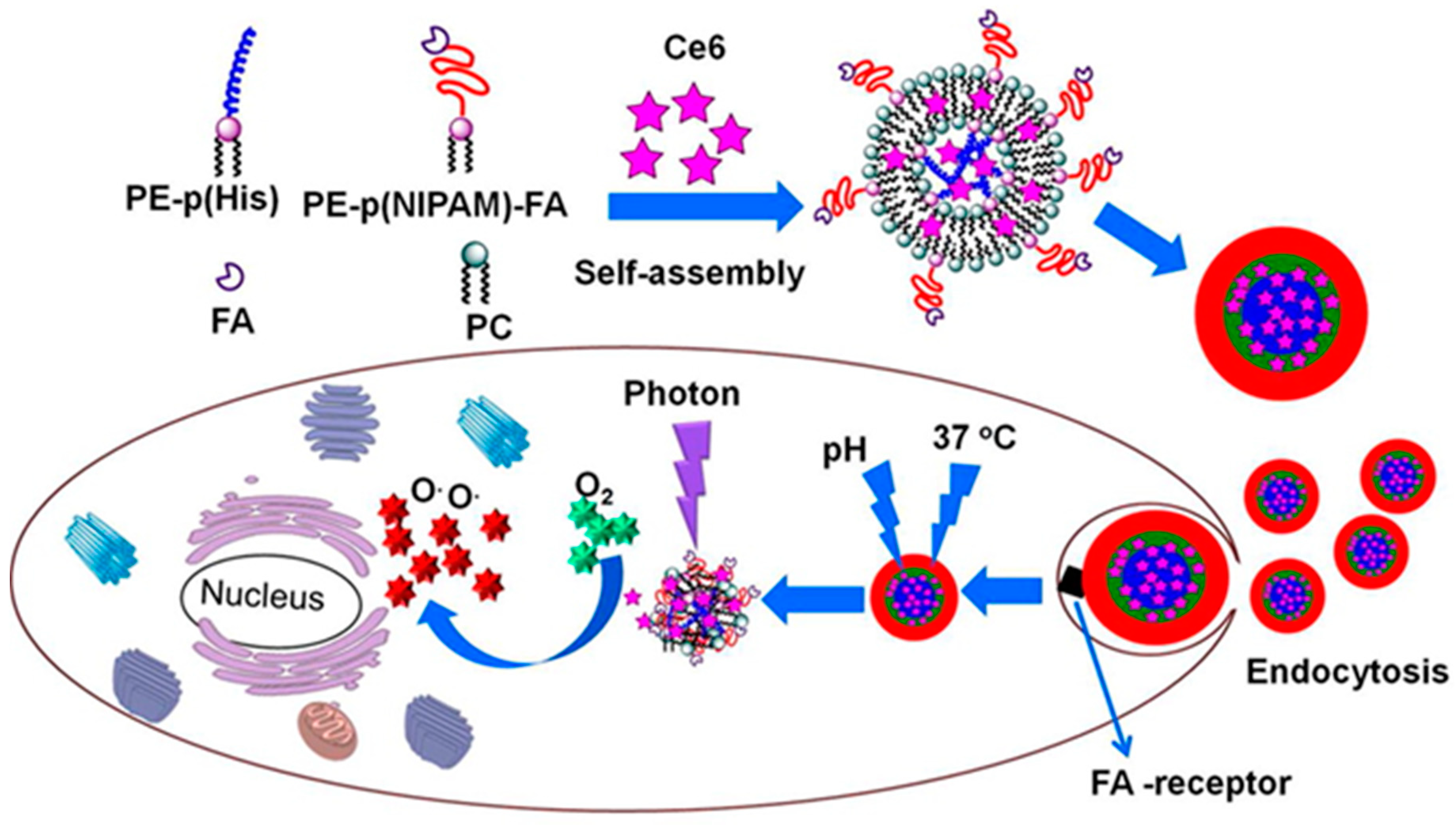
| Ce6 | Tumor pH | PE-pHis40 and PE- pNIPAm40-FA | [77] | |
| Arg | Polyarginine-KLA peptide | Mitochondria | Polyarginine-KLA | [100] |
| Docetaxel | Intracellular tumor | PArg nanocapsules | [83] | |
| Dox | Tumor pH | PAA-g-PEG/PArg | [106] | |
| Insulin | Intestine | pArg nano capsule | [107] | |
| Tyr | Dox | Breast tumor | PEG-b-pTyr-lipoic acid | [124] |
| Dox | Colorectal tumor | PEG-pTyr/cRGD-functionalized-PEG-pTyr | [125] | |
| Pro | Nile blue A | pH 5.2 | pNIPAm/pPro hydrogel | [128] |
| Trp | testosterone | pH 2.0 | Poly(γ-benzyl-l-glutamate)-graft-poly(tryptophan) | [130] |
| 8-aniline-naphthalene-1-sulfonate & pyrene | pH 5.2 | Poly(N-hydroxyethyl-l-glutamate)-b-poly(l-tryptophan) | [131] |
| Polymers a | Micelle Types | Diameter | Ref. |
|---|---|---|---|
| Citraconylated-pAsp | Sphere | 60 nm | [149] |
| FA-polyaspartylhydrazide | Vesicle | 105–113 nm | [152] |
| pAsp-co-polylacticacid-DPPE | Sphere | 219–281 nm | [158] |
| mPEG-pAsp | Vesicle | 50 nm | [159] |
| PHEA-g-C18-NH2 | Sphere | 9 nm | [160] |
| PEG-b-pAsp | Sphere | 200 nm | [163] |
| P(MPC)-b-pAsp | Vesicle | ~100 nm | [170] |
| PLGA-pNIPAm-HEMA | Sphere | 45 nm | [180] |
| PLGA-co-PLA-DPPE | Sphere | 172–200 nm | [187] |
| Chitosan-N-arginine-PLGA | Sphere | 260 nm | [189] |
| PLGA-chitosan | Sphere | 210 nm | [190] |
| Type of AA | Payload | Target/pH | Polymer a | Ref. |
|---|---|---|---|---|
| Asp | DFS | Colon | pAsp-l-pNIPAm | [146] |
| 5-Fluoruracil | Intestine | Konjac glucomannan-pAsp hydrogel | [147] | |
| Ocular drug | Eye | Thiolated PAsp | [149] | |
| Glutathione or oxytocin | Colon | Succinic derivative of insulin cross linked pAspHz | [152] | |
| DTX | Drug resistant cancer cell | PEG-b-PAsp | [162] | |
| Glu | Dox | Intracellular tumor | mPEG-b-p(LGA-co-CELG) | [182] |
| Adriamycin hydrochloride | Tumor pH | Star-block-copolymer of PEI, PLGA and PEG | [183] | |
| Dox | Lysosome | PLGA-co-PLA-DPPE | [187] |
© 2019 by the authors. Licensee MDPI, Basel, Switzerland. This article is an open access article distributed under the terms and conditions of the Creative Commons Attribution (CC BY) license (http://creativecommons.org/licenses/by/4.0/).
Share and Cite
Augustine, R.; Kalva, N.; Kim, H.A.; Zhang, Y.; Kim, I. pH-Responsive Polypeptide-Based Smart Nano-Carriers for Theranostic Applications. Molecules 2019, 24, 2961. https://doi.org/10.3390/molecules24162961
Augustine R, Kalva N, Kim HA, Zhang Y, Kim I. pH-Responsive Polypeptide-Based Smart Nano-Carriers for Theranostic Applications. Molecules. 2019; 24(16):2961. https://doi.org/10.3390/molecules24162961
Chicago/Turabian StyleAugustine, Rimesh, Nagendra Kalva, Ho An Kim, Yu Zhang, and Il Kim. 2019. "pH-Responsive Polypeptide-Based Smart Nano-Carriers for Theranostic Applications" Molecules 24, no. 16: 2961. https://doi.org/10.3390/molecules24162961
APA StyleAugustine, R., Kalva, N., Kim, H. A., Zhang, Y., & Kim, I. (2019). pH-Responsive Polypeptide-Based Smart Nano-Carriers for Theranostic Applications. Molecules, 24(16), 2961. https://doi.org/10.3390/molecules24162961