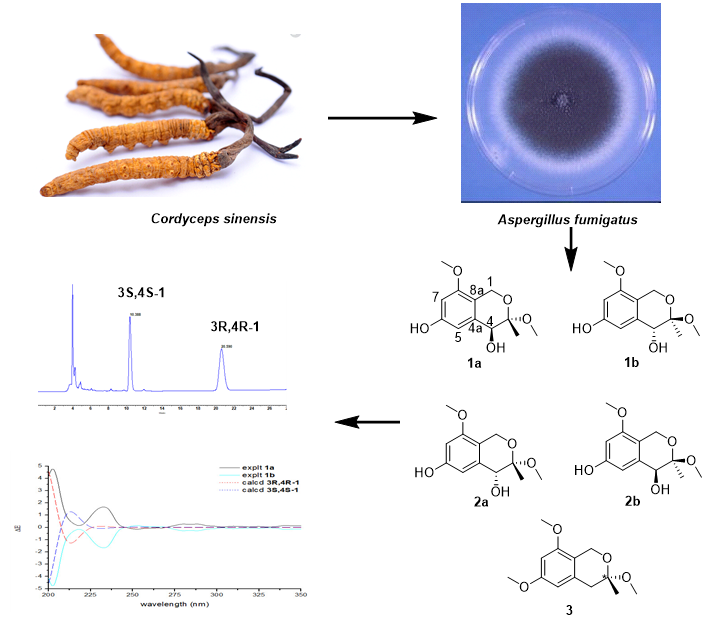
Novel Polyketides Produced by the Endophytic Fungus Aspergillus Fumigatus from Cordyceps Sinensis

 ,
,  ,
,
Abstract
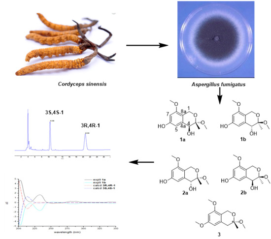
1. Introduction
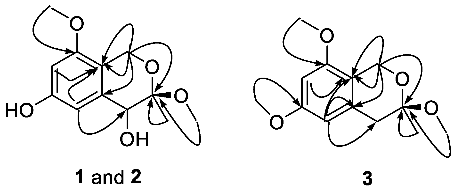
2. Results and Discussion
3. Materials and Methods
3.1. General Experimental Procedures
3.2. Fungal Material
3.3. Fractionation and Isolation
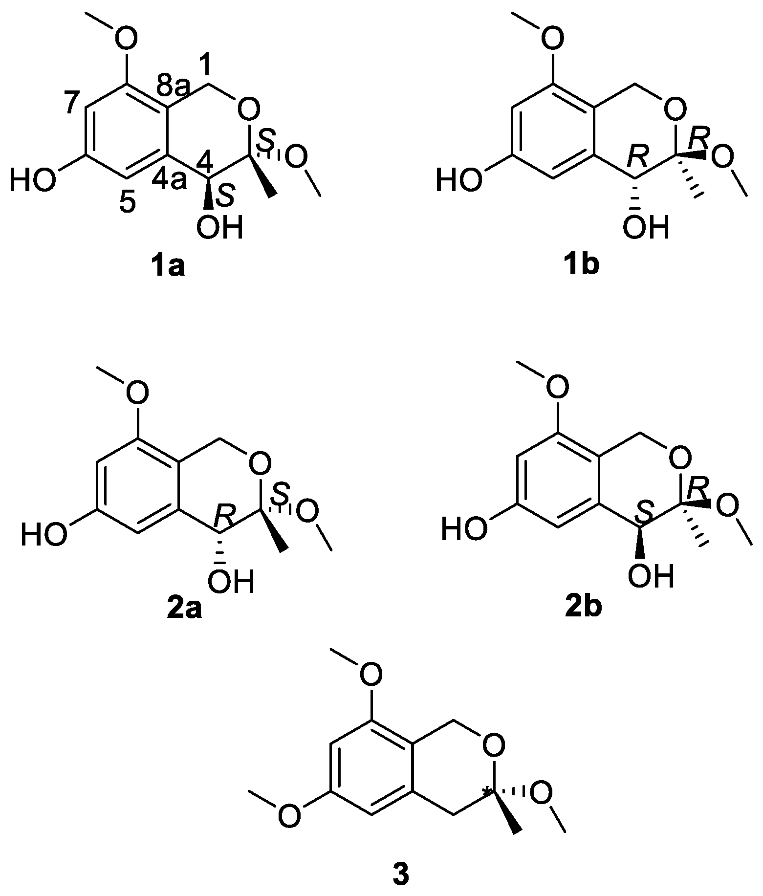
3.3.1. 3R,4S-3,8-Dimethoxy-3-methylisochromane-4,6-diol (1a)
3.3.2. 3R,4R-3,8-Dimethoxy-3-methylisochromane-4,6-diol (1b)
3.3.3. 3S,4R-3,8-Dimethoxy-3-methylisochromane-4,6-diol (2a)
3.3.4. 3R,4S-3,8-Dimethoxy-3-methylisochromane-4,6-diol (2b)
3.3.5. 3,6,8-Trimethoxy-3-methylisochromane (3)
3.4. MTT Assay
4. Conclusions
Supplementary Materials
Author Contributions
Funding
Acknowledgments
Conflicts of Interest
References
- Haas, H. Fungal siderophore metabolism with a focus on Aspergillus fumigatus. Nat. Prod. Rep. 2014, 31, 1266–1276. [Google Scholar] [CrossRef] [PubMed]
- Kwon-Chung, K.J.; Sugui, J.A. Aspergillus fumigatus—What makes the species a ubiquitous human fungal pathogen? PLoS Pathog. 2013, 9, e1003743. [Google Scholar] [CrossRef] [PubMed]
- Chu, M.; Mierzwa, R.; He, L.; Xu, L.; Patel, M.; Patel, D.; Chan, T.M. Structure of Sch 528647: A new antitumor antibiotic related to Fumagillin. J. Antibiot. 2001, 54, 1096–1099. [Google Scholar] [CrossRef] [PubMed]
- Wang, Y.; Li, D.H.; Li, Z.L.; Sun, Y.J.; Hua, H.M.; Liu, T.; Bai, J. Terpenoids from the Marine-Derived Fungus Aspergillus fumigatus YK-7. Molecules 2016, 21, 31. [Google Scholar] [CrossRef] [PubMed]
- Cutler, H.G.; Lauren, D.R.; Wilkins, A.L.; Holland, P.T.; Hill, R.A.; Dugan, F.M. Ruakuric acid: A natural product from Aspergillus fumigatus. Phytochemistry 1996, 43, 209–214. [Google Scholar] [CrossRef]
- Wang, F.Z.; Fang, Y.C.; Zhu, T.J.; Zhang, M.; Lin, A.Q.; Gu, Q.Q.; Zhu, W.M. Seven new prenylated indole diketopiperazine alkaloids from holothurian-derived fungus Aspergillus fumigatus. Tetrahedron 2008, 64, 7986–7991. [Google Scholar] [CrossRef]
- Magotra, A.; Kumar, M.; Kushwaha, M.; Awaschi, P.; Raina, C.; Gupta, A.P.; Shah, B.A.; Gandhi, S.G.; Chaubey, A. Epigenetic modifier induced enhancement of fumiquinazoline C production in Aspergillus fumigatus (GA-L7): An endophytic fungus from Grewia asiatica L. ABM Express 2017, 7, 43. [Google Scholar] [CrossRef] [PubMed]
- Shi, Y.S.; Zhang, Y.; Chen, X.Z.; Zhang, N.; Liu, Y.B. Metabolites produced by the endophytic fungus Aspergillus fumigatus from the stem of Erythrophloeum fordii oliv. Molecules 2015, 20, 10793–10799. [Google Scholar] [CrossRef] [PubMed]
- Sin, N.; Meng, L.H.; Wang, M.Q.W.; Wen, J.J.; Bornmann, W.G.; Crews, C.M. The anti-angiogenic agent fumagillin covalently binds and inhibits the methionine aminopeptidase, MetAP-2. Proc. Natl. Acad. Sci. USA 1997, 94, 6099–6103. [Google Scholar] [CrossRef] [PubMed]
Sample Availability: Samples of the compounds 1a, 1b, and 3 are available from the authors. |


| Position | 1 a | 2 | 3 | |||
|---|---|---|---|---|---|---|
| δH | δC | δH | δC | δH | δC | |
| 1 | 4.41, d, 15.3 Hz 4.46, d, 15.3 Hz | 60.7 | 4.56, d, 15.2 Hz 4.43, d, 15.2 Hz | 60.6 | 4.65, d, 15.1 Hz 4.44, d, 15.1 Hz | 60.2 |
| 2 | - | - | - | - | - | - |
| 3 | - | 101.5 | - | 99.5 | - | 98.8 |
| 4 | 4.00, s | 70.4 | 4.36, s | 72.6 | 2.85, d, 16.4 Hz 2.73, d, 16.4 Hz | 39.8 |
| 4a | - | 137.3 | - | 138.6 | - | 134.4 |
| 5 | 6.38, d, 2.2 Hz | 109.0 | 6.63, d, 2.1 Hz | 105.5 | 6.33, d, 2.1 Hz | 105.5 |
| 6 | - | 157.2 | - | 158.5 | - | 161.0 |
| 7 | 6.33, d, 2.2 Hz | 99.0 | 6.28, d, 2.1 Hz | 97.9 | 6.33 d, 2.1 Hz | 96.9 |
| 8 | - | 158.4 | - | 156.8 | - | 157.4 |
| 8a | - | 114.2 | - | 114.6 | - | 115.2 |
| 3-Me | 1.46, s | 19.1 | 1.49, s | 20.5 | 1.44, s | 23.4 |
| 3-OMe | 3.30, s | 49.8 | 3.31, s | 49.1 | 3.28, s | 48.8 |
| 6-OMe | - | - | - | - | 3.78 | 55.7 |
| 8-OMe | 3.76, s | 55.8 | 3.75, s | 55.8 | 3.72 | 55.8 |
© 2018 by the authors. Licensee MDPI, Basel, Switzerland. This article is an open access article distributed under the terms and conditions of the Creative Commons Attribution (CC BY) license (http://creativecommons.org/licenses/by/4.0/).
Share and Cite
Guo, D.-L.; Li, X.-H.; Feng, D.; Jin, M.-Y.; Cao, Y.-M.; Cao, Z.-X.; Gu, Y.-C.; Geng, Z.; Deng, F.; Deng, Y. Novel Polyketides Produced by the Endophytic Fungus Aspergillus Fumigatus from Cordyceps Sinensis. Molecules 2018, 23, 1709. https://doi.org/10.3390/molecules23071709
Guo D-L, Li X-H, Feng D, Jin M-Y, Cao Y-M, Cao Z-X, Gu Y-C, Geng Z, Deng F, Deng Y. Novel Polyketides Produced by the Endophytic Fungus Aspergillus Fumigatus from Cordyceps Sinensis. Molecules. 2018; 23(7):1709. https://doi.org/10.3390/molecules23071709
Chicago/Turabian StyleGuo, Da-Le, Xiao-Hua Li, Dan Feng, Meng-Ying Jin, Yu-Mei Cao, Zhi-Xing Cao, Yu-Cheng Gu, Zhao Geng, Fang Deng, and Yun Deng. 2018. "Novel Polyketides Produced by the Endophytic Fungus Aspergillus Fumigatus from Cordyceps Sinensis" Molecules 23, no. 7: 1709. https://doi.org/10.3390/molecules23071709
APA StyleGuo, D.-L., Li, X.-H., Feng, D., Jin, M.-Y., Cao, Y.-M., Cao, Z.-X., Gu, Y.-C., Geng, Z., Deng, F., & Deng, Y. (2018). Novel Polyketides Produced by the Endophytic Fungus Aspergillus Fumigatus from Cordyceps Sinensis. Molecules, 23(7), 1709. https://doi.org/10.3390/molecules23071709