Angelica Stem: A Potential Low-Cost Source of Bioactive Phthalides and Phytosterols

Abstract

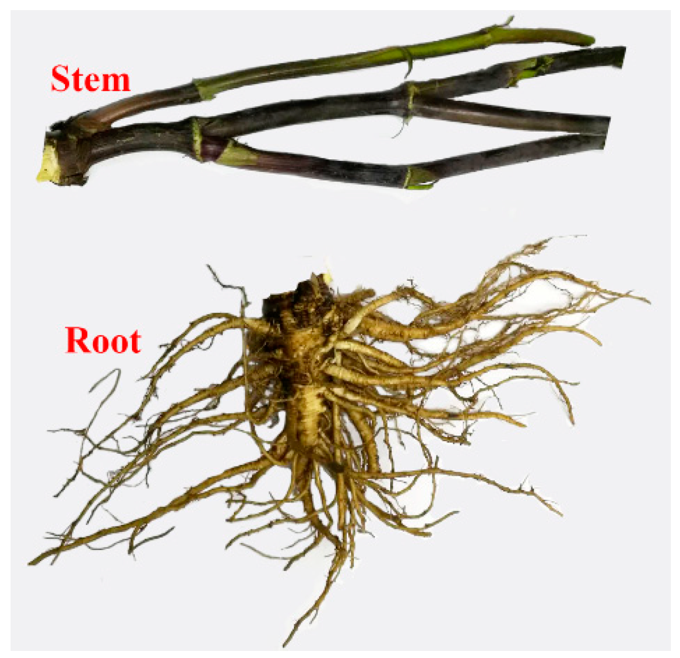
1. Introduction
2. Results and Discussion
2.1. Extraction and Purification of Extracts
2.2. GC/MS Analysis
2.2.1. Identification of Phytochemicals
2.2.2. Quantitative Analysis
2.3. Coniferyl Ferulate Analysis by HPLC–MS/MS
3. Materials and Methods
3.1. Materials and Chemicals
3.2. Isolation and Purification of Oily Extracts
3.3. GC–MS Procedure
3.3.1. Derivatization and GC–MS Analysis
3.3.2. Qualitative Analysis
3.4. HPLC–MS/MS Procedure
3.4.1. Multiple Reaction Monitoring (MRM) for Coniferyl Ferulate Analysis
3.4.2. Sample Analysis by LC–MS/MS
4. Conclusions
Supplementary Materials
Author Contributions
Funding
Conflicts of Interest
References
- Atanasov, A.G.; Waltenberger, B.; Pferschy-Wenzig, E.M.; Linder, T.; Wawrosch, C.; Uhrin, P.; Temml, V.; Wang, L.M.; Schwaiger, S.; Heiss, E.H.; et al. Discovery and resupply of pharmacologically active plant-derived natural products: A review. Biotechnol. Adv. 2015, 33, 1582–1614. [Google Scholar] [CrossRef] [PubMed]
- Kinghorn, A.D.; Pan, L.; Fletcher, J.N.; Chai, H.Y. The relevance of higher plants in lead compound discovery programs. J. Nat. Prod. 2011, 74, 1539–1555. [Google Scholar] [CrossRef] [PubMed]
- Tan, H.S.; Hu, D.D.; Song, J.Z.; Xu, Y.; Cai, S.F.; Chen, Q.L.; Meng, Q.W.; Li, S.L.; Chen, S.L.; Mao, Q.; et al. Distinguishing radix Angelica sinensis from different regions by HS-SFME/GC–MS. Food Chem. 2015, 186, 200–206. [Google Scholar] [CrossRef] [PubMed]
- Wei, W.L.; Zeng, R.; Gu, C.M.; Qu, Y.; Huang, L.F. Angelica sinensis in China-a review of botanical profile, ethnopharmacology, phytochemistry and chemical analysis. J. Ethnopharmacol. 2016, 190, 116–141. [Google Scholar] [CrossRef] [PubMed]
- Lu, Q.; Qiu, T.Q.; Yang, H. Ligustilide inhibits vascular smooth muscle cells proliferation. Eur. J. Pharmacol. 2006, 542, 136–140. [Google Scholar] [CrossRef] [PubMed]
- Saw, C.L.L.; Wu, Q.; Su, Z.Y.; Wang, H.; Yang, Y.H.; Xu, X.T.; Huang, Y.; Khor, T.O.; Kong, A.N.T. Effects of natural phytochemicals in Angelica sinensis (Danggui) on Nrf2-mediated gene expression of phase II drug metabolizing enzymes and anti-inflammation. Biopharm. Drug Dispos. 2013, 34, 303–311. [Google Scholar] [CrossRef] [PubMed]
- Yin, J.; Wang, C.; Mody, A.; Bao, L.; Hung, S.H.; Svoronos, S.A.; Tseng, Y. The effect of Z-ligustilide on the mobility of human glioblastoma T98G cells. PLoS ONE 2013, 8, e66598. [Google Scholar] [CrossRef] [PubMed]
- Zhang, L.; Du, J.R.; Wang, J.; Yu, D.K.; Chen, Y.S.; He, Y.; Wang, C.Y. Z-ligustilide extracted from radix Angelica sinensis decreased platelet aggregation induced by ADP ex vivo and arterio-venous shunt thrombosis in vivo in rats. Yakugaku Zasshi 2009, 129, 855–859. [Google Scholar] [CrossRef] [PubMed]
- Ho, C.C.; Kumaran, A.; Hwang, L.S. Bio-assay guided isolation and identification of anti-Alzheimer active compounds from the root of Angelica sinensis. Food Chem. 2009, 114, 246–252. [Google Scholar] [CrossRef]
- Jia, Y.; He, Y.; Lu, F.C. The structure-antioxidant activity relationship of dehydrodiferulates. Food Chem. 2018, 269, 480–485. [Google Scholar] [CrossRef] [PubMed]
- Ronchetti, D.; Impagnatiello, F.; Guzzetta, M.; Gasparini, L.; Borgatti, M.; Gambari, R.; Ongini, E. Modulation of NOS expression by a nitric oxide-releasing derivative of the natural antioxidant ferulic acid in activated RAW 264.7 macrophages. Eur. J. Pharmacol. 2006, 532, 162–169. [Google Scholar] [CrossRef] [PubMed]
- Yan, J.J.; Cho, J.Y.; Kim, H.S.; Kim, K.L.; Jung, J.S.; Huh, S.O.; Suh, H.W.; Kim, Y.H.; Song, D.K. Protection against beta-amyloid peptide toxicity in vivo with long-term administration of ferulic acid. Br. J. Pharmacol. 2001, 133, 89–96. [Google Scholar] [CrossRef] [PubMed]
- Chen, C.; Wu, C.H.; Lu, X.H.; Li, S.J. Coniferyl ferulate, a strong inhibitor of glutathione-S-transferase isolated from Radix Angelicae sinensis, to reverses multidrug resistance and down-regulation of P-glycoprotein. Acta Pharmacol. Sin. 2013, 34, 21–22. [Google Scholar] [CrossRef]
- Chou, S.C.; Everngam, M.C.; Sturtz, G.; Beck, J.J. Antibacterial activity of components from Lomatium californicum. Phytother. Res. 2006, 20, 153–156. [Google Scholar] [CrossRef] [PubMed]
- Li, S.Y.; Yu, Y.; Li, S.P. Identification of antioxidants in essential oil of radix angelicae sinensis using HPLC coupled with DAD-MS and ABTS-based assay. J. Agric. Food Chem. 2007, 55, 3358–3362. [Google Scholar] [CrossRef] [PubMed]
- Zhou, Y.N.; Fu, W.W.; Liu, P. Coniferyl ferulate attenuated liver fibrosis through Inhibition of transforming growth factor-B receptor. In Proceedings of the 65th Annual Meeting of the American Association for the Study of Liver Diseases, Boston, MA, USA, 7 November 2014; pp. 571A–572A. [Google Scholar]
- Gao, Q.; Li, J.; Cheung, J.K.H.; Duan, J.; Ding, A.; Cheung, A.W.H.; Zhao, K.; Li, W.Z.; Dong, T.T.; Tsim, K.W.K. Verification of the formulation and efficacy of danggui buxue tang (a decoction of radix astragali and radix angelicae sinensis): An exemplifying systematic approach to revealing the complexity of Chinese herbal medicine formulae. Chin. Med. 2007, 2, 12. [Google Scholar] [CrossRef] [PubMed]
- Zhou, G.S.; Yang, N.Y.; Tang, Y.P.; Duan, J.A.; Jiang, S.; Yan, H.; Guo, S.; Song, B.S.; He, Z.Q. Chemical constituents from the aerial parts of Angelica sinensis and their bioactivities. Chin. J. Nat. Med. 2012, 10, 295–298. [Google Scholar] [CrossRef]
- Xie, J.J.; Lu, J.; Qian, Z.M.; Yu, Y.; Duan, J.A.; Li, S.P. Optimization and comparison of five methods for extraction of coniferyl ferulate from Angelica sinensis. Molecules 2009, 14, 555–565. [Google Scholar] [CrossRef] [PubMed]
- Zhang, X.L.; Liu, L.F.; Zhu, L.Y.; Bai, Y.J.; Mao, Q.; Li, S.L.; Chen, S.L.; Xu, H.X. A high performance liquid chromatography fingerprinting and ultra high performance liquid chromatography coupled with quadrupole time-of-flight mass spectrometry chemical profiling approach to rapidly find characteristic chemical markers for quality evaluation of dispensing granules, a case study on Chuanxiong Rhizoma. J. Pharm. Biomed. Anal. 2014, 88, 391–400. [Google Scholar] [CrossRef] [PubMed]
- Bai, Y.J.; Kong, M.; Xu, J.D.; Zhang, X.L.; Zhou, S.S.; Wang, X.N.; Liu, L.F.; Li, S.L. Effect of different drying methods on the quality of Angelicae Sinensis Radix evaluated through simultaneously determining four types of major bioactive components by high performance liquid chromatography photodiode array detector and ultra-high performance liquid chromatography quadrupole time-of-flight mass spectrometry. J. Pharm. Biomed. Anal. 2014, 94, 77–83. [Google Scholar] [CrossRef] [PubMed]
- Zou, J.; Chen, G.D.; Zhao, H.; Huang, Y.; Luo, X.; Xu, W.; He, R.R.; Hu, D.; Yao, X.S.; Gao, H. Triligustilides A and B: Two pairs of phthalide trimers from Angelica sinensis with a complex polycyclic skeleton and their activities. Org. Lett. 2018, 20, 884–887. [Google Scholar] [CrossRef] [PubMed]
- Boerjan, W.; Ralph, J.; Baucher, M. Lignin biosynthesis. Annu. Rev. Plant Biol. 2003, 54, 519–546. [Google Scholar] [CrossRef] [PubMed]
- Itoh, A.; Isoda, K.; Kondoh, M.; Kawase, M.; Kobayashi, M.; Tamesada, M.; Yagi, K. Hepatoprotective effect of syringic acid and vanillic acid on concanavalin a-induced liver injury. Biol. Pharm. Bull. 2009, 32, 1215–1219. [Google Scholar] [CrossRef] [PubMed]
- Itoh, A.; Isoda, K.; Kondoh, M.; Kawase, M.; Watari, A.; Kobayashi, M.; Tamesada, M.; Yagi, K. Hepatoprotective effect of syringic acid and vanillic acid on CCl4-induced liver injury. Biol. Pharm. Bull. 2010, 33, 983–987. [Google Scholar] [CrossRef] [PubMed]
- Gugliucci, A.; Bastos, D.H.M.; Schulze, J.; Souza, M.F.F. Caffeic and chlorogenic acids in Ilex paraguariensis extracts are the main inhibitors of AGE generation by methylglyoxal in model proteins. Fitoterapia 2009, 80, 339–344. [Google Scholar] [CrossRef] [PubMed]
- Khadem, S.; Marles, R.J. Monocyclic phenolic acids; hydroxy- and polyhydroxybenzoic acids: Occurrence and recent bioactivity studies. Molecules 2010, 15, 7985–8005. [Google Scholar] [CrossRef] [PubMed]
- Epps, S.V.R.; Petrujkic, B.T.; Sedej, I.; Krueger, N.A.; Harvey, R.B.; Beier, R.C.; Stanton, T.B.; Phillips, T.D.; Anderson, R.C.; Nisbet, D.J. Comparison of anti-campylobacter activity of free thymol and thymol-beta-d-glucopyranoside in absence or presence of beta-glycoside-hydrolysing gut bacteria. Food Chem. 2015, 173, 92–98. [Google Scholar] [CrossRef] [PubMed]
- Phillips, K.M.; Ruggio, D.M.; Ashraf-Khorassani, M. Phytosterol composition of nuts and seeds commonly consumed in the United States. J. Agric. Food Chem. 2005, 53, 9436–9445. [Google Scholar] [CrossRef] [PubMed]
- Berges, R.R.; Windeler, J.; Trampisch, H.J.; Senge, T. Randomised, placebo-controlled, double-blind clinical trial of beta-sitosterol in patients with benign prostatic hyperplasia. Beta-sitosterol study group. The Lancet 1995, 345, 1529–1532. [Google Scholar] [CrossRef]
- Gabay, O.; Sanchez, C.; Salvat, C.; Chevy, F.; Breton, M.; Nourissat, G.; Wolf, C.; Jacques, C.; Berenbaum, F. Stigmasterol: A phytosterol with potential anti-osteoarthritic properties. Osteoarthr. Cartil. 2010, 18, 106–116. [Google Scholar] [CrossRef] [PubMed]
- Kmiecik, D.; Korczak, J.; Rudzinska, M.; Gramza-Michalowska, A.; Hes, M.; Kobus-Cisowska, J. Stabilisation of phytosterols by natural and synthetic antioxidants in high temperature conditions. Food Chem. 2015, 173, 966–971. [Google Scholar] [CrossRef] [PubMed]
- Ling, W.H.; Jones, P.J. Dietary phytosterols: A review of metabolism, benefits and side effects. Life Sci. 1995, 57, 195–206. [Google Scholar] [CrossRef]
- Dai, X.L.; Pang, L.; Zhang, Z.; Yang, C.F.; Li, Y.M. Development of a sensitive LC-MS/MS method for quantification of coniferyl ferulate and its metabolite coniferyl alcohol in rat plasma: Application to a pharmacokinetic study. J. Pharm. Biomed. Anal. 2017, 146, 201–205. [Google Scholar] [CrossRef] [PubMed]
- Zhou, S.S.; Xu, J.; Tsang, C.K.; Yip, K.M.; Yeung, W.P.; Zhao, Z.Z.; Zhu, S.; Fushimi, H.; Chang, H.Y.; Chen, H.B. Comprehensive quality evaluation and comparison of Angelica sinensis radix and Angelica acutiloba radix by integrated metabolomics and glycomics. J. Food Drug Anal. 2018, 26, 1122–1137. [Google Scholar] [CrossRef] [PubMed]
- Lu, F.C.; Ralph, J. Facile synthesis of 4-hydroxycinnamyl p-coumarates. J. Agric. Food Chem. 1998, 46, 2911–2913. [Google Scholar] [CrossRef]
Sample Availability: Sample of coniferyl ferulate is available from the corresponding author. |


| Peak No. | RT (min) | Chemical Name | Formula | Relative Intensity of MS Ions | Stem | Root |
|---|---|---|---|---|---|---|
| 1 | 17.46 | Cinnamic acid | C9H8O2 | 220(27), 205(93), 161(100), 131(73), 103(62) | + | - |
| 2 | 18.98 | p-hydroxybenzoic acid | C7H6O3 | 282(23), 267(100), 223(98), 193(49), 126(13) | + | - |
| 3 | 19.20 | p-hydroxybenzeneacetic acid | C8H8O3 | 296(14), 252(23), 179(26), 164(18), 73(100) | + | - |
| 4 | 19.60 | Butylphthalide | C12H14O2 | 190(3), 172(3), 133(100), 105(33), 77(16) | - | + |
| 5 | 20.00 | Z-butylidenephthalide | C12H12O2 | 188(21), 159(100), 146(43), 131(42), 103(35) | + | + |
| 6 | 20.92 | E-butylidenephthalide | C12H12O2 | 188(21), 159(100), 146(43), 131(48), 103(39) | - | + |
| 7 | 21.00 | 4-hydroxyphenyl-1, 2-ethanediol | C8H10O3 | 267(100), 193(7), 147(16) | + | - |
| 8 | 21.16 | Z-ligustilide | C12H14O2 | 190(55), 161(80), 148(100), 133(24), 105(70) | + | + |
| 9 | 21.66 | Vanillic acid | C8H8O4 | 312(44), 297(100), 282(36), 267(72), 253(48) | + | + |
| 10 | 22.43 | E-ligustilide | C12H14O2 | 190(54), 161(83), 148(100), 105(82), 77(36) | + | + |
| 11 | 22.76 | Protocatechuic acid | C7H6O4 | 370(37), 355(27), 311(25), 193(100), 165(7) | + | - |
| 12 | 23.38 | Myristic acid | C14H28O2 | 285(67), 145(19), 132(35), 117(100) | + | + |
| 13 | 24.20 | Senkyunolide G | C12H14O3 | 278(11), 249(93), 221(100), 205(22), 131(16) | + | + |
| 14 | 25.32 | Pentadecanoic acid | C15H30O2 | 299(74), 132(36), 129(37), 117(100) | + | + |
| 15 | 25.48 | Dibutyl phthalate | C16H22O4 | 223(5), 205(6), 149(100) | + | + |
| 16 | 27.23 | Palmitic acid | C16H32O2 | 328(5), 313(67), 132(42), 117(100) | + | + |
| 17 | 27.26 | Senkyunolide I | C12H16O4 | 368(19), 353(11), 252(100), 237(31), 223(19) | + | + |
| 18 | 28.05 | Ferulic acid | C10H10O4 | 338(100), 323(92), 308(74), 293(63), 249(52) | + | + |
| 19 | 28.88 | Caffeic acid | C9H8O4 | 396(54), 381(19), 307(8), 219(100), 191(14) | + | - |
| 20 | 29.05 | Heptadecanoic acid | C17H34O2 | 342(5), 327(71), 145(23) 129(41), 117(100) | + | + |
| 21 | 29.27 | Senkyunolide H | C12H16O4 | 368(20), 353(16), 252(100), 237(32), 147(45) | + | + |
| 22 | 30.01 | Mannonolactone | C6H10O6 | 319(14), 305(5), 229(7), 129(9), 73(100) | + | + |
| 23 | 30.26 | Linoelaidic acid | C18H32O2 | 352(4), 337(66), 262(44), 177(51), 73(100) | + | + |
| 24 | 30.35 | Z-oleic acid | C18H34O2 | 354(3), 339(47), 129(71) 117(98), 73(100) | + | + |
| 25 | 30.52 | E-oleic acid | C18H34O2 | 354(5), 339(83), 129(78), 117(100), 75(80) | + | + |
| 26 | 30.82 | Stearic acid | C18H36O2 | 356(7), 341(69), 132(42), 129(43)117(100) | + | + |
| 27 | 32.55 | Butyl 9,12-octadecadienoate | C22H40O2 | 336(6), 263(25), 178(18), 135(29), 109(42) | - | + |
| 28 | 33.60 | Eicosanoic acid | C20H40O2 | 384(10), 369(75), 132(44), 117 (100) | + | + |
| 29 | 34.78 | Thymol-β-d-glucopyranoside | C16H24O6 | 361(100), 271(23), 243(26), 169(40), 147(30) | + | - |
| 30 | 35.18 | Monopalmitin | C19H38O4 | 371(100), 239(27), 203(23), 147(36), 129(16) | + | + |
| 31 | 35.75 | Behenic acid | C22H44O2 | 412(12), 379(71), 132 (47), 117(100) | + | + |
| 32 | 45.64 | Campesterol | C28H48O | 472(28), 382(83), 343(60), 255(28), 129(100) | + | - |
| 33 | 46.30 | Stigmasterol | C29H48O | 484(25), 394(43), 255(41), 129(54), 83(100) | + | + |
| 34 | 48.11 | β-sitosterol | C29H50O | 486(33), 396(88), 357(60), 255(29), 129(100) | + | + |
| Analytes | Stem | Root | ||
|---|---|---|---|---|
| mg/g Extracts | mg/g Stem | mg/g Extracts | mg/g Root | |
| Z-butylidenephthalide | 4.20 ± 0.31 | 0.074 ± 0.005 | 7.73 ± 0.78 | 0.26 ± 0.03 |
| E-butylidenephthalide | nd 1 | nd | 4.86 ± 0.54 | 0.16 ± 0.02 |
| Z-ligustilide | 28.60 ± 1.24 | 0.51 ± 0.02 | 63.04 ± 3.06 | 2.08 ± 0.10 |
| E-ligustilide | 4.16 ± 0.10 | 0.076 ±0.002 | 10.94 ± 0.47 | 0.36 ± 0.02 |
| Senkyunolide I | 11.53 ± 3.21 | 0.21 ± 0.06 | 34.30 ± 8.50 | 1.13 ± 0.28 |
| Senkyunolide H | nd | nd | 17.71 ±0.37 | 0.58 ± 0.02 |
| Cinnamic acid | 3.15 ± 0.88 | 0.057 ± 0.018 | nd | nd |
| p-hydroxybenzoic acid | 3.48 ± 0.13 | 0.064 ± 0.002 | nd | nd |
| Vanillic acid | 1.16 ± 0.12 | 0.021± 0.002 | nd | nd |
| Protocatechuic acid | 3.00 ±0.26 | 0.055 ±0.004 | nd | nd |
| Caffeic acid | 2.00 ±0.15 | 0.036 ±0.002 | nd | nd |
| Ferulic acid | 7.10 ± 0.56 | 0.13 ± 0.01 | 18.18 ± 0.53 | 0.60 ± 0.02 |
| Campesterol | 4.93 ± 0.12 | 0.088 ±0.002 | nd | nd |
| Stigmasterol | 14.03 ± 0.41 | 0.26 ± 0.01 | 3.94 ± 0.18 | 0.13 ± 0.01 |
| β-sitosterol | 56.21 ± 1.20 | 1.01 ± 0.02 | 11.21 ± 0.75 | 0.37 ± 0.03 |
| Analytes | Stem | Root | ||
|---|---|---|---|---|
| mg/g Extract | mg/g Stem | mg/g Extract | mg/g Root | |
| Ferulic acid | 5.42 ± 0.65 | 0.098 ± 0.010 | 17.57 ± 0.91 | 0.58 ± 0.03 |
| Coniferyl ferulate | 1.16 ± 0.26 | 0.021 ± 0.004 | 15.76 ± 0.03 | 0.52 ± 0.01 |
© 2018 by the authors. Licensee MDPI, Basel, Switzerland. This article is an open access article distributed under the terms and conditions of the Creative Commons Attribution (CC BY) license (http://creativecommons.org/licenses/by/4.0/).
Share and Cite
Zhao, C.; Jia, Y.; Lu, F. Angelica Stem: A Potential Low-Cost Source of Bioactive Phthalides and Phytosterols. Molecules 2018, 23, 3065. https://doi.org/10.3390/molecules23123065
Zhao C, Jia Y, Lu F. Angelica Stem: A Potential Low-Cost Source of Bioactive Phthalides and Phytosterols. Molecules. 2018; 23(12):3065. https://doi.org/10.3390/molecules23123065
Chicago/Turabian StyleZhao, Chengke, Yuan Jia, and Fachuang Lu. 2018. "Angelica Stem: A Potential Low-Cost Source of Bioactive Phthalides and Phytosterols" Molecules 23, no. 12: 3065. https://doi.org/10.3390/molecules23123065
APA StyleZhao, C., Jia, Y., & Lu, F. (2018). Angelica Stem: A Potential Low-Cost Source of Bioactive Phthalides and Phytosterols. Molecules, 23(12), 3065. https://doi.org/10.3390/molecules23123065