Article

Icariin Metabolism by Human Intestinal Microflora

Metalloenzyme Research Group and Department of Integrative Plant Science, Chung-Ang University, Anseong 17546, Korea
*
Author to whom correspondence should be addressed.
Molecules 2016, 21(9), 1158; https://doi.org/10.3390/molecules21091158
Submission received: 4 August 2016 / Revised: 25 August 2016 / Accepted: 26 August 2016 / Published: 31 August 2016

Abstract

:
Icariin is a major bioactive compound of Epimedii Herba, a traditional oriental medicine exhibiting anti-cancer, anti-inflammatory and anti-osteoporosis activities. Recently, the estrogenic activities of icariin drew significant attention, but the published scientific data seemed not to be so consistent. To provide fundamental information for the study of the icaritin metabolism, the biotransformation of icariin by the human intestinal bacteria is reported for the first time. Together with human intestinal microflora, the three bacteria Streptococcus sp. MRG-ICA-B, Enterococcus sp. MRG-ICA-E, and Blautia sp. MRG-PMF-1 isolated from human intestine were reacted with icariin under anaerobic conditions. The metabolites including icariside II, icaritin, and desmethylicaritin, but not icariside I, were produced. The MRG-ICA-B and E strains hydrolyzed only the glucose moiety of icariin, and icariside II was the only metabolite. However, the MRG-PMF-1 strain metabolized icariin further to desmethylicaritin via icariside II and icaritin. From the results, along with the icariin metabolism by human microflora, it was evident that most icariin is quickly transformed to icariside II before absorption in the human intestine. We propose the pharmacokinetics of icariin should focus on metabolites such as icariside II, icaritin and desmethylicaritin to explain the discrepancy between the in vitro bioassay and pharmacological effects.

Graphical Abstract

1. Introduction

Epimedii Herba is the aerial part of the plant species belonging to the genus Epimedium, including E. koreanum Nakai, E. brevicornum Maximowicz, E. pubescens Maximowicz, E. wushanense T. S. Ying, and E. sagittatum Maximowicz (Berberidaceae). It is also known as horny goat weed or yin yang huo and is listed as an herbal medicine in the pharmacopeias of Korea, Japan and China. It has been traditionally used for various biological activities, such as treating impotence, delaying aging, enhancing bone formation, and improving the function of the cardiovascular system [1]. A major bioactive ingredient of Epimedii Herba is prenylflavone glycosides with an 8-prenyl group on the kaempferol skeleton. Until now, more than 20 derivatives of 8-prenylkaempferol 3,7-O-diglycosides, including icariin, epimedin A, epimedin B, epimedin C, epimedokoreanoside I, and sagittetoside B, have been reported from E. koreanum (Figure 1) [2].
Icariin is the index compound of Epimedii Herba (KP), and the biological activities, pharmacological effects, pharmacokinetics and metabolism of icariin were extensively studied in the animal models [3]. Along with the immune-enhancing effects [4], the phytoestrogenic activity [5] and osteogenic effects [6,7] of icariin were also reported. However, the estrogenic activity of icariin did not directly correlate with the expected pharmacological effects, such as the anti-osteoporosis and neuroprotective activities measured at different levels of experiments [8,9]. For example, icariin was the weakest phytoestrogen compared with icaritin and Epimedium extract based on an in vitro estrogen receptor mediated estrogenic assay, although it showed the strongest estrogenic activity in ovariectomized rats [10]. Accordingly, the metabolic pathway of icariin in a non-classical system, such as the digestive tract, has become more important.
Based on the pharmacokinetic study of icariin in rats, icariin is reported to be readily absorbed by the body and metabolized. The metabolism of icariin resulted in 19 phase I metabolites in rat plasma [11], but significant biliary excretion and reabsorption to the body make the pharmacokinetics of icariin more complicated [12]. Recently, Zhou et al. reported that icariin is hydrolyzed to icaritin via icariside I or II by the rat intestinal flora [13]. On the contrary, few reports on the icariin metabolism in the human body are available. Furthermore, there have been no reports regarding the human intestinal metabolism of icariin yet.
In our research group, the biotransformation of flavonoids by human intestinal bacteria has been extensively studied [14,15]. We believe the identification of intestinal metabolites of dietary flavonoids produced by human intestinal bacteria can help to explain the pharmacological effects of flavonoids that cannot be observed from the in vitro bioassay [16]. Among the beneficial biological activities of Epimedii Herba, a strong estrogenic effect of icariin drew our attention, because the molecular structure of icariin bearing two monosaccharides appeared too hydrophilic to be a strong phytoestrogen. To study the metabolism of icariin in the human intestine, the biotransformation of icariin by human intestinal microflora and the isolated bacteria Streptococcus sp. MRG-ICA-B, Enterococcus sp. MRG-ICA-E, and Blautia sp. MRG-PMF-1 was performed under anaerobic conditions. Based on the identified metabolites, the metabolic pathway of icariin in the human intestine was proposed for the first time in this report.

2. Results

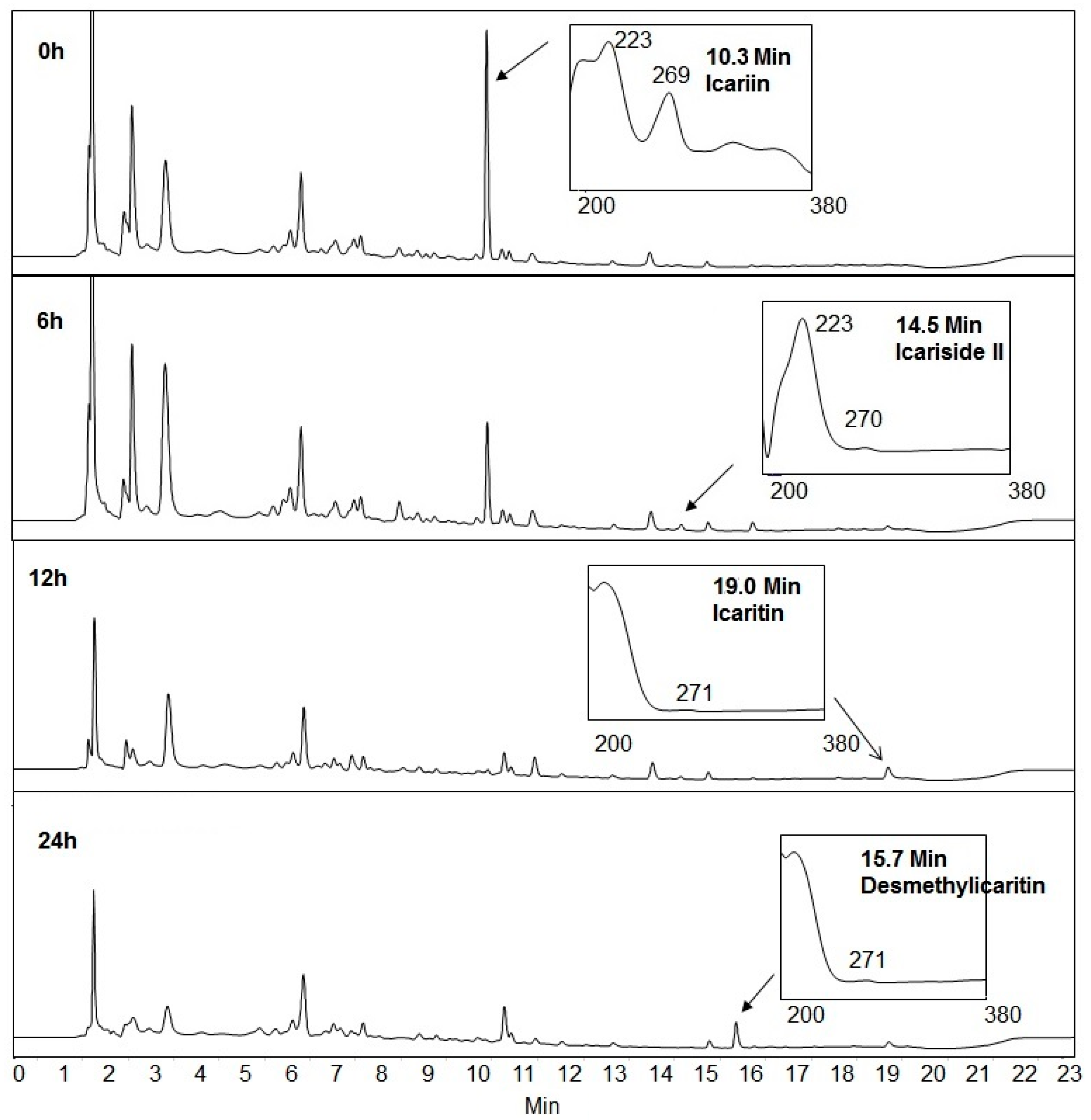
2.1. Icariin Metabolism by Human Intestinal Microflora

Icariin was added to the three different human intestinal microflora prepared from human fecal samples. Icariin (0.1 mM) was completely metabolized and the metabolites, such as icariside II, icaritin and desmethylicaritin, were identified from HPLC chromatograms as shown at Figure 2. Even though we cannot exclude a possible selective inhibition against specific bacteria, we could not observe any inhibitory effects of icariin, even at higher concentrations, for the growth of intestinal microflora. Therefore, we have screened three bacteria that metabolize icariin to investigate the detailed metabolic pathway.

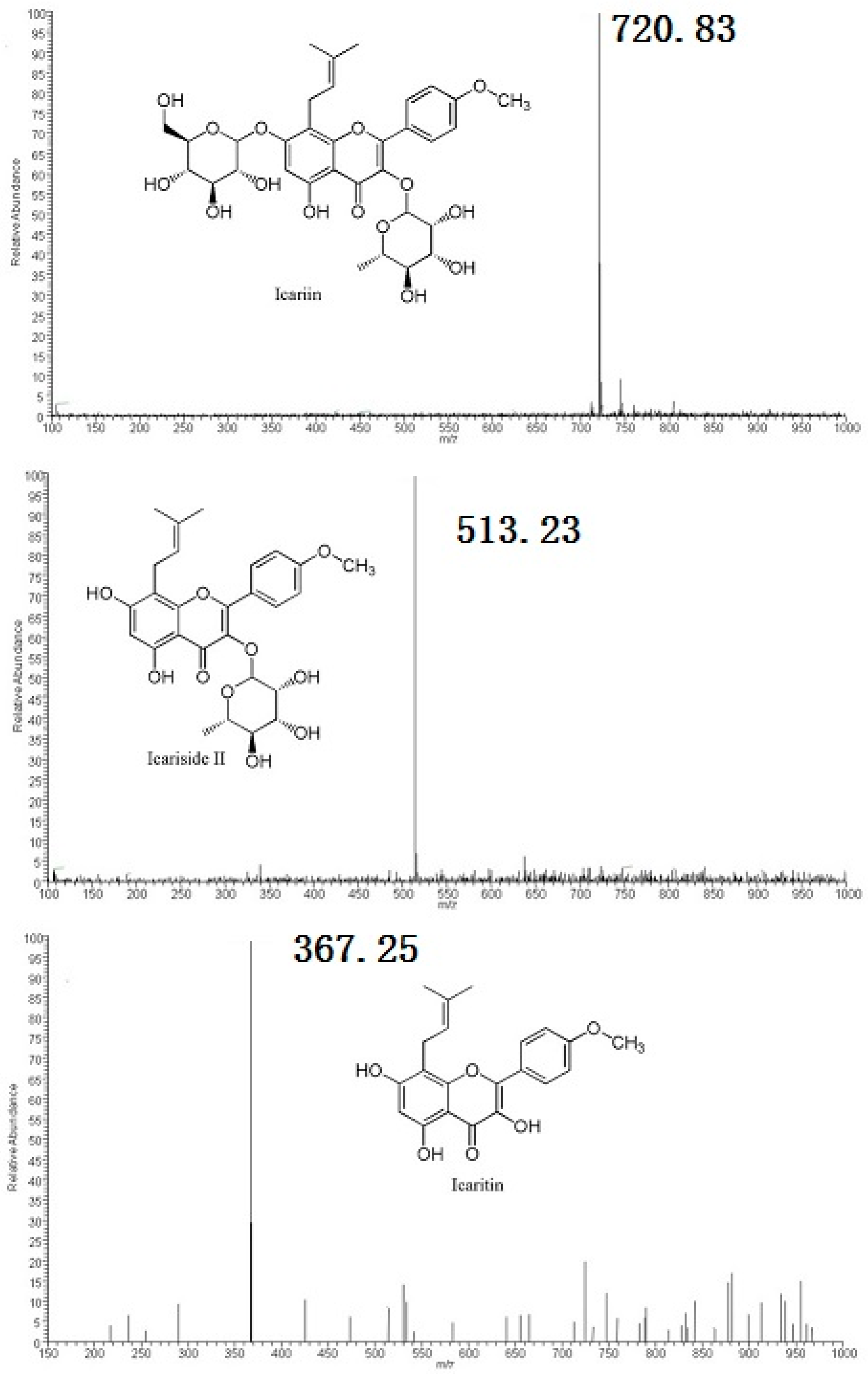
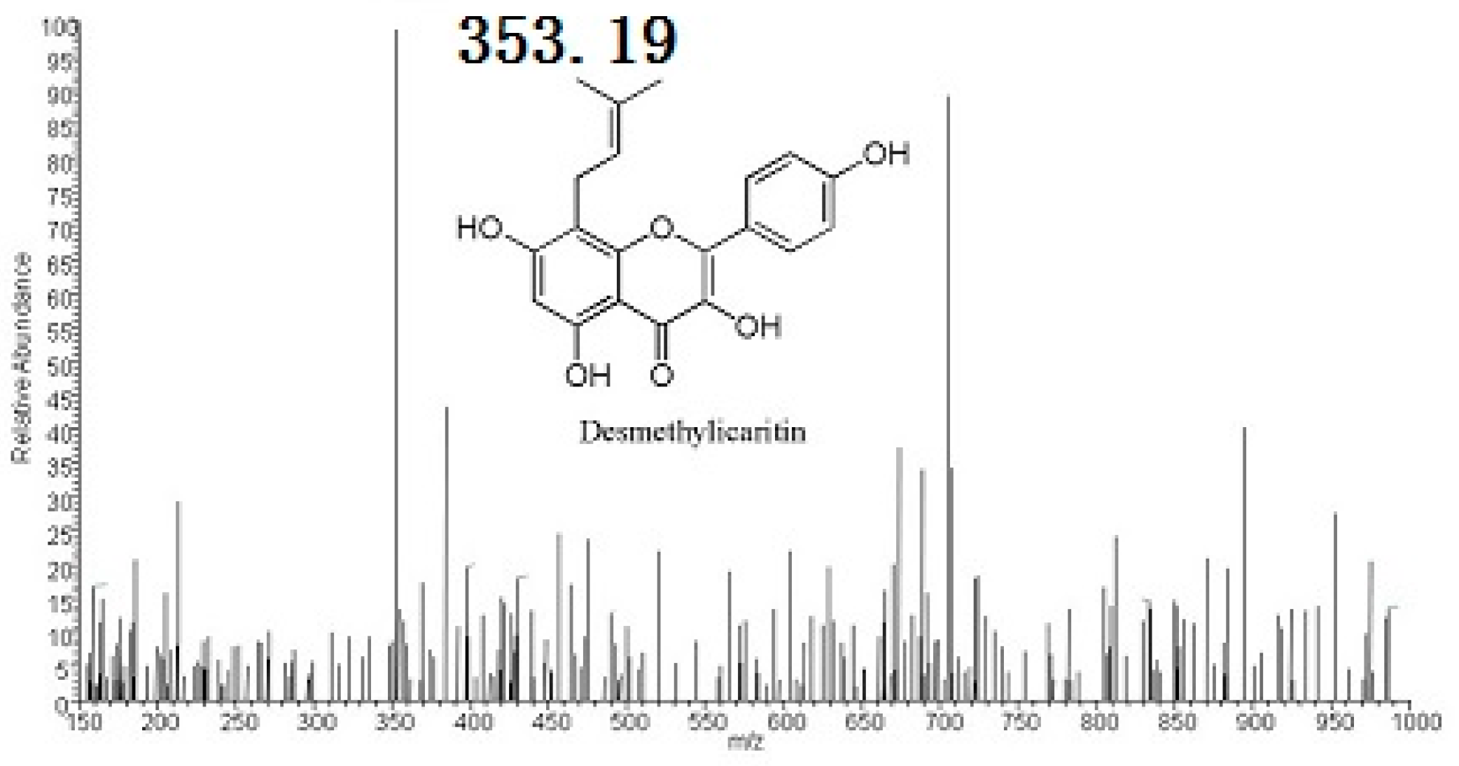
2.2. Metabolites Resulting from Icariin Metabolism by Intestinal Bacteria

Icariin conversions by Streptococcus sp. MRG-ICA-B and Enterococcus sp. MRG-ICA-E yielded single metabolites at the retention time of 14.5 min, which were identical to HPLC and ESI-MS analyses. On the ESI-MS analysis, icariin showed a parent peak at m/z 720.89 which corresponded to the [M + CO2H − H] species (Figure 3). The molecular ion [M − H] peak of the metabolite at rt = 14.5 min was found at m/z 513.29 from the ESI-MS spectrum. It was smaller than that of icariin by 162 Da, which indicated the cleavage of glucose (Figure 3). Therefore, the metabolite was assigned as icariside II, which also was confirmed by comparison with the standard compound. Icariin was also metabolized by Blautia sp. MRG-PMF-1 (Figure 4). Along with icariside II, two new metabolites at the retention times of 19.0 min and 15.7 min were observed sequentially from the HPLC analysis. The molecular ion [M + H]+ peak of the metabolite at rt = 19.0 min was found at m/z 369.21 from the ESI-MS spectrum, which was smaller than that of icariside II by 144 Da, indicating the cleavage of rhamnose. The molecular ion [M − H] peak of the metabolite at rt = 15.7 min was found at m/z 353.19 from the ESI-MS spectrum, and indicated the cleavage of the methyl group from the other metabolite at rt = 19.0 min (Figure 3). Two metabolites produced only by the MRG-PMF1 strain were identified as icaritin (rt = 19.0 min) and desmethylicaritin (rt = 15.7 min), respectively.

2.3. Biotransformation Rate of Icariin by Intestinal Bacteria

Bacterial icariin conversion was studied in a time-dependent manner by HPLC analysis. Apparently, the deglycosylation of icariin by Streptococcus sp. MRG-ICA-B was first-order and completed in 96 h (Figure 5a). Icariin biotransformation by Blautia sp. MRG-PMF-1 was much faster and more complicated, and the concentrations of icariside II and icaritin were the highest at 12 h. Icariin was completely converted within a day and desmethylicaritin was the only metabolite found in the medium after two days (Figure 5b).

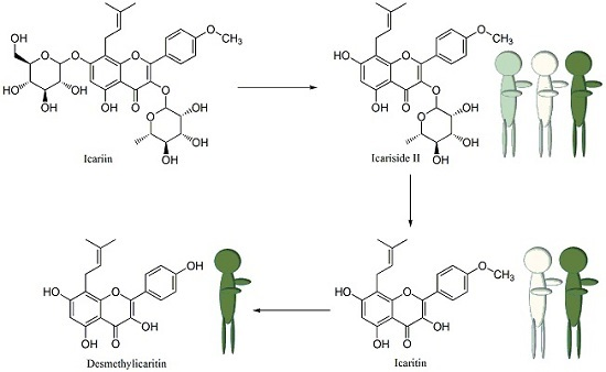
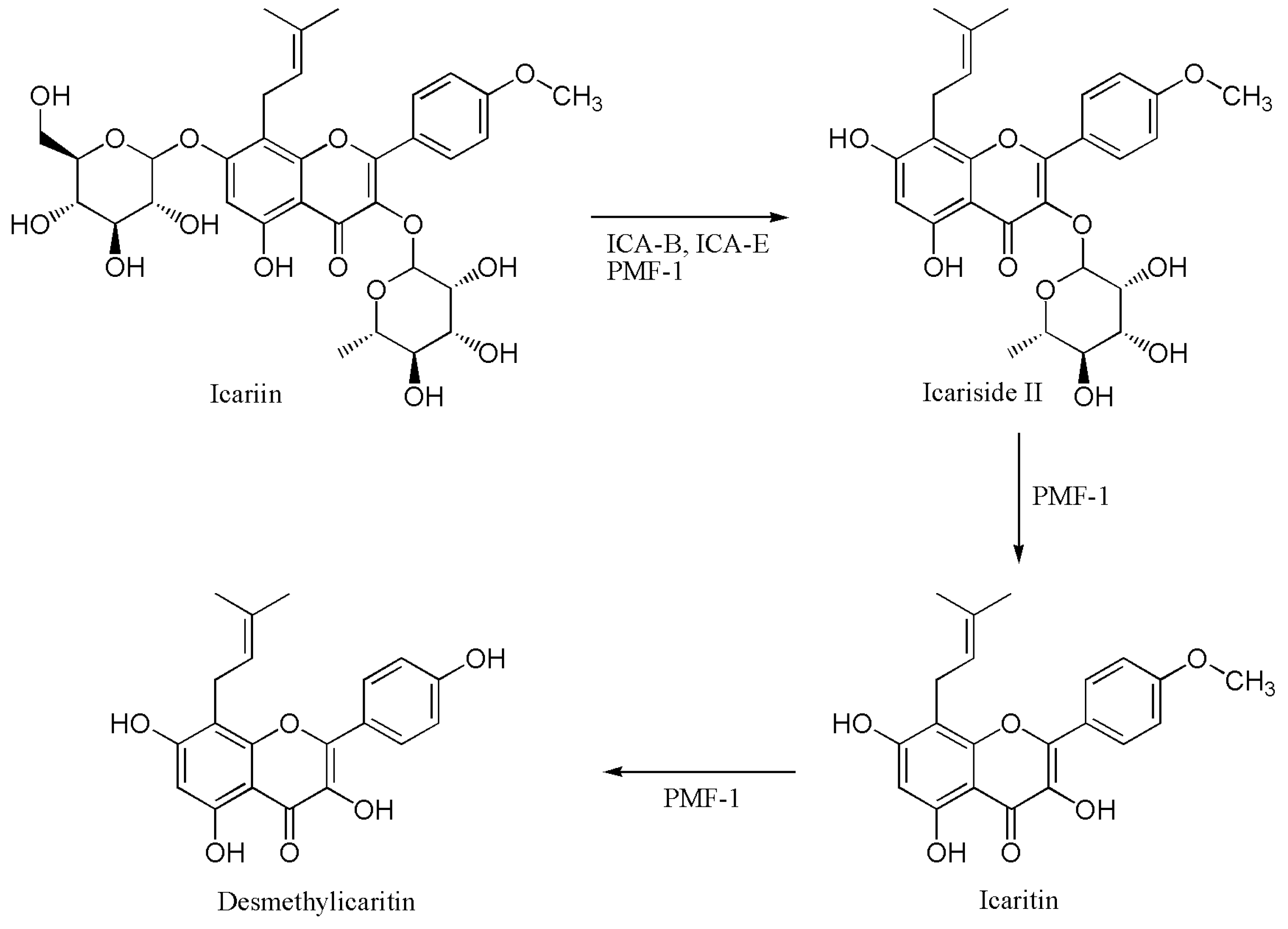
2.4. Proposed Metabolic Pathway of Icariin in Human Intestine

Icariin biotransformation by three human intestinal bacteria showed that icariin was converted to icariside II, icaritin and desmethylicaritin (Figure 6). Recently, icariin biotransformation in the rat intestine was reported, and icariside I and II were identified as metabolites resulting in icaritin [13]. However, we could not observe the formation of icariside I by human intestinal bacteria.

3. Discussion

Most bioactive polyphenolics are metabolized by the bacteria maintaining the symbiotic ecosystem in the human intestine, before absorption in the body [16]. We have been studying the microbial metabolism of flavonoids with isolated human intestinal bacteria to provide the biochemical information. For example, it is found that strong estrogenic S-equol is stereospecifically produced by human intestinal bacteria through a series of biochemical reactions including deglycosylation, reductions, and dihydroxylation from daidzin [17,18,19,20]. It is also known by many research groups, including us, that bioactive polyphenolic compounds can be biotransformed to different metabolites, depending on the microbiota harbored by the host. In the case of the daidzein metabolism, O-desmethylangolensin and 2R-(4-hydroxyphenyl)propionic acid, together with S-equol, were produced depending on the host microbiota [21,22]. The emerging research area of flavonoid metabolism by human intestinal bacteria provided a new paradigm in the area of pharmacokinetic studies. At the same time, it brought up the issue that many in vitro pharmacological activity measurements should be performed with the microbial metabolites which actually interact with the in vivo biochemical receptors.
In this report, the metabolism of icariin by human intestinal bacteria was investigated to provide information on whether icariin is the actual phytoestrogen responsible for biological activity in the body. Human intestinal microflora quickly metabolized 0.1 mM of icariin to icariside II within an hour as observed from the mixed cells metabolism. The concentration of icariin was chosen to represent the actual intake amount of Epimedii Herba. Similar results were observed from the icariin metabolism by rabbit intestinal flora [23]. When icariin was reacted with the isolated bacteria, icariside II was the only metabolite produced by the two bacteria Streptococcus sp. MRG-ICA-B and Enterococcus sp. MRG-ICA-E. Icariside II is also a constituent of E. koreanum and exhibits anti-hepatotoxic activity as strong as silybin [24]. The icariin metabolism by Blautia sp. MRG-PMF-1 produced further hydrolyzed products, icaritin and desmethylicaritin, which were reported to exhibit estrogenic effects by acting on the estrogen receptor [8,25]. Icaritin was recently reported to show a strong anti-cancer activity at concentrations of a few μM [26,27]. Besides, desmethylicaritin showed significant anti-adipogenesis activity [28].
Regardless of the important biological activities of icaritin and desmethylicaritin, the roles of human intestinal bacteria in the formation of these metabolites were not clear. In this report, we have confirmed the formation of icaritin and desmethylicaritin by the human intestinal bacterium Blautia sp. MRG-PMF1. Based on our experiments, the icariin metabolism in the human intestine looked similar to those reported from animal models. However, icariside I was not detected from our works. Furthermore, it should be also noted that individual variations in the icariin metabolism are expected due to the different host microbiota of individuals.

4. Materials and Methods

The experimental protocol was evaluated and approved by the Institutional Review Board of Chung-Ang University (Approval Number: 1041078-201502-BR-029-01).

4.1. Chemicals and Bacteria

Icariin, icariside II and icaritin were purchased from Sigma-Aldrich (St. Louis, Mo, USA). HPLC grade acetonitrile and water were from Burdick & Jackson (Muskegon, MI, USA). Ethyl acetate and acetic acid were purchased from Fisher (Pittsburg, PA, USA). Gifu anaerobic medium (GAM) was from Nissui Pharmaceutical Co. (Tokyo, Japan). The GAM broth was prepared following the manufacturer’s instructions and every GAM plate contained 15 g/L agar in GAM broth. N,N-Dimethylformamide (99.5%, DMF) was from Samchun Pure Chemicals (Gyeonggi-do, Korea).
Human intestinal microflora and three human intestinal bacteria isolated from our laboratory, Streptococcus sp. MRG-ICA-B, Enterococcus sp. MRG-ICA-E, and Blautia sp. MRG-PMF1 (GenBank accession number: KT585282, KT583836, and KJ078647, respectively) were used in this experiment. Preparation of intestinal mixed cells, bacterial growth and substrate conversion experiments were performed under the anaerobic conditions, according to the published method [15,29].

4.2. General HPLC Methods

To monitor the reaction products, Finnigan Surveyor Plus HPLC-DAD system (Thermo Scientific) equipped with a C18 reversed-phase column (Hypersil GOLD 5 μm, 4.6 by 100 mm; Thermo Scientific) were employed. The mobile phase was composed of deionized water with 0.1% acetic acid (solvent A) and acetonitrile with 0.1% acetic acid (solvent B). Following the injection of 10 μL of analyte, the elution profile started with 90% of A for 1 min. The solvent gradient was then changed linearly to 40% of A over 11 min, 20% of A over next 3 min, and kept for 2 min. The eluent flow rate was 1.0 ml/min.

4.3. Icariin Metabolism by Human Intestinal Bacteria

Bacteria were grown to OD 0.8 at 600 nm in 99 μL of GAM broth media, and 1 μL of icariin (10 mM in DMF) was added to the each medium. After incubation for 24 h, 1 mL of ethyl acetate was added to stop the biotransformation. The mixture was then vortexed for 20 s and centrifuged (10,770× g) for 1 min. The supernatant (800 µL) was taken and dried under reduced pressure. The residue was then dissolved in 100 µL of DMF, and a 10 µL aliquot was injected for HPLC.

4.4. Structural Analyses of Icariin Metabolites by HPLC-MS

Icariin metabolites were analyzed by a HPLC-MS system. Dionex Ultimate 3000 HPLC system (Thermo Scientific) equipped with a C18 reversed-phase column (Kinetex, 100 × 2.1 mm, 1.7 µL, Phenomenex, Torrance, CA, USA) and a diode array detector (DAD) was coupled to a Thermo Fisher Scientific LCQ fleet instrument (Thermo Scientific) for electrospray ionization mass spectrometry (ESI-MS) analysis. The mobile phase was composed of deionized water with 0.1% formic acid (solvent A) and acetonitrile with 0.1% formic acid (solvent B). Gradient for the eluent began with 80% of A (0–1 min) and increased to 70% of A (1–5 min), to 80% of A (5–12 min), and finally to 90% of A (12–30 min). The flow rate was 0.3 mL/min. ESI condition: spray voltage, 5.4 kV; sheath gas, 15 arbitrary units; auxiliary gas, five arbitrary units; heated capillary temperature, 275 °C; capillary voltage, 27 V; and tube lens, 100 V.

4.5. Biotransformation Kinetics

Icariin was used as substrates for biotransformation kinetics study of Streptococcus sp. MRG-ICA-B and Blautia sp. MRG-PMF-1 in an anaerobic chamber. When the growth of bacteria in GAM broth media reached OD 1.6 (MRG-ICA-B) and 1.5 (MRG-PMF-1) at 600 nm, 100 µL of the cell culture was transferred to 10 mL of fresh GAM broth medium. Biotransformation was initiated by adding 100 µL of icariin (10 mM in DMF). To monitor the conversion rate, 100 µL of the reaction medium were taken regularly and allocated into Eppendorf tubes. The allocated media were extracted and analyzed by HPLC as described above.

Acknowledgments

This work was supported under the framework of international cooperation program managed by the National Research Foundation of Korea (NRF-2015K2A1A2068137) and by the Korean government (MSIP) (NRF-2015R1A1A3A04001198).

Author Contributions

M.K. and J.H. conceived and designed the experiments; H.W. and M.K. performed the experiments and analyzed the data; J.H. wrote the paper.

Conflicts of Interest

The authors declare no conflict of interest. The founding sponsors had no role in the design of the study; in the collection, analyses, or interpretation of data; in the writing of the manuscript, and in the decision to publish the results.

References

  1. Tang, W.; Eisenbrand, G. Handbook of Chinese Medicinal Plants; Wiley-VCH: Weinheim, Germany, 2011. [Google Scholar]
  2. Wang, L.; Wang, X.; Kong, L. Automatic authentication and distinction of Epimedium koreanum and Epimedium wushanense with HPLC fingerprint analysis assisted by pattern recognition techniques. Biochem. Syst. Ecol. 2012, 40, 138–145. [Google Scholar] [CrossRef]
  3. Li, C.; Li, Q.; Mei, Q.; Lu, T. Pharmacological effects and pharmacokinetic properties of icariin, the major bioactive component in Herba Epimedii. Life Sci. 2015, 126, 57–68. [Google Scholar] [CrossRef] [PubMed]
  4. Kim, S.J.; Park, M.S.; Ding, T.; Wang, J.; Oh, D.H. Biological activities of isolated icariin from Epimedium koreanum Nakai. J. Korean Soc. Food Sci. Nutr. 2011, 40, 1397–1403. [Google Scholar] [CrossRef]
  5. Ma, H.R.; Wang, J.; Chen, Y.F.; Chen, H.; Wang, W.S.; Aisa, H.A. Icariin and icaritin stimulate the proliferation of SKBr3 cells through the GPER1-mediated modulation of the EGFR-MAPK signaling pathway. Int. J. Mol. Med. 2014, 33, 1627–1634. [Google Scholar] [CrossRef] [PubMed]
  6. Hsieh, T.P.; Sheu, S.Y.; Sun, J.S.; Chen, M.H.; Liu, M.H. Icariin isolated from Epimedium pubescens regulates osteoblasts anabolism through BMP-2, SMAD4, and Cbfa1 expression. Phytomedicine 2010, 17, 414–423. [Google Scholar] [CrossRef] [PubMed]
  7. Zhai, Y.K.; Guo, X.Y.; Ge, B.F.; Zhen, P.; Ma, X.N.; Zhou, J.; Ma, H.P.; Xian, C.J.; Chen, K.M. Icariin stimulates the osteogenic differentiation of rat bone marrow stromal cells via activating the PI3K-AKT-eNOS-NO-cGMP-PKG. Bone 2014, 66, 189–198. [Google Scholar] [CrossRef] [PubMed]
  8. Ye, H.Y.; Lou, Y.J. Estrogenic effects of two derivatives of icariin on human breast cancer MCF-7 cells. Phytomedicine 2005, 12, 735–741. [Google Scholar] [CrossRef] [PubMed]
  9. Zhu, J.T.T.; Choi, R.C.Y.; Chu, G.K.Y.; Cheung, A.W.H.; Gao, Q.T.; Li, J.; Jiang, Z.Y.; Dong, T.T.X.; Tsim, K.W.K. Flavonoids possess neuroprotective effects on cultured pheochromocytoma PC12 cells: A comparison of different flavonoids in activating estrogenic effect and in preventing β-amyloid-induced cell death. J. Agric. Food Chem. 2007, 55, 2438–2445. [Google Scholar] [CrossRef] [PubMed]
  10. Kang, H.K.; Choi, Y.H.; Kwon, H.; Lee, S.B.; Kim, D.H.; Sung, C.K.; Park, Y.I.; Dong, M.S. Estrogenic/antiestrogenic activities of a Epimedium koreanum extract and its major components: In vitro and in vivo studies. Food Chem. Toxicol. 2012, 50, 2751–2759. [Google Scholar] [CrossRef] [PubMed]
  11. Qian, Q.; Li, S.L.; Sun, E.; Zhang, K.R.; Tan, X.B.; Wei, Y.J.; Fan, H.W.; Cui, L.; Jia, X.B. Metabolite profiles of icariin in rat plasma by ultra-fast liquid chromatography coupled to triple-quadrupole/time-of-flight mass spectrometry. J. Pharm. Biomed. Anal. 2012, 66, 392–398. [Google Scholar] [CrossRef] [PubMed]
  12. Wu, Y.T.; Lin, C.W.; Lin, L.C.; Chiu, A.W.; Chen, K.K.; Tsai, T.H. Analysis of biliary excretion of icariin in rats. J. Agric. Food Chem. 2010, 58, 9905–9911. [Google Scholar] [CrossRef] [PubMed]
  13. Zhou, J.; Chen, Y.; Wang, Y.; Gao, X.; Qu, D.; Liu, C. A comparative study on the metabolism of Epimedium koreanum Nakai-Prenyllated flavonoids in rats by an intestinal enzyme (lactase phlorizin hydrolase) and intestinal flora. Molecules 2014, 19, 177–203. [Google Scholar] [CrossRef] [PubMed]
  14. Kim, M.; Lee, J.; Han, J. Deglycosylation of isoflavone C-glycosides by newly isolated human intestinal bacteria. J. Sci. Food Agric. 2015, 95, 1925–1931. [Google Scholar] [CrossRef] [PubMed]
  15. Kim, M.; Kim, N.; Han, J. Deglycosylation of flavonoid O-glucosides by human intestinal bacteria Enterococcus sp. MRG-2 and Lactococcus sp. MRG-IF-4. Appl. Biol. Chem. 2016, 59, 443–449. [Google Scholar] [CrossRef]
  16. Serra, A.; Macià, A.; Romero, M.P.; Reguant, J.; Ortega, N.; Motilva, M.J. Metabolic pathways of the colonic metabolism of flavonoids (flavonols, flavones and flavanones) and phenolic acids. Food Chem. 2012, 130, 383–393. [Google Scholar] [CrossRef]
  17. Kim, M.; Kim, S.I.; Han, J.; Wang, X.L.; Song, D.G.; Kim, S.U. Stereospecific biotransformation of dihydrodaidzein into (3S)-equol by the human intestinal bacterium Eggerthella strain Julong 732. Appl. Environ. Microbiol. 2009, 75, 3062–3068. [Google Scholar] [CrossRef] [PubMed]
  18. Kim, M.; Marsh, E.N.G.; Kim, S.U.; Han, J. Conversion of (3S,4R)-tetrahydrodaidzein to (3S)-equol by THD reductase: Proposed mechanism involving a radical intermediate. Biochemistry 2010, 49, 5582–5587. [Google Scholar] [CrossRef] [PubMed]
  19. Kim, M.; Won, D.; Han, J. Absolute configuration determination of isoflavan-4-ol stereoisomers. Bioorg. Med. Chem. Lett. 2010, 20, 4337–4341. [Google Scholar] [CrossRef] [PubMed]
  20. Park, H.Y.; Kim, M.; Han, J. Stereospecific microbial production of isoflavanones from isoflavones and isoflavone glucosides. Appl. Microbiol. Biotechnol. 2011, 91, 1173–1181. [Google Scholar] [CrossRef] [PubMed]
  21. Kim, M.; Han, J. Absolute configuration of (−)-2-(4-hydroxyphenyl)propionic acid: Stereochemistry of soy isoflavone metabolism. Bull. Korean Chem. Soc. 2014, 35, 1883–1886. [Google Scholar] [CrossRef]
  22. Kim, M.; Han, J. Chiroptical study and absolute configuration of (−)-O-DMA produced from daidzein metabolism. Chirality 2014, 26, 434–437. [Google Scholar] [CrossRef] [PubMed]
  23. Yao, Z.H.; Liu, M.Y.; Dai, Y.; Zhang, Y.; Qin, Z.F.; Tu, F.J.; Yao, X.S. Metabolism of Epimedium-derived flavonoid glycosides in intestinal flora of rabbits and its inhibition by gluconolactone. Chin. J. Natl. Med. 2011, 9, 461–465. [Google Scholar]
  24. Cho, N.J.; Sung, S.H.; Lee, H.S.; Jeon, M.H.; Kim, Y.C. Anti-hepatotoxic activity of icariside II, a constituent of Epimedium koreanum. Arch. Pharm. Res. 1995, 18, 289–292. [Google Scholar] [CrossRef]
  25. Wang, Z.Q.; Lou, Y.J. Proliferation-stimulating effects of icaritin and desmethylicaritin in MCF-7 cells. Eur. J. Pharmacol. 2004, 504, 147–153. [Google Scholar] [CrossRef] [PubMed]
  26. Sun, L.; Chen, W.; Qu, L.; Wu, J.; Si, J. Icaritin reverses multidrug resistance of HepG2/ADR human hepatoma cells via downregulation of MDR1 and P-glycoprotein expression. Mol. Med. Rep. 2013, 8, 1883–1887. [Google Scholar] [PubMed]
  27. Wu, T.; Wang, S.; Wu, J.; Lin, Z.; Sui, Z.; Xu, X.; Shimizu, N.; Chen, B.; Wang, X. Icaritin induces lytic cytotoxicity in extranodal NK/T-cell lymphoma. J. Exp. Clin. Cancer Res. 2015, 34, 17. [Google Scholar] [CrossRef] [PubMed]
  28. Wang, X.L.; Wang, N.; Zheng, L.Z.; Xie, X.H.; Yao, D.; Liu, M.Y.; Yao, Z.H.; Dai, Y.; Zhang, G.; Yao, X.S.; et al. Phytoestrogenic molecule desmethylicaritin suppressed adipogenesis via Wnt/β-catenin signaling pathway. Eur. J. Pharmacol. 2013, 714, 254–260. [Google Scholar] [CrossRef] [PubMed]
  29. Kim, M.; Kim, N.; Han, J. Metabolism of Kaempferia parviflora polymethoxyflavones by human intestinal bacterium Bautia sp. MRG-PMF1. J. Agric. Food Chem. 2014, 62, 12377–12383. [Google Scholar] [CrossRef] [PubMed]
  • Sample Availability: Samples of icariin, icariside II, and icaritin are available from the authors and commercial sources.
Figure 1. Molecular structures of the major prenylflavonoids isolated from Epimedii Herba.
Molecules 21 01158 g001
Figure 2. HPLC chromatograms of icariin biotransformation products. Each chromatogram was obtained from the different human intestinal microflora. Microflora a, b and c showed icariside II, icaritin and desmethylicaritin formation, respectively, after 48 h.
Molecules 21 01158 g002
Figure 3. MS spectra of icariin and its metabolites. Thermo Fisher Scientific LCQ fleet instrument (Thermo Scientific, Waltham, MA, USA) was used for electrospray ionization mass spectrometry (ESI-MS) analysis. ESI condition: spray voltage, 5.4 kV; sheath gas, 15 arbitrary units; auxiliary gas, five arbitrary units; heated capillary temperature, 275 °C; capillary voltage, 27 V; and tube lens, 100 V.
Molecules 21 01158 g003aMolecules 21 01158 g003b
Figure 4. HPLC chromatogram changes at 270 nm absorption over icariin metabolism by Blautia sp. MRG-PMF1.
Molecules 21 01158 g004
Figure 5. Time-dependent biotransformation of icariin by (a) Streptococcus sp. MRG-ICA-B and (b) Blautia sp. MRG-PMF-1.
Molecules 21 01158 g005
Figure 6. Proposed metabolic pathway of icariin in human intestine.
Molecules 21 01158 g006

