
Phytochemical Profile and Evaluation of the Biological Activities of Essential Oils Derived from the Greek Aromatic Plant Species Ocimum basilicum, Mentha spicata, Pimpinella anisum and Fortunella margarita

 ,
,  , , ,
, , ,  ,
,  , and
, and 
Abstract
:
1. Introduction
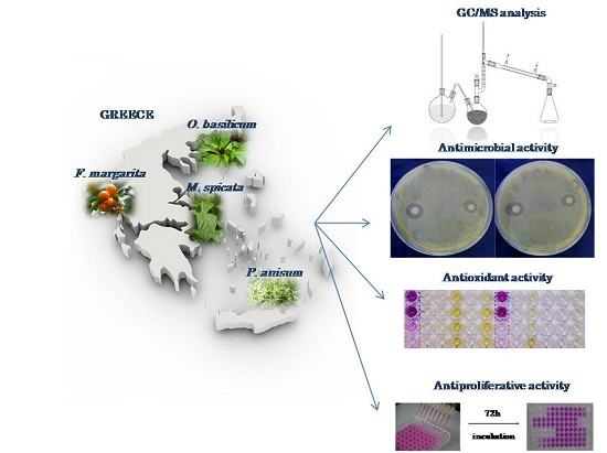
2. Results and Discussion
2.1. GC/MS Analysis of Essential Oils
2.2. Antimicrobial Activity of Essential Oils
2.3. In Vitro Antioxidant Capacity of Essential Oils
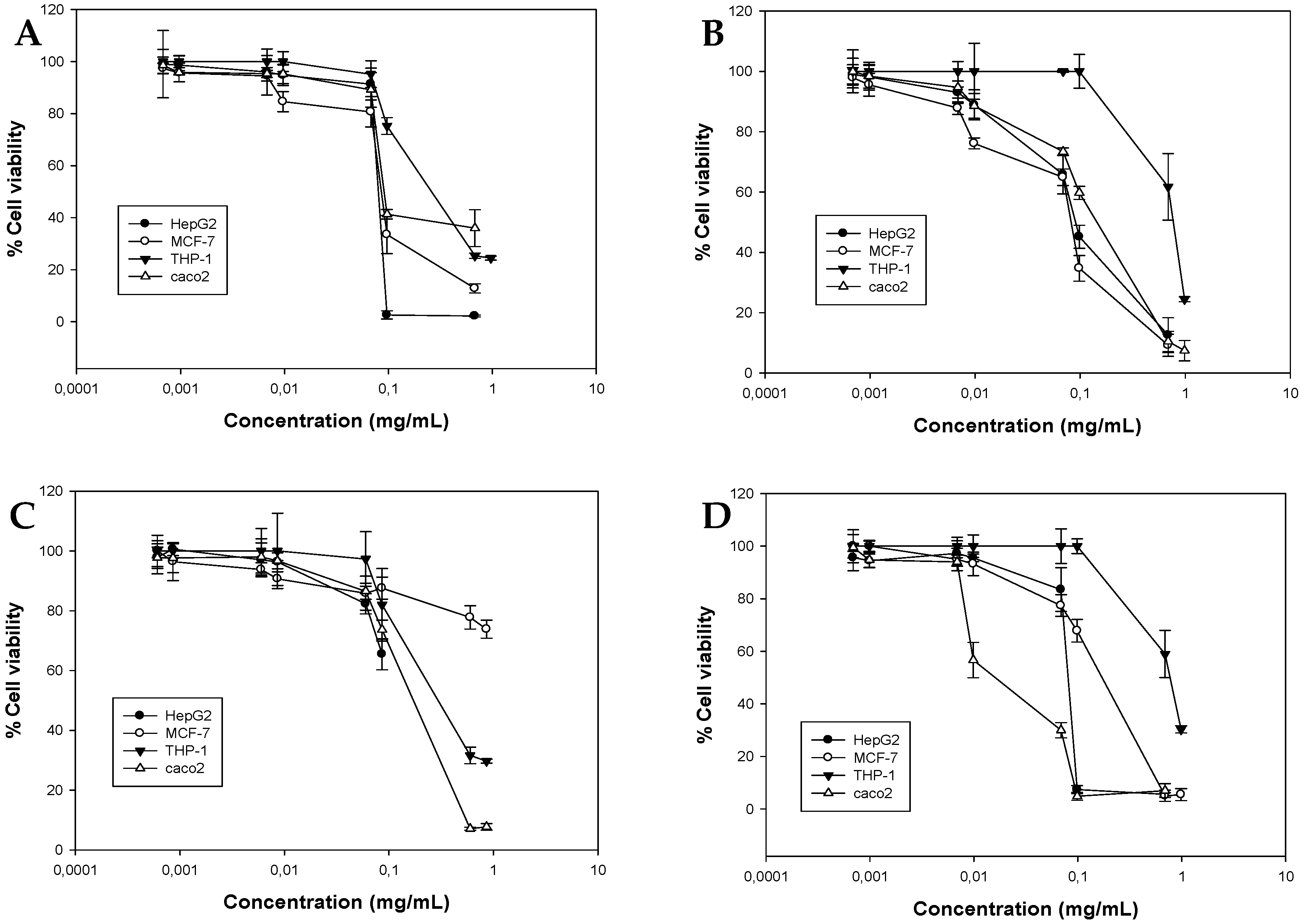
2.4. Antiproliferative Activity of Essential Oils
3. Materials and Methods
3.1. Essential Oil Extraction and GC/MS Analysis
3.2. Microbial Strains
3.3. Antimicrobial Assays
3.3.1. Disk Diffusion Assay
3.3.2. Determination of Minimum Inhibitory Concentration (MIC) and Non-Inhibitory Concentration (NIC)
3.4. Cell Lines and Cell Cultures
3.5. Antioxidant Activity
3.5.1. DPPH Assay
3.5.2. ABTS Assay
3.6. Cell Viability Assays
3.6.1. Sulforhodamine B Assay
3.6.2. XTT Cell Viability Assay
3.7. Data Analysis
4. Conclusions
Acknowledgments
Author Contributions
Conflicts of Interest
References
- Bakkali, F.; Averbeck, S.; Averbeck, D.; Idaomar, M. Biological effects of essential oils—A review. Food Chem. Toxicol. 2008, 46, 446–475. [Google Scholar] [CrossRef] [PubMed]
- Edris, A.E. Pharmaceutical and therapeutic potentials of essential oils and their individual volatile constituents: A review. Phytother. Res. 2007, 21, 308–323. [Google Scholar] [CrossRef] [PubMed]
- Tongnuanchan, P.; Benjakul, S. Essential oils: Extraction, bioactivities, and their uses for food preservation. J. Food Sci. 2014, 79, R1231–R1249. [Google Scholar] [CrossRef] [PubMed]
- Gautam, N.; Mantha, A.K.; Mittal, S. Essential oils and their constituents as anticancer agents: A mechanistic view. Biomed. Res. Int. 2014, 2014. [Google Scholar] [CrossRef] [PubMed]
- Huang, X.Y.; Wang, H.C.; Yuan, Z.; Li, A.; He, M.L.; Ai, K.X.; Zheng, Q.; Qin, H.L. Gemcitabine combined with gum mastic causes potent growth inhibition and apoptosis of pancreatic cancer cells. Acta Pharmacol. Sin. 2010, 31, 741–745. [Google Scholar] [CrossRef] [PubMed]
- Carnesecchi, S.; Langley, K.; Exinger, F.; Gosse, F.; Raul, F. Geraniol, a component of plant essential oils, sensitizes human colonic cancer cells to 5-Fluorouracil treatment. J. Pharmacol. Exp. Ther. 2002, 301, 625–630. [Google Scholar] [CrossRef] [PubMed]
- Dangkong, D.; Limpanasithikul, W. Effect of citral on the cytotoxicity of doxorubicin in human B-lymphoma cells. Pharm. Biol. 2015, 53, 262–268. [Google Scholar] [CrossRef] [PubMed]
- Ravizza, R.; Gariboldi, M.B.; Molteni, R.; Monti, E. Linalool, a plant-derived monoterpene alcohol, reverses doxorubicin resistance in human breast adenocarcinoma cells. Oncol. Rep. 2008, 20, 625–630. [Google Scholar] [CrossRef] [PubMed]
- Sak, K. Chemotherapy and dietary phytochemical agents. Chemother. Res. Pract. 2012, 2012. [Google Scholar] [CrossRef] [PubMed]
- Burt, S. Essential oils: Their antibacterial properties and potential applications in foods—A review. Int. J. Food Microbiol. 2004, 94, 223–253. [Google Scholar] [CrossRef] [PubMed]
- Lianou, A.; Koutsoumanis, K.P. Strain variability of the behavior of foodborne bacterial pathogens: A review. Int. J. Food Microbiol. 2013, 167, 310–321. [Google Scholar] [CrossRef] [PubMed]
- Battey, A.S.; Duffy, S.; Schaffner, D.W. Modeling yeast spoilage in cold-filled ready-to-drink beverages with Saccharomyces cerevisiae, Zygosaccharomyces bailii, and Candida lipolytica. Appl. Environ. Microb. 2002, 68, 1901–1906. [Google Scholar] [CrossRef]
- Garcia, D.; Ramos, A.J.; Sanchis, V.; Marin, S. Predicting mycotoxins in foods: A review. Food Microb. 2009, 26, 757–769. [Google Scholar] [CrossRef] [PubMed]
- Lambert, R.J.W.; Lambert, R. A model for the efficacy of combined inhibitors. J. Appl. Microbiol. 2003, 95, 734–743. [Google Scholar] [CrossRef] [PubMed]
- Chorianopoulos, N.; Lambert, R.J.W.; Skandamis, P.N.; Evergetis, E.T.; Haroutounian, S.A.; Nychas, G.J.E. A newly developed assay to study the minimum inhibitory concentration of Satureja spinosa essential oil. J. Appl. Microbiol. 2006, 100, 778–786. [Google Scholar] [CrossRef] [PubMed]
- Borrego, S.; Valdés, O.; Vivar, I.; Lavin, P.; Guiamet, P.; Battistoni, P.; Gómez de Saravia, S.; Borges, P. Essential oils of plants as biocides against microorganisms isolated from Cuban and Argentine documentary heritage. ISRN Microbiol. 2012, 826786. [Google Scholar] [CrossRef] [PubMed]
- Settanni, L.; Palazzolo, E.; Guarrasi, V.; Aleo, A.; Mammina, C.; Moschetti, G.; Germanà, M.A. Inhibition of foodborne pathogen bacteria by essential oils extracted from citrus fruits cultivated in Sicily. Food Control. 2012, 26, 326–330. [Google Scholar] [CrossRef]
- Chorianopoulos, N.; Kalpoutzakis, E.; Aligiannis, N.; Mitaku, S.; Nychas, G.J.E.; Haroutounian, S.A. Essential oils of Satureja, Origanum and Thymus species: Chemical composition and antibacterial activities against foodborne pathogens. J. Agric. Food Chem. 2004, 52, 8261–8267. [Google Scholar] [CrossRef] [PubMed]
- Djenane, D.; Yangüela, J.; Montañés, L.; Djerbal, M.; Roncalés, P. Antimicrobial activity of Pistacia lentiscus and Satureja montana essential oils against Listeria monocytogenes CECT 935 using laboratory media: Efficacy and synergistic potential in minced beef. Food Control. 2011, 22, 1046–1053. [Google Scholar] [CrossRef]
- Rattanachaikunsopon, P.; Phumkhachorn, P. Antimicrobial activity of basil (Ocimum basilicum) oil against Salmonella enteritidis in vitro and in food. Biosci. Biotechnol. Biochem. 2010, 74, 1200–1204. [Google Scholar] [CrossRef] [PubMed]
- Lixandru, B.E.; Drăcea, N.O.; Dragomirescu, C.C.; Drăgulescu, E.C.; Coldea, I.L.; Anton, L.; Dobre, E.; Rovinaru, C.; Codiţă, I. Antimicrobial activity of plant essential oils against bacterial and fungal species involved in food poisoning and/or food decay. Roum. Arch. Microbiol. Immunol. 2010, 69, 224–230. [Google Scholar] [PubMed]
- Wang, Y.W.; Zeng, W.C.; Xu, P.Y.; Lan, Y.J.; Zhu, R.X.; Zhong, K.; Huang, Y.N.; Gao, H. Chemical composition and antimicrobial activity of the essential oil of kumquat (Fortunella crassifolia Swingle) peel. Int. J. Mol. Sci. 2012, 13, 3382–3393. [Google Scholar] [CrossRef] [PubMed]
- Nazzaro, F.; Fratianni, F.; De Martino, L.; Coppola, R.; de Feo, V. Effect of essential oils on pathogenic bacteria. Pharmaceuticals 2013, 6, 1451–1474. [Google Scholar] [CrossRef] [PubMed]
- Afoulous, S.; Ferhout, H.; Raoelison, E.G.; Valentin, A.; Moukarzel, B.; Couderc, F.; Bouajila, J. Chemical composition and anticancer, antiinflammatory, antioxidant and antimalarial activities of leaves essential oil of Cedrelopsis grevei. Food Chem. Toxicol. 2013, 56, 352–362. [Google Scholar] [CrossRef] [PubMed]
- Bayala, B.; Bassole, I.H.; Gnoula, C.; Nebie, R.; Yonli, A.; Morel, L.; Figueredo, G.; Nikiema, J.B.; Lobaccaro, J.M.; Simpore, J. Chemical composition, antioxidant, anti-inflammatory and antiproliferative activities of essential oils of plants from Burkina Faso. PLoS ONE 2014, 9, e92122. [Google Scholar] [CrossRef] [PubMed]
- Yu, L.; Haley, S.; Perret, J.; Harris, M.; Wilson, J.; Qian, M. Free radical scavenging properties of wheat extracts. J. Agric. Food Chem. 2002, 50, 1619–1624. [Google Scholar] [CrossRef] [PubMed]
- Dawidowicz, A.L.; Olszowy, M. Does antioxidant properties of the main component of essential oil reflect its antioxidant properties? The comparison of antioxidant properties of essential oils and their main components. Nat. Prod. Res. 2014, 28, 1952–1963. [Google Scholar] [CrossRef] [PubMed]
- Kathirvel, P.; Ravi, S. Chemical composition of the essential oil from basil (Ocimum basilicum Linn.) and its in vitro cytotoxicity against HeLa and HEp-2 human cancer cell lines and NIH 3T3 mouse embryonic fibroblasts. Nat. Prod. Res. 2012, 26, 1112–1118. [Google Scholar] [CrossRef] [PubMed]
- Trevisan, M.T.; Vasconcelos Silva, M.G.; Pfundstein, B.; Spiegelhalder, B.; Owen, R.W. Characterization of the volatile pattern and antioxidant capacity of essential oils from different species of the genus Ocimum. J. Agric. Food Chem. 2006, 54, 4378–4382. [Google Scholar] [CrossRef] [PubMed]
- Hussain, A.I.; Anwar, F.; Sherazi, S.T.H.; Przybylski, R. Chemical composition, antioxidant and antimicrobial activities of basil (Ocimum basilicum) essential oils depends on seasonal variations. Food Chem. 2008, 108, 986–995. [Google Scholar] [CrossRef] [PubMed]
- Politeo, O.; Jukic, M.; Milos, M. Chemical composition and antioxidant capacity of free volatile aglycones from basil (Ocimum basilicum L.) compared with its essential oil. Food Chem. 2007, 101, 379–385. [Google Scholar] [CrossRef]
- Shirazi, M.T.; Gholami, H.; Kavoosi, G.; Rowshan, V.; Tafsiry, A. Chemical composition, antioxidant, antimicrobial and cytotoxic activities of Tagetes minuta and Ocimum basilicum essential oils. Food Sci. Nutr. 2014, 2, 146–155. [Google Scholar] [CrossRef] [PubMed]
- Snoussi, M.; Noumi, E.; Trabelsi, N.; Flamini, G.; Papetti, A.; de Feo, V. Mentha spicata essential oil: Chemical composition, antioxidant and antibacterial activities against planktonic and biofilm cultures of Vibrio spp. Strains. Molecules 2015, 20, 14402–14424. [Google Scholar] [CrossRef] [PubMed]
- Sönmez, A.Y.; Bilen, S.; Alak, G.; Hisar, O.; Yanık, T.; Biswas, G. Growth performance and antioxidant enzyme activities in rainbow trout (Oncorhynchus mykiss) juveniles fed diets supplemented with sage, mint and thyme oils. Fish Physiol. Biochem. 2015, 41, 165–175. [Google Scholar] [CrossRef] [PubMed]
- Jayaprakasha, G.K.; Murthy, K.N.; Demarais, R.; Patil, B.S. Inhibition of prostate cancer (LNCaP) cell proliferation by volatile components from Nagami kumquats. Planta Med. 2012, 78, 974–980. [Google Scholar] [CrossRef] [PubMed]
- Singh, G.; Kapoor, I.P.S.; Singh, P.; de Heluani, C.S.; Catalan, C.A.N. Chemical composition and antioxidant potential of essential oil and oleoresins from anise seeds (Pimpinella anisum L.). Int. J. Essent. Oil Ther. 2008, 2, 122–130. [Google Scholar]
- Jamshidzadeh, A.; Heidari, R.; Razmjou, M.; Karimi, F.; Moein, M.R.; Farshad, O.; Akbarizadeh, A.R.; Shayesteh, M.R. An in vivo and in vitro investigation on hepatoprotective effects of Pimpinella anisum seed essential oil and extracts against carbon tetrachloride-induced toxicity. Iran J. Basic Med. Sci. 2015, 18, 205–211. [Google Scholar] [PubMed]
- Al-Kalaldeh, J.Z.; Abu-Dahab, R.; Afifi, F.U. Volatile oil composition and antiproliferative activity of Laurus nobilis, Origanum syriacum, Origanum vulgare, and Salvia triloba against human breast adenocarcinoma cells. Nutr. Res. 2010, 30, 271–278. [Google Scholar] [CrossRef] [PubMed]
- Loizzo, M.R.; Menichini, F.; Tundis, R.; Bonesi, M.; Nadjafi, F.; Saab, A.M.; Frega, N.G.; Menichini, F. Comparative chemical composition and antiproliferative activity of aerial parts of Salvia leriifolia Benth. and Salvia acetabulosa L. essential oils against human tumor cell in vitro models. J. Med. Food 2010, 13, 62–69. [Google Scholar] [PubMed]
- Saab, A.M.; Tundis, R.; Loizzo, M.R.; Lampronti, I.; Borgatti, M.; Gambari, R.; Menichini, F.; Esseily, F.; Menichini, F. Antioxidant and antiproliferative activity of Laurus nobilis L. (Lauraceae) leaves and seeds essential oils against K562 human chronic myelogenous leukaemia cells. Nat Prod Res. 2012, 26, 1741–1745. [Google Scholar] [CrossRef] [PubMed]
- Manosroi, J.; Dhumtanom, P.; Manosroi, A. Anti-proliferative activity of essential oil extracted from Thai medicinal plants on KB and P388 cell lines. Cancer Lett. 2006, 235, 114–120. [Google Scholar] [CrossRef] [PubMed]
- Zu, Y.; Yu, H.; Liang, L.; Fu, Y.; Efferth, T.; Liu, X.; Wu, N. Activities of ten essential oils towards Propionibacterium acnes and PC-3, A-549 and MCF-7 cancer cells. Molecules 2010, 15, 3200–3210. [Google Scholar] [CrossRef] [PubMed]
- Hussain, A.I.; Anwar, F.; Nigam, P.S.; Ashraf, M.; Gilani, A.H. Seasonal variation in content, chemical composition and antimicrobial and cytotoxic activities of essential oils from four Mentha species. J. Sci. Food Agric. 2010, 90, 1827–1836. [Google Scholar] [CrossRef] [PubMed]
- De Silva, T. (Ed.) A Manual on the Essential Oil Industry; Publications Sales Office of United Nations Industrial Development Organization: Vienna, Austria, 1995; p. 86.
- Mitropoulou, G.; Fitsiou, E.; Stavropoulou, E.; Papavassilopoulou, E.; Vamvakias, M.; Pappa, A.; Oreopoulou, A.; Kourkoutas, Y. Composition, antimicrobial, antioxidant, and antiproliferative activity of Origanum dictamnus (dittany) essential oil. Microb. Ecol. Health Dis. 2015, 6, 26543. [Google Scholar]
- Anestopoulos, I.; Kavo, A.; Tentes, I.; Kortsaris, A.; Panayiotidis, M.; Lazou, A.; Pappa, A. Silibinin protects H9c2 cardiac cells from oxidative stress and inhibits phenylephrine-induced hypertrophy: Potential mechanisms. J. Nutr. Biochem. 2013, 4, 586–594. [Google Scholar] [CrossRef] [PubMed]
- Re, R.; Pellegrini, N.; Proteggente, A.; Pannala, A.; Yang, M.; Rice-Evans, C. Antioxidant activity applying an improved ABTS radical cation decolorization assay. Free Radic. Biol. Med. 1999, 26, 1231–1237. [Google Scholar] [CrossRef]
- Vichai, V.; Kirtikara, K. Sulforhodamine B colorimetric assay for cytotoxicity screening. Nat. Protoc. 2006, 1, 1112–1116. [Google Scholar] [CrossRef] [PubMed]
- Roehm, N.W.; Rodgers, G.H.; Hatfield, S.M.; Glasebrook, A.L. An improved colorimetric assay for cell proliferation and viability utilizing the tetrazolium salt XTT. J. Immunol. Methods 1991, 142, 257–265. [Google Scholar] [CrossRef]
- Sample Availability: Samples of the essential oils are available from the authors.


| Compounds | KRI * | Mentha spicata (% Area) | Ocimum basilicum (% Area) | Pimpinella anisum (% Area) | Fortunella margarita (% Area) |
|---|---|---|---|---|---|
| cis-3-Hexenol | 811 | 0.014 | Trace | ||
| Thujene | 915 | Trace | |||
| α-Pinene | 922 | 0.670 | 0.069 | 0.081 | 0.743 |
| Camphene | 927 | Trace | |||
| Sabinene | 953 | 0.060 | 0.133 | ||
| Methyl heptenone | 954 | 0.134 | |||
| Oct-1-en-3-ol | 955 | 0.001 | |||
| β-Pinene | 958 | 1.450 | 0.038 | 0.054 | 0.019 |
| Myrcene | 973 | 0.039 | 0.010 | 2.680 | |
| α-Phellandrene | 981 | 0.089 | 0.073 | ||
| δ-3-Carene | 990 | 0.020 | |||
| α-Terpinene | 997 | Trace | |||
| p-Cymene | 1004 | 0.001 | 0.088 | Trace | |
| β-Phellandrene | 1004 | 0.016 | |||
| 2-Ethylhexenol | 1006 | 0.134 | |||
| 1,8-Cineole | 1010 | 0.020 | |||
| Limonene | 1011 | 8.410 | 0.020 | 0.035 | 93.784 |
| cis-Ocimene | 1016 | 0.016 | 0.001 | ||
| trans-Ocimene | 1018 | 0.001 | Trace | 0.019 | |
| γ-Terpinene | 1030 | 0.061 | 0.034 | 0.023 | |
| Epoxylinalool I | 1049 | 0.167 | Trace | ||
| Alc C8 | 1050 | 0.001 | |||
| Thujone | 1057 | 0.001 | |||
| Dehydro-p-cymene | 1062 | 0.013 | Trace | ||
| Epoxylinalool II | 1064 | 0.149 | |||
| Terpinolene | 1070 | 0.046 | 0.014 | ||
| Linalol | 1086 | 18.400 | 0.278 | 0.118 | |
| Octen-1-en-3-yl acetate | 1087 | 0.001 | |||
| trans-p-Menthene-2.3-dien-1-ol | 1105 | 0.018 | |||
| Camphor | 1108 | 0.022 | |||
| cis-p-Menthene-2.8-diene-1-ol | 1115 | 0.017 | |||
| p-vinylanisole | 1118 | 0.017 | |||
| Menthone | 1124 | 0.130 | 0.033 | ||
| iso-Menthone | 1133 | 0.040 | 0.017 | ||
| Borneol | 1138 | Trace | |||
| Menthol | 1150 | 0.190 | 0.240 | ||
| p-Cymenol | 1151 | 0.016 | |||
| Terp-1-ene-4-ol | 1152 | 0.020 | |||
| Dihydrocarvone | 1160 | 0.200 | |||
| Dihydrocarveol | 1160 | 0.130 | |||
| α-Terpineol | 1168 | 0.003 | 0.012 | 0.026 | |
| 3-Hexenyl butyrate | 1168 | 0.003 | |||
| Epoxyphellandrene | 1171 | Trace | |||
| 8-Cumenol | 1172 | Trace | |||
| trans-Carveol | 1177 | 0.014 | |||
| Methyl chavicol | 1177 | 74.920 | 1.525 | ||
| Decanal | 1178 | 0.015 | |||
| Octyl acetate | 1191 | 0.028 | 0.055 | ||
| cis-Carveol | 1197 | 0.011 | |||
| Nerol | 1203 | 0.040 | |||
| Neral (cis-citral) | 1205 | 0.200 | |||
| Anisaldehyde | 1207 | 0.110 | 0.545 | ||
| Carvone | 1217 | 85.410 | 0.023 | ||
| Piperitone | 1218 | 0.001 | |||
| cis-Anethole | 1218 | 0.435 | |||
| Geraniol | 1231 | Trace | |||
| Perilla aldehyde | 1233 | 0.019 | |||
| Geranial (trans-citral) | 1237 | 0.519 | |||
| trans-Anethole | 1265 | 0.028 | 88.130 | ||
| Isobornyl acetate | 1277 | 0.001 | |||
| Dihydrocarvenyl acetate | 1304 | 0.130 | |||
| δ-Elemene | 1327 | 0.149 | 0.022 | ||
| Eugenol | 1331 | 0.059 | |||
| Anisyl methyl ketone | 1339 | 0.025 | |||
| α-Longipinene | 1339 | 0.061 | |||
| Neryl acetate | 1340 | 0.014 | |||
| α-Cubebene | 1344 | Trace | |||
| Cyclosativene | 1357 | 0.041 | |||
| Geranyl acetate | 1358 | 0.111 | |||
| Ylangene | 1360 | 0.050 | |||
| Methyl eugenol | 1365 | 0.049 | |||
| α-Copaene | 1366 | 0.029 | 0.016 | ||
| β-Bourbonene | 1371 | 0.040 | 0.019 | 0.033 | |
| β-Elemene | 1378 | 0.098 | 0.023 | ||
| p-Menth-1-en-9-yl acetate | 1399 | 0.007 | |||
| Caryophyllene | 1403 | 0.070 | 0.273 | 0.010 | |
| Methoxypropiophenone | 1402 | 0.048 | |||
| Bergamotene | 1424 | 0.509 | |||
| a Farnesene | 1427 | 0.054 | 0.054 | ||
| α-Himachalene | 1431 | 0.381 | |||
| Humulene | 1436 | 0.154 | 0.008 | ||
| cis-β-Farnesene | 1438 | 0.219 | |||
| Dehydro-neo-isolongifolene | 1443 | 0.079 | |||
| Methyl-isoeugenol | 1446 | 0.088 | |||
| ar-Curcumene | 1460 | 0.025 | 0.091 | ||
| γ-Himachalene | 1460 | 4.155 | |||
| δ-Germacrene | 1462 | 0.025 | 1.343 | ||
| trans-β-Farnesene | 1468 | 0.025 | |||
| Zingiberene | 1478 | 0.570 | |||
| Bicyclogermacrene | 1479 | 0.246 | |||
| β-Chimachalene | 1481 | 0.243 | |||
| α-Mourolene | 1483 | Trace | |||
| Myristicin | 1487 | 0.045 | |||
| β-Bisabolene | 1492 | 0.097 | 0.473 | ||
| Calamenene | 1496 | 0.019 | |||
| Valencene | 1501 | 0.009 | |||
| δ-Cadinene | 1504 | 0.091 | 0.053 | ||
| p-Methoxycinnamic ald | 1507 | 0.572 | |||
| α-Calacorene | 1516 | Trace | |||
| α-Bisabolene | 1525 | 1.068 | |||
| β-Germacrene | 1533 | 0.039 | |||
| Caryophyllene oxide | 1551 | 0.135 | |||
| 1,5,5,8-Tetramethyl-12-oxabicyclo[9.1.0]dodeca-3.7-diene | 1575 | 0.053 | |||
| Pseudo-isoeugenyl-2-methyl butyrate | 1833 | 4.155 |
| Essential Oil | 5 log cfu/mL Initial Inoculum | |||||
|---|---|---|---|---|---|---|
| S. enteritidis | S. typhimurium | E. coli | S. epidermidis | S. aureus | L. monocytogenes | |
| Spearmint | 14 ± 0.5 | 13 ± 0.5 | 13 ± 0.3 | 15 ± 0.3 | 13 ± 0.3 | 11 ± 0.5 |
| Sweet basil | 12 ± 0.5 | 13 ± 0.7 | 13 ± 0.5 | 17 ± 0.3 | 15 ± 0.5 | 13 ± 0.3 |
| Kumquat | 0 | 0 | 0 | 0 | 0 | 0 |
| Anise | 0 | 0 | 0 | 0 | 0 | 0 |
| Ciproxin | 31 ± 0.3 | 37 ± 0.3 | 34 ± 0.5 | 35 ± 0.3 | 33 ± 0.3 | 33 ± 0.5 |
| Essential Oil | 7 log cfu/mL Initial Inoculum | |||||
| S. enteritidis | S. typhimurium | E. coli | S. epidermidis | S. aureus | L. monocytogenes | |
| Spearmint | 10 ± 0.5 | 10 ± 0.5 | 10 ± 0.5 | 10 ± 0.5 | 10 ± 0.5 | 10 ± 0.5 |
| Sweet basil | 10 ± 0.5 | 10 ± 0.5 | 10 ± 0.5 | 10 ± 0.5 | 10 ± 0.5 | 10 ± 0.5 |
| Kumquat | 0 | 0 | 0 | 0 | 0 | 0 |
| Anise | 0 | 0 | 0 | 0 | 0 | 0 |
| Ciproxin | 25 ± 0.5 | 25 ± 0.3 | 30 ± 0.5 | 25 ± 0.5 | 26 ± 0.3 | 23 ± 0.3 |
| Essential Oil | Inoculum (log cfu/mL) | |
|---|---|---|
| 5 | 7 | |
| Spearmint | 35 ± 0.5 | 27 ± 0.5 |
| Sweet basil | 20 ± 0.7 | 16 ± 0.7 |
| Kumquat | 29 ± 0.7 | 24 ± 0.5 |
| Anise | 16 ± 0.7 | 13 ± 0.5 |
| Amphotericin B | 24 ± 0.3 | 20 ± 0.3 |
| Essential Oil | 1 Day | 2 Days | 3 Days |
|---|---|---|---|
| Spearmint | 40 ± 0.5 | 25 ± 0.5 | 0 |
| Sweet basil | 15 ± 0.5 | 10 ± 0.7 | 0 |
| Kumquat | 18 ± 0.3 | 0 | 0 |
| Anise | 40 ± 0.7 | 20 ± 0.5 | 0 |
| Amphotericin B | 22 ± 0.5 | 20 ± 0.5 | 19 ± 0.3 |
| Microbial Species | Spearmint | Sweet Basil | Ciproxin | |||
|---|---|---|---|---|---|---|
| MIC | NIC | MIC | NIC | MIC | NIC | |
| S. enteritidis | 1960 ± 9 | 600 ± 9 | 4270 ± 29 | 2000 ± 20 | 0.976 ± 0.001 | 0.957 ± 0.001 |
| S. typhimurium | 3670 ± 22 | 1280 ± 29 | 3880 ± 33 | 2660 ± 12 | 0.979 ± 0.001 | 0.964 ± 0.001 |
| E. coli | 1980 ± 33 | 580 ± 11 | 2410 ± 10 | 1500 ± 19 | 0.984 ± 0.001 | 0.956 ± 0.002 |
| S. epidermidis | 2590 ± 14 | 610 ± 20 | 4190 ± 23 | 1570 ± 10 | 0.979 ± 0.002 | 0.957 ± 0.002 |
| S. aureus | 2530 ± 20 | 650 ± 20 | 5720 ± 20 | 1020 ± 11 | 0.982 ± 0.002 | 0.963 ± 0.003 |
| L. monocytogenes | 2480 ± 15 | 710 ± 12 | 5369 ± 29 | 1650 ± 18 | 0.978 ± 0.001 | 0.968 ± 0.002 |
| Essential Oil (Highest Concentration Used) | % DPPH Inhibition | % ABTS Inhibition | ABTS (μmolesEA/g) * |
|---|---|---|---|
| Kumquat (43 mg/mL) | 34.5 ± 0.07 | 6.7 ± 0.1 | 326.2 ± 0.05 |
| Spearmint (4.8 mg/mL) | 6 ± 1.45 | 53.2 ± 0.02 | 9833.3 ± 10.5 |
| Basil (49 mg/mL) | 14.5 ± 0.01 | 43.7 ± 0.03 | 834.3 ± 3.4 |
| Anise (48.5 mg/mL) | 48 ± 0.07 | 18.6 ± 0.03 | 383.5 ± 6 |
| Ascorbic acid (0.11 mg/mL) | 76.5 ± 0.002 | 96.5 ± 0.001 | - |
| EC50 (mg/mL) | ||||
|---|---|---|---|---|
| HepG2 | Caco2 | MCF-7 | THP-1 | |
| Sweet basil | 0.18 ± 0.028 | 0.071 ± 0.0032 | 0.17 ± 0.022 | 0.67 ± 0.00214 |
| Kumquat | n.d. | 0.1 ± 0.027 | n.d. | 0.1 ± 0.0023 |
| Spearmint | 0.22 ± 0.038 | 0.162 ± 0.0035 | 0.284 ± 0.02 | 0.71 ± 0.004 |
| Anise | 0.39 ± 0.0282 | 0.25 ± 0.04 | 0.3 ± 0.01 | 0.11 ± 0.00067 |
| Etoposide | 0.00065 ± 0.000063 | 0.0073 ± 0.00063 | 0.00167 ± 0.00041 | 0.00045 ± 0.000013 |
© 2016 by the authors. Licensee MDPI, Basel, Switzerland. This article is an open access article distributed under the terms and conditions of the Creative Commons Attribution (CC-BY) license ( http://creativecommons.org/licenses/by/4.0/).
Share and Cite
Fitsiou, E.; Mitropoulou, G.; Spyridopoulou, K.; Tiptiri-Kourpeti, A.; Vamvakias, M.; Bardouki, H.; Panayiotidis, M.Ι.; Galanis, A.; Kourkoutas, Y.; Chlichlia, K.; et al. Phytochemical Profile and Evaluation of the Biological Activities of Essential Oils Derived from the Greek Aromatic Plant Species Ocimum basilicum, Mentha spicata, Pimpinella anisum and Fortunella margarita. Molecules 2016, 21, 1069. https://doi.org/10.3390/molecules21081069
Fitsiou E, Mitropoulou G, Spyridopoulou K, Tiptiri-Kourpeti A, Vamvakias M, Bardouki H, Panayiotidis MΙ, Galanis A, Kourkoutas Y, Chlichlia K, et al. Phytochemical Profile and Evaluation of the Biological Activities of Essential Oils Derived from the Greek Aromatic Plant Species Ocimum basilicum, Mentha spicata, Pimpinella anisum and Fortunella margarita. Molecules. 2016; 21(8):1069. https://doi.org/10.3390/molecules21081069
Chicago/Turabian StyleFitsiou, Eleni, Gregoria Mitropoulou, Katerina Spyridopoulou, Angeliki Tiptiri-Kourpeti, Manolis Vamvakias, Haido Bardouki, Mihalis Ι. Panayiotidis, Alex Galanis, Yiannis Kourkoutas, Katerina Chlichlia, and et al. 2016. "Phytochemical Profile and Evaluation of the Biological Activities of Essential Oils Derived from the Greek Aromatic Plant Species Ocimum basilicum, Mentha spicata, Pimpinella anisum and Fortunella margarita" Molecules 21, no. 8: 1069. https://doi.org/10.3390/molecules21081069
APA StyleFitsiou, E., Mitropoulou, G., Spyridopoulou, K., Tiptiri-Kourpeti, A., Vamvakias, M., Bardouki, H., Panayiotidis, M. Ι., Galanis, A., Kourkoutas, Y., Chlichlia, K., & Pappa, A. (2016). Phytochemical Profile and Evaluation of the Biological Activities of Essential Oils Derived from the Greek Aromatic Plant Species Ocimum basilicum, Mentha spicata, Pimpinella anisum and Fortunella margarita. Molecules, 21(8), 1069. https://doi.org/10.3390/molecules21081069