Article

Apple-Derived Extracellular Vesicles Interact with Skin-Resident Cells and Their Skin Distribution Is Enhanced by Microneedling

1
Symrise Srl, 35129 Padua, Italy
2
Casadei Clinic, 30174 Venice, Italy
3
Department of Medical Sciences, University of Ferrara, 44121 Ferrara, Italy
4
Maria Cecilia Hospital, GVM Care & Research, 48033 Cotignola, Italy
5
Consorzio Melinda, 38023 Cles, Italy
*
Author to whom correspondence should be addressed.
J. Aesthetic Med. 2026, 2(3), 14; https://doi.org/10.3390/jaestheticmed2030014
Submission received: 23 March 2026 / Revised: 9 June 2026 / Accepted: 2 July 2026 / Published: 10 July 2026

Abstract

Background: The cosmetic industry is a rapidly expanding, high-value sector that drives continuous research into biologically active compounds and advanced delivery technologies aimed at improving skin health. Objective: To investigate the biological activity of apple-derived extracellular vesicles (ADEVs) and to evaluate strategies to enhance the transcutaneous penetration of ADEV-based formulations. Methods: In vitro, purified ADEVs were characterized in terms of size distribution and concentration. Their internalization was assessed in different cell types, and gene expression analysis was performed in treated cellular models. Ex vivo, PKH26-labeled ADEVs were applied to human surgical skin samples, either alone or in combination with microneedling (MN) using different procedural protocols. Fluorescence distribution was quantified in both epidermal and dermal compartments. Results: ADEV characterization showed a size range of 102–160 nm and a particle concentration of 9.48 × 10¹¹ particles/mL, together with morphological features consistent with EVs. ADEVs demonstrated efficient cellular internalization in vitro and modulated the expression of selected target genes (CDH5, NOS3, KLF2, and KLF4). In the ex vivo model, fluorescence signal associated with PKH26-labeled ADEV-based formulations was detected within human skin layers. A significant treatment effect was observed in both epidermal (F = 33.57, p < 0.001) and dermal compartments (F = 7.57, p = 0.018), with the highest fluorescence signal consistently detected when EV application was preceded by MN (MN + EVs). Notably, ADEV-based formulation alone also induced a significant increase in the epidermis compared with untreated controls, although of lower magnitude than that observed following MN pre-treatment. Conclusions: These findings support a controlled evaluation of microneedling as a strategy to enhance the distribution of ADEV-based formulations within human skin, highlighting the importance of application sequence. More broadly, this work contributes to the development of standardized and reproducible delivery approaches, while underscoring the need for further methodological refinement to achieve vesicle-specific tracking in complex tissues.

1. Introduction

The cosmetic industry represents a high-value-added sector that is continuously expanding and driving substantial research efforts toward the development of novel products and innovative application technologies. Both the discovery of new substances or formulations capable of improving the wellbeing of the skin and its appendages, and the development of methods suitable for enhancing the transcutaneous absorption of such products, are crucial aspects for ensuring treatment effectiveness.
Extracellular vesicles (EVs), defined as particles that are released from cells, are delimited by a lipid bilayer and cannot replicate on their own [1], have emerged as one of the most compelling biological platforms in contemporary regenerative and aesthetic medicine. EVs are nanoscale lipid bilayer–enclosed particles actively released into the extracellular milieu and carrying a complex cargo of bioactive molecules, including lipids, peptides, proteins, and regulatory microRNAs (miRNAs) [2]. Their molecular composition is highly source-dependent and reflects the biological origin and metabolic state of the producing cells or organisms. Increasing evidence indicates that EVs exert pleiotropic effects on skin biology, modulating key processes such as tissue regeneration, wound healing, inflammatory responses, extracellular matrix (ECM) remodeling, and scar quality—mechanisms of direct relevance to aesthetic and reconstructive surgery [3,4].
Within the heterogeneous population of EVs, exosomes represent a distinct and biologically specialized subclass, originating from the endosomal compartment and released following the fusion of multivesicular bodies with the plasma membrane [1]. Exosomes are characterized by a size range of approximately 30–150 nm and a conserved molecular architecture enriched in specific membrane lipids and regulatory nucleic acids, conferring high stability and efficient intercellular signaling capacity [2,5]. Unlike larger microvesicles or apoptotic bodies, exosomes function as key mediators of paracrine and autocrine communication, capable of transferring functional molecular cargo to recipient cells and modulating gene expression, inflammatory signaling, angiogenesis, and ECM homeostasis [3,4]. These properties have positioned exosomes as a particularly attractive vesicle population for regenerative and translational applications involving cutaneous repair and tissue remodeling.
In recent years, exosome-based strategies have rapidly gained momentum in aesthetic and regenerative surgery, driven by the need for cell-free, biologically active interventions capable of enhancing surgical outcomes while minimizing regulatory and safety constraints. Exosomes are increasingly investigated as bioactive tools able to recapitulate many of the regenerative and immunomodulatory effects traditionally attributed to cell-based therapies, without the risks associated with live-cell transplantation [6]. In aesthetic practice, their application is primarily aimed at improving wound healing, modulating post-procedural inflammation, enhancing skin quality, and supporting ECM regeneration—key determinants of optimal surgical and cosmetic results [6,7]. Their compatibility with minimally invasive delivery approaches further strengthens their translational appeal.
However, despite the growing clinical enthusiasm, the current body of literature remains affected by several critical limitations. These include substantial heterogeneity in EV sources, isolation and characterization methods, limited standardization of dosing and delivery protocols, and a relative scarcity of robust mechanistic and histological data [7,8]. In particular, direct evidence demonstrating the ability of exosomes to penetrate the skin barrier and interact with deeper cutaneous compartments remains limited. Many studies rely predominantly on in vitro models or indirect clinical observations, underscoring the need for well-designed investigations capable of elucidating exosome behavior within human skin in a controlled and reproducible manner.
In parallel with the increasing interest in exosome-based formulations, physical delivery techniques aimed at overcoming the intrinsic barrier function of the skin have gained widespread adoption. Among these, microneedling (MN) has emerged as a versatile and clinically established approach [7]. MN devices consist of micron-scale needles that generate controlled microchannels within the epidermis and superficial dermis, thereby facilitating transdermal delivery of pharmacological agents, biologics, and cosmeceutical compounds. A wide range of MN technologies has been developed and validated for clinical use, including solid, hollow, and motorized systems with adjustable penetration depth and density [9,10,11].
Clinically, MN-assisted topical treatments have been explored across multiple aesthetic indications, including modulation of the dermo–epidermal junction in aging skin, improvement of skin texture and pore appearance, and management of pigmentary disorders such as melasma [12,13,14]. While these studies consistently report enhanced clinical outcomes when MN is combined with topical formulations, the biological mechanisms underlying these effects, particularly at the histological and ultrastructural levels, remain not completely understood. Notably, the extent to which MN facilitates the penetration of nanoscale bioactive vesicles and influences their spatial distribution within the skin has not been systematically investigated.
In the present study, we focused on the potential of apple-derived extracellular vesicles (ADEVs) isolated from apples using a proprietary extraction technology [15]. These ADEVs have previously demonstrated significant biological activity, including attenuation of the NF-κB–mediated inflammatory pathway and the preservation of ECM integrity through increased collagen synthesis and reduced matrix degradation [16,17]. Based on these results, we investigated the ability of ADEVs to drive intracellular processes in vitro, including the modulation of the gene expression profile of different cell types, and to penetrate more deeply through the skin barrier of ex vivo skin when vesicle application was supported by MN procedures. By integrating EV biology with controlled MN techniques, this study aims to provide mechanistic and histological evidence supporting a biologically driven, minimally invasive approach to cutaneous regeneration with direct relevance to aesthetic surgical practice.

2. Materials and Methods

2.1. Extracellular Vesicle Preparation and Characterization

2.1.1. Extracellular Vesicles Derived from Apple

Apple-derived extracellular vesicles (ADEVs) were isolated from DOP Golden Delicious apples (Malus domestica) cultivated in Val di Non, Trentino (Italy), following the procedures described in Trentini et al. [15].

2.1.2. Tunable Resistive Pulse Sensing

Particle quantification was performed using the qNANO Gold System (Izon Science Ltd., Cambridge, MA, USA) based on Tunable Resistive Pulse Sensing (TRPS). This technique provided detailed information on the size distribution of particles. An NP100 nanopore (Izon Science Ltd., Cambridge, MA, USA) was used and calibrated to a stretch of 49 mm, verified using a digital caliper. Nanopore installation, wetting, and cleaning were performed according to the manufacturer’s instructions using recommended reagents. Measurements were conducted at two applied pressure settings (10 and 20 Pa), as defined by the qNANO instrument. During acquisition, the particle rate was maintained above 200 particles per minute, and a minimum of 500 events was recorded per measurement. Calibration particles (CPC100, Izon Science Ltd., Cambridge, MA, USA), at a concentration of 1.7 × 10¹³ particles/mL, were used at both pressure settings, ensuring a maximum current deviation below 5%. All measurements were performed in triplicate to ensure consistency and reproducibility.

2.1.3. Scanning Electron Microscopy

ADEV morphology was examined by scanning electron microscopy (SEM). Isolated vesicles were fixed in glutaraldehyde, deposited onto glass coverslips, and dehydrated through graded ethanol solutions. Samples were then gold-coated and visualized under high-vacuum conditions using a Zeiss EVO 40 SEM (Zeiss, Oberkochen, Germany) operating in secondary electron mode at 12 kV.

2.2. ADEV Cellular Uptake and Functional Impact

2.2.1. In Vitro Cultures

Human umbilical vein endothelial cells (HUVECs; Thermo Fisher Scientific, Waltham, MA, USA) were cultured in an EBMTM-2 basal medium (Lonza Group AG, Basel, Switzerland) supplemented with EGMTM-2 SingleQuotsTM Supplements (Lonza Group AG, Basel, Switzerland) at a density of 5 × 10⁶/cm2. Primary human fibroblast cells (Thermo Fisher Scientific, Waltham, MA, USA) were cultured in a Dulbecco’s Modified Eagle Medium (DMEM) basal medium (Lonza Group AG, Basel, Switzerland) supplemented with 5% fetal bovine serum (Lonza Group AG, Basel, Switzerland) at a density of 5 × 10⁶/cm2.

2.2.2. Ca2+ Signaling Analysis

Intracellular Ca2+ dynamics were assessed using the ratiometric fluorescent Ca2+ indicator Fura-2 AM (Invitrogen, Thermo Fisher Scientific Division, Carlsbad, CA, USA). Primary human dermal fibroblasts grown on 24 mm glass coverslips were incubated for 30 min at 37 °C in Krebs–Ringer buffer (KRB; Lonza Group AG, Basel, Switzerland) containing 1 mM Ca2+ and supplemented with 2.5 µM Fura-2 AM, 0.02% Pluronic F-68 (Sigma-Aldrich, Merck Group, St. Louis, MO, USA), and 0.1 mM sulfinpyrazone (Sigma-Aldrich, Merck Group, St. Louis, MO, USA). When required, extracellular Ca2+ was chelated by adding 5 µM EGTA (Lonza Group AG, Basel, Switzerland). After incubation, coverslips were washed and maintained in either 1 mM Ca2+/KRB or 1 mM Ca2+/KRB containing 5 µM EGTA. Live-cell imaging was performed at 37 °C using an open Leyden chamber mounted on a temperature-controlled stage. Immediately prior to analysis, cells were treated with ADEVs and excited alternately at 340 and 380 nm using an Olympus xCellence multiple-wavelength fluorescence microscopy system (Olympus, Hachioji, Tokyo, Japan) equipped with an ORCA-ER CCD camera (Hamamatsu Photonics, Shizuoka, Japan) and a UPlanFLN 40× oil-immersion objective (Olympus, Hachioji, Tokyo, Japan). Cytosolic Ca2+ variations were expressed as fluorescence emission ratios over time.

2.2.3. Confocal Microscopy Analysis of ADEV Labeling and Intracellular Uptake

To evaluate ADEV internalization by human primary dermal fibroblasts, vesicles were labeled in green using the lipophilic membrane dye DiO (DiOC18(3), Invitrogen, Thermo Fisher Scientific Division, Carlsbad, CA, USA) according to the manufacturer’s instructions with minor modifications. ADEVs were incubated with the dye for 15 min at 37 °C, and the unbound excess was then removed by washing with phosphate-buffered saline (PBS), followed by ultracentrifugation at 37,500 rpm (180,000× g) for 1 h at 8 °C in an Ultracentrifuge Optima XE-90 with a type 70 Ti rotor (Beckman Coulter Inc., Brea, CA, USA). For staining, human primary dermal fibroblasts were seeded on 13 mm coverslips and then treated with labeled ADEVs overnight (~16 h). After incubation, cells were fixed with 4% paraformaldehyde and stained with Alexa Fluor™ 594 Phalloidin (Invitrogen, Thermo Fisher Scientific Division, Carlsbad, CA, USA) to label F-actin filaments. Coverslips were subsequently mounted onto glass slides using the mounting medium Fluoromount-G with DAPI (Invitrogen, Thermo Fisher Scientific Division, Carlsbad, CA, USA) to stain cell nuclei. Fluorescence images were acquired using a BC43 Benchtop Confocal Microscope (Oxford Instruments—Andor Technology, Belfast, Northern Ireland, UK) equipped with a 60× Plan Apochromat oil immersion objective.

2.2.4. RNA Extraction and Gene Expression Analysis by qRT-PCR

To evaluate EV-induced modulation of the gene expression profile in vitro, total RNA was extracted using the TRIzol/chloroform method. Briefly, cells were lysed in 500 μL of TRIzol reagent (Thermo Fisher Scientific, Waltham, MA, USA), followed by the addition of 100 μL of chloroform. Samples were centrifuged at 12,000× g for 15 min at 4 °C, after which the aqueous phase was recovered, and RNA was precipitated with isopropanol. RNA pellets were resuspended in 20 μL of nuclease-free water. RNA concentration and purity were assessed using a NanoDrop™ spectrophotometer (Thermo Fisher Scientific, Waltham, MA, USA). For complementary DNA (cDNA) synthesis, 500 ng of total RNA per sample was reverse-transcribed using the SensiFAST cDNA Synthesis Kit (Meridian Bioscience, Boston, MA, USA) in a final reaction volume of 20 μL. Quantitative real-time PCR was performed using the SensiFAST SYBR No-ROX Master Mix (Meridian Bioscience, Boston, MA, USA) on a Rotor-Gene® Q thermocycler (Qiagen, Hilden, Germany). The amplification protocol consisted of an initial enzyme activation step at 95 °C for 2 min, followed by 40 cycles of:
  • denaturation at 95 °C for 5 s,
  • annealing/extension at 60 °C for 20–30 s (data acquisition step).
At the end of the amplification, a melting curve analysis was performed from 60 °C to 95 °C, with incremental increases of 0.5 °C, to verify the specificity of the amplified products.
Gene expression analysis was conducted using human gene-specific primers (Thermo Fisher Scientific, Waltham, USA) for:
  • Krüppel-Like Factor2 (hKLF2) (for: TGGGCATTTTTGGGCTACCT; rev: GCTGCCCTCCATCAAACTCT);
  • Krüppel-Like Factor4 (hKLF4) (for: TCCAGTGCCAAAAATGCGAC; rev: CCCTCATCGGGAAGACAGTG);
  • Cadherin 5 (hCDH5) (for: ATGACAATGCCCCGGAGTTT; rev: TGTTGGCCGTGTTATCGTGA);
  • Nitric Oxide Synthase 3 (hNOS3) (for: CGGCATCACCAGGAAGAAGA; rev: GCCATACAGGATTGTCGCCT);
  • Glyceraldehyde-3-Phosphate Dehydrogenase (hGAPDH) (for: GCCGTCTAGAAAAACCTGCC; rev: AAAGTGGTCGTTGAGGGCAA).
Relative gene expression levels were calculated using the 2ΔΔCt method, with GAPDH serving as the internal reference gene. Fold change was determined by comparing gene expression in the ADEV-treated group to the untreated control group. Statistical significance was assessed by applying an unpaired Student’s t-test to ΔCt values between experimental groups. Each experiment was performed 3 times.

2.3. Assessment of the Distribution of PKH26-Labeled ADEV-Based Formulations in an Ex Vivo Human Skin Model

2.3.1. Human Skin Sample Collection and Preparation

Human skin samples were obtained as discarded tissue from elective plastic surgery procedures, with no further intended clinical use. All donors provided written informed consent prior to tissue collection, in accordance with institutional ethical guidelines and the principles of the Declaration of Helsinki. Immediately after excision, skin specimens were placed in sterile plastic containers containing DMEM (Gibco, Thermo Fisher Scientific Division, Grand Island, NY, USA) supplemented with penicillin-streptomycin (Sigma-Aldrich, Merck Group, St. Louis, MO, USA). Samples were maintained at 4 °C during transport and promptly transferred to the laboratory for processing.
Upon arrival, skin tissues were carefully prepared by removing the hypodermal layer, preserving the epidermal and dermal layers. Each specimen was then trimmed into standardized sections measuring approximately 5 × 5 cm, which were subsequently allocated to the different experimental conditions described in the following sections (Section 2.3.2, Section 2.3.3, Section 2.3.4 and Section 2.3.5) and exposed to the treatments indicated.

2.3.2. Ex Vivo Skin Experimental Design

The study employed a randomized complete block design, in which each of the three donors served as a biological block and received all treatment conditions. The experimental unit was defined as the 5 × 5 cm skin area assigned to a specific treatment within each donor (Section 2.3.1). Biopsies and histological sections obtained from the same treated area were considered subsamples rather than independent experimental replicates. Treatment effects were analyzed using mixed-effects models (Section 2.3.8) that accounted for the hierarchical structure of the data, with sections nested within biopsies, biopsies nested within treated areas, and treated areas nested within donors.
For donors 2 and 3 only, an additional microneedling (MN) treatment was performed using a greater needle penetration depth than the standard treatment (1.0 mm vs. 0.75 mm). Because this exploratory condition was applied to only a limited subset of biological replicates, the results are presented descriptively without formal statistical inference.

2.3.3. Ex Vivo Skin Treatment with ADEVs

ADEVs were previously labeled with the red fluorescent tracer PKH26 (Sigma-Aldrich, Merck Group, St. Louis, MO, USA) according to the manufacturer’s instructions to enable subsequent microscopic analysis. After the staining protocol, the excess dye was removed by adding PBS, followed by ultracentrifugation at 37,400 rcf for 30 min using an Optima L-70 ultracentrifuge (Beckman Coulter Inc., Brea, CA, USA), equipped with a type 70 Ti rotor. After removing the supernatant, the pellet was resuspended in PBS to obtain a 100× concentrated preparation of labeled vesicles. The latter was then diluted to 1× in PBS (Gibco, Thermo Fisher Scientific, Grand Island, NY, USA) to obtain a final concentration of 1 × 10⁸ particles/mL. This diluted ADEV solution was topically applied to the epidermal surface at a dosage of 15 µL/cm2, corresponding to 1.5 × 10⁶/cm2.

2.3.4. Microneedling Protocols

MN was performed using a 1NEED Pro microneedling pen (Campomats Human Technology, Riccione, Italy). The device was operated at the lowest oscillation frequency setting (level 1), corresponding to the manufacturer-defined needle actuation frequency, using 24-needle cartridges. The only parameter that varied among the experiments was the needle depth, which was set to either 0.75 or 1 mm, while all other device settings were kept constant.
Due to the limited surface area of the skin biopsies, the MN procedure was performed on a larger area (5 × 5 cm) prior to punching. Since each skin sample had a square shape and the 24-needle cartridge (Dr. Pen EU, Zoggelstraat 9, Uden, 5404NC, The Netherlands) had a diameter of 10 mm, five parallel passes were performed for all samples to ensure homogeneous coverage of the entire skin surface.
MN was performed by applying the minimal pressure required to maintain consistent contact between the device and the skin surface while avoiding any appreciable compression of the skin. To ensure reproducibility and inter-experimental consistency, all procedures were carried out by the same operator, applying a controlled manual load.

2.3.5. Skin Punching and Ex Vivo Culture and Biopsy Collection

Following treatment, two skin biopsies were obtained for each experimental group, from the 5 × 5 cm skin sections using an electronic punch device (Nouvag AG, model TCM3000BL, Goldach, Switzerland). The biopsies were then transferred to an ex vivo culture system and maintained for 120 min, with the epidermal surface exposed to air and the dermal side immersed in modified William’s E culture medium (Sigma-Aldrich, Merck Group, St. Louis, MO, USA).
Biopsies were cultured under standard conditions (37 °C, 5% CO2, humidified atmosphere) in six-well plates, with two biopsies per well and a medium volume of 2.5 mL per well.

2.3.6. Histology

After 2 h of culture, skin biopsies were immediately frozen at −80 °C. Afterwards, samples were embedded in cryogel medium (Surgipath FSC22, Leica Biosystems, Nussloch, Germany), cryofixed in liquid nitrogen, and sectioned at 7 μm using a Leica CM1950 cryostat (Leica Microsystems GmbH, Wetzlar, Germany).

2.3.7. Fluorescence Microscopy

The sections were mounted with Fluoromount aqueous mounting medium (Merck KGaA, Darmstadt, Germany) containing DAPI for nuclear counterstaining, and images were acquired at 200× magnification with a 20× objective using a Leica THUNDER Imager (Leica Microsystems GmbH, Wetzlar, Germany) equipped with a Monochrome DFC 9000 GT camera (Leica Microsystems GmbH, Wetzlar, Germany). For each experimental group, 8–12 digital images obtained from two skin biopsies were acquired using the Leica Application Suite X (LAS X) software platform (version 3.7.6.25997; Leica Microsystems GmbH, Wetzlar, Germany). Fluorescence intensity was then analyzed using the Java-based image processing program ImageJ (version 1.54t; NIH, Bethesda, MD, USA), and the data collected were normalized to the selected area.

2.3.8. Statistical Analysis

Prior to formal analysis, an exploratory data assessment was conducted to identify potential outliers. Outlier detection was performed on the original, non-normalized measurements using standardized residuals from the fitted linear mixed-effects model. Observations with an absolute standardized residual greater than 3 were considered potential outliers [18,19].
Given the hierarchical structure of the experimental design, with multiple sections derived from each biological replicate, statistical inference was conducted at the level of the biological replicate (donor), while biopsies and sections were treated as nested observations. Treatment effects were evaluated using linear mixed-effects models fitted with the lme4 package [20], with statistical significance assessed using Satterthwaite’s approximation implemented in the lmerTest package [21]. Treatment was included as a fixed effect, whereas donor was included as a random effect to account for the repeated-measures block design and inter-donor variability.
Estimated marginal means (EMMs) and Tukey-adjusted pairwise comparisons were obtained using the emmeans package [22].
As a non-parametric confirmation of the results obtained from the mixed-effects models, Friedman tests were also applied to assess the robustness of the observed effects without assuming normality of the data or residuals.
All statistical analyses were performed using R (version 4.6.0; R Foundation for Statistical Computing, Vienna, Austria).

3. Results

3.1. Apple-Derived Extracellular Vesicles Interact with Skin-Resident Cells

Apple-derived extracellular vesicles (ADEVs) were extensively characterized to confirm their classification as extracellular vesicles (EVs) and to assess their biological activity, in accordance with current practices for plant-derived vesicles.
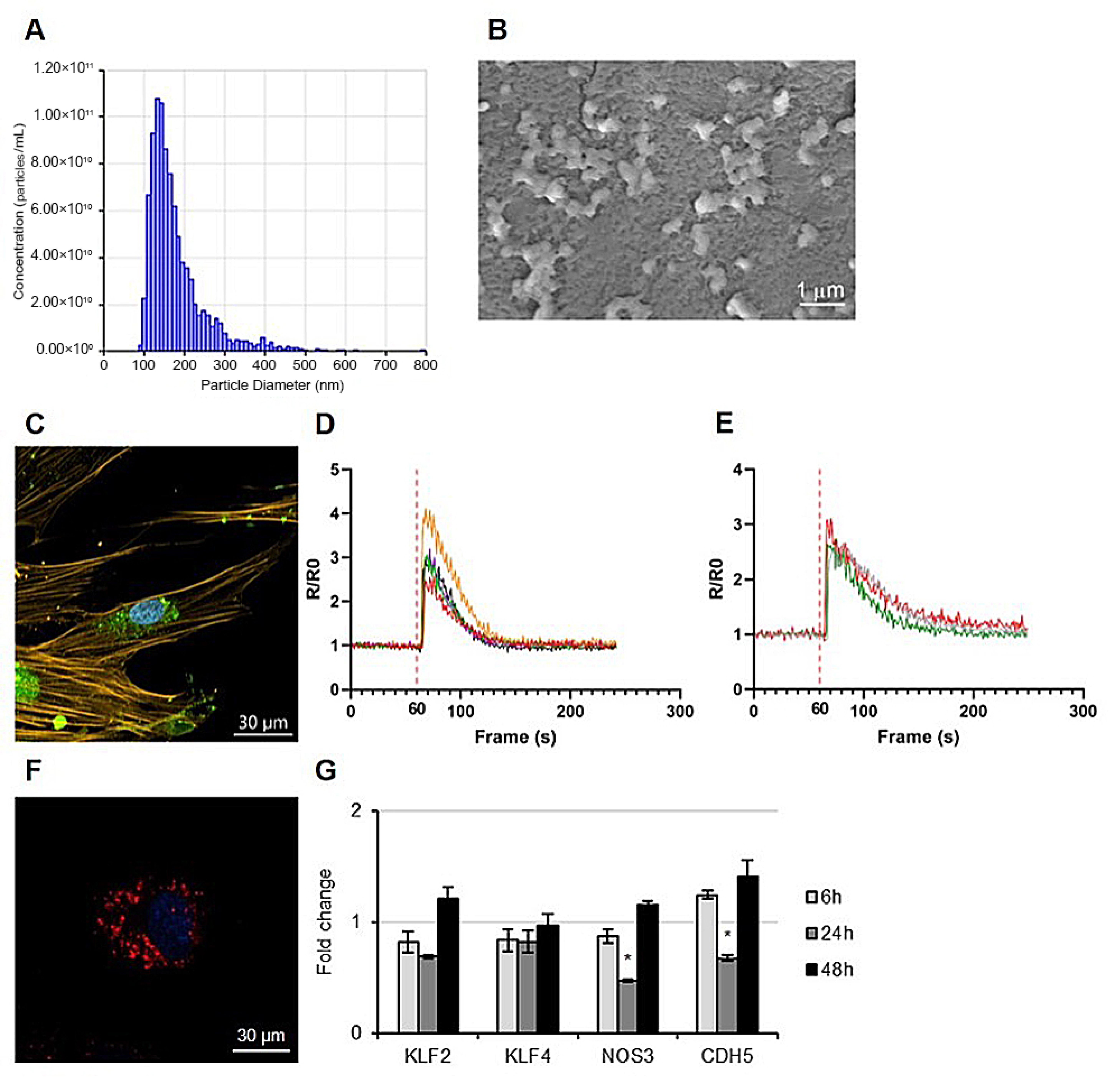
Particle size analysis demonstrated a diameter distribution ranging approximately from 102 to 160 nm, which is consistent with the accepted size range for EVs, and a particle concentration of 9.48 × 10¹¹ particles/mL (Figure 1A). Morphological assessment by scanning electron microscopy (SEM) revealed the presence of vesicles with a typical rounded, cup-shaped nanostructure, further supporting their correct identification and structural integrity (Figure 1B).
The safety of ADEVs was demonstrated by microscopy analysis and functional assays. Fluorescence microscopy showed the presence of fluorescently labeled EVs within dermal fibroblasts and endothelial cells (Figure 1C,F), providing evidence that ADEVs are recognized and internalized by skin-resident cells. Functional characterization was performed by evaluating intracellular calcium signaling, a key indicator of biological activity, and cell–vesicle interactions. Exposure to ADEVs elicited a measurable cytosolic Ca2+ response, indicating that these vesicles are biologically active and capable of triggering intracellular signaling pathways (Figure 1D,E).
Overall, the characterization approach adopted in this study follows current practices for plant-derived vesicles, using methodologies that are appropriate for their specific biological features.
To further characterize the effects of ADEVs, we analyzed the expression of selected genes associated with endothelial function and vascular regulation, including Cadherin 5 (CDH5), Nitric Oxide Synthase 3 (NOS3), Krüppel-Like Factor 2 (KLF2), and Krüppel-Like Factor 4 (KLF4) (Figure 1G).
CDH5, a key component of endothelial adherens junctions, showed only minor, in-consistent variations over time without a clear trend of downregulation (Figure 1G). Similarly, NOS3 levels exhibited limited fluctuations, with no sustained decrease observed across time points. KLF2 displayed a slight increase, whereas KLF4 expression remained stable under the tested conditions (Figure 1G).
Overall, these results indicate that ADEVs do not markedly affect the expression of genes associated with endothelial identity and function, despite minor time-dependent fluctuations.

3.2. Microneedling (MN) Enhances the Penetration and Distribution of PKH26-Labeled ADEV-Based Formulations Within Human Skin

To further investigate ADEV–skin interactions in a tissue context, PKH26-labeled ADEVs were applied to an ex vivo human skin model to evaluate fluorescence distribution within skin compartments.
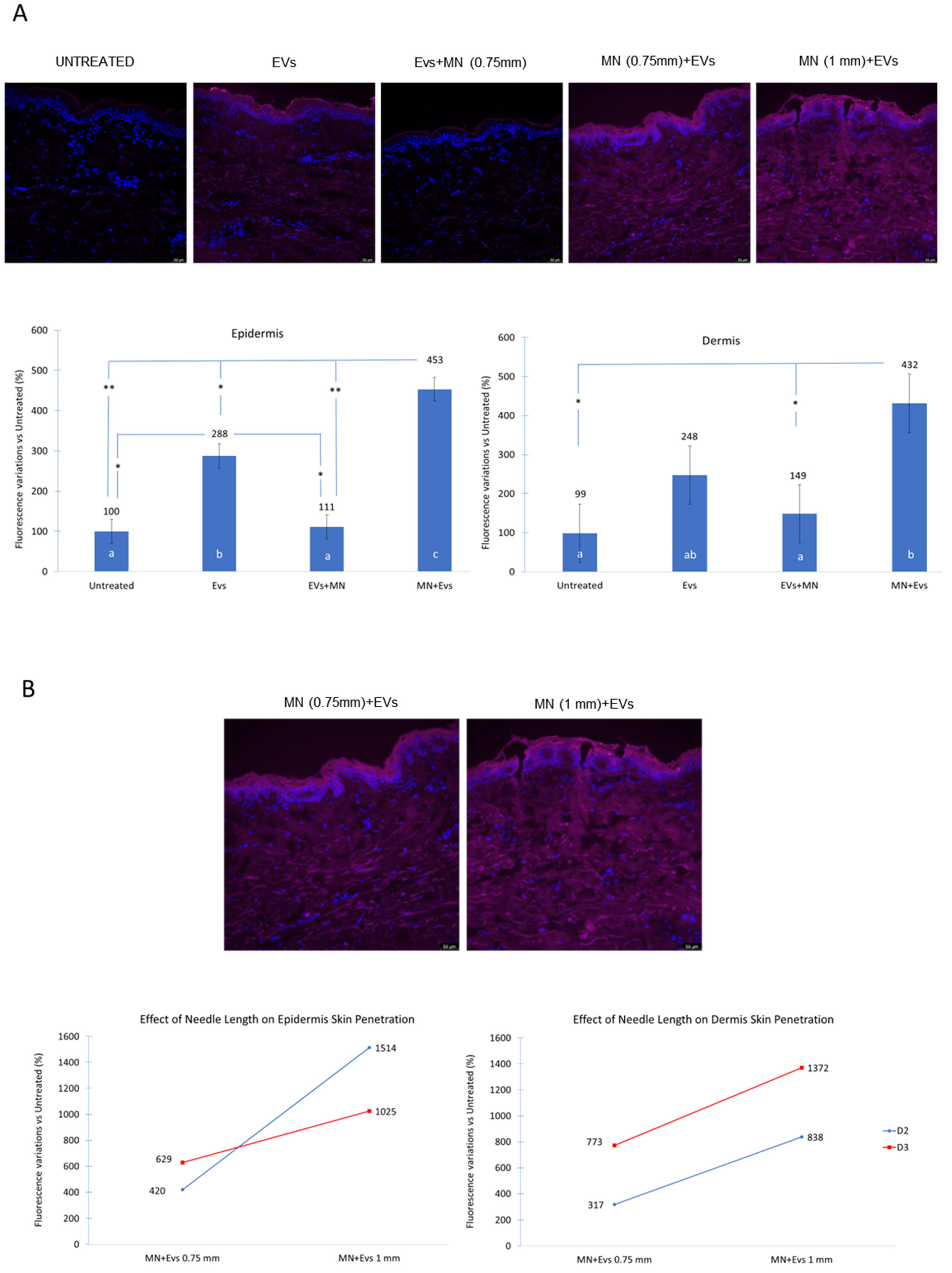
After treatment, an increase in fluorescence signal was observed across the stratum corneum (SC) (Figure 2A). However, while only a modest increase in fluorescence was observed in the dermis, a statistically significant increase was detected in the epidermis for the EVs condition compared with untreated controls (p = 0.021) (Figure 2A).
Hereafter, the term “EVs” is used as a simplified label for the PKH26-labeled ADEV-based formulation applied in the ex vivo experiments.
Based on the established effects of MN on skin permeability, we hypothesized that MN could enhance the cutaneous penetration of the PKH26-labeled formulation.
The MN procedure was standardized across experiments, including oscillation frequency, needle cartridge, and application pressure, while needle depth was used as the only variable parameter.
Notably, the sequence of application influenced the outcome. MN performed after ADEV application (EVs + MN) resulted in reduced fluorescence signal in both epidermis and dermis, whereas MN performed prior to ADEV application (MN + EVs) led to a marked enhancement of signal intensity (Figure 2A).
In the dermis, treatment significantly affected fluorescence distribution (F = 7.57, p = 0.018), with the highest response observed in the MN + EVs group (431.6 ± s.e. 74.7; values expressed as percent change relative to the untreated control), followed by EVs (247.9 ± 74.5), and EVs + MN (149.2 ± 74.6).
Post hoc analysis showed that MN + EVs elicited significantly higher responses than both EVs + MN (p = 0.037) and untreated samples (p = 0.018), whereas no other pairwise comparisons were statistically significant (Figure 2A).
A consistent pattern was observed in the epidermis, where a significant treatment effect was also detected (F = 33.57, p = 0.0001154). Estimated marginal means indicated the highest response for MN + EVs (453 ± 29), followed by EVs (288 ± 30), EVs + MN (111 ± 30), and untreated controls (100 ± 30). Tukey-adjusted comparisons confirmed significantly higher responses for MN + EVs compared to EVs + MN (p = 0.0015) and untreated samples (p = 0.0011). In addition, the ADEV-based formulation showed significantly higher values than both EVs + MN (p = 0.028) and untreated controls (p = 0.021), whereas no difference was observed between EVs + MN and untreated samples (Figure 2A).
These findings were further supported by non-parametric Friedman tests performed separately for each skin compartment, confirming significant differences among treatments in both epidermis and dermis (χ2 = 8.2, df = 3, p = 0.042).
Overall, these results indicate that MN enhances the distribution of the PKH26-labeled formulation within the skin layers, with a strong dependence on the application sequence.
Furthermore, we investigated whether the distribution of the PKH26-labeled formulation could be further enhanced by increasing microneedle depth (Figure 2B). Specifically, we compared the effect of 0.75 mm vs. 1 mm needle length in a subset of donors (D2 and D3), focusing on the MN + EVs condition. A higher fluorescence signal was observed at increased needle depth (Figure 2B).
However, given the limited number of biological replicates, this analysis is presented as exploratory and hypothesis-generating rather than confirmatory.

4. Discussion

EVs are rapidly emerging as one of the most attractive biological tools in aesthetic and regenerative medicine. Their increasing clinical adoption is driven by the recent promising results obtained in improving skin quality, accelerating wound healing, modulating inflammation, and enhancing post-procedural outcomes through a cell-free, biologically active approach [23,24].
Among the different sources, plant-derived EVs have gained growing attention due to their scalability, favorable safety profile, low immunogenicity, and suitability for topical and minimally invasive applications [2]. Despite their widespread commercial availability and experimental clinical use, there is still limited direct experimental evidence directly demonstrating EV-associated transport across the human skin barrier and establishing biological interactions with deeper cutaneous compartments, although this represents a critical factor for their application in aesthetic surgery. From a clinical standpoint, experts are not only interested in the way EVs “work” but also in the mechanistic insight through which they exert their effects. In particular, the ability of EVs to cross the stratum corneum, reach biologically relevant targets, and influence processes such as dermal remodeling and microcirculation represents a decisive factor for the committed integration into aesthetic surgery protocols.
The present study was designed to address some of these clinically important questions by combining EV size characterization with a controlled evaluation of delivery strategies. ADEVs were thoroughly characterized in terms of size distribution, molecular features, and biological activity, confirming their functional relevance for skin biology.
Since recent studies have highlighted the anti-inflammatory and extracellular matrix-protective roles of EVs [24] and emerging evidence suggests similar effects for apple-derived extracellular vesicles [8,25], we investigated the interaction between ADEVs and skin-resident cells. Our in vitro findings indicate that ADEVs are internalized by these cells and retain biological activity, with indications of modulation of intracellular signaling pathways, including genes potentially implicated in endothelial function and vascular regulation, particularly in the context of wound healing [26,27]. Specifically, CDH5, KLF2, and NOS3 are reported to play important roles in vasculogenesis and endothelial and vascular homeostasis [28,29,30,31,32,33,34]. The observed modulation of these genes, although moderate in magnitude, suggests that ADEVs are not only internalized by skin-resident cells involved in key physiological functions of the skin but also may contribute to modest changes in gene expression.
While these findings are not sufficient, on their own, to support broad mechanistic conclusions, they provide preliminary evidence that warrants further investigation. In particular, future studies should expand the range of molecular targets and experimental models examined in order to better elucidate the mechanisms underlying ADEV–cell interactions and their potential implications for skin health.
To reinforce the data uncovered by our in vitro studies, we investigated the ability of ADEV-based formulations to interact with and distribute across the skin barrier, while also investigating the possibility of boosting this ability through the application of a widely used technique like microneedling (MN).
Both in aesthetic and dermatological clinical practices, MN is adopted as a safe and minimally invasive therapeutic procedure [10,11,12,13,14] as well as a transdermal delivery system for drugs or bioactive substances to be administered to patients [11,35]. For their part, cosmetics mainly take advantage of MN to increase skin permeation and thus enhance topical skincare product absorption by passing through the microchannels generated by the microneedles [9]. As highlighted by previous classifications, MN may be approached by modulating the procedural parameters, such as needle depth, speed of the device, and treatment application sequence, which are essential to optimize given the substantial changes in the procedure’s efficacy.
Using a “poke and patch” approach, one of the most common techniques in cosmetic and aesthetic applications, we evaluated the distribution of PKH26-labeled ADEVs within the skin layers (see the Section “Interpretative limitations” for technical details) and tested different protocols in which they were combined with MN. Under baseline conditions, we observed an increase in the fluorescence signal within the epidermal compartment, suggesting a certain degree of interaction of the labeled preparation with the stratum corneum. However, the signal detected in the dermis remained limited.
In contrast, combining ADEVs with MN resulted in an overall increase in the fluorescence signal in both epidermal and dermal compartments, consistently observed across all donors included in the study. In particular, MN applied as the pre-treatment significantly enhanced the distribution of the PKH26-labeled preparation within the skin. This effect was further enhanced when MN pre-treatment was performed using an increased needle length (1 mm vs. 0.75 mm), supporting the role of procedural parameters in modulating the efficiency of MN-assisted delivery.
Importantly, the fluorescence signal used in this study reflects the distribution of the PKH26-labeled vesicle preparation and does not, by itself, provide definitive evidence of vesicle-specific localization within tissue compartments.
Nonetheless, within the controlled comparative framework adopted here, the observed differences between treatments remain informative in assessing how MN modulates the distribution behavior of the applied formulation.
Interestingly, MN performed after topical application (EVs + MN) resulted in a reduction in the fluorescence signal in both the epidermis and dermis, in contrast to the enhancement observed when MN was applied prior to ADEV administration. This suggests that the timing of the temporary disruption of the tissue, mechanically induced by the application of the MN, is a critical factor to consider in a protocol including treatment with EVs.
A plausible explanation is that the generation of MN-induced microchannels after topical ADEV administration may induce fluid dislocation or compromise the stability of diffusion gradients, thereby limiting the effective penetration of the applied formulation.
Similarly, in the case of mechanical skin manipulation techniques like massage and ultrasound, a negative impact on the transdermal delivery of some types of treatments has been described under specific tested conditions [36]. Importantly, our data highlight that ADEV-based formulations behave differently from conventional topical actives, corroborating the need for evidence-based procedural protocols for use in vivo.
Taken together, these results suggest that, in aesthetic medicine, the selection of the appropriate delivery strategy and the timing of the execution of a mechanical procedure like MN carry similar weight and may have implication as decisive as those of the choice of the specific bioactive to deliver. Vice versa, indiscriminate combination or improper sequencing of the protocol steps could potentially invalidate the benefits of the therapy adopted.

Interpretative Limitations

A limitation of this study is related to the use of the lipophilic fluorescent dye PKH26 for EV labelling. Although free dye was removed from the EV preparations prior to application, PKH26 may still undergo redistribution or non-specific association with lipid-rich components in biological tissues such as the skin.
Therefore, the fluorescent signal detected in the skin layers cannot be unequivocally attributed to intact EVs and should be interpreted as the distribution of PKH26-labelled vesicle preparations. In addition, the absence of a free PKH26 dye control does not allow the estimation of the contribution of potential non-vesicle-associated dye behaviour within the experimental system.
It is important to note that this study was primarily designed within a biophysical and mechanistic framework aimed at investigating the impact of MN-assisted delivery on the cutaneous penetration and spatial distribution of EV-based formulations, rather than providing definitive evidence of EV-specific cellular uptake. Within this context, the fluorescence signal is intended as a proxy for the distribution of the labelled formulation across the skin layers.
Future studies should further validate these findings using complementary and orthogonal EV labelling strategies to improve specificity and confirm EV-associated uptake mechanisms.

5. Conclusions

Collectively, this research provides a more solid insight into the behavior of plant-derived EVs in human skin by demonstrating a biologically relevant interaction between ADEVs and skin-resident cells.
Furthermore, our findings indicate that the fluorescence signal associated with PKH26-labeled ADEV-based formulations can be detected across the skin layers, with this distribution being significantly enhanced through rational optimization of the delivery strategy (e.g., microneedling). These data support a more informed integration of EV-based approaches into cosmetic and aesthetic clinical practice.
In conclusion, our results are well aligned with the growing demand for biologically driven and minimally invasive solutions, contributing to the development of more standardized and reproducible approaches for skin-targeted applications.

Author Contributions

Conceptualization, A.C., B.Z. and M.M. (Marco Massironi); methodology, M.M. (Michele Massironi), C.R., A.C. and B.Z.; validation, M.M. (Marco Massironi) and B.Z.; investigation, M.M. (Michele Massironi), C.R., M.P.C. and L.F.; resources, L.L. and A.C.; writing—original draft preparation, C.R., B.Z., M.P.C. and L.F.; writing—review and editing, C.R.; supervision, M.M. (Marco Massironi) and B.Z.; project administration, M.M. (Marco Massironi). All authors have read and agreed to the published version of the manuscript.

Funding

This research received no external funding.

Institutional Review Board Statement

Human skin samples used in this study were obtained exclusively as surgical discards from aesthetic procedures and were originally intended for disposal. Tissue removal was performed solely for clinical purposes, without any modification of the surgical procedure and without introducing additional risks to the patients. All samples were obtained with written informed consent and were fully anonymized prior to transfer to our laboratory. As the study did not involve human subjects or any form of clinical experimentation and was based exclusively on anonymized discarded human material, approval by an institutional ethics committee was not required.

Informed Consent Statement

Concerning skin sample collection, informed consent from patients was obtained and retained by the clinic where the procedures took place, while our laboratory has no direct contact with the donors. Tissue samples were transferred anonymously, with only the donor’s sex and year of birth provided for research purposes.

Data Availability Statement

The data that support the findings of this study are available from the corresponding author upon reasonable request, subject to restrictions due to proprietary considerations.

Acknowledgments

We gratefully acknowledge Francesca Cestaro (Numeraway Srl) for her valuable support in statistical analysis and data interpretation. The authors declare that no generative AI tools were used in the preparation of this manuscript.

Conflicts of Interest

The authors declare that the research was conducted in the absence of any commercial or financial relationships that could be construed as a potential conflict of interest. Symrise AG is a global company that produces and markets cosmetics. Consorzio Melinda is a consortium of apple producers from which the plant-derived vesicles (ADEVs) were obtained. These entities had no role in the study design, data collection, analysis, or interpretation of the results.

References

  1. Welsh, J.A.; Goberdhan, D.C.; O’Driscoll, L.; Théry, C.; Witwer, K.W. MISEV2023: An Updated Guide to EV Research and Applications. J. Extracell. Vesicle 2024, 13, e12416. [Google Scholar] [CrossRef] [PubMed]
  2. Subha, D.; Harshnii, K.; Madhikiruba, K.G.; Nandhini, M.; Tamilselvi, K.S. Plant Derived Exosome-like Nanovesicles: An Updated Overview. Plant Nano Biol. 2023, 3, 100022. [Google Scholar] [CrossRef]
  3. Chigozie, V.U. Exosomal Bioactive Molecules: Molecular Regulation, Functional Heterogeneity, and Translational Implications in Disease Diagnostics and Therapeutics. Clin. Exp. Med. 2026, 26, 179. [Google Scholar] [CrossRef] [PubMed]
  4. Qing, L.; Chen, H.; Tang, J.; Jia, X. Exosomes and Their microRNA Cargo: New Players in Peripheral Nerve Regeneration. Neurorehabilit. Neural Repair 2018, 32, 765–776. [Google Scholar] [CrossRef] [PubMed]
  5. Gurung, S.; Perocheau, D.; Touramanidou, L.; Baruteau, J. The exosome journey: From biogenesis to uptake and intracellular signalling. Cell Commun. Signal. 2021, 19, 47. [Google Scholar] [CrossRef] [PubMed] [PubMed Central]
  6. Sileo, L.; Cavaleri, M.P.; Lovatti, L.; Pezzotti, G.; Ferroni, L.; Zavan, B. Dermatologically Tested Apple-Derived Extracellular Vesicles: Safety, Anti-Aging, and Soothing Benefits for Skin Health. J. Cosmet. Dermatol. 2025, 24, e70254. [Google Scholar] [CrossRef] [PubMed]
  7. Dhaliwal, N.K.; Moothathamby, T.; Sokhal, B.S.; Gkini, M.A. The Use of Microneedling with Exosomes in Dermatology: A Systematic Review. J. Cosmet. Dermatol. 2026, 25, e70881. [Google Scholar] [CrossRef] [PubMed]
  8. Ferroni, L.; Zavan, B. Plant-Derived Extracellular Vesicles in Cosmetics: Building a Framework for Safety, Efficacy, and Quality. Cosmetics 2025, 12, 252. [Google Scholar] [CrossRef]
  9. Hamed, R.; Abu Nahia, B.J.; Alkilani, A.Z.; Al-Adhami, Y.; Obaidat, R. Recent Advances in Microneedling-Assisted Cosmetic Applications. Cosmetics 2024, 11, 51. [Google Scholar] [CrossRef]
  10. McCrudden, M.T.C.; McAlister, E.; Courtenay, A.J.; González-Vázquez, P.; Raj Singh, T.R.; Donnelly, R.F. Microneedle Applications in Improving Skin Appearance. Exp. Dermatol. 2015, 24, 561–566. [Google Scholar] [CrossRef] [PubMed]
  11. Kochhar, J.S.; Tan, J.J.Y.; Kwang, Y.C.; Kang, L. Recent Trends in Microneedle Development & Applications in Medicine and Cosmetics (2013–2018). In Microneedles for Transdermal Drug Delivery; Kochhar, J.S., Tan, J.J.Y., Kwang, Y.C., Kang, L., Eds.; Springer International Publishing: Cham, Switzerland, 2019; pp. 95–144. [Google Scholar]
  12. Lee, Y.S. Preliminary Histological Evidence of Epidermal and DEJ Remodeling with Microneedling-Assisted Topical Exosome Therapy: A Single-Subject Case Report. Clin. Cosmet. Investig. Dermatol. 2025, 18, 2377–2385. [Google Scholar] [CrossRef] [PubMed]
  13. De Oliveira Ferraz Gominho, B.; Medeiros, A.C.F. Controlled Clinical Trial Evaluating the Efficacy of Microneedling Combined with Exosomes Versus Microneedling Alone in the Treatment of Melasma. Preprints 2025, 2025050934. [Google Scholar] [CrossRef]
  14. Wan, J.; Yoon, S.E.; Yi, K.-H. The Efficacy of Combined Exosome (Exodew) and Microneedling Treatment for Facial Pore Reduction and Skin Texture Improvement. Plast. Reconstr. Surg. Glob. Open 2025, 13, e6849. [Google Scholar] [CrossRef] [PubMed]
  15. Trentini, M.; Zanolla, I.; Tiengo, E.; Zanotti, F.; Sommella, E.; Merciai, F.; Campiglia, P.; Licastro, D.; Degasperi, M.; Lovatti, L.; et al. Link between Organic Nanovescicles from Vegetable Kingdom and Human Cell Physiology: Intracellular Calcium Signalling. J. Nanobiotechnol. 2024, 22, 68. [Google Scholar] [CrossRef] [PubMed]
  16. Trentini, M.; Zanolla, I.; Zanotti, F.; Tiengo, E.; Licastro, D.; Dal Monego, S.; Lovatti, L.; Zavan, B. Apple Derived Exosomes Improve Collagen Type I Production and Decrease MMPs during Aging of the Skin through Downregulation of the NF-κB Pathway as Mode of Action. Cells 2022, 11, 3950. [Google Scholar] [CrossRef] [PubMed]
  17. Trentini, M.; Zanotti, F.; Tiengo, E.; Camponogara, F.; Degasperi, M.; Licastro, D.; Lovatti, L.; Zavan, B. An Apple a Day Keeps the Doctor Away: Potential Role of miRNA 146 on Macrophages Treated with Exosomes Derived from Apples. Biomedicines 2022, 10, 415. [Google Scholar] [CrossRef] [PubMed]
  18. Field, A.; Miles, J.; Field, Z. Discovering Statistics Using R; Sage Publicatons: London, UK, 2012. [Google Scholar]
  19. Zuur, A.F.; Ieno, E.N.; Walker, N.; Saveliev, A.A.; Smith, G.M. Mixed Effects Models and Extensions in Ecology with R; Statistics for Biology and Health; Springer: New York, NY, USA, 2009. [Google Scholar]
  20. Bates, D.; Mächler, M.; Bolker, B.; Walker, S. Fitting Linear Mixed-Effects Models Using Lme4. J. Stat. Softw. 2015, 67, 1–48. [Google Scholar] [CrossRef]
  21. Kuznetsova, A.; Brockhoff, P.B.; Christensen, R.H.B. lmerTest Package: Tests in Linear Mixed Effects Models. J. Stat. Softw. 2017, 82, 1–26. [Google Scholar] [CrossRef]
  22. Lenth, R. Emmeans: Estimated Marginal Means, Aka Least-Squares Means, version 2.0.3; CRAN: Online, 2026. [Google Scholar] [CrossRef]
  23. Park, S.Y.; Yi, K.-H. Exosome-Mediated Advancements in Plastic Surgery: Navigating Therapeutic Potential in Skin Rejuvenation and Wound Healing. Plast. Reconstr. Surg.–Glob. Open 2024, 12, e6021. [Google Scholar] [CrossRef] [PubMed]
  24. Yuan, S.; Jin, M.; Zhang, Y.; Zhang, M.; Yuan, M.; Ding, X.; Wang, J. Extracellular Vesicles in Skin: Biological Function and Therapeutic Potential. Int. J. Nanomed. 2025, 20, 11211–11233. [Google Scholar] [CrossRef] [PubMed]
  25. Burshtein, J.; Wei, J.; Majewska, L.; Schlesinger, T. Plant-Derived Extracellular Vesicles in Dermatology: A Review of Emerging Therapeutic Applications. J. Clin. Aesthet. Dermatol. 2026, 19, 15–21. [Google Scholar] [PubMed]
  26. Narauskaitė, D.; Vydmantaitė, G.; Rusteikaitė, J.; Sampath, R.; Rudaitytė, A.; Stašytė, G.; Aparicio Calvente, M.I.; Jekabsone, A. Extracellular Vesicles in Skin Wound Healing. Pharmaceuticals 2021, 14, 811. [Google Scholar] [CrossRef] [PubMed]
  27. Wu, W.; Zhang, B.; Wang, W.; Bu, Q.; Li, Y.; Zhang, P.; Zeng, L. Plant-Derived Exosome-Like Nanovesicles in Chronic Wound Healing. Int. J. Nanomed. 2024, 19, 11293–11303. [Google Scholar] [CrossRef] [PubMed]
  28. Dekker, R.J.; van Thienen, J.V.; Rohlena, J.; de Jager, S.C.; Elderkamp, Y.W.; Seppen, J.; de Vries, C.J.M.; Biessen, E.A.L.; van Berkel, T.J.C.; Pannekoek, H.; et al. Endothelial KLF2 Links Local Arterial Shear Stress Levels to the Expression of Vascular Tone-Regulating Genes. Am. J. Pathol. 2005, 167, 609–618. [Google Scholar] [CrossRef] [PubMed]
  29. Pautz, A.; Li, H.; Kleinert, H.; Pautz, A.; Li, H.; Kleinert, H. Regulation of NOS Expression in Vascular Diseases. Front. Biosci. (Landmark Ed.) 2021, 26, 85–101. [Google Scholar] [CrossRef] [PubMed]
  30. Sauteur, L.; Krudewig, A.; Herwig, L.; Ehrenfeuchter, N.; Lenard, A.; Affolter, M.; Belting, H.-G. Cdh5/VE-Cadherin Promotes Endothelial Cell Interface Elongation via Cortical Actin Polymerization during Angiogenic Sprouting. Cell Rep. 2014, 9, 504–513. [Google Scholar] [CrossRef] [PubMed]
  31. Richards, M.; Nwadozi, E.; Pal, S.; Martinsson, P.; Kaakinen, M.; Gloger, M.; Sjöberg, E.; Koltowska, K.; Betsholtz, C.; Eklund, L.; et al. Claudin5 Protects the Peripheral Endothelial Barrier in an Organ and Vessel-Type-Specific Manner. eLife 2022, 11, e78517. [Google Scholar] [CrossRef] [PubMed]
  32. Lin, Z.; Natesan, V.; Shi, H.; Dong, F.; Kawanami, D.; Mahabeleshwar, G.H.; Atkins, G.B.; Nayak, L.; Cui, Y.; Finigan, J.H.; et al. Kruppel-Like Factor 2 Regulates Endothelial Barrier Function. Arterioscler. Thromb. Vasc. Biol. 2010, 30, 1952–1959. [Google Scholar] [CrossRef] [PubMed]
  33. Sugiura, S.; Yoshida, H.; Sugiura, H.; Uehara, M.; Sugiura, Y.; Maruo, Y.; Hayashi, Y.; Yamamoto, T.; Kato, T.; Fujimoto, N.; et al. Increased Intracellular Stress Responses and Decreased KLF2 in Adult Patients with Atopic Dermatitis. Cell Stress Chaperones 2025, 30, 84–99. [Google Scholar] [CrossRef] [PubMed]
  34. Man, M.; Wakefield, J.S.; Mauro, T.M.; Elias, P.M. Role of Nitric Oxide in Regulating Epidermal Permeability Barrier Function. Exp. Dermatol. 2022, 31, 290–298. [Google Scholar] [CrossRef] [PubMed]
  35. Saha, I.; Rai, V.K. Hyaluronic Acid Based Microneedle Array: Recent Applications in Drug Delivery and Cosmetology. Carbohydr. Polym. 2021, 267, 118168. [Google Scholar] [CrossRef] [PubMed]
  36. Kaushik, V.; Keck, C.M. Influence of Mechanical Skin Treatment (Massage, Ultrasound, Microdermabrasion, Tape Stripping and Microneedling) on Dermal Penetration Efficacy of Chemical Compounds. Eur. J. Pharm. Biopharm. 2021, 169, 29–36. [Google Scholar] [CrossRef] [PubMed]
Figure 1. Characterization of apple-derived extracellular vesicles (ADEVs) and analysis of their interaction with skin-resident cells. (A) ADEV size distribution and concentration measured by Tunable Resistive Pulse Sensing (TRPS). (B) Representative scanning electron microscopy (SEM) image showing the morphology of isolated ADEVs. Scale bar: 1 µm. (C) Fluorescence microscopy image of primary human dermal fibroblasts after incubation with green-labelled ADEVs, demonstrating vesicle internalization. Cell nuclei are shown in blue and ADEVs in green. Scale bar: 30 µm. (D,E) Intracellular Ca2+ response in human dermal fibroblasts following stimulation with bradykinin (positive control) and ADEVs, respectively. The red dashed line indicates the time of stimulus application. (F) Representative microscopy image of primary human endothelial cells after incubation with red-labelled ADEVs. Scale bar: 30 µm. (G) Bar plots show RT-qPCR results of mRNA expression levels of the following genes associated with endothelial identity, vascular homeostasis, and barrier function: hKLF2, hKLF4, hNOS3, and hCDH5. Fold change calculated as the ratio of gene expression in ADEV-treated cells relative to untreated control cells, which were set at 1; * indicates a statistically significant difference compared with untreated control cells. Each experiment was performed three times.
Jaestheticmed 02 00014 g001
Figure 2. Microneedling (MN) enhances the distribution of PKH26-labeled ADEV-based formulations within human skin. (A) Representative images of skin sections (scale bar: 50 µm) under each experimental condition, including the untreated control, PKH26-labeled ADEVs alone, and MN applied before or after PKH26-labeled ADEVs (needle depth = 0.75 mm), together with corresponding bar plots showing the estimated marginal means (EMMs) of fluorescence intensity. Data are expressed as the percentage change relative to the untreated control group and quantified in both the epidermis (left) and the dermis (right). For each biological replicate (donor), fluorescence values were calculated as the mean of multiple sections/images. Each data point represents one biological replicate. Bar plots display EMMs derived from the mixed-effects model, and error bars represent corresponding standard errors of the EMMs. Different letters beneath the bars (a–c) indicate statistically significant differences among groups based on mixed-effects model analysis followed by Tukey-adjusted post hoc comparisons (* p < 0.05; ** p < 0.01). (B) Representative images (scale bar: 50 µm) and corresponding line plots comparing fluorescence intensity following MN treatment at different needle depths (0.75 mm and 1.0 mm) in a subset of donors (Donors 2 and 3). The MN + PKH26-labeled ADEV condition at 0.75 mm is shown in both panels to enable direct visual comparison. Fluorescence intensity is expressed as the percentage change relative to the untreated control group and quantified in both the epidermis (left) and the dermis (right). This comparison is presented as exploratory, and no formal statistical inference is provided. Color legend: DAPI-stained nuclei are shown in blue, PKH26-labeled ADEVs in magenta, and the line plot colors identify Donor 2 (blue) and Donor 3 (red).
Jaestheticmed 02 00014 g002
Disclaimer/Publisher’s Note: The statements, opinions and data contained in all publications are solely those of the individual author(s) and contributor(s) and not of MDPI and/or the editor(s). MDPI and/or the editor(s) disclaim responsibility for any injury to people or property resulting from any ideas, methods, instructions or products referred to in the content.