Brief Report

The CK2/ECE1c Partnership: An Unveiled Pathway to Aggressiveness in Cancer

by
Karla Villalobos-Nova
,
María de los Ángeles Toro
,
Pablo Pérez-Moreno
,
Ignacio Niechi
and
Julio C. Tapia
*
Laboratorio de Transformación Celular, Programa de Biología Celular y Molecular, Instituto de Ciencias Biomédicas, Facultad de Medicina, Universidad de Chile, Santiago 8380453, Chile
*
Author to whom correspondence should be addressed.
These authors contributed equally to this work.
Current address: Instituto de Bioquímica y Microbiología, Facultad de Ciencias, Universidad Austral de Chile, Valdivia 5110566, Chile.
Kinases Phosphatases 2024, 2(1), 1-8; https://doi.org/10.3390/kinasesphosphatases2010001
Submission received: 4 November 2023 / Revised: 25 November 2023 / Accepted: 29 November 2023 / Published: 19 December 2023
(This article belongs to the Special Issue Past, Present and Future of Protein Kinase CK2 Research)

Abstract

:
The endothelin-1 (ET1) peptide has a pathological role in the activation of proliferation, survival and invasiveness pathways in different cancers. ET1’s effects rely on its activation by the endothelin-converting enzyme-1 (ECE1), which is expressed as four isoforms, differing only in their cytoplasmic N-terminuses. We already demonstrated in colorectal cancer, glioblastoma, and preliminarily lung cancer, that the isoform ECE1c heightens aggressiveness by promoting cancer stem cell traits. This is achieved through a non-canonical ET1-independent mechanism of enhancement of ECE1c’s stability upon CK2-dependent phosphorylation at S18 and S20. Here, a K6 residue is presumably responsible for ECE1c ubiquitination as its mutation to R impairs proteasomal degradation. However, how phosphorylation enhances ECE1c’s stability and how this translates into aggressiveness are still open questions. In this brief report, by swapping residues to either phospho-mimetic or phospho-resistant amino acids, we propose that the N-terminus may also be phosphorylated at Y5 and/or T9 by an unknown kinase(s). In addition, N-terminus phosphorylation may lead to a blockage of K6 ubiquitination, increasing ECE1c’s stability and presumably activating the Wnt/β-catenin signaling pathway. Thus, a novel CK2/ECE1c partnership may be emerging to promote aggressiveness and thus become a biomarker of poor prognosis and a potential therapeutic target for several cancers.

Graphical Abstract

1. Introduction

Cancer stem cells (CSC) are a marginal cell population responsible for genesis, metastasis, and relapse after chemotherapy in many tumors [1,2,3,4,5,6]. In recent years, various signaling pathways have garnered attention due to their capacity to provoke CSC development and therefore promote aggressiveness traits. One such signaling pathway, the endothelin-1 (ET1) axis, has been highlighted as playing a pathological role in cancer etiology. In its physiological role, the ET1 axis contributes to the regulation of vascular tone, mainly through binding to its ETBR receptor. On the other hand, the stimulation of its ETAR receptor leads to the pathological activation of cancer pathways [7,8,9]. The ET1 peptide is activated by the cleaving of the endothelin-converting enzyme-1 (ECE1), a membrane-bound protease constituted by three domains: a short N-terminal cytoplasmic domain (which defines four isoforms), followed by a transmembrane domain, and finally a large extracellular C-terminal catalytic domain [9,10,11,12].
ECE1 expression has been found to be elevated in patients with various cancers, and increased mRNA and/or protein levels have been detected in several cancer cell lines, with the ECE1c being the most-expressed isoform [13,14,15]. High levels of ET1 have also been identified in patients with colorectal cancer, suggesting that ECE1 may participate in the disease process [16,17,18]. Furthermore, an early study reported the increased invasiveness triggered by isoform ECE1c in prostate cancer cells [19]. However, the role of this and of the other ECE1 isoforms in cancer hallmarks beyond invasiveness was an open question for many years.

2. Results and Discussion

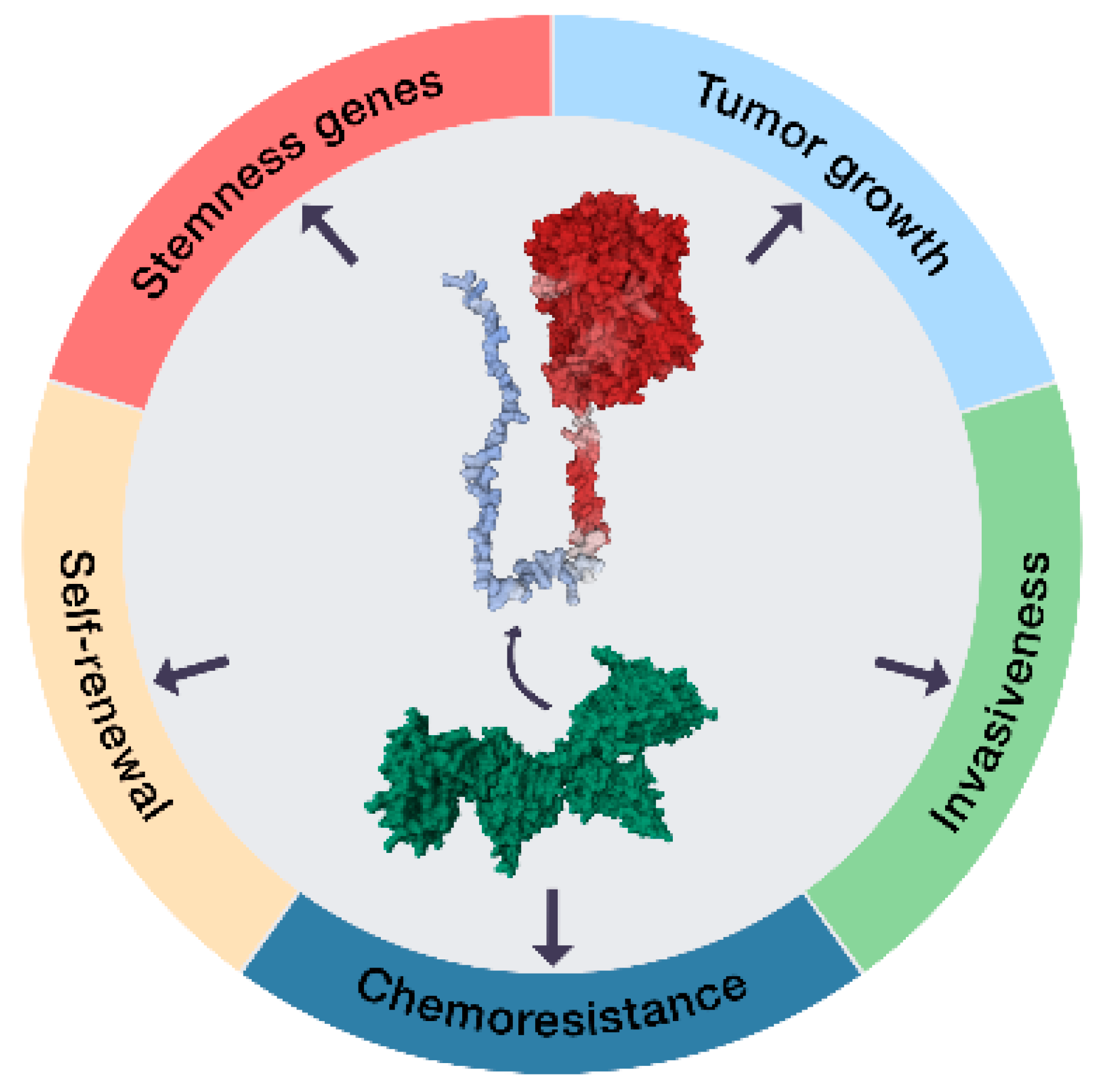
2.1. The ECE1c-Dependent Acquisition of a CSC Phenotype

A breakthrough arrived in 2015, when Niechi et al. first reported that isoform ECE1c leads to aggressiveness in colorectal cancer cells [20]. Importantly, these authors demonstrated that the protein kinase CK2 can phosphorylate ECE1c in its N-terminal cytoplasmic domain, greatly enhancing its stability [20]. In fact, the literature shows that CK2 may regulate the stability of many proteins. For example, CK2’s phosphorylation of c-myc prevents its proteasomal degradation, enhancing the transcription of genes involved in several cancer hallmarks [21]. Likewise, CK2’s phosphorylation of OTUB1 promotes its nuclear deubiquitinating activity and stabilization of chromatin-binding proteins [22]. Indeed, increased CK2 mRNA and protein levels have been found in many types of tumors [23,24,25,26,27]. The findings from Niechi et al. take on special relevance in colorectal cancer, as CK2 regulates many traits related to aggressiveness in this disease, such as survival, resistance to death, metabolic changes, angiogenesis, DNA repair, and tumor growth [28,29]. In consequence, the existence of a non-canonical ET1-independent mechanism underlying cancer cell aggressiveness, occurring through the CK2-dependent phosphorylation of ECE1c, is emerging as an interesting possibility (Figure 1).

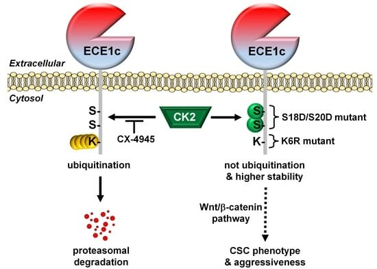
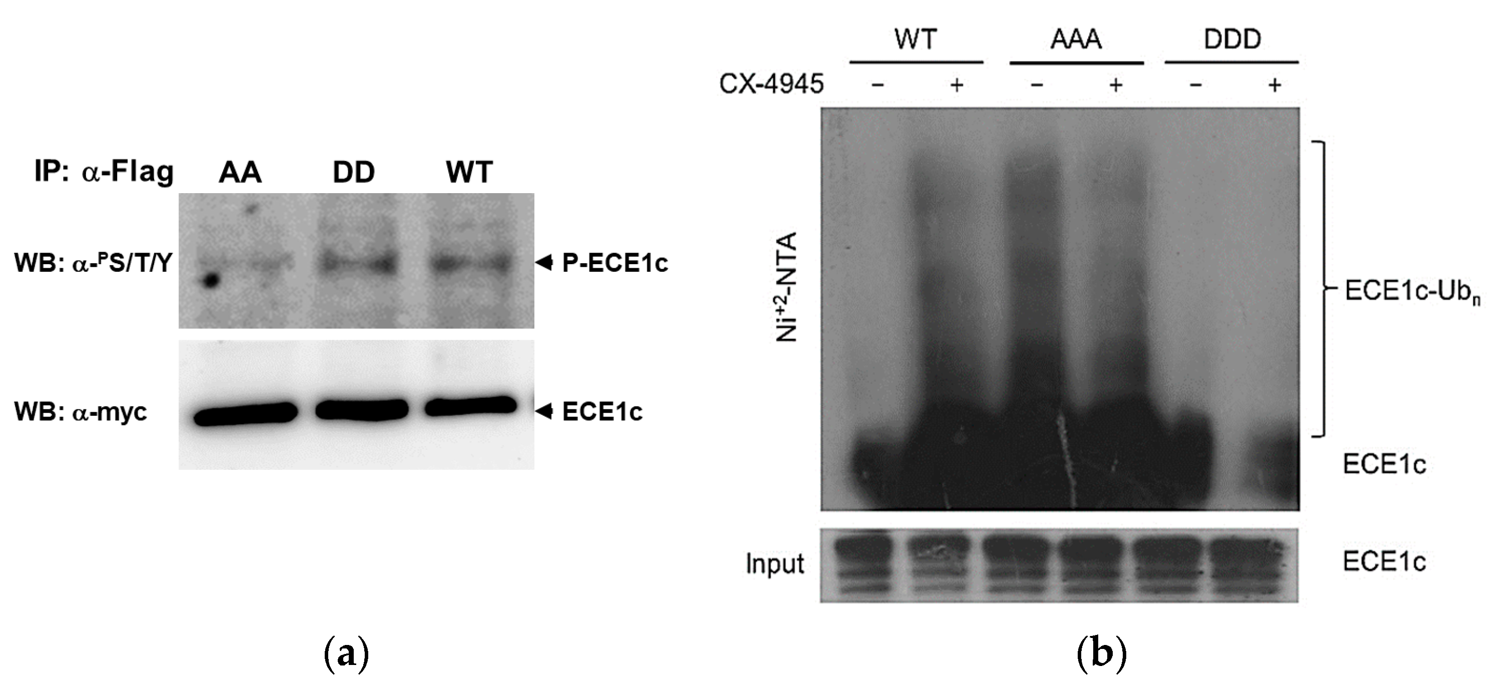
2.2. CK2’s Phosphorylation of ECE1c and Aggressiveness

The N-terminus of ECE1c is highly conserved in several species, displaying almost a complete identity [15]. It contains three conserved putative phosphorylation sites for CK2: T9, S18, and S20, which are predicted to be excellent substrates by the NetPhos 3.1 software (www.cbs.dtu.dk; accessed 31 July 2023). By designing biphospho-mimetic (i.e., ECE1cS18D/S20D) and biphospho-resistant (i.e., ECE1cS18A/S20A) mutants at both serines, Pérez-Moreno et al. delved deeper into the mechanism(s) that may enhance the stability of ECE1c. These authors clearly demonstrated that S18 and S20 residues are bona fide CK2 phosphorylation sites, which greatly enhance the stability of ECE1c but also lead to an increased invasiveness and other aggressiveness-associated traits in colorectal cancer cells. Surprisingly, while T9 was an excellent putative substrate for CK2, it was not in fact phosphorylated in vitro by this kinase [33]. However, phosphorylation at T9 (or even at Y5, according to NetPhos 3.1) is greatly disfavored when S18/S20 are replaced by alanines, while phosphorylation is significantly enhanced when serines are swapped for phospho-mimetic aspartates (Figure 2a). Moreover, CHO-K1 cells—which express negligible ECE1 levels—were used to show that the triphospho-mimetic ECE1cDDD mutant displays significant resistance to ubiquitination compared to its counterpart, ECE1cAAA (Figure 2b).
The above findings are consistent with a potential hierarchical process in which phospho-serines are a requisite for phosphorylation at Y5 or T9. Alternatively, as reported for some substrates of CK2, the regulatory β subunit may have a docking function in T9 phosphorylation in a given cell context [34,35,36]. This docking event has been observed, for instance, with the inhibitor of cyclin-dependent kinases, p27KIP1, whose phosphorylation via CK2 only occurs in the presence of the β subunit [36]. Finally, it is also plausible that while S18/S20 are CK2 substrates, another kinase(s) actually phosphorylates at Y5 or T9. In this regard, the EGF receptor, although with a low score of 0.389 as predicted by NetPhos 3.1, may phosphorylate ECE1c at Y5. Likewise, PKA with a score of 0.656 may also phosphorylate it at T9.
But beyond phosphorylating Y5/T9, how does ECE1c’s phosphorylation translate into enhanced protein stability? The simplest answer is that CK2 phosphorylation induces a conformational change at the N-terminus—a phenomenon already reported for other proteins [36]—that can block protein degradation. On the other hand, many proteins are ubiquitinated in a lysine-dependent fashion to be further degraded by the proteasome [37,38]. In fact, ECE1c contains a conserved K6 residue at its N-terminus [15]. Moreover, the proteasomal inhibitor MG-132 blocks the degradation triggered by the specific CK2 inhibitor silmitasertib in colorectal cancer cells [20]. Thus, CK2 phosphorylation may be linked to greater ECE1c stability by somehow impeding ubiquitination at K6.
To confirm the above hypothesis and correlate increased protein stability with enhanced aggressiveness, Pérez-Moreno et al. mutated K6 to arginine (i.e., ECE1cK6R) to block ubiquitination. As anticipated, protein stability was dramatically increased but, surprisingly and importantly, this mutant protein led to the occurrence of a CSC-like phenotype and a significantly greater aggressiveness of the colorectal cancer cells both in vitro and in vivo [30]. Notably, comparable results have been observed in other cancer models, such as glioblastoma [31] and lung cancer [32].
In terms of the mechanism that could explain the enhanced stability of ECE1c, a conformational change in the phosphorylated N-terminus may prevent K6 ubiquitination. Another potential mechanism is the binding of an adaptor to the phospho-sites. Adaptors that bind to either phospho-Thr or phospho-Ser include proteins containing FHA-, WW-, PoloBox-, or WD40-domains; 14-3-3 proteins; and leucine-rich repeats [39,40,41,42]. Nevertheless, whether the underlying mechanism leading to aggressiveness is shared among other types of cancer of different tumor origins remains another interesting but unsolved question.

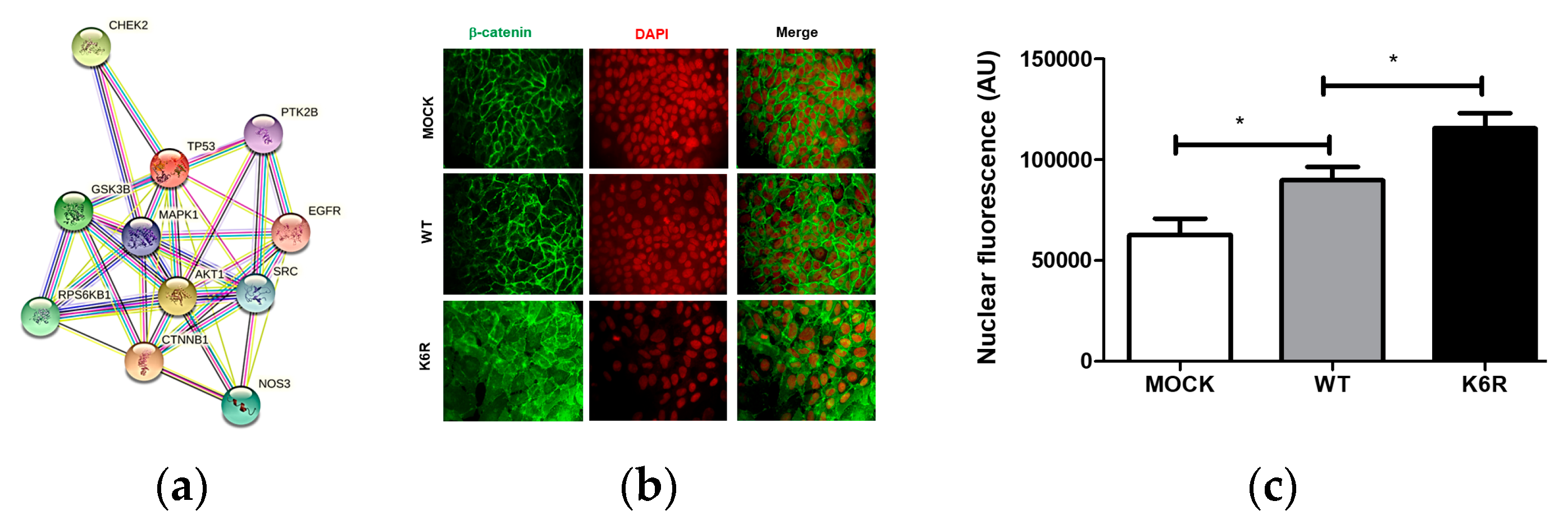
2.3. The Non-Canonical ET1 Regulation of Signaling Proteins

The effect of the ECE1cK6R mutant has been observed to be independent of its canonical role in activating the ET1 peptide. Levels of this peptide in culture media of ECE1cK6R-expressing cells were indistinguishable from those of ECE1cWT-expressing cells originating from colorectal cancer [30], glioblastoma [31] or lung cancer [32]. Taken together with data obtained using ECE1c inhibitors, ETAR silencers, ETAR antagonists, and ET1 supply (for review [15]), these findings indicate that the promotion of an ECE1c-driven CSC phenotype occurs in parallel to, if not at the expense of, ET1 activation and binding to its ETAR receptor.
Consistent with the above findings, studies using an array of human phospho-proteins from colorectal cancer cells showed that the expression of the highly stable biphospho-mimetic ECE1cDD mutant—functionally similar to ECE1cK6R—promotes significant changes in some signaling proteins (Figure 3a). These proteins include β-catenin, AKT1, SRC, and GSK3β, which are known regulators of the canonical Wnt signaling pathway involved in CSC genesis [1,2,3,4,5,6]. Upon activation, β-catenin migrates to the nuclei and transactivates the expression of several genes related with stemness, proliferation, differentiation, death, etc. [43,44,45]. Our data show that the expression of the super-stable ECE1cK6R mutant promotes nuclear localization of β-catenin in colorectal cancer cells (Figure 3b,c), which strongly suggests an activation of the canonical Wnt signaling pathway. In fact, we have already determined a significantly augmented expression of many canonical Wnt target genes as a consequence of the expression of both super-stable ECE1cK6R and biphospho-mimetic ECE1cS18D/S20D enzymes in colorectal cancer cells, such as the stemness genes c-Myc, CD44, Lgr5, Nanog and Snail [30], as well as the proliferation-related genes CCND1 (cyclin-D1) and BIRC5 (survivin) [33], respectively. Notably, whether the same nuclear localization of β-catenin following the expression of the super-stable ECE1cK6R occurs in glioblastoma and lung cancer cells is an interesting but yet unsolved issue, since a group of canonical Wnt target genes, such as c-Myc, CD44, MMP9, Oct-4, Snail, Sox-2 and Twist, have been found to be augmented in these cancer models [31,32].
Finally, an in silico analysis predicted that all of the proteins detected in the above proteome array may interact with one or more proteins (Figure 3a). However, when ECE1c was incorporated into this analysis, no interactions were predicted with any of the proteins, with the exception of eNOS, which is related to the regulatory role of ECE1 in vascular tone [15]. Moreover, no genetic mutations of Y5, K6, T9, S18, or S20 residues in cancer patients have been reported in the literature [30]. However, it is well known that several signaling proteins are aberrantly elevated in colorectal and other cancers, including CK2 and β-catenin [27,29,45,46]. Thus, whether ECE1c indeed contributes to cancer aggressiveness in a CK2-dependent post-translational manner is an issue that certainly merits continued research.

3. Materials and Methods

Phosphorylation assay. Cells were treated for 3 h with 25 μM MG-132 (Tocris, Bristol, UK). One milligram of cell lysates was immunoprecipitated with 10 μg of rabbit anti-Flag antibody (Cell Signaling Technology, Danvers, MA, USA), and then ECE1c proteins phosphorylated at any S/T/Y were detected by Western blot with a mouse anti-pS/pT/pY pan-antibody (Abcam, Cambridge, UK). Total immunoprecipitated ECE1c was detected by WB with a mouse anti-Myc antibody (Cell Signaling Technology, Danvers, MA, USA).
Ubiquitination assay. Cells were transfected with 5 μg of a plasmid encoding His6-tagged ubiquitin. After 16 h of growing in normal conditions, cells were treated, or not treated, for 24 h with 25 μM of the CK2 inhibitor CX-4945 (ApexBio Technology, Houston, TX, USA) and incubated the last 3 h with 25 μM MG-132 (Cayman, Ann Arbor, MI, USA). His6-ubiquitinated proteins from 1 mg of each lysate were pulled down with a Ni+2-NTA resin (Thermo Fisher Scientific, Waltham, MA, USA), separated by SDS-PAGE, and detected by Western Blot with a rabbit anti-ECE1 pan-antibody (Abcam, Cambridge, UK).
Proteome assay. Cell lysates were used in a Human Proteome Profiler Array (R&D Systems, Minneapolis, MN, USA) following manufacturer’s instructions. Phospho-proteins whose levels showed significant variations for ECE1cDD vs. ECE1cWT or ECE1cAA vs. ECE1cWT were analyzed by STRING for protein–protein interactions (https://string-db.org/; accessed on 20 May 2021).
Confocal immunofluorescent microscopy. Cells were grown on glass coverslips under normal conditions for 48 h. After rinsing with PBS, cells were fixed in 4% paraformaldehyde/PBS for 30 min and permeabilized with 0.1% Triton X-100 for 10 min. Cells were incubated with a mouse anti-β-catenin antibody (Becton Dickinson, NJ, USA) followed of an AF488-conjugated secondary antibody (Cell Signaling Technology, Danvers, MA, USA) and DAPI for nuclei. Cells were further mounted onto slides with mowiol (Sigma-Aldrich, St. Louis, MO, USA) and then visualized with an LSM-400 Carl Zeiss confocal microscope Axiovert (Zeiss Group, Jena, Germany) following excitation at 488 nm.

4. Conclusions

A novel CK2/ECE1c functional partnership may be emerging from a hidden non-canonical mechanism that promotes aggressiveness. As tumors progress, the N-terminus of ECE1c is phosphorylated at S18/S20 by CK2 and presumably at Y5/T9 by an unknown kinase(s). This phosphorylation leads to a blockage of ubiquitination at K6, improving the stability of ECE1c, activating CSC-associated signaling pathway(s), and consequently leading to poor prognosis in patients. Therefore, more research is necessary to uncover how this CK2/ECE1c relationship modulates aggressiveness in cancer cells. Undoubtedly, experiments in vitro with cancer cell lines and in vivo with mice will help to define the elusive molecular mechanism(s) governing poor prognosis in cancer patients.

Author Contributions

Conceptualization, J.C.T.; methodology, P.P.-M. and I.N.; investigation, K.V.-N., M.d.l.Á.T., P.P.-M. and I.N.; writing—original draft preparation, J.C.T.; writing—review and editing, P.P.-M. and I.N.; project administration and funding acquisition, J.C.T. All authors have read and agreed to the published version of the manuscript.

Funding

This research was supported by Vicerrectoría de Investigación y Desarrollo de la Universidad de Chile VID ENL10/21; Líneas de apoyo a la investigación financiadas por el ICBM 2021; FONDECYT grant 1220353.

Institutional Review Board Statement

Not applicable.

Data Availability Statement

Data are contained within the article.

Acknowledgments

We thank Ulises Urzua (Universidad de Chile) and Verónica A. Burzio (Universidad Andrés Bello) for their valuable support.

Conflicts of Interest

The authors declare no conflict of interest.

References

  1. Ajani, J.A.; Song, S.; Hochster, H.S.; Steinberg, I.B. Cancer Stem Cells: The Promise and the Potential. Semin. Oncol. 2015, 42, S3–S17. [Google Scholar] [CrossRef] [PubMed]
  2. Barker, N.; Ridgway, R.A.; Van Es, J.H.; Van De Wetering, M.; Begthel, H.; van den Born, M.; Danenberg, E.; Clarke, A.R.; Sansom, O.J.; Clevers, H. Crypt stem cells as the cells-of-origin of intestinal cancer. Nature 2009, 457, 608–611. [Google Scholar] [CrossRef] [PubMed]
  3. Fanali, C.; Lucchetti, D.; Farina, M.; Corbi, M.; Cufino, V.; Cittadini, A.; Sgambato, A. Cancer stem cells in colorectal cancer from pathogenesis to therapy: Controversies and perspectives. World J. Gastroenterol. 2014, 20, 923–942. [Google Scholar] [CrossRef] [PubMed]
  4. Jordan, C.T.; Guzman, M.L.; Noble, M. Cancer Stem Cells. N. Engl. J. Med. 2006, 355, 1253–1261. [Google Scholar] [CrossRef] [PubMed]
  5. Kreso, A.; Dick, J.E. Evolution of the Cancer Stem Cell Model. Cell Stem Cell 2014, 14, 275–291. [Google Scholar] [CrossRef]
  6. Nguyen, L.V.; Vanner, R.; Dirks, P.; Eaves, C.J. Cancer stem cells: An evolving concept. Nat. Rev. Cancer 2012, 12, 133–143. [Google Scholar] [CrossRef] [PubMed]
  7. Barton, M.; Yanagisawa, M. Endothelin: 20 Years from Discovery to Therapy. Can J. Physiol. Pharmacol. 2008, 86, 485–498. [Google Scholar] [CrossRef]
  8. Kawanabe, Y.; Nauli, S.M. Endothelin. Cell. Mol. Life Sci. 2011, 68, 195–203. [Google Scholar] [CrossRef]
  9. Rosanò, L.; Spinella, F.; Bagnato, A. Endothelin 1 in cancer: Biological implications and therapeutic opportunities. Nat. Rev. Cancer 2013, 13, 637–651. [Google Scholar] [CrossRef]
  10. Bagnato, A.; Rosanò, L. The endothelin axis in cancer. Int. J. Biochem. Cell Biol. 2008, 40, 1443–1451. [Google Scholar] [CrossRef]
  11. Khimji, A.-K.; Rockey, D.C. Endothelin—Biology and disease. Cell. Signal. 2010, 22, 1615–1625. [Google Scholar] [CrossRef] [PubMed]
  12. Jafri, F.; Ergul, A. Nuclear Localization of Endothelin-Converting Enzyme-1: Subisoform Specificity. Arter. Thromb. Vasc. Biol. 2003, 23, 2192–2196. [Google Scholar] [CrossRef] [PubMed]
  13. Funke-Kaiser, H.; Thomas, A.; Bremer, J.; Kovacevic, S.D.; Scheuch, K.; Bolbrinker, J.; Theis, S.; Lemmer, J.; Zimmermann, A.; Zollmann, F.S.; et al. Regulation of the major isoform of human endothelin-converting enzyme-1 by a strong housekeeping promoter modulated by polymorphic microsatellites. J. Hypertens. 2003, 21, 2111–2124. [Google Scholar] [CrossRef] [PubMed]
  14. Lindenau, S.; von Langsdorff, C.; Saxena, A.; Paul, M.; Orzechowski, H.-D. Genomic organisation of the mouse gene encoding endothelin-converting enzyme-1 (ECE-1) and mRNA expression of ECE-1 isoforms in murine tissues. Gene 2006, 373, 109–115. [Google Scholar] [CrossRef] [PubMed]
  15. Tapia, J.C.; Niechi, I. Endothelin-converting enzyme-1 in cancer aggressiveness. Cancer Lett. 2019, 452, 152–157. [Google Scholar] [CrossRef]
  16. Arun, C.; London, N.; Hemingway, D. Prognostic Significance of Elevated Endothelin-1 Levels in Patients with Colorectal Cancer. Int. J. Biol. Markers 2004, 19, 32–37. [Google Scholar] [CrossRef]
  17. Simpson, R.A.; Dickinson, T.; Porter, K.E.; London, N.J.M.; Hemingway, D.M. Raised levels of plasma big endothelin 1 in patients with colorectal cancer. Br. J. Surg. 2000, 87, 1409–1413. [Google Scholar] [CrossRef]
  18. Arun, C.; Swift, B.; Porter, K.; West, K.; London, N.; Hemingway, D. The Role of Big Endothelin-1 in Colorectal Cancer. Int. J. Biol. Markers 2002, 17, 268–274. [Google Scholar] [CrossRef]
  19. Lambert, L.A.; Whyteside, A.R.; Turner, A.J.; Usmani, B.A. Isoforms of endothelin-converting enzyme-1 (ECE-1) have opposing effects on prostate cancer cell invasion. Br. J. Cancer 2008, 99, 1114–1120. [Google Scholar] [CrossRef]
  20. Niechi, I.; Silva, E.; Cabello, P.; Huerta, H.; Carrasco, V.; Villar, P.; Cataldo, L.R.; Marcelain, K.; Armisen, R.; Varas-Godoy, M.; et al. Colon cancer cell invasion is promoted by protein kinase CK2 through increase of endothelin-converting enzyme-1c protein stability. Oncotarget 2015, 6, 42749–42760. [Google Scholar] [CrossRef]
  21. Duncan, J.S.; Litchfield, D.W. Too much of a good thing: The role of protein kinase CK2 in tumorigenesis and prospects for therapeutic inhibition of CK2. Biochim. Biophys. Acta BBA Proteins Proteom. 2008, 1784, 33–47. [Google Scholar] [CrossRef] [PubMed]
  22. Herhaus, L.; Perez-Oliva, A.B.; Cozza, G.; Gourlay, R.; Weidlich, S.; Campbell, D.G.; Pinna, L.A.; Sapkota, G.P. Casein kinase 2 (CK2) phosphorylates the deubiquitylase OTUB1 at Ser 16 to trigger its nuclear localization. Sci. Signal. 2015, 8, ra35. [Google Scholar] [CrossRef] [PubMed]
  23. St-Denis, N.A.; Litchfield, D.W. From Birth to Death: The Role of Protein Kinase CK2 in the Regulation of Cell Proliferation and Survival. Cell. Mol. Life Sci. 2009, 66, 1817–1829. [Google Scholar] [CrossRef] [PubMed]
  24. Litchfield, D.W. Protein kinase CK2: Structure, regulation and role in cellular decisions of life and death. Biochem. J. 2003, 369, 1–15. [Google Scholar] [CrossRef] [PubMed]
  25. Ruzzene, M.; Pinna, L.A. Addiction to protein kinase CK2: A common denominator of diverse cancer cells? Biochim. Biophys. Acta BBA—Proteins Proteom. 2010, 1804, 499–504. [Google Scholar] [CrossRef] [PubMed]
  26. Liang, L.; Qu, L.; Ding, Y. Protein and mRNA Characterization in Human Colorectal Carcinoma Cell Lines with Different Metastatic Potentials. Cancer Investig. 2007, 25, 427–434. [Google Scholar] [CrossRef] [PubMed]
  27. Lin, K.-Y.; Tai, C.; Hsu, J.-C.; Li, C.-F.; Fang, C.-L.; Lai, H.-C.; Hseu, Y.-C.; Lin, Y.-F.; Uen, Y.-H. Overexpression of Nuclear Protein Kinase CK2 α Catalytic Subunit (CK2α) as a Poor Prognosticator in Human Colorectal Cancer. PLoS ONE 2011, 6, e17193. [Google Scholar] [CrossRef] [PubMed]
  28. Borgo, C.; Ruzzene, M. Role of protein kinase CK2 in antitumor drug resistance. J. Exp. Clin. Cancer Res. 2019, 38, 287. [Google Scholar] [CrossRef]
  29. Borgo, C.; D’amore, C.; Sarno, S.; Salvi, M.; Ruzzene, M. Protein kinase CK2: A potential therapeutic target for diverse human diseases. Signal Transduct. Target. Ther. 2021, 6, 183. [Google Scholar] [CrossRef]
  30. Pérez-Moreno, P.; Indo, S.; Niechi, I.; Huerta, H.; Cabello, P.; Jara, L.; Aguayo, F.; Varas-Godoy, M.; Burzio, V.A.; Tapia, J.C. Endothelin-converting enzyme-1c promotes stem cell traits and aggressiveness in colorectal cancer cells. Mol. Oncol. 2020, 14, 347–362. [Google Scholar] [CrossRef]
  31. Niechi, I.; Erices, J.I.; Carrillo-Beltrán, D.; Uribe-Ojeda, A.; Torres, Á.; Rocha, J.D.; Uribe, D.; Toro, M.A.; Villalobos-Nova, K.; Gaete-Ramírez, B.; et al. Cancer Stem Cell and Aggressiveness Traits Are Promoted by Stable Endothelin-Converting Enzyme-1c in Glioblastoma Cells. Cells 2023, 12, 506. [Google Scholar] [CrossRef] [PubMed]
  32. Almarza, C.; González, M.; Toro, M.; Villalobos-Nova, K.; Niechi, I.; Silva-Pavez, E.; López-Muñoz, R.A.; Varas-Godoy, M.; Burzio, V.A.; Jara, L.; et al. Cisplatin-Resistance and Aggressiveness Are Triggered upon Expression of a Super-Stable Endothelin-Converting Enzyme-1c in Lung Cancer Cells. Unpublished manuscript.
  33. Pérez-Moreno, P.; Quezada-Meza, C.; Chavez-Almarza, C.; Niechi, I.; Silva-Pavez, E.; Trigo-Hidalgo, C.; Aguayo, F.; Jara, L.; Cáceres-Verschae, A.; Varas-Godoy, M.; et al. Phosphorylation of Endothelin-Converting Enzyme-1c at Serines 18 and 20 by CK2 Promotes Aggressiveness Traits in Colorectal Cancer Cells. Front. Oncol. 2020, 10, 1004. [Google Scholar] [CrossRef] [PubMed]
  34. Meggio, F.; Pinna, L.A. One-thousand-and-one substrates of protein kinase CK2? FASEB J. 2003, 17, 349–368. [Google Scholar] [CrossRef] [PubMed]
  35. Marin, O.; Sarno, S.; Boschetti, M.; Pagano, M.A.; Meggio, F.; Ciminale, V.; D’Agostino, D.M.; Pinna, L.A. Unique features of HIV-1 Rev protein phosphorylation by protein kinase CK2 (‘casein kinase-2’). FEBS Lett. 2000, 481, 63–67. [Google Scholar] [CrossRef] [PubMed]
  36. Tapia, J.C.; Bolanos-Garcia, V.M.; Sayed, M.; Allende, C.C.; Allende, J.E. Cell cycle regulatory protein p27KIP1 is a substrate and interacts with the protein kinase CK2. J. Cell. Biochem. 2004, 91, 865–879. [Google Scholar] [CrossRef]
  37. Swatek, K.N.; Komander, D. Ubiquitin modifications. Cell Res. 2016, 26, 399–422. [Google Scholar] [CrossRef] [PubMed]
  38. Dikic, I. Proteasomal and Autophagic Degradation Systems. Annu. Rev. Biochem. 2017, 86, 193–224. [Google Scholar] [CrossRef]
  39. Smerdon, S.J.; Yaffe, M.B. Recognition of Phospho-Serine/Threonine Phosphorylated Proteins by Phos-pho-Serine/Threonine-Binding Domains. In Handbook of Cell Signaling, 2nd ed.; Academic Press: Cambridge, MA, USA, 2010; Volume 2, pp. 539–550. [Google Scholar] [CrossRef]
  40. Yaffe, M.B.; Smerdon, S.J. PhosphoSerine/Threonine Binding Domains: You Can’t pSERious? Structure 2001, 9, R33–R38. [Google Scholar] [CrossRef]
  41. Almawi, A.W.; Matthews, L.A.; Guarné, A. FHA domains: Phosphopeptide binding and beyond. Prog. Biophys. Mol. Biol. 2017, 127, 105–110. [Google Scholar] [CrossRef]
  42. Fu, H.; Subramanian, R.R.; Masters, S.C. 14-3-3 Proteins: Structure, Function, and Regulation. Annu. Rev. Pharmacol. Toxicol. 2000, 40, 617–647. [Google Scholar] [CrossRef]
  43. Wang, B.; Li, X.; Liu, L.; Wang, M. β-Catenin: Oncogenic role and therapeutic target in cervical cancer. Biol. Res. 2020, 53, 33. [Google Scholar] [CrossRef] [PubMed]
  44. Tominaga, J.; Fukunaga, Y.; Abelardo, E.; Nagafuchi, A. Defining the function of β-catenin tyrosine phosphorylation in cadherin-mediated cell–cell adhesion. Genes Cells 2008, 13, 67–77. [Google Scholar] [CrossRef] [PubMed]
  45. Nusse, R.; Clevers, H. Wnt/β-Catenin Signaling, Disease, and Emerging Therapeutic Modalities. Cell 2017, 169, 985–999. [Google Scholar] [CrossRef] [PubMed]
  46. Lian, H.; Su, M.; Zhu, Y.; Zhou, Y.; Soomro, S.H.; Fu, H. Protein Kinase CK2, a Potential Therapeutic Target in Carcinoma Management. Asian Pac. J. Cancer Prev. 2019, 20, 23–32. [Google Scholar] [CrossRef]
Figure 1. Stable ECE1c leads to CSC traits in various cancer models. Schematic representation of the hallmarks associated with CSCs observed in vitro upon stabilization of ECE1c. Protein stability of ECE1c has been experimentally enhanced by the following mutations at its N-terminus: K6R (ubiquitination site, putative) and S18D/S20D (CK2 phosphorylation sites). As a consequence, traits associated with a CSC phenotype, such as expression of stemness genes, self-renewal, tumor growth, invasiveness and drug resistance, have been found to be significantly augmented in colorectal cancer [30], glioblastoma [31] and lung cancer [32] cells. ECE1 three-dimensional structure (upper) corresponds to a computational prediction (AF-P42892-F1) of isoform B obtained from AlphaFold (https://alphafold.ebi.ac.uk/; accessed on 30 October 2023). The 3D structure is shown in molecular surface representation and is colored by uncertainty/disorder. The 3D structure of human CK2 holoenzyme (lower) corresponds to accession 1JWH obtained from the Protein Data Bank (PDB; https://www.rcsb.org/; accessed on 30 October 2023) and is depicted in molecular surface mode and colored by model index.
Kinasesphosphatases 02 00001 g001
Figure 2. Phosphorylation of ECE1c blocks its ubiquitination. (a) DLD-1 cells expressing either Myc/Flag-tagged ECE1c wild-type (WT) or mutants (AA: S18A/S20A; DD: S18D/S20D) were treated with MG-132. Cell lysates were immunoprecipitated (IP) with an anti-Flag antibody and then ECE1c proteins phosphorylated at any S/T/Y were detected by Western blot (WB) with an anti-pS/pT/pY pan-antibody. Total immunoprecipitated ECE1c was detected by WB with an anti-Myc antibody. (b) CHO-K1 cells expressing ECE1cWT, ECE1cAAA (i.e., T9A/S18A/S20A), or ECE1cDDD (i.e., T9D/S18D/S20D) were transfected with a plasmid encoding His6-tagged ubiquitin. Cells were treated, or not treated, with the CK2 inhibitor CX-4945 to promote ECE1c degradation. His6-ubiquitinated proteins of each lysate were pulled down with a Ni+2-NTA resin, separated by SDS-PAGE, and detected by WB with an anti-ECE1 pan-antibody (upper). Total ECE1c levels at input are also shown (lower).
Kinasesphosphatases 02 00001 g002
Figure 3. CSC-linked signaling proteins are regulated by P-ECE1c. (a) Lysates from DLD-1 cells expressing ECE1cDD, ECE1cAA, or ECE1cWT were used in a Human Proteome Profiler Array. Phospho-proteins whose levels showed significant variations for ECE1cDD vs. ECE1cWT or ECE1cAA vs. ECE1cWT were: EGF receptor (EGFR), ERK1 (MAPK1), GSK3β, β-catenin (CTNNB1), AKT1, SRC, p53 (TP53), S6K (RPS6KB1), Chk2 (CHEK2), Pyk2 (PTK2B), and eNOS (NOS3). Proteins were analyzed by STRING for protein–protein interactions, including: “known” (cyan: curated databases; pink: experimentally determined), “predicted” (blue: gene co-occurrence), and “other” interactions (green: text mining; black: co-expression; grey: protein homology). (b) DLD-1 cells expressing ECE1cWT, ECE1cK6R, or mock were grown under normal conditions for 48 h. β-catenin was detected by confocal IF microscopy with an anti-β-catenin antibody followed of an AF488-conjugated secondary antibody and DAPI for nuclei. (c) Nuclear fluorescence (AU: arbitrary units) was quantified in the three clone cells at “b” using the Image J software. Values were plotted as mean ± SE from at least three independent experiments. * p ≤ 0.05.
Kinasesphosphatases 02 00001 g003
Disclaimer/Publisher’s Note: The statements, opinions and data contained in all publications are solely those of the individual author(s) and contributor(s) and not of MDPI and/or the editor(s). MDPI and/or the editor(s) disclaim responsibility for any injury to people or property resulting from any ideas, methods, instructions or products referred to in the content.

