Review

Exploring Protein Kinase CK2 Substrate Recognition and the Dynamic Response of Substrate Phosphorylation to Kinase Modulation

by
Luca Cesaro
1,
Angelica Maria Zuliani
1,
Valentina Bosello Travain
2 and
Mauro Salvi
1,*
1
Department of Biomedical Sciences, University of Padova, 35131 Padova, Italy
2
Department of Molecular Medicine, University of Padova, 35131 Padova, Italy
*
Author to whom correspondence should be addressed.
Kinases Phosphatases 2023, 1(4), 251-264; https://doi.org/10.3390/kinasesphosphatases1040015
Submission received: 2 August 2023 / Revised: 29 September 2023 / Accepted: 2 October 2023 / Published: 7 October 2023
(This article belongs to the Special Issue Past, Present and Future of Protein Kinase CK2 Research)

Abstract

:
Protein kinase CK2 (formerly known as casein kinase 2 or II), a ubiquitous and constitutively active enzyme, is widely recognized as one of the most pleiotropic serine/threonine kinases. It plays a critical role in numerous signaling pathways, with hundreds of bona fide substrates. However, despite considerable research efforts, our understanding of the entire CK2 substratome and its functional associations with the majority of these substrates is far from being completely deciphered. In this context, we aim to provide an overview of how CK2 recognizes its substrates. We will discuss the pros and cons of the existing methods to manipulate CK2 activity in cells, as well as exploring the dynamic response of substrate phosphorylation to CK2 modulation.

1. Introduction

PhosphoSitePlus, the most up-to-date and most comprehensive database of post-translational modifications, collects nearly 300,000 non-redundant Ser/Thr/Tyr phosphosites [1]. The majority of these phosphosites have been identified by large-scale mass spectrometry analysis, but the kinases responsible for their generation have been determined only for a few of them. Protein kinase CK2 is one of the most pleiotropic kinases (767 human bona fide phosphosite substrates identified to date according to the PhosphoSitePlus database—https://www.phosphosite.org, accessed on 1 May 2023). CK2’s ubiquity and constitutive activity contribute to its versatility, allowing it to participate in a wide range of cellular signaling events [2,3,4,5,6,7,8,9]. Consequently, CK2 has attracted significant attention as a potential therapeutic target for various human pathologies [10,11,12,13,14,15,16,17,18,19,20]. In a previous study, we proposed that CK2 might be responsible for generating at least 20% of the total human phosphoproteome [21]. However, it is important to acknowledge that this percentage may have been overestimated due to the promiscuity of the CK2 consensus sequence, which may affect the accuracy of substrate prediction [22]. Nevertheless, it is widely recognized that a significant number of CK2 substrates remain unidentified, highlighting the fact that our understanding of CK2 functions is still incomplete.
CK2 is the acronym for casein kinase to denote its ability to efficiently phosphorylate casein in vitro, but the true physiological kinase has been lately identified as Fam20C, the kinase responsible for generating the majority of Ser/Thr phosphosites in secreted proteins [23,24]. The term CK2 does not refer to a single enzyme but rather to a heterotetrameric complex composed of two catalytic subunits (α and/or α′) and two regulatory subunits (β). The two catalytic subunits α and α′ (encoded by the two different genes, CSNK2A1 and CSNK2A2, respectively) may differ in tissue distribution but share a high structural homology. These two isoforms share approximately 90% identity within their catalytic domains differing mainly in the C-terminal domain. However, there is evidence that in addition to the tetrameric form, CK2 can also exist as monomeric catalytic enzymes and regulatory proteins. Crystallographic analysis and cell experiments have suggested that CK2 complex formation is a dynamic process, allowing for the presence of both tetrameric and monomeric forms [25,26]. Furthermore, the expression levels of CK2 subunits may not always be balanced [27,28,29], further supporting the existence of monomeric CK2 catalytic and regulatory subunits. In addition, it has been proposed that the CK2β regulatory subunit may have CK2-independent functions by acting as a partner of other kinases, potentially contributing to diverse signaling pathways beyond CK2-mediated phosphorylation [30,31].

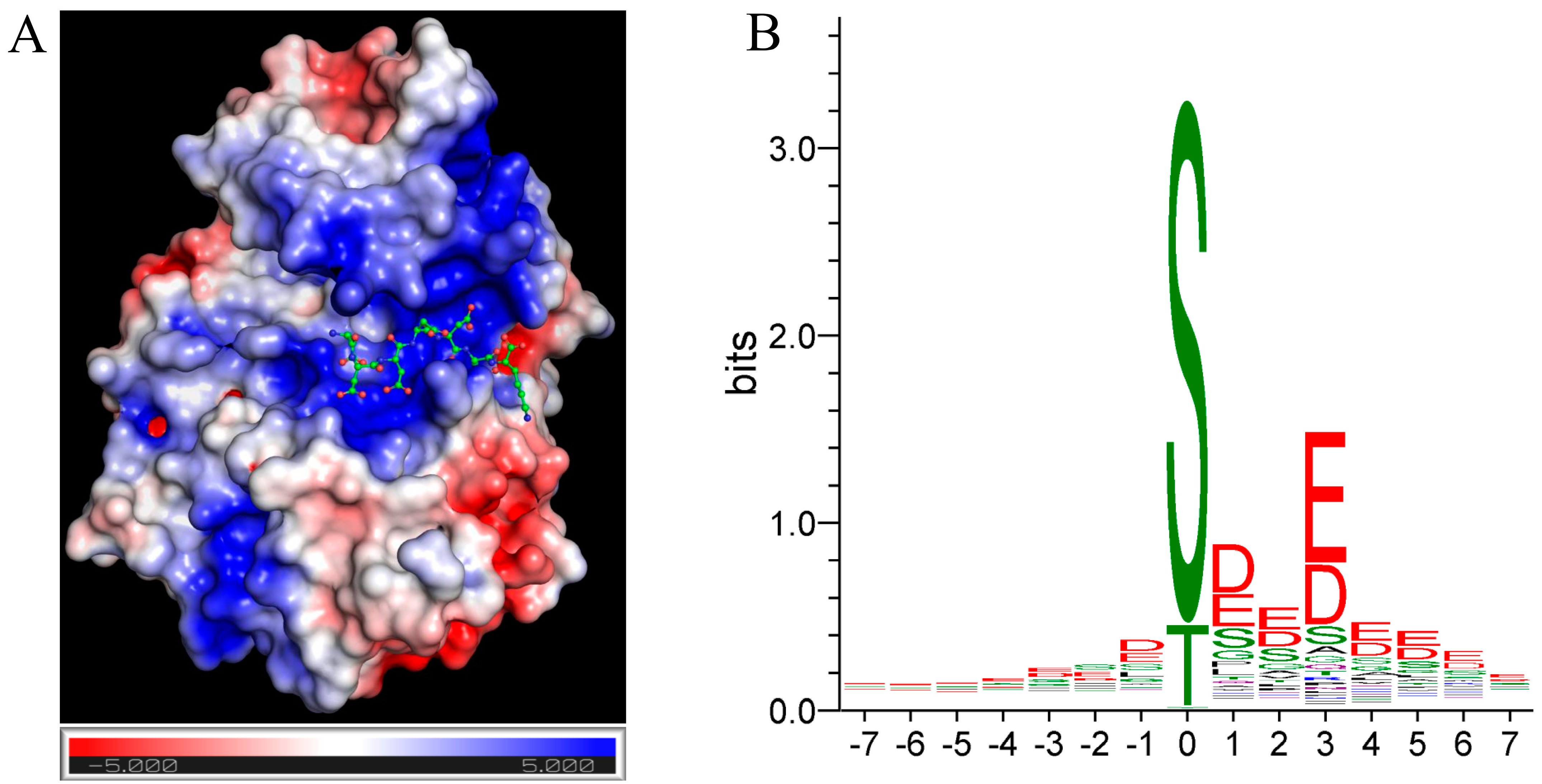
2. CK2 Consensus Sequence

The recognition mechanism by which kinases identify their substrates has been extensively examined and discussed in previous reviews [32,33,34]. Briefly, successful phosphorylation depends on some key factors. First, the kinase and its substrate must come into contact, which is only possible if they share the same subcellular localization. In addition, the proximity of kinase and substrate can be facilitated by kinase docking motifs outside the catalytic domain functioning by tethering enzyme and substrate. Secondly, the target site on the substrate must interact with the catalytic domain of the kinase. This interaction requires the presence of specific determinants surrounding the target site, which vary depending on the structural characteristics of the catalytic fold. These specific determinants are commonly referred to as “kinase consensus sequences”. Recent research has shown that approximately 60% of the Ser/Thr kinases can be classified into three distinct motif classes: proline-directed (requiring a Pro in the +1 position), basophilic and acidophilic kinases [35]. The importance of specific determinants surrounding the target site varies between different kinases, with serine/threonine kinases placing greater emphasis on these determinants compared to tyrosine kinases. Indeed, for tyrosine kinases the presence of docking sites that facilitate proximity between the substrate and the kinase is generally considered to be the most critical factor [36].
How does CK2 recognize its substrates? CK2 localizes in a variety of subcellular compartments, including the nucleus, cytosol, and plasma membrane. The two β-regulatory subunits present in the tetrameric CK2 complex play an important role in substrate recruitment and phosphorylation. As the catalytic subunits of CK2 are constitutively active, as mentioned above, the β-subunits do not function to switch the activity on/off. Instead, the primary function of the β-subunits is to act as a docking protein, facilitating the interaction between the kinase and its substrate [37]. Based on the dependence on the β-subunit, a classification of CK2 substrates as β-dependent or β-independent has been proposed [38]. For example, the downregulation or knockout of the β-subunit is sufficient to completely inhibit the endogenous phosphorylation of S129 Akt and a wide range of other CK2 substrates [39].
CK2 is widely recognized as an acidophilic kinase, indicating a preference for phosphorylation sites surrounded by acidic residues at both the N- and C-termini. The crystal structure of protein kinase CK2α shown in Figure 1A highlights its substrate-binding region, which is characterized by a positively charged electrostatic surface (blue zone). Interestingly, the presence of a proline residue in the +1 position, typically associated with proline-directed kinases, inhibits CK2 activity [33]. In general, a detailed analysis of the kinase consensus sequence can be obtained by analyzing the phosphorylation sites of a peptide library or, better still, by analyzing the bona fide substrates of the kinase once a sufficient number has been identified. In the case of CK2, 767 substrates have been identified in humans according to the PhosphoSitePlus database (https://www.phosphosite.org, accessed on 1 May 2023). Online tools such as WebLogo [40] or Two-Sample Logo [41] can help analyze the primary structure of peptide substrates and identify the most recurrent residues around the target amino acid. These tools are graphical representations of an amino acid multiple sequence alignment that provide an informative representation of sequence motifs, where the height of the stack represents the degree of sequence conservation, and the height of the individual residue within the stack represents its relative frequency at that position. The Weblogo analysis of CK2 phosphosites presented in Figure 1B is the graphical representation of the amino acids surrounding 767 CK2 phosphorylation sites. A closer examination of the selected frame (+7, −7) reveals that acidic residues are favored at all positions, with particular significance at position +3 (69% of substrates containing it) and at position +1 (51% of substrates containing it), confirming the general CK2 consensus sequence as S/T[E/D]x[E/D]. The CK2 substrate logo generated in 2009 [21] with a lower number of substrates (433) shows significant similarity to the logo generated in this study. In fact, the most frequent residues are the same in all positions between −7 and +7 in the two logos. This similarity suggests that the identified sequence motifs and conservation patterns are robust and consistent, and the generated logo represents the definitive fingerprint of the CK2 kinase. How useful are these considerations? In our work from 2009 [21], by analyzing the entire human phosphoproteome we assigned a certain number of substrates to CK2 only according to this CK2 fingerprint. We also demonstrated in vitro and, in some cases, in cell phosphorylation by CK2 for some of these putative substrates, confirming the reliability of the approach. Based on this observation, we proposed that approximately 20% of the phosphoproteome could be attributed to CK2 [21]. However, a more recent analysis suggests that a significative proportion of the phosphopeptides that have the CK2 consensus sequence also have consensus sequences for other kinases, suggesting that these sites could be phosphorylated by multiple kinases or by kinases other than CK2, thus revising the conclusion of our previous analysis [22].
Despite this, the identification of the CK2 consensus sequence in a substrate remains the first approach to understanding whether a site could be a direct substrate of CK2.

3. Is a Phosphopeptide Containing the CK2 Consensus Sequence a True CK2 Substrate?

While the consensus sequence is important for substrate recognition by CK2, experimental verification of CK2 phosphorylation is necessary. The first step in verifying direct CK2 phosphorylation of a substrate is to perform in vitro phosphorylation. In vitro phosphorylation is usually performed with the single recombinant catalytic subunit. However, the outcome of in vitro phosphorylation could differ in the presence or absence of the regulatory CK2β subunits, which play an important role in facilitating the correct binding of the substrate to the catalytic domain. The requirement for the β-subunit is not universal and previous studies have classified CK2 substrates into β-dependent and β-independent categories based on their reliance on CK2β for efficient phosphorylation (see [38] for details).
Identifying CK2 as the kinase responsible for phosphorylating a specific site on a protein in the cell and understanding the functional significance of this phosphorylation event require the ability to detect the phosphorylation site and assess the changes following manipulation of CK2 activity in the cell. The assessment of site-specific phosphorylation in a protein in cells typically requires the development of specific tools. One potential approach to detecting and quantifying the phosphorylation state of the protein is to generate phosphospecific antibodies that specifically recognize and bind to the phosphorylated site of interest for use in immunoblotting and/or immunofluorescence. Another method involves the use of Phos-tag, a chemical compound that binds selectively to phosphorylated residues and can be incorporated into polyacrylamide gels allowing the visualization of the phosphorylation states of a protein by Western blotting [43], the reliability of which, however, appears to be substrate-specific. In some cases, radioactive orthophosphate incorporation or phosphostaining molecules such as ProQdiamond [44] can be used to study the changes in phosphorylation of the whole protein after immunoprecipitation and Western blotting. In addition to these approaches, mass spectrometry plays a crucial role in the identification and quantification of phosphorylation sites in a single protein or in whole-cell lysate after phosphosite enrichment [45]. However, a comprehensive discussion of these techniques and their applications is beyond the scope of this review.
Here, we will describe and critically discuss the methods available to modulate the activity of protein kinase CK2 in the cell.

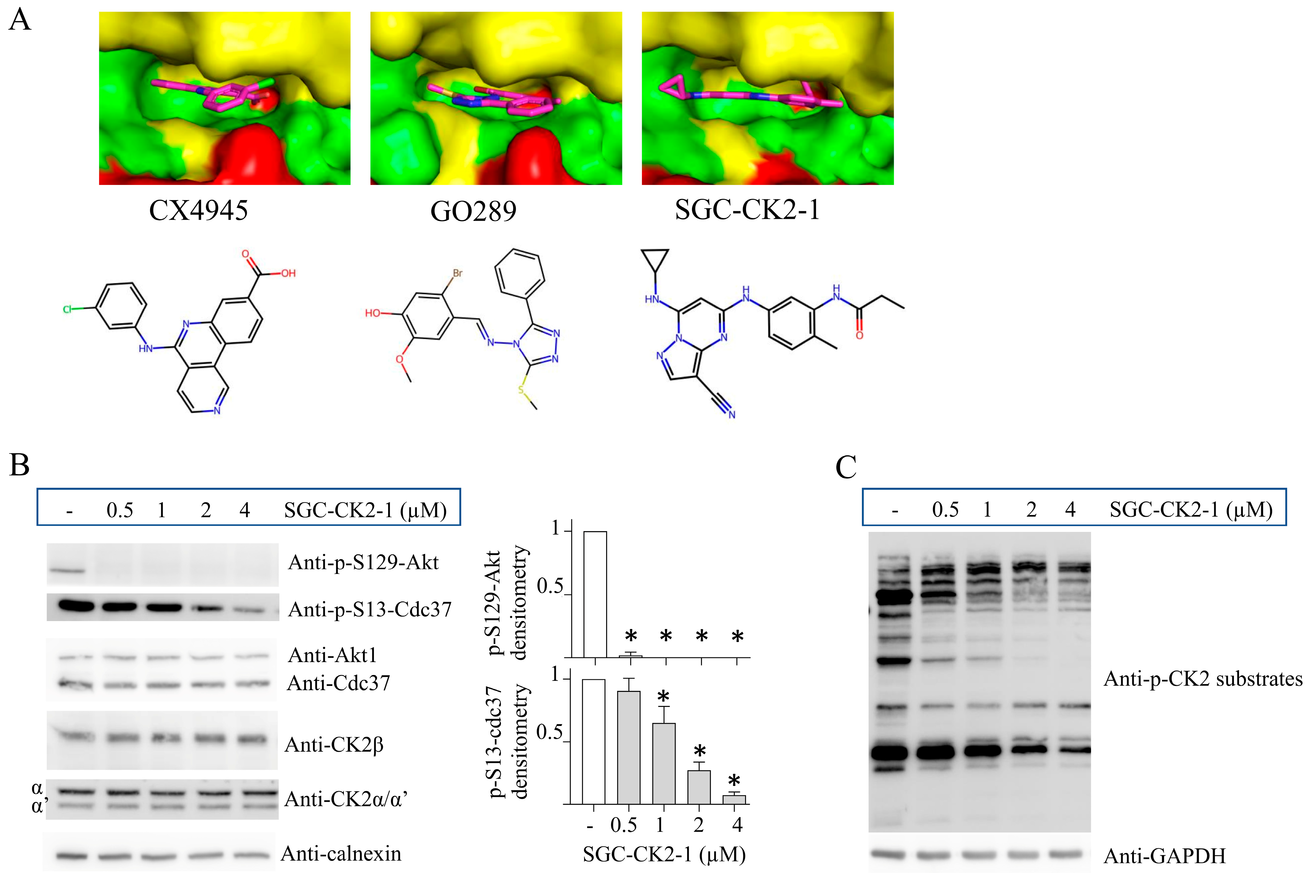
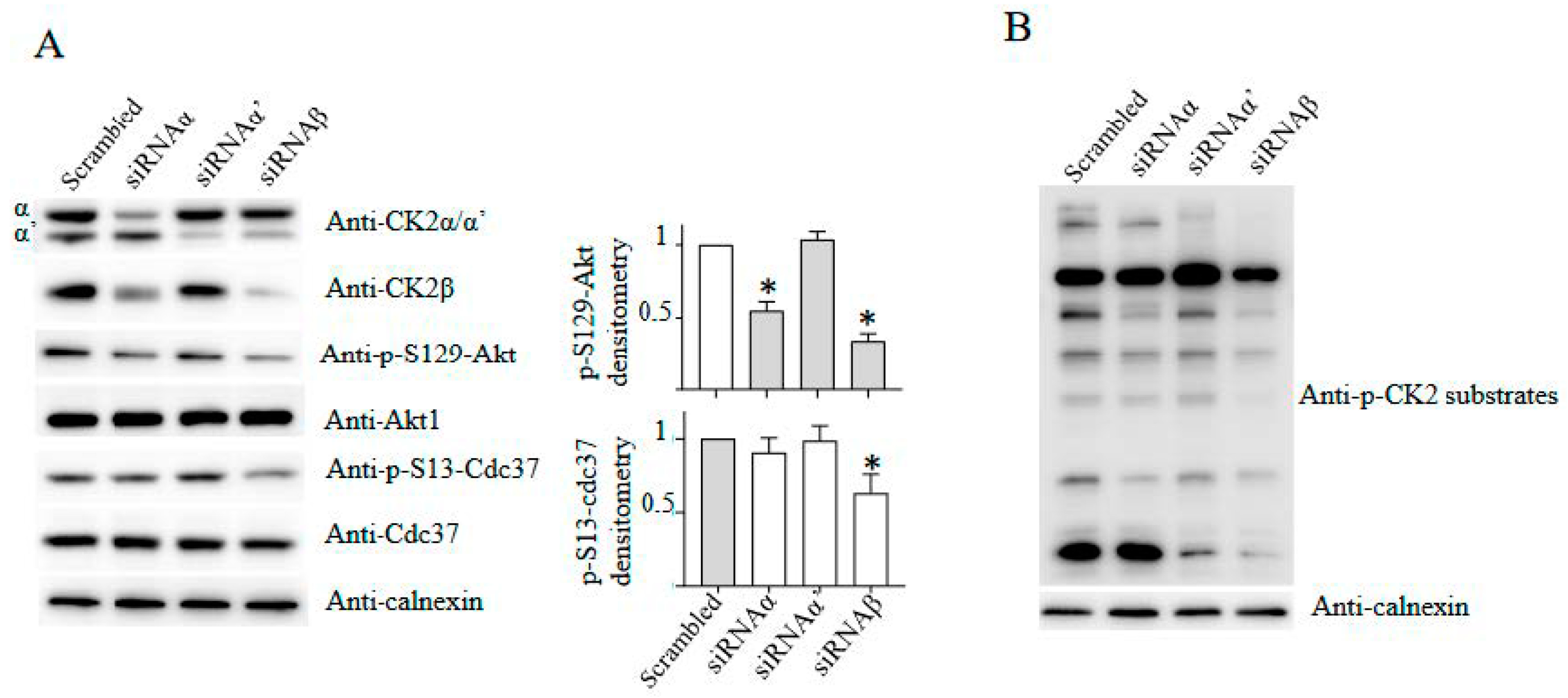
4. CK2 Knockdown, Knockout and Inhibition: Advantages and Disadvantages

Modulating CK2 activity in cells is crucial to establish the link between CK2 and the regulation of physiological functions of a target protein. Transient overexpression of the catalytic or regulatory subunits is readily achievable, subject to limitations in transfection efficiency that depend on the cell line. However, as CK2 is constitutively active [37], reducing its activity is likely to be the most effective approach to identify the functional consequences of protein phosphorylation. Furthermore, although transient kinase overexpression provides some information, it can be misleading as it generally results in the expression of a large amount of kinases, which may be mislocalized and phosphorylate substrates that are not typically phosphorylated at physiological levels.
Two different and complementary approaches can be used to reduce kinase activity: inhibition of the catalytic activity or knockdown/knockout of the protein kinase expression. Although both lead to a reduction in total kinase activity in the cell, these approaches are not equivalent and can give different results, as in the second case kinase-independent effects are still possible due to the binding interactions of the inactive kinase.
Inhibition of CK2 using small chemical molecules is the simplest and most efficient method of reducing CK2 activity in cells. Several inhibitors targeting CK2 have been identified over the years and Table 1 lists the most important compounds with their Ki/IC50 (for a comprehensive list of CK2 inhibitors please refer to [46,47]). The most commonly used inhibitors in the past, such as 4,5,6,7-tetrabromobenzotriazole (TBB) or 2-dimethylamino-4,5,6,7-tetrabromo-benzimidazole (DMAT), exhibited numerous off-target effects and have largely been discarded. The discovery of CX-4945 [48] (also known as Silmitasertib) has represented a breakthrough for its potency and specificity compared to previous molecules and has until recently been considered the inhibitor of choice for targeting CK2 in cells. However, over the years it has been shown that this molecule also inhibits other kinases with high potency including PIM-1 [49], GSK3β [50] and DYRK1A [50,51]. It is also worth mentioning other off-target effects of CX4945: it acts as a splicing regulator [52] and as an inducer of methuosis [53,54], which are two phenomena independent of CK2 activity.
Recently, new specific CK2 inhibitors have been developed, such as GO289 [68] and SGC-CK2-1 [64]. The latter is the most specific CK2 inhibitor to date and the compound of choice for dissecting the cellular functions of CK2. The superior efficacy of SGC-CK2-1 compared to CX4945 has been highlighted in a recent comparative phosphoproteomic analysis [69]. However, it should be emphasized that no inhibitor is completely free of off-target effects. Therefore, the use of at least two different structurally unrelated inhibitors is always recommended [70]. The use of the two inhibitors CX-4945 and GO289, in addition to SGC-CK2-1, seems to be one of the best options. These three inhibitors are structurally unrelated (see structure in Figure 2A), and even if they bind to the same regions of CK2 (the catalytic pocket), they interact with different residues (Figure 2A). Another general consideration supporting the use of different inhibitors is that despite their in vitro efficacy, the inhibitors may have different subcellular distributions, preferentially affecting specific pools of the kinase (see, for example, the recent study in ref. [71]), suggesting that the potency of each inhibitor may differ depending on the type of substrate.
How do we know when treatment with a small molecule inhibitor has reached its effect in the cell? Determining the endpoint of small molecule inhibitor treatment depends on the desired outcome, such as inhibition efficiency of kinase activity or downstream signaling events.
The degree of CK2 inhibition can be measured by incubating the cell lysate with radioactive ATP and a specific peptide substrate (such as R3AD2SD5, which is phosphorylated by both the tetrameric and monomeric forms of CK2 [72]) or a protein substrate (such as casein). This assay has also been used for non-covalent inhibitors demonstrating the persistence of inhibition in diluted cell extracts [73]. However, a successful inhibition of most of the kinase’s activity in cells does not necessarily reflect an immediate effect on the phosphorylation of specific substrates. Indeed, it should be emphasized that CK2 is a constitutively active kinase [37] and its signaling involves both phosphorylation and dephosphorylation events. The dephosphorylation rate at a given phosphosite is not constant. Some phosphosites may have a rapid turnover, while others may have a longer half-life, resulting in variable rates of dephosphorylation when CK2 activity is reduced.
In addition, different phosphosites may respond differently to the same level of CK2 inhibition. Some phosphosites may be more sensitive to changes in CK2 activity, while others may require a stronger inhibition. For example, in lymphoblastic cells, treatment with the CK2 inhibitor CX-5011 can lead to a more pronounced dephosphorylation of pS129Akt compared to pS13 Cdc37 [74]. Furthermore, the same concentration of a CK2 inhibitor, CX-4945 or GO289, can differentially affect the phosphorylation of CK2 substrates in C2C12 cells, as demonstrated using an antibody specific for p-CK2 substrates [75]. Similarly, in genetically engineered C2C12 cells with less than 10% of total CK2 activity [76,77], the phosphorylation of S529 NF-kBp65 and S129 Akt is reduced to a much greater extent than the phosphorylation of pS13 Cdc37 [78]. This phenomenon is further illustrated in Figure 2B,C, which display a similar scenario in human bronchial 16-HBE cells that were treated with increasing doses of the CK2 inhibitor SGC-CK2-1 (Figure 2B). p-S129 Akt, disappears completely at the lower concentration of the inhibitor, whereas S13 Cdc-37 phosphorylation [79] is more resistant to CK2 inhibition exhibiting a dose-dependent response. This observation can also be applied to other substrates. Indeed, it is possible to detect phosphosites that display different responses to the same concentration of CK2 inhibitor by using a mixture of phospho-CK2 substrate-specific antibodies that recognize endogenous proteins containing the CK2 pS/pTDXE motif (Figure 2C). These findings imply that the optimal dosage of inhibitor is substrate-specific and not uniform within the same cellular context.
The second approach to reducing CK2 activity is to target the expression of the protein. Two methods can be used to modulate CK2 protein expression: RNA interference (RNAi) (gene knockdown) or CRISPR tools (gene knockout). RNAi for gene silencing can be achieved using several RNAi platforms, including small interfering RNA (siRNA), short hairpin RNA (shRNA), miRNA, and antisense oligonucleotides (ASO).
Several miRNAs have been identified that target the mRNA of CK2 subunits and may provide insight into the mechanisms behind altered CK2 expression in conditions including cancer [80], ischemia [81] and cell senescence [82,83]. For a comprehensive list of all identified miRNAs that affect CK2 subunits, please refer to [37]. A recent study revealed that the long non-coding RNA KCNQ1OT1 regulates CK2α expression in senescence and caloric restriction by targeting miR-760 [84]. Manipulation of miRNA expression could theoretically be an approach to modulate cellular CK2 expression. However, the limitations of this approach are as follows: (i) this type of regulation is cell-specific, (ii) each miRNA generally has several different targets [85], and (iii) a single mRNA can be targeted by a variety of miRNAs [85], indicating that a single miRNA often has a limited effect on a specific target. For instance, a 50% downregulation of CK2α in colon cancer cells requires the simultaneous transfection of four different miRNAs [86].
The most common method for gene silencing is the transient transfection of specific siRNA oligo targeting the mRNA of one of the three subunits. Downregulation of CK2α, α′, β is easily achieved, and several siRNAs that effectively work have been described in the literature. The advantage of this method is its ease of use, adaptability to different cell types, and efficacy even in hard-to-transfect cells (as siRNAs are smaller and easier to transfect than plasmids). This method leads to a reduction in mRNA expression or a block in its translation, so the effect of a siRNA on the cellular amount of protein is closely linked to its stability.
The choice of which subunit to target depends on the specific research question and the desired outcome. siRNA could be designed to target one or both catalytic subunits and the β-regulatory subunit. Downregulation of a single catalytic subunit typically abolishes no more than 40–60% of the total CK2 activity [28,88]. It is worth noting that CK2 catalytic subunits may be highly redundant, and knockdown of a single catalytic subunit may do not guarantee the inhibition of the phosphorylation of the protein of interest. The situation is different when a substrate is specifically recognized by one or the other catalytic subunit (see below). The downregulation of the only β-regulatory subunit could instead have a dramatic effect on the phosphorylation of the substrate, but again the effect is substrate-dependent.
Despite the potential efficacy of siRNA downregulation, it is important to note that the downregulation is never complete, and a variable residual amount of the targeted protein is still present.
Complete abolition of protein expression is possible by gene editing (CRISPR tools). We generated for the first time CK2 knockout clonal cells [76]. However, although knockout of the single CK2 subunit is generally achievable in different cell types [76,88,89,90,91], to our knowledge, a double knockout of the catalytic subunits has never been achieved. Despite our efforts, we have only been able to obtain clones with minimal CK2 activity (less than 10% of the total) in C2C12 cells expressing a truncated form of CK2α′ and without CK2α [77]. We have hypothesized that a minimal level of activity is required for cell viability [77,87], meaning that it may not be possible to generate clones that are completely devoid of CK2 activity.
Even though CRISPR tools allow the complete abolition of the protein expression, there are two major drawbacks to this approach. First, it is more labor-intensive, because it requires the selection of the gene-edited clones, which takes about 1–3 months depending on the cell proliferation rate. Another important limitation is that a clonal cell population is generated, and not all clones show the same phenotype. This variability is due to the clonal selection starting from a single cell and the potential impact of the selection procedures, especially when the kinase the expression of which has been knocked out has key roles in cell functions. Cells can overcome the lack of protein in a stochastic manner. Therefore, it is necessary to test different clones to validate the involvement of the knocked-out protein in a specific biological phenomenon.
The advantage of the knockdown/knockout methods over inhibitor treatments is the ability to uncover the distinct biological roles of each CK2 subunit. Indeed, if in some cases CK2 catalytic subunits are highly redundant, as discussed above, isoform-specific roles have also been described [92,93,94]. It has also been shown that knockout of a specific subunit may have a greater effect on the phosphorylation level of some CK2 substrates [76,88]. The same is observed with siRNA downregulation of individual CK2 subunits, as shown in Figure 3. Indeed, the phosphorylation level of CK2 substrates changes not as a consequence of the overall reduction in the kinase but depending on which subunit is downregulated (Figure 3). For example, p-S129-AKT is reduced by CK2α or CK2β downregulation, but it is not affected by CK2α′ downregulation. On the contrary, p-S13-Cdc37 is only reduced by CK2β downregulation (Figure 3A). Moreover, the Western blotting experiments performed on the p-CK2 substrates provide compelling evidence that the phosphorylation of specific phosphosites is generated by different CK2 isoforms (Figure 3B). However, it is important to consider the interrelated expression of individual CK2 subunits, as shown in Figure 3A. In the context of 16-HBE cells, the knockdown of CK2α leads to the downregulation of CK2β expression, and conversely, the downregulation of CK2β leads to the downregulation of CK2α′. This interdependence between CK2 subunits has been observed in different cell types, both with knockdown and knockout experiments [76,88]. Interestingly, some of the effects are cell-specific. For example, in C2C12 cells, the downregulation of CK2β increases the expression of CK2α [76]. These findings highlight the complexity of CK2 regulation and of the interactions between subunits, which can vary depending on the cellular context.

5. Conclusions

The exploration of the entire CK2 substratome and the functional implications of CK2 phosphorylation events remains an active area of research. Several tools have been developed to establish the relationship between substrates and CK2, which is crucial for unraveling the physiological functions of this kinase. The use of CK2-specific inhibitors is probably the most feasible approach to improve our understanding of the functions of this kinase. Recently, the use of specific inhibitors in combination with the expression of inhibitor-resistant CK2 mutants has been shown to be a valuable tool for the identification of CK2 substrates [69]. Ongoing research and development efforts are focused on improving the potency and specificity of CK2 inhibitors. Currently the most effective inhibitors are ATP-competitive molecules, with SGC-CK2-1 being the most specific one available to date. An intensive line of research is also working on the development of inhibitors targeting different domains of CK2, including allosteric inhibitors [95,96]. Nevertheless, we emphasize the need for cautious interpretation when using kinase inhibitors. It is important to note that the dephosphorylation of different substrates may have different time courses and/or require different inhibitor concentrations. Simply demonstrating that a kinase inhibitor reduces the phosphorylation of a single substrate may not be sufficient to suggest the block of the entire CK2 signaling.
Furthermore, knockdown/knockout tools serve as valuable complementary approaches that offer significant advantages in elucidating the isoform-specific effects of the catalytic isoforms (CK2α and CK2α′) and the regulatory CK2β on the phosphorylation of CK2 substrates. Although some evidence has been generated over the years, a clear understanding of the redundancy and specificity of each catalytic isoform and the definition of the entire CK2β-dependent substratome still remain elusive. This aspect becomes even more critical in light of the recent discovery of specific mutations in the CSNK2A1 gene associated with Okur–Chung Neurodevelopmental Syndrome (OCNDS) and mutations in the CSNK2B gene associated with Poirier–Bienvenu Neurodevelopmental Syndrome (POBINDS) [19,97]. A predictive analysis of OCNDS and POBINDS variants suggests that mutations in CK2α and CK2β have functional and structural implications [68]. Fifteen distinct OCNDS missense mutations in CK2α result in varying degrees of loss of kinase activity when expressed as recombinant purified proteins [98], while the most common mutation (K198R) is responsible for altered substrate specificity of CK2α [99,100]. To gain deeper insights into disease mechanisms, further investigations are warranted to comprehensively characterize the CK2 subunit-specific effects and their implications in these neurodevelopmental syndromes.

Funding

This research received no external funding.

Institutional Review Board Statement

Not applicable.

Informed Consent Statement

Not applicable.

Data Availability Statement

Not applicable.

Conflicts of Interest

The authors declare no conflict of interest.

References

  1. Hornbeck, P.V.; Zhang, B.; Murray, B.; Kornhauser, J.M.; Latham, V.; Skrzypek, E. PhosphoSitePlus, 2014: Mutations, PTMs and Recalibrations. Nucleic Acids Res. 2015, 43, D512–D520. [Google Scholar] [CrossRef] [PubMed]
  2. D’Amore, C.; Salizzato, V.; Borgo, C.; Cesaro, L.; Pinna, L.A.; Salvi, M. A Journey through the Cytoskeleton with Protein Kinase CK2. Curr. Protein Pept. Sci. 2019, 20, 547–562. [Google Scholar] [CrossRef] [PubMed]
  3. Hashemolhosseini, S. The Role of Protein Kinase CK2 in Skeletal Muscle: Myogenesis, Neuromuscular Junctions, and Rhabdomyosarcoma. Neurosci. Lett. 2020, 729, 135001. [Google Scholar] [CrossRef]
  4. Montenarh, M. Protein Kinase CK2 and Angiogenesis. Adv. Clin. Exp. Med. 2014, 23, 153–158. [Google Scholar] [CrossRef] [PubMed]
  5. Nuñez de Villavicencio-Diaz, T.; Rabalski, A.J.; Litchfield, D.W. Protein Kinase CK2: Intricate Relationships within Regulatory Cellular Networks. Pharmaceuticals 2017, 10, 27. [Google Scholar] [CrossRef]
  6. Montenarh, M.; Götz, C. Protein Kinase CK2 and Ion Channels (Review). Biomed. Rep. 2020, 13, 55. [Google Scholar] [CrossRef]
  7. St-Denis, N.A.; Litchfield, D.W. Protein Kinase CK2 in Health and Disease: From Birth to Death: The Role of Protein Kinase CK2 in the Regulation of Cell Proliferation and Survival. Cell. Mol. Life Sci. 2009, 66, 1817–1829. [Google Scholar] [CrossRef] [PubMed]
  8. Götz, C.; Montenarh, M. Protein Kinase CK2 in Development and Differentiation. Biomed. Rep. 2017, 6, 127–133. [Google Scholar] [CrossRef] [PubMed]
  9. Kanki, T.; Kurihara, Y.; Jin, X.; Goda, T.; Ono, Y.; Aihara, M.; Hirota, Y.; Saigusa, T.; Aoki, Y.; Uchiumi, T.; et al. Casein Kinase 2 Is Essential for Mitophagy. EMBO Rep. 2013, 14, 788–794. [Google Scholar] [CrossRef]
  10. Borgo, C.; D’Amore, C.; Sarno, S.; Salvi, M.; Ruzzene, M. Protein Kinase CK2: A Potential Therapeutic Target for Diverse Human Diseases. Signal Transduct. Target. Ther. 2021, 6, 183. [Google Scholar] [CrossRef] [PubMed]
  11. Quezada Meza, C.P.; Ruzzene, M. Protein Kinase CK2 and SARS-CoV-2: An Expected Interplay Story. Kinases Phosphatases 2023, 1, 141–150. [Google Scholar] [CrossRef]
  12. Baier, A.; Szyszka, R. CK2 and Protein Kinases of the CK1 Superfamily as Targets for Neurodegenerative Disorders. Front. Mol. Biosci. 2022, 9, 916063. [Google Scholar] [CrossRef] [PubMed]
  13. White, A.; McGlone, A.; Gomez-Pastor, R. Protein Kinase CK2 and Its Potential Role as a Therapeutic Target in Huntington’s Disease. Biomedicines 2022, 10, 1979. [Google Scholar] [CrossRef] [PubMed]
  14. Halloran, D.; Pandit, V.; Nohe, A. The Role of Protein Kinase CK2 in Development and Disease Progression: A Critical Review. J. Dev. Biol. 2022, 10, 31. [Google Scholar] [CrossRef]
  15. Ampofo, E.; Nalbach, L.; Menger, M.D.; Montenarh, M.; Götz, C. Protein Kinase CK2-A Putative Target for the Therapy of Diabetes Mellitus? Int. J. Mol. Sci. 2019, 20, 4398. [Google Scholar] [CrossRef]
  16. Trembley, J.H.; Kren, B.T.; Afzal, M.; Scaria, G.A.; Klein, M.A.; Ahmed, K. Protein Kinase CK2—Diverse Roles in Cancer Cell Biology and Therapeutic Promise. Mol. Cell. Biochem. 2023, 478, 899–926. [Google Scholar] [CrossRef] [PubMed]
  17. Strum, S.W.; Gyenis, L.; Litchfield, D.W. CSNK2 in Cancer: Pathophysiology and Translational Applications. Br. J. Cancer 2022, 126, 994–1003. [Google Scholar] [CrossRef] [PubMed]
  18. Castello, J.; Ragnauth, A.; Friedman, E.; Rebholz, H. CK2-An Emerging Target for Neurological and Psychiatric Disorders. Pharmaceuticals 2017, 10, 7. [Google Scholar] [CrossRef]
  19. Unni, P.; Friend, J.; Weinberg, J.; Okur, V.; Hochscherf, J.; Dominguez, I. Predictive Functional, Statistical and Structural Analysis of CSNK2A1 and CSNK2B Variants Linked to Neurodevelopmental Diseases. Front. Mol. Biosci. 2022, 9, 851547. [Google Scholar] [CrossRef]
  20. Hong, H.; Benveniste, E.N. The Immune Regulatory Role of Protein Kinase CK2 and Its Implications for Treatment of Cancer. Biomedicines 2021, 9, 1932. [Google Scholar] [CrossRef] [PubMed]
  21. Salvi, M.; Sarno, S.; Cesaro, L.; Nakamura, H.; Pinna, L.A. Extraordinary Pleiotropy of Protein Kinase CK2 Revealed by Weblogo Phosphoproteome Analysis. Biochim. Biophys. Acta 2009, 1793, 847–859. [Google Scholar] [CrossRef] [PubMed]
  22. Cesaro, L.; Pinna, L.A. Prevalence and Significance of the Commonest Phosphorylated Motifs in the Human Proteome: A Global Analysis. Cell Mol. Life Sci. 2020, 77, 5281–5298. [Google Scholar] [CrossRef] [PubMed]
  23. Salvi, M.; Cesaro, L.; Tibaldi, E.; Pinna, L.A. Motif Analysis of Phosphosites Discloses a Potential Prominent Role of the Golgi Casein Kinase (GCK) in the Generation of Human Plasma Phospho-Proteome. J. Proteome Res. 2010, 9, 3335–3338. [Google Scholar] [CrossRef] [PubMed]
  24. Tagliabracci, V.S.; Wiley, S.E.; Guo, X.; Kinch, L.N.; Durrant, E.; Wen, J.; Xiao, J.; Cui, J.; Nguyen, K.B.; Engel, J.L.; et al. A Single Kinase Generates the Majority of the Secreted Phosphoproteome. Cell 2015, 161, 1619–1632. [Google Scholar] [CrossRef]
  25. Filhol, O.; Nueda, A.; Martel, V.; Gerber-Scokaert, D.; Benitez, M.J.; Souchier, C.; Saoudi, Y.; Cochet, C. Live-Cell Fluorescence Imaging Reveals the Dynamics of Protein Kinase CK2 Individual Subunits. Mol. Cell. Biol. 2003, 23, 975–987. [Google Scholar] [CrossRef]
  26. Niefind, K.; Guerra, B.; Ermakowa, I.; Issinger, O.G. Crystal Structure of Human Protein Kinase CK2: Insights into Basic Properties of the CK2 Holoenzyme. EMBO J. 2001, 20, 5320–5331. [Google Scholar] [CrossRef]
  27. Chua, M.M.J.; Lee, M.; Dominguez, I. Cancer-Type Dependent Expression of CK2 Transcripts. PLoS ONE 2017, 12, e0188854. [Google Scholar] [CrossRef]
  28. Salizzato, V.; Zanin, S.; Borgo, C.; Lidron, E.; Salvi, M.; Rizzuto, R.; Pallafacchina, G.; Donella-Deana, A. Protein Kinase CK2 Subunits Exert Specific and Coordinated Functions in Skeletal Muscle Differentiation and Fusogenic Activity. FASEB J. 2019, 33, 10648–10667. [Google Scholar] [CrossRef]
  29. Guerra, B.; Siemer, S.; Boldyreff, B.; Issinger, O.-G. Protein Kinase CK2: Evidence for a Protein Kinase CK2β Subunit Fraction, Devoid of the Catalytic CK2α Subunit, in Mouse Brain and Testicles. FEBS Lett. 1999, 462, 353–357. [Google Scholar] [CrossRef]
  30. Bibby, A.C.; Litchfield, D.W. The Multiple Personalities of the Regulatory Subunit of Protein Kinase CK2: CK2 Dependent and CK2 Independent Roles Reveal a Secret Identity for CK2β. Int. J. Biol. Sci. 2005, 1, 67–79. [Google Scholar] [CrossRef]
  31. Guerra, B.; Issinger, O.-G.; Wang, J.Y.J. Modulation of Human Checkpoint Kinase Chk1 by the Regulatory Beta-Subunit of Protein Kinase CK2. Oncogene 2003, 22, 4933–4942. [Google Scholar] [CrossRef]
  32. Ubersax, J.A.; Ferrell, J.E. Mechanisms of Specificity in Protein Phosphorylation. Nat. Rev. Mol. Cell. Biol. 2007, 8, 530–541. [Google Scholar] [CrossRef]
  33. Pinna, L.A.; Ruzzene, M. How Do Protein Kinases Recognize Their Substrates? Biochim. Biophys. Acta 1996, 1314, 191–225. [Google Scholar] [CrossRef] [PubMed]
  34. de Oliveira, P.S.L.; Ferraz, F.A.N.; Pena, D.A.; Pramio, D.T.; Morais, F.A.; Schechtman, D. Revisiting Protein Kinase–Substrate Interactions: Toward Therapeutic Development. Sci. Signal. 2016, 9, re3. [Google Scholar] [CrossRef] [PubMed]
  35. Johnson, J.L.; Yaron, T.M.; Huntsman, E.M.; Kerelsky, A.; Song, J.; Regev, A.; Lin, T.-Y.; Liberatore, K.; Cizin, D.M.; Cohen, B.M.; et al. An Atlas of Substrate Specificities for the Human Serine/Threonine Kinome. Nature 2023, 613, 759–766. [Google Scholar] [CrossRef] [PubMed]
  36. Salvi, M.; Cesaro, L.; Pinna, L.A. Variable Contribution of Protein Kinases to the Generation of the Human Phosphoproteome: A Global Weblogo Analysis. Biomol. Concepts 2010, 1, 185–195. [Google Scholar] [CrossRef] [PubMed]
  37. Borgo, C.; D’Amore, C.; Cesaro, L.; Sarno, S.; Pinna, L.A.; Ruzzene, M.; Salvi, M. How Can a Traffic Light Properly Work If It Is Always Green? The Paradox of CK2 Signaling. Crit. Rev. Biochem. Mol. Biol. 2021, 56, 321–359. [Google Scholar] [CrossRef] [PubMed]
  38. Pinna, L.A. Protein Kinase CK2: A Challenge to Canons. J. Cell Sci. 2002, 115, 3873–3878. [Google Scholar] [CrossRef]
  39. Borgo, C.; Franchin, C.; Cesaro, L.; Zaramella, S.; Arrigoni, G.; Salvi, M.; Pinna, L.A. A Proteomics Analysis of CK2β(-/-) C2C12 Cells Provides Novel Insights into the Biological Functions of the Non-Catalytic β Subunit. FEBS J. 2019, 286, 1561–1575. [Google Scholar] [CrossRef]
  40. Schneider, T.D.; Stephens, R.M. Sequence Logos: A New Way to Display Consensus Sequences. Nucleic Acids Res. 1990, 18, 6097–6100. [Google Scholar] [CrossRef]
  41. Vacic, V.; Iakoucheva, L.M.; Radivojac, P. Two Sample Logo: A Graphical Representation of the Differences between Two Sets of Sequence Alignments. Bioinformatics 2006, 22, 1536–1537. [Google Scholar] [CrossRef] [PubMed]
  42. Jurrus, E.; Engel, D.; Star, K.; Monson, K.; Brandi, J.; Felberg, L.E.; Brookes, D.H.; Wilson, L.; Chen, J.; Liles, K.; et al. Improvements to the APBS Biomolecular Solvation Software Suite. Protein Sci. 2018, 27, 112–128. [Google Scholar] [CrossRef] [PubMed]
  43. Kinoshita, E.; Kinoshita-Kikuta, E.; Takiyama, K.; Koike, T. Phosphate-Binding Tag, a New Tool to Visualize Phosphorylated Proteins. Mol. Cell. Proteom. 2006, 5, 749–757. [Google Scholar] [CrossRef] [PubMed]
  44. Martin, K.; Steinberg, T.H.; Cooley, L.A.; Gee, K.R.; Beechem, J.M.; Patton, W.F. Quantitative Analysis of Protein Phosphorylation Status and Protein Kinase Activity on Microarrays Using a Novel Fluorescent Phosphorylation Sensor Dye. Proteomics 2003, 3, 1244–1255. [Google Scholar] [CrossRef]
  45. Urban, J. A Review on Recent Trends in the Phosphoproteomics Workflow. From Sample Preparation to Data Analysis. Anal. Chim. Acta 2022, 1199, 338857. [Google Scholar] [CrossRef]
  46. Borgo, C.; Ruzzene, M. Protein Kinase CK2 Inhibition as a Pharmacological Strategy. Adv. Protein Chem. Struct. Biol. 2021, 124, 23–46. [Google Scholar] [CrossRef]
  47. Chen, Y.; Wang, Y.; Wang, J.; Zhou, Z.; Cao, S.; Zhang, J. Strategies of Targeting CK2 in Drug Discovery: Challenges, Opportunities, and Emerging Prospects. J. Med. Chem. 2023, 66, 2257–2281. [Google Scholar] [CrossRef]
  48. Pierre, F.; Chua, P.C.; O’Brien, S.E.; Siddiqui-Jain, A.; Bourbon, P.; Haddach, M.; Michaux, J.; Nagasawa, J.; Schwaebe, M.K.; Stefan, E.; et al. Discovery and SAR of 5-(3-Chlorophenylamino)Benzo[c][2,6]Naphthyridine-8-Carboxylic Acid (CX-4945), the First Clinical Stage Inhibitor of Protein Kinase CK2 for the Treatment of Cancer. J. Med. Chem. 2011, 54, 635–654. [Google Scholar] [CrossRef]
  49. Bogusz, J.; Zrubek, K.; Rembacz, K.P.; Grudnik, P.; Golik, P.; Romanowska, M.; Wladyka, B.; Dubin, G. Structural Analysis of PIM1 Kinase Complexes with ATP-Competitive Inhibitors. Sci. Rep. 2017, 7, 13399. [Google Scholar] [CrossRef]
  50. Grygier, P.; Pustelny, K.; Nowak, J.; Golik, P.; Popowicz, G.M.; Plettenburg, O.; Dubin, G.; Menezes, F.; Czarna, A. Silmitasertib (CX-4945), a Clinically Used CK2-Kinase Inhibitor with Additional Effects on GSK3β and DYRK1A Kinases: A Structural Perspective. J. Med. Chem. 2023, 66, 4009–4024. [Google Scholar] [CrossRef]
  51. Kim, H.; Lee, K.-S.; Kim, A.-K.; Choi, M.; Choi, K.; Kang, M.; Chi, S.-W.; Lee, M.-S.; Lee, J.-S.; Lee, S.-Y.; et al. A Chemical with Proven Clinical Safety Rescues Down-Syndrome-Related Phenotypes in through DYRK1A Inhibition. Dis. Model Mech. 2016, 9, 839–848. [Google Scholar] [CrossRef]
  52. Kim, H.; Choi, K.; Kang, H.; Lee, S.-Y.; Chi, S.-W.; Lee, M.-S.; Song, J.; Im, D.; Choi, Y.; Cho, S. Identification of a Novel Function of CX-4945 as a Splicing Regulator. PLoS ONE 2014, 9, e94978. [Google Scholar] [CrossRef]
  53. Lertsuwan, J.; Lertsuwan, K.; Sawasdichai, A.; Tasnawijitwong, N.; Lee, K.Y.; Kitchen, P.; Afford, S.; Gaston, K.; Jayaraman, P.-S.; Satayavivad, J. CX-4945 Induces Methuosis in Cholangiocarcinoma Cell Lines by a CK2-Independent Mechanism. Cancers 2018, 10, 283. [Google Scholar] [CrossRef]
  54. D’Amore, C.; Moro, E.; Borgo, C.; Itami, K.; Hirota, T.; Pinna, L.A.; Salvi, M. “Janus” Efficacy of CX-5011: CK2 Inhibition and Methuosis Induction by Independent Mechanisms. Biochim. Biophys. Acta BBA Mol. Cell Res. 2020, 1867, 118807. [Google Scholar] [CrossRef]
  55. Zandomeni, R.O. Kinetics of Inhibition by 5,6-Dichloro-1-β-d-Ribofuranosylbenzimidazole on Calf Thymus Casein Kinase II. Biochem. J. 1989, 262, 469–473. [Google Scholar] [CrossRef]
  56. Andrzejewska, M.; Pagano, M.A.; Meggio, F.; Brunati, A.M.; Kazimierczuk, Z. Polyhalogenobenzimidazoles: Synthesis and Their Inhibitory Activity against Casein Kinases. Bioorganic Med. Chem. 2003, 11, 3997–4002. [Google Scholar] [CrossRef]
  57. Cozza, G.; Girardi, C.; Ranchio, A.; Lolli, G.; Sarno, S.; Orzeszko, A.; Kazimierczuk, Z.; Battistutta, R.; Ruzzene, M.; Pinna, L.A. Cell-Permeable Dual Inhibitors of Protein Kinases CK2 and PIM-1: Structural Features and Pharmacological Potential. Cell. Mol. Life Sci. 2014, 71, 3173–3185. [Google Scholar] [CrossRef]
  58. Pagano, M.A.; Andrzejewska, M.; Ruzzene, M.; Sarno, S.; Cesaro, L.; Bain, J.; Elliott, M.; Meggio, F.; Kazimierczuk, Z.; Pinna, L.A. Optimization of Protein Kinase CK2 Inhibitors Derived from 4,5,6,7-Tetrabromobenzimidazole. J. Med. Chem. 2004, 47, 6239–6247. [Google Scholar] [CrossRef]
  59. McCarty, M.F.; Assanga, S.I.; Lujan, L.L. Flavones and Flavonols May Have Clinical Potential as CK2 Inhibitors in Cancer Therapy. Med. Hypotheses 2020, 141, 109723. [Google Scholar] [CrossRef]
  60. Urolithin as a Converging Scaffold Linking Ellagic Acid and Coumarin Analogues: Design of Potent Protein Kinase CK2 Inhibitors—Cozza—2011—ChemMedChem—Wiley Online Library. Available online: https://chemistry-europe.onlinelibrary.wiley.com/doi/10.1002/cmdc.201100338 (accessed on 20 September 2023).
  61. Yim, H.; Lee, Y.H.; Lee, C.H.; Lee, S.K. Emodin, an Anthraquinone Derivative Isolated from the Rhizomes of Rheum Palmatum, Selectively Inhibits the Activity of Casein Kinase II as a Competitive Inhibitor. Planta Medica 1999, 65, 9–13. [Google Scholar] [CrossRef]
  62. Meggio, F.; Pagano, M.A.; Moro, S.; Zagotto, G.; Ruzzene, M.; Sarno, S.; Cozza, G.; Bain, J.; Elliott, M.; Deana, A.D.; et al. Inhibition of Protein Kinase CK2 by Condensed Polyphenolic Derivatives. An in Vitro and in Vivo Study. Biochemistry 2004, 43, 12931–12936. [Google Scholar] [CrossRef]
  63. Cozza, G.; Mazzorana, M.; Papinutto, E.; Bain, J.; Elliott, M.; di Maira, G.; Gianoncelli, A.; Pagano, M.A.; Sarno, S.; Ruzzene, M.; et al. Quinalizarin as a Potent, Selective and Cell-Permeable Inhibitor of Protein Kinase CK2. Biochem. J. 2009, 421, 387–395. [Google Scholar] [CrossRef]
  64. Wells, C.I.; Drewry, D.H.; Pickett, J.E.; Tjaden, A.; Krämer, A.; Müller, S.; Gyenis, L.; Menyhart, D.; Litchfield, D.W.; Knapp, S.; et al. Development of a Potent and Selective Chemical Probe for the Pleiotropic Kinase CK2. Cell Chem. Biol. 2021, 28, 546–558. [Google Scholar] [CrossRef]
  65. Vangrevelinghe, E.; Zimmermann, K.; Schoepfer, J.; Portmann, R.; Fabbro, D.; Furet, P. Discovery of a Potent and Selective Protein Kinase CK2 Inhibitor by High-Throughput Docking. J. Med. Chem. 2003, 46, 2656–2662. [Google Scholar] [CrossRef]
  66. Pagano, M.A.; Poletto, G.; Di Maira, G.; Cozza, G.; Ruzzene, M.; Sarno, S.; Bain, J.; Elliott, M.; Moro, S.; Zagotto, G.; et al. Tetrabromocinnamic Acid (TBCA) and Related Compounds Represent a New Class of Specific Protein Kinase CK2 Inhibitors. Chembiochem 2007, 8, 129–139. [Google Scholar] [CrossRef]
  67. Battistutta, R.; Cozza, G.; Pierre, F.; Papinutto, E.; Lolli, G.; Sarno, S.; O’Brien, S.E.; Siddiqui-Jain, A.; Haddach, M.; Anderes, K.; et al. Unprecedented Selectivity and Structural Determinants of a New Class of Protein Kinase CK2 Inhibitors in Clinical Trials for the Treatment of Cancer. Biochemistry 2011, 50, 8478–8488. [Google Scholar] [CrossRef]
  68. Oshima, T.; Niwa, Y.; Kuwata, K.; Srivastava, A.; Hyoda, T.; Tsuchiya, Y.; Kumagai, M.; Tsuyuguchi, M.; Tamaru, T.; Sugiyama, A.; et al. Cell-Based Screen Identifies a New Potent and Highly Selective CK2 Inhibitor for Modulation of Circadian Rhythms and Cancer Cell Growth. Sci. Adv. 2019, 5, eaau9060. [Google Scholar] [CrossRef]
  69. Menyhart, D.; Gyenis, L.; Jurcic, K.; Roffey, S.E.; Puri, A.; Jovanovic, P.; Szkop, K.J.; Pittock, P.; Lajoie, G.; Axtman, A.D.; et al. Comparison of CX-4945 and SGC-CK2-1 as Inhibitors of CSNK2 Using Quantitative Phosphoproteomics: Triple SILAC in Combination with Inhibitor-Resistant CSNK2. Curr. Res. Chem. Biol. 2023, 3, 100041. [Google Scholar] [CrossRef]
  70. Cohen, P. Guidelines for the Effective Use of Chemical Inhibitors of Protein Function to Understand Their Roles in Cell Regulation. Biochem. J. 2009, 425, 53–54. [Google Scholar] [CrossRef]
  71. Birus, R.; El-Awaad, E.; Ballentin, L.; Alchab, F.; Aichele, D.; Ettouati, L.; Götz, C.; Le Borgne, M.; Jose, J. 4,5,7-Trisubstituted Indeno[1,2-b]Indole Inhibits CK2 Activity in Tumor Cells Equivalent to CX-4945 and Shows Strong Anti-Migratory Effects. FEBS Open Bio 2022, 12, 394–411. [Google Scholar] [CrossRef]
  72. Marin, O.; Meggio, F.; Pinna, L.A. Design and Synthesis of Two New Peptide Substrates for the Specific and Sensitive Monitoring of Casein Kinases-1 and -2. Biochem. Biophys. Res. Commun. 1994, 198, 898–905. [Google Scholar] [CrossRef] [PubMed]
  73. Ruzzene, M.; Penzo, D.; Pinna, L.A. Protein Kinase CK2 Inhibitor 4,5,6,7-Tetrabromobenzotriazole (TBB) Induces Apoptosis and Caspase-Dependent Degradation of Haematopoietic Lineage Cell-Specific Protein 1 (HS1) in Jurkat Cells. Biochem. J. 2002, 364, 41–47. [Google Scholar] [CrossRef] [PubMed]
  74. Zanin, S.; Borgo, C.; Girardi, C.; O’Brien, S.E.; Miyata, Y.; Pinna, L.A.; Donella-Deana, A.; Ruzzene, M. Effects of the CK2 Inhibitors CX-4945 and CX-5011 on Drug-Resistant Cells. PLoS ONE 2012, 7, e49193. [Google Scholar] [CrossRef] [PubMed]
  75. Borgo, C.; Cesaro, L.; Hirota, T.; Kuwata, K.; D’Amore, C.; Ruppert, T.; Blatnik, R.; Salvi, M.; Pinna, L.A. Comparing the Efficacy and Selectivity of Ck2 Inhibitors. A Phosphoproteomics Approach. Eur. J. Med. Chem. 2021, 214, 113217. [Google Scholar] [CrossRef]
  76. Borgo, C.; Franchin, C.; Scalco, S.; Bosello-Travain, V.; Donella-Deana, A.; Arrigoni, G.; Salvi, M.; Pinna, L.A. Generation and Quantitative Proteomics Analysis of CK2α/α’(-/-) Cells. Sci. Rep. 2017, 7, 42409. [Google Scholar] [CrossRef] [PubMed]
  77. Borgo, C.; D’Amore, C.; Cesaro, L.; Itami, K.; Hirota, T.; Salvi, M.; Pinna, L.A. A N-Terminally Deleted Form of the CK2α’ Catalytic Subunit Is Sufficient to Support Cell Viability. Biochem. Biophys. Res. Commun. 2020, 531, 409–415. [Google Scholar] [CrossRef] [PubMed]
  78. Franchin, C.; Borgo, C.; Cesaro, L.; Zaramella, S.; Vilardell, J.; Salvi, M.; Arrigoni, G.; Pinna, L.A. Re-Evaluation of Protein Kinase CK2 Pleiotropy: New Insights Provided by a Phosphoproteomics Analysis of CK2 Knockout Cells. Cell. Mol. Life Sci. 2018, 75, 2011–2026. [Google Scholar] [CrossRef]
  79. Miyata, Y.; Nishida, E. CK2 Binds, Phosphorylates, and Regulates Its Pivotal Substrate Cdc37, an Hsp90-Cochaperone. Mol. Cell. Biochem. 2005, 274, 171–179. [Google Scholar] [CrossRef]
  80. Feliciano, A.; Castellvi, J.; Artero-Castro, A.; Leal, J.A.; Romagosa, C.; Hernández-Losa, J.; Peg, V.; Fabra, A.; Vidal, F.; Kondoh, H.; et al. MiR-125b Acts as a Tumor Suppressor in Breast Tumorigenesis via Its Novel Direct Targets ENPEP, CK2-α, CCNJ, and MEGF9. PLoS ONE 2013, 8, e76247. [Google Scholar] [CrossRef]
  81. Liang, Y.; Xu, J.; Wang, Y.; Tang, J.-Y.; Yang, S.-L.; Xiang, H.-G.; Wu, S.-X.; Li, X.-J. Inhibition of MiRNA-125b Decreases Cerebral Ischemia/Reperfusion Injury by Targeting CK2α/NADPH Oxidase Signaling. Cell. Physiol. Biochem. 2018, 45, 1818–1826. [Google Scholar] [CrossRef]
  82. Lee, Y.-H.; Kim, S.Y.; Bae, Y.-S. Upregulation of MiR-760 and MiR-186 Is Associated with Replicative Senescence in Human Lung Fibroblast Cells. Mol. Cells 2014, 37, 620–627. [Google Scholar] [CrossRef]
  83. Song, J.; Bae, Y.-S. CK2 Down-Regulation Increases the Expression of Senescence-Associated Secretory Phenotype Factors through NF-ΚB Activation. Int. J. Mol. Sci. 2021, 22, 406. [Google Scholar] [CrossRef] [PubMed]
  84. Lee, Y.; Bae, Y.-S. Long Non-Coding RNA KCNQ1OT1 Regulates Protein Kinase CK2 Via MiR-760 in Senescence and Calorie Restriction. Int. J. Mol. Sci. 2022, 23, 1888. [Google Scholar] [CrossRef] [PubMed]
  85. Naeli, P.; Winter, T.; Hackett, A.P.; Alboushi, L.; Jafarnejad, S.M. The Intricate Balance between MicroRNA-Induced MRNA Decay and Translational Repression. FEBS J. 2023, 290, 2508–2524. [Google Scholar] [CrossRef] [PubMed]
  86. Kim, S.Y.; Lee, Y.-H.; Bae, Y.-S. MiR-186, MiR-216b, MiR-337-3p, and MiR-760 Cooperatively Induce Cellular Senescence by Targeting α Subunit of Protein Kinase CKII in Human Colorectal Cancer Cells. Biochem. Biophys. Res. Commun. 2012, 429, 173–179. [Google Scholar] [CrossRef]
  87. Borgo, C.; Cesaro, L.; Hirota, T.; Kuwata, K.; D’Amore, C.; Ruppert, T.; Blatnik, R.; Salvi, M.; Pinna, L.A. Analysis of the Phosphoproteome of CK2α(-/-)/Δα’ C2C12 Myoblasts Compared to the Wild-Type Cells. Open Biol. 2023, 13, 220220. [Google Scholar] [CrossRef]
  88. Lettieri, A.; Borgo, C.; Zanieri, L.; D’Amore, C.; Oleari, R.; Paganoni, A.; Pinna, L.A.; Cariboni, A.; Salvi, M. Protein Kinase CK2 Subunits Differentially Perturb the Adhesion and Migration of GN11 Cells: A Model of Immature Migrating Neurons. Int. J. Mol. Sci. 2019, 20, 5951. [Google Scholar] [CrossRef]
  89. Di Maira, G.; Gentilini, A.; Pastore, M.; Caligiuri, A.; Piombanti, B.; Raggi, C.; Rovida, E.; Lewinska, M.; Andersen, J.B.; Borgo, C.; et al. The Protein Kinase CK2 Contributes to the Malignant Phenotype of Cholangiocarcinoma Cells. Oncogenesis 2019, 8, 61. [Google Scholar] [CrossRef]
  90. D’Amore, C.; Borgo, C.; Bosello-Travain, V.; Vilardell, J.; Salizzato, V.; Pinna, L.A.; Venerando, A.; Salvi, M. Deciphering the Role of Protein Kinase CK2 in the Maturation/Stability of F508del-CFTR. Biochim. Biophys. Acta BBA Mol. Basis Dis. 2020, 1866, 165611. [Google Scholar] [CrossRef]
  91. Schmitt, B.M.; Boewe, A.S.; Götz, C.; Philipp, S.E.; Urbschat, S.; Oertel, J.; Menger, M.D.; Laschke, M.W.; Ampofo, E. CK2 Activity Mediates the Aggressive Molecular Signature of Glioblastoma Multiforme by Inducing Nerve/Glial Antigen (NG)2 Expression. Cancers 2021, 13, 1678. [Google Scholar] [CrossRef]
  92. Vilk, G.; Saulnier, R.B.; St Pierre, R.; Litchfield, D.W. Inducible Expression of Protein Kinase CK2 in Mammalian Cells. Evidence for Functional Specialization of CK2 Isoforms. J. Biol. Chem. 1999, 274, 14406–14414. [Google Scholar] [CrossRef] [PubMed]
  93. Xu, X.; Toselli, P.A.; Russell, L.D.; Seldin, D.C. Globozoospermia in Mice Lacking the Casein Kinase II Alpha’ Catalytic Subunit. Nat. Genet. 1999, 23, 118–121. [Google Scholar] [CrossRef]
  94. Lou, D.Y.; Dominguez, I.; Toselli, P.; Landesman-Bollag, E.; O’Brien, C.; Seldin, D.C. The Alpha Catalytic Subunit of Protein Kinase CK2 Is Required for Mouse Embryonic Development. Mol. Cell. Biol. 2008, 28, 131–139. [Google Scholar] [CrossRef] [PubMed]
  95. Prudent, R.; Cochet, C. New Protein Kinase CK2 Inhibitors: Jumping out of the Catalytic Box. Chem. Biol. 2009, 16, 112–120. [Google Scholar] [CrossRef] [PubMed]
  96. Iegre, J.; Atkinson, E.L.; Brear, P.D.; Cooper, B.M.; Hyvönen, M.; Spring, D.R. Chemical Probes Targeting the Kinase CK2: A Journey Outside the Catalytic Box. Org. Biomol. Chem. 2021, 19, 4380–4396. [Google Scholar] [CrossRef]
  97. Ballardin, D.; Cruz-Gamero, J.M.; Bienvenu, T.; Rebholz, H. Comparing Two Neurodevelopmental Disorders Linked to CK2: Okur-Chung Neurodevelopmental Syndrome and Poirier-Bienvenu Neurodevelopmental Syndrome-Two Sides of the Same Coin? Front. Mol. Biosci. 2022, 9, 850559. [Google Scholar] [CrossRef]
  98. Dominguez, I.; Cruz-Gamero, J.M.; Corasolla, V.; Dacher, N.; Rangasamy, S.; Urbani, A.; Narayanan, V.; Rebholz, H. Okur-Chung Neurodevelopmental Syndrome-Linked CK2α Variants Have Reduced Kinase Activity. Hum. Genet. 2021, 140, 1077–1096. [Google Scholar] [CrossRef]
  99. Caefer, D.M.; Phan, N.Q.; Liddle, J.C.; Balsbaugh, J.L.; O’Shea, J.P.; Tzingounis, A.V.; Schwartz, D. The Okur-Chung Neurodevelopmental Syndrome Mutation CK2K198R Leads to a Rewiring of Kinase Specificity. Front. Mol. Biosci. 2022, 9, 850661. [Google Scholar] [CrossRef]
  100. Werner, C.; Gast, A.; Lindenblatt, D.; Nickelsen, A.; Niefind, K.; Jose, J.; Hochscherf, J. Structural and Enzymological Evidence for an Altered Substrate Specificity in Okur-Chung Neurodevelopmental Syndrome Mutant CK2αLys198Arg. Front. Mol. Biosci. 2022, 9, 831693. [Google Scholar] [CrossRef]
Figure 1. CK2 substrate recognition. (A) Electrostatic potential mapped onto the molecular surface of CK2α (PDB ID: 6SPW) and the binding of a peptide substrate (D6K derived from ARC3140 inhibitor). The image was generated with PyMol 2.5 Software (Schrodinger, LLC., New York, NY, USA). The electrostatic surface has been calculated with APBS [42]. (B) Web logo analysis of CK2-validated phosphosites. A total of 767 non-redundant phosphosites have been collected from the PhosphoSitePlus database (https://www.phosphosite.org, accessed on 1 May 2023) and analyzed. Weblogo was generated using WebLogo 3: Public Beta.
Kinasesphosphatases 01 00015 g001
Figure 2. Recent CK2-specific inhibitors. (A) Molecular structure of the latest CK2-specific inhibitors and their binding to the CK2 ATP-binding pocket. (B,C) Effect of increasing doses of SGC-CK2-1 on CK2 substrates. 16-HBE cells have been treated with crescent doses of SGC-CK2-1 or with vehicle (DMSO) for 24 h. Cells have been lysed as described in [87] and loaded in SDS-PAGE. The phosphorylation of CK2 substrates (S129-Akt and S13-Cdc37 in panel (B), proteins containing the CK2 S/TDXE motif in panel (C)) have been assessed using Western blotting. The following antibodies have been used: anti-p-S129-Akt (ab133458), anti-Akt (sc-5298), anti-p-S13-Cdc37 (ab108360), anti-cdc-37 (sc-13129), anti-CK2β (ab76025), anti-CK2α/α′ (MCA3031Z), anti CK2-p-substrates (CS# 8738). Calnexin (sc-46669) and GAPDH (ABS16) are the loading controls. The graph in panel (B) represents the densitometric quantification of the immunostained bands p-S129-Akt and p-S13-Cdc37 (mean ± SD, n = 3; * p < 0.05).
Kinasesphosphatases 01 00015 g002
Figure 3. Silencing of CK2 subunits. 16-HBE cells have been treated for 72h with siRNA targeting CK2α, CK2α′, CK2β or scramble siRNA as control (siRNA sequences are reported in [88]). After incubation, cells have been lysed as described in [87] and loaded in SDS-PAGE. The CK2 protein amount and the phosphorylation of CK2 substrates have been assessed by Western blotting. The antibodies have been described in Figure 2. The graph in panel (A) represents the densitometric quantification of the immunostained bands p-S129-Akt and p-S13-Cdc37 (mean ± SD, n = 3; * p < 0.05). Calnexin (sc-46669) are the loading controls.
Kinasesphosphatases 01 00015 g003
Table 1. Most commonly used CK2 inhibitors. For a comprehensive list of CK2 inhibitors please refer to [46,47].
ATP-Competitive InhibitorsKi or IC50References
Polyhalogenated benzimidazole and benzotriazole derivatesDRBKi = 23 μM[55]
TBBIC50 = 1.6 μM[56]
TDBIC50 = 0.032 μM[57]
TBIIC50 = 0.5 μM[58]
DMATIC50 = 0.14 μM[58]
FlavonoidsQuercetinIC50 = 0.55 μM[59]
ApigeninIC50 = 0.8 μM[59]
FisetinIC50 = 0.35 μM[59]
CoumarinsEllagic acidIC50 = 40 nM[60]
AntraquinonesEmodinKi = 1.85 μM[61]
MNXKi = 0.8 μM[62]
MNAKi = 0.78 μM[62]
QuinalizarinKi = 0.06 μM[63]
Pyrazolo[1,5-a]pyrimidine derivativesSGC-CK2-1IC50 = 36 nM (CK2α)
IC50 = 16 nM (CK2α′)
[64]
Carboxyl acid derivativesIQAIC50 = 0.39 μM[65]
TBCAIC50 = 110 nM[66]
CX-5011IC50 = 2.3 nM[67]
CX-4945IC50 = 1 nM[48]
Other compoundsGO289IC50 = 7 nM[68]
Disclaimer/Publisher’s Note: The statements, opinions and data contained in all publications are solely those of the individual author(s) and contributor(s) and not of MDPI and/or the editor(s). MDPI and/or the editor(s) disclaim responsibility for any injury to people or property resulting from any ideas, methods, instructions or products referred to in the content.

