
Review

Microsurgical Interventions for Cancer-Related Lymphedema

by
Aurora M. Kareh
*,
Brielle Weinstein
and
Nicholas J. Panetta
Department of Plastic and Reconstructive Surgery, Morsani College of Medicine, University of South Florida, Tampa, FL 33602, USA
*
Author to whom correspondence should be addressed.
Lymphatics 2026, 4(2), 18; https://doi.org/10.3390/lymphatics4020018
Submission received: 6 January 2026 / Revised: 25 February 2026 / Accepted: 17 March 2026 / Published: 30 March 2026
(This article belongs to the Special Issue Contemporary Multidisciplinary Management of Lymphatic Disease)

Abstract

Lymphedema is a chronic, incurable disease affecting patients who undergo high-risk cancer treatments. Advances in microsurgical techniques have paved the way for the development of techniques that can prevent or treat this unrelenting condition. In this article we discuss microsurgical interventions for the prevention and treatment of lymphedema, as well as the role of robotics in lymphatic surgery.

1. Introduction

Lymphedema is a chronic, progressive condition characterized by impaired drainage of protein-rich lymph fluid, leading to debilitating swelling, lipogenesis, and soft tissue fibrosis [1]. In developed countries, lymphedema most commonly occurs secondary to iatrogenic injury to the lymphatic system during the treatment of various malignancies, most commonly breast cancer. Major risk factors for the development of breast cancer-related lymphedema (BCRL) include invasive cancer, age older than 65 years, axillary lymph node dissection (ALND), regional radiation therapy (RT), taxane-based chemotherapy, high body mass index, subclinical edema, and cellulitis [2,3,4,5].
BCRL is estimated to affect up to 40% of patients who have a history of high-risk breast cancer [5]. Diagnosis of BCRL is not immediate after the inciting event. In patients without adjuvant RT, onset of lymphedema peaks at around 6 to 12 months; in those that have received adjuvant RT, onset is at around 18 to 24 months [6]. Once it develops, lymphedema requires lifelong management with compression and dedicated lymphedema therapy. Diagnostic criteria for lymphedema vary widely and may be based on interlimb volume difference, interlimb circumference difference, a relative volume change of the same limb, changes in bioimpedance, lymphoscintigraphy, indocyanine green lymphangiography (ICG), or patient symptomatology, depending on the institution [7].
While there is no cure for lymphedema, microsurgical advances have created opportunities for prevention and treatment of this debilitating, chronic disease. Here, we present a review of microsurgical techniques that are used for the prevention and treatment of lymphedema including immediate lymphatic reconstruction (ILR), delayed lymphovenous bypass (LVB), vascularized lymph node transfer (VLNT), and the application of robotic microsurgery in lymphatic surgery.

2. Prevention of Lymphedema: Immediate Lymphatic Reconstruction

2.1. Lymphedema Prevention in Breast Cancer

Prevention of lymphedema via immediate lymphovenous bypass at the time of lymph node dissection was originally described by Boccardo et al. in 2009 and has since been adopted by microsurgeons worldwide in the setting of breast, urological, gynecological, soft tissue, and cutaneous malignancies [8,9]. This operation occurs immediately after removal of the lymph nodes. The goal is to identify and re-route lymphatic channels that were transected during the nodal extirpation to preserve lymphatic drainage from the adjacent extremity.
Prophylactic ILR begins with reverse mapping of the lymphatic channels that drain the affected extremity using isosulfan blue, indocyanine green, or fluorescein dye. During the lymph node dissection, dyed channels are identified, and length into the lymph nodes is preserved. After completion of the lymph node dissection, transected channels are anastomosed to a nearby vein; in axillary dissections for the treatment of breast cancer, this is typically a branch of the axillary vein. Anastomoses may be done in an end-to-end, end-to-side, sleeve, or arborized fashion. Technique may influence lymphedema rates, but studies controlling for anastomosis type in ILR have not been performed. Patency is confirmed by visualization of isosulfan blue, indocyanine green, or fluorescein dye flowing proximally into the vein. This technique re-establishes flow of lymphatic fluid from the extremity into the venous circulation to prevent accumulation of fluid in the soft tissues of the adjacent extremity.
Numerous studies have demonstrated significant reductions in BCRL rates after ILR. In the original description in 2009, Boccardo et al. performed ILR in 18 patients and found a lymphedema rate of 0% on lymphoscintigraphy at 6 and 12 months after surgery [8]. Their follow-up study in 2014 demonstrated a BCRL rate of 4.05% in 78 patients, citing a steep learning curve as the reason for lymphedema development in three patients [10].
More recent studies have further demonstrated the benefit of ILR after ALND [11]. A retrospective series of 66 patients who underwent ILR demonstrated a lymphedema rate of 6% at a mean 250-day follow-up compared to 44% in a retrospective control group that underwent ALND with no ILR [12]. In a retrospective review of 90 patients who underwent ILR over a 4-year period, of which 87% underwent adjuvant radiotherapy (RT), only 9% of patients met criteria for a diagnosis of lymphedema at the conclusion of the study period [13]. This is a significant reduction from the 33.4% lymphedema risk in patients undergoing ALND and RT without ILR [14]. In a randomized controlled trial comparing patients who underwent ILR after axillary dissection with those who did not, the cumulative lymphedema incidence at 24 months after surgery was 9.5% in the ILR group vs. 32% in the non-ILR group [15]. A meta-analysis from 2022 which included four studies with control and treatment groups demonstrated a risk reduction of developing BCRL of 0.22 in the ILR group and a number needed to treat of four in order to prevent one case of BCRL [16].
Patients with obesity, defined as a BMI of 30 kg/m2 or greater, may have higher rates of BCRL after ILR. Wainwright et al. found an overall BCRL rate of 9.4% in 341 patients who underwent ILR. Subgroup analysis demonstrated a 14% rate of BCRL in patients with BMI > 30 kg/m2 compared to 6.5% in patients with BMI < 30 kg/m2 [5]. This study suggests that, despite the higher risk of developing BCRL, ILR may still be protective in this high-BMI population, although potentially less so than in patients with normal BMI.
The short-term benefit to ILR in BCRL has been clearly demonstrated in the literature [3,4,15]. Long-term studies that elucidate whether ILR fully prevents lymphedema or whether it merely delays progression to lymphedema are forthcoming.

2.2. Lymphedema Prevention in Soft Tissue Sarcoma

The majority of studies exploring the benefits of ILR discuss prevention of upper-extremity lymphedema due to the high prevalence of breast cancer in today’s population. However, it is important to note that the second highest incidence of lymphedema occurs in the soft tissue sarcoma population [17]. Over 40% of sarcomas occur in the extremities, and the lower extremities are disproportionately affected compared to the upper extremities [18,19]. The venous and lymphatic systems of the lower extremities are exposed to higher pressures than those of the upper extremities, making the management of lower-extremity lymphedema much more difficult. This is particularly true in patients with sarcoma in the medial thigh, as this location has a dense lymphatic network within the soft tissue [20].
The primary treatment of soft tissue sarcomas is radical resection. Although nodal metastases and lymph node dissection are not routine in these malignancies, RT is often recommended in high-grade tumors in neoadjuvant or adjuvant settings.
Studies assessing rates of clinically significant lymphedema in patients who have undergone soft tissue sarcoma resection have reported lymphedema rates in the range of 9–30% [17,21]. Risk factors for the development of lymphedema after sarcoma treatment include high-dose RT, RT field length < 35 cm, tumor depth, tumors < 5 cm, and lower-extremity tumor location [21].
Multiple studies have reported on the efficacy of ILR in lower-extremity soft tissue sarcomas, primarily for high-risk tumors involving the thigh. Uyulmaz et al. performed ILR in 11 patients who underwent resection of soft tissue sarcomas of the medial thigh. Nineteen patients underwent ILR via immediate lymphovenous bypass as described above for BCRL. Two patients that had no suitable vein for LVB underwent primary lympho-lymphatic anastomosis, and two patients that had neither veins nor lymphatic channels available within the wound underwent extralesional LVB. Four of 11 patients (36%) developed clinical lymphedema; one patient developed progressive lymphedema, and three patients developed moderate lymphedema treated with compression [17].
A retrospective cohort study compared patients undergoing medial thigh tumor resection who underwent ILR with patients undergoing resection with no lymphatic reconstruction [22]. In this study, all ILR procedures were performed at the foot or superior edge of the knee via extralesional LVB. Eight patients underwent ILR at the time of tumor resection: three patients underwent LVB at the knee and foot, while five patients underwent LVB in the foot alone. Of these patients, none developed pitting edema or a difference in limb circumference at 6 months postoperatively. One patient developed mild swelling of the affected leg after RT, but circumferential measurements and ICG remained normal. In the group that underwent resection with no lymphatic reconstruction, 45% of patients reported lower-extremity swelling at 22 months. The authors recommended performing ILR in select patients who are at high risk of developing lymphedema after soft tissue tumor resection [22].
Indications for pursuing ILR in patients undergoing sarcoma resection have not been firmly established. Early reports have demonstrated decreased lymphedema rates in the early postoperative period, which may be beneficial with regards to wound healing. Additional studies are needed to explore the long-term benefit of ILR in this patient population.
In patients undergoing sarcoma resection with large anticipated soft tissue defects in which critical lymphatic channels have been resected, vascularized lymph vessel transfer (VLVT) may also be considered for the prevention of lymphedema. VLVT involves the transfer of skin and subcutaneous tissue with lymph channel orientation, or lymph axiality, that is similar to that of the defect site. Inset of the VLVT lymphatic channels within 2 cm of the recipient site lymph vessels is critical for the prevention of lymphedema [23]. VLVT provides an option for both soft tissue reconstruction and lymphatic channel reconstruction without microsurgical or supermicrosurgical techniques.

2.3. Lymphedema Prevention in Other Malignancies

Prophylactic LVB for gynecologic and urologic cancers may be considered after inguinal lymph node dissection for the prevention of lower-extremity lymphedema. ILR in these cases has shown favorable results, although patient sample sizes are small and long-term results are limited [24,25]. ILR for cutaneous malignancies, however, remains highly controversial due to the potential for systemic spread of these cancers from the lymphatic system through the bypass.

2.4. Prophylactic Vascularized Lymph Node Transfer

Multiple centers have begun exploring prophylactic VLNT at the time of lymph node dissection or at the time of autologous breast reconstruction in those who are at high risk of developing upper-extremity lymphedema [26,27,28,29]. VLNT will be further discussed in the Section 3. Additional studies establishing indications for prophylactic VLNT are needed.

3. Treatment of Lymphedema

Once lymphedema develops, there is no definitive cure. There are multiple surgical treatment options, including microsurgical and non-microsurgical interventions, that may decrease the severity of symptoms, severity of disease, or therapy burden.
The mainstay of lymphedema treatment postoperatively is complete decongestive therapy (CDT). This specialized therapy, led by a certified lymphedema therapist, is split into decongestive and maintenance phases and involves multiple treatment modalities including compression, manual lymphatic massage, lymphatic pumps, and skincare.
Operative treatment modalities for lymphedema include ablative and physiologic procedures. Ablative procedures aim to remove skin and/or subcutaneous fat from the affected limb with the goal of decreasing limb volume and facilitating CDT. Ablative procedures include liposuction, subcutaneous soft tissue excision, and the Charles procedure, the last of which has largely fallen out of favor [1]. Physiologic procedures aim to re-establish normal lymphatic flow by redirecting excess lymphatic fluid centrally.
In this section we discuss the treatment of fluid-dominant disease with physiologic microsurgical procedures. There is no standard treatment algorithm for these physiologic procedures that is agreed upon by all lymphedema centers, as the management of lymphedema requires a patient-centered approach that focuses on the specific characteristics of their disease.

3.1. Delayed Lymphovenous Bypass

Microsurgical lymphovenous bypass for the treatment of lymphedema was initially described in 1977 [30]. Since that time, improvements in technology and refinements in microsurgical technique have paved the way for modern supermicrosurgery, defined as anastomosis of vessels that are less than 0.8 mm in diameter. In this procedure, lymphatic fluid is re-routed from an abnormal lymphatic channel to a nearby venule to decrease the fluid burden of the affected limb.
Traditionally, delayed LVB is considered in patients who have early fluid-dominant disease and adequate targets for bypass. More recent studies have demonstrated the feasibility of delayed LVB in patients with advanced-stage lymphedema with the addition of advanced imaging modalities such as magnetic resonance lymphangiography and ultrasonography [31,32]. Patients undergo ICG in the preoperative setting to visualize suitable channels that demonstrate interruption of normal lymph flow or terminate in dermal backflow. This is typically repeated on the day of surgery for precise incision planning. Visualization of nearby venules may be performed with the assistance of a near-infrared vein finder or ultrasonography. An incision is made where a selected lymphatic and patent venule are near one another. The incision is infiltrated with local anesthetic, and 0.1 cc of isosulfan blue dye may be injected 2 cm distal to the planned incision for ease with identification of the lymphatic channels. The incision is made, vessels are identified, and a bypass is performed between the distal lymphatic and proximal venule. The most common techniques for anastomosis in LVB are end-to-end or end-to-side anastomoses, although intussusception of multiple lymphatics into a vein may also be performed [33]. There are currently no head-to-head studies examining the superiority of one anastomotic technique over another. However, it is critical to confirm anastomotic patency by visualization of isosulfan blue dye or lymphatic fluid flowing into the venule after completion of the anastomosis.
Delayed LVB is associated with decreased limb volume measurements, fewer cellulitis episodes, and improved quality of life [34]. A randomized controlled trial assessing 100 women with early-stage lymphedema who underwent either delayed LVB or CDT demonstrated improved physical and mental function in patients who underwent LVB, with 40% completely or partially stopping the use of compression garments. There was no significant difference or improvement in limb volume or limb circumference in either group [35]. In a prospective study assessing 140 patients over a 24 mo period there were improvements in lymphedema staging, pain, heaviness, quality of life, and general health. There was a reduction in the number of days and hours per day that patients wore compression garments from before to 24 months after LVB in both upper- and lower-extremity groups. Interestingly, delayed LVB may not change limb volume or circumference measurements despite improvements in quality-of-life measures and time spent in compression [34].
Delayed LVB has also demonstrated some benefit for patients with lower-extremity lymphedema after soft tissue sarcoma resection, although protocols for treatment in this population are not yet well established. One study reported delayed LVB for management of lymphedema and lymphorrhea in two patients after sarcoma resection from the medial thigh. LVBs were performed 1 week and 1 month after tumor resection. Both patients demonstrated symptomatic improvement after lymphatic bypass [36].
If the patient does not improve or has persistent symptoms after LVB, repeat imaging may be considered to evaluate for the presence of new lymphatic targets that were not previously visualized. If there are no suitable channels for bypass or if the disease is too severe, VLNT may be considered.

3.2. Vascularized Lymph Node Transfer

Vascularized lymph node transfer involves the transfer of a free flap containing lymph nodes and a vascular pedicle to an area affected by lymphedema. Depending on the donor site selected, the flap may also contain a skin and subcutaneous tissue component. In the early postoperative period, the VLNT serves as a lymphatic pump that shunts excess fluid into the venous circulation. Over time, the transferred nodes promote lymphangiogenesis via increased expression of vascular endothelial growth factor [37,38].
Indications for VLNT vary by institution and are controversial [38]. Traditionally VLNT was reserved for patients with advanced fluid-dominant lymphedema that lacked functioning lymphatic vessels for bypass on imaging or those who demonstrated minimal improvement after LVB [1,39]. However, given the wide variability of practice patterns in lymphatic surgery, VLNT may be considered in earlier disease, in combination with LVB, or in a prophylactic manner after ALND [27,28,29].
There are numerous donor sites available for VLNT, with all options demonstrating equivalent improvements in lymphedema postoperatively [40]. Common donor sites include submental, supraclavicular, lateral thoracic, gastroepiploic, mesenteric, and groin lymph nodes [38,40]. The risks of donor site morbidity must be weighed carefully when selecting donor nodes. Among these risks is the development of secondary lymphedema distal to the lymph node donor site, which could be devastating in a patient already suffering from lymphedema in another limb. Omental/gastroepiploic lymph node transfer has become increasingly popular in the last decade [41]. The intra-abdominal nodes are an attractive choice for VLNT due to the low risk of secondary lymphedema; to our knowledge, secondary lymphedema has never been reported after harvest of the gastroepiploic lymph nodes. The omentum may be harvested laparoscopically or robotically and may be split into two flaps for dual-level VLNT or bilateral-extremity VLNT in cases of bilateral lymphedema. The disadvantages of the omental flap include entry into the peritoneal cavity, the lack of a cutaneous component, and a risk of pancreatitis [38,40]. Submental and supraclavicular nodes also carry a low risk of secondary lymphedema but are harvested at the cost of scarring in visible areas. The inguinal lymph nodes offer a much more hidden scar but have the highest risk of secondary lymphedema amongst the listed options.
Recipient site selection in the lymphedematous limb remains controversial. The VLNT may be anastomosed and inset in the proximal limb, typically at or near the site of lymph node dissection, or in the distal limb, where disease is usually more severe. Proximal placement of the transferred lymph nodes allows for scar release and resurfacing with well-vascularized tissue. However, prior surgery and radiation therapy may make identification of adequate recipient vessels in this hostile area considerably more difficult. Placement of the VLNT in a non-anatomic location in the distal limb maximizes the pump mechanism of the flap and may exhibit better volume reduction compared to more proximal placement of the flap [38]. A major disadvantage of distal flap placement is the aesthetic appearance of the limb, as there is typically a visible scar, skin graft, or skin paddle when flaps are placed distally where there is paucity of soft tissue. Recipient location selection should be made in a collaborative approach with the patient given these advantages and disadvantages.
Lymph node transfer has shown positive results in the management of lymphedema [42]. In a systematic review of 17 studies analyzing VLNT for BCRL, those that reported volumetric outcomes demonstrated a mean reduction in limb volume difference between limbs of 40.31% after VLNT [43]. A randomized controlled trial comparing VLNT to a control group demonstrated significant reductions in limb volume, infection, subjective symptoms, and overall cost in the VLNT group [44]. VLNT has also been associated with significant improvements in overall quality of life, pain, and heaviness using a validated lymphedema quality-of-life questionnaire [45]. In a study comparing CDT, LVB, and VLNT with and without breast reconstruction, VLNT demonstrated greater improvements in limb circumferential differences, circumferential reduction rates, and cellulitis when compared to LVB or CDT [46].

3.3. Combination Procedures

Given that lymphedema is a sequela of high-risk breast cancer treatments, patients with lymphedema will often seek breast reconstruction. There have been conflicting reports on the effect breast reconstruction has on the development of BCRL. Some studies have reported a lower incidence of BCRL or longer time to development of lymphedema in patients who have undergone breast reconstruction alone without lymphedema-specific interventions [47,48]. It is hypothesized that the transfer of vascularized tissue for breast reconstruction can bridge damaged lymphatics and address scarring after cancer interventions [48]. However, other studies have found no association between the incidence of lymphedema and breast reconstruction [46,49].
Given these conflicting reports in the literature, a recent meta-analysis aimed to define the relative risk of lymphedema after breast reconstruction. The authors analyzed 11 studies which encompassed 10,403 breasts. Their findings demonstrated a significantly lower risk of lymphedema in patients who underwent breast reconstruction compared with those who underwent mastectomy and breast conservation therapy combined. In subgroup analysis, the authors also found a significantly lower risk of lymphedema in patients undergoing breast reconstruction compared with mastectomy alone but found no difference in BCRL rates between breast reconstruction and breast conservation therapy groups [50].
In patients seeking both treatment of BCRL and breast reconstruction, VLNT may be performed simultaneously with autologous breast reconstruction. This may be performed either as a single flap containing both soft tissue and nodes (i.e., abdominal-based flap with groin lymph nodes or latissimus dorsi flap with lateral thoracic lymph nodes) or two distinct flaps, with one providing soft tissue reconstruction and the other containing nodes [39]. Abdominal-based reconstruction is most often accomplished with a deep inferior epigastric artery perforator (DIEP) flap or muscle-sparing transverse rectus abdominis myocutaneous flap with groin lymph nodes.
The lymph node basin of the groin used for VLNT is supplied by the superficial circumflex iliac artery and vein. When performing an abdominal-based free flap combined with lymph nodes from the groin, reverse lymphatic mapping with Technetium or isosulfan blue and ICG is essential to spare the lymph nodes critical to lower-extremity lymphatic drainage, thus avoiding the development of donor site lymphedema. Typically the superficial groin lymph nodes are harvested with the flap, while the deep groin lymph nodes are preserved to drain the lower extremity [51]. Markings for the abdominal flap are similar to those for a standard abdominal-based free flap, but all vessels should be dissected to maximum length to facilitate tension-free microvascular anastomosis. Additional arterial or venous anastomoses may need to be performed for perfusion of the lymph nodes depending on the perfusion of the nodes based on the deep inferior epigastric system [51].

4. Robotic-Assisted Microsurgery

The use of robotics in the treatment of lymphedema is relatively new, with early studies describing successful robotic-assisted microsurgical procedures for extremity lymphedema, central lymphatic disorders, and breast lymphedema [52,53,54,55]. The MUSA (Microsure, Eindhoven, The Netherlands) was introduced in 2020 as the first dedicated microsurgical robotic system [52]. Since that time multiple systems have emerged, with only the Symani Surgical System (Medical Microinstruments, Inc., Wilmington, DE, USA) approved by the FDA for use in the United States.
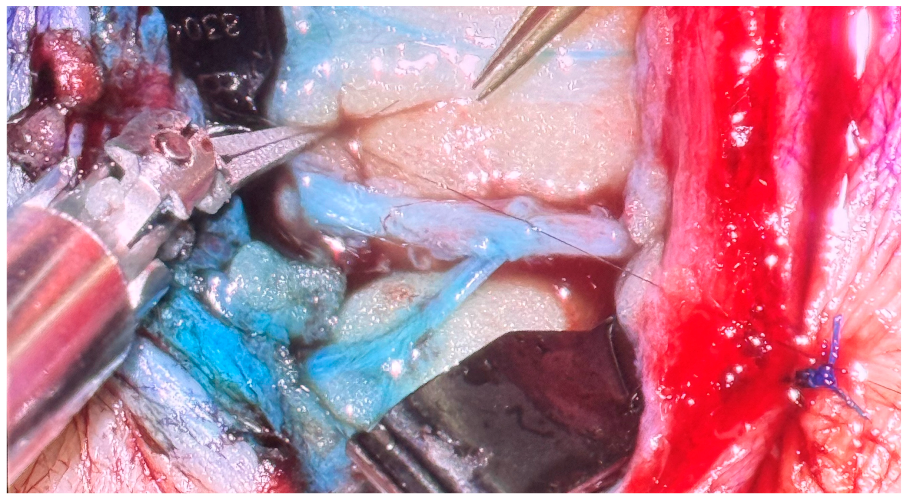
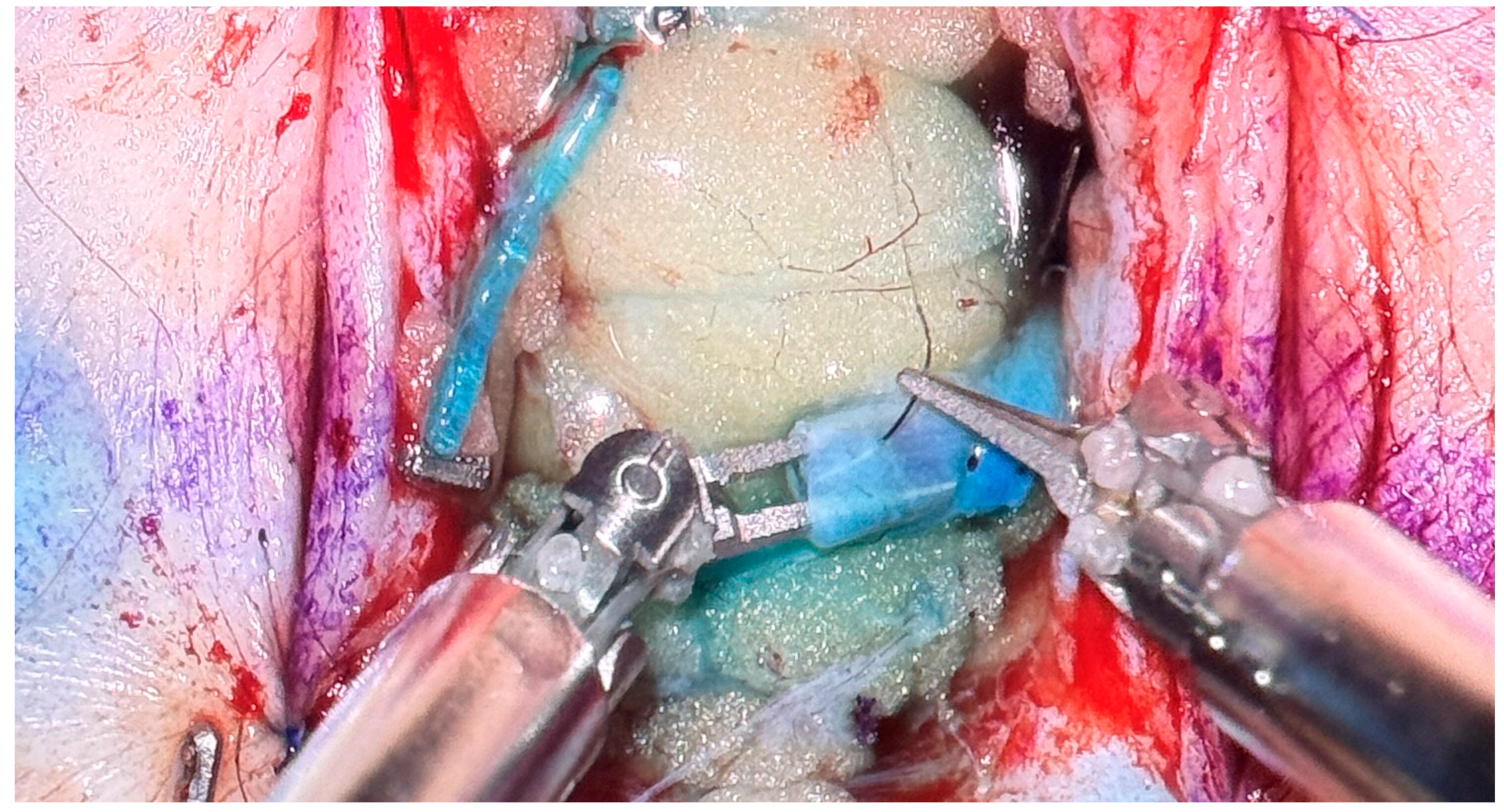
The success of LVB relies heavily on technical precision given the size and delicate nature of the lymphatic channels and venules; likewise, successful supermicrosurgical anastomosis of vascular structures depends on anastomotic patency. The microsurgical robot provides numerous advantages compared to manual microsurgery in technically demanding anastomoses [53]. Features of the microsurgical robot such as tremor reduction and motion scaling, or slowing down of the surgeon’s hand movements, decrease inefficient movement and increase surgical precision [52,53]. Additionally, the Symani Surgical System provides an augmented range of motion with seven degrees of freedom compared to manual microsurgery. Intraoperative photos of a robotic LVB performed at our institution can be seen in Figure 1 and Figure 2.
In their study of 10 robotic-assisted microsurgical anastomoses, Lindenblatt et al. found the robot to provide high-accuracy suture placement, particularly in small, fragile vessels or vessels with notable size mismatch [53]. The robot may also provide an advantage in deep anatomical planes that are otherwise difficult to reach in manual surgery, opening the door to perform bypasses in areas that were previously too small or distant to consider [56].
A systematic review analyzing 13 studies in which 225 robotic LVBs were performed demonstrated comparable patient outcomes between robot-assisted and manual groups in both upper- and lower-extremity groups. There was a significantly longer procedure time in the robotic-assisted groups, which was attributed to the steep learning curve associated with use of the robot [57].
Disadvantages of the robot include the high cost of the robotic system, increased operating time, need for training of the surgeon and operating room staff, time required to prepare the surgical field, lack of manual feedback, and steep learning curve for the operator [52,57,58]. Further studies are needed to establish a cost–benefit analysis and to refine indications for use of the robot in lymphatic surgery.

5. Conclusions

Lymphedema is associated with decreased quality of life in patients that have already undergone rigorous cancer treatments. Although lymphedema has no cure, microsurgical treatments have shown promising outcomes in the prevention and treatment of this chronic disease. Further studies are needed to evaluate long-term outcomes and refine surgical indications for these technically demanding procedures.

Funding

This research received no external funding.

Institutional Review Board Statement

The study was conducted in accordance with the Declaration of Helsinki, and approved by the Institutional Review Board (or Ethics Committee) of University of South Florida Study 007686 (Approval Date: 7 July 2025).

Informed Consent Statement

Informed consent was obtained from all subjects involved in the study. Written informed consent has been obtained from the patient to include photographs published in this paper.

Data Availability Statement

No new data were created or analyzed in this study.

Conflicts of Interest

Kareh has no conflicts of interest to disclose. Weinstein is a consultant for Integra LifeSciences. Panetta is a consultant for Medical Microinstruments and Johnson & Johnson. The funders had no role in the design of the study; in the collection, analyses, or interpretation of data; in the writing of the manuscript; or in the decision to publish the results.

Abbreviations

The following abbreviations are used in this manuscript:
BCRLBreast cancer-related lymphedema
ALNDAxillary lymph node dissection
RTRadiation therapy
ILRImmediate lymphatic reconstruction
LVBLymphovenous bypass
VLNTVascularized lymph node transfer
ICGIndocyanine green lymphangiography
CDTComplex decongestive therapy

References

  1. Kareh, A.M.; Xu, K.Y. Surgical Management of Lymphedema. Mo. Med. 2020, 117, 143–148. [Google Scholar]
  2. Gillespie, T.C.; Sayegh, H.E.; Brunelle, C.L.; Daniell, K.M.; Taghian, A.G. Breast cancer-related lymphedema: Risk factors, precautionary measures, and treatments. Gland Surg. 2018, 7, 379–403. [Google Scholar] [CrossRef] [Scilit]
  3. Li, M.X.; Zhang, J.; Howard, M.A.; Teven, C.M. Efficacy of Immediate Lymphatic Reconstruction in Prevention of Breast Cancer-Related Lymphedema: A Systematic Review and Meta-Analysis. Microsurgery 2025, 45, e70109. [Google Scholar] [CrossRef] [Scilit]
  4. Weinstein, B.; Le, N.K.; Robertson, E.; Zimmerman, A.; Tavares, T.; Tran, T.; Laronga, C.; Panetta, N.J. Reverse Lymphatic Mapping and Immediate Microsurgical Lymphatic Reconstruction Reduces Early Risk of Breast Cancer-Related Lymphedema. Plast. Reconstr. Surg. 2022, 149, 1061–1069. [Google Scholar] [CrossRef] [Scilit] [PubMed]
  5. Wainwright, D.J.; Le, N.K.; Weinstein, B.; West, W.; Tavares, T.; Panetta, N.J. The Impact of Obesity on Success of Immediate Lymphatic Reconstruction for Prevention of Breast Cancer-Related Lymphedema. Ann. Plast. Surg. 2024, 92, S437–S440. [Google Scholar] [CrossRef] [Scilit] [PubMed]
  6. McDuff, S.G.R.; Mina, A.I.; Brunelle, C.L.; Salama, L.; Warren, L.E.G.; Abouegylah, M.; Swaroop, M.; Skolny, M.N.; Asdourian, M.; Gillespie, T.; et al. Timing of Lymphedema After Treatment for Breast Cancer: When Are Patients Most At Risk? Int. J. Radiat. Oncol. Biol. Phys. 2019, 103, 62–70. [Google Scholar] [CrossRef] [Scilit] [PubMed]
  7. Wagner, B.D.; Rubin, J.; Boe, L.A.; Diwan, R.; Bloomfield, E.; Giles, C.; Rochlin, D.H.; Yoshimatsu, H.; Mehrara, B.J.; Coriddi, M.R. Are We Underestimating Breast Cancer-Related Lymphedema? The Impact of Diagnostic Thresholds and Compression Therapy. Ann. Surg. Oncol. 2026, 33, 77–85. [Google Scholar] [CrossRef] [Scilit]
  8. Boccardo, F.; Casabona, F.; De Cian, F.; Friedman, D.; Villa, G.; Bogliolo, S.; Ferrero, S.; Murelli, F.; Campisi, C. Lymphedema microsurgical preventive healing approach: A new technique for primary prevention of arm lymphedema after mastectomy. Ann. Surg. Oncol. 2009, 16, 703–708. [Google Scholar] [CrossRef] [Scilit]
  9. Friedman, R.; Ismail Aly, M.A.; Fanning, J.E.; Pardo, J.A.; Johnson, A.R.; Lee, B.T.; James, T.; Singhal, D. Immediate lymphatic reconstruction: Lessons learned over eight years. J. Plast. Reconstr. Aesthet. Surg. 2024, 94, 1–11. [Google Scholar] [CrossRef] [Scilit]
  10. Boccardo, F.; Casabona, F.; De Cian, F.; Friedman, D.; Murelli, F.; Puglisi, M.; Campisi, C.C.; Molinari, L.; Spinaci, S.; Dessalvi, S.; et al. Lymphatic microsurgical preventing healing approach (LYMPHA) for primary surgical prevention of breast cancer-related lymphedema: Over 4 years follow-up. Microsurgery 2014, 34, 421–424, Correction in Microsurgery 2015, 35, 83. [Google Scholar] [CrossRef] [Scilit]
  11. Pons, G.; Martínez-Jaimez, P.; Condrea, S.; Masia, J. Immediate lymphatic reconstruction with targeted lymphatic axillary repair. J. Plast. Reconstr. Aesthet. Surg. 2025, 101, 134–140. [Google Scholar] [CrossRef] [Scilit]
  12. Le, N.K.; Liu, L.; Jesus Cruz, R.; Parikh, J.; Rotatori, R.M.; Wainwright, D.J.; Weinstein, B.; Tavares, T.; Panetta, N.J. Efficacy of Immediate Lymphatic Reconstruction in Prevention of Breast Cancer-Related Lymphedema. Ann. Plast. Surg. 2023, 90, S363–S365. [Google Scholar] [CrossRef] [Scilit] [PubMed]
  13. Granoff, M.D.; Fleishman, A.; Shillue, K.; Johnson, A.R.; Ross, J.; Lee, B.T.; Teller, P.; James, T.A.; Singhal, D. A 4-Year Institutional Experience of Immediate Lymphatic Reconstruction. Plast. Reconstr. Surg. 2023, 152, 773e–778e. [Google Scholar] [CrossRef] [Scilit]
  14. Johnson, A.R.; Kimball, S.; Epstein, S.; Recht, A.; Lin, S.J.; Lee, B.T.; James, T.A.; Singhal, D. Lymphedema Incidence After Axillary Lymph Node Dissection: Quantifying the Impact of Radiation and the Lymphatic Microsurgical Preventive Healing Approach. Ann. Plast. Surg. 2019, 82, S234–S241. [Google Scholar] [CrossRef] [Scilit]
  15. Coriddi, M.; Dayan, J.; Bloomfield, E.; McGrath, L.; Diwan, R.; Monge, J.; Gutierrez, J.; Brown, S.; Boe, L.; Mehrara, B. Efficacy of Immediate Lymphatic Reconstruction to Decrease Incidence of Breast Cancer-related Lymphedema: Preliminary Results of Randomized Controlled Trial. Ann. Surg. 2023, 278, 630–637. [Google Scholar] [CrossRef] [Scilit]
  16. Hill, W.K.F.; Deban, M.; Platt, A.; Rojas-Garcia, P.; Jost, E.; Temple-Oberle, C. Immediate Lymphatic Reconstruction during Axillary Node Dissection for Breast Cancer: A Systematic Review and Meta-analysis. Plast. Reconstr. Surg. Glob. Open 2022, 10, e4291. [Google Scholar] [CrossRef] [Scilit] [PubMed]
  17. Uyulmaz, S.; Grünherz, L.; Giovanoli, P.; Fuchs, B.; Lindenblatt, N. Primary Lymphovenous Anastomosis After Extended Soft Tissue Resection in the Medial Thigh for Reduction of Lymphocele and Lymphedema. Ann. Plast. Surg. 2024, 93, 221–228. [Google Scholar] [CrossRef] [Scilit]
  18. Schreiber, D.; Bell, R.S.; Wunder, J.S.; O’Sullivan, B.; Turcotte, R.; Masri, B.A.; Davis, A.M. Evaluating function and health related quality of life in patients treated for extremity soft tissue sarcoma. Qual. Life Res. 2006, 15, 1439–1446. [Google Scholar] [CrossRef] [Scilit] [PubMed]
  19. Chen, K.Y.; Ahlquist, S.; Gajewski, C.; Nelson, S.; Bernthal, N.M.; Wessel, L.E. Distal Upper Extremity Sarcoma Epidemiology: A Single-Institution Review. Hand, 2025; Epub ahead of printing. [CrossRef] [Scilit]
  20. Wan, R.; Hussain, A.; Kuruoglu, D.; Houdek, M.T.; Moran, S.L. Prophylactic lymphaticovenous anastomosis (LVA) for preventing lymphedema after sarcoma resection in the lower limb: A report of three cases and literature review. Microsurgery 2023, 43, 273–280. [Google Scholar] [CrossRef] [Scilit]
  21. Friedmann, D.; Wunder, J.S.; Ferguson, P.; O’Sullivan, B.; Roberge, D.; Catton, C.; Freeman, C.; Saran, N.; Turcotte, R.E. Incidence and Severity of Lymphoedema following Limb Salvage of Extremity Soft Tissue Sarcoma. Sarcoma 2011, 2011, 289673. [Google Scholar] [CrossRef] [Scilit]
  22. Wagner, J.M.; Dadras, M.; Ufton, D.; Huber, J.; Wallner, C.; Sogorski, A.; von Glinski, M.; Hong, J.P.; Lehnhardt, M.; Behr, B. Prophylactic lymphaticovenous anastomoses for resection of soft tissue tumors of the thigh to prevent secondary lymphedema-a retrospective comparative cohort analysis. Microsurgery 2022, 42, 239–245. [Google Scholar] [CrossRef] [Scilit]
  23. Yamamoto, T.; Yamamoto, N.; Kageyama, T.; Sakai, H.; Fuse, Y.; Tsukuura, R. Lymph-interpositional-flap transfer (LIFT) based on lymph-axiality concept: Simultaneous soft tissue and lymphatic reconstruction without lymph node transfer or lymphatic anastomosis. J. Plast. Reconstr. Aesthet. Surg. 2021, 74, 2604–2612. [Google Scholar] [CrossRef] [Scilit] [PubMed]
  24. Goldman, C.; Lee, H.; Tom, L.; Krasnow, R. Microsurgical treatment of lower extremity lymphedema: A multidisciplinary approach to improve morbidity in advanced penile cancer patients. Urol. Oncol. 2022, 40, 113.e1–113.e8. [Google Scholar] [CrossRef] [Scilit]
  25. Morotti, M.; Menada, M.V.; Boccardo, F.; Ferrero, S.; Casabona, F.; Villa, G.; Campisi, C.; Papadia, A. Lymphedema microsurgical preventive healing approach for primary prevention of lower limb lymphedema after inguinofemoral lymphadenectomy for vulvar cancer. Int. J. Gynecol. Cancer 2013, 23, 769–774. [Google Scholar] [CrossRef] [Scilit]
  26. Francis, A.M.; Kopplin, N.G.; Chang, E.I. The MD Anderson Algorithm for Lymphedema Management. J. Clin. Med. 2025, 14, 1851. [Google Scholar] [CrossRef] [Scilit]
  27. Ciudad, P.; Escandón, J.M.; Manrique, O.J.; Gutierrez-Arana, J.; Mayer, H.F. Lymphedema prevention and immediate breast reconstruction with simultaneous gastroepiploic vascularized lymph node transfer and deep inferior epigastric perforator flap: A case report. Microsurgery 2022, 42, 617–621. [Google Scholar] [CrossRef] [Scilit]
  28. Brown, S.; Kokosis, G.; Graziano, F.D.; Haran, O.; Smith-Montes, E.; Zivanovic, O.; Ariyan, C.E.; Coit, D.G.; Coriddi, M.; Mehrara, B.J.; et al. Immediate Lymphatic Reconstruction with Vascularized Omentum Lymph Node Transplant: Reducing the Risk of Both Painful Contracture and Lymphedema. Plast. Reconstr. Surg. Glob. Open 2024, 12, e5747. [Google Scholar] [CrossRef] [Scilit]
  29. Yoshimatsu, H.; Karakawa, R.; Fuse, Y.; Yano, T. Simultaneous Lymphatic Superficial Circumflex Iliac Artery Perforator Flap Transfer from the Zone 4 Region in Autologous Breast Reconstruction Using the Deep Inferior Epigastric Artery Perforator Flap: A Proof-of-Concept Study. J. Clin. Med. 2022, 11, 534. [Google Scholar] [CrossRef] [Scilit]
  30. O’Brien, B.M.; Sykes, P.; Threlfall, G.N.; Browning, F.S. Microlymphaticovenous anastomoses for obstructive lymphedema. Plast. Reconstr. Surg. 1977, 60, 197–211. [Google Scholar] [CrossRef] [Scilit] [PubMed]
  31. Cha, H.G.; Oh, T.M.; Cho, M.J.; Pak, C.S.J.; Suh, H.P.; Jeon, J.Y.; Hong, J.P. Changing the Paradigm: Lymphovenous Anastomosis in Advanced Stage Lower Extremity Lymphedema. Plast. Reconstr. Surg. 2021, 147, 199–207. [Google Scholar] [CrossRef] [Scilit] [PubMed]
  32. Yoshimatsu, H.; Cho, M.J.; Inoue, K.; Karakawa, R.; Fuse, Y.; Hayashi, A.; Yano, T. Combining indocyanine green lymphography and ultrasonography findings to identify lymphatic vessels in advanced-stage lymphedema: The milestone and swirl sign approach. J. Plast. Reconstr. Aesthet. Surg. 2025, 102, 134–141. [Google Scholar] [CrossRef] [Scilit]
  33. Forte, A.J.; Sisti, A.; Huayllani, M.T.; Boczar, D.; Cinotto, G.; Ciudad, P.; Manrique, O.J.; Lu, X.; McLaughlin, S. Lymphaticovenular anastomosis for breast cancer-related upper extremity lymphedema: A literature review. Gland. Surg. 2020, 9, 539–544. [Google Scholar] [CrossRef] [Scilit] [PubMed]
  34. Thomas, M.; Pike, C.; Humphreys, I.; Bragg, T.; Ghattaura, A. Impact and outcomes after lymphaticovenous anastomosis for 150 cases of lymphoedema followed up over 24 months. J. Plast. Reconstr. Aesthet. Surg. 2023, 85, 104–113. [Google Scholar] [CrossRef] [Scilit] [PubMed]
  35. Jonis, Y.M.J.; Wolfs, J.; Hummelink, S.; Tielemans, H.J.P.; Keuter, X.H.A.; van Kuijk, S.; Ulrich, D.J.O.; van der Hulst, R.; Qiu, S.S. The 6 month interim analysis of a randomized controlled trial assessing the quality of life in patients with breast cancer related lymphedema undergoing lymphaticovenous anastomosis vs. conservative therapy. Sci. Rep. 2024, 14, 2238. [Google Scholar] [CrossRef] [Scilit]
  36. Kobayashi, H.; Iida, T.; Yamamoto, T.; Ikegami, M.; Shinoda, Y.; Tanaka, S.; Kawano, H. Lymphaticovenous Anastomoses for Lymphedema Complicated by Severe Lymphorrhea Following Resection of Soft-Tissue Sarcomas of the Adductor Compartment: A Report of Two Cases. JBJS Case Connect. 2017, 7, e80. [Google Scholar] [CrossRef] [Scilit]
  37. Salibian, A.A.; Yu, N.; Patel, K.M. Staging Approaches to Lymphatic Surgery: Techniques and Considerations. J. Surg. Oncol. 2025, 131, 12–21. [Google Scholar] [CrossRef] [Scilit]
  38. Pappalardo, M.; Patel, K.; Cheng, M.H. Vascularized lymph node transfer for treatment of extremity lymphedema: An overview of current controversies regarding donor sites, recipient sites and outcomes. J. Surg. Oncol. 2018, 117, 1420–1431. [Google Scholar] [CrossRef] [Scilit]
  39. Almadani, H.; Lu, J.; Bokhari, S.; How-Volkman, C.; Brazio, P.S. Simultaneous Vascularized Lymph Node Transfer and Breast Reconstruction: A Systematic Review. J. Clin. Med. 2025, 14, 1694. [Google Scholar] [CrossRef] [Scilit] [PubMed]
  40. Chang, E.I.; Chu, C.K.; Hanson, S.E.; Selber, J.C.; Hanasono, M.M.; Schaverien, M.V. Comprehensive Overview of Available Donor Sites for Vascularized Lymph Node Transfer. Plast. Reconstr. Surg. Glob. Open 2020, 8, e2675. [Google Scholar] [CrossRef] [Scilit] [PubMed]
  41. Cho, M.J.; Flores Garcia, J.; Myung, Y.; Cha, H.G.; Hayashi, A.; Hong, J.P.; Skoracki, R. Evolving Role of Lymphedema Surgery on Breast Reconstruction: A Systematic Review and Multi-Institutional Algorithmic Approach. J. Clin. Med. 2024, 13, 6518. [Google Scholar] [CrossRef] [Scilit]
  42. Rannikko, E.H.; Pajula, S.; Suominen, S.H.; Kiiski, J.; Mani, M.R.; Halle, M.; Kaartinen, I.S.; Lahdenperä, O.; Arnardottir, T.H.; Kauhanen, S.M.; et al. Phase II Study Shows the Effect of Adenoviral Vascular Endothelial Growth Factor C and Lymph Node Transfer in Lymphedema. Plast. Reconstr. Surg. 2025, 155, 256e–267e. [Google Scholar] [CrossRef] [Scilit] [PubMed]
  43. Winters, H.; Tielemans, H.J.P.; Paulus, V.; Hummelink, S.; Slater, N.J.; Ulrich, D.J.O. A systematic review and meta-analysis of vascularized lymph node transfer for breast cancer-related lymphedema. J. Vasc. Surg. Venous Lymphat. Disord. 2022, 10, 786–795.e1. [Google Scholar] [CrossRef] [Scilit]
  44. Dionyssiou, D.; Demiri, E.; Tsimponis, A.; Sarafis, A.; Mpalaris, V.; Tatsidou, G.; Arsos, G. A randomized control study of treating secondary stage II breast cancer-related lymphoedema with free lymph node transfer. Breast Cancer Res. Treat. 2016, 156, 73–79. [Google Scholar] [CrossRef] [Scilit] [PubMed]
  45. Gratzon, A.; Schultz, J.; Secrest, K.; Lee, K.; Feiner, J.; Klein, R.D. Clinical and Psychosocial Outcomes of Vascularized Lymph Node Transfer for the Treatment of Upper Extremity Lymphedema After Breast Cancer Therapy. Ann. Surg. Oncol. 2017, 24, 1475–1481. [Google Scholar] [CrossRef] [Scilit] [PubMed]
  46. Engel, H.; Lin, C.Y.; Huang, J.J.; Cheng, M.H. Outcomes of Lymphedema Microsurgery for Breast Cancer-related Lymphedema with or Without Microvascular Breast Reconstruction. Ann. Surg. 2018, 268, 1076–1083. [Google Scholar] [CrossRef] [Scilit]
  47. Lopez Penha, T.R.; Voogd, A.C.; Heuts, E.M.; Ijsbrandy, C.; Hendrix, N.A.; von Meyenfeldt, M.F.; van der Hulst, R.R. Reduced prevalence of lymphedema in patients with reconstructive breast surgery. Breast J. 2014, 20, 671–673. [Google Scholar] [CrossRef] [Scilit]
  48. Card, A.; Crosby, M.A.; Liu, J.; Lindstrom, W.A.; Lucci, A.; Chang, D.W. Reduced incidence of breast cancer-related lymphedema following mastectomy and breast reconstruction versus mastectomy alone. Plast. Reconstr. Surg. 2012, 130, 1169–1178. [Google Scholar] [CrossRef] [Scilit]
  49. Crosby, M.A.; Card, A.; Liu, J.; Lindstrom, W.A.; Chang, D.W. Immediate breast reconstruction and lymphedema incidence. Plast. Reconstr. Surg. 2012, 129, 789e–795e. [Google Scholar] [CrossRef] [Scilit]
  50. Laustsen-Kiel, C.M.; Hansen, L.; Ørholt, M.; Zhang, S.M.; Krezdorn, N.; Vester-Glowinski, P.V.; Damsgaard, T.E. The impact of breast reconstruction compared with no reconstruction on breast cancer-related lymphedema: A systematic review and meta-analysis. Surgery 2025, 187, 109649. [Google Scholar] [CrossRef] [Scilit]
  51. Schaverien, M.V.; Chang, E.I. Combined deep inferior epigastric artery perforator flap with vascularized groin lymph node transplant for treatment of breast cancer-related lymphedema. Gland. Surg. 2021, 10, 460–468. [Google Scholar] [CrossRef] [Scilit]
  52. von Reibnitz, D.; Weinzierl, A.; Barbon, C.; Gutschow, C.A.; Giovanoli, P.; Grünherz, L.; Lindenblatt, N. 100 anastomoses: A two-year single-center experience with robotic-assisted micro- and supermicrosurgery for lymphatic reconstruction. J. Robot. Surg. 2024, 18, 164. [Google Scholar] [CrossRef] [Scilit]
  53. Lindenblatt, N.; Grünherz, L.; Wang, A.; Gousopoulos, E.; Barbon, C.; Uyulmaz, S.; Giovanoli, P. Early Experience Using a New Robotic Microsurgical System for Lymphatic Surgery. Plast. Reconstr. Surg. Glob. Open 2022, 10, e4013. [Google Scholar] [CrossRef] [Scilit]
  54. van Mulken, T.J.M.; Schols, R.M.; Scharmga, A.M.J.; Winkens, B.; Cau, R.; Schoenmakers, F.B.F.; Qiu, S.S.; van der Hulst, R. First-in-human robotic supermicrosurgery using a dedicated microsurgical robot for treating breast cancer-related lymphedema: A randomized pilot trial. Nat. Commun. 2020, 11, 757. [Google Scholar] [CrossRef] [Scilit]
  55. Kukreja-Pandey, S.; Ozmen, B.B.; Schwarz, G.S. Robotic-Assisted Lymphatic Supermicrosurgery for Breast Lymphedema-A Case Report and Literature Review. Ann. Plast. Surg. 2025, 95, 105–110. [Google Scholar] [CrossRef] [Scilit]
  56. Weinzierl, A.; Barbon, C.; Gousopoulos, E.; von Reibnitz, D.; Giovanoli, P.; Grünherz, L.; Lindenblatt, N. Benefits of robotic-assisted lymphatic microsurgery in deep anatomical planes. JPRAS Open 2023, 37, 145–154. [Google Scholar] [CrossRef] [Scilit] [PubMed]
  57. Morkuzu, S.; Ozmen, B.B.; Buyuker, C.; Ozturk, A.; Fidan, E.A.; Ozdemir, M.; Djohan, R.S.; Schwarz, G.S.; Levin, L.S. Robotic-Assisted Lymphovenous Anastomosis: A Systematic Review of Surgical Outcomes. Microsurgery 2025, 45, e70136. [Google Scholar] [CrossRef] [Scilit] [PubMed]
  58. Barbon, C.; Grünherz, L.; Uyulmaz, S.; Giovanoli, P.; Lindenblatt, N. Exploring the learning curve of a new robotic microsurgical system for microsurgery. JPRAS Open 2022, 34, 126–133. [Google Scholar] [CrossRef] [Scilit] [PubMed]
Figure 1. Robotic-assisted delayed lymphovenous bypass, prior to anastomosis.
Lymphatics 04 00018 g001
Figure 2. Robotic-assisted delayed lymphovenous bypass, securing anastomosis with 11-0 nylon suture.
Lymphatics 04 00018 g002
Disclaimer/Publisher’s Note: The statements, opinions and data contained in all publications are solely those of the individual author(s) and contributor(s) and not of MDPI and/or the editor(s). MDPI and/or the editor(s) disclaim responsibility for any injury to people or property resulting from any ideas, methods, instructions or products referred to in the content.