Article

Estrogen-Responsive Gene Modulation by Mentha pulegium L. Extract in Uterine and Ovarian Tissues of Immature Rat


by
Lorraine Sallah
1,*,
Patrick W. Narkwa
2,
Seth A. Domfeh
3,
Peter N. Coffie
4,5,
Patience N. Ansong
6,
Cynthia A. Danquah
7,
Kofi O. Owusu-Daaku
5,* and
Babatunde M. Duduyemi
8
1
Department of Physiology, School of Medical Sciences, Kwame Nkrumah University of Science and Technology, PMB, Kumasi, Ghana
2
Department of Clinical Microbiology, Kwame Nkrumah University of Science and Technology, PMB, Kumasi, Ghana
3
Department of Biochemistry and Biotechnology, Faculty of Biosciences, Kwame Nkrumah University of Science and Technology, PMB, Kumasi, Ghana
4
Department of Physiology, Accra College of Medicine, P.O. Box CT 9828, Accra, Ghana
5
Department of Theoretical and Applied Biology, College of Science, Kwame Nkrumah University of Science and Technology, PMB, Kumasi, Ghana
6
Department of Nursing and Midwifery, Garden City University, P.O. Box 12755, Kumasi, Ghana
7
Department of Pharmacology, Faculty of Pharmacy and Pharmaceutical Sciences, Kwame Nkrumah University of Science and Technology, PMB, Kumasi, Ghana
8
Department of Pathology, University of Sierra Leone Teaching Hospital Complex, Freetown, Sierra Leone
*
Authors to whom correspondence should be addressed.
Appl. Biosci. 2026, 5(3), 64; https://doi.org/10.3390/applbiosci5030064
Submission received: 19 February 2026 / Revised: 21 May 2026 / Accepted: 3 June 2026 / Published: 1 August 2026
(This article belongs to the Special Issue Plant Natural Compounds: From Discovery to Application (2nd Edition))

Abstract

Mentha pulegium L. is reported to contain phytochemicals known to bind to estrogen receptors and modulate estrogenic effects. A hydroethanolic leaf extract of Mentha pulegium L. (MPE) was prepared, and its effects on uterine and ovarian tissues in immature rats were investigated, focusing on transcriptional endpoint-related estrogenic activity. Female Sprague Dawley rats were treated with varying doses of MPE alone or in combination with estradiol for seven days. Gene expression analysis was performed using reverse transcriptase-quantitative polymerase chain reaction (RT-qPCR) to evaluate the effect of MPE on estrogen-responsive biomarkers: Calbindin-D9k (CaBP-9k), Progesterone receptor (Pgr), Trefoil factor 1 (pS2), Intestinal calcium-binding protein integral (Icabp), Integral membrane-associated protein-1 (Itmap1) and Complement component 3 (CC3) genes. In ovarian tissues, MPE treatment decreased CaBP-9k and Icapb expression, with CC3 showing significant decreases in the 200 mg/kg group. Treatments with MPE and estradiol significantly reduced the expression of all estrogen-responsive genes compared to estradiol treatment. In uterine tissues, 1000 mg/kg MPE increased CaBP-9k and pS2 expression significantly but decreased Icapb, CC3, pS2, and Itmap across all treatment groups significantly. Combined estradiol treatment with MPE (500 and 1000) mg/kg showed significantly low CaBP-9k and CC3 expressions. Increased expression of Icapb, Itmap, and pS2 was observed when combined estradiol treatments with MPE (500 and 1000) mg/kg were compared to estradiol treatment. MPE influenced the expression of specific genes in the uterus and ovaries and thus may exhibit endocrine-modulatory activity by multiple mechanisms of action, highlighting its potential complexity in modulating estrogenic responses.

1. Introduction

Estrogen mediates physiological and pathological processes in the reproductive, cardiovascular, skeletal, endocrine, neurological, and immunological systems through the estrogen receptor (ER), which is an important mediator of estrogen’s biological effects and activities in cells. Estrogen receptors, ERα and ERβ, regulate cellular gene expression by activating and repressing gene transcription [1]. Some transcriptional events connected to cell cycle alterations are fully or partially controlled at the messenger ribonucleic acid (mRNA) level. Changes in gene expression may have temporary or sporadic effects on various biological processes, including protein synthesis and modification, DNA replication, RNA synthesis, cell cycle, and ATP transport [2]. Hence, it is essential to understand the complex regulatory processes influencing gene expression in the uterine endometrium and ovaries, and their structure and function, fertility, and reproductive pathologies.
Phytoestrogens are a diverse group of secondary metabolites found in plants with a weaker affinity for ERs and estrogenic activity. These natural, environmentally plant-derived compounds have selective estrogen receptor modulator properties and ER-subtype selectivity [3]. They are structurally and functionally similar to synthetic estrogens, mimicking estrogens with a variety of biochemical and biological properties. Even though much weaker than estradiol, they bind to the ER, induce a dual regulatory effect, and exert both tissue-dependent estrogen-like and anti-estrogen-like effects [4,5]. Phytoestrogens may act as estrogen antagonists, alter the pattern of synthesis and metabolism of endogenous hormones, modify hormone receptor properties, and inhibit endogenous estrogenic effects [3].
Mentha pulegium L., commonly known as Pennyroyal, is an aromatic perennial herb of the mint family with a wide distribution all over as one of the world’s oldest herbs. It is an ornamental and medicinal plant rich in phytochemicals [6]. Its rich reservoir includes identified diverse bioactive compounds that include alkaloids, flavonoids, phenolic acids, terpenes, sterols, saponins, caffeic acid conjugates, rosmarinic acid, pulegone, piperitenone, menthone, and neo-menthol [7,8,9,10,11]. These bioactive compounds have a wide variety of pharmacological activities, including the modulation of estrogen receptors, and can cause fluctuations in estrogen levels, increasing concern about plant-derived endocrine modulators, reproductive safety, and their implications for reproductive health [12].
M. pulegium has traditionally been used for decades for various purposes [12], and from scientific investigations into its traditional uses, various authors have demonstrated its antioxidant effects [13,14], muscle relaxant effects [15], antimicrobial effects [15,16], antibacterial and antiradical effects [17], antifungal activity [14], and antiemetic and insecticidal properties [12,18], antiseptic properties [19], and anti-inflammatory [20], anticandidal and anticholinergic properties [18]. Despite its recognized medicinal benefits, Mentha pulegium, especially its essential oils, exhibits notable toxic effects [18]. Nonetheless, Mentha pulegium is generally regarded as safe when used at recommended doses; while high doses of the essential oil have been associated with toxicity, these effects are largely dose-dependent and linked to specific metabolites such as menthofuran [19,20].
A recent evaluation conducted by the Expert Panel of the Flavor and Extract Manufacturers Association validated pennyroyal oil’s standing in terms of generally recognized as safe and the crude extract of M. pulegium is widely reputed as safe to ingest based on the recommended usage levels and in restricted quantities [19]. Sallah et al. in 2024 also reported a safety margin of up to 1000 mg/kg intake with no observable signs of physical, neurotoxicity, autonomic or central nervous system (CNS)-related toxic symptoms [11]. Despite its widespread use in traditional medicine for reproductive conditions, its molecular mechanisms of action and its estrogenic or endocrine-disrupting potential are insufficiently characterized. Mentha pulegium exhibits these properties largely because of its wide safety margin.
Based on its reported use as a menstrual regulator [8] and abortifacient [11,19], its component phytoestrogens may have the ability to modulate estrogen-sensitive gene expressions in the uterine and ovarian tissues, hence the need to characterize the activity of the hydroethanolic leaf extract of Mentha pulegium L. (MPE). Many studies on phytoestrogenic plant extracts have predominantly employed receptor-binding assays, uterotrophic responses, or basic biochemical indices, providing limited insight into underlying molecular mechanisms [19].
Therefore, this study investigates the effects of MPE in an immature rat model using transcriptional profiling as molecular endpoints that also indicate estrogenic activity. It aimed specifically to determine the effects on mRNA levels of CaBP-9k, Pgr, pS2, Icabp, Itmap1, and CC3 genes, establishing potential agonistic and antagonistic properties of MPE and potentially elucidating the estrogenic and antiestrogenic activity of MPE using their expressional levels in the uterus and the ovaries of prepubertal rats. Measuring the expression levels of these genes’ mRNA transcripts would provide insights into gene expression patterns and cellular responses to MPE treatments. Evaluating transcriptional endpoints of estrogenic activity provides molecular-level validation of endocrine activity and strengthens the biological relevance and translational value of phytoestrogen research preceding measurable phenotypic changes. This contributes to the safety evaluation and pharmacological understanding of Mentha pulegium.

2. Materials and Methods

2.1. Study Setting

This study was conducted in the Departments of Clinical Microbiology and Pharmacology, Kwame Nkrumah University of Science and Technology (KNUST), Ghana.

2.2. Plant Material and Extraction

Coarse powdered leaves of Mentha pulegium L. collected at the late vegetative growth stage were obtained from Relish, an authorized herbal shop located in Accra, Ghana, and authenticated and deposited at the Herbarium, Department of Pharmacognosy, Kwame Nkrumah University of Science and Technology, with specimen voucher number (KNUST/HM1/2019/L011). The plant material was subjected to extraction by soaking in 7.5 L of 70% ethanol for 72 h. The hydroethanolic extract was decanted and concentrated at reduced pressure below 40 °C using a SolventVap rotary evaporator (Across International LLC, SE05.110, Livingston, NJ 07039, USA) to obtain a dry semi-solid extract (130.6 g) with a percentage yield of 10.9%. In glass specimen containers, MPE was stored in a refrigerator (4 °C) until experimental testing. A solution of MPE was prepared by mixing it with distilled water to obtain the respective dosages used.

2.3. Quantitative and Qualitative Phytochemical Analysis

The dried leaves of M. pulegium and the hydroethanolic leaf extract of M. pulegium (MPE) were subjected to standard qualitative methods. These chemical tests were carried out to detect the presence of different phytoconstituents. Chemical reactions were executed on aliquots of the plant extracts. The reactions were based on a liquid–liquid partition with solvents in which chemical reactions produced colorimetric changes, fluorescence, or precipitates of a specific color. The presence or absence of phytochemical constituents tested for were tannins, flavonoids, saponins, alkaloids, glycosides, coumarins, triterpenoids, and sterols using standard methods illustrated in Table 1.
The hydroethanolic leaf extract of Mentha pulegium (MPE) was subjected to quantitative analysis by standard methods described below.

2.3.1. Determination of Total Phenolic Content

Total phenolic content of MPE was determined using the Folin–Ciocalteu method. Varying concentrations of the extract (0.1–1 mg/mL) were mixed with 0.4 mL Folin–Ciocalteu reagent and 4 mL sodium carbonate solution. The reaction mixture was made up to 10 mL with distilled water and incubated at room temperature for 90 min. Absorbance was measured spectrophotometrically, and catechol was used to generate the calibration curve. Results were expressed as mg catechol equivalents per gram dry weight of extract (mg CE/g DW) [25].

2.3.2. Determination of Alkaloid Content

Alkaloid content of MPE was determined using the bromocresol green (BCG) method. An mL of the extract was mixed with 5 mL phosphate buffer (pH 4.7) and 5 mL BCG solution, then extracted with 4 mL chloroform. The absorbance of the chloroform layer was measured spectrophotometrically against a blank containing phosphate buffer and BCG solution without extract. Atropine was used as the standard reference compound. Results were expressed as mg atropine equivalents per gram of dry extract (mg ATE/g DW) [25].

2.3.3. Determination of Flavonoid Content

Total flavonoid content of MPE was determined using the aluminum chloride colorimetric method with catechin as the standard. One mL MPE was mixed with 4 mL distilled water, then 0.3 mL 5% sodium nitrite and 0.3 mL 10% aluminum chloride were added. After incubation at room temperature for 6 min, 2 mL of 1 M sodium hydroxide was added. The absorbance of the reaction mixture was measured spectrophotometrically against a blank. Results were expressed as mg catechin equivalents per gram dried extract (mg CE/g DE) [25].

2.3.4. Determination of Steroid Content

Steroid content of MPE was determined by colorimetric assay. Briefly, 1 mL of the extract was mixed with 2 mL of 4 N sulphuric acid, 2 mL of 0.5% (w/v) iron(III) chloride, and 0.5 mL of 0.5% (w/v) potassium hexacyanoferrate (III) solution. The reaction mixture was heated in a water bath at 70 ± 2 °C for 30 min with intermittent shaking, then diluted to volume with distilled water. Absorbance was measured spectrophotometrically against a reagent blank [25]. Results were expressed as mg cholesterol equivalents per gram (mg CE/g DW).

2.4. Animals and Experimental Design

Immature female Sprague Dawley (SD) rats were used as a developmental model to examine the potential biological effects of MPE. The institutional Animal Research Ethics Committee (AREC), KNUST approved the protocol under approval number KNUST 0045.
Pregnant SD rats were procured from the Noguchi Memorial Institute for Medical Research, Accra, and housed at standard conditions at the Department of Pharmacology’s Animal House KNUST. They were observed for overt signs of ill health. Immature female rats (not exceeding postnatal day 25) obtained as offspring were weaned and weighed. By random sampling, rats were allocated to eight (8) groups of eight (8) rats each and allowed to acclimatize for three days. The LD50 for Mentha pulegium was estimated to be above 1000 mg/kg [11]. Therefore, experimental doses were calculated using an estimated LD50 of 2000 mg/kg following the OECD 2001 acute toxicity guidelines. The doses were calculated as fractions (1/10, ¼ and ½) of the LD50 to obtain 200, 500 and 1000 mg/kg, respectively [26]. Experimental rats were then treated daily with MPE (200, 500 or 1000) mg/kg by oral gavage, 4 μg/kg of estradiol (E2) subcutaneously, or with a combination of MPE (200, 500 or 1000) mg/kg, and E2 (4 μg/kg, Sigma, St. Louis, MO, USA) for seven consecutive days. Control groups were administered either in distilled water (naïve control: NC) or E2 (4 μg/kg) dissolved in corn oil (positive control) (Table 2). All the procedures and methods used in this study were in accordance with the National Institute of Health Guidelines for Care and Use of Laboratory Animals (NIH publication No. 85-23, revised 1985), Organization for Economic Cooperation and Development (OECD) guidelines for animal care, and the OECD Guideline for Testing of Chemicals 2009. The study also adhered to the ARRIVE (Animal Research: Reporting of In Vivo Experiments) guidelines in carrying out the research and reporting the findings.

2.5. RNA Isolation and Synthesis of Complementary DNA (cDNA)

All experimental rats were sacrificed 24 h after the last administration (eighth day) by cervical dislocation, and their uteri and ovaries were rapidly excised and placed in Eppendorf tubes for immediate storage. The excised tissues were immediately frozen in liquid nitrogen for RNA isolation. The isolated tissues were homogenized using nuclease-free water. RNA extraction and purification were done using the Quick-RNA Miniprep Plus Kit (Zymo Research Corp., Irvine, CA, USA) according to the manufacturer’s protocol. The quantity and purity of the total RNA were determined using the NanoDrop ND-1000 Spectrophotometer (Thermo Fischer Scientific Inc, Waltham, MA, USA), version 3.8.1. All samples exhibited an absorbance ratio of 1.98–2.15 at A260/280. Primer specificity was confirmed by melt curve analysis following amplification, which demonstrated a single, sharp peak for each target, indicating specific amplification without detectable primer-dimer formation or non-specific products. First-strand cDNA was synthesized from template RNA samples of all groups by mixing the template RNA with Random Primer Mix, ProtoScript II Reaction, ProtoScript II Enzyme, and Nuclease-free Water (ProtoScript II First-Strand cDNA Synthesis Kit) from New England Biolabs, Ipswich, MA, USA. The cDNA synthesis was performed according to the manufacturer’s protocol.

2.6. Quantitative Polymerase Chain Reaction (qPCR)

Primers for the quantification of mRNA of each gene were obtained from Biomers.net GmbH, Ulm, Germany. The oligonucleotide primers for endogenous control (18s rRNA) and target genes (CaBP-9k, Pgr, pS2, Icabp, Itmap1, and CC3) are shown in Table 3. Quantitative real-time PCR with SYBR green detection was performed with a StepOnePlus Real-Time PCR System. Each sample contained a final PCR reaction mixture of 20 µL made up of 10 µL Luna Universal qPCR Master Mix (New England Biolabs, Ipswich, MA, USA), 0.5 µL forward primer, 0.5 µL reverse primer, 7 µL nuclease-free water, and 2 µL cDNA template. The expression of all target genes was normalized using the 18s rRNA expression levels in each tissue sample using the change in the cycle threshold method (ΔΔCT). Samples were run in triplicate in 96-well multiwell plates for each treatment for each gene. The Applied Biosystems real-time instrument (StepOnePlus™ Real-Time PCR System, Thermo Fisher Scientific Inc., Waltham, MA, USA) was programmed with its indicated thermocycling protocol, and the SYBER green mix (Thermo Scientific Inc., Waltham, MA, USA) was used. The qPCR was performed at 95 °C initial denaturation (enzyme activation) for 60 s, denaturation at 95 °C for 15 s, and extension (annealing temperature) at 60 °C for 30 s. Per the manufacturer’s protocol, the cycle was repeated 40–45 times to obtain the cycle threshold CT. Change in CT was calculated using ΔΔCT method relative to the 18 s to derive fold change relative to the control group.

2.7. Relative Gene Expression and Statistical Analysis

Relative expressions were evaluated by calculating the change in cycle threshold (ΔΔCT) for each sample using MS Excel 2016 and analyzing the data by expressing them in log10 mean ± standard deviation (SD) using the GraphPad Prism software, version 8. Gene expression analysis was performed using mRNA obtained from both the uterus and ovaries of eight animals per group (n = 8 biological replicates), and each qPCR reaction was run in a triplicate. Comparison between treatments was done using a one-way analysis of variance (ANOVA) followed by Dunnett’s post hoc analysis. A p-value less than 0.05 was accepted as statistically significant in all comparisons.

3. Results

3.1. Qualitative and Quantitative Phytochemical Analysis of Mentha pulegium

The phytochemical screening performed in this study identified major classes of phytochemicals present in the extract rather than specific individual compounds; therefore, further chromatographic analyses such as HPLC or LC-MS are required to characterize the bioactive constituents responsible for the observed effects.
Qualitative phytochemical analysis of both Mentha pulegium L. (MP) and its extract (MPE) showed the presence of tannins, flavonoids, saponins, alkaloids, glycosides, coumarins, triterpenoids, and sterols (Table 4).
The quantitative analysis showed the highest amount to be phenolics (294.6 ± 12.10 extract). Quantitatively, amounts of flavonoids, alkaloids, and steroids were 112.5 ± 2.43, 0.89 ± 0.1, and 0.68 ± 0.04, respectively (Table 5).
All the results obtained for alkaloids, flavonoids, phenols, and steroids were compared with standard chemicals Atropine, Catechin, Catechol, and Cycloartenol, respectively.

3.2. Effects of MPE and E2 Treatments on Expression Levels of mRNA Transcripts in Ovarian Tissues

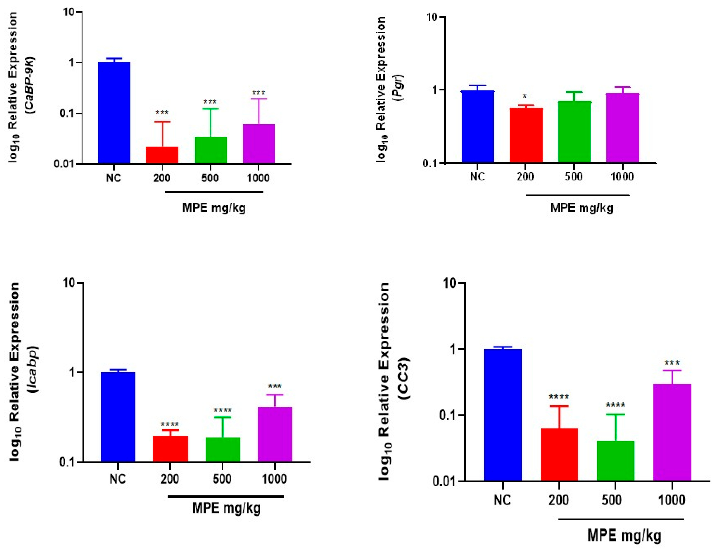
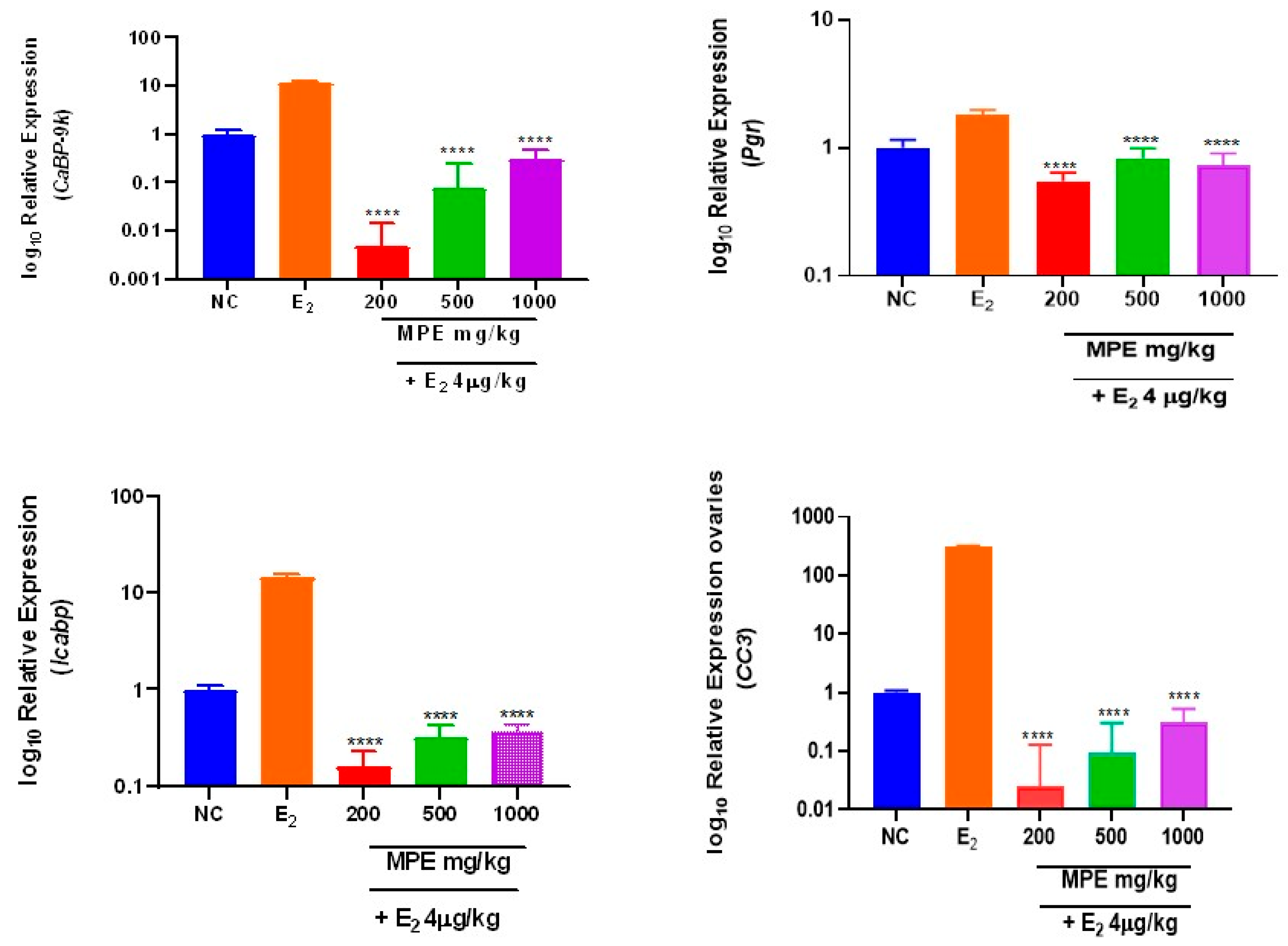
Oral administration of MPE significantly downregulated the relative expression of CaBP-9k in ovarian tissues (p = 0.0003) at all treatment levels compared to the naïve control (Figure 1). The relative expression of Icabp in the ovaries was significantly lower in 200, 500, and 1000 mg/kg at p < 0.0001, p < 0.0001 and p = 0.0005, respectively (Figure 1). The relative expression of Pgr was downregulated significantly only in the 200 mg/kg treatment group (p = 0.0479) compared to the naïve control (Figure 1). The relative expression of CC3 was significantly lower at all treatment levels (200, 500, and 1000 mg/kg) at p < 0.0001, p < 0.0001, and p = 0.0007, respectively (Figure 1). In rats treated with a combination of (200, 500, and 1000) mg/kg MPE and 4 µg/kg E2, the expression of all the estrogen-responsive genes studied reduced significantly (p < 0.0001) compared to E2 treatments alone (Figure 2).

3.3. Effects of MPE and E2 Treatments on Expression Levels of mRNA Transcripts in Uterine Tissues

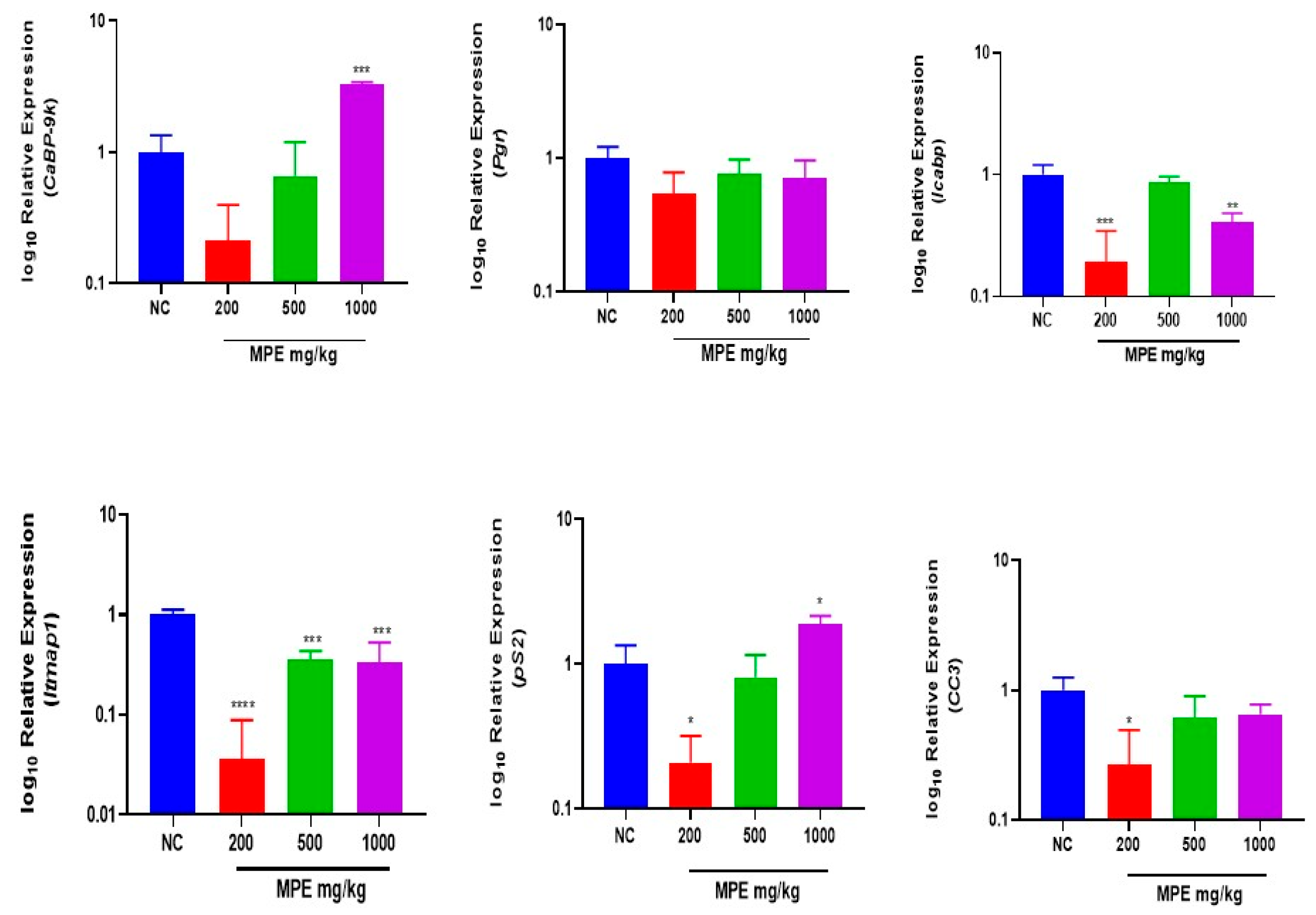
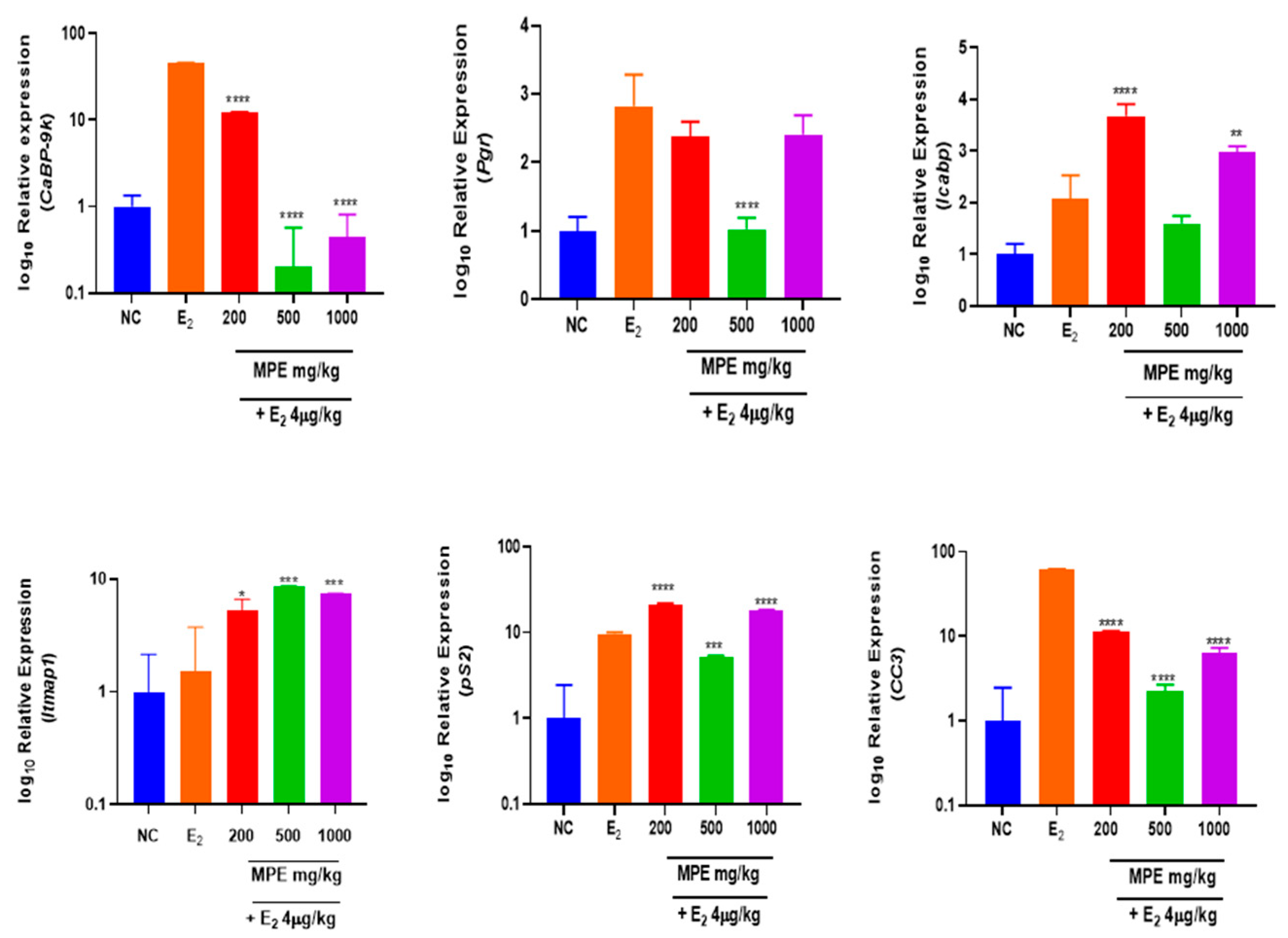
Administration of 1000 mg/kg MPE resulted in a significant increase (p = 0.0001) in CaBP-9k expression in the uterine tissues (Figure 3). However, co-treatment of 1000 mg/kg MPE with E2 significantly reversed the E2-induced and 1000 mg/kg-induced increases in uterine CaBP-9k mRNA expression levels (p < 0.0001) (Figure 4). A combined treatment of 500 mg/kg MPE with estradiol decreased the expression of Pgr compared to the estradiol-treated group at p = 0.0006 (Figure 4). The relative expression of Icabp in the uterus was significantly lower than that of the naïve control in the 200 and 1000 mg/kg MPE treatment groups at p = 0.0003 and p = 0.0027, respectively (Figure 3). However, the combination of the MPE and estradiol increased expression for the 200 and 1000 mg/kg combined treatments at p < 0.0001 and p = 0.0051 (Figure 4). Treatments with 200, 500, and 1000 mg/kg MPE resulted in a relatively low expression of Itmap1 significantly at p = 0.0002, p = 0.003, and p = 0.0023, respectively (Figure 3), but was markedly upregulated in combined treatments at p = 0.0124 for 200 mg/kg, p = 0.0002 for 500 and p = 0.0006 for 1000 mg/kg (Figure 4). The pS2 gene in the uterus of the 200 mg/kg MPE-treated group was downregulated, with relative expressions found to be significantly lower than that of the naïve untreated control group at p = 0.0435. In contrast, 1000 mg/kg was significantly higher (p = 0.0471) (Figure 3). The administration of 200 mg/kg MPE decreased relative expression significantly (p = 0.0129) in CC3 expression compared to the control (Figure 3). A significant decrease (p < 0.0001) in the expression of CC3 was observed in all combined treatment groups when compared to estradiol-treated groups (Figure 4).

4. Discussion

Phytochemical screening in this study has confirmed the presence of some bioactive compounds that have been reported to have a range of different biochemical and physiological effects [30,31]. The presence of tannins, flavonoids, saponins, alkaloids, glycosides, coumarins, triterpenoids, and sterols, as was reported by some studies on the chemical composition of the genus Mentha L. (Lamiaceae), was shown in this study [12].
Mentha plants, including Mentha pulegium, are rich in flavonoids and phenolics, as also reported in this study’s quantitative analysis, with phenolic and flavonoid content showing the highest concentrations. Similar phytochemical profiles have been reported in hydroalcoholic extracts of aerial parts of M. pulegium and related Mentha species, although variations in concentration have been observed among studies [15].
A lyophilized extract obtained from a hydroalcoholic extract of whole aerial flowering parts also contained 13.04% of polyphenols and 5.22% of flavonoids [7]. These differences may be attributed to the extraction solvent, plant part analyzed, the plants’ vegetative phase geographical origin, soil composition, seasonal variation, environmental conditions, and the existence of different chemotypes [8].
The bioactive components from the qualitative and quantitative phytochemical screening in this study have been found to possess anti-ovulatory and anti-fertility properties (phenolics, phytosteroids, and saponins) that affect estrous cycle regulation (Flavonoids, alkaloids, and saponins), conception, and reproduction [32]. In explaining the action or mechanism in which these secondary metabolites act, Younas and others, quoting several researchers from 1998 to 2015, stated that these metabolites specifically modulate molecular targets such as neuroreceptors, ion pumps, and channels, as well as some cytoskeleton and/or enzyme-degrading neurotransmitters [33].
However, the biological effects of these compounds are influenced by factors such as dosage, duration of exposure, route of administration, and endogenous hormonal status. Consequently, variations in phytochemical composition reported across studies may result in differences in pharmacological outcomes, highlighting the need for further studies to characterize the active constituents and clarify their mechanisms of action.
Expressions of genes studied at the transcriptional profiling level were modified (increased or decreased) by exposure to MPE, demonstrating that exposure during prepubertal maturation may change or affect the expression profiles of the uterus and ovaries. Although gene expression in response to estrogens varies by tissue and stages of life and is sex-specific, it is possible to establish transcript profiles indicative of its mechanisms of action. Therefore, all genes understudied may not be directly associated with the uterotrophic and estrogenic responses found in the peripubertal uterus, but may have essential roles in other biological phenomena and are worth noting, which is a fact stated by several studies [34,35,36]. According to the transcriptional profiles of genes in the estradiol (E2) and genes in MPE-treated, which demonstrate different levels of regulation, MPE may have substantially lower estrogenic efficacy than estradiol. As expected, administering E2 to the E2-treated group produced the most sensitive gene expression regulation, also observed in a study by [37]. Estradiol binds to the ER and causes a displacement of some chaperone proteins. The estrogen–ER complexes act as transcription factors by binding to specific ERE sequences in the target genes’ promotes, triggering numerous transcriptional reactions and activities related to female reproduction, a phenomenon stated by [38]. The seven-day intragastric administration of MPE showed modest estrogenic efficacy in vivo, with expression of the study genes comparable to E2. Explaining this activity, the phytoestrogens in MPE could be weakly estrogenic and may have a lower affinity to bind to both ER types. This induces some estrogen-responsive genes and the expression of various estrogenic and antiestrogenic effects [39,40].
M. pulegium extracts, which are known to contain flavonoids, phenolics, alkaloids, and tannins, may underlie and contribute to the observed biological effects.
Gene regulation by MPE in ovaries: The importance of estrogen is efficiently documented in reproductive endocrinology and ovarian function in females [41]. Various reports by [42] has established its critical role in ovarian folliculogenesis. Their research indicates that estrogen receptor beta (Erβ) regulates gene expression before puberty and early postnatal life. Lee et al., in 2021, demonstrated that ERβ is crucial in controlling the ovary’s gonadotropin responses, as well as the fact that a subset of gonadotropin-induced genes necessary for follicular development, oocyte maturation, and ovulation is dependent on Erβ [43]. Dysregulation, therefore, can disrupt folliculogenesis and impair granulosa cell responsiveness to FSH [42]. Administration of E2 has been reported by [44] to jumpstart sexual maturation and the onset of puberty in immature rats despite the absence of gonadotropins, which are known to be essential in the formation and activation of primordial follicles and the early stages of follicle development to the preantral phases, also indicating that ER-dependent neurotropic processes govern gonadotrophin-releasing hormone release [43,45]. Intraovarian actions of estradiol ultimately enhance follicular responsiveness to gonadotropins, resulting in increased aromatase activity and estrogen synthesis, resulting in the modification of ER levels and DNA synthesis [46,47].
Based on this, in elucidating the estrogenic nature of MPE, this study identifies the possible roles that may be significant to the structural and functional development of the ovary in rats, as genes that are understudied may have direct biological estrogen targets. Identifying ER subtypes for these specific estrogen-dependent ovarian genes could also provide more precise mechanisms of ovarian function and novel targets for developing MPE as a fertility regulator.
Compared to the untreated rat ovaries, the notable reductions in the expression of mRNA for all genes in the immature ovaries were dose-related, in which higher doses were associated with increased expression levels. The significant drops in gene expression with the combined treatments show antagonism and may indicate ERβ-mediated action in the ovaries, as evidence indicates that E2 controls primordial and primary follicle growth and folliculogenesis through ERβ in young rats [48]. The effects of E2 exposure on gene transcription were stronger than those of MPE exposure, suggesting that MPE would have a lesser impact on phenotypic, with ERβ-mediated activity in immature rat ovaries.
Progesterone Receptors (Pgr) are essential effectors of ER signaling and mediate progesterone’s physiological effects, which play a central role in reproductive events. Their expression is a prognostic marker of ER action used clinically in breast and reproductive tract malignancies [49]. In contrast to several studies in which immature rats treated with estrogen implants, estradiol, or hypophysectomized estrogen treatments showed no Pgr expression [50,51,52], E2-treated rats in this study showed some expression. Some researchers proposed an indirect function for estrogen, which is required for follicle growth and maintenance, in their ability to express Pgr mRNA as the reason for the subsequent induction of Prgs [53]. Levels of Pgr were also significantly decreased in Letrozole-induced PCOS Aloe vera-treated ovaries [54], as Pgr was downregulated in all MPE doses, with 200 mg/kg showing significance. Downregulation observed with combined treatments may indicate binding to the same receptor sites and some antagonism.
Contrary to this study’s findings, CC3 was found to be among the most upregulated among 450 E-dependent differentially expressed ovarian genes in a study by [55], whose biological functions are highly related to the functional and structural development (organ morphogenesis, complement activation, organ development and regulation of multicellular processes) of the ovary. It was found to have direct biological targets of estrogenic action [55]. However, MPE showed some antagonistic activity, inhibiting E2 action at all doses, which may also indicate ER-targeted action. Calbindin-D9k, a reputable biomarker of potential estrogenic activity [56], is also a cytosolic calcium-binding protein expressed in uterine, placental, and other organ tissues [57]; however, its expression in the ovaries has not been reported. It could have the same role in calcium transport as reported by [58], although this study found some downregulated expressions in the ovaries.
Although this study did not gather the complete information to fully understand how these estrogenic biomarkers act in totality in the immature ovary, the data presented shows that changes in their expressions may be integral to the signal transduction pathway of estrogens and potentially could be used to generate assays to evaluate reproductive toxicities. The base information gathered could be used to develop a testable hypothesis to better understand the molecular pathways associated with their exposure to chemicals with estrogenic properties. Examining ovarian gene expression after MPE exposure provides new insight into the effects of MPE on the immature rat ovary.
Gene regulation by MPE in uterus: Uterine expression of the various genes indicated some estrogenic activity; however, MPE showed differences in its action on the expression of these genes individually. This study may attribute the observed results to the complex regulatory processes influencing gene expression in the endometrium. Several other factors, including menstrual cycle events, hormonal regulation influences, and the dynamic and changing cellular composition of endometrial structure and function, could explain the differences observed in uterine gene expression, as stated by [2,59] in their respective studies. Therefore, in this study, using the whole excised tissue and the different patterns of ER expression and tissue gene expression signature of each cell has played an important role in the response of rat uterus to the phytoestrogen component of MPE and E2, which is also explained by [60] in another study. The selective affinity of some phytoestrogens has been reported [61] and this could also account for the differential gene expression patterns observed in this study. In addition, endogenous estrogens and phytoestrogens may not exert identical effects on uterine gene expression [62].
Eliciting an estrogenic response at the gene expression level also differs with the concentration of phytoestrogens present. For some phytoestrogens, the saturation of receptors by active compounds and higher concentrations may lead to the phenomenon of downregulation. This hormesis-type of function is common to many phytoestrogens, which is due to the saturation of receptors leading to the downregulation of receptors induced by the higher concentration [3]. However, experimental animals may need to be exposed to higher levels of phytoestrogens in developing female rats. Coumarin phytoestrogens, a component of MPE, have been reported to have a weaker affinity to both ERα and ERβ than 17β-estradiol. However, it is stated that their selective affinity for ERβ is greater than ERα, and it could be attributed to the activity of coumarins in MPE [61]. In vivo studies show that some endocrine-disrupting chemicals (EDs) and estrogenic compounds, like E2, significantly increase the expression of uterine Calbindin-D9k and the underlying mechanisms in its expression in the myometrium and stromal cells of the rat uterus are reported to be associated with the direct stimulatory effects of E2 and the indirect inhibitory effects of progesterone [63]. Estradiol, as shown in this study, initiates CaBP-9k expression [64]. Strong enough evidence exists about the induction of uterine CaBP-9k in immature rats by E2 and some EDs via the ERα pathway, not Erβ [65].
In this study, mRNA levels varied and did not change significantly except for 1000 mg/kg MPE. Per the primary function of CaBP-9K in myometrial activity related to intracellular calcium level control [65], its significant expression at 1000 mg/kg MPE could therefore mean efficient regulation of the homeostasis of solutes and water in the uterine epithelial cells [62]. Based on several research findings reported by [27], indicating the in vivo estrogenic potential of methylparaben and ethylparaben, the effect of MPE on gene expression can be likened to that of these parabens that mimic estrogen. Similarly, the expression of CaBP-9k was significantly upregulated by the highest doses of the parabens [27], as observed with 1000 mg/kg MPE. However, the increase was weak compared with the increase induced by E2 treatment, as was reported for long alkyl chain paraben treatments by [63]. A similar significant expression was found in immature rats with Triclosan, an endocrine disruptor with estrogen-like properties. The notable downregulation of CaBP-9k evoked an antagonistic outcome similar to the concurrent exposure to Triclosan and fluoride [66]. Significant CaBP-9K expression by 1000 mg/kg MPE, as seen with octamethylcyclotetrasiloxane (an endocrine disruptor due to its estrogenic properties), suggests that high doses of MPE possess estrogenic properties. However, a similar downregulation of CaBP-9K expression in combined treatments with E2 also occurred in octamethylcyclotetrasiloxane and tetrabromodiphenyl Ether (an endocrine disruptor) treated following ICI 182,780 exposure [64,67], indicating the weak estrogenicity of MPE that Erα may mediate. The ability of MPE (500 and 1000) mg/kg to reduce or reverse CaBP-9K expressional levels in the combined treatments may have resulted from its antagonistic and antiestrogenic effect through ER-dependent pathways in an immature uterus.
Even though Pgr plays a vital role in regulating cell differentiation and proliferation, which are essential for gland development, reproductive proficiency during the reproductive cycle, efficient implantation, and decidualization in pregnancy [48,68,69], MPE cannot be considered as efficacious as E2 in activating Pgr as an ER target gene in the rat uterine tissue. This was confirmed in the combined treatment with E2 and MPE (500 mg/kg), where at this dose level, MPE could either antagonize or block the activation of the Pgr gene by E2. Similarly, this was also found for the lyophilized powder of dried Korean-grown Opuntia ficus indica (L.) Mill fruits [70], which contain high amounts of flavonoids and glycosides, which were also found in M. pulegium extracts [6,11]. The induction of Pgr mRNA may be the result of a classical estrogenic response that could be downregulated due to potential antiestrogenic activity at 500 mg/kg MPE in combination with E2 [64,71] with the effect of 2-Methoxyestradiol, which is an endogenous metabolite of E2 and 17-ethylestradiol. This could result from decreased binding to and signaling through the ERα pathway, which is blocked by antiestrogenic activity.
The pS2 gene encodes a member of the trefoil factor family group, also reported to be a member of the estrogen-inducible gene family. Estrogenic stimulation increases pS2 expression at transcriptional levels [41]. In a similar study using Flemingia strobilia extract, which contains flavonoids and flavonoid glycosides, mRNA expression of pS2 significantly increased after treatment [28]. This was consistent with our observed expression of ps2 in the 1000 mg/kg MPE-treated group. A similar increase in pS2 gene induction was also reported, however, in MCF-7 cells [70]. Increases seen in both combinations of 200 mg/kg and 1000 mg/kg MPE with E2 could mean that MPE at these doses could have either an additive or potentiating effect on pS2 expression levels in the uterine tissue. This further indicates that MPE has weak estrogenic activity and could possibly transactivate ER, as Lupeol, a triterpenoid in Millettia macophylla [3]. As was suggested for Lupeol, MPE might also act through alternate mechanisms, either by inhibiting or activating specific enzymes involved in estrogenic response [3]. The significantly reduced the expression of pS2 at 200 mg/kg MPE, although its interaction with E2 suggests a synergistic effect on pS2 expression in the uterus. Similar findings have been reported regarding the effects of triterpenoids lupenone and lupeol on the growth-regulating estrogen receptor binding-1 gene expression, where an antiestrogenic effect was only seen at low concentrations, while it was synergistic with E2 at higher concentrations [3].
The downregulated expression of Icabp in the uterus with MPE only and an upregulation with MPE combined with estradiol indicate its potentiating or additive effect, but it has some antiestrogenic properties when administered alone. Contradicting these results, all doses of methylparaben and ethylparaben increased the expression of Icabp [27]. Using the uterine tissue to determine the ability of MPE to regulate the transcriptional activity of endogenous ERs through the expression of Itmap1, MPE treatment alone resulted in a downregulation of Itmap1 across all three experimental doses, contrasting a study using parabens [27]. However, in all combined treatments with E2, significant increases in the E2-dependent Itmap1 gene expression further indicate that MPE could be a weak estrogen with a potentiating or additive effect when administered with E2. The gene ontology for integral membranes includes Itmap1, which has been linked to reproduction and was initially identified as a novel zona pellucida protein highly expressed in a mid-to-late gestation mouse uterus only. It may have an important role in late-pregnancy uterine events [72,73]. However, in this study, Itmap1 was expressed in a non-gravid immature rat uterus but was significantly reduced compared to the untreated naïve controls. Nevertheless, its effect in a combined treatment shows that MPE could potentiate the effect of E2 in the immature uterus.
The expression of CC3 at all treatment levels was downregulated at the transcriptional levels in the uterus either significantly (200 mg/kg MPE) or non-significantly (500 and 1000 mg/kg MPE). Based on similarities in phytoestrogen content [74], the effect of MPE on CC3 expression could be compared to the effect of Ficus deltoidea in the uterus. However, studies with Ficus deltoidea upregulated CC3 expression [75], unlike MPE, where CC3 expression levels were dissimilar to those shown by estradiol and the naïve control. Per the role of CC3 in stimulating local inflammatory responses by activating complement pathways and promoting innate immunity, this observation suggests that, in contrast to natural estrogen, MPE may possibly induce some immune suppression in the uterus. Contrary to findings in this study, Triclosan, a phenolic compound which increased CC3 mRNA expression in immature rats was reported to delay vaginal opening and had some reproductive toxicity in rats [76].
Estradiol significantly increased CC3 expression levels, but in co-treatments with MPE, these effects are minimized; therefore, the partially inhibited E2-induced increase in CC3 mRNA levels for all doses indicates possible antagonism and activity via ER-dependent pathways. Similarly to a study that sought to determine the effect of Genistein (a weak estrogen or an anti-estrogen) on CC3 expression in a combination treatment with E2 [77], the analysis of CC3 expression provides an additional piece of evidence of MPE exhibiting some antagonistic properties.
The observed transcriptional changes in estrogen-responsive genes may suggest that MPE exerts a dose-related modulation of estrogen receptor signaling pathways. While MPE alone appears to mimic estrogenic activity by regulating target genes, its ability to modify estradiol-induced transcription in co-treatment conditions indicates potential competitive or antagonistic interaction at the receptor level. Such effects are well-documented for phytoestrogens, which may act as selective modulators exhibiting both estrogenic and antiestrogenic properties depending on the hormonal milieu.

5. Conclusions

This research has demonstrated that MPE may show some estrogenic activity and can induce CaBP-9k, Pgr, pS2, Icabp, Itmap1, and CC3 in the uterus and ovaries; however, MPE showed differences in its action on the expression of these individual genes, with its in vivo action involving ER and ER-mediated pathways. This communicates distinct altered expressions and differential effects in the uterus and ovaries at the transcriptional level. These transcriptional changes may be early indicators of potential endocrine activity rather than definitive proof of physiological outcomes. The observed gene expression changes should be interpreted as indicative trends, and further studies that include and validate multiple reference genes, including 18S rRNA, should be conducted to confirm these findings.
However, regarding the varying mRNA levels of estrogen-related genes in this study, MPE may be involved in disrupting reproductive function through both agonistic and antagonistic mechanisms. It may exhibit endocrine-modulatory activity with multiple mechanisms of action, highlighting its potential complexity in modulating estrogenic responses. These results also provide new insights into the estrogenic effects of MPE at a critical developmental stage of the female reproductive system.

6. Limitations and Recommendations

Not all transcript levels may have been translated into proteins; therefore, further studies are recommended to include the evaluation of protein levels either by Western blotting or immunohistochemistry.
RNA quality assessment in this study relied on NanoDrop spectrophotometry, assessing RNA purity rather than total integrity (RIN values), which is recommended.
Also, future studies should integrate classical uterotrophic endpoints, including uterine weight and histopathological assessment, alongside transcriptional profiling to establish whether the observed molecular alterations translate into physiologically relevant estrogenic effects.

Author Contributions

Conceptualization, L.S.; methodology, L.S., P.W.N., S.A.D. and B.M.D.; data curation, L.S., P.N.C., P.N.A., P.W.N. and S.A.D.; statistical analysis, P.N.C., S.A.D. and L.S.; writing—original draft preparation, L.S., P.N.C. and P.N.A.; writing—review and editing, L.S., S.A.D., C.A.D., K.O.O.-D. and B.M.D.; supervision, C.A.D. and K.O.O.-D.; funding acquisition, L.S. All authors have read and agreed to the published version of the manuscript.

Funding

This research was partially funded by KNUST/Building Stronger Universities III (Grant number: KNUST-BSUIII).

Institutional Review Board Statement

The study was approved by the Animal Research Ethics Committee (AREC) of KNUST (approval number: KNUST0045, date of approval: 23 August 2023).

Informed Consent Statement

Not applicable.

Data Availability Statement

Data will be made available upon request from the authors.

Acknowledgments

We acknowledge the contribution of George Asumeng-Koffour of blessed memory for his role in conceptualization and design as a supervisor, Fulgencios Somkang and the technical staff at the Pharmacology Department, as well as the technical staff of the Department of Clinical Microbiology, whose contribution greatly influenced the completion of this project. Also, we acknowledge the contribution of Wireko Bannor Manasseh and Roberta Y. Asiwu for their technical support.

Conflicts of Interest

The authors declare no conflicts of interest.

Abbreviations

The following abbreviations are used in this manuscript:
CaBP-9kCalbindin-D9k
PgrProgesterone
pS2Trefoil factor 1
IcabpIntestinal Calcium-Binding Protein
Itmap1Integral membrane-associated protein-1
CC3Complement Component 3

References

  1. Hilton, H.N.; Clarke, C.L.; Graham, J.D. Estrogen and progesterone signalling in the normal breast and its implications for cancer development. Mol. Cell. Endocrinol. 2018, 466, 2–14. [Google Scholar] [CrossRef] [Scilit]
  2. Fertuck, K.C.; Eckel, J.E.; Gennings, C.; Zacharewski, T.R. Identification of temporal patterns of gene expression in the uteri of immature, ovariectomized mice following exposure to ethynylestradiol. Physiol. Genom. 2004, 15, 127–141. [Google Scholar] [CrossRef] [Scilit]
  3. Zingue, S.; Ntsa, D.M.; Nde, C.B.M.; Michel, T.; Ndinteh, D.T.; Clyne, C.; Njamen, D. Lupeol, the major compound of the dichloromethane extract of Millettia macrophylla Benth (Fabaceae), displays estrogenic effects in ovariectomized rats. Phytother. Res. 2019, 33, 949–957. [Google Scholar] [CrossRef] [Scilit] [PubMed]
  4. Kim, S.H.; Park, M.J. Effects of phytoestrogen on sexual development. Korean J. Pediatr. 2012, 55, 265–271. [Google Scholar] [CrossRef] [Scilit] [PubMed]
  5. Jawaid, T.; Awasthi, A.; Kamal, M. Estrogenic Activity of a Hydro-Alcoholic Extract of Bambusa Arundinaceae Leaves on Female Wistar Rats. J. Adv. Pharm. Technol. Res. 2015, 6, 19–24. [Google Scholar] [CrossRef] [Scilit] [PubMed]
  6. El-Gazar, A.A.; Emad, A.M.; Ragab, G.M.; Rasheed, D.M. Mentha pulegium L. (Pennyroyal, Lamiaceae) Extracts Impose Abortion or Fetal-Mediated Toxicity in Pregnant Rats; Evidenced by the Modulation of Pregnancy Hormones, MiR-520, MiR-146a, TIMP-1 and MMP-9 Protein Expressions, Inflammatory State, Certain Relate. Toxins 2022, 14, 347. [Google Scholar] [CrossRef] [Scilit] [PubMed]
  7. Teixeira, B.; Marques, A.; Ramos, C.; Batista, I.; Serrano, C.; Matos, O.; Neng, N.R.; Nogueira, J.M.; Saraiva, J.A.; Nunes, M.L. European pennyroyal (Mentha pulegium) from Portugal: Chemical composition of essential oil and antioxidant and antimicrobial properties of extracts and essential oil. Ind. Crops Prod. 2012, 36, 81–87. [Google Scholar] [CrossRef] [Scilit]
  8. Casiglia, S.; Bruno, M.; Fontana, G.; Senatore, F. Chemical composition of the essential oil of Mentha pulegium growing wild in Sicily and its activity on microorganisms affecting historical art crafts. Nat. Prod. Commun. 2017, 12, 1311–1315. [Google Scholar] [CrossRef] [Scilit]
  9. Salem, N.; Sriti, J.; Bachrouch, O.; Msaada, K.; Khammassi, S.; Hammami, M.; Selmi, S.; Boushih, E.; Ouertani, M.; Hachani, N.; et al. Phenological stage effect on phenolic composition and repellent potential of Mentha pulegium against Tribolium castaneum and Lasioderma serricorne. Asian Pac. J. Trop. Biomed. 2018, 8, 207–216. [Google Scholar] [CrossRef] [Scilit]
  10. Mondher, B. Comparative study of the chemical properties and composition of the Mentha pulegium L. of Algerian origin and that of Tunisian origin. Int. J. Agric. Sci. Food Technol. 2020, 6, 30–36. [Google Scholar] [CrossRef] [Scilit]
  11. Sallah, L.; Coffie, P.N.; Babatunde, D.M.; Danquah, C.A.; Baah, F.K.; Mante, P.K.; Owusu-Daaku, K.O. Anti-implantation and uterotonic properties of Mentha pulegium L. in female Sprague-Dawley rats. Int. J. Sci. Rep. 2024, 10, 346–355. [Google Scholar] [CrossRef] [Scilit]
  12. Fatiha, B.; Madani, K.; Chibane, M.; Duez, P. Chemical Composition and Biological Activities of Mentha Species. In Aromatic and Medicinal Plants—Back to Nature; InTechOpen: Incheon, Republic of Korea, 2017; pp. 47–79. [Google Scholar] [CrossRef] [Scilit] [PubMed]
  13. Ouakouak, H.; Chohra, M.; Denane, M. Chemical Composition, Antioxidant Activities of the Essential Oil of Mentha pulegium L, South East of Algeria. Int. Lett. Nat. Sci. 2015, 39, 49–55. [Google Scholar] [CrossRef] [Scilit]
  14. Soukaina, K.; Safa, Z.; Soukaina, H.; Hicham, C.; Bouchra, C. Choline chloride-based deep eutectic solvents (NADES): Potential use as green extraction media for polyphenols from Mentha pulegium, antioxidant activity, and antifungal activity. Microchem. J. 2024, 199, 110174. [Google Scholar] [CrossRef] [Scilit]
  15. Aires, A.; Marrinhas, E.; Carvalho, R.; Dias, C.; Saavedra, M.J. Phytochemical composition and antibacterial activity of hydroalcoholic extracts of Pterospartum tridentatum and Mentha pulegium against staphylococcus aureus isolates. Biomed Res. Int. 2016, 2016, 5201879. [Google Scholar] [CrossRef] [Scilit] [PubMed]
  16. Yusuf, U.; Muhammad, M. Antibacterial Properties of Mentha pulegium. South Asian Res. J. Appl. Med. Sci. 2019, 1, 43–45. [Google Scholar] [CrossRef] [Scilit]
  17. El-Naggar, H.M.; Osman, A.R. Enhancing growth and bioactive metabolites characteristics in Mentha pulegium L. via silicon nanoparticles during in vitro drought stress. BMC Plant Biol. 2024, 24, 657. [Google Scholar] [CrossRef] [Scilit] [PubMed]
  18. Kelidari, H.R.; Alipanah, H.; Roozitalab, G.; Ebrahimi, M.; Osanloo, M. Anticancer effect of solid-lipid nanoparticles containing Mentha longifolia and Mentha pulegium essential oils: In vitro study on human melanoma and breast cancer cell lines. Biointerface Res. Appl. Chem. 2022, 12, 2128–2137. [Google Scholar] [CrossRef] [Scilit]
  19. Gerenutti, M.; Modesto, L.; Carrara, V.A.; Magalhães, S.A.; Paulo, S. Maternal exposure to aqueous extract of Mentha pulegium L. inducing toxicity to embryo development in rats. Afr. J. Pharm. Pharmacol. 2014, 8, 609–614. Available online: https://academicjournals.org/journal/AJPP/article-full-text-pdf/356DC8D45287 (accessed on 2 June 2026).
  20. Assaggaf, H.; El Hachlafi, N.; Elbouzidi, A.; Taibi, M.; Benkhaira, N.; El Kamari, F.; Alnasseri, S.M.; Laaboudi, W.; Bouyahya, A.; Ardianto, C.; et al. Unlocking the combined action of Mentha pulegium L. essential oil and Thym honey: In vitro pharmacological activities, molecular docking, and in vivo anti-inflammatory effect. Heliyon 2024, 10, e31922. [Google Scholar] [CrossRef] [Scilit] [PubMed]
  21. Jennie, A.; Bejads, J.; Srinivas, K. Phytochemical studies and GC-MS analysis of the plant elephantopus scaber Address for correspondence. J. Glob. Trends Pharm. Sci. 2014, 5, 1499–1503. [Google Scholar]
  22. Francesca, I.; Mera, G.; Estefanía, D.; Falconí, G.; Córdova, V.M. Secondary metabolites in plants: Main classes, phytochemical analysis and pharmacological activities. Rev. Bionatura 2019, 4, 1000–1009. [Google Scholar] [CrossRef] [Scilit]
  23. Shaikh, J.R.; Patil, M. Qualitative tests for preliminary phytochemical screening: An overview. Int. J. Chem. Stud. 2020, 8, 603–608. [Google Scholar] [CrossRef] [Scilit]
  24. Wani, J.A.; Achur, R.N.; Nema, R.K. Phytochemical Screening and Aphrodisiac Activity of Asparagus racemosus. Int. J. Pharm. Sci. Drug Res. 2011, 3, 112–115. [Google Scholar] [CrossRef] [Scilit]
  25. Madhu, M.; Sailaja, V.; Satyadev, T.; Satyanarayana, M.V. Quantitative phytochemical analysis of selected medicinal plant species by using various organic solvents. J. Pharmacogn. Phytochem. 2016, 5, 25–29. [Google Scholar] [CrossRef] [Scilit]
  26. Organisation for Economic Cooperation and Development (OECD). Test guideline 425: Acute Oral Toxicity—Up-and-Down Procedure. OECD Guidel. Test. Chem. 2022, 425, 26. [Google Scholar] [CrossRef] [Scilit]
  27. Sun, L.; Yu, T.; Guo, J.; Zhang, Z.; Hu, Y.; Xiao, X.; Sun, Y.; Xiao, H.; Li, J.; Zhu, D.; et al. The Estrogenicity of Methylparaben and Ethylparaben at Doses Close to the Acceptable Daily Intake in Immature Sprague—Dawley Rats; Nature Publishing Group: London, UK, 2016; Volume 6. [Google Scholar] [CrossRef] [Scilit] [PubMed]
  28. Jeong, S.Y.; Chang, M.; Choi, S.-H.; Oh, S.-R.; Wu, H.-H.; Zhu, Y.; Gao, X.-M.; Wang, X.; Zhang, B.; Lim, D.-S.; et al. Estrogenic Effects of Phytoestrogens Derived from Flemingia Strobilifera in MCF-7 Cells and Immature Rats; Pharmaceutical Society of Korea: Seoul, Republic of Korea, 2018; Volume 41. [Google Scholar] [CrossRef] [Scilit] [PubMed]
  29. Naciff, J.M.; Overmann, G.J.; Torontali, S.M.; Carr, G.J.; Tiesman, J.P.; Daston, G.P. Impact of the phytoestrogen content of laboratory animal feed on the gene expression profile of the reproductive system in the immature female rat. Environ. Health Perspect. 2004, 112, 1519–1526. [Google Scholar] [CrossRef] [Scilit] [PubMed]
  30. Yakubu, M.; Adeshina, A.; Oladiji, A.; Ma, A.; Ob, O.; Ga, J.; Awo, O.; Aj, A. Abortifacient Potential of Aqueous Extract of Senna alata Leaves in Rats. J. Reprod. Contracept. 2010, 21, 163–177. [Google Scholar] [CrossRef] [Scilit]
  31. Arora, S.; Saini, M. Gas Chromatography Mass Spectrometry Profiling in Methanolic and Ethyl—Acetate Root and Stem Extract of Corbichonia decumbens (Forssk) Exell from Thar Desert of Rajasthan, India. Pharmacogn. Res. 2017, 9, 48–52. [Google Scholar] [CrossRef] [Scilit] [PubMed]
  32. Khan, M.E.; Williams, E.T.; Abel, A.; Toma, I. Effects of the aqueous and methanolic leaf extracts of Pteridium acquilinum (linnaeus) on some female rats hormones. Direct Res. J. Public Health Environ. Technol. 2016, 1, 8–13. [Google Scholar]
  33. Younas, M.; Hano, C. cancer remedy: Current understanding and future. RSC Adv. 2018, 8, 29714–29744. [Google Scholar] [CrossRef] [Scilit] [PubMed]
  34. Daston, G.P.; Naciff, J.M. Gene expression changes related to growth and differentiation in the fetal and juvenile reproductive system of the female rat: Evaluation of microarray results. Reprod. Toxicol. 2005, 19, 381–394. [Google Scholar] [CrossRef] [Scilit] [PubMed]
  35. Daston, G.P.; Naciff, J.M. Predicting developmental toxicity through toxicogenomics. Birth Defects Res. Part. C—Embryo Today Rev. 2010, 90, 110–117. [Google Scholar] [CrossRef] [Scilit] [PubMed]
  36. Wall, E.H.; Hewitt, S.C.; Case, L.K.; Lin, C.Y.; Korach, K.S.; Teuscher, C. The role of genetics in estrogen responses: A critical piece of an intricate puzzle. FASEB J. 2014, 28, 5042–5054. [Google Scholar] [CrossRef] [Scilit] [PubMed]
  37. Heikaus, S.; Winterhager, E.; Traub, O.; Grümmer, R. Responsiveness of endometrial genes connexin26, connexin43, C3 and clusterin to primary estrogen, selective estrogen receptor modulators, phyto- and xenoestrogens. J. Mol. Endocrinol. 2002, 29, 239–249. [Google Scholar] [CrossRef] [Scilit] [PubMed]
  38. Tang, Z.R.; Zhang, R.; Lian, Z.X.; Deng, S.L.; Yu, K. Estrogen-receptor expression and function in female reproductive disease. Cells 2019, 8, 1123. [Google Scholar] [CrossRef] [Scilit] [PubMed]
  39. Hopert, A.C.; Bayer, A.; Frank, K.; Strunck, E.; Wünsche, W.; Vollmer, G. Characterization of estrogenicity of phytoestrogens in an endometrial-derived experimental model. Environ. Health Perspect. 1998, 106, 581–586. [Google Scholar] [CrossRef] [PubMed]
  40. Basu, P.; Maier, C. Phytoestrogens and breast cancer: In vitro anticancer activities of isoflavones, lignans, coumestans, stilbenes and their analogs and derivatives. Biomed. Pharmacother. 2018, 107, 1648–1666. [Google Scholar] [CrossRef] [Scilit] [PubMed]
  41. Kajita, S.; Suzuki, E.; Saegusa, M. Progesterone receptor expression correlates with estrogen receptor α but not β and pS2 status in ovarian carcinomas: Relation to apoptosis, cell proliferation, p27Kip1 expression, and survival. Kitasato Med. J. 2012, 42, 22–32. Available online: https://www.kitasato-u.ac.jp/ktms/kaishi/pdf/KMJ42-1/KMJ42-1p022-032.pdf (accessed on 2 June 2026).
  42. Zalewski, A.; Cecchini, E.L.; Deroo, B.J. Expression of extracellular matrix components is disrupted in the immature and adult estrogen receptor β-null mouse ovary. PLoS ONE 2012, 7, e29937. [Google Scholar] [CrossRef] [Scilit] [PubMed]
  43. Lee, E.B.; Praveen Chakravarthi, V.; Wolfe, M.W.; Karim Rumi, M.A. ERβ regulation of gonadotropin responses during folliculogenesis. Int. J. Mol. Sci. 2021, 22, 10348. [Google Scholar] [CrossRef] [Scilit] [PubMed]
  44. Rasier, G.; Parent, A.S.; Gérard, A.; Lebrethon, M.C.; Bourguignon, J.P. Early maturation of gonadotropin-releasing hormone secretion and sexual precocity after exposure of infant female rats to estradiol or dichlorodiphenyltrichloroethane. Biol. Reprod. 2007, 77, 734–742. [Google Scholar] [CrossRef] [Scilit] [PubMed]
  45. Mayer, C.; Acosta-Martinez, M.; Dubois, S.L.; Wolfe, A.; Radovick, S.; Boehm, U.; Levine, J.E. Timing and completion of puberty in female mice depend on estrogen receptor α-signaling in kisspeptin neurons. Proc. Natl. Acad. Sci. USA 2010, 107, 22693–22698. [Google Scholar] [CrossRef] [Scilit] [PubMed]
  46. Hewitt, S.C.; Couse, J.F.; Korach, K.S. Estrogen receptor knockout mice: What their phenotypes reveal about mechanisms of estrogen action. Breast Cancer Res. 2000, 2, 345–352. [Google Scholar] [CrossRef] [Scilit] [PubMed]
  47. Xu, X.L.; Huang, Z.Y.; Yu, K.; Li, J.; Fu, X.W.; Deng, S.L. Estrogen Biosynthesis and Signal Transduction in Ovarian Disease. Front. Endocrinol. 2022, 13, 827032. [Google Scholar] [CrossRef] [Scilit] [PubMed]
  48. Chauvin, S.; Cohen-Tannoudji, J.; Guigon, C.J. Estradiol Signaling at the Heart of Folliculogenesis: Its Potential Deregulation in Human Ovarian Pathologies. Int. J. Mol. Sci. 2022, 23, 512. [Google Scholar] [CrossRef] [Scilit] [PubMed]
  49. Diep, C.H.; Ahrendt, H.; Lange, C.A.; Lange, C.A.; Ph, D. Progesterone induces progesterone receptor gene (PGR) expression via rapid activation of protein kinase pathways required for cooperative estrogen receptor alpha (ER) and progesterone receptor (PR) genomic action at ER/PR target genes. Steroids 2016, 114, 48–58. [Google Scholar] [CrossRef] [Scilit] [PubMed]
  50. Ing, N.H.; Tornesi, M.B. Estradiol up-regulates estrogen receptor and progesterone receptor gene expression in specific ovine uterine cells. Biol. Reprod. 1997, 56, 1205–1215. [Google Scholar] [CrossRef] [Scilit] [PubMed]
  51. Peluso, J.J.; Pru, J.K. Non-canonical progesterone signaling in granulosa cell function. Reproduction 2014, 147, 169–178. [Google Scholar] [CrossRef] [Scilit] [PubMed]
  52. Shin, Y.Y.; Kang, E.J.; Jeong, J.S.; Kim, M.J.; Jung, E.; Jeung, E.; An, B. Pregnenolone as a potential candidate for hormone therapy for female reproductive disorders targeting ERβ. Mol. Reprod. Dev. 2019, 86, 109–117. [Google Scholar] [CrossRef] [Scilit] [PubMed]
  53. Park, O.K.; Mayo, K.E. Transient expression of progesterone receptor messenger RNA in ovarian granuiosa cells after the preovulatory luteinizing hormone surge. Mol. Endocrinol. 1991, 5, 967–978. [Google Scholar] [CrossRef] [Scilit] [PubMed]
  54. Dey, A.; Dhadhal, S.; Maharjan, R.; Nagar, P.S.; Nampoothiri, L. Partially purified non-polar phytocomponents from Aloe barbadensis Mill. gel restores metabolic and reproductive comorbidities in letrozole-induced polycystic ovary syndrome rodent model- an “in-vivo” study. J. Ethnopharmacol. 2022, 291, 115161. [Google Scholar] [CrossRef] [Scilit] [PubMed]
  55. Liew, S.H.; Sarraj, M.A.; Drummond, A.E.; Findlay, J.K. Estrogen-Dependent Gene Expression in the Mouse Ovary. PLoS ONE 2011, 6, e14672. [Google Scholar] [CrossRef] [Scilit] [PubMed]
  56. Moon, B.; Kim, W.; Park, C.H.; Oh, S.M. Ginkgo Biloba Extract (EGb761) Did Not Express Estrogenic Activity in an Immature Rat Uterotrophic Assay. Environ. Health Toxicol. 2018, 33, e2018016. [Google Scholar] [CrossRef] [Scilit] [PubMed]
  57. Christakos, S.; Mady, L.J.; Dhawan, P. Calbindin-D28K and Calbindin-D9K and the Epithelial Calcium Channels TRPV5 and TRPV6, 4th ed.; Elsevier Inc.: Amsterdam, The Netherlands, 2018; Volume 1. [Google Scholar] [CrossRef] [Scilit]
  58. Sim, J.Y.; Jung, E.M.; Yoo, Y.M.; Choi, K.C.; Jeung, E.B. Transcriptional and translational expression of calbindin-D9k in the duodenum, kidney and uterus of a female canine model. J. Vet. Sci. 2010, 11, 15–19. [Google Scholar] [CrossRef] [Scilit] [PubMed]
  59. Mortlock, S.; Mckinnon, B.; Montgomery, G.W. Genetic Regulation of Transcription in the Endometrium in Health and Disease. Front. Reprod. Health 2022, 3, 795464. [Google Scholar] [CrossRef] [Scilit] [PubMed]
  60. Nephew, K.P.; Long, X.; Osborne, E.; Burke, K.A.; Ahluwalia, A.; Bigsby, R.M. Effect of estradiol on estrogen receptor expression in rat uterine cell types. Biol. Reprod. 2000, 62, 168–177. [Google Scholar] [CrossRef] [Scilit] [PubMed]
  61. Wang, T.; Wang, Y.; Zhuang, X.; Luan, F.; Zhao, C.; Cordeiro, M.N.D.S. Interaction of coumarin phytoestrogens with ERα and ERβ: A molecular dynamics simulation study. Molecules 2020, 25, 1165. [Google Scholar] [CrossRef] [Scilit] [PubMed]
  62. Heneweer, M.; Houtman, R.; Poortman, J.; Groot, M.; Maliepaard, C.; Peijnenburg, A. Estrogenic effects in the immature rat uterus after dietary exposure to ethinylestradiol and zearalenone using a systems biology approach. Toxicol. Sci. 2007, 99, 303–314. [Google Scholar] [CrossRef] [Scilit] [PubMed]
  63. Yang, H.; Nguyen, T.T.; An, B.S.; Choi, K.C.; Jeung, E.B. Synergistic effects of parabens on the induction of calbindin-D9k gene expression act via a progesterone receptor-mediated pathway in GH3 cells. Hum. Exp. Toxicol. 2012, 31, 134–144. [Google Scholar] [CrossRef] [Scilit] [PubMed]
  64. Lee, J.S.; Kim, Y.K.; Yang, H.; Kang, H.Y.; Ahn, C.; Jeung, E.B. Two faces of the estrogen metabolite 2-methoxyestradiol in vitro and in vivo. Mol. Med. Rep. 2015, 12, 5375–5382. [Google Scholar] [CrossRef] [Scilit] [PubMed]
  65. Choi, K.C.; Leung, P.C.K.; Jeung, E.B. Biology and physiology of Calbindin-D9k in female reproductive tissues: Involvement of steroids and endocrine disruptors. Reprod. Biol. Endocrinol. 2005, 3, 66. [Google Scholar] [CrossRef] [Scilit] [PubMed]
  66. Abd-Elhakim, Y.M.; Mohammed, A.T.; Ali, H.A. Impact of subchronic exposure to triclosan and/or fluoride on estrogenic activity in immature female rats: The expression pattern of calbindin-D9k and estrogen receptor α genes. J. Biochem. Mol. Toxicol. 2018, 32, 6–13. [Google Scholar] [CrossRef] [Scilit] [PubMed]
  67. Dang, V.H.; Choi, K.C.; Jeung, E.B. Tetrabromodiphenyl ether (BDE 47) evokes estrogenicity and calbindin-D9k expression through an estrogen receptor-mediated pathway in the uterus of immature rats. Toxicol. Sci. 2007, 97, 504–511. [Google Scholar] [CrossRef] [Scilit] [PubMed]
  68. Grimm, S.L.; Hartig, S.M.; Edwards, D.P. Review: Progesterone Receptor Signaling Mechanisms AC NU. J. Mol. Biol. 2016, 428, 3831–3849. [Google Scholar] [CrossRef] [Scilit] [PubMed]
  69. Marquardt, R.M.; Kim, T.H.; Shin, J.H.; Jeong, J.W. Progesterone and estrogen signaling in the endometrium: What goes wrong in endometriosis? Int. J. Mol. Sci. 2019, 20, 3822. [Google Scholar] [CrossRef] [Scilit] [PubMed]
  70. An, B.H.; Jeong, H.; Zhou, W.; Liu, X.; Kim, S.; Jang, C.Y.; Kim, H.-S.; Sohn, J.; Park, H.-J.; Sung, N.-H.; et al. Evaluation of the Biological Activity of Opuntia ficus indica as a Tissue- and Estrogen Receptor Subtype-Selective Modulator. Phyther. Res. 2016, 30, 971–980. [Google Scholar] [CrossRef] [Scilit] [PubMed]
  71. Pinto, M.P.; Medina, R.A.; Owen, G.I. 2-Methoxyestradiol and Disorders of Female Reproductive Tissues. Horm. Cancer 2014, 5, 274–283. [Google Scholar] [CrossRef] [Scilit] [PubMed]
  72. Kasik, J.W. A cDNA cloned from pregnant mouse uterus exhibits temporo-spatial expression and predicts a novel protein. Biochem J. 1998, 950, 947–950. [Google Scholar] [CrossRef] [Scilit]
  73. Imamura, T.; Asada, M.; Vogt, S.K.; Rudnick, D.A.; Lowe, M.E.; Muglia, L.J. Protection from pancreatitis by the zymogen granule membrane protein integral membrane-associated protein-1. J. Biol. Chem. 2002, 277, 50725–50733. [Google Scholar] [CrossRef] [Scilit] [PubMed]
  74. Zekri, N.; Amalich, S.; Boughdad, A.; Alaoui El Belghiti, M.; Zair, T. Phytochemical study and insecticidal activity of Mentha pulegium L. oils from Morocco against Sitophilus Oryzae. Mediterr. J. Chem. 2013, 2, 607–619. [Google Scholar] [CrossRef] [Scilit]
  75. Zaid, S.S.M.; Othman, S.; Kassim, N.M. Protective role of Mas Cotek (Ficus deltoidea) against the toxic effects of bisphenol A on morphology and sex steroid receptor expression in the rat uterus. Biomed. Pharmacother. 2021, 140, 111757. [Google Scholar] [CrossRef] [Scilit] [PubMed]
  76. Jung, E.M.; An, B.S.; Yang, H.; Choi, K.C.; Jeung, E.B. Biomarker genes for detecting estrogenic activity of endocrine disruptors via estrogen receptors. Int. J. Environ. Res. Public Health 2012, 9, 698–711. [Google Scholar] [CrossRef] [Scilit] [PubMed]
  77. Diel, P.; Hertrampf, T.; Seibel, J.; Laudenbach-Leschowsky, U.; Kolba, S.; Vollmer, G. Combinatorial effects of the phytoestrogen genistein and of estradiol in uterus and liver of female Wistar rats. J. Steroid Biochem. Mol. Biol. 2006, 102, 60–70. [Google Scholar] [CrossRef] [Scilit] [PubMed]
Figure 1. Relative expressions of CaBP-9k, Pgr, Icabp, and CC3 genes in the ovaries of rats treated with MPE for 7 days. Relative expression normalized using 18s rRNA and expressed as fold change. Each data point represents an independent biological replicate (n = 8 per group), with each sample analyzed in technical triplicate. Values are expressed as log10 mean (ΔΔCT) ± SD, followed by Dunnett’s post hoc test. Values are significantly different from naïve controls at (* p < 0.05), (*** p < 0.001) and (**** p < 0.0001).
Applbiosci 05 00064 g001
Figure 2. Relative expressions of CaBP-9k, Pgr, Icabp, and CC3 genes in the ovaries of rats treated with a combination of MPE and estradiol (E2) for 7 days. Relative expression normalized using 18s rRNA and expressed as fold change. Each data point represents an independent biological replicate (n = 8 per group), with each sample analyzed in technical triplicate. Values are expressed as log10 mean (ΔΔCT) ± SD, followed by Dunnett’s post hoc test (n = 8). Values are significantly different from E2 at (**** p < 0.0001).
Applbiosci 05 00064 g002
Figure 3. Relative expressions of CaBP-9k, Pgr, Icabp, Itmap1, pS2, and CC3 genes in the uterus of rats treated with MPE for 7 days. Relative expression normalized using 18s rRNA and expressed as fold change. Each data point represents an independent biological replicate (n = 8 per group), with each sample analyzed in technical triplicate. Values are expressed as log10 mean (ΔΔCT) ± SD, followed by Dunnett’s post hoc test (n = 8). Values are significantly different from naïve controls at (* p < 0.05), (** p < 0.01), (*** p < 0.001) and (**** p < 0.0001).
Applbiosci 05 00064 g003
Figure 4. Relative expressions of CaBP-9k, Pgr, Icabp, Itmap1, pS2, and CC3 genes in the uterus of rats treated with a combination of MPE and estradiol (E2) for 7 days. Relative expression normalized using 18s rRNA and expressed as fold change. Each data point represents an independent biological replicate (n = 8 per group), with each sample analyzed in technical triplicate. Values are expressed as log10 mean (ΔΔCT) ± SD, followed by Dunnett’s post hoc test (n = 8). Values are significantly different from E2 at (* p < 0.05), (** p < 0.01), (*** p < 0.001), (**** p < 0.0001).
Applbiosci 05 00064 g004
Table 1. Preliminary qualitative phytochemical screening of the dried leaves of M. pulegium and the hydroethanolic leaf extract of M. pulegium (MPE).
PhytoconstituentsTest (Reactants)Observation If PositiveReference
Tannins Lead Acetate Test (Lead acetate solution)White precipitate[21]
FlavonoidsShinoda Test (95% ethanol, HCL and magnessium turnings)Pink coloration[21]
Saponins Froth test (Water)Formation of 1 cm layer of foam[22]
Alkaloids Dragendorff’s test (Solution of Potassium bismuth iodide) and
Mayer’s test (Potassium mercuric iodide)
A red precipitate
A yellow precipitate
[22]
Glycosides Keller–Killiani Test (Glacial acetic acid, 5% FeCl3, Suphuric acid)Reddish brown color at junction of two liquids and upper layer turned bluish green)[21]
Coumarins NaOH test (Plant extract + 10% NaOH + Chloroform) Yellow color appearance [23]
Triterpenoids Thionyl chloride solution (Thionyl chloride)Formation of pink color[24]
Sterols Libermann Burchard test (Chloroform, acetic anhydride, concentrated sulphuric acid)Golden Yellow appearance[22]
Table 2. Groups and the various treatments for experimental animals showing doses of a hydroethanolic leaf extract of Mentha pulegium (MPE) and Estradiol.
GroupTreatmentNumber (N)
Group 1 (Naïve Control)Distilled water8
Group 2 (MPE Low Dose)200 mg/kg MPE8
Group 3 (MPE Medium Dose)500 mg/kg MPE8
Group 4 (MPE High dose)1000 mg/kg MPE8
Group 5 (Estradiol) 4 μg/kg 17-β-Estradiol8
Group 6 (MPE + Estradiol)200 mg/kg MPE + 4 μg/kg 17-β-Estradiol8
Group 7 (MPE + Estradiol)500 mg/kg MPE + 4 μg/kg 17-β-Estradiol8
Group 8 (MPE + Estradiol)1000 mg/kg MPE + 4 μg/kg 17-β-Estradiol8
Table 3. List of reverse (R) and forward (F) primers used for amplifying estrogen-responsive genes in rat uteri and ovaries for qPCR analysis.
Gene Primers: Sequences of Primers; from 5′ to 3′References Organ
CaBP-9kF 5′-AAGAGCATTTTTCAAAAATA-3′
R 5′-GTCTCAGAATTTGCTTTATT-3′
[27]Uterus and ovary
PgrF 5′-GATGGAAGGGCAGCATAACTATTT-3′
R 5′-ACAGCACTTTCTCAGACGACATG-3′
[27]Uterus and ovary
pS2F 5′-GGAAAAGGGTTGCTGTTTTG-3′
R 5′-ACAGGTGTGTATGAAGCAGGTG-3′
[28]uterus
IcabpF 5′-CTGGATAAGAACGATGATGGAGAA-3′
R 5′-GGTGGTGTCGGAGCTCCTT-3′
[27]uterus and ovary
Itmap1F 5′CTATTTCTTTTCCTCTGGTACCACTATTC-3′
R 5′-AGGGTGTGGCCTTGGATAATT-3′
[27]uterus
CC3F 5′-CGTGAGCAGCACAGAAGAGA-3′
R 5′-CCAGGTGGTGATGGAATCTT-3′
[29]uterus and ovary
Table 4. Preliminary phytochemical screening of Mentha pulegium.
ConstituentHydroethanolic Extract (MPE)Coarse Powdered Leaves
Tannins++
Flavonoids++
Saponins++
Alkaloids++
Glycosides++
Coumarins++
Triterpenoids++
Sterols++
+ indicates the presence of phytochemical constituents.
Table 5. Quantitative analysis of some phytochemical constituents in a hydroethanolic leaf extract of M. pulegium.
TestAmount Unit
Alkaloids0.89 ± 0.1mg ATE/g DW
Total Phenolics294.6 ± 12.10mg CE/g DW
Flavonoid112.5 ± 2.43mg CE/g DE
Steroids0.68 ± 0.04mg CE/g DW
Values are represented as mean ± SD (n = 3).
Disclaimer/Publisher’s Note: The statements, opinions and data contained in all publications are solely those of the individual author(s) and contributor(s) and not of MDPI and/or the editor(s). MDPI and/or the editor(s) disclaim responsibility for any injury to people or property resulting from any ideas, methods, instructions or products referred to in the content.