Article

Hydroxyapatite–Magnesium Bioceramics: Synthesis and Mechanical–Chemical Characterization

by
Elizabeth Refugio-García
1,
Zaira Itzel Bedolla-Valdez
2,
Alfredo Emiliano Chávez-Pantiga
1,
Gerardo Vázquez-Huerta
1,
José Guadalupe Miranda-Hernández
3,
Carlos Adrián Calles-Arriaga
4,
José Amparo Rodríguez-García
4 and
Enrique Rocha-Rangel
4,*
1
Materials Department, Universidad Autónoma Metropolitana, Av. San Pablo 180, Col. Reynosa-Tamaulipas, Ciudad de Mexico 02200, Mexico
2
Advanced Materials Division, Instituto Potosino de Investigación Científica y Tecnológica, Camino a la Presa de San José 2055, Col. Lomas 4a Sección, San Luis Potosi 78216, Mexico
3
Industrial Engineering Department, Centro Universitario UAEM Valle de México, Atizapan de Zaragoza 54500, Mexico
4
Research Department, Universidad Politécnica de Victoria, Parque Científico y Tecnológico de Tamaulipas, Av. Nuevas Tecnologías 5902, Ciudad Victoria 87138, Mexico
*
Author to whom correspondence should be addressed.
Appl. Biosci. 2026, 5(3), 61; https://doi.org/10.3390/applbiosci5030061
Submission received: 20 January 2026 / Revised: 23 June 2026 / Accepted: 10 July 2026 / Published: 15 July 2026
(This article belongs to the Topic Advances in Biomaterials—2nd Edition)

Abstract

Hydroxyapatite (HA) is widely used in biomedical applications due to its biocompatibility and chemical similarity to the mineral phase of bone; however, its low mechanical strength limits its structural use. In this work, HA ceramics with different Mg additions (0, 0.5, 1, 3, 5, and 10% by weight) were prepared using the powder processing technique. The mixtures were homogenized, conformed and sintered at 1100 °C. The incorporation of intermediate Mg concentrations produced an increase in fracture toughness compared to pure HA. The best mechanical performance was obtained with the formulation containing 5% Mg by weight, achieving a hardness of 319 HV, a porosity of 12.92% and a fracture toughness of 4.06 MPa·m0.5, comparable to those reported for human cortical bone, indicating its potential for applications in moderately loaded bone implants. The findings indicate that magnesium functions as a reinforcing component in the ceramic matrix, mitigating critical defects and thereby contributing to the improved toughness of Mg-containing hydroxyapatite ceramics. The polarization resistance results show that the incorporation of low fractions by weight of magnesium (1% Mg) adjusts the electrochemical behavior of the material, while higher increases in its concentration cause a deterioration of this property.

1. Introduction

Hydroxyapatite (HA), Ca10(PO4)6(OH)2, is a calcium phosphate biomaterial extensively used in orthopedic and dental applications due to its chemical similarity to the mineral phase of bone, excellent biocompatibility and osteoconductivity [1,2,3]. However, the clinical use of monolithic HA remains limited in load-bearing applications because of its intrinsic brittleness, low fracture toughness and poor fatigue resistance [4]. Therefore, improving the mechanical and functional performance of HA while preserving its biological properties remains an important challenge in calcium phosphate biomaterials research.
Two main strategies have been explored to overcome these limitations: reinforcement through composite formation and physicochemical modification by ionic substitution within the HA lattice. HA-based composites reinforced with metals, oxides or bioactive phases have shown improvements in mechanical behavior and bioactivity [5,6,7]. Alternatively, ionic substitution has been investigated as a route to modify crystallinity, solubility and biological response while maintaining the apatite structure [8,9].
Among the biologically relevant ions, magnesium (Mg2+) has attracted particular interest because it is naturally present in bone tissue and is associated with osteogenesis and bone remodeling processes [10,11,12]. Previous studies have reported that Mg incorporation into HA may influence crystal size, crystallinity and dissolution behavior [13,14]. Due to the smaller ionic radius of Mg2+ compared with Ca2+, several authors have proposed that magnesium incorporation can affect the HA crystal structure [15]. Consequently, Mg-containing HA materials have been synthesized using different processing routes, including precipitation, sol–gel and hydrothermal methods [13,16].
In addition to chemical synthesis routes, magnesium-containing HA materials can also be produced by powder mixing and thermal processing using metallic magnesium additions. Depending on processing conditions, these systems may contain multiple phases, including HA, magnesium oxide and residual magnesium. However, the distinction between true lattice substitution and composite-type phase formation remains unclear in several reported studies [17,18,19]. As a result, the relationship between magnesium addition, phase evolution and functional properties in powder-processed HA systems is still not fully understood.
The present study investigates HA-magnesium mixtures with controlled Mg additions processed by powder metallurgy. The work focuses on the evaluation of phase constitution, microstructure, mechanical behavior and electrochemical response as a function of magnesium content. The working hypothesis is that increasing Mg addition modifies the phase composition and microstructural characteristics of the system, which in turn influences its mechanical and electrochemical behavior. This study searches to enhance the understanding of the mechanical and electrochemical properties of magnesium-containing hydroxyapatite (HA) materials fabricated through solid-state processing routes.

2. Methodology

The powders used for the preparation of the biomaterials were hydroxyapatite and magnesium (particle size: 5–10 μm, purity: 99.9% (Sigma-Aldrich, Naucalpan, Estado de México, México). The studied chemical compositions are listed in Table 1. The Mg/Ca molar ratios corresponding to the nominal Mg contents were estimated by converting the weight percentage of Mg into molar amounts using the atomic mass of Mg (24.305 g·mol−1). The remaining cationic fraction was assumed to correspond to Ca within the hydroxyapatite structure (Ca10(PO4)6(OH)2). This approach allows an approximate evaluation of the level of Mg substitution in the HAp lattice, although the possible formation of secondary Mg-containing phases at higher doping levels should not be excluded. The ratio Mg/Ca for each composition is also shown in Table 1. The ratio Ca/P is 1.67.
The initial hydroxyapatite–magnesium powder mixtures were ground in a ball mill (Lab Mill-8000, Gilson Company, Inc., Lewis Center, OH, USA) at 400 rpm for 9 h. Zirconia milling media with a diameter of 3 mm were used. The milling process was performed under dry conditions, with the addition of 1 mL of isopropyl alcohol as a process control agent. A powder-to-ball weight ratio of 1:20 was employed. The milled powders were subsequently compacted into cylindrical pellets by uniaxial pressing using a hardened steel die under an applied pressure of 200 MPa. The cylindrical samples measured 1 cm in diameter and 1 cm in height. The pellets were sintered in an electric furnace (VHT 8, Nabertherm, GmbH, Lilienthal, Germany) at 1000 °C with a dwell time of 1 h. The sintering process was conducted under vacuum, using a heating rate of 10 °C/min. After completion of the thermal cycle, the furnace was switched off and the samples were allowed to cool naturally inside the furnace. After sintering, the physical characterization of the composites was performed. (For the study, five samples of each composition were prepared; the results presented here are the average of all of them.) The density was determined according to Archimedes’ principle, following the ASTM B962-15 standard [20]. Fracture toughness was evaluated using the indentation fracture method [21], applying the equation proposed by Evans [22]. Microhardness measurements were carried out in accordance with the ASTM E384-16 standard [23]. For this analysis, twelve indentations were performed at different locations on each sample, and the reported values correspond to the average of these measurements. Microhardness tests were conducted using a microhardness tester (Emco-Test, DuraScan 200, QMN Equipment Inc., Mississauga, ON, Canada), using a force of 9.8 newtons for 15 s of penetration. The crystalline structure was analyzed by X-ray diffraction (XRD) using a SmartLab RIGAKU (STEM-LE-1086-LC, STEMART, Shirley, NY, USA) powder diffractometer equipped with Cu Kα radiation (λ = 1.5406 Å), scanning over a 2θ range of 20–80°. The XRD patterns of the alloys were analyzed using X’Pert HighScore Plus (v2.2b) software. The surface morphology, elemental composition, and spatial distribution of elements were investigated by energy-dispersive X-ray spectroscopy (EDS) coupled to a FEI ESEM Quanta FEG-250 scanning electron microscope (Thermo Fisher, Waltham, MA USA). Electron micrographs were acquired using the same instrument at magnifications between 500× and 2000×. All analyses were performed at an accelerating voltage of 20 kV, employing a backscattered electron (BSE) detector under low vacuum. Prior to characterization, all samples were affixed onto aluminum stubs using conductive carbon. Prior to observation, the samples were ground and polished using standard metallographic procedures to obtain a flat and reflective surface. For the degradation resistance assessment, the samples were immersed in a physiological solution with an isotonic composition of 0.9% sodium chloride (NaCl) for 30 min prior to testing in order to ensure surface saturation. Electrochemical measurements were carried out using a potentiostat–galvanostat (IM7X, Zahner-Elektrik GmbH & Co., Kronach, Germany) in a conventional three-electrode cell configuration. The HA–Mg composites served as the working electrodes (WE), an Ag/AgCl electrode in 3 M NaCl was used as the reference electrode (RE), and a graphite rod acted as the counter electrode (CE). Electrochemical impedance spectroscopy (EIS) was employed to evaluate the polarization resistance (Rp), providing insight into the degradation behavior of the samples. The EIS measurements were per-formed by applying a sinusoidal perturbation of ±10 mV over a frequency range from 100 kHz to 100 mHz. Prior to each measurement, the system was allowed to reach a stable open-circuit potential.

3. Results

3.1. Density and Porosity

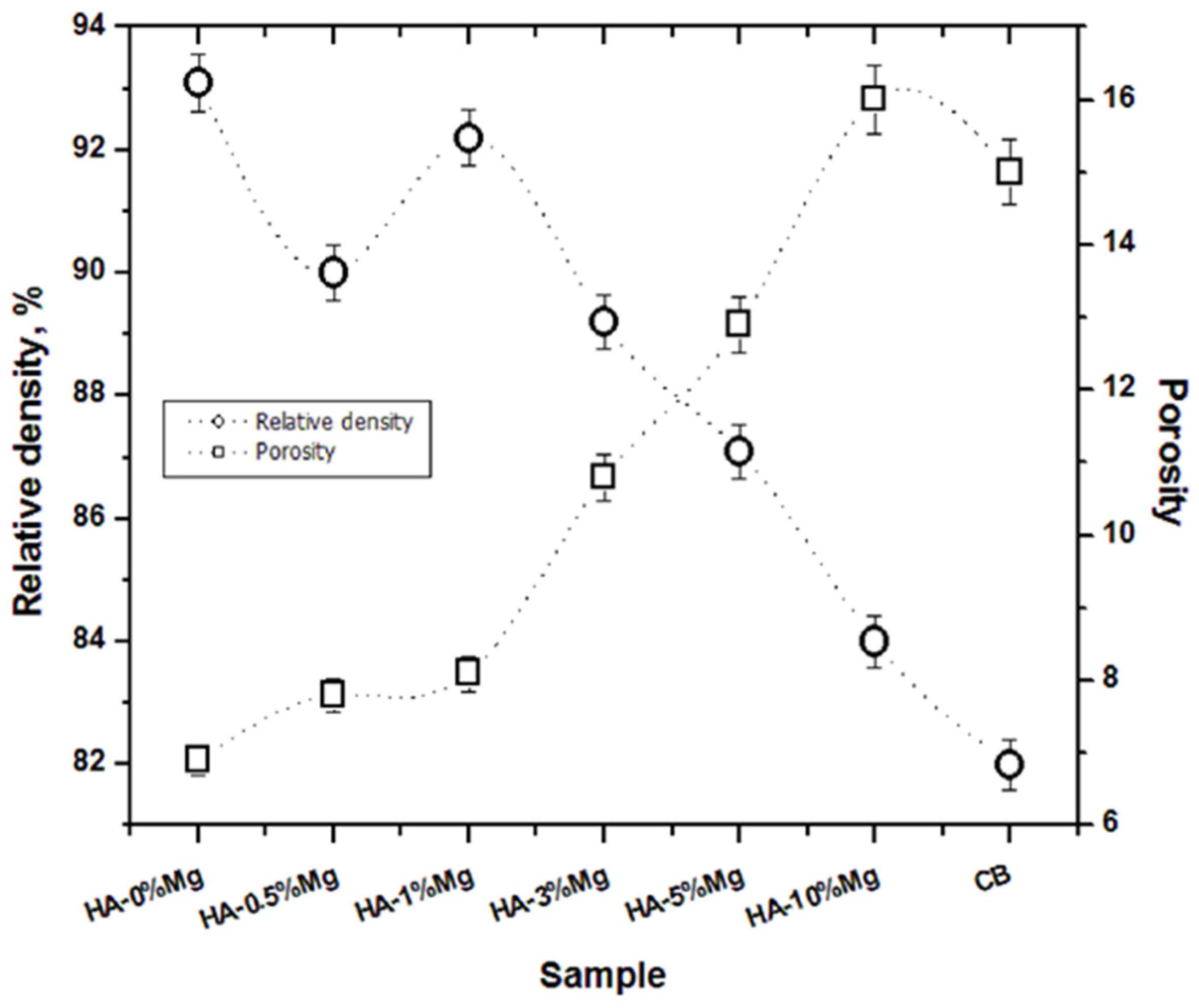
Figure 1 presents the relative density and porosity of the sintered ceramics as a function of magnesium content, with the corresponding error bars for each studied composition. An inverse relationship is observed, with increasing magnesium content leading to a decrease in relative density and a corresponding increase in porosity, indicating a reduction in densification during sintering. Although high density is generally associated with improved mechanical properties, a controlled level of porosity is desirable for biomedical applications because it can promote cell infiltration, vascularization, and bone ingrowth. It should be noted that these biological benefits depend not only on the overall porosity but also on pore size, interconnectivity, and scaffold architecture, which play a key role in osseointegration and tissue regeneration [24,25]. However, these structural features were not evaluated in the present study and are beyond its scope. Cortical bone (CB) exhibits porosity values of approximately 15%, which are slightly higher than those obtained for ceramics containing 3, 5, and 10% magnesium. This similarity suggests that the developed ceramics possess porosity levels compatible with bone tissue, potentially favoring bone ingrowth and reducing stress-shielding effects. Therefore, despite the decrease in relative density with increasing magnesium content, the resulting porosity, particularly at lower magnesium concentrations, is considered suitable for the intended biomedical application. The values for cortical bone density and porosity were included in the figure solely for comparative purposes.

3.2. Crystalline Structure

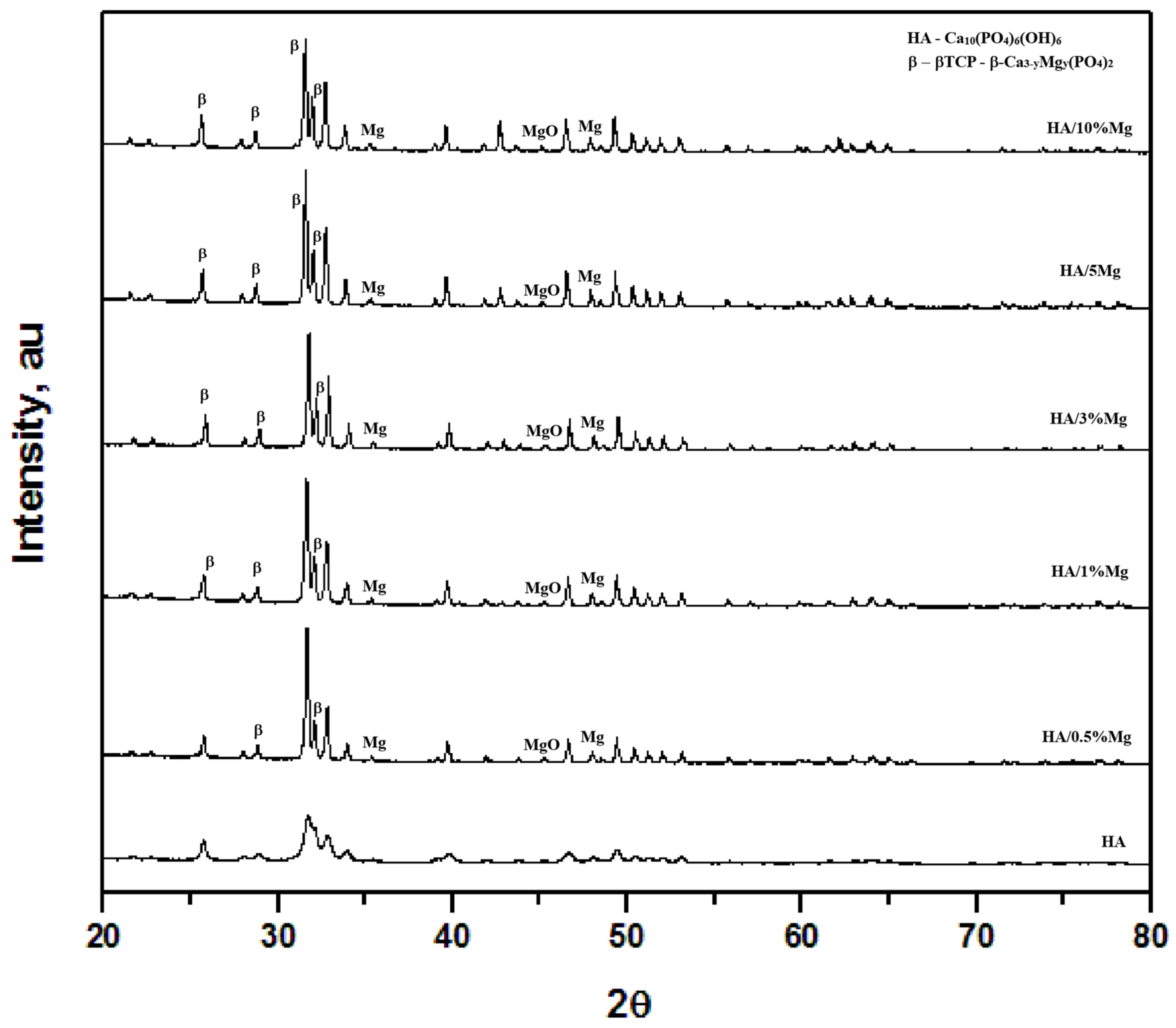
Figure 2 presents the X-ray diffraction (XRD) patterns of Mg-containing hydroxyapatite samples sintered at 1000 °C, allowing evaluation of the phase evolution associated with magnesium addition. The diffraction pattern of pure HA exhibits sharp and well-defined peaks corresponding predominantly to hydroxyapatite, indicating high crystallinity after sintering. As Mg content increases, additional diffraction peaks become evident. Peaks located at 2θ values of approximately 36.6° and 47.8° can be associated with the (101) and (102) planes of metallic Mg, suggesting that part of the added magnesium remains as a secondary phase after processing. Likewise, the low-intensity peak observed near 45° may correspond to MgO formation, likely related to partial oxidation during sintering at high temperatures.
The XRD patterns also reveal the progressive appearance of secondary calcium phosphate phases with increasing Mg addition. Diffraction peaks located at 2θ values near 26.4°, 28.2°, 33.6° and 35.3° are consistent with β-tricalcium phosphate (β-TCP), indicating partial HA decomposition during thermal treatment. Similar phase evolution has been previously reported in Mg-containing calcium phosphate systems processed at temperatures between 700 and 1200 °C [26,27,28]. In this system, Mg acts as a destabilizing agent for HA, promoting its conversion into more thermodynamically stable and soluble calcium phosphate phases, as described by Reaction (1).
Ca 10 P O 4 6 O H 2   Mg 2 + ,   T 3   β -C a 3 P O 4 2   +   CaO   +   H 2 O
Although magnesium incorporation into the HA structure has been discussed in previous studies due to the smaller ionic radius of Mg2+ relative to Ca2+, the present results do not allow definitive confirmation of lattice substitution. Therefore, the XRD and EDS analyses are interpreted only as evidence of magnesium incorporation within the material system and associated phase evolution. Additional high-resolution structural or spectroscopic techniques would be required to conclusively determine the extent of substitutional incorporation. Furthermore, a slight reduction in peak intensity and broadening of some reflections can be qualitatively observed as Mg content increases, which may be associated with reduced crystallinity and inhibited grain growth during sintering. Similar behavior has been reported in Mg-containing calcium phosphate ceramics and is commonly related to microstructural refinement effects [29]. For pure hydroxyapatite using the peak located in 2 theta angles of 31.8 which corresponds to the (211) plane of hydroxyapatite, and the Scherrer formula, the crystallite size was estimated to be 252 nm. The lattice parameters were calculated as a = b = 9.421 Å, c = 6.892 Å. On the other hand, for the sample 5% Mg-modified hydroxyapatite and using the same peak in 2 theta angles of 31.8, the crystallite size was estimated to be 231 nm, whereas the lattice parameters were calculated as a = b = 9.413 Å, c = 6.812 Å. The incorporation of Mg2+ into the hydroxyapatite structure induces a slight contraction of the unit cell parameters, as evidenced by the decrease in both a and c lattice constants. This behavior can be attributed to the substitution of Ca2+ ions by the smaller Mg2+ ions, which generates local lattice distortions and reduces the average interatomic distances within the crystal structure. Additionally, the observed decrease in crystallite size suggests that Mg2+ hinders crystal growth during sintering, increasing structural disorder and reducing crystallinity.
In a general way, the XRD results demonstrate that magnesium addition modifies the phase composition of the HA-based system, promoting the formation of secondary phases such as β-TCP and Mg-containing compounds. These structural changes are expected to influence the microstructural and functional behavior of the materials, which is further discussed in the following sections.

3.3. Microstructure

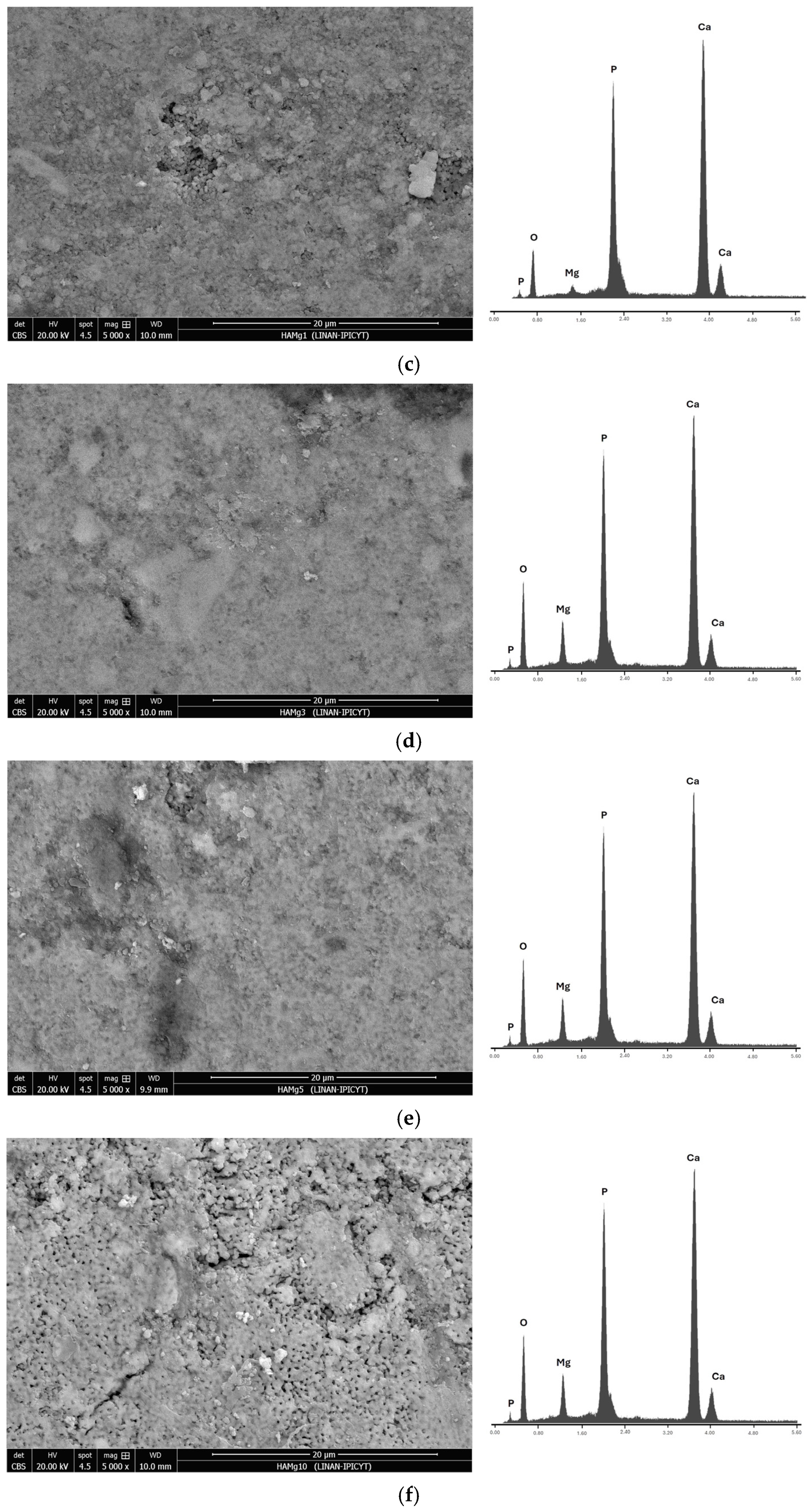
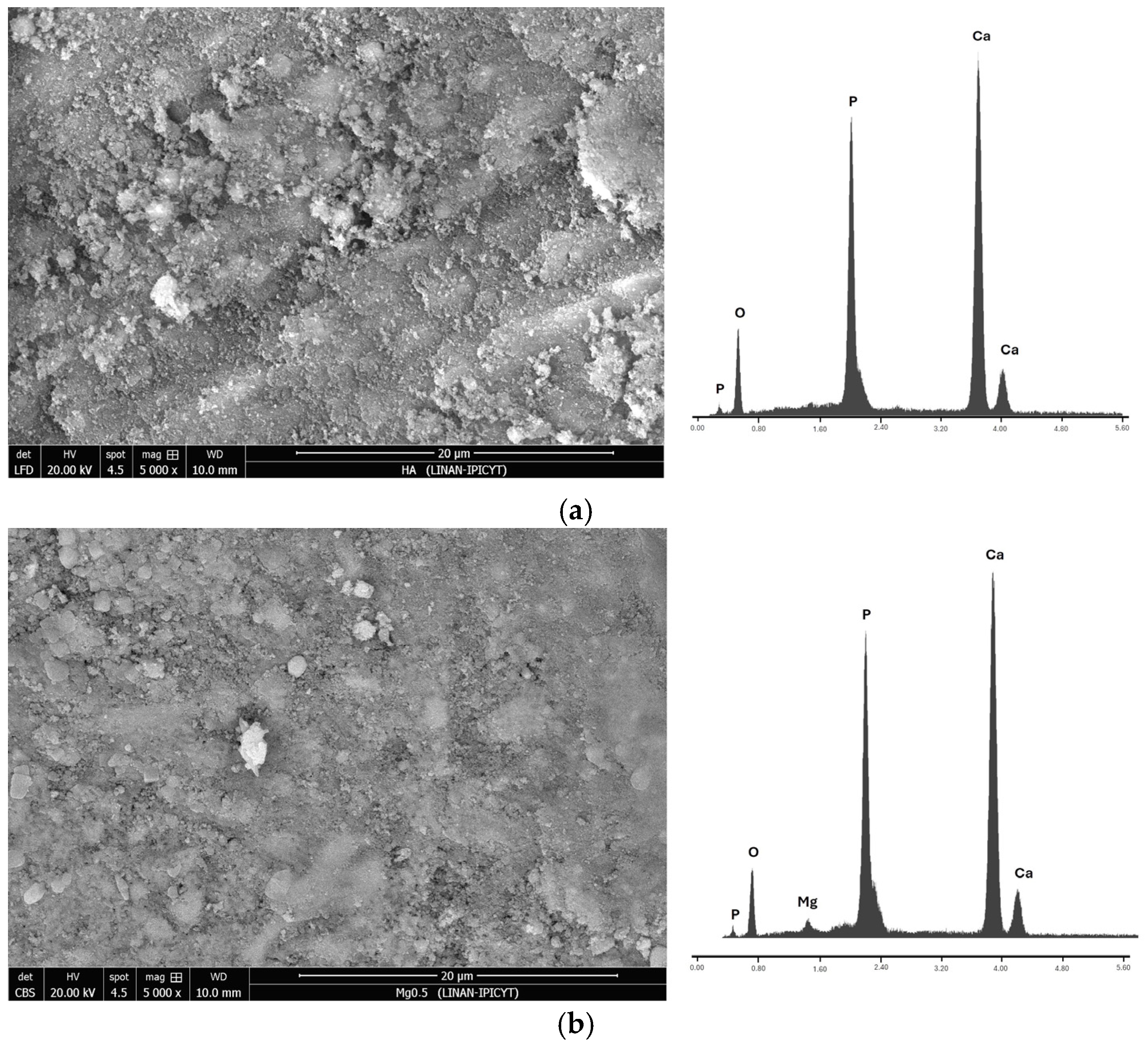
Figure 3 presents the microstructures of the Mg-containing hydroxyapatite ceramics observed by scanning electron microscopy (SEM). To identify the chemical elements present in the samples, energy-dispersive X-ray spectroscopy (EDS) analyses were also performed, and the corresponding spectra are also included in Figure 3.
The SEM micrographs reveal agglomerated particles with rough and irregular morphologies, which are characteristic of ceramics produced by powder-processing routes. Although the microstructure appears relatively homogeneous at the micrometer scale, the SEM resolution does not allow a precise evaluation of grain refinement or grain boundary features. Likewise, no evident large-scale segregation is observed; however, the presence of minor secondary phases detected by XRD cannot be excluded from SEM observations due to their low concentration and limited spatial resolution.
The EDS spectra confirm the presence of the main constituent elements of hydroxyapatite, namely Ca, P, and O, together with weaker Mg signals associated with Mg incorporation into the material. Since the EDS analysis is mainly qualitative, the results only suggest the presence of Mg within the samples and do not conclusively demonstrate a fully homogeneous elemental distribution at the microscale.
As the Mg content increases, slight morphological changes can be observed, including a tendency toward smaller and more compact agglomerates. This behavior may be related to the presence of Mg during the sintering process, which could influence diffusion and grain growth mechanisms. Although Mg2+ has a smaller ionic radius than Ca2+ and has been reported to substitute for Ca2+ in hydroxyapatite, the EDS analysis performed in this study only confirms the presence of Mg and does not provide direct evidence of crystallographic substitution or homogeneous incorporation within the HA lattice. Therefore, any potential effect of Mg on crystal growth should be interpreted cautiously. Furthermore, these observations are limited by the resolution of the SEM analysis. In addition, samples with higher Mg contents exhibit a slightly more porous appearance, which may be related to the inhibitory effect of Mg on densification during thermal treatment. Despite this, the porosity distribution remains relatively uniform, without evidence of critical macroscopic defects.
These observations are consistent with the XRD analysis, which indicates that Mg incorporation occurs mainly within the hydroxyapatite structure, while also revealing the presence of minor secondary phases such as MgO and β-TCP at higher Mg contents. Therefore, the combined SEM, EDS, and XRD results suggest that Mg affects both the structural and morphological evolution of hydroxyapatite, although additional high-resolution analyses would be necessary to fully confirm elemental homogeneity and grain boundary effects.

3.4. Mapping

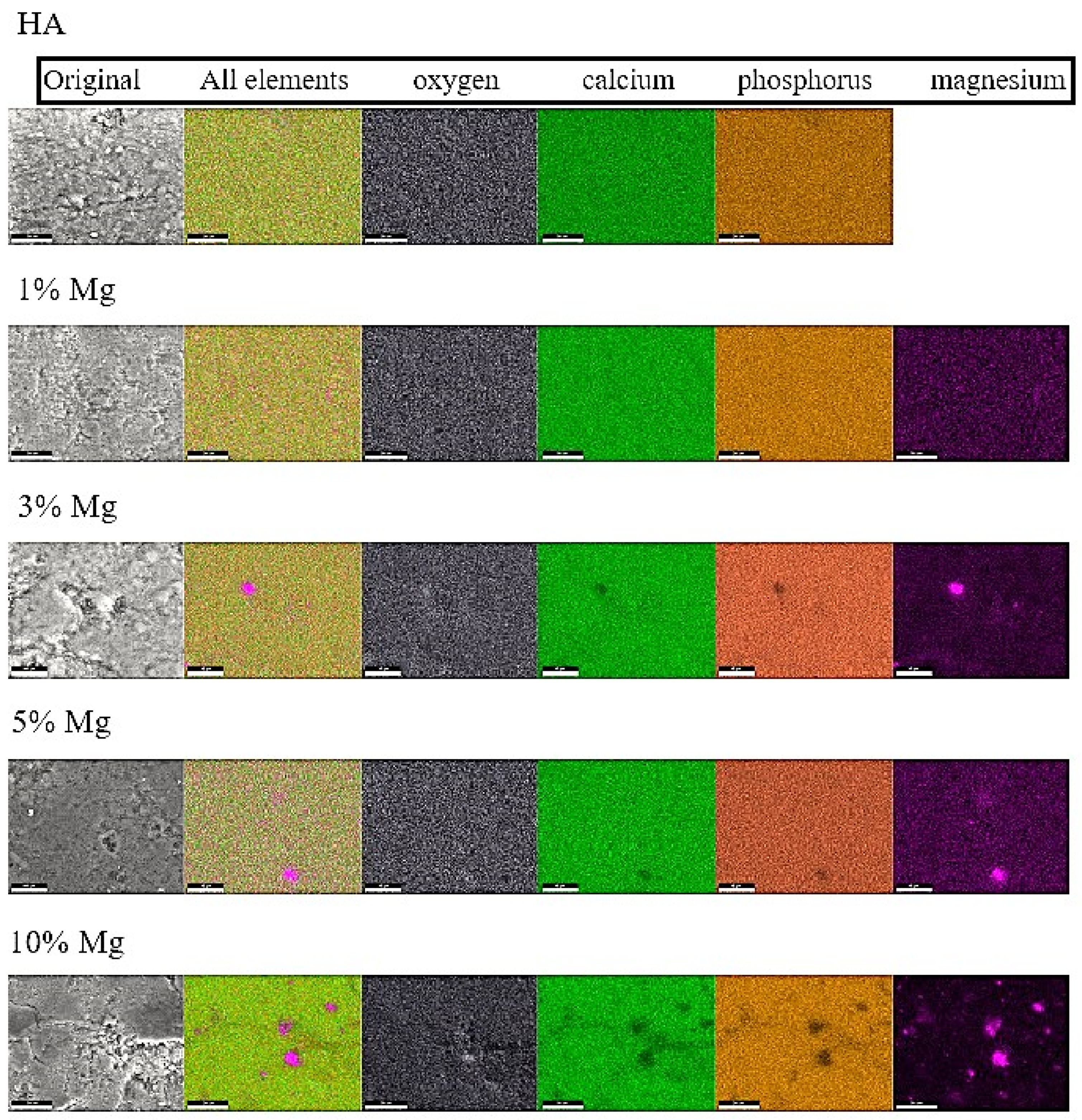
Figure 4 presents the elemental mapping results of Mg-containing hydroxyapatite ceramics, showing the spatial distribution of the characteristic elements of the apatite matrix (Ca, P and O), which confirms the preservation of the material composition after the doping process. In addition, the presence of Mg signals in the doped samples demonstrates that magnesium is successfully incorporated into the material.
At low and moderate Mg concentrations, the elemental maps reveal a relatively homogeneous distribution of Mg throughout the analyzed regions, suggesting an adequate dispersion of the dopant without evident large Mg-rich agglomerates. These observations are consistent with the SEM micrographs, where no pronounced microstructural contrasts associated with segregated regions are observed, and with the XRD results, which show limited evidence of secondary phases at low Mg contents.
It should be noted that elemental mapping only provides information regarding the spatial distribution of the elements and does not directly confirm crystallographic substitution within the hydroxyapatite lattice. Therefore, the possible incorporation of Mg2+ ions through partial substitution of Ca2+ sites is proposed based on the combined interpretation of XRD results, phase evolution, and previous reports in the literature. Such substitution may induce local lattice distortions due to the smaller ionic radius of Mg2+ compared with Ca2+, which could contribute to the formation of β-TCP-related phases at higher Mg concentrations.
As the Mg content increases, the elemental maps exhibit regions with less homogeneous Mg distribution, indicating the onset of localized enrichment or segregation phenomena. This behavior suggests that the solubility limit of Mg in the hydroxyapatite structure may be exceeded at high doping levels, favoring the formation of Mg-rich regions or secondary phases, in agreement with the structural changes observed by XRD. Although the mapping resolution does not allow a definitive identification of segregated crystalline phases, the results qualitatively support the trend toward increasing heterogeneity at elevated Mg concentrations.
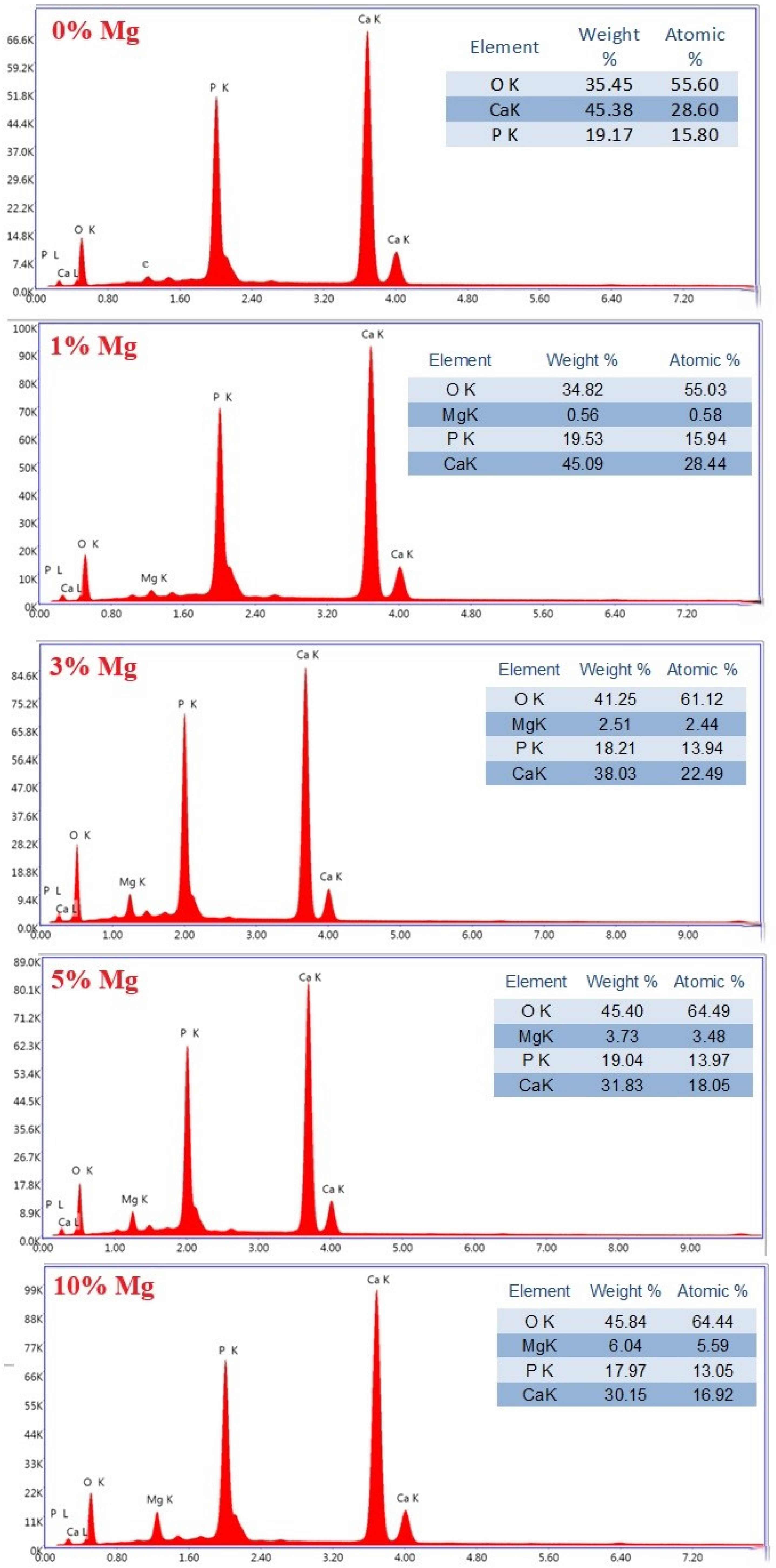
In addition to elemental mapping, quantitative EDS analyses were performed on representative regions of each sample (Figure 5). Typical EDS spectra and the corresponding elemental compositions are provided in this figure, confirming the presence of Mg and the overall chemical composition of the Mg-doped hydroxyapatite ceramics.

3.5. Microhardness and Fracture Toughness

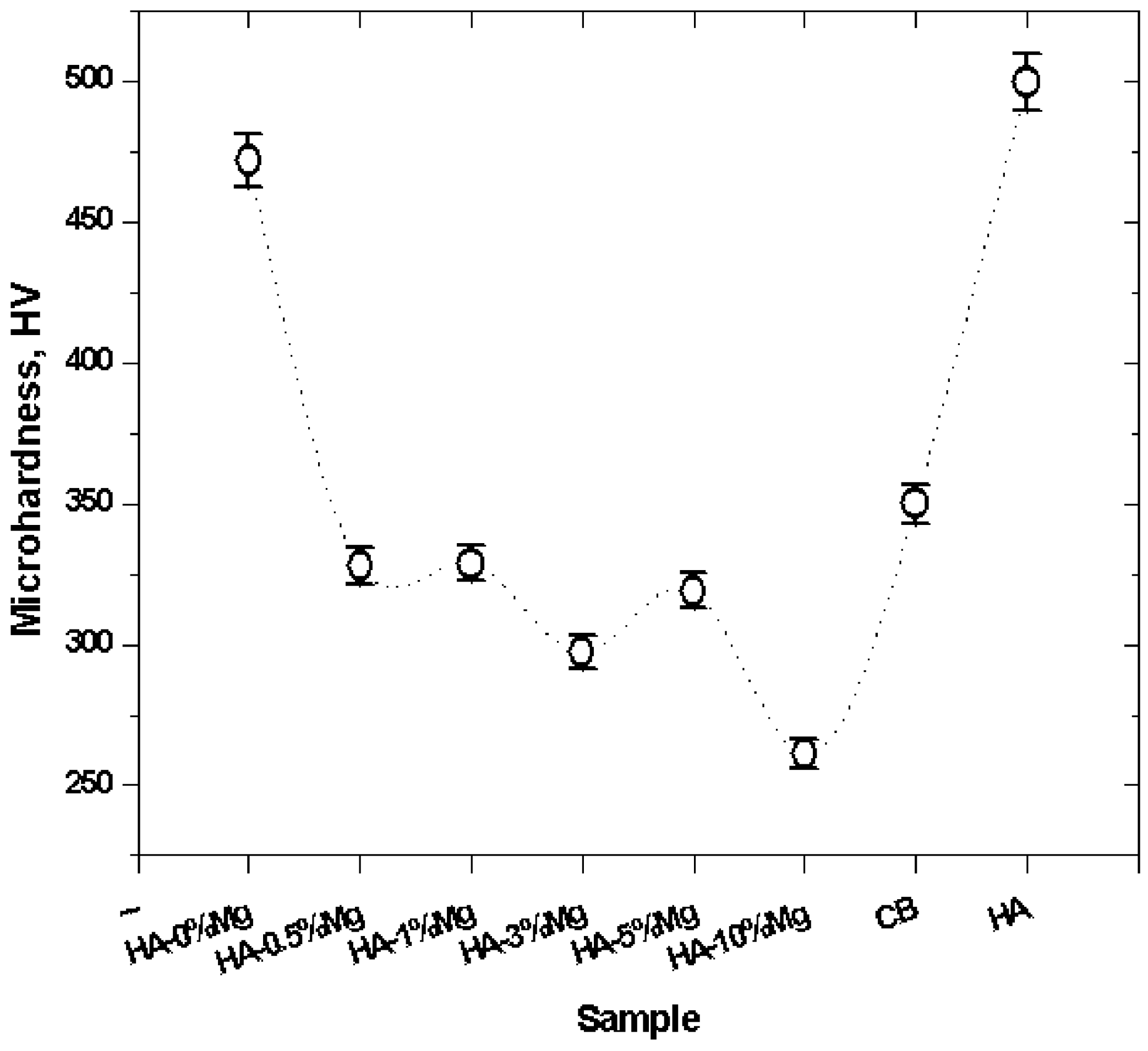
Figure 6 presents the microhardness values of the Mg-containing hydroxyapatite ceramics and the corresponding error bars associated with the standard deviation of each studied composition. A progressive decrease in microhardness is observed with increasing magnesium content, which is consistent with the reduction in relative density and the increase in porosity reported in Figure 1, as well as with the microstructural changes observed in Figure 3. This trend indicates that magnesium incorporation influences the densification behavior during sintering and consequently affects the mechanical response of the ceramic matrix. At low magnesium contents (0.5–1 wt%), the microhardness values decrease moderately, approaching those reported for cortical bone. This behavior may be associated with the homogeneous distribution of magnesium within the matrix and the moderate porosity developed at these compositions, which could improve the mechanical compatibility of the material with natural bone tissue. In contrast, higher magnesium contents (≥3 wt%) produce a more pronounced reduction in microhardness, correlating with the increased porosity and microstructural heterogeneity observed in the SEM analysis. These structural features likely reduce the resistance of the material to localized deformation under indentation loads. For comparison purposes, the microhardness values reported in the literature for pure hydroxyapatite and cortical bone are also included. The lower hardness values measured in the Mg-containing hydroxyapatite ceramics highlight the significant influence of magnesium addition on tailoring the mechanical response of hydroxyapatite-based ceramics.
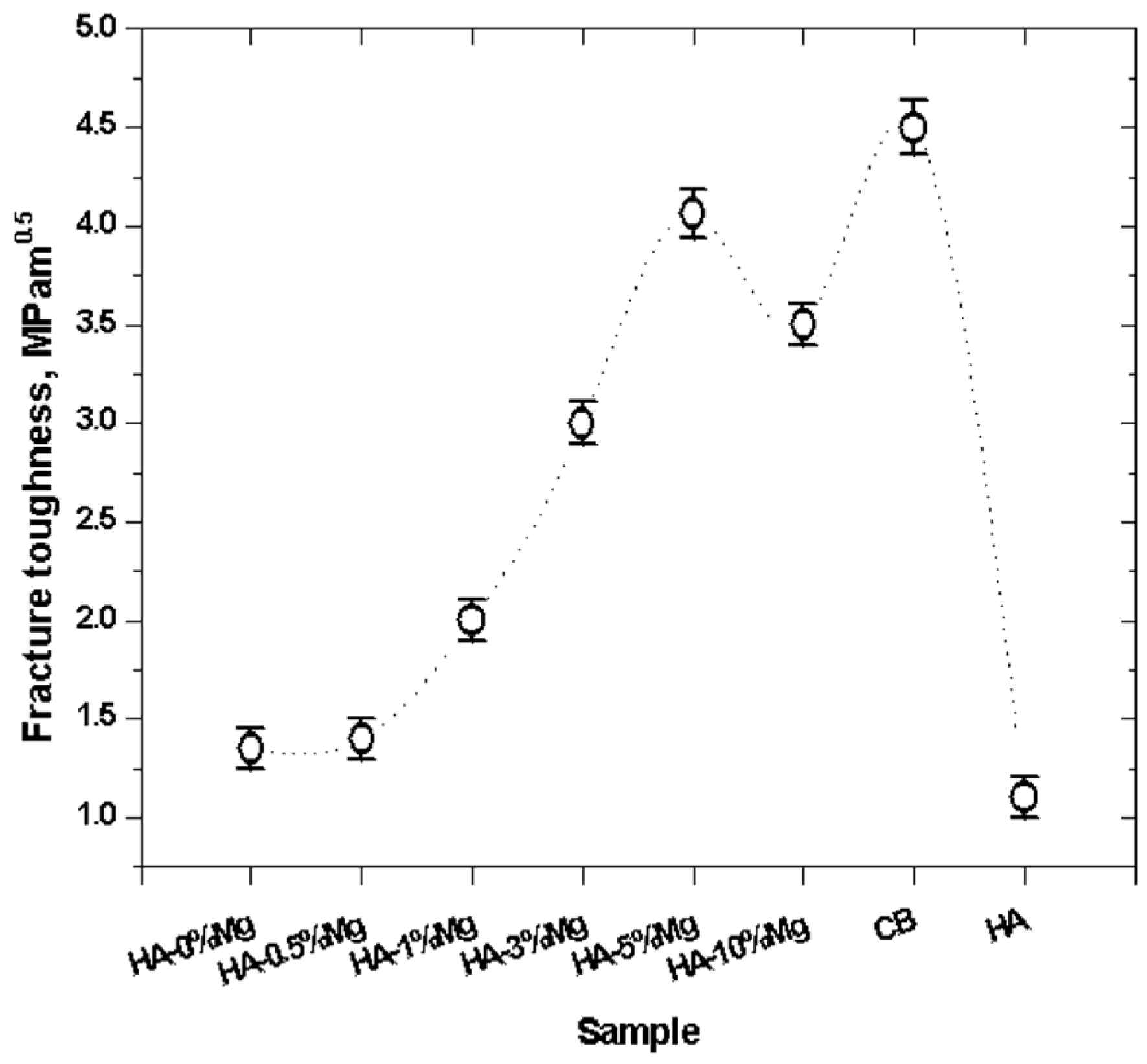
Figure 7 presents the fracture toughness values of Mg-containing hydroxyapatite ceramics together with reference values reported for cortical bone and pure hydroxyapatite. Furthermore, Figure 7 includes error bars corresponding to the standard deviation of each studied composition. An increase in fracture toughness is observed with increasing magnesium content up to 5 wt% Mg, followed by a decrease at 10 wt% Mg. Nevertheless, the toughness value at 10 wt% Mg remains higher than those measured for pure HA and low-magnesium compositions. These results suggest the existence of an optimal magnesium concentration in the range of 3–5 wt% for improving fracture resistance. The increase in fracture toughness at intermediate magnesium contents is consistent with the microstructural characteristics observed in Figure 3 and the densification behavior presented in Figure 1. Samples containing up to 5 wt% Mg exhibit a relatively homogeneous microstructure with moderate porosity, which may contribute to improved resistance against crack propagation under indentation loading. In contrast, the reduction in fracture toughness at 10 wt% Mg correlates with the higher porosity and increased microstructural heterogeneity observed in this composition. These features may facilitate crack initiation and reduce the structural integrity of the ceramic matrix. When considered together with the microhardness results, an inverse tendency between hardness and fracture toughness can be identified. Increasing magnesium content decreases microhardness, mainly due to reduced densification and increased porosity, while fracture toughness improves at intermediate Mg concentrations. This behavior suggests that magnesium addition modifies the balance between rigidity and damage tolerance in the hydroxyapatite ceramics. In particular, the samples containing 3–5 wt% Mg exhibit fracture toughness values closer to those of cortical bone, indicating a potentially favorable mechanical compatibility for biomedical applications.
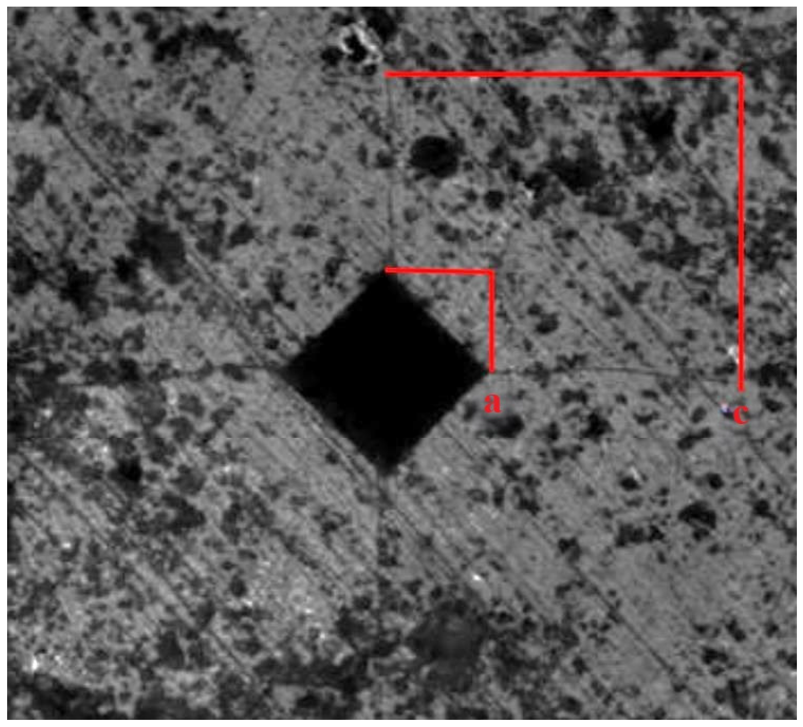
Figure 8 shows a typical indentation footprint and the cracks formed at its corners; the average values of a (indentation size) and c (crack length) were used to determine the fracture toughness value.

3.6. Electrochemical Impedance Spectroscopy

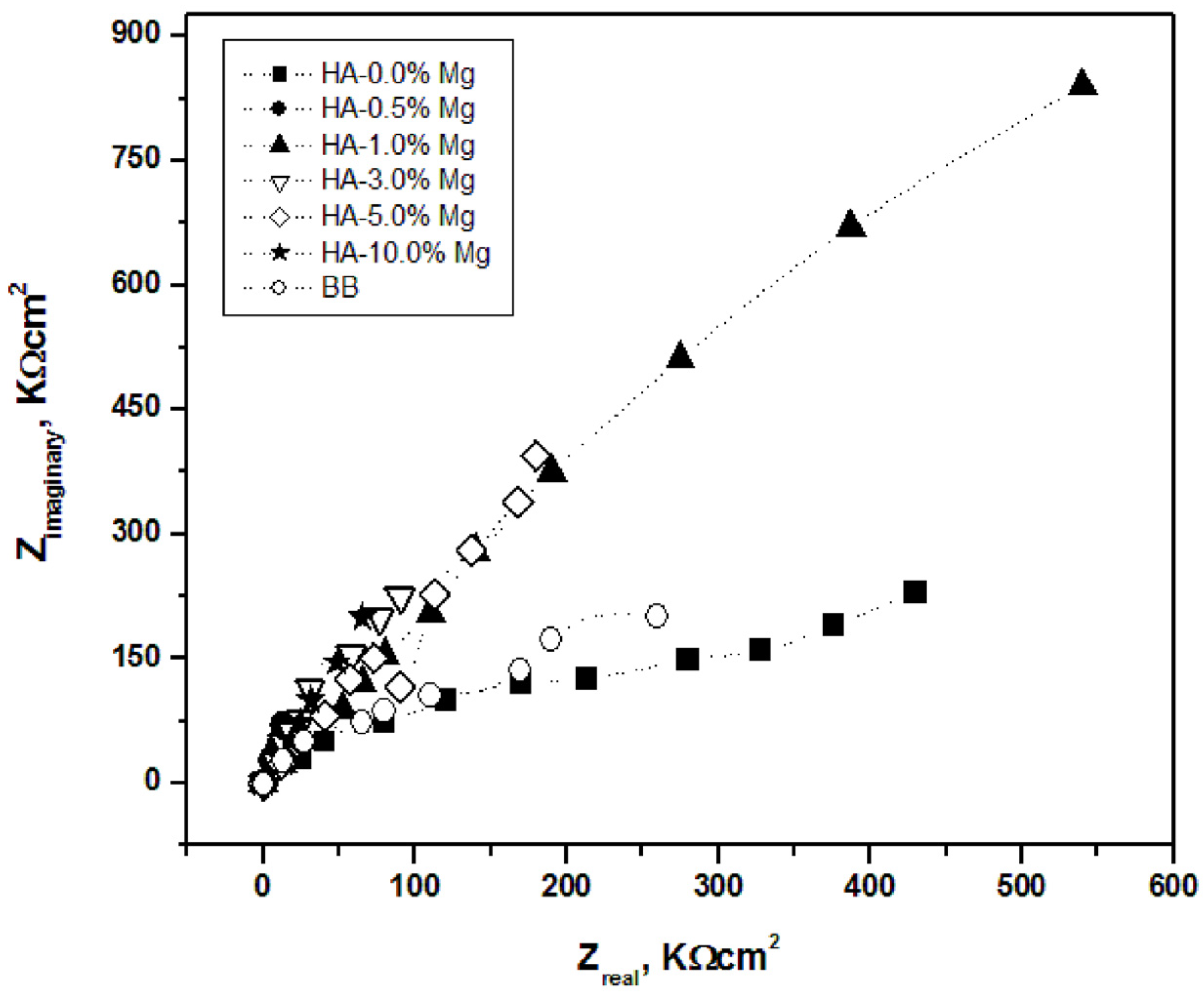
Figure 9 presents the Nyquist plots obtained for Mg-containing hydroxyapatite ceramics and bovine bone immersed in a physiological 0.9 wt% NaCl solution. All spectra exhibit depressed and incomplete semicircles, indicating non-ideal electrochemical behavior commonly associated with heterogeneous ceramic surfaces and distributed relaxation processes. Owing to the incomplete development of the capacitive loops within the measured frequency range, a rigorous determination of the polarization resistance (Rp) by equivalent circuit fitting was not performed. Therefore, the diameter of the observed semicircular response and its extrapolated intercept on the real impedance axis were used only as comparative indicators of the electrochemical response among the studied samples.
The impedance response does not show a linear dependence on magnesium content. The HA–1 wt% Mg sample exhibits the largest semicircle diameter, suggesting the highest resistance to ionic transport and the greatest electrochemical stability among the evaluated compositions. Samples containing 3 and 5 wt% Mg also display relatively high impedance responses, whereas pure hydroxyapatite and bovine bone show the smallest semicircular features, indicating lower resistance to electrolyte penetration. The samples containing 0.5 and 10 wt% Mg exhibit intermediate behavior. These tendencies are consistent with the densification and microstructural characteristics discussed previously. Lower magnesium contents promote more homogeneous microstructures with moderate porosity, whereas higher magnesium concentrations are associated with increased porosity and microstructural heterogeneity. Since porosity strongly affects electrolyte penetration into ceramic materials, these structural differences are likely responsible for the observed differences in impedance response. Nevertheless, because no equivalent-circuit fitting was conducted, the present analysis should be regarded as qualitative, and the reported values should be interpreted only as comparative indicators rather than absolute electrochemical parameters.

4. Discussion

The results demonstrate that magnesium acts as a multifunctional modifier in hydroxyapatite ceramics, simultaneously affecting densification behavior, phase stability, microstructural evolution, and functional properties. A clear inverse relationship is observed between Mg content and relative density, accompanied by a progressive increase in porosity. This behavior indicates that magnesium hinders densification during sintering, which is consistent with its role as a lattice-distorting ion and its influence on diffusion processes. From a structural perspective, XRD analysis confirms that Mg addition induces significant modifications in the hydroxyapatite system. In addition to partial incorporation of Mg into the HA lattice, the presence of secondary phases such as β-tricalcium phosphate (β-TCP) suggests partial thermal decomposition of HA promoted by magnesium. This phase transformation leads to a biphasic HA/β-TCP system. Previous studies have suggested that such biphasic compositions may provide a balance between the chemical stability of HA and the higher resorbability of β-TCP. However, the present study did not evaluate biological performance, and therefore no direct conclusions regarding their biological advantages can be drawn. The observed reduction in crystallinity and peak broadening further supports the role of Mg in inhibiting crystal growth and promoting structural disorder. These structural changes are reflected in the microstructural evolution observed by SEM. At low and intermediate Mg contents, the microstructure remains relatively homogeneous with moderately compact agglomerates, while higher Mg concentrations lead to increased porosity and more heterogeneous morphologies. Elemental mapping confirms that Mg is relatively well distributed at low concentrations, whereas at higher contents a tendency toward local enrichment appears, suggesting that the solubility limit of Mg in the HA lattice may be approached or exceeded. The mechanical response is strongly governed by these structural and microstructural changes. Microhardness decreases progressively with Mg content, which is primarily attributed to reduced densification and increased porosity. In contrast, fracture toughness shows a non-linear trend, increasing up to 3–5 wt% Mg and decreasing at higher concentrations. This behavior suggests that moderate Mg additions promote mechanisms such as crack deflection and energy dissipation associated with a refined and moderately porous microstructure. However, excessive Mg content leads to structural heterogeneity that facilitates crack initiation and propagation. Electrochemical impedance results further highlight the strong dependence of ionic transport resistance on microstructural features. The highest impedance response is observed for intermediate Mg contents (~1 wt%), which correspond to more compact and homogeneous structures. In contrast, higher Mg concentrations reduce impedance due to increased porosity, which facilitates electrolyte penetration. Although the analysis remains qualitative due to the absence of equivalent-circuit fitting, the trends clearly indicate that microstructure plays a dominant role in electrochemical stability. Although the observed increase in porosity and the formation of a biphasic HA/β-TCP system have been reported in the literature as characteristics that may influence biological interactions, the present study did not include biological evaluations such as cell viability, bioactivity, protein adsorption, or in vivo osseointegration assays. Therefore, any discussion regarding potential bone ingrowth, osteoconductivity, or overall biomedical performance should be considered speculative. The present work is limited to establishing the relationships between magnesium content, phase evolution, microstructure, mechanical behavior, and electrochemical response. Future studies incorporating in vitro and in vivo biological assessments will be necessary to determine whether the structural and physicochemical modifications observed here translate into improved biological performance.

5. Conclusions

Magnesium incorporation significantly modifies the structural, microstructural, mechanical, and electrochemical properties of hydroxyapatite ceramics, demonstrating its influence on the physicochemical behavior of the material.
Increasing Mg content reduces densification and increases open porosity. The porosity values obtained at low and intermediate Mg contents are within the range reported for cortical bone; however, the biological implications of this similarity were not evaluated in the present study.
Mg promotes partial destabilization of hydroxyapatite during sintering, leading to the formation of a biphasic HA/β-TCP system, reduced crystallinity, and inhibited crystal growth associated with lattice distortion.
Magnesium influences microstructural evolution, producing relatively homogeneous microstructures at low Mg contents and increasing porosity and heterogeneity as the Mg concentration increases.
Microhardness decreases progressively with increasing Mg content, whereas fracture toughness reaches its highest values at intermediate Mg concentrations (3–5 wt%), indicating a non-linear relationship between Mg addition and mechanical performance.
Electrochemical impedance measurements indicate that specimens containing approximately 1 wt% Mg exhibit the highest impedance response, while higher Mg contents are associated with lower impedance, consistent with the increased porosity and electrolyte penetration observed in the microstructural analysis.
This study establishes the influence of magnesium on the structural, mechanical, and electrochemical behavior of hydroxyapatite ceramics. However, since no biological assays were performed, the results should not be interpreted as direct evidence of enhanced biological performance or osseointegration. Future work will focus on in vitro and in vivo evaluations to validate the biological implications of the physicochemical changes identified in this study.

Author Contributions

Conceptualization, E.R.-G. and E.R.-R.; methodology, E.R.-G., A.E.C.-P., G.V.-H. and J.G.M.-H.; validation, E.R.-G., A.E.C.-P., G.V.-H. and J.G.M.-H.; software, J.G.M.-H.; formal analysis, E.R.-G. and E.R.-R.; investigation, E.R.-G., Z.I.B.-V., G.V.-H. and J.G.M.-H.; resources, E.R.-G., Z.I.B.-V. and E.R.-R.; data curation, C.A.C.-A. and J.A.R.-G.; writing—original draft preparation, E.R.-G., Z.I.B.-V., A.E.C.-P., G.V.-H. and E.R.-R.; writing—review and editing, C.A.C.-A. and J.A.R.-G.; visualization, E.R.-G., G.V.-H. and E.R.-R.; supervision, E.R.-G. and E.R.-R.; project administration, E.R.-G., G.V.-H. and E.R.-R.; funding acquisition, E.R.-G., Z.I.B.-V. and E.R.-R. All authors have read and agreed to the published version of the manuscript.

Funding

This research received no external funding.

Institutional Review Board Statement

Not applicable.

Informed Consent Statement

Not applicable.

Data Availability Statement

The original contributions presented in this study are included in the article. Further inquiries can be directed to the corresponding author.

Acknowledgments

The support from LINAN and IPICYT, as well as the technical assistance of Ana Iris Peña-Maldonado and Ignacio Guadalupe Becerril Juárez in SEM characterization, and Beatriz Adriana Rivera Escoto in X-ray diffraction measurements, are gratefully acknowledged.

Conflicts of Interest

The authors declare no conflicts of interest.

Abbreviations

The following abbreviations are used in this manuscript:
HAHydroxyapatite
CBCortical bone
BBBovine bone
HVHardness Vickers
SPSSpark plasma sintering
CECounter electrode
EISElectrochemical impedance spectroscopy
RpPolarization resistance
wtWeight

References

  1. Dorozhkin, S.V. Calcium Orthophosphate (CaPO4)-Based Bioceramics: Preparation, Properties, and Applications. Coatings 2022, 12, 1380. [Google Scholar] [CrossRef]
  2. Bose, S.; Tarafder, S. Calcium phosphate ceramic systems in growth factor and drug delivery for bone tissue engineering: A review. Acta Biomater. 2012, 8, 1401–1421. [Google Scholar] [CrossRef] [PubMed]
  3. Ielo, I.; Calabrese, G.; De Luca, G.; Conoci, S. Recent Advances in Hydroxyapatite-Based Biocomposites for Bone Tissue Regeneration in Orthopedics. Int. J. Mol. Sci. 2022, 23, 9721. [Google Scholar] [CrossRef]
  4. Wang, W.; Yeung, K.W.K. Bone Grafts and Biomaterials Substitutes for Bone Defect Repair: A Review. Bioact. Mater. 2017, 2, 224–247. [Google Scholar] [CrossRef] [PubMed]
  5. Motlagh, S.R.; Alirezaei, Z.; Parvizi, S. Perspective Chapter: Metal Matrix Composites for Biomedical Applications—Design, Processing, and Performance, Biomedical Engineering. In Advances in Regenerative Medicine and Tissue Engineering; IntechOpen: London, UK, 2024. [Google Scholar] [CrossRef]
  6. Singh, G.; Singh, S.; Sidhu, S.S. Recent Progress in Hydroxyapatite-Based Composite Coatings for Biomedical Applications. In Advances in Materials and Manufacturing; Singh, S., Singh, I., Eds.; ICDMT 2024; Lecture Notes in Mechanical Engineering; Springer: Singapore, 2025. [Google Scholar] [CrossRef]
  7. Venkatesan, J.; Kim, S.K. Nano-hydroxyapatite composite biomaterials for bone tissue engineering—A review. J. Biomed. Nanotechnol. 2014, 10, 3124–3140. [Google Scholar] [CrossRef] [PubMed]
  8. Menon, K.; Nagaraj, A.; Sudha, A.; Kalagatur, N.K.; Subramaniam, S.; Samiappan, S. Synthesis and characterization of hydroxyapatite biocomposite using Syzygium aromaticum for biomedical application. Next Nanotechnol. 2025, 8, 100202. [Google Scholar] [CrossRef]
  9. Ressler, A.; Žužić, A.; Ivanišević, I.; Kamboj, N.; Ivanković, H. Ionic substituted hydroxyapatite for bone regeneration applications: A review. Open Ceram. 2021, 6, 100122. [Google Scholar] [CrossRef]
  10. Rude, R.K.; Gruber, H.E. Magnesium Deficiency and Osteoporosis: Animal and Human Observations. J. Nutr. Biochem. 2004, 15, 710–716. [Google Scholar] [CrossRef] [PubMed]
  11. Castiglioni, S.; Cazzaniga, A.; Albisetti, W.; Maier, J.A.M. Magnesium and Osteoporosis: Current State of Knowledge and Future Research Directions. Nutrients 2013, 5, 3022–3033. [Google Scholar] [CrossRef] [PubMed]
  12. Chen, Y.; Zhang, S.; Bai, J.; Yang, Y.; Wang, Y.; Zhou, Y.; Jiang, W.; Wang, J.; Zhu, J.; Zhu, C.; et al. Magnesium-based biomaterials for coordinated tissue repair: A comprehensive overview of design strategies, advantages, and challenges. J. Magnes. Alloys 2024, 12, 3025–3061. [Google Scholar] [CrossRef]
  13. Bystrov, V.S.; Paramonova, E.V.; Avakyan, L.A.; Eremina, N.V.; Makarova, S.V.; Bulina, N.V. Effect of Magnesium Substitution on Structural Features and Properties of Hydroxyapatite. Materials 2023, 16, 5945. [Google Scholar] [CrossRef] [PubMed]
  14. Omema, U.; Khalid, H.; Chaudhry, A.A. Magnesium-substituted hydroxyapatite. In Handbook of Ionic Substituted Hydroxyapatites; Woodhead Publishing Series in Biomaterials; Khan, A.S., Chaudhry, A.S., Eds.; Woodhead Publishing: Sawston, UK, 2020; pp. 197–216. [Google Scholar] [CrossRef]
  15. Chirică, I.M.; Enciu, A.-M.; Tite, T.; Dudău, M.; Albulescu, L.; Iconaru, S.L.; Predoi, D.; Pasuk, I.; Enculescu, M.; Radu, C.; et al. The Physico-Chemical Properties and Exploratory Real-Time Cell Analysis of Hydroxyapatite Nanopowders Substituted with Ce, Mg, Sr, and Zn (0.5–5 at.%). Materials 2021, 14, 3808. [Google Scholar] [CrossRef] [PubMed]
  16. Singh, J.; Singh, H.; Batra, U. Magnesium Doped Hydroxyapatite: Synthesis, Characterization and Bioactivity Evaluation. In Biomaterials Science: Processing, Properties, and Applications V: Ceramic Transactions, Volume 254; Narayan, R., Bose, S., Bandyopadhyay, A., Eds.; The American Ceramic Society: Westeville, OH, USA, 2015. [Google Scholar] [CrossRef]
  17. Vaiani, L.; Boccaccio, A.; Uva, A.E.; Palumbo, G.; Piccininni, A.; Guglielmi, P.; Cantore, S.; Santacroce, L.; Charitos, I.A.; Ballini, A. Ceramic Materials for Biomedical Applications: An Overview on Properties and Fabrication Processes. J. Funct. Biomater. 2023, 14, 146. [Google Scholar] [CrossRef] [PubMed]
  18. Okasha, A.T.; Abdel-Khalek, A.A.; Rudayni, H.A.; Al Zoubi, W.; Alfassam, H.E.; Allam, A.A.; Abukhadra, M.R. Synthesis and characterization of Mg-hydroxyapatite and its cellulose hybridized structure as enhanced bio-carrier of oxaliplatin drug; equilibrium and release kinetics. RSC Adv. 2023, 13, 30151–30167. [Google Scholar] [CrossRef] [PubMed]
  19. Noviyanti, A.R.; Rahayu, I.; Fauzia, R.P.; Risdiana. The effect of Mg concentration to mechanical strength of hydroxyapatite derived from eggshell. Arab. J. Chem. 2021, 14, 103032. [Google Scholar] [CrossRef]
  20. ASTM B962-15; Standard Test Methods for Density of Compacted or Sintered Powder Metallurgy (PM) Products Using Archimedes’ Principle. ASTM International: West Conshohocken, PA, USA, 2015.
  21. Rocha-Rangel, E. Fracture Toughness Determinations by Means of Indentation Fracture. In Nanocomposites with Unique Properties and Applications in Medicine and Industry; John, C., Ed.; IntechOpen: London, UK, 2011; pp. 1–18. [Google Scholar] [CrossRef] [PubMed]
  22. Evans, A.G.; Charles, E.A. Fracture Toughness Determination by Indentation. J. Am. Ceram. Soc. 1976, 59, 371–372. [Google Scholar] [CrossRef]
  23. ASTM E384-16; Standard Test Method for Microindentation Hardness of Materials. ASTM International: West Conshohocken, PA, USA, 2016.
  24. Mukasheva, F.; Adilova, L.; Dyussenbinov, A.; Yernaimanova, B.; Abilev, M.; Akilbekova, D. Optimization of scaffold pore size for tissue engineering: Insights into various tissue types. Front. Bioeng. Biotechnol. 2024, 12, 1444986. [Google Scholar] [CrossRef] [PubMed]
  25. Toosi, S.T.; Javid-Naderi, M.J.; Tamayol, A.; Ebrahimzadeh, M.H.; Yaghoubian, S.; Mousavi Shaegh, S.A. Additively manufactured porous scaffolds by design for treatment of bone defects. Front. Bioeng. Biotechnol. 2024, 11, 1252636. [Google Scholar] [CrossRef] [PubMed]
  26. Jerban, S.; Ma, Y.; Wan, L.; Searleman, A.C.; Jang, H.; Sah, R.L.; Chang, E.Y.; Du, J. Collagen Proton Fraction from Ultrashort Echo Time Magnetization Transfer (UTE-MT) MRI Modelling Correlates Significantly with Cortical Bone Porosity Measured with Micro-Computed Tomography (μCT). NMR Biomed. 2019, 32, e4099. [Google Scholar] [CrossRef] [PubMed]
  27. Matić, T.; Zebić, M.L.; Miletić, V.; Cvijović-Alagić, I.; Petrović, R.; Janaćković, D.; Veljović, D. Sr, Mg Co-Doping of Calcium Hydroxyapatite: Hydrothermal Synthesis, Processing, Characterization and Possible Application as Dentin Substitutes. Ceram. Int. 2022, 48, 11155–11165. [Google Scholar] [CrossRef]
  28. Rabadjieva, D.; Gergulova, R.; Sezanova, K.; Kovacheva, D.; Titorenkova, R. Mg, Zn Substituted Calcium Phosphates—Thermodynamic Modeling, Biomimetic Synthesis in the Presence of Low-Weight Amino Acids and High Temperature Properties. Materials 2023, 16, 6638. [Google Scholar] [CrossRef] [PubMed]
  29. Salamanca, E.; Pan, Y.H.; Sun, Y.S.; Hsueh, H.W.; Dorj, O.; Yao, W.L.; Lin, J.C.Y.; Teng, N.C.; Watanabe, I.; Abe, S.; et al. Magnesium Modified-Tricalcium Phosphate Induces Cell Osteogenic Differentiation In Vitro and Bone Regeneration In Vivo. Int. J. Mol. Sci. 2022, 23, 1717. [Google Scholar] [CrossRef] [PubMed]
  30. Mirzaali, M.J.; Schwiedrzik, J.J.; Thaiwichai, S.; Best, J.P.; Michler, J.; Zysset, P.K.; Wolframa, U. Mechanical Properties of Cortical Bone and Their Relationships with Age, Gender, Composition and Microindentation Properties in the Elderly. Bone 2016, 93, 196–211. [Google Scholar] [CrossRef] [PubMed]
Figure 1. Relative density and porosity values obtained for each sintered ceramic. CB—cortical bone.
Applbiosci 05 00061 g001
Figure 2. X-ray diffraction patterns of Mg-containing hydroxyapatite ceramics sintered at 1000 °C.
Applbiosci 05 00061 g002
Figure 3. Microstructures and EDS spectra of the different hydroxyapatite–magnesium ceramics obtained by scanning electron microscopy: (a) HA-0%Mg, (b) HA-0.5%Mg, (c) HA-1%Mg, (d) HA-3%Mg, (e) HA-5%Mg, (f) HA-10%Mg.
Applbiosci 05 00061 g003aApplbiosci 05 00061 g003b
Figure 4. EDS Element Distribution Maps of hydroxyapatite with different additions of Mg. The figure shows, from top to bottom, the maps of all Mg-containing hydroxyapatite compositions studied.
Applbiosci 05 00061 g004
Figure 5. Typical EDS spectra for mapping and the corresponding elemental compositions.
Applbiosci 05 00061 g005
Figure 6. Microhardness values obtained for each sintered ceramic. CB—cortical bone [30], HA [3].
Applbiosci 05 00061 g006
Figure 7. Fracture toughness values obtained for each sintered ceramic. CB—cortical bone [30], HA [3].
Applbiosci 05 00061 g007
Figure 8. Typical indentation footprint and the cracks formed at its corners. a: indentation size, c: crack length.
Applbiosci 05 00061 g008
Figure 9. Impedances of hydroxyapatite samples with different magnesium contents. BB—bovine bone.
Applbiosci 05 00061 g009
Table 1. Composition of different prepared formulations.
CompositionMagnesium
(Weight %)
Hydroxyapatite
Weight %
Mg/Ca
Molar Ratio
HA-0%MgMg 01000.0000
HA-0.5%MgMg 0.599.50.0083
HA-1%MgMg 1990.0168
HA-3%MgMg 3970.0880
HA-5%MgMg 5950.0899
HA-10%MgMg 10900.1976
Disclaimer/Publisher’s Note: The statements, opinions and data contained in all publications are solely those of the individual author(s) and contributor(s) and not of MDPI and/or the editor(s). MDPI and/or the editor(s) disclaim responsibility for any injury to people or property resulting from any ideas, methods, instructions or products referred to in the content.