1. Introduction
Phytate (InsP6), a polyphosphate serving as phosphorus storage in numerous plants, particularly cereals and oilseeds [
1,
2,
3], is subject to ongoing scientific controversy. Its classification as an antinutrient stems from its high binding affinity for divalent and trivalent cations, which form complexes that sequester essential minerals such as iron, zinc, and calcium, rendering them biologically unavailable [
4,
5]. Conversely, beneficial properties have been documented, including antioxidant and anti-inflammatory activities, anticancer effects, and enhanced absorption of pharmacologically active substances [
1,
4,
6,
7,
8,
9], for review see [
10]. Clinical evidence demonstrates that dietary phytate intake results in elevated phytate levels in plasma and urine [
6,
11], confirming systemic bioavailability. However, the precise mechanisms governing both cellular uptake and transepithelial absorption remain unresolved.
The molecule’s highly negative charge at physiological pH theoretically precludes passive diffusion across lipid membranes, and no specific transporter has been identified [
1,
6,
12]. Windhorst et al. (2013) proposed a cellular uptake mechanism involving non-receptor-mediated endocytosis in tumor cells, whereby phytate-metal complexes bind to the glycocalyx and trigger internalization followed by lysosomal dephosphorylation [
12]. Additionally, phytate may undergo dephosphorylation by phytases, yielding lower inositol phosphates (InsP
5, InsP
4, InsP
3) prior to absorption [
13]. However, neither mechanism explains transepithelial transport from the intestinal lumen into the systemic circulation, leaving a critical knowledge gap regarding how dietary phytate achieves systemic bioavailability.
The paracellular pathway represents an alternative route for transepithelial transport. Paracellular permeability is regulated by tight junctions—multiprotein complexes comprising occludin, claudins, and ZO-1 proteins that seal the intercellular space between adjacent enterocytes [
14,
15,
16]. Tight junction integrity is critically dependent on extracellular calcium availability. Tomita et al. (1996) demonstrated that ethylenediaminetetraacetic acid (EDTA), a calcium chelator, increases paracellular permeability in intestinal epithelial monolayers by depleting extracellular calcium, activating protein kinase C, and consequently disrupting tight junction complexes [
14]. Given that phytate exhibits comparable calcium-chelating properties to EDTA due to its chemical structure, similar barrier-disrupting effects are mechanistically plausible. Indeed, in 2015 Fu et al. reported that phytate reduces the expression of tight junction proteins (occludin, ZO-1, claudin-1) in Caco-2 monolayers in a calcium-dependent manner [
15]. However, this study did not investigate whether the observed alterations in tight junction protein expression translate into functional barrier disruption permitting phytate permeation. The present study addresses this gap by investigating calcium-dependent phytate transport across the intestinal epithelial barrier.
The calcium-phytate interaction is governed by stoichiometry and determines complex solubility. The phytate molecule myo-inositol-1,2,3,4,5,6-hexakisphosphate shows in solution an axial–equatorial–axial configuration at positions 1, 2, and 3, conferring exceptional stability and strong negative charge at neutral pH [
3]. This structural feature dictates that monocalcium phytate (Ca
1-phytate) and dicalcium phytate (Ca
2-phytate) complexes remain soluble, whereas calcium-to-phytate molar ratios of 3:1 or higher induce precipitation of insoluble Ca
3-phytate complexes across a wide pH range [
3,
6,
8]. At this stoichiometry, neither calcium nor phosphate is available for absorption. Consequently, the calcium concentration in the intestinal lumen determines both phytate solubility and potential for transepithelial transport.
To investigate these relationships, differentiated Caco-2 cell monolayers were employed as an established model of the intestinal epithelial barrier that forms functional tight junctions [
17]. Differentiated Caco-2 monolayers are a well-known in vitro intestinal model and a well-established approach in studies of drug and other small molecule absorption and permeability. These cells express tight junction proteins and exhibit microvilli, as well as many transporters and enzymes. There is a large body of evidence showing a reasonable association between Caco-2 cells and human intestinal absorption for many small molecules. However, this model system cannot replicate the full complexity of the human intestine; therefore, several limitations must be taken into account. The study examined in which form—soluble or precipitated—phytate exists as a function of calcium concentration, to what extent phytate can traverse the intestinal barrier under these conditions, and whether cellular uptake occurs. Two calcium concentrations were tested: a low concentration (2.1 µM) representing conditions where phytate remains soluble and calcium depletion may compromise tight junction integrity, and a physiological concentration (1.8 mM) where Ca
3-phytate precipitation is favored but tight junctions remain intact. Different phytate concentrations (0.17, 0.55, 1.66 mM) were selected to provide calcium-to-phytate ratios spanning the transition from soluble to insoluble complexes. Transepithelial phytate flux was quantified by HPLC, paracellular barrier integrity was assessed using Lucifer yellow (LY) as a validated permeability marker of comparable molecular weight to phytate, and phytate distribution between apical medium, basolateral medium, and cell-associated fractions was determined following 24 h incubation.
2. Materials and Methods
2.1. Materials
Phytic acid dodecasodium salt and Lucifer yellow dipotassium salt were obtained from Sigma-Aldrich (Steinheim, Germany). The human colon adenocarcinoma cell line Caco-2 was purchased from the German Collection of Microorganisms and Cell Cultures (Braunschweig, Germany). Eagle’s minimal essential medium (EMEM, normal calcium, 1.8 mM), MEM-Eagle Joklik’s Formulation (SMEM, low calcium, 2.1 µM), non-essential amino acids (NEAA), phosphate-buffered saline (PBS, pH 7.4), and penicillin/streptomycin were obtained from Lonza (Verviers, Belgium), and fetal calf serum (FCS) from PAA (Pashing, Austria). Fluorescein sodium salt was obtained from Fluka (Steinheim, Germany).
2.2. Cell Culture
Caco-2 cells were routinely cultivated in 75 cm2 cell culture flasks from Corning (Corning, NY, USA) in EMEM containing 10% (v/v) FCS, 1% NEAA, 1% glutamine, 50 U/mL penicillin G and 50 U/mL streptomycin. Cells were maintained at 37 °C and 5% CO2 in a humidified atmosphere. The cell culture medium was changed three times a week.
2.3. Culturing of Caco-2 Cells on Semi-Permeable Inserts
Permeability of phytate over intestinal epithelia was investigated using differentiated Caco-2 cells grown on semi-permeable membranes. The handling of the cells on inserts, as well as a testing of the monolayer’s integrity were described in detail by Briviba et al. (2018) [
17].
2.4. Incubation of Cells with Phytate
Two media, EMEM and SMEM (with additives to match the same concentration of components of EMEM), which differ in the concentrations of calcium, were used to incubate the cells with phytate. EMEM contains 1.8 mM calcium, and SMEM contains 2.1 µM calcium. Hence EMEM will be referred to as medium with a normal calcium concentration (normalCa), because it is very close to the physiological concentration in the extracellular fluid, while SMEM will be referred to as medium with lower calcium concentration (lowCa), representing a low calcium intake and/or a high intake of other calcium-binding compounds like tannins and other polyphenols. Phytate stock solution was diluted to final phytate concentrations of 0.17, 0.55, and 1.66 mM with cell culture medium. The pH was controlled and, if necessary, adjusted to pH 7.4. These solutions were added to the transwell inserts (apical compartment). The basal compartment contained the corresponding medium only. Inserts that did not contain cells served as a control to measure the stability of phytate during incubation. To control the integrity of the tight junction, a cell culture medium containing Lucifer yellow (10 mg/mL) and phytate at concentrations indicated was added to the apical compartment.
While the average colon transit time is known between 16 and 29 h [
18], for phytate, which is often associated with high-fiber foods, an incubation time of 24 h at 37 °C in the cell culture incubator with 5% CO
2 was chosen, representing a realistic residence time. Then, the samples were collected, and the phytate concentrations were estimated in the apical and basolateral chambers by HPLC. The cells were washed twice with PBS without calcium and magnesium, lysed by freezing/thawing and the concentration of phytate was measured using HPLC.
To measure the soluble phytate the samples were estimated directly, without the addition of HCl. To measure the total phytate concentration (soluble and insoluble) 0.58 M HCl was added to the samples to dissolve the precipitated phytate and the samples were centrifuged (2700× g 20 min) and filtrated.
2.5. Analysis of Inositol Phosphates
Phytate was estimated on Mono-Q (HR5/5, Pharmacia, Freiburg, Germany) using an HCl gradient (0.005–0.5 mol/L) with post-column derivatization by Fe
3+(FeCl
3/NaCl) as described by Schlemmer et al. (2009) with minor modifications [
6].
2.6. Statistical Analysis
The statistical calculations were performed using analysis of variance (ANOVA) followed by Tukey’s test or Dunn’s test (nonparametric analysis) (SigmaPlot 14.0, Systat Software Inc., San Jose, CA, USA) or exact permutation test (R version 4.5.0). When normal distribution and/or homoscedasticity were not met, data transforming (e.g., square function) was performed. The significance level was set at p < 0.05. Results are given as mean ± SD.
3. Results
To investigate the intestinal absorption of phytate dependent on the calcium concentration, differentiated Caco-2 cells cultured on permeable membranes were incubated with phytate, added to the apical compartment, in cell culture medium either with low (2.1 µM) or normal calcium (1.8 mM) concentrations. It is well known that phytate forms soluble and insoluble complexes with calcium depending on the calcium/phytate ratio and the pH-value. In this study, we differentiated between these two forms and estimated the concentration of soluble and total (soluble plus insoluble) phytate in the apical and basal compartments, as well as in the cell fraction after 24 h of incubation.
The recovery of total phytate as the sum of all three fractions (apical and basal compartment and cell-fraction) varied between 84 and 92% in medium with low calcium and 72 and 91% in medium with normal calcium, respectively.
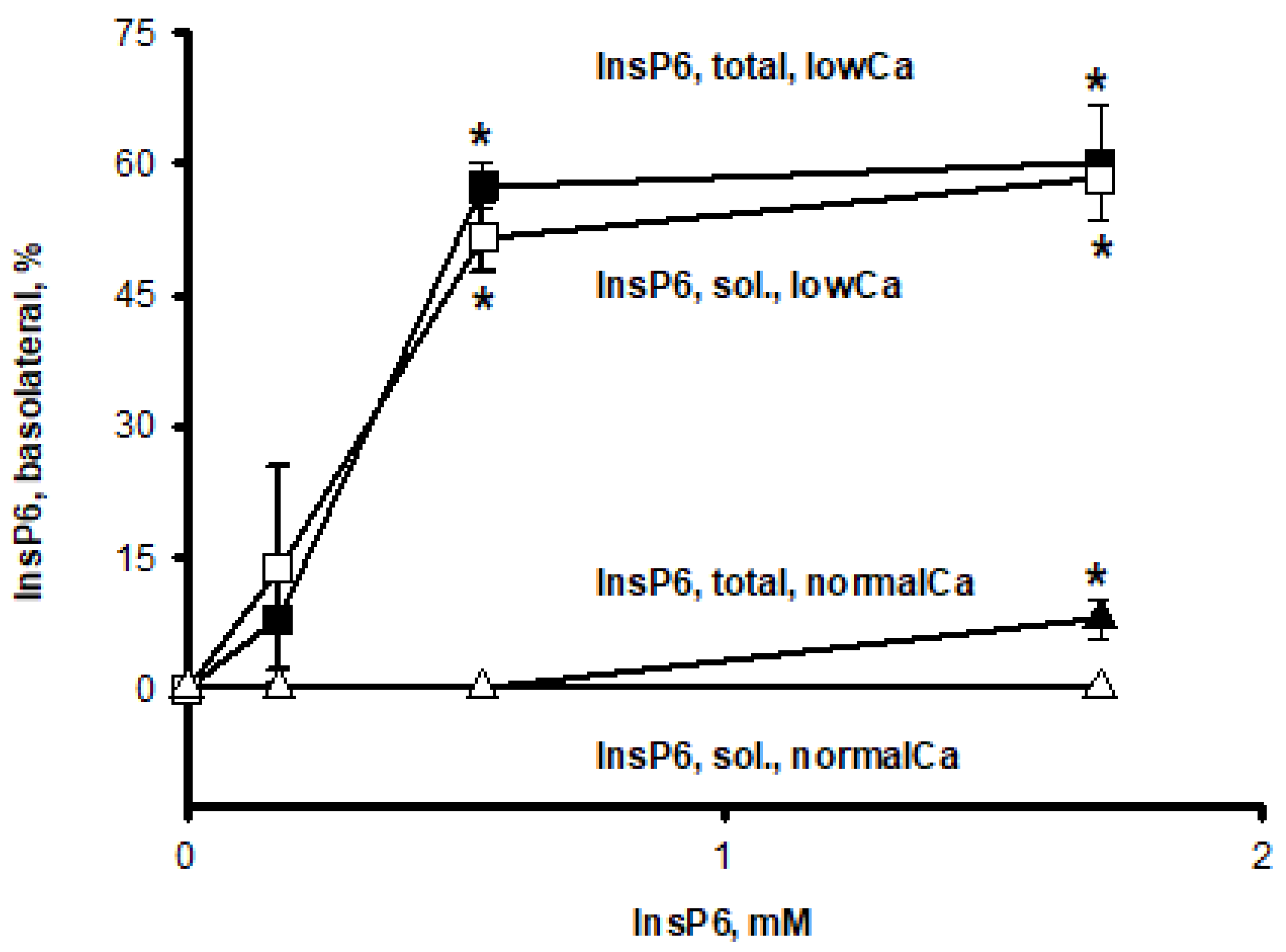
In the low calcium medium, a statistically significant permeability of phytate from the apical to the basal compartment was observed starting from the initial apical phytate concentration of 0.55 mM (exact permutation test,
p < 0.05) (
Figure 1). Due to the different volumes of the apical (1.5 mL) and basolateral (2.6 mL) compartments, equilibrium in the concentration between these two compartments is reached when the amount of phytate recaches nearly 63% of the initial phytate amount in the basolateral compartment. At 1.66 mM, the permeability of phytate was about 60% of the initial apical amount, indicating disruption of the barrier function of the Caco-2 cell monolayer. There was no significant difference (ANOVA,
p < 0.05) between phytate in the soluble form and total phytate indicating that almost all phytate was in a soluble form in the low calcium medium.
A significant lower permeability of phytate was observed in the medium containing normal calcium concentration. There was no measurable permeability at the phytate concentration of 0.17 and 0.55 mM. Only at the phytate concentration of 1.66 mM a statistically significant permeability (exact permutation test,
p < 0.05) of total phytate was observed (
Figure 1). No soluble phytate was detected, indicating that all phytate that reached the basolateral side at normal calcium levels became insoluble. Furthermore,
Table 1 shows the amount of phytate in the soluble form as the sum of all three fractions (apical, basal, cell-fraction). In the normal calcium medium at the concentration of 0.17 mM, all phytate was insoluble, and at 0.55 mM, only about 7% were soluble. At the highest concentration of 1.66 mM 42% of total phytate was in a soluble form (
Table 1).
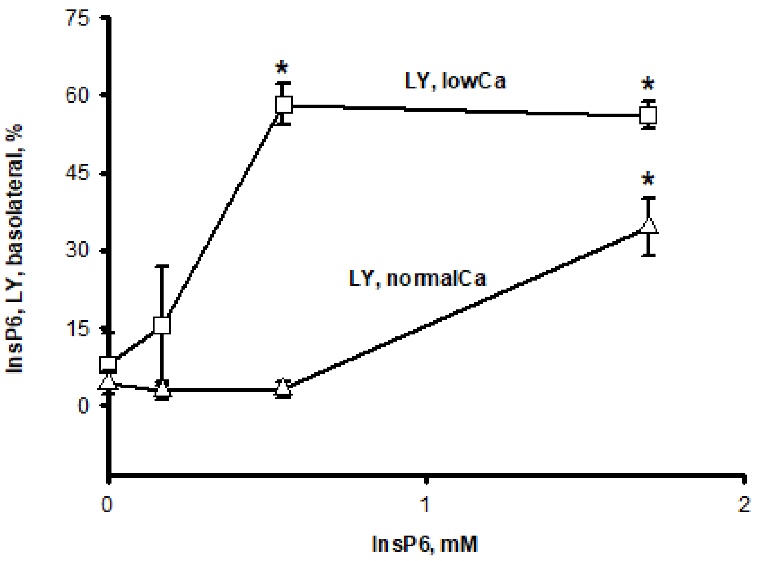
To observe the effect of phytate on tight junction disruption, Lucifer yellow, a marker of paracellular permeation, was co-incubated with phytate. The observed increase in phytate (
Figure 1) in the basal compartment correlated well with an increase in Lucifer yellow (
Figure 2). At the low calcium concentration, a statistically significant amount of Lucifer yellow could already permeate to the basolateral compartment from a phytate concentration of 0.55 mM and above. Even at normal calcium, the permeation of Lucifer yellow was observed, but only at the concentration of 1.66 mM phytate (ANOVA, Dunn’s test,
p < 0.05). This observation indicated disruptions of tight junctions by phytate, which cause the paracellular permeation.
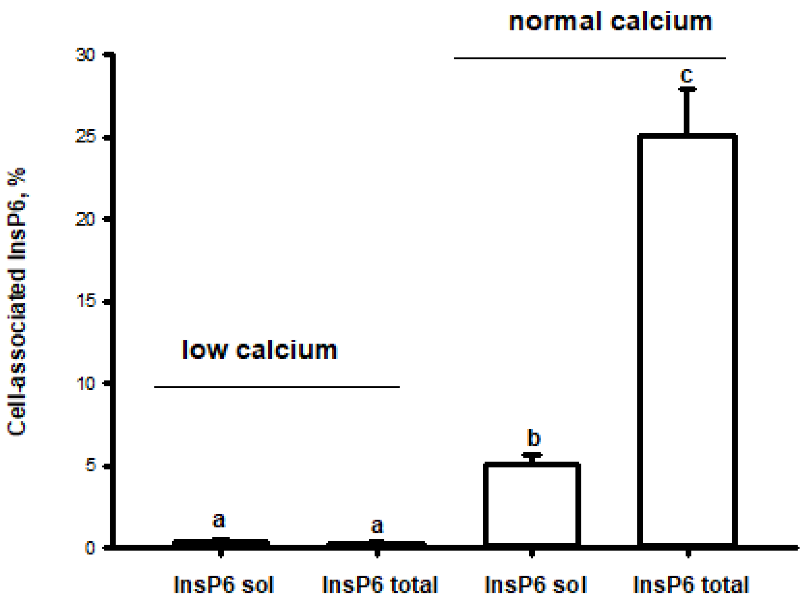
The incubation with phytate up to 0.55 mM did not result in any detectable phytate in the cell fraction in either medium (
Figure 3). In the low calcium medium, only about 0.3% of phytate apical amount was detected in the cell fraction when cells were incubated with 1.66 mM phytate. Whereby nearly all phytate was in a soluble form. In contrast, incubation in the normal calcium medium with 1.66 mM phytate led to an increase up to 2.4% of phytate in soluble form and about 25% as total phytate (soluble plus insoluble form) (
Figure 3). This observation could indicate the pronounced formation of insoluble aggregates of phytate with calcium and adherence to the cell monolayer.
4. Discussion
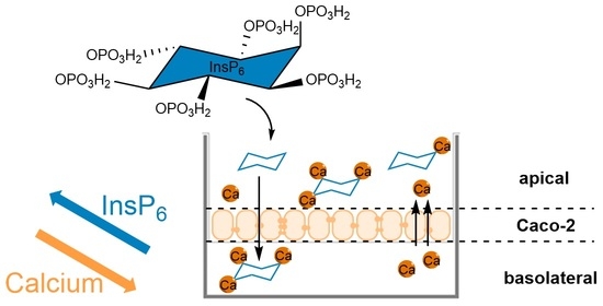
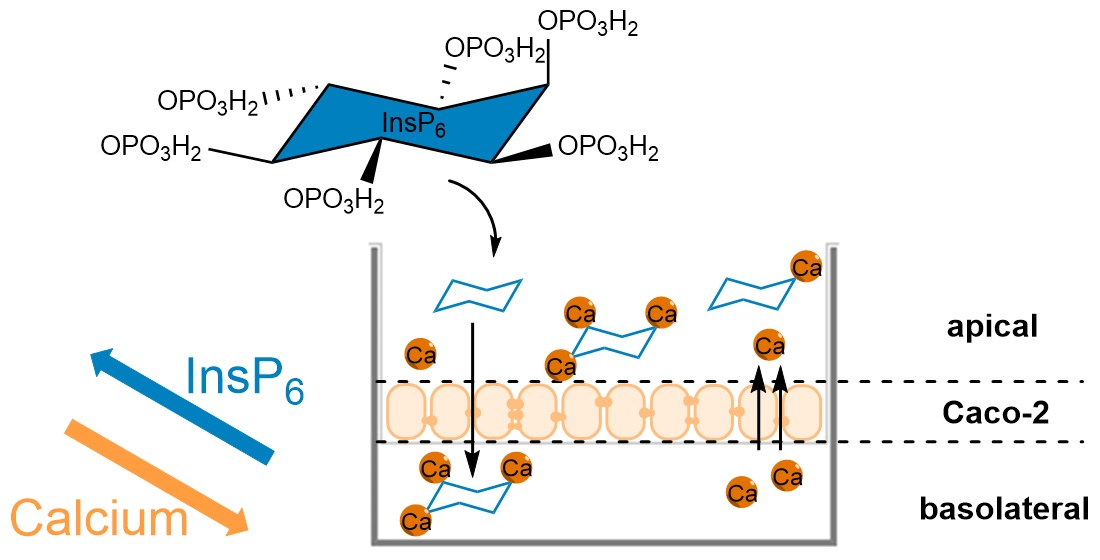
Here we have shown that phytate can pass the intestinal epithelium via paracellular pathway. Considering calcium’s role in paracellular permeability [
16] and the very high affinity of phytate to calcium [
7], it seems a logical consequence that phytate, depending on its concentration, can cause disruption of tight junctions and pass the monolayer. This conclusion is in line with the data reported by Fu et al. in 2015 [
15], demonstrating that phytate can decrease the integrity of Caco-2 cell monolayers by downregulating the expression of some tight junction proteins such as occludin, ZO-1, and claudin-1, and that calcium ions can partially inhibit the effect of phytate. However, neither cell-association nor transepithelial transport of phytate was investigated by Fu et al. (2015) [
15].
Especially at the low calcium concentration (2.1 µM), phytate at 0.55 mM and above effectively induces the diffusion of Lucifer yellow and itself across the Caco-2 monolayer. An increase in permeability was observed with increasing phytate concentration. Phytate stays in a soluble form at the low calcium concentration in both apical and basal compartments. In contrast, the loss of tight junction integrity was only observed at the highest concentration measured at the normal calcium concentration. This loss of integrity is mainly caused by the part of 42% phytate that was still soluble in the normal calcium medium. Due to its high density of negatively charged phosphate groups, phytate strongly interacts with calcium, which has a strong influence on phytate’s solubility in various conditions [
19]. Graf (1983) [
7] demonstrated with a potentiometric measurement that the number of calcium bounds to phytate never extended to three due to precipitation of the complex by the addition of a third Ca
2+ ion, even at low pH values. This observation was independent of the phytate concentration over a 100-fold concentration range [
7].
Thus, the higher the calcium concentration, the lower the solubility of the calcium–phytate complex. In the precipitated form neither phytate nor calcium are considered to be bioavailable [
6]. Therefore, only soluble phytate, Ca
1-phytate or Ca
2-phytate, can bind calcium from the tight junction and, consequently, can cause the disruption of tight junctions. When phytate reaches the basolateral compartment at normal calcium concentration, it precipitates into an insoluble form. This shows that, even at physiological extracellular calcium concentrations, phytate will precipitate immediately upon reaching the basolateral side.
As described above, phytate cannot pass through the membrane at physiological pH due to its chemical properties, and no transporter has been identified so far. Nevertheless, there are indications that phytate can be taken up into cells via endocytosis [
6,
12,
20]. In the present work we observed a significant amount of phytate in the cell fraction. However, we were not able to distinguish between the extracellular and intracellular form. Particularly in a medium with normal calcium and 1.66 mM phytate we observed that about 25% of phytate was found cell-associated but only about 2.4% was in a soluble form, showing a high precipitation of phytate and possible adherence to the cell surface. This is in line with the suggestions of Phillippy in 2006 [
21], who examined an increase in cell-associated calcium in Caco-2 cells with higher concentrations of InsP6, assuming that this enhancement could also be an artifact, resulting from calcium–phytate complexes binding to the surface of the cells. It seems that at the beginning of the incubation, all phytate was soluble in the apical compartment (1.66 mM phytate vs. 1.8 mM calcium) but, apparently, all calcium was bound by phytate forming Ca
1-phytate and Ca
2-phytate soluble calcium complexes. This reduces the calcium concentration in the apical compartment. Lucifer yellow permeation indicates a paracellular flux; phytate from apical to basal, and likely calcium from basal to apical. Therefore, it is possible that the intense formation of insoluble calcium-phytate-precipitates occur in the paracellular space. And we were not able to remove these precipitates by rinsing with PBS. This is in line with our observation that at the same concentration of phytate but in a medium with low calcium, only 0.3% of phytate was found in the cell fraction and in a soluble form. In addition, the reflux of calcium from the basolateral to the apical side leads to further precipitation of phytate in the apical compartment, which can be associated with the cell surface. This is in line with Windhorst et al. (2013), who described that the higher the calcium concentration, the more association of the calcium–phytate complex with the cell surface will occur [
12]. In particular, the phytate complex showed a high binding affinity to the glycocalyx.
Thus, it is possible to have a low calcium concentration in the intestinal tract (apical) in the physiological conditions but not on the basal side. The calcium concentration in the extracellular fluid is tightly controlled, because it is essential for the function of a number of organs. Therefore, even if phytate passes the intestinal epithelium via the paracellular pathway, it will completely precipitate in the basolateral compartment at physiological pH and at physiological extracellular calcium concentration.