Article

Widespread Abundance of Capillaria hepatica Among Wild Rodents Captured at Wet Markets in Medan, Indonesia: Indicating Highly Potential Zoonosis Transmission


1
Department of Parasitology, Faculty of Medicine, Universitas Sumatera Utara, Medan 20155, Indonesia
2
Faculty of Public Health, Universitas Sumatera Utara, Medan 20155, Indonesia
3
Department of Microbiology, Faculty of Medicine, Universitas Sumatera Utara, Medan 20155, Indonesia
4
Master Program in Biomedical Sciences, Faculty of Medicine, Universitas Sumatera Utara, Medan 20155, Indonesia
5
Department of Parasitology, Faculty of Medicine, Universitas Imelda Medan, Medan 20239, Indonesia
6
Undergraduate Program in Medicine, Faculty of Medicine, Universitas Sumatera Utara, Medan 20155, Indonesia
7
Faculty of Forestry, Universitas Sumatera Utara, Medan 20353, Indonesia
8
Research Center for Veterinary Science, Research Organization for Health, National Research and Innovation Agency, Cibinong, Bogor 16911, Indonesia
9
Department of Pediatrics, Faculty of Medicine, Universitas Sumatera Utara, Medan 20155, Indonesia
*
Author to whom correspondence should be addressed.
Zoonotic Dis. 2026, 6(2), 24; https://doi.org/10.3390/zoonoticdis6020024
Submission received: 12 April 2026 / Revised: 19 May 2026 / Accepted: 28 May 2026 / Published: 10 June 2026

Simple Summary

Capillaria hepatica is a zoonotic nematode primarily infecting the livers of mammals. In humans, it causes hepatic capillariasis, a relatively uncommon but potentially severe disease. Transmission occurs when embryonated eggs are ingested from contaminated soil, food, or water, directly linking environmental exposure to infection risk. Wild rodents serve as the main reservoir hosts for the parasite in environment. The wet market, as part of the local food security, is a potential hotspot for transmission by fomite through contaminated environments. The study was done in Medan, the capital of North Sumatra province, Indonesia. We examined 150 wild rodents captured at three popular wet markets in Medan and the surrounding residential area for the presence of C. hepatica in their liver tissue using microscopy and molecular methods, with added spatial display. Most rodents were infected with C. hepatica, particularly the brown rat (Rattus norvegicus), followed by the Asian house rat (Rattus tanezumi). The findings indicate a significant risk of environmental contamination and an increased risk of transmission to humans in the adjacent community.

Abstract

Background: Capillaria hepatica is a zoonotic nematode primarily infecting rodents. In humans, it causes hepatic capillariasis, a disease that resembles hepatitis. Rodents act as primary reservoirs and key amplifiers of transmission by ingesting the embryonated eggs in contaminated soil/food/water. The wet market is a potential hotspot for transmission through contaminated environments. Methods: We aimed to detect C. hepatica among wild rodents from three popular wet markets in Medan, Indonesia: Simpang Limun, Petisah, and Melati. Bait traps were placed near market stalls and nearby residences. Rodent species were identified based on morphology. Liver tissue was microscopically examined and further confirmed with PCR targeting the C. hepatica 18S rRNA gene. The spatial display was produced through a GIS approach. Results: From 150 rodents, the species captured were Rattus norvegicus (70.7%) and Rattus tanezumi (29.3%), with R. norvegicus as the most infected (95/106). Overall prevalence was 79.3%, with Petisah having the highest local prevalence (48/51). Positive rodents were found at market stalls and in nearby residential areas. Conclusions: We found a high prevalence of C. hepatica among wild rodents in the three wet markets in Medan, suggesting a potential threat of transmission to humans nearby.

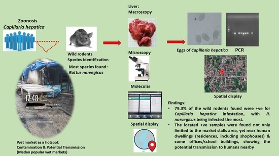
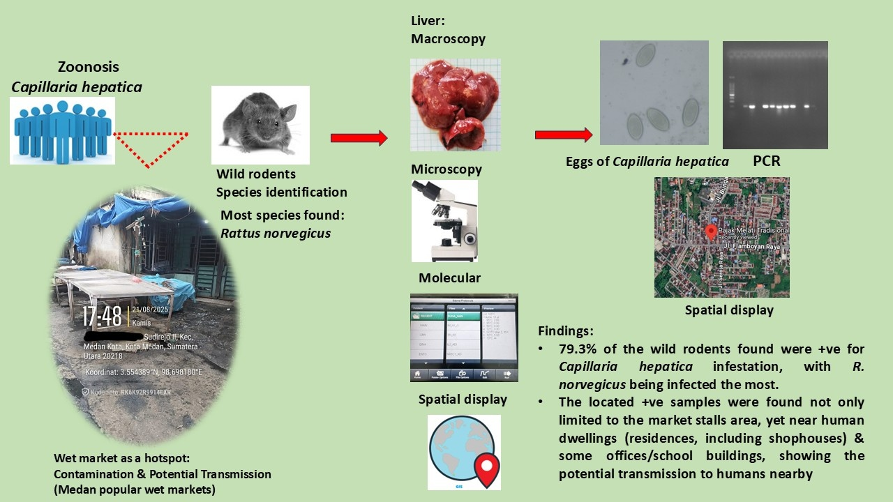
Graphical Abstract

1. Introduction

Over 60% of human infections originate from zoonotic sources. This encompasses a diverse array of bacteria, viruses, fungi, and parasites. Interactions between people and animals facilitate the transmission of zoonoses [1]. Synanthropic animals are wildlife that inhabit areas near human settlements and derive benefits from anthropogenically altered habitats, such as urban areas, agricultural lands, or residences. Among the synanthropic animals that play important roles in zoonosis transmission in human environments are wild rodents [2]. They have swift reproductive cycles and extensive occupancy, frequently interacting with humans and the surrounding environment. Rodents are not only considered pests and harmful to agriculture and property but also act as reservoirs of various zoonotic pathogens. Within this group, the species Rattus norvegicus, known as the brown rat, and Rattus tanezumi, also known as the Asian house rat, are commonly studied for their roles in zoonotic diseases. They are linked to diseases such as plague, hantavirus pulmonary syndrome, leptospirosis, scrub typhus, and numerous other zoonotic diseases [3,4,5,6]. As zoonosis reservoirs, they substantially facilitate the transmission and propagation of zoonosis through multiple direct and indirect mechanisms. Transmission may occur by bites, inhalation of pathogens from rodent feces, or consumption of food and drink contaminated by rodent excreta. For Capillaria hepatica, the transmission is thought to occur mainly through rodents’ carcasses contaminating the environment [7,8,9,10,11,12,13].
Capillaria hepatica, also known as Calodium hepaticum, is a parasitic nematode that primarily infects rodents’ livers. C. hepatica exhibits a wide host range, encompassing at least 80 species of rodents from the family Muridae and infecting a minimum of 24 other mammalian families. It can cause symptoms similar to those of hepatitis, but also others. Due to its location, it often goes undiagnosed, but in many cases it is also asymptomatic. Reports in humans found that it can cause the zoonotic hepatic capillariasis, with clinical presentations similar to those of acute or subacute hepatitis [7]. Symptoms may include fever, abdominal pain, jaundice, hepatomegaly, and eosinophilia. Heavy infection can lead to liver fibrosis or hepatic failure. Hepatic capillariasis is frequently mistaken for various liver illnesses, including viral hepatitis, bacterial liver abscess, and liver tumors, due to its varied and unusual clinical presentations. Furthermore, this pathogen may also induce pulmonary symptoms, including cough, expectoration, and chest pain, potentially due to ectopic larval migration into the lungs [9,14,15]. Confirmatory diagnosis is made through biopsy/autopsy as the gold standard, serology tests, and imaging such as Magnetic Resonance Imaging (MRI), Computed Tomography (CT), and ultrasound. Diagnosis can be challenging due to the infection’s non-specific clinical presentation. Treatment includes administration of albendazole with corticosteroid [9,16,17]. Considered rare, records of human infection have been documented globally throughout Europe, the Americas, Asia, Africa, and Oceania. Infections can be grouped into pseudo-infections (or spurious infections) and true infections [7,9,12,17,18].
Among the primary risk factors for human infestation are poor sanitary practices, inadequate hygiene, high population density, and contact with rodents or other animals. Individuals, both children and adults, who engage in dirt consumption (geophagia, pica), as well as children exhibiting soil-to-hand-to-mouth contact, are particularly vulnerable [8,9].
Rodents act as the primary reservoirs for C. hepatica in the environment, maintaining the parasite’s life cycle and serving as key amplifiers of environmental contamination. Capillariasis occurs when eggs are ingested; the larvae then migrate to the liver, where they mature, causing inflammation, fibrosis, and tissue damage. Transmission to humans and other incidental hosts occurs through ingestion of embryonated eggs in contaminated soil, food, or water, particularly in regions with high rodent density and inadequate sanitation. Distinct from other nematodes, C. hepatica completes its entire development in the liver of its host, where adult worms lay unembryonated eggs. These eggs are only released into the environment upon the host’s death, decomposition, or predation, at which point they embryonate in soil and become infective. Spurious infections arise when unembryonated eggs are swallowed, usually by predators (cats, dogs) or scavengers of rodent carcasses, pass through the intestine, and are subsequently excreted with feces. Although spurious infection is not a true infection, the eggs excreted can become embryonated in the environment and become a source of infection. Therefore, the abundant presence of C. hepatica in the main reservoir hosts, such as rodents found near human settlements, suggested a highly contaminated environment and a potential public health threat [7,8,9,10,11,12,13,17,19].
Changes in the global environment increase the number of rodents near human dwellings and, in turn, potentially increase the risk of C. hepatica transmission. Capillaria hepatica infection is commonly observed in rodents, particularly in Rattus spp. A few studies revealed the presence of this parasite among rodents from different areas of Indonesia [20,21,22,23,24]. However, the phenomenon observed remains poorly understood. Furthermore, there were no documented cases of C. hepatica infection in humans in the country. This absence may be attributed to underreporting or misdiagnosis. The wet market, as part of the locals’ food security, is a potential hotspot for transmission through contaminated environments. We aimed to identify the occurrence of C. hepatica infestations among wild rodents found in the three popular wet markets in Medan and their zoonotic implications. As part of local food security, it is essential to monitor the occurrence of hotspots for emerging disease transmission in these wet markets.

2. Materials and Methods

2.1. Study Sites and the Captured Rodents

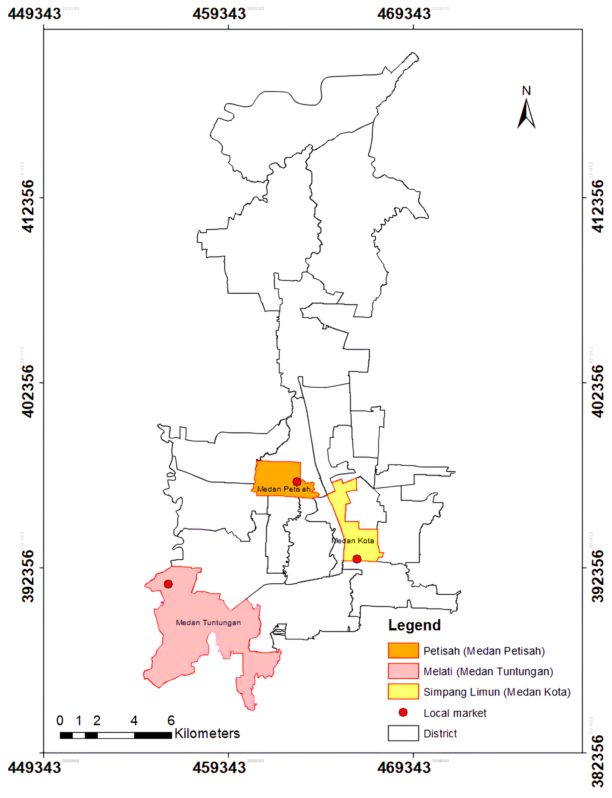
The rodent samples were collected from three popular wet markets in Medan: Simpang Limun (serving the area of Medan Kota district), Petisah (Medan Petisah district), and Melati (Medan Tuntungan district) in August–September 2025 (Figure 1). Medan, the capital of North Sumatra, Indonesia, has a tropical climate, and an area of 281.99 km2. It lies between 3°27′ and 3°47′ of North Latitude, 98°35′ and 98°44′ of East Longitude, and a height 2.5–37.5 m above sea level (masl), and is bordered by the Regency of Deli Serdang. Medan Kota district has a domain of 5.75 km2, covering 2.04% of Medan’s total area, and a 27 masl height, plus a population of 84,779. Medan Petisah covers 1.87% of Medan area, with a width of 5.28 km2 and height of 26 masl, plus 72,599 residents. Medan Tuntungan covers 25.16 km2 area, or around 8.92% of Medan, with an average height of 56 masl, and 101,069 residents [25]. The ethical approval for the study was obtained from the Health Research Ethics Committee of Universitas Sumatera Utara (protocol number 2603268-KEPK-USU).
These three popular wet markets have market stalls selling mostly fresh produce, groceries, and cooked food, with some clothing retailers. The neighborhood area (within 200 m of the markets) includes residential areas (houses and mixed-use/shophouses), offices, and schools. Traps were placed on the crowded market stalls and the nearby human dwellings for three weeks. Trap installation was located next to the stalls, as well as inside and outside of the buildings for human dwellings. The single-use iron live trap applied was rectangular, measuring 34 cm × 20 cm × 15 cm. Dried salted fish and other residual cooked food (e.g., fried chicken) were used as bait. Each site was equipped with 100 traps, installed daily at 6 p.m. and collected at 7 a.m. the next morning. Rodents were captured live inside the trap until collection, then transported to the laboratory for further processing.

2.2. Data Collection and Microscopic Examination

Rodents were manually restrained by lightly grasping their neck scruff prior to euthanasia to facilitate the safe and effective delivery of the intramuscular injection. Rodents were euthanized via intramuscular injection of ketamine and xylazine. After anesthesia administration, the sex, body mass, body length, tail length, ear length, and hind leg length of the captured rodents were recorded for identification, following the protocol described by Herbreteau et al. [26]. The captured rodents were identified based on these morphological characteristics [26,27]. After the postmortem, liver samples were collected and visually inspected for visible white spot lesions. The suspected tissue was then dissected, placed in a tube, incubated in pepsin-HCl solution, and examined under a light microscope (CX21 binocular microscope, Olympus®, Tokyo, Japan) for the presence of ova and parasites. Another piece of the tissue was placed in a tube containing 70% ethanol and stored at −20 °C for further DNA analysis.

2.3. Molecular Confirmation of Capillaria hepatica

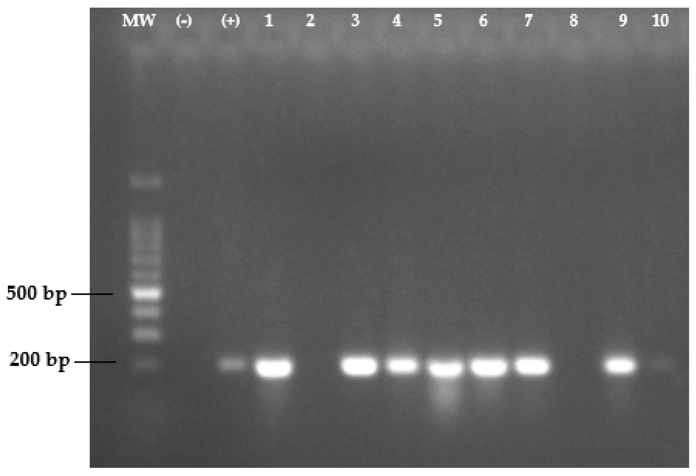
After thawing frozen liver samples, 25 mg of tissue with obvious lesions was manually homogenized in phosphate-buffered saline, and DNA was extracted using the Genomic DNA mini kit for tissue (Geneaid, Geneaid Biotech Ltd., New Taipei City, Taiwan) according to the manufacturer’s instructions. PCR amplification was performed using GoTaq® Green Master Mix (Promega Corporation, Madison, WI, USA) with two published primers (233F: 5′-CGG TTC GCT GTT CAG TTG TT-3′ and 436R: 5′-TGC TGC CTT CCT TGG ATG TA-3′) targeting the highly conserved gene for 18S ribosomal RNA of Capillaria hepatica (GenBank acc. no. LC425008) [28]. The amplification was performed using the Bio-Rad T100 thermal cycler (Bio-Rad Laboratories, Hercules, CA, USA), with the following conditions: denaturation at 95 °C for 2 min; 35 cycles of 30 s at 95 °C, 30 s at 56 °C, and 30 s at 72 °C; followed by a final extension at 72 °C for 5 min. The PCR products (204 bp) were confirmed by 1.5% agarose gel electrophoresis.

2.4. Data Analysis

A spatial display approach was employed to visualize the distribution of rodent occurrence in relation to key environmental and human activity areas. Field data on rodent presence were collected using geographic coordinates and subsequently processed within a Geographic Information System (GIS) environment. The visualization incorporated primary spatial layers: the local wet markets and human settlements. The settlements included residences (houses) and daily activity sites such as offices and educational facilities, particularly schools attended by children. Rodent occurrence points were mapped and overlaid with these layers for spatial visualization and identified areas of overlap.
All data were tabulated in Microsoft Excel (Version 2508, Microsoft, Redmon, WA, USA) and descriptively analyzed using tables, charts, and percentage calculations.

3. Results

3.1. Captured Rodents, C. hepatica Prevalence, and Spatial Display

A total of 150 wild rodents were captured in these three popular wet markets in Medan. We identified two rodent species, R. norvegicus and R. tanezumi, based on morphological findings. The most captured species was R. norvegicus (70.7%), followed by R. tanezumi (29.3%). Each captured point was grouped into three categories: market stalls, houses/shophouses, and the school/office area. The study performed a descriptive comparison of the rodent species captured, showing the value of each species identified in the examined areas (Table 1).
The Simpang Limun wet market yielded 49 rodent samples, of which 27 were identified as R. norvegicus, and the remaining 22 were described as R. tanezumi. The Petisah market had 51 rodents, comprising 40 R. norvegicus and 11 R. tanezumi. The Melati market collected 50 rodent samples, of which 39 were identified as R. norvegicus and 11 as R. tanezumi. Overall, molecular examination revealed that R. norvegicus demonstrated high infestation, with 95 of 106 samples testing positive for C. hepatica. The overall prevalence of C. hepatica in the captured rodents was 79.3%, with Petisah having the highest local prevalence (48/51).
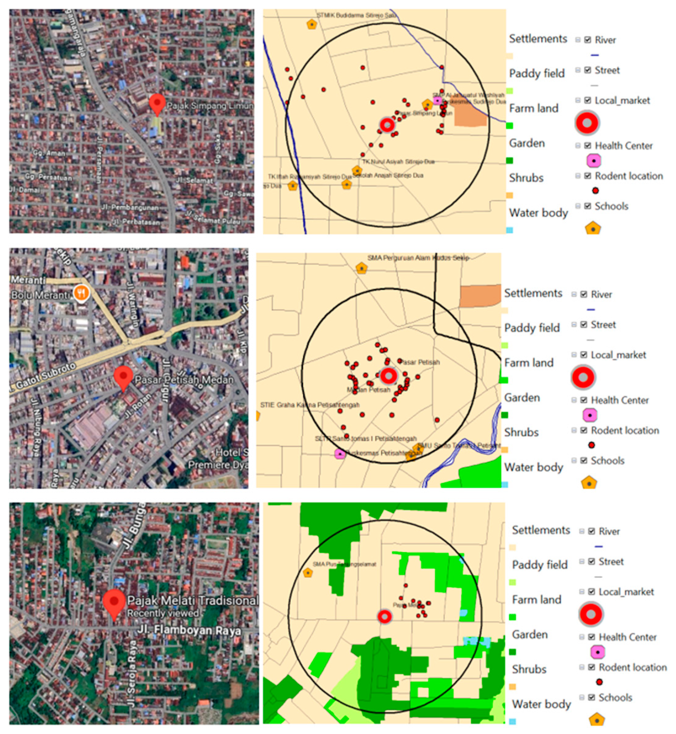
In the spatial display at Simpang Limun, most of the infected rodents were found in the market stalls and house areas. However, one positive rodent was found in the nearby primary school. This is an important finding, considering that C. hepatica transmission risk is more prominent in children. For Petisah, C. hepatica-infected rodents were mostly found in market stalls. Additionally, three cases were found in the nearby office area. The Melati wet market also showed that most positive samples came from rodents found near the market stall areas, yet no cases were found near the school/office area (Figure 2). An additional note in the findings was that the identified office areas for the captured rodents were informal offices, where the building structure was either a house or shophouse being used as office, and that they were not listed in the GIS system as established offices; hence, the spatial layer display for the appointed offices was minimal/barely demonstrated.

3.2. Microscopy and Molecular Findings

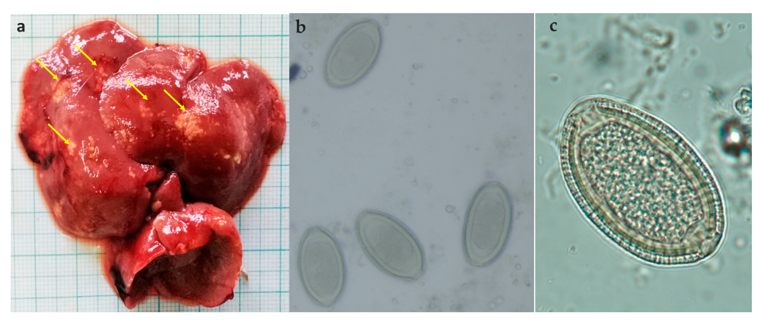
Macroscopic examination of the removed livers from the rodent samples consistently revealed yellowish to milky lesions on the liver surface, suggesting the presence of C. hepatica. Microscopy revealed distinctive ellipsoid-shaped eggs, with numerous joining pits which looked like striations on the shells, and shallow polar plugs (Figure 3).
Using the 18S rRNA-based genomic amplification approach, we detected a 204 bp band in positive C. hepatica samples (Figure 4). Microscopy was slightly inferior to PCR, detecting 118 of 119 positive cases. All the positive rodents were found at market stalls and in nearby residential areas.

4. Discussion

4.1. Sites of Trapping

The three wet markets in Medan, where the study was conducted, are popular, with large volumes of visitors and high trading activity in fresh produce and other food. The selection of these three sites was determined by various considerations, including their role in serving communities in an urban setting, their close proximity to human dwellings, and their location in a relatively densely populated region. Wet markets are part of local food security, enabling communities to access affordable food and goods for daily needs. On the other hand, abundant food supplies from leftovers and waste are available for rodents residing in these wet markets, providing a suitable environment for their population’s growth. High population density will also facilitate close contact between rodents and humans, thereby elevating the risk of zoonosis transmission. Inadequate waste management and poor hygiene maintenance are challenges commonly found in wet markets, thereby further promoting the growth of rodent populations [29,30,31]. A similar situation was found in these wet markets, where insufficient waste management is combined with a lack of hygiene upkeep in resource-limited urban settings.
The Simpang Limun wet market is situated in crowded area and near human dwellings, including houses, schools, and offices. Additionally, some of the houses nearby, particularly those in the approximate area of this market, are shophouses. Shophouses in Medan are mixed-use buildings commonly found in urban settings, often with constricted spaces. Some of them are two- to three-story buildings with the ground floor used for commercial space and the upper levels for living areas. Some are one-story buildings with the front area used for trading and the back for living [32]. This kind of setting is more convenient and efficient for the vendors. A similar situation is found in Petisah wet market, which is also densely populated. Shophouses are commonly found in this area, and based on our findings, they provide a significant shelter for the captured rodents’ population, although not as prominent as the market stalls. The wet market Melati, compared to the two previous places, has a lower human population density and a wider area [25]. Fewer shophouses are found in Melati than in the other two places. Most of the captured rodents in these three markets were from market stalls, followed by the houses/shophouses area, with fewer being found in the school/office area. The number of captured rodents in the houses/shophouses category in Simpang Tuntungan and Petisah was higher in shophouse buildings. However, in Melati, fewer rodents were captured in houses/shophouses, and they were mostly obtained from the house structure buildings. Market stalls and shophouses can provide an abundant source of food for these rodents. Previous studies on rodents and zoonotic infections in urban settings observed a similar pattern [1,3,22,30,33]. The urban rat population was sustained by the high density of residential zones surrounding the market, ongoing human activity, abundant food resources, and inadequate waste control in the market. These factors facilitate the exposure and transmission of rodents and the zoonotic pathogens they carry to humans.

4.2. Species of the Captured Rodents

In all three markets, R. norvegicus has been shown to be the more prominent species, followed by R. tanezumi. Rattus norvegicus is among the most abundant and ubiquitous urban rodent species considered pests worldwide. Rattus tanezumi is indigenous to Asia; however, due to global trade, it has spread worldwide. Aside from being agricultural pests and causing structural damage to building construction, both are also associated with various zoonotic pathogens distributed globally [34,35,36]. The capture of these rodents suggested a correlation between human activities and their presence in these areas.
The sewage rat R. norvegicus, popularly known as the brown rat, is prevalent in human habitats worldwide due to its extensive distribution and diverse dietary adaptability, enabling swift acclimatization to its environment. Well adapted for close contact with humans and rarely found in the wild, this species is prevalent in metropolitan regions with lower socioeconomic levels, and in areas where natural soil remains accessible for burrowing [29]. Thus, the wet markets in Medan municipality are well-suited as habitats for R. norvegicus. Additionally, numerous studies have established the presence of R. norvegicus in public spaces and its role as a reservoir for various zoonotic infections [3,33,34]. Rattus tanezumi, known also by the name the Asian house rat, is a widespread commensal rodent found throughout East and Southeast Asia, and has recently expanded worldwide. In addition to being agricultural pests and causing property damage, R. tanezumi is a potent reservoir for numerous zoonotic diseases owing to its migratory behavior and varied habitats, as documented in previous studies [3,33,37]. In this study, our findings also showed similar results, where R. norvegicus was found as the most frequent species in public spaces, followed by R. tanezumi.
Our findings also showed that R. norvegicus was the most infested species with C. hepatica. Of the 119 rodents infected with C. hepatica, the infestation percentage for R. norvegicus was 89.6%. Other studies have documented similar results, showing the high prevalence of C. hepatica in R. norvegicus, indicating that this species is one of the main hosts of C. hepatica [10,11,19,38]. Interestingly, our findings showed the highest prevalence of C. hepatica in captured rodents at 79.3%, compared with other published studies reporting prevalence between 21.11% and 52%, indicating the abundant presence of this pathogen at the study site.
In Simpang Limun, the number of C. hepatica-infested rodents captured in the market stalls and houses/shophouses area was not much different (15 vs. 13). Most of the positive rodents were R. norvegicus. Of note, one captured rodent infested with this pathogen was found near the primary school area. As transmission of C. hepatica is usually through contaminated environments, such as soil/food/water, and children are particularly at risk [8,9]. This finding indicates the likelihood of C. hepatica transmission to these schoolchildren. In Petisah, the infested captured rodents were dominated by R. norvegicus, and all individuals of this species were infected with C. hepatica. Most of the infested rodents were captured in market stalls, followed by shophouses, suggesting that shophouses can provide accommodating shelter for C. hepatica-infested rodents. In the office area, we found three R. norvegicus rodents, all infected with C. hepatica. From the Melati market, the infected captured rodents were also dominated by R. norvegicus. They were mostly found in market stalls. Six infested rodent samples were captured in the house buildings, and one was captured in the shophouses. No rodents were found in the school/office category. As Melati is located in the Medan Tuntungan district, which spans a wider area, it has a relatively lower population density than the other two districts, and fewer shophouse structures were found in this area. Overall, Petisah has the highest local prevalence for C. hepatica-infested rodents (94.1%). Petisah resides in a densely populated area. Other studies also reported a higher prevalence of C. hepatica-infested rodents in more densely populated areas, including in urban settings [10,11,13]. Rodents choose specific urban environments based on the accessibility of suitable shelter, food, and water. Among these three elements, food availability is believed to be the most significant. In urban settings, particularly in densely populated areas, inadequately stored or improperly disposed of food and organic waste serve as the primary food source for rats. These three wet markets, particularly Petisah, have been shown to be ideal places for the occupancy of these C. hepatica-infested rodents [29].

4.3. Microscopy and Molecular PCR Results

The macroscopic analysis of the excised livers from the rodent’s autopsy consistently demonstrated yellowish to milky lesions. During macroscopic examination, the suspected captured rodents’ livers consistently showed distinct feature of milky lesions, suggesting C. hepatica infestation. Microscopy and molecular identification were also performed on similar cuts of distinctive lesions from the autopsy of each rodent’s liver. Microscopy showed a slightly lower sensitivity than PCR, identifying 118 of 119 positive cases.
The gold standard for detecting C. hepatica is liver biopsy or autopsy. The morphology of the egg structure observed under microscopy in the biopsy/autopsy is also quite distinctive and straightforward, with ellipsoid-shaped eggs, striated shells, and shallow polar plugs [9,16]. Other methods, such as serology, imaging, and molecular identifications, were developed because biopsy/autopsy is invasive. The molecular examination using the 18s rRNA target gene of C. hepatica was initially developed for the detection in an asymptomatic patient, and has been used since then in other published studies [11,28]. The slightly lower microscopy sensitivity compared to PCR in our findings was probably due to the selection of the autopsy tissue for further examination by these two methods. Selecting the suspected milky liver lesions from the autopsy for examination improved microscopy performance.

4.4. Zoonosis Perspective

Wild rodents can host numerous zoonotic pathogens that can be transmitted to humans, including parasites, viruses, and bacteria. Hepatic capillariasis is considered a rare and neglected disease in humans. So far, 197 cases have been documented sporadically worldwide, including from Europe, Asia, the Americas, Africa, and the Oceania. From that, 97 were confirmed as true infections, while the rest were suspected to be spurious infections [7,12,13,14,17,18,28,39].
The clinical signs of hepatic capillariasis are nonspecific, with symptoms including fever, fatigue, anorexia, nausea, vomiting, abdominal discomfort, and diarrhea. Infection may also induce pulmonary manifestations, such as cough, expectoration, and chest pain, associated with the larva migration in the pulmonary system. Hepatomegaly, discomfort in the hepatic area, and jaundice can also be found. Severe infection can lead to liver fibrosis, hepatic failure, and can even be fatal. Hepatic capillariasis is frequently mistaken for various other liver illnesses, including viral hepatitis, bacterial liver abscess, and liver tumors, due to its atypical clinical presentations. Low awareness among the community regarding this disease, the lack of information from extensive systematic epidemiological studies, the challenge in diagnosis, where the gold standard is the invasive biopsy/autopsy, and the failure to identify moderate or asymptomatic infections, all combined, are contributory factors in the misdiagnosis and underreporting of the disease. Historically, no recorded cases of C. hepatica infection in humans from Indonesia have been reported. However, reports of C. hepatica infestation in rodents were reported from different areas of Indonesia [23,24,38]. Furthermore, hepatitis is of growing concern in Medan municipality [40]. Hence, it is suggested that human cases are underreported and/or misdiagnosed in Indonesia.
This study has some limitations. Because we focused only on detecting C. hepatica in rodents at the study location, we qualitatively identified C. hepatica-infected rodents based on the presence or absence of the pathogen. We did not measure parasite/egg density or perform histopathological examination and the grading of the liver’s damage. We also did not measure contamination levels of C. hepatica in soil/food/water at the study location, nor did we examine the roles of other animal predators (cats, dogs) and scavengers in the area through spurious infection modes to amplify contamination and transmission. Furthermore, we did not include secondary data on suspected undifferentiated hepatitis cases among humans from the study location to investigate their association with hepatic capillarias. In addition, due to limited resources, we did not continue the PCR identification with nucleotide sequencing to enrich the study findings. Nevertheless, this is the first study to demonstrate the presence of C. hepatica-infected rodents in public spaces in Medan municipality, the capital city of North Sumatra province, suggesting that the study location was substantially contaminated and highlighting the potential of rodents and this pathogen to cause zoonotic transmission to humans in the area, posing a significant public health issue.

5. Conclusions

The present study revealed that wild rodents, particularly R. norvegicus and R. tanezumi, which are prevalent in urban areas, may act as the main reservoirs for C. hepatica transmission to humans in the study location. Furthermore, the widespread presence of these infested rodents in the studied markets and nearby human dwellings suggested substantial potential for environmental contamination by C. hepatica and an imminent threat of human transmission. Urban public spaces, such as wet markets, are highly conducive to becoming the habitats of these infested rodents. Improved waste disposal, increased hygiene practices, improved architectural design, and proper storage of food and leftovers to control the population of these rodents are important [29,30,31]. Active routine surveillance to monitor this pathogen in the environment and its association with human infection is also advisable, as C. hepatica presence has been documented in the area. Additionally, the control and monitoring efforts should include other zoonotic pathogens that harbor rodents as their main reservoirs.

Author Contributions

Conceptualization: S.V.H. and A.H.W.; methodology: S.V.H., R.L., S.A., H., M.A., M.P., D.M.D., A.Z., A.H.W. and I.N.D.L.; validation: S.V.H. and A.H.W.; formal analysis: S.V.H., A.H.W. and A.Z.; investigation: S.V.H., H., M.A., M.P., D.M.D. and A.H.W.; resources: S.V.H., A.H.W. and I.N.D.L.; data curation: S.V.H. and A.H.W.; writing—original draft preparation: S.V.H.; writing—review, editing, and supervision: S.V.H., R.L., S.A., A.Z., A.H.W. and I.N.D.L.; Project Administration and Funding Acquisition: S.V.H. All authors have read and agreed to the published version of the manuscript.

Funding

This work was supported by research funding from the Directorate of Research and Community Service, Directorate General of Higher Education, Indonesian Ministry of Higher Education, Science, and Technology, in 2025 to SVH (main contract number 112/C3/DT.05.00/PL/2025, subsequent contract number: 17/UN5.4.10.K/PT.01.03/KP-DPPM/2025). The APC was discounted for the journal as this was an invited manuscript submission through an invitation to INDL.

Institutional Review Board Statement

The ethical approval for the study was obtained from the Health Research Ethics Committee of Universitas Sumatera Utara (protocol number 2603268-KEPK-USU, approved on 25 July 2025).

Informed Consent Statement

Not Applicable.

Data Availability Statement

The original contributions presented in this study are included in the article. Further inquiries can be directed to the corresponding author.

Acknowledgments

The authors would like to thank the Master Program in Biomedical Sciences, Faculty of Medicine, Universitas Sumatera Utara, for the support of the research collaboration.

Conflicts of Interest

The authors declare no conflicts of interest.

Abbreviations

The following abbreviations are used in this manuscript:
GISGeographic information system
kmKilometer
mMeter
cmCentimeter
maslMeter above sea level
amAnte meridiem
pmPost meridiem
DNADeoxyribonucleic acid
rRNARibosomal ribonucleic acid
PCRPolymerase chain reaction
CCelsius
minsMinutes
sSeconds
bpBase pair

References

  1. Rahman, M.T.; Sobur, M.A.; Islam, M.S.; Ievy, S.; Hossain, M.J.; Zowalaty, M.E.E.; Rahman, A.M.M.T.; Ashour, H.M. Zoonotic Diseases: Etiology, Impact, and Control. Microorganisms 2020, 8, 1405. [Google Scholar] [CrossRef]
  2. Islam, M.M.; Farag, E.; Mahmoudi, A.; Hassan, M.M.; Mostafavi, E.; Enan, K.A.; Al-Romaihi, H.; Atta, M.; El Hussein, A.R.M.; Mkhize-Kwitshana, Z. Rodent-Related Zoonotic Pathogens at the Human–Animal–Environment Interface in Qatar: A Systematic Review and Meta-Analysis. Int. J. Environ. Res. Public Health 2021, 18, 5928. [Google Scholar] [CrossRef]
  3. Mohd-Qawiem, F.; Nur-Fazila, S.H.; Ain-Fatin, R.; Yong, Q.H.; Nur-Mahiza, M.I.; Yasmin, A.R. Detection of Zoonotic-Borne Parasites in Rattus spp. in Klang Valley, Malaysia. Vet. World 2022, 15, 1006–1014. [Google Scholar] [CrossRef]
  4. Santi, M.; Astuti, E.P.; Yuliasih, Y.; Widawati, M. Infestation of Leptospira in Rodents at the Perimeter Area in “Pelabuhan Ratu” Port, Sukabumi District. In Proceedings of the 1st International Conference for Health Research—BRIN (ICHR 2022); Atlantis Press International B.V.: Dordrecht, The Netherlands, 2023; pp. 957–966. [Google Scholar] [CrossRef]
  5. Chen, Y.L.; Guo, X.G.; Ding, F.; Lv, Y.; Yin, P.W.; Song, W.Y.; Zhao, C.F.; Zhang, Z.W.; Fan, R.; Peng, P.Y.; et al. Infestation of Oriental House Rat (Rattus tanezumi) with Chigger Mites Varies along Environmental Gradients across Five Provincial Regions of Southwest China. Int. J. Environ. Res. Public Health 2023, 20, 2203. [Google Scholar] [CrossRef]
  6. Herbreteau, V.; Bordes, F.; Jittapalapong, S.; Supputamongkol, Y.; Morand, S. Rodent-Borne Diseases in Thailand: Targeting Rodent Carriers and Risky Habitats. Infect. Ecol. Epidemiol. 2012, 2, 18637. [Google Scholar] [CrossRef] [PubMed]
  7. Fuehrer, H.P.; Igel, P.; Auer, H. Capillaria hepatica in Man-an Overview of Hepatic Capillariosis and Spurious Infections. Parasitol. Res. 2011, 109, 969–979. [Google Scholar] [CrossRef] [PubMed]
  8. Pal, M.; Gutama, K.P. Capillariasis as an Emerging Foodborne Helminthic Disease. In Encyclopedia of Food Safety, 2nd ed.; Elsevier: Amsterdam, The Netherlands, 2024; pp. 556–563. [Google Scholar] [CrossRef]
  9. Liu, J.; Liu, R.; Huang, J.; Liu, Q.; Cui, J.; Yu, H. Capillaria hepatica—A Neglected Zoonotic Parasite. Vet. Sci. 2026, 13, 100. [Google Scholar] [CrossRef] [PubMed]
  10. Roqueplo, C.; Lepidi, H.; Medkour, H.; Laidoudi, Y.; Marié, J.-L.; Davoust, B. Enzootic Hepatic Capillariasis (Calodium hepaticum) in Street Rats (Rattus norvegicus) from Marseille City, France. Pathogens 2020, 9, 1048. [Google Scholar] [CrossRef]
  11. Sandy, J.; Uea-Anuwong, T.; Kiu, L.H.; Lee, L.K.F.; Abdullah, S.; Magouras, I. Capillaria hepatica (syn. Calodium hepaticum) Infection and Factors Influencing Infection Carriage in Rats (Rattus spp.) in Hong Kong. One Health 2024, 19, 100878. [Google Scholar] [CrossRef]
  12. Klisiowicz, D.d.R.; Reifur, L.; Shimada, M.K.; Haidamak, J.; Cognialli, R.C.R.; Ferreira, T. High Occurrence of Calodium hepaticum (syn. Capillaria hepatica) Spurious Infection in a Village in the Atlantic Forest of Southern Brazil. Memórias Inst. Oswaldo Cruz 2014, 109, 371–373. [Google Scholar] [CrossRef]
  13. Da Rocha, E.J.G.; De Almeida Basano, S.; De Souza, M.M.; Honda, E.R.; De Castro, M.B.; Colodel, E.M.; Silva, J.C.D.E.; Barros, L.P.; Rodrigues, E.S.; Camargo, L.M.A. Study of the Prevalence of Capillaria hepatica in Humans and Rodents in an Urban Area of the City of Porto Velho, Rondônia, Brazil. Rev. Inst. Med. Trop. São Paulo 2015, 57, 39–46. [Google Scholar] [CrossRef]
  14. Ewing, G.M.; Tilden, I.L. Capillaria hepatica: Report of Fourth Case of True Human Infestation. J. Pediatr. 1956, 48, 341–348. [Google Scholar] [CrossRef]
  15. Juncker-Voss, M.; Prosl, H.; Lussy, H.; Enzenberg, U.; Auer, H.; Nowotny, N. Serological Detection of Capillaria hepatica by Indirect Immunofluorescence Assay. J. Clin. Microbiol. 2000, 38, 431–433. [Google Scholar] [CrossRef] [PubMed]
  16. Wang, L.; Zhang, Y.; Deng, Y.; Li, X.; Zheng, X.; Wang, F.; Zou, Y. Clinical and Laboratory Characterizations of Hepatic Capillariasis. Acta Trop. 2019, 193, 206–210. [Google Scholar] [CrossRef]
  17. Choirunnisa, I.R.; Suwanti, L.T.; Mufasirin; Puspitasari, H. Prevalence and Clinical Characteristics of Human Hepatic Capillariasis in Asia: Review. Media Kedokt. Hewan 2026, 37, 98–113. [Google Scholar] [CrossRef]
  18. Marcelo, L.; Camargo, A.; De, J.; Almeida, S.; Janaina De Souza Vera, L.; Di Tarique, P.; Barreto, C.; Kang Tourinho, E.; Maria De Souza, M. Capillariaisis (Trichurida, Trichinellidae, Capillaria hepatica) in the Brazilian Amazon: Low Pathogenicity, Low Infectivity and a Novel Mode of Transmission. Parasites Vectors 2010, 3, 11. [Google Scholar] [CrossRef]
  19. Quilla, M.H.D.P.; Paller, V.G.V. Histopathological Features and Prevalence of Capillaria hepatica Infection in Rattus spp. in Philippine Mount Makiling Forest Reserve and Its Adjacent Areas. J. Parasit. Dis. 2020, 44, 338–348. [Google Scholar] [CrossRef]
  20. Brown, R.J.; Carney, W.P.; Van Peenen, P.F.D.; Cross, J.H.; Sulianti Saroso, J. Capillariasis in Wild Rats of Indonesia. Southeast Asian J. Trop. Med. Public Health 1975, 6, 219–222. [Google Scholar] [PubMed]
  21. Wiroreno, W. Nematode Parasites of Rats in West Java, Indonesia. Southeast Asian J. Trop. Med. Public Health 1978, 9, 520–525. [Google Scholar]
  22. Abuzahra, M.; Suwanti, L.T.; Yunus, M. A Decade of Research on Ectoparasites and Endoparasites in Wild Rats in Indonesia (2015–2025): A Review. Open Vet. J. 2025, 15, 2329–2342. [Google Scholar] [CrossRef] [PubMed]
  23. Herawati, A. Prevalence of Infection of Endoparasites and Its Intensity in Small Mammals’ Liver Captured from Irrigated Rice Crop Area and Villages. In Proceedings of the 5th Universitas Ahmad Dahlan Public Health Conference (UPHEC 2019), Yogyakarta, Indonesia, 3–4 July 2019; pp. 141–147. [Google Scholar] [CrossRef]
  24. Herawati, N.A.; Sudarmaji. Diversity of Rodent Species and Its Potency as the Vector for Transmitting Rodent Borne Parasitic Disease in Households. In BIO Web of Conferences; EDP Sciences: Les Ulis, France, 2021; Volume 33. [Google Scholar] [CrossRef]
  25. Badan Pusat Statistik Kota Medan. Kota Medan Dalam Angka 2025. 28 February 2025. Available online: https://medankota.bps.go.id/id/publication/2025/02/28/1d05cd4a4df521885f54c685/kota-medan-dalam-angka-2025.html (accessed on 22 March 2026).
  26. Herbreteau, V.; Jittapalapong, S.; Rerkamnuaychoke, W.; Chaval, Y.; Cosson, J.F.; Morand, S. Protocols for Field and Laboratory Rodent Studies; Kasetsart University: Bangkok, Thailand, 2011; pp. 3–33. [Google Scholar]
  27. Yuliadi, B.; Muhidin; Indriani, S. Tikus Jawa, Teknik Survei di Bidang Kesehatan; Ristiyanto, Ahmadi, A.S., Eds.; Lembaga Penerbit Badan Penelitian dan Pengembangan Kesehatan RI: Jakarta, Indonesia, 2013.
  28. Manor, U.; Doviner, V.; Kolodziejek, J.; Weidinger, P.; Dagan, A.; Ben-Haim, M.; Rokah, M.; Nowotny, N.; Boleslavsky, D. Capillaria hepatica (syn. Calodium hepaticum) as a Cause of Asymptomatic Liver Mass. Am. J. Trop. Med. Hyg. 2021, 105, 204–206. [Google Scholar] [CrossRef] [PubMed]
  29. Feng, A.Y.T.; Himsworth, C.G. The Secret Life of the City Rat: A Review of the Ecology of Urban Norway and Black Rats (Rattus norvegicus and Rattus rattus). Urban Ecosyst. 2014, 17, 149–162. [Google Scholar] [CrossRef]
  30. Traweger, D.; Travnitzky, R.; Moser, C.; Walzer, C.; Bernatzky, G. Habitat Preferences and Distribution of the Brown Rat (Rattus norvegicus Berk.) in the City of Salzburg (Austria): Implications for an Urban Rat Management. J. Pest Sci. 2006, 79, 113–125. [Google Scholar] [CrossRef]
  31. Clapperton, B.K. A Review of the Current Knowledge of Rodent Behaviour in Relation to Control Devices; Science & Technical Publishing, Department of Conservation: Wellington, New Zealand, 2006. [Google Scholar]
  32. Zahrah, W.; Ginting, N.; Aulia, D.N.; Marisa, A. “Shop as Home”: The Meaning of Built Environment to Quality of Life of Shop Houses Residents in Medan, Indonesia. Civ. Eng. Archit. 2021, 9, 932–948. [Google Scholar] [CrossRef]
  33. Paramasvaran, S.; Sani, R.A.; Hassan, L.; Kaur, H.; Krishnasamy, M.; Jeffery, J.; Raj, S.; Ghazali, S.M.; Hock, L.K. Endo-Parasite Fauna of Rodents Caught in Five Wet Markets in Kuala Lumpur and Its Potential Zoonotic Implications. Trop. Biomed. 2009, 26, 67–72. [Google Scholar] [PubMed]
  34. Kusumarini, S.; Yesica, R.; Bagus, I.; Rama Wisesa, G.; Hermanto, J.; Nurholizah, Y.; Trinastuti, M.W. Detection of Ecto and Endoparasites Among Wild Rats from Urban Area in Blimbing, Malang, East Java. Acta Vet. Indones. 2021, 95–101. [Google Scholar] [CrossRef]
  35. Blasdell, K.; Bordes, F.; Chaisiri, K.; Chaval, Y.; Claude, J.; Cosson, J.F.; Latinne, A.; Michaux, J.; Morand, S.; Pagès, M.; et al. Progress on Research on Rodents and Rodent-Borne Zoonoses in South-East Asia. Wildl. Res. 2015, 42, 98–107. [Google Scholar] [CrossRef]
  36. Ganasen, T.; Mohd-Azami, S.N.I.; Khoo, J.J.; Peng, T.L.; Johari, J.; Sahimin, N.; Ya’cob, Z.; Abubakar, S.; Loong, S.K. Rodent-Borne Zoonotic Diseases in Southeast Asia: A Narrative Review. Trop. Biomed. 2025, 42, 100–122. [Google Scholar] [CrossRef]
  37. Tijjani, M.; Majid, R.A.; Abdullahi, S.A.; Unyah, N.Z. Detection of Rodent-Borne Parasitic Pathogens of Wild Rats in Serdang, Selangor, Malaysia: A Potential Threat to Human Health. Int. J. Parasitol. Parasites Wildl. 2020, 11, 174–182. [Google Scholar] [CrossRef]
  38. Supriadi, S.; Oktaviana, D.; Nofisulastri, N. Deteksi Cacing Capillaria hepatica Pada Organ Hati Tikus. Biosci. J. Ilm. Biol. 2021, 9, 651. [Google Scholar] [CrossRef]
  39. Aghdam, M.K.; Karimi, A.; Amanati, A.; Ghoroubi, J.; Khoddami, M.; Shamsian, B.S.; Shamsizadeh, A.; Far, S.Z. Capillaria hepatica, a Case Report and Review of the Literatures. Arch. Pediatr. Infect. Dis. 2015, 3, e19398. [Google Scholar] [CrossRef]
  40. Dinas Kesehatan Kota Medan. Profil Kesehatan Medan 2024. 2025. Available online: https://drive.google.com/file/d/11Q4pFRQxdZU5mHCtg1ovLe2b-p9JLy-O/view (accessed on 22 March 2026).
Figure 1. Locations of rodent collection: the wet markets Petisah (in Medan Petisah district), Simpang Limun (in Medan Kota district), and Melati (in Medan Tuntungan district).
Zoonoticdis 06 00024 g001
Figure 2. The spatial display of the distribution of C. hepatica-infected rodents across the three popular wet markets in Medan, with added layers of settlements around to show potential transmission to nearby humans. Appearance in order (from top to bottom): Simpang Limun, Petisah, and Melati.
Zoonoticdis 06 00024 g002
Figure 3. (a) Macroscopic appearance of the rodent’s infected liver with milky spot lesions on the surface (arrows), (b) microscopic appearance of C. hepatica eggs from the rodent’s liver under 400× magnification, and (c) under 1000× magnification.
Zoonoticdis 06 00024 g003
Figure 4. Gel electrophoresis result of the amplified 18s rRNA target gene for C. hepatica, with a 204 bp product. MW: Molecular Weight, (-): control negative, (+): control positive, and samples in number.
Zoonoticdis 06 00024 g004
Table 1. Prevalence of C. hepatica infection in captured rodents from the locations of three wet markets in Medan.
No.Wet Market LocationCategoryCaptured Rodents
R. norvegicusR. tanezumiLocal
Prevalence
+n+n
1.Simpang Limun
(n: 49)
Market stalls114154111559.2%
Houses & shophouses9211437
School/office101000
n:2162781422
2.Petisah
(n: 51)
Market stalls2402462894.1%
Houses & shophouses13013213
School/office303000
n:400408311
3.Melati
(n: 50)
Market stalls2923163984%
Houses & shophouses538202
School/office000000
n:345398311
Total captured
Rodents
1509511106242044
Total positive for Capillaria hepatica95/10624/44
Percentage89.6%54.5%
Overall prevalence:119/150 (79.3%)
n: subtotal of captured rodents, +: rodent positive for C. hepatica, −: rodent negative for C. hepatica.
Disclaimer/Publisher’s Note: The statements, opinions and data contained in all publications are solely those of the individual author(s) and contributor(s) and not of MDPI and/or the editor(s). MDPI and/or the editor(s) disclaim responsibility for any injury to people or property resulting from any ideas, methods, instructions or products referred to in the content.