Proceeding Paper

Enterococcus faecalis Biofilm: A Clinical and Environmental Hazard †

by
Bindu Sadanandan
* and
Kavyasree Marabanahalli Yogendraiah
Department of Biotechnology, M S Ramaiah Institute of Technology, Bengaluru 560054, Karnataka, India
*
Author to whom correspondence should be addressed.
Presented at the 4th International Electronic Conference on Antibiotics, 21–23 May 2025; Available online: https://sciforum.net/event/ECA2025.
Med. Sci. Forum 2025, 35(1), 5; https://doi.org/10.3390/msf2025035005
Published: 5 August 2025
(This article belongs to the Proceedings of The 4th International Electronic Conference on Antibiotics)

Abstract

This review explores the biofilm architecture and drug resistance of Enterococcus faecalis in clinical and environmental settings. The biofilm in E. faecalis is a heterogeneous, three-dimensional, mushroom-like or multilayered structure, characteristically forming diplococci or short chains interspersed with water channels for nutrient exchange and waste removal. Exopolysaccharides, proteins, lipids, and extracellular DNA create a protective matrix. Persister cells within the biofilm contribute to antibiotic resistance and survival. The heterogeneous architecture of the E. faecalis biofilm contains both dense clusters and loosely packed regions that vary in thickness, ranging from 10 to 100 µm, depending on the environmental conditions. The pathogenicity of the E. faecalis biofilm is mediated through complex interactions between genes and virulence factors such as DNA release, cytolysin, pili, secreted antigen A, and microbial surface components that recognize adhesive matrix molecules, often involving a key protein called enterococcal surface protein (Esp). Clinically, it is implicated in a range of nosocomial infections, including urinary tract infections, endocarditis, and surgical wound infections. The biofilm serves as a nidus for bacterial dissemination and as a reservoir for antimicrobial resistance. The effectiveness of first-line antibiotics (ampicillin, vancomycin, and aminoglycosides) is diminished due to reduced penetration, altered metabolism, increased tolerance, and intrinsic and acquired resistance. Alternative strategies for biofilm disruption, such as combination therapy (ampicillin with aminoglycosides), as well as newer approaches, including antimicrobial peptides, quorum-sensing inhibitors, and biofilm-disrupting agents (DNase or dispersin B), are also being explored to improve treatment outcomes. Environmentally, E. faecalis biofilms contribute to contamination in water systems, food production facilities, and healthcare environments. They persist in harsh conditions, facilitating the spread of multidrug-resistant strains and increasing the risk of transmission to humans and animals. Therefore, understanding the biofilm architecture and drug resistance is essential for developing effective strategies to mitigate their clinical and environmental impact.

1. Introduction

Enterococcus faecalis is a Gram-positive, opportunistic pathogen frequently implicated in healthcare-associated infections [1]. Although several members of the Enterococcus genus are associated with nosocomial infections, E. faecalis remains one of the most clinically significant due to its robust adaptability, high virulence, and remarkable ability to acquire antibiotic resistance [2]. It can survive under harsh environmental conditions, colonize medical devices, and evade both immune responses and antimicrobial therapies, primarily through the formation of biofilms [3]. Like many Enterococcus species, E. faecalis can colonize indwelling medical devices, owing to its robust ability to form biofilms—structured, surface-attached multicellular aggregates [2,3]. In E. faecalis, biofilm formation is a well-characterized feature that significantly contributes to its pathogenicity, resilience under antimicrobial stress, and persistence in environmental reservoirs—although biofilm formation is also observed in other Enterococcus species and common among many biofilm-forming bacterial pathogens [4].
Genetic studies have identified multiple virulence factors that are either unique to or enriched in E. faecalis, including the enterococcal surface protein (Esp), endocarditis and biofilm-associated pili (Ebp), cytolysin, aggregation substance, secreted antigen A (SagA), and microbial surface components recognizing adhesive matrix molecules. These are regulated by quorum-sensing systems such as the Faecalis system regulator (fsr) locus and are more comprehensively characterized in E. faecalis compared to other Enterococcus species [5].
The global spread of multidrug-resistant E. faecalis, particularly vancomycin-resistant strains (VRE), has been linked to hospital environments, zoonotic reservoirs, and wastewater ecosystems, raising serious public health concerns regarding its environmental survival and resistance transmission pathways [6]. Biofilm-associated antibiotic tolerance arises from reduced drug penetration, metabolic heterogeneity, and the presence of mobile genetic elements, all of which complicate treatment and promote the persistence of infection. In diverse environmental niches, such as wastewater, hospital infrastructure, and food-processing surfaces, E. faecalis forms highly resilient biofilms that withstand adverse physicochemical conditions. These biofilms serve as reservoirs for multidrug-resistant strains and facilitate their transmission to humans and animals [7,8].
To highlight the clinical importance of E. faecalis biofilms, a comparative overview is provided of major biofilm-forming ESKAPE (Enterococcus faecium, Staphylococcus aureus, Klebsiella pneumoniae, Acinetobacter baumannii, Pseudomonas aeruginosa, Enterobacter species) pathogens like Staphylococcus aureus and Pseudomonas aeruginosa, and Escherichia coli as a relevant non-ESKAPE comparator. Biofilm formation is a widely conserved strategy among clinically relevant pathogens and contributes significantly to chronic and device-associated infections. S. aureus forms biofilms rich in polysaccharide intercellular adhesin, facilitating persistence on indwelling devices and prosthetic materials [9]. P. aeruginosa produces highly structured, mucoid biofilms regulated by complex quorum-sensing systems (Las, Rhl, and Pqs), particularly in chronic lung and wound infections. E. coli, especially uropathogenic strains, develop biofilms through curli fibers and type 1 fimbriae, contributing to catheter-associated urinary tract infections [10]. In contrast, E. faecalis forms moderately structured yet highly persistent biofilms, driven by Esp and Ebp proteins, and uniquely combines intrinsic and acquired antibiotic tolerance, making its infections particularly difficult to treat [11].
To overcome the biofilm-associated infections, emerging, and advanced approaches are being explored for disrupting biofilm structure and enhancing antibiotic efficacy, such as conventional antimicrobial therapies, nanoparticle-based antimicrobials, quorum-sensing inhibitors, CRISPR-based gene editing, bacteriophage therapy, enzyme-based treatments, anti-biofilm surface coatings, bioelectric or sound-based techniques, and energy-based disruption methods, to combat this persistent pathogen [12].
Despite the extensive research on E. faecalis, several critical knowledge gaps remain. One major unresolved issue is the lack of effective treatments capable of eradicating established E. faecalis biofilms, which are inherently tolerant to antibiotics and immune responses. This tolerance is not solely due to genetic resistance but also biofilm-specific adaptations such as reduced metabolic activity, altered microenvironments, and protective extracellular matrices. Additionally, the mechanisms by which E. faecalis persists and adapts in diverse environmental settings, including wastewater, medical devices, and host tissues, remain incompletely understood. Recent studies have revealed novel resistance elements, horizontal gene transfer events, and surface proteins that facilitate both environmental survival and hospital transmission [13,14,15]. Recent meta-analyses have confirmed the co-occurrence of virulence genes (esp, gelE) with resistance determinants vancomycin (vanA) and erythromycin ribosomal methylase (ermB) in clinical and environmental E. faecalis isolates. These findings underscore horizontal gene transfer as a key driver of global dissemination [16,17,18]. Addressing these gaps is essential for developing targeted interventions and for improving infection control strategies.
While multiple reviews have examined enterococcal virulence or antibiotic resistance in general, this article presents a focused and updated review of the biofilm-forming capacity of E. faecalis, including its biofilm architecture, genetic control, resistance dynamics, environmental persistence, and current treatment strategies [19,20]. Importantly, it highlights the knowledge gaps in our understanding of its biofilm biology, such as the specific reasons for its persistent environmental survival and the failure of many antibiofilm interventions. Unlike broader reviews, this work concentrates specifically on E. faecalis, comparing general enterococcal traits versus species-specific features where relevant, and integrates both clinical and environmental perspectives [21,22].
To ensure comprehensive and transparent coverage of the literature, we conducted a systematic search using NCBI PubMed, Scopus, Web of Science, and Google Scholar, focusing on peer-reviewed articles published from Jan 2010 to June 2025. Keywords used included “Enterococcus faecalis biofilm,” “biofilm architecture,” “virulence,” “resistance,” “treatment,” and “environmental survival.” Inclusion criteria were as follows: (i) studies published in English, (ii) peer-reviewed research specifically addressing E. faecalis biofilm structure, pathogenesis, or treatment strategies, and (iii) studies involving clinical or environmental isolates. Exclusion criteria eliminated articles that focused primarily on other Enterococcus species without E. faecalis-specific findings, as well as non-English or non-peer-reviewed sources. Therefore, the scope of this review selectively includes high-impact studies published from Jan 2010 to June 2025 on E. faecalis biofilm architecture, developmental stages, regulatory genetics, virulence factors, global transmission, antibiotic resistance, and experimental biofilm disruption strategies. It is not an exhaustive catalog of all biofilm studies, but rather a curated synthesis aimed at identifying critical knowledge gaps and future research directions that bridge clinical microbiology and environmental health.

2. Stages of Biofilm Formation in Enterococcus faecalis

Biofilm formation in E. faecalis is a complex, multi-stage process involving several coordinated genetic and biochemical pathways. The stages of the biofilm formation in E. faecalis is shown in Figure 1. It generally includes the following stages:
Adhesion: This is the initial step that involves the adhesion of planktonic (free-floating) cells to a surface. E. faecalis uses several surface-associated structures, such as aggregation substance (Agg), which is a pheromone-inducible surface protein that promotes adherence to host cells and abiotic surfaces. Esp plays a vital role in primary attachment and biofilm maturation. Ebp helps in mediating adhesion to host tissues and medical device surfaces. Adhesins such as lipoteichoic acid and other surface-associated molecules assist in electrostatic and hydrophobic interactions.
Microcolony Formation: Once attached, the cells begin to divide and form microcolonies. During this phase, intercellular communication and matrix production begin. E. faecalis utilizes the fsr quorum-sensing system, which regulates the production of gelatinase (gelE) and serine protease (sprE), key enzymes involved in biofilm development. Autolysins (AtlA) are the enzymes that contribute to the release of extracellular DNA (eDNA), which helps in cell–cell adhesion and matrix stability.
Maturation: The maturing biofilm becomes increasingly structured, forming layers of cells embedded in an extracellular polymeric substance (EPS) composed of polysaccharides, proteins, extracellular DNA, and lipids. This matrix protects the cells from external threats and facilitates nutrient exchange.
Dispersion: In response to environmental cues (e.g., nutrient depletion or immune pressure), cells can leave the biofilm to colonize new niches. This stage is critical for infection dissemination and recurrence [23,24,25].

3. Architecture of Enterococcus faecalis Biofilm

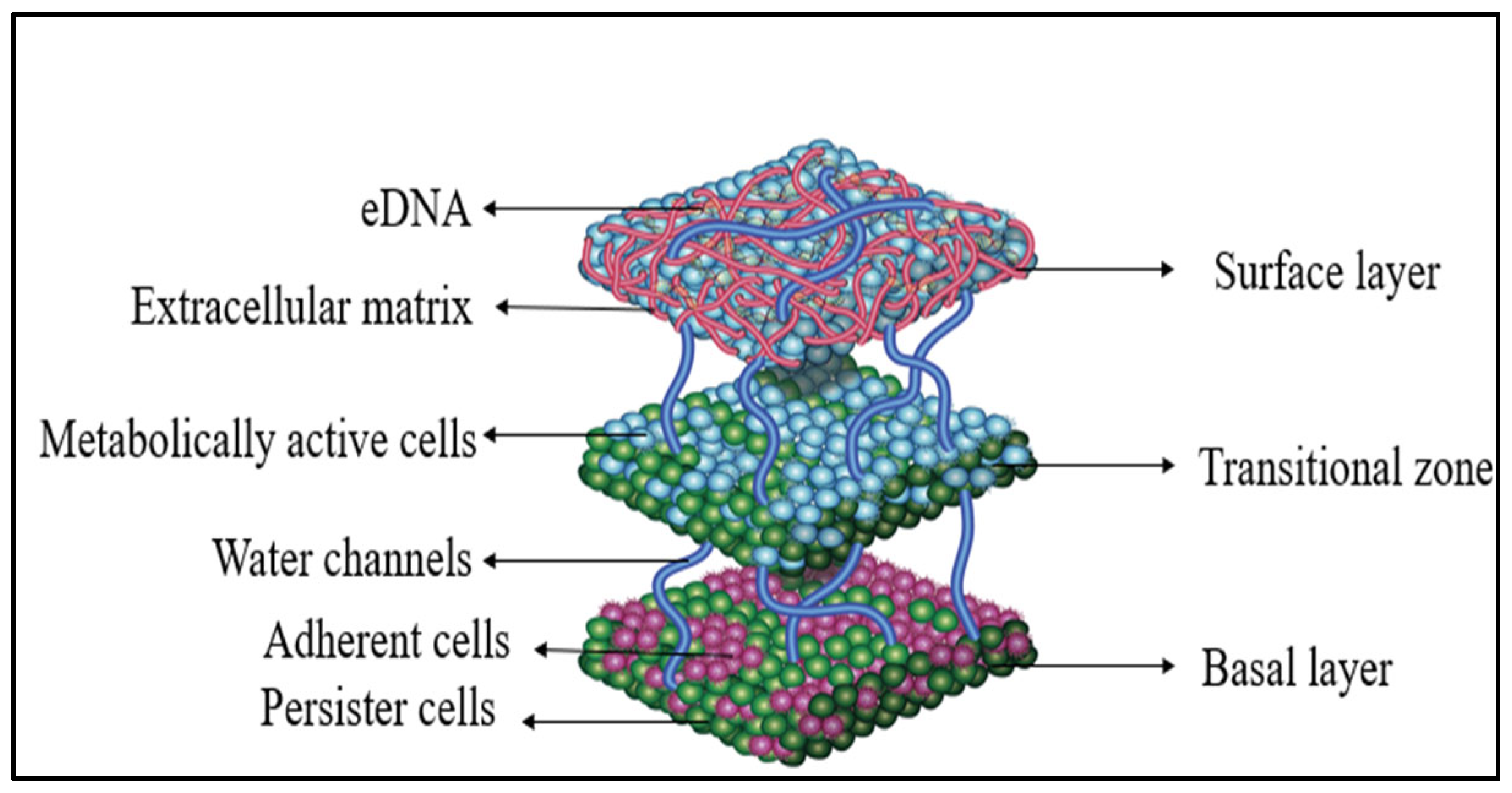
The E. faecalis biofilm displays a stratified 3D architecture with distinct layers, as shown in Figure 2. At the bottom layer (basal layer), cells are in direct contact with the underlying surface, such as tissue, a catheter, or abiotic material. This region mainly consists of the initially adherent cells and is often enriched with persister cells—dormant, highly antibiotic-tolerant cells that contribute to biofilm resilience and chronic infection. The extracellular matrix (ECM) in this layer is typically thinner or partially degraded, with less eDNA unless enhanced by local cell lysis. Above this is the middle layer (transitional zone), where ECM and cell density remain high, but eDNA levels are reduced compared to the top layer. This zone contains a mix of metabolically active and dormant cells and functions as a diffusion gradient layer, where oxygen and nutrient levels start to decline, affecting gene expression and metabolic activity. The topmost layer (surface layer) is rich in ECM components such as polysaccharides, proteins, and eDNA. This layer acts as a protective shield against environmental stresses, including antimicrobial agents and immune responses. Cells here are usually metabolically active due to their better access to oxygen and nutrients, and eDNA is especially abundant in this region as well as at the biofilm periphery, contributing to structural integrity and horizontal gene transfer. Overall, eDNA distribution, matrix density, and cell activity change with depth, creating a heterogeneous biofilm architecture that promotes survival under hostile conditions and complicates treatment efforts [26,27].

4. Clinical Hazards Caused by E. faecalis Biofilm

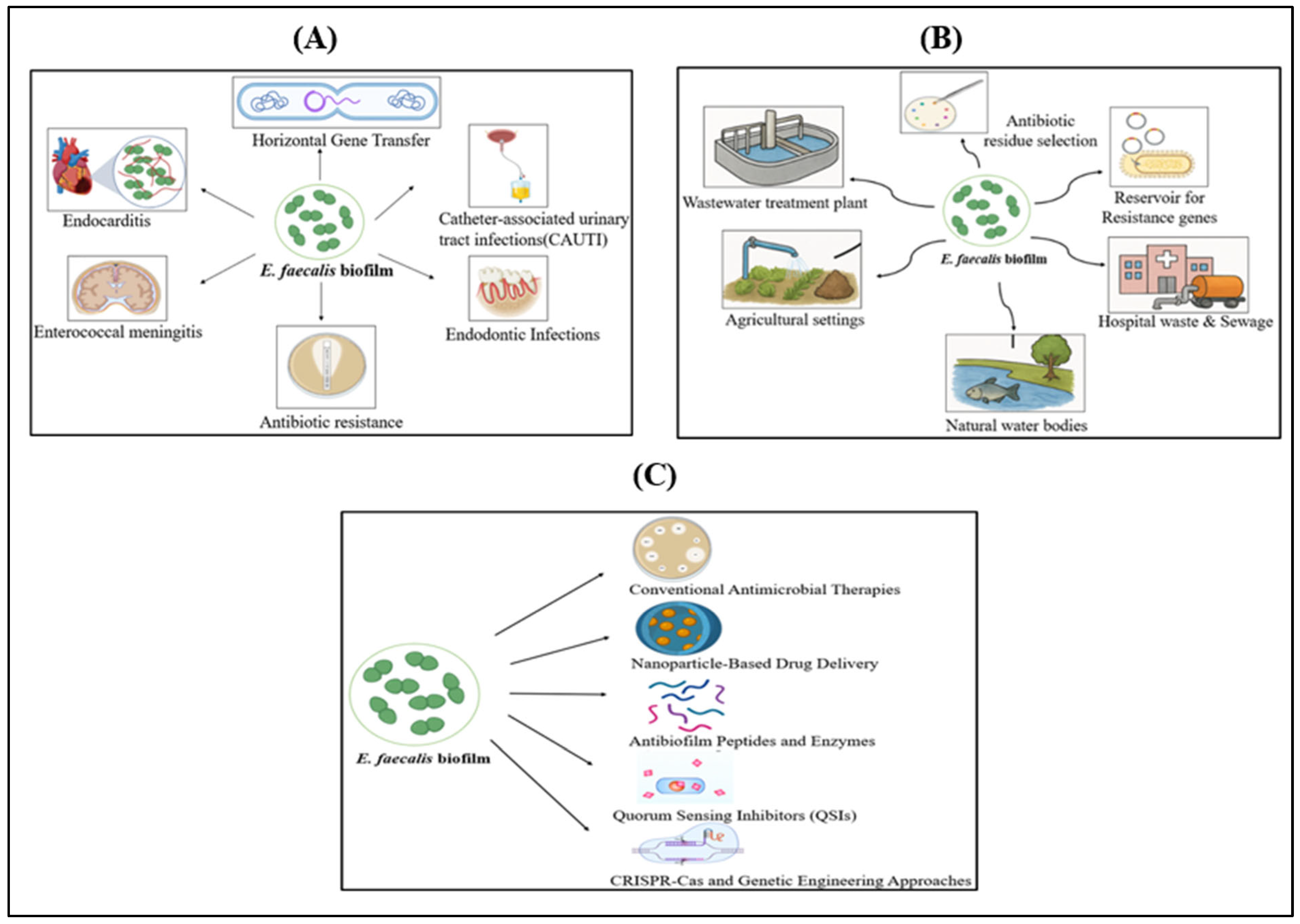
E. faecalis biofilms pose serious clinical challenges due to their ability to evade the host immune system and resist antibiotic treatment, leading to chronic infections, treatment failures, and increased healthcare costs [28]. These biofilms are implicated in urinary tract infections, endocarditis, and persistent endodontic infections, as shown in Figure 3A. Within the biofilm, E. faecalis exhibits an altered metabolism, reduced growth rates, and a protective ECM that limits antibiotic penetration and immune cell access, necessitating prolonged therapy or device removal. In endodontics, its survival in dentinal tubules and resistance to high pH medicaments contribute to root canal failure. Moreover, biofilm-associated strains facilitate the horizontal transfer of resistance genes, promoting multidrug resistance in hospital environments. Notably, biofilm-embedded E. faecalis can be up to 1000 times more resistant to antibiotics like ampicillin, vancomycin, and linezolid compared to planktonic cells [29,30,31].

5. Environmental Hazards Caused by E. faecalis Biofilm

E. faecalis biofilms poses significant environmental risks due to their persistence in diverse habitats such as water systems, food-processing environments, and agricultural settings, as shown in Figure 3B. In aquatic systems, biofilm formation on surfaces within drinking water pipelines and wastewater treatment facilities enhances E. faecalis survival against chlorination and other disinfection methods, contributing to recurring contamination and potential public health risks [32,33]. In food industries, biofilms on equipment surfaces serve as persistent contamination sources, reducing product shelf life and increasing the risk of foodborne transmission [34].
Agricultural runoff and manure use facilitate the environmental spread of E. faecalis, particularly strains carrying antibiotic resistance genes like vanA and ermB. These biofilms support horizontal gene transfer, raising concerns about resistance dissemination through the food chain and water sources [35]. Furthermore, environmental biofilms may serve as reservoirs for multidrug-resistant strains that can re-enter clinical settings, emphasizing the need for monitoring under the One Health approach [36]. Collectively, E. faecalis biofilms contribute to environmental contamination, resistance gene propagation, and ecosystem imbalance.

6. Emerging Strategies for Prevention and Treatment of E. faecalis Biofilm

Treating E. faecalis biofilms remains a major challenge, especially in endodontics, implant-related, and hospital-acquired infections. Their resistance to antibiotics and immune responses has prompted the need for advanced strategies, as shown in Figure 3C.

6.1. Conventional Antimicrobial Therapies

The conventional treatment of E. faecalis infections relies on antibiotics such as ampicillin, vancomycin, and linezolid. Ampicillin, a β-lactam antibiotic, is preferred due to its high affinity for penicillin-binding proteins, effectively disrupting cell wall synthesis [37]. However, biofilm-embedded E. faecalis shows marked tolerance to β-lactams due to limited drug penetration, metabolic dormancy, and persister cells [38]. Combination therapies, such as ampicillin with ceftriaxone, have demonstrated synergistic effects, especially in cases resistant to aminoglycosides [39]. In endodontics, agents like calcium hydroxide, chlorhexidine, and sodium hypochlorite are used but have a limited impact on mature biofilm [40]. This highlights the need for more effective and adjunctive strategies to combat biofilm-associated E. faecalis infections.

6.2. Nanoparticle-Based Antimicrobial Delivery

Nanoparticle-based delivery systems offer an effective strategy against biofilm-associated infections, enhancing drug stability, solubility, and targeted delivery. Their small size allows for better biofilm penetration and sustained release. Some nanoparticles, like silver and zinc oxide, also possess intrinsic antimicrobial activity by disrupting bacterial membranes. These systems can bypass resistance mechanisms and improve efficacy against drug-resistant strains [41].

6.3. Plant-Based Alternative Therapy

Plant-derived compounds such as flavonoids and essential oils exhibit strong, low-toxicity antibacterial activity by disrupting cell structures. Garlic, clove, cinnamon, and Indian gooseberry were found to be effective in inhibiting bacterial growth [42].

6.4. Antibiofilm Peptides

Antibiofilm peptides and enzymes offer promising alternatives against E. faecalis biofilms. Peptides like nisin, LL-37, and β-defensins disrupt bacterial membranes and inhibit biofilm gene expression, especially in the early stages [43].

6.5. Enzyme-Based Biofilm Disruption

Enzymatic agents effectively disrupt E. faecalis biofilms by degrading key matrix components. DNases break down extracellular DNA, weakening the structural integrity of the biofilm. Proteases degrade matrix proteins and adhesins, reducing cell adhesion. Dispersin B targets and cleaves specific polysaccharides, while amylases hydrolyze glucans that help stabilize the biofilm. These enzymes enhance the penetration and efficacy of antimicrobial treatments by dismantling the protective biofilm barrier [44].

6.6. Quorum Sensing Inhibitors (QSIs)

Quorum-sensing inhibitors (QSIs) disrupt bacterial communication, reducing E. faecalis biofilm formation and virulence without promoting resistance. The key system in E. faecalis is the fsr quorum-sensing pathway, which regulates enzymes like gelE and sprE involved in biofilm development. Compounds like ambuic acid inhibit the fsr system, suppressing virulence factor production and impairing biofilm formation [45].

6.7. Anti-Biofilm Surface Coatings

Surface engineering approaches, such as coating medical devices with silver nanoparticles, antimicrobial peptides, or quorum-sensing inhibitors, aim to prevent the initial adhesion and subsequent biofilm formation of E. faecalis [46,47].

6.8. CRISPR-Cas and Genetic Engineering Approaches

Recent advances in genome editing, particularly CRISPR-Cas systems, offer promising strategies to combat E. faecalis biofilm infections. CRISPR has been used to selectively disrupt key biofilm and virulence genes such as gelE, sprE, and components of the fsr quorum-sensing system, leading to reduced matrix production, impaired biofilm structure, and lower pathogenicity [48,49].

7. Advanced Strategies for Disrupting E. faecalis Biofilm

7.1. Bacteriophage Therapy

The use of bacteriophages and their endolysins has emerged as a promising strategy to combat E. faecalis biofilms, especially in light of increasing antibiotic resistance. Bacteriophages can penetrate and replicate within bacterial biofilms, causing targeted lysis without disturbing the host microbiota. Recent phage isolates, such as vB_EfaS_HEf13 (lytic Siphoviridae phage) and EFLK1 (Myoviridae family), have shown strong lytic activity against both planktonic and biofilm-embedded E. faecalis strains [50]. In addition, phage-derived endolysins (PlyV12, LysEF-P10) demonstrate potent antibiofilm effects by degrading peptidoglycan independently of host cell viability, making them effective against dormant biofilm cells and persisters [51]. These agents can act synergistically with antibiotics, resensitizing biofilms to conventional drugs and reducing the risk of resistance development. However, limitations such as their narrow host range, potential immune reactions, and delivery challenges still need to be addressed through bioengineering and formulation strategies. As antibiotic options diminish, phage therapy and endolysins represent a vital area of translational research for treating E. faecalis biofilm-associated infections.

7.2. Energy-Based Disruption Methods

  • Bioelectric and Acoustic Methods
Bioelectric effects, including both alternating current (AC) and direct current (DC) electric fields, as well as ultrasound-based techniques, have shown promise in dispersing biofilm structures and enhancing antimicrobial uptake. AC and DC electric fields assist drug delivery by increasing cell membrane permeability and disrupting the biofilm matrix through electroporation and electrostatic forces. Similarly, ultrasound promotes biofilm disruption through mechanical shear forces and cavitation, improving antibiotic penetration and effectiveness [52,53].
  • Shock Wave Therapy
Shock wave therapy, particularly low-energy extracorporeal shock waves, is a novel method for disrupting E. faecalis biofilms. These high-pressure acoustic pulses generate shear forces and cavitation, leading to mechanical disruption of the biofilm matrix. In endodontic applications, shock wave-enhanced irrigation has been shown to improve the removal of E. faecalis biofilms and enhanced antimicrobial penetration [54].
  • Non-Thermal Plasma (NTP)
Recently, NTP has emerged as a powerful, non-contact antimicrobial tool capable of inactivating E. faecalis biofilms through reactive oxygen species (ROS) and reactive nitrogen species (RNS). NTP induces oxidative damage to biofilm cells and disrupts ECM components without causing thermal injury to surrounding tissues, making it suitable for wound care and surface decontamination [55]. These approaches, especially when used in combination, offer exciting alternatives for biofilm management, particularly against drug-resistant and device-associated E. faecalis infections [56].
  • Photodynamic therapy (PDT)
PDT is a light-based antimicrobial approach that disrupts E. faecalis biofilms by using a photosensitizer (methylene blue), visible light, and oxygen to generate ROS. These ROS damage bacterial cells and break down the biofilm matrix. PDT is especially useful in endodontics and offers a non-invasive, resistance-free method to enhance disinfection [57].

8. Conclusions

E. faecalis biofilms present significant clinical and environmental threats due to their complex architecture, multidrug tolerance, and robust virulence mechanisms. Their involvement in chronic, device-associated infections and their persistence in diverse environmental reservoirs underscore the urgent need for integrated strategies to mitigate their impact. Given the capacity of E. faecalis to transmit between humans, animals, and environmental sources, a collaborative One Health approach—bridging human health, veterinary, and environmental sectors—is essential to monitor, control, and prevent its spread.
Future research should focus on unraveling the genetic and molecular underpinnings of biofilm resilience, developing targeted antibiofilm agents, including bacteriophages, endolysins, and enzyme-based therapies, and improving detection methods in clinical and environmental settings. Additionally, understanding the ecology of E. faecalis in wastewater, agricultural, and food-processing environments supports better infection control and sanitation policies. By integrating clinical microbiology, public health surveillance, and environmental monitoring, more effective and sustainable interventions can be developed to address the growing threat posed by E. faecalis biofilms.

Author Contributions

B.S.; conceptualization, validation, visualization, supervision, review and editing, K.M.Y.; writing—original draft. All authors have read and agreed to the published version of the manuscript.

Funding

This research received no external funding.

Institutional Review Board Statement

Not applicable.

Informed Consent Statement

Not applicable.

Data Availability Statement

The data that support the findings of this study are available from the corresponding author upon reasonable request.

Acknowledgments

Kavyasree Marabanahalli Yogendraiah thanks the Management of M S Ramaiah Institute of Technology for the Ramaiah Doctoral Fellowship.

Conflicts of Interest

The authors declare no conflicts of interest.

Abbreviations

ECMExtracellular matrix
EspEnterococcal surface protein
QSIsQuorum-sensing inhibitors
AggAggregation substance
EbpEndocarditis and biofilm-associated pili
fsrFaecalis system regulator
AtlAAutolysins
GelEGelatinase
SprESerine protease
eDNAExtracellular DNA
EPSExtracellular polymeric substance
vanAVancomycin
ermBErythromycin ribosomal methylase
NTPNon-Thermal Plasma
PDTPhotodynamic therapy

References

  1. Archambaud, C.; Nunez, N.; da Silva, R.A.; Kline, K.A.; Serror, P. Enterococcus faecalis: An overlooked cell invader. Microbiol. Mol. Biol. Rev. 2024, 88, e00069-24. [Google Scholar]
  2. Sadanandan, B.; Vaniyamparambath, V.; Lokesh, K.N.; Shetty, K.; Joglekar, A.P.; Ashrit, P.; Hemanth, B. Candida albicans biofilm formation and growth optimization for functional studies using response surface methodology. J. Appl. Microbiol. 2022, 132, 3277–3292. [Google Scholar] [CrossRef] [PubMed]
  3. Ashrit, P.; Sadanandan, B.; Kyathsandra Natraj, L.; Shetty, K.; Vaniyamparambath, V.; Raghu, A.V. A microplate-based Response Surface Methodology model for growth optimization and biofilm formation on polystyrene polymeric material in a Candida albicans and Escherichia coli co-culture. Polym. Adv. Technol. 2022, 33, 2872–2875. [Google Scholar] [CrossRef]
  4. Sadanandan, B.; Ashrit, P.; Nataraj, L.K.; Shetty, K.; Jogalekar, A.P.; Vaniyamparambath, V.; Hemanth, B. High-throughput comparative assessment of biofilm formation of Candida glabrata on polystyrene material. Korean J. Chem. Eng. 2022, 39, 1277–1286. [Google Scholar] [CrossRef]
  5. Willett, J.L.E.; Dunny, G.M. Insights into Ecology, Pathogenesis, and Biofilm Formation of Enterococcus faecalis from Functional Genomics. Microbiol. Mol. Biol. Rev. 2025, 89, e00081-23. [Google Scholar] [CrossRef] [PubMed]
  6. Guan, L.; Beig, M.; Wang, L.; Navidifar, T.; Moradi, S.; Tabaei, F.M.; Teymouri, Z.; Moghadam, M.A.; Sedighi, M. Global Status of Antimicrobial Resistance in Clinical Enterococcus faecalis Isolates: Systematic Review and Meta-Analysis. Ann. Clin. Microbiol. Antimicrob. 2024, 23, 80. [Google Scholar] [CrossRef]
  7. Ghazvinian, M.; Asgharzadeh Marghmalek, S.; Gholami, M.; Amir Gholami, S.; Amiri, E.; Goli, H.R. Antimicrobial resistance patterns, virulence genes, and biofilm formation in enterococci strains collected from different sources. BMC Infect. Dis. 2024, 24, 274. [Google Scholar] [CrossRef]
  8. Hashem, Y.A.; Abdelrahman, K.A.; Aziz, R.K. Phenotype–Genotype Correlations and Distribution of Key Virulence Factors in Enterococcus faecalis Isolated from Patients with Urinary Tract Infections. Infect. Drug Resist. 2021, 14, 1713–1723. [Google Scholar] [CrossRef]
  9. Peng, Q.; Tang, X.; Dong, W.; Sun, N.; Yuan, W. A Review of Biofilm Formation of Staphylococcus aureus and Its Regulation Mechanism. Antibiotics 2022, 12, 12. [Google Scholar] [CrossRef]
  10. Chadha, J.; Harjai, K.; Chhibber, S. Revisiting the Virulence Hallmarks of Pseudomonas aeruginosa: A Chronicle through the Perspective of Quorum Sensing. Environ. Microbiol. 2022, 24, 2630–2656. [Google Scholar] [CrossRef]
  11. Hakim, T.A.; Zaki, B.M.; Mohamed, D.A.; El-Shatoury, E.H.; Farrag, H.A. Novel Strategies for Vancomycin-Resistant Enterococcus faecalis Biofilm Control: Bacteriophage (vB_EfaS_ZC1), Propolis, and Their Combined Effects in an Ex Vivo Endodontic Model. Ann. Clin. Microbiol. Antimicrob. 2025, 24, 24. [Google Scholar] [CrossRef]
  12. Erol, H.B.; Kaskatepe, B.; Gocmen, D.; Ziraman, F.G. The Treatment of Enterococcus faecalis-Related Root Canal Biofilms with Phage Therapy. Microb. Pathog. 2024, 197, 107081. [Google Scholar] [CrossRef]
  13. Azzam, A.; Elkafas, H.; Khaled, H.; Ashraf, A.; Yousef, M.; Elkashef, A.A. Prevalence of Vancomycin-Resistant Enterococci (VRE) in Egypt (2010–2022): A Systematic Review and Meta-Analysis. J. Egypt. Public Health Assoc. 2023, 98, 8. [Google Scholar] [CrossRef] [PubMed]
  14. Silva, V.; Freitas, C.; Ribeiro, J.; Igrejas, G.; Poeta, P. Comparative Analysis of Antibiotic Resistance and Biofilm Formation in Enterococcus spp. across One Health Domains. FEMS Microbes. 2025, 6, xtaf005. [Google Scholar] [CrossRef] [PubMed]
  15. Michaelis, C.; Grohmann, E. Horizontal Gene Transfer of Antibiotic Resistance Genes in Biofilms. Antibiotics 2023, 12, 328. [Google Scholar] [CrossRef] [PubMed]
  16. Boeder, A.M.; Spiller, F.; Carlstrom, M.; Izídio, G.S. Enterococcus faecalis: Implications for Host Health. World J. Microbiol. Biotechnol. 2024, 40, 190. [Google Scholar] [CrossRef]
  17. Mubarak, A.G.; El-Zamkan, M.A.; Younis, W.; Saleh, S.O.; Abd-Elhafeez, H.H.; Yoseef, A.G. Phenotypic and genotypic characterization of Enterococcus faecalis and Enterococcus faecium isolated from fish, vegetables, and humans. Sci. Rep. 2024, 14, 21741. [Google Scholar] [CrossRef]
  18. Namaki Kheljan, M.; Teymorpour, R.; Peeri Doghaheh, H.; Arzanlou, M. Antimicrobial biocides susceptibility and tolerance-associated genes in Enterococcus faecalis and Enterococcus faecium isolates collected from human and environmental sources. Curr. Microbiol. 2022, 79, 170. [Google Scholar] [CrossRef]
  19. Șchiopu, P.; Toc, D.A.; Colosi, I.A.; Costache, C.; Ruospo, G.; Berar, G.; Todea, D.A. An overview of the factors involved in biofilm production by the Enterococcus genus. Int. J. Mol. Sci. 2023, 24, 11577. [Google Scholar] [CrossRef]
  20. Gaeta, C.; Marruganti, C.; Ali, I.A.; Fabbro, A.; Pinzauti, D.; Santoro, F.; Grandini, S. The presence of Enterococcus faecalis in saliva as a risk factor for endodontic infection. Front. Cell Infect Microbiol. 2023, 13, 1061645. [Google Scholar] [CrossRef]
  21. Radford-Smith, D.E.; Anthony, D.C. Vancomycin Resistant E. faecium: Addressing Global and Clinical Challenges. Antibiotics 2025, 14, 522. [Google Scholar]
  22. Samani, R.J.; Tajbakhsh, E.; Momtaz, H.; Samani, M.K. Prevalence of virulence genes and antibiotic resistance pattern in Enterococcus faecalis isolated from urinary tract infection in Shahrekord, Iran. Rep. Biochem. Mol. Biol. 2021, 10, 50. [Google Scholar] [CrossRef]
  23. Nkemngong, C.; Teska, P. Biofilms, mobile genetic elements and the persistence of pathogens on environmental surfaces in healthcare and food processing environments. Front. Microbiol. 2024, 15, 1405428. [Google Scholar] [CrossRef] [PubMed]
  24. Oli, A.K.; Javaregowda, P.K.; Jain, A.; Kelmani, C.R. Mechanism involved in biofilm formation of Enterococcus faecalis. In Focus on Bacterial Biofilms; Das, T., Ed.; IntechOpen: London, UK, 2022; Chapter 5; pp. 179–200. ISBN 978-1-80355-797-4. [Google Scholar]
  25. Woitschach, F.; Kloss, M.; Schlodder, K.; Borck, A.; Grabow, N.; Reisinger, E.C.; Sombetzki, M. Bacterial adhesion and biofilm formation of Enterococcus faecalis on zwitterionic methylmethacrylate and polysulfones. Front. Cell Infect. Microbiol. 2022, 12, 868338. [Google Scholar] [CrossRef] [PubMed]
  26. Rather, M.A.; Gupta, K.; Mandal, M. Microbial biofilm: Formation, architecture, antibiotic resistance, and control strategies. Braz. J. Microbiol. 2021, 52, 1701–1718. [Google Scholar] [CrossRef] [PubMed]
  27. Cattoir, V. The multifaceted lifestyle of enterococci: Genetic diversity, ecology and risks for public health. Curr. Opin. Microbiol. 2022, 65, 73–80. [Google Scholar] [CrossRef]
  28. Khan, N.; Ishfaq, M.; Mufti, I.U.; Ishfaq, S. A Short Review on Enterococcus faecalis. Pak. J. Sci. Ind. Res. Ser. B Biol. Sci. 2024, 67, 298–310. [Google Scholar]
  29. Miller, W.R.; Munita, J.M.; Arias, C.A. Mechanisms of Antibiotic Resistance in Enterococci. Expert Rev. Anti-Infect. Ther. 2014, 12, 1221–1236. [Google Scholar] [CrossRef]
  30. Mohamed, J.A.; Huang, D.B.; Murray, B.E. High-Level Aminoglycoside Resistance in Enterococci. Antimicrob. Agents Chemother. 2023, 67, e00733-23. [Google Scholar]
  31. Thomas, V.C.; Hiromasa, Y.; Harms, N.; Thurlow, L.R.; Tomich, J.; Hancock, L.E. A Signaling Peptide-Dependent Quorum Sensing System Regulates Enterococcus faecalis Biofilm Formation and Virulence. J. Bacteriol. 2023, 205, e00362-22. [Google Scholar]
  32. García-Aljaro, C.; Ballesté, E.; Muniesa, M.; Jofre, J. Determination of crAssphage in water samples and applicability for tracking human faecal pollution. Microb. Biotechnol. 2017, 10, 1775–1780. [Google Scholar] [CrossRef]
  33. Zdragas, A.; Partheniou, P.; Kotzamanidis, C.; Psoni, L.; Koutita, O.; Moraitou, E.; Tzanetakis, N.; Yiangou, M. Molecular characterization of low-level vancomycin-resistant enterococci found in coastal water of Thermaikos Gulf, Northern Greece. Water Res. 2008, 42, 1274–1280. [Google Scholar] [CrossRef] [PubMed]
  34. Arias, C.A.; Murray, B.E. The Rise of the Enterococcus: Beyond Vancomycin Resistance. Nat. Rev. Microbiol. 2012, 10, 266–278. [Google Scholar] [CrossRef] [PubMed]
  35. Lu, Y.; Jiang, Y.; Tang, Y.; Zhou, Q.; Hu, B. Lysozyme Reduces Biofilm Viability of Vancomycin-Resistant Enterococcus faecalis via Non-Enzymatic Antimicrobial Activity. Front. Microbiol. 2022, 13, 963431. [Google Scholar]
  36. He, Z.; Zhang, Y.; Sun, Y.; Li, X. Lysozyme-Induced Reduction of Enterococcus faecalis Biofilms and Enhancement of Antibiotic Susceptibility. J. Dent. Res. Rev. 2023, 14, 34–42. [Google Scholar]
  37. Hemmati, F.; Rezaee, M.A.; Ebrahimzadeh, S.; Yousefi, L.; Nouri, R.; Kafil, H.S.; Gholizadeh, P. Novel strategies to combat bacterial biofilms. Mol. Biotechnol. 2021, 63, 569–586. [Google Scholar] [CrossRef]
  38. Yang, S.; Meng, X.; Zhen, Y.; Baima, Q.; Wang, Y.; Jiang, X.; Xu, Z. Strategies and mechanisms targeting Enterococcus faecalis biofilms associated with endodontic infections: A comprehensive review. Front. Cell. Infect. Microbiol. 2024, 14, 1433313. [Google Scholar] [CrossRef]
  39. Zhou, W.; Sun, Y.; Liu, J.; Zhang, T. Antibiofilm Strategies Combining Enzymatic Degradation and Antimicrobial Agents against Multidrug-Resistant Pathogens. J. Biomed. Sci. 2023, 30, 12. [Google Scholar]
  40. Wang, Y.; Fang, L.; Wang, P.; Qin, L.; Jia, Y.; Cai, Y.; Liu, F.; Zhou, H.; Wang, S. Antibacterial Effects of Silica Nanoparticles Loading Nano-Silver and Chlorhexidine in Root Canals Infected by Enterococcus faecalis. J. Endod. 2025, 51, 54–63. [Google Scholar] [CrossRef]
  41. Wu, B.; Zhou, Z.; Hong, X.; Xu, Z.; Xu, Y.; He, Y.; Chen, S. Novel Approaches on Root Canal Disinfection Methods Against E. faecalis. J. Oral Microbiol. 2025, 17, 2475947. [Google Scholar] [CrossRef]
  42. Sadanandan, B.; Vijayalakshmi, V.; Ashrit, P.; Babu, U.V.; Sharath Kumar, L.M.; Sampath, V.; Shetty, K.; Joglekar, A.P.; Awaknavar, R. Aqueous Spice Extracts as Alternative Antimycotics to Control Highly Drug Resistant Extensive Biofilm Forming Clinical Isolates of Candida albicans. PLoS ONE 2023, 18, e0281035. [Google Scholar] [CrossRef] [PubMed]
  43. Vasconcelos, M.A.; da Silva, B.R.; Andrade, A.L.; de Souza, J.V.; Cavalcante, R.M.S.; de Almeida, J.A.C.; Oliveira, M.P. Antimicrobial and Antibiofilm Activity of Synthetic Peptide [W7]KR12-KAEK Against Enterococcus faecalis Strains. Curr. Microbiol. 2023, 80, 325. [Google Scholar] [CrossRef] [PubMed]
  44. Güneş, B.; Akçelik, N. The Role of eDNA in Biofilm Structure of Enterococcus faecalis and Investigation of the Efficiency of Enzyme and Antibiotic Application in Biofilm Eradication. Microbiol. Bull. 2022, 56, 606–619. [Google Scholar]
  45. Shao, C.; Bai, J.; Gao, C.; Wang, Y.; Zhang, Y. Quorum Sensing and Biofilm Formation in Enterococcus faecalis: Potential Targets for Infection Control. Front. Cell. Infect. Microbiol. 2022, 12, 857611. [Google Scholar]
  46. Sadanandan, B.; Vijayalakshmi, V.; Shetty, K.; Rathish, A.; Shivkumar, H.; Gundreddy, M.; Narendra, N.K.; Devaiah, N.M. In Situ Aqueous Spice Extract-Based Antifungal Lock Strategy for Salvage of Foley’s Catheter Biofouled with Candida albicans Biofilm Gel. Gels 2025, 11, 23. [Google Scholar] [CrossRef]
  47. Sewid, A.H.; Sharaf, M.; El-Demerdash, A.S.; Ragab, S.M.; Al-Otibi, F.O.; Yassin, M.T.; Liu, C.G. Hexagonal Zinc Oxide Nanoparticles: A Novel Approach to Combat Multidrug-Resistant Enterococcus faecalis Biofilms in Feline Urinary Tract Infections. Front. Cell. Infect. Microbiol. 2025, 14, 1505469. [Google Scholar] [CrossRef]
  48. Hullahalli, K.; Rodrigues, M.; Palmer, K.L. CRISPR-Cas-Mediated Genome Editing and Antimicrobial Strategies in Enterococcus faecalis: Progress and Perspectives. Front. Microbiol. 2023, 14, 1139215. [Google Scholar]
  49. Rodriguez, A.M.; Wang, B.; Liu, Y.; Lu, T.K. Phage-Delivered CRISPR-Cas Systems for Precision Antimicrobials against Drug-Resistant Enterococcus faecalis. Nat. Commun. 2023, 14, 3264. [Google Scholar]
  50. Khalifa, L.; Gelman, D.; Shlezinger, M.; Dessal, A.L.; Coppenhagen-Glazer, S.; Beyth, S.; Hazan, R.; Yerushalmy, O.; Que, Y.A.; Ben-Porat, S.; et al. Defeating Antibiotic- and Phage-Resistant Enterococcus faecalis Using a Phage Cocktail In Vitro and in a Clot Model. Front. Microbiol. 2018, 9, 326. [Google Scholar] [CrossRef]
  51. Tesema, M.Y. Outlooks of Endolysins with Innolysins Therapeutic Potentials against Antimicrobial Resistance. Discov. Med. 2025, 2, 188. [Google Scholar] [CrossRef]
  52. Zhu, M.; Dang, J.; Dong, F.; Zhang, Y.; Liu, Y.; Huang, C. Antimicrobial and Cleaning Effects of Ultrasonic-Mediated Plasma-Loaded Microbubbles on Enterococcus faecalis Biofilm: An In Vitro Study. BMC Oral Health 2023, 23, 133. [Google Scholar] [CrossRef] [PubMed]
  53. Albalawi, T. Strategies for Detecting Bacterial Biofilms: Unveiling the Hidden World of Microbial Aggregates. Egypt. J. Soil Sci. 2024, 64, 1069–1096. [Google Scholar] [CrossRef]
  54. Assadian, H.; Fathollahi, S.; Pourhajibagher, M.; Solimei, L.; Benedicenti, S.; Chiniforush, N. Effectiveness of Activated Sodium Hypochlorite Irrigation by Shock Wave-Enhanced Emission Photoacoustic Streaming, Sonic and Ultrasonic Devices in Removing Enterococcus faecalis Biofilm from Root Canal System. J. Clin. Med. 2024, 13, 6278. [Google Scholar] [CrossRef]
  55. Scholtz, V.; Vaňková, E.; Kašparová, P.; Premanath, R.; Karunasagar, I.; Julák, J. Non-Thermal Plasma Treatment of ESKAPE Pathogens: A Review. Front. Microbiol. 2021, 12, 737635. [Google Scholar] [CrossRef]
  56. Gupta, T.T.; Ayan, H. Application of Non-Thermal Plasma on Biofilm: A Review. Appl. Sci. 2019, 9, 3548. [Google Scholar] [CrossRef]
  57. López-Jiménez, L.; Fusté, E.; Martínez-Garriga, B.; Arnabat-Domínguez, J.; Vinuesa, T.; Viñas, M. Effects of Photodynamic Therapy on Enterococcus faecalis Biofilms. Lasers Med. Sci. 2015, 30, 1519–1526. [Google Scholar] [CrossRef]
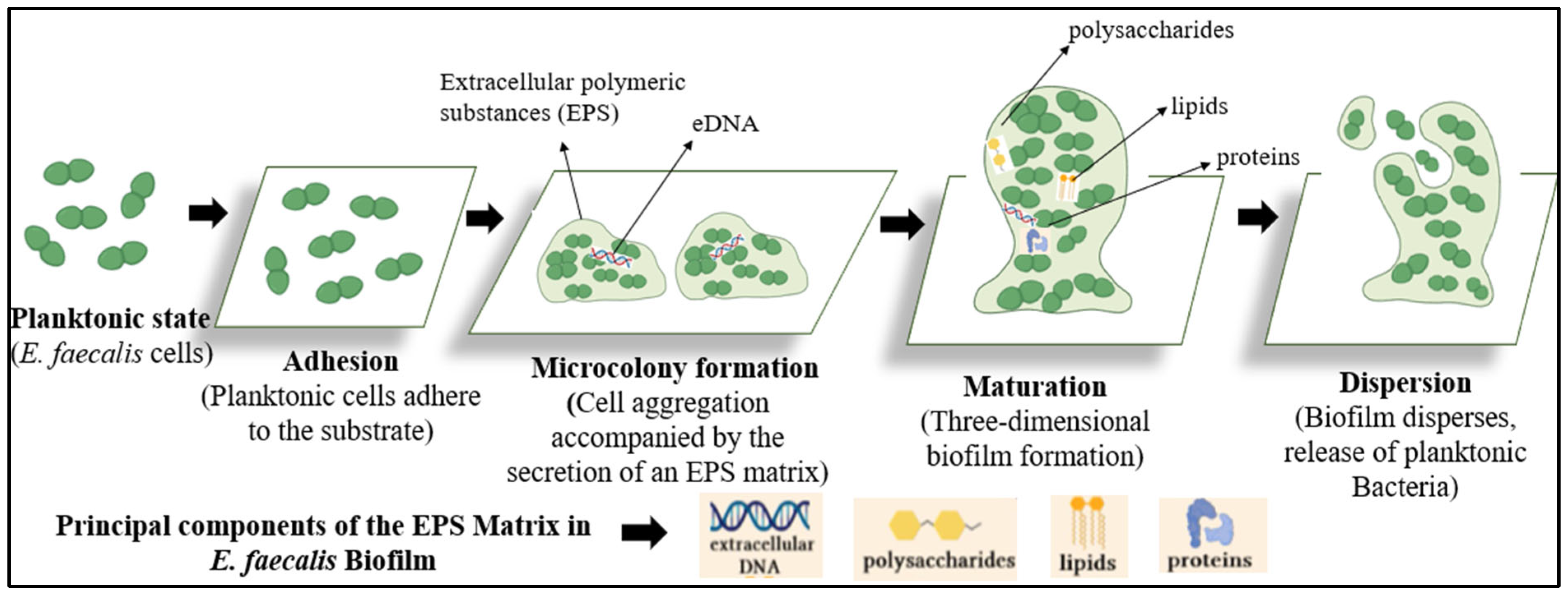
Figure 1. Stages of the formation of E. faecalis biofilm.
Msf 35 00005 g001
Figure 2. Three-dimensional stacks of E. faecalis biofilm.
Msf 35 00005 g002
Figure 3. (A) Clinical hazards caused by E. faecalis biofilm; (B) Environmental hazards caused by E. faecalis biofilm; (C) Emerging strategies to prevent E. faecalis biofilm.
Msf 35 00005 g003
Disclaimer/Publisher’s Note: The statements, opinions and data contained in all publications are solely those of the individual author(s) and contributor(s) and not of MDPI and/or the editor(s). MDPI and/or the editor(s) disclaim responsibility for any injury to people or property resulting from any ideas, methods, instructions or products referred to in the content.