Next Article in Journal
Design and Synthesis of Fused Derivatives of 7-Hydroxycoumarin (Umbelliferone) with the Flavonol Quercetin and the Flavone Luteolin-Analysis of Their Antioxidant and Physicochemical Properties
Previous Article in Journal
Mitophagy-Inducing Nanocarriers Restore Mitochondrial Quality and Cell Functions in Senescent Retinal Pigment Epithelial Cells
 
 
Correction published on 3 March 2026, see Oxygen 2026, 6(1), 5.
Article

The Role of Zinc Oxide Nanoparticles in Boosting Tomato Leaf Quality and Antimicrobial Potency

by
Mostafa Ahmed
1,2,*,
Sally I. Abd-El Fatah
3,
Abdulrhman Sayed Shaker
4,5,
Zoltán Tóth
6,* and
Kincső Decsi
6
1
Department of Agricultural Biochemistry, Faculty of Agriculture, Cairo University, Giza 12613, Egypt
2
Festetics Doctoral School, Institute of Agronomy, Georgikon Campus, Hungarian University of Agriculture and Life Sciences, 8360 Keszthely, Hungary
3
Food Toxicology and Contaminants Department, National Research Centre, Dokki, Cairo 12622, Egypt
4
Department of Microbiology, Faculty of Agriculture, Cairo University, Giza 12613, Egypt
5
School of Environmental and Conservation Sciences, Murdoch University, Murdoch, WA 6150, Australia
6
Institute of Agronomy, Georgikon Campus, Hungarian University of Agriculture and Life Sciences, 8360 Keszthely, Hungary
*
Authors to whom correspondence should be addressed.
Submission received: 2 December 2025 / Revised: 2 January 2026 / Accepted: 5 January 2026 / Published: 8 January 2026 / Corrected: 3 March 2026

Abstract

Salt stress is a major agricultural issue. A promising modern agriculture method is the foliar treatment of zinc oxide nanoparticles (ZnONPs). This approach has shown promise in boosting challenged tomato yields, fruit quality, and leaf extract antibacterial activity against pathogens. A greenhouse experiment was conducted. The previously synthesized and characterized ZnONPs were used to alleviate the harmful effects of NaCl stress. Tomato fruit weight from different treatments was determined, and the gas–liquid chromatography device was used to observe the changes in fatty acid production. The antimicrobial activities of the aqueous and diethyl ether extracts from tomato leaves were determined against six bacterial and six fungal strains. The plants that were salinity-stressed and sprayed with 0.075 and 0.15 g/L ZnONPs showed a better improvement compared to the salinity-stressed plants. Also, the sprayed plants that were not stressed at all showed promising results compared to the control and the other different treatments. Through the process of molecular docking, it was shown that caffeic acid, ferulic acid, p-coumaric acid, sinapic acid, and apigenin-7-glucoside are essential chemicals that possess antibacterial and antifungal effects against the DNA Gyrase inhibitor and the sterol 14-alpha demethylase (CYP51) enzyme, respectively. It is concluded that salt stress can negatively affect the growth, quality, and variant plant features. However, the foliar application of ZnONPs is able to overcome those adverse effects in the stressed plants, and enhance the non-stressed as well.

1. Introduction

Salinity is one of the environmental stresses that elicit varied responses in plants, including a reduction in seed germination rate, leaf area, chlorophyll content, ion absorption, and growth improvement, alongside an increase in antioxidant levels and soluble substances, as well as the activation of specific genes [1,2,3]. Under the duress of salinity, photosynthesis diminishes, leading to a reduction in ion absorption and glucose degradation, citrate cycle, and respiratory functions, hence increasing the likelihood of reactive oxygen species (ROS) formation. The concentrations of osmolytes, flavonoids, alkaloids, and the antioxidative enzymes are often elevated in the cells of plants exhibiting salt tolerance to mitigate the ROS [4,5,6,7].
Tomatoes are considered an important food source. They include various secondary metabolites, such as dehydrotomatine, which help plants resist bacteria, viruses, fungi, and nematodes [8,9]. Tomato plants subjected to salt stress may exhibit altered leaf extract compositions, potentially affecting their efficacy in combating pathogens. Certain studies indicated that salt stress can induce leaves to synthesize increased levels of chemicals, including antioxidants and phenolic compounds [1,10,11,12]. This may enhance the leaves’ efficacy in combating pathogens. Conversely, additional studies indicated that salt stress might adversely affect certain plant traits, thereby diminishing their capacity to combat pathogens [13].
Zinc oxide nanoparticles are widely recognized for their ability to eliminate pathogens and inhibit microbial proliferation by penetrating the cell membrane. The oxidative stress is responsible for damaging the matrix lipids, molecular recognition carbohydrates, functional proteins, DNA, and other vital biomolecules [14]. It has been asserted that the antibacterial properties of bulk zinc oxide solution arise from the external generation of a reactive oxygen (RO) molecule, hydrogen peroxide (H2O2) [15]. Researchers have investigated the potential hazards of nanoparticles when they emit deleterious ions [16,17,18]. Zinc oxide is amphoteric like the amino acids, indicating its capacity to react with both acids and bases to produce Zn2+ ions.
The pathogens have developed resistance to metronidazole, and the disease has many distressing negative effects, including allergy, vomiting, increased susceptibility to cervical cancer, infertility, and a role in human immunodeficiency virus (HIV) transmission. Novel and effective therapeutic agents are needed [19]. Dehydrotomatine and alpha-tomatine are glycoalkaloids that are produced by tomato plants (Lycopersicon esculentum), and these glycoalkaloids have the potential to defend tomato plants from parasites such as bacteria, fungus, viruses, and insects [20].
Friedman’s 2013 study showed that α-tomatine effectively targets the membranes of human cancer cells, resulting in their leakage and subsequent death [21]. Serratì et al. (2020) clearly showed its efficacy in inhibiting the metastatic capability of melanoma cells [22]. Friedman et al. (2000) identified a significant association concerning the “complexation” of cholesterol [23]. The tomatine–cholesterol complex is excessively large for absorption, resulting in its expulsion, which subsequently reduces plasma LDL cholesterol levels. Diosa-Toro et al. (2019) found that the aglycone version (tomatidine) is particularly effective against mosquito-borne viruses by disrupting the final stages of the viral life cycle [24]. Dyle et al. (2014) identified tomatidine as a notable inhibitor of muscle atrophy, emphasizing a unique connection [25]. This suggests a possible therapeutic use of tomato glycoalkaloids in treating age-related muscle atrophy (sarcopenia) or disease-induced cachexia.
ZnONPs, owing to their biodegradable and antibacterial properties in nanostructured form, are a feasible tool for controlling diseases in agriculture [26,27]. They have been successfully tested as antibacterial agents, with high activity against various microorganisms [28]. ZnONPs have been found to protect tomato plants from the tobacco mosaic virus (TMV) [29].
Utilizing molecular docking, we investigated the antibacterial components of tomato extracts in order to gain a better understanding of the mechanisms by which they exert their effects on human diseases. Molecular docking was utilized in order to ascertain the many sorts of contacts and angles that occur between a ligand and the target protein receptor that is present in the microorganisms that are the subject of this inquiry. The structure-based drug design (SBDD) methodology, which is critical for a wide variety of biological and metabolic processes [30,31], is a significant component of this methodology.
The present study is regarded as a supplementary investigation of tomato plants previously cultivated in a greenhouse [32]. We hypothesized that the external application of ZnONPs would alleviate the detrimental effects of salt stress by modifying the plant’s antioxidant systems and photosynthetic apparatus. It is suggested that this physiological stability triggered a metabolic shift towards the overproduction of specific secondary metabolites. These metabolic changes would be manifested as an enhanced antibacterial profile in the plant extracts. The objectives of this investigation were to examine salt-stressed tomato leaves treated with ZnONPs on the production levels of the fatty acids, which also presented the yield of tomato fruits, and estimate the antimicrobial activities of the different polarity extracts from the sprayed leaves.

2. Materials and Methods

2.1. Experimental Design

On 29 May 2023, 549 Kecskeméti tomato seeds were sown in a plastic seedling plate. Thirty days post-sowing (DAS), when the seedlings exhibited 3 to 4 true leaves, they were transferred to pots measuring 28 cm in diameter and 28 cm in depth on 27 June 2023. On the tenth day following transplantation, 0.15 M sodium chloride was administered to the soil to induce salt stress. Following salt stress, ZnO nanoparticles were applied via foliar spray three times at 10-day intervals at concentrations of 0.075 or 0.15 g/L. The treatment conditions were as follows: T1 Control (distilled water ‘dw’), T2 (dw + 0.075 g/L ZnO nanoparticles), T3 (dw + 0.15 g/L ZnO nanoparticles), T4 (0.15 M sodium chloride), T5 (0.15 M NaCl, 0.075 g/L ZnO nanoparticles), T6 (0.15 M NaCl, 0.15 g/L ZnO nanoparticles). All the treatments were replicated four times. Tomatoes were collected on 26 September 2023 [32,33].
Tomato plants were subjected to salinity stress using a 0.15 M NaCl solution. This presented a sufficiently elevated level to constitute a significant, although non-lethal, challenge. Cuartero and Fernández-Munoz (1999) asserted that tomato development often decelerates at electrical conductivities exceeding 2.5 dS/m, but a concentration of 0.15 M (about 15 dS/m) is an appropriate level for investigating stress-mitigation strategies without inducing immediate mortality [34].
The selected ZnONPs (0.075 and 0.15 g/L) were evaluated within the hormetic zone identified in the literature [35]. The concentrations were maintained below the phytotoxic threshold [36,37], typically exceeding 500 mg/L for Solanum lycopersicum. Foliar application was chosen to avoid soil-zinc immobilization, so enabling direct nutrient absorption. Our data, which indicate an increase in biomass, confirm that these doses functioned as mitigators rather than additional stressors.

2.2. Formation and Assessment of the Optical and Crystallographic Properties of ZnONPs

Following the approach detailed in our previous research [32], the chemical synthesis and characterization of ZnONPs were conducted. Zinc oxide nanoparticles (ZnONPs) were synthesized by precipitating an aqueous solution of zinc nitrate hexahydrate (3.04%) with sodium hydroxide (0.4%). This procedure was executed to produce ZnO nanoparticles. For foliar application, the white crystals were solubilized in distilled water using ultrasonic vibrations. This led to the synthesis of ZnONPs in varying concentrations (75 and 150 mg L−1).

2.3. Crude Aqueous and Diethyl Ether Extraction

The powdered plant material (10 g) from the tomato leaves was obtained by crude aqueous extraction using deionized water and diethyl ether. The aqueous extract was filtered using a Buchner funnel, centrifuged at 10,000 rpm, and then concentrated in a freeze dryer. The obtained semi-solid extracts (residue) were kept in a freezer at −20 °C until further use. The diethyl ether extract was obtained using continuous mixing in a shaker. It was filtered using Whatman No. 1 filter paper. The extract (filtrate) was concentrated at 40 °C under reduced pressure using a rotary evaporator and then kept in a glass flask. The semi-solid diethyl ether extract (residue) obtained was stored in a refrigerator for further use.

2.4. Fatty Acid Profile

Christie (1993) stated that fatty acid methyl esters (FAMEs) are produced by transesterifying the whole lipid with 2% sulfuric acid in methanol [38]. We used gas chromatography equipment with a flame ionization detector and a DB5 silica capillary column (60 m × 0.32 mm i.d.) to obtain a peak at the fatty acids. The oven was programmed to go from 45 °C to 60 °C at a rate of 1 °C per minute. It was then set to go up from 60 °C to 240 °C at a pace of 3 °C per minute. Helium, the carrier gas, was added at a rate of 1 mL every hour. The detector temperature was set to 250 °C and the injector temperature was set at 230 °C [38].

2.5. Evaluating Dry Matter (DM), Crude Protein (CP), Ash Contents, and Neutral and Acid Detergent Fibers (NDF and ADF) in Tomato Leaves Using NIRS™ DS 2500 FOSS

The plant sample’s moisture level was used to figure out the five criteria: dry matter, protein, and ash contents, and detergent fibers (acid and neutral). The calibrations work for sample temperatures between 10 and 30 °C. Soest et al. (1991) utilized amylase that could withstand heat to find both types of fibers (acid and neutral) [39]. Dry matter, crude protein in leaves, and ash content were measured using A.O.A.C. methods 930.15, 990.03, and 923.03 [40].

2.6. Assessment of the Antimicrobial Activity

2.6.1. Examined Microorganisms

Two Gram-positive bacteria, Bacillus cereus EMCC 1080 and Staphylococcus aureus ATCC 13565, and four Gram-negative bacteria, Pseudomonas aeruginosa NRRL B-272, Salmonella typhi ATCC 25566, Escherichia coli O157:H7 ATCC 51659, and Listeria monocytogenes LMD 7726, were obtained from the holding company for biological products and vaccines; VACSERA, Egypt. We grew stock cultures on nutrient agar slants at 37 °C for 1 day and then stored them in the refrigerator until needed. For the antifungal assay, Fusarium proliferatum MPVP 328, Fusarium verticilloides ITEM 10027, Aspergillus niger SSWT 2999, Aspergillus carbonarius ITAL 204, Aspergillus ochraceus ITAL 14, and Aspergillus flavus NRRL 3357 were used. The Cranfield University Applied Mycology Department provided the fungal isolates. We grew stock cultures on nutrient agar slants at 25 °C for 5 days and then stored them in the refrigerator for later use.

2.6.2. Investigational Microbiological Medium

The nutritional medium of agar was prepared in 1 L of distilled water and contained meat and yeast extracts at weights of 1 and 2 g, respectively. It also contained peptone, NaCl, and agar, with 5, 5, and 15 g, respectively. For the bacterial disc diffusion experiment, the pH was measured to 7.4 ± 0.2 at 37 °C [41]. In 1 L of distilled water, tryptic soya broth (TSB) contained casein pancreatic digest, soybean papaic digest, dextrose, NaCl, and K2HPO4, with 17, 3, 2.5, 5, and 2.5 g. The pH was adjusted to 7.3 ± 0.2 at 25 °C [41]. Potato dextrose agar medium (PDA) contained 200 + 15 + 20 g of potato, glucose, and agar, respectively, in 1 L of distilled water [42].

2.6.3. The Protocol of Disc Diffusion to Determine the Zone of Inhibition

To prepare the 0.5 McFarland standard, combine 500 µL of 1.175% (w/v) barium chloride dihydrate (BaCl2·2H2O) with 99.5 mL (0.0995 L) of 1% (v/v) sulfuric acid (H2SO4). To maintain the barium sulfate precipitate in suspension, continuous stirring is required. A spectrophotometer calibrated to a wavelength of 625 nm assesses the optical density of the standard to ensure accuracy. The permissible absorbance range is 0.08 to 0.1. Prior to utilization, the standard must be homogenized with a vortex mixer to ensure a hazy and uniform appearance. To prepare the bacterial sample, a single loopful of each bacterial type is extracted from a 24 h nutritional agar slant and transferred into a tube containing 4 to 5 mL of TSB. The broth culture is thereafter maintained at 35 °C for a duration of 2 to 6 h. The incubation procedure persists until the turbidity of the broth culture aligns with the density of the 0.5 McFarland standard, indicating approximately 1.5 × 10⁸ CFU/mL of bacteria present.
We used the Kirby–Bauer disc diffusion method to find out how sensitive tomato extracts were to different types of bacteria [43,44,45]. The disc diffusion method assessed bacterial susceptibility on 20 mL nutrient agar plates. TSB bacteria were evenly placed on agar surfaces using sterile cotton brushes to ensure simultaneous growth. In the treatment groups, extract fractions were mixed with 1000 µL of DMSO to reach a final concentration of 5 mg/mL. To remove excess solvent, Whatman No. 1 sterile 6 mm filter paper discs were soaked in the extract and dried aseptically. The prepared discs were placed on inoculated agar using sterile forceps. DMSO was the negative control and tetracycline (0.5 mg/mL) the positive control. The plates were incubated at 37 °C for 24 h. Following incubation, we used a millimeter ruler or digital caliper to measure the overall diameter of the zones of inhibition, including the 6 mm disc, to assess antimicrobial activity.
To maximize growth, the fungal isolates were grown on potato dextrose agar (PDA) at 25 °C for five days. To ensure uniform dispersion, a spore suspension was produced in 0.01% (v/v) Tween 80. To create a confluent lawn, a sterile glass rod was used to evenly distribute 0.05 mL of fungal inoculum over yeast extract supplement (YES) agar plates. The disc diffusion method determined microorganism antibacterial susceptibility. The sterile 6 mm filter paper discs were soaked in the extracts and dried in a sterile atmosphere, as in antibacterial testing. The loaded discs were placed on the inoculated YES medium with sterile forceps. DMSO was the negative control while 1000 U/mL nystatin was the positive control. All plates were 25 °C for 48 h. To evaluate extract activity against fungus, we measured the diameter of the clear zones of inhibition (mm) surrounding each disc [46].

2.6.4. Estimation of Minimal Concentration Causing Inhibition (MIC)

The bacterial minimal concentration causing inhibition was determined using Perrucci et al. (2004)’s method [47]. The tube dilution method was used to estimate the MIC [48]. To fulfill the inocula of 108 cfu/mL, we performed a dilution of the 1-day bacterial strain culture in 0.01 L of TSB using the 0.5 McFarland standard solution. Each extract was added to culture tubes at variant concentrations in DMSO. We added 100 µL of bacterial cell suspension to each tube and incubated them at 37 °C for 24 h. Turbidity indicates the growth of the inoculum in the broth, whereas the extract’s minimal concentration of inhibition (MIC) is the lowest quantity that inhibited the growth of the tested strain of bacteria or fungi.
For the fungus’s minimal concentration causing inhibition, a mixture of crude extracts, 0.5 mL of 0.1% (v/v) tween 80, and 9.5 mL of molten PDA was mixed at 45 °C and placed in 6 cm Petri dishes. We placed 3 µL of fungal solution (108 cfu/mL; 0.5 McFarland standard solution) in the center of the plates. The plates were kept at 25 °C for 2 days. After incubation, the MIC was determined [49].

2.7. The Docking of Molecules Characterization

A molecular docking study was conducted using the AutoDock (SWISS Dock, (version 1.2.0)) and Discovery Studio Visualizer (version 25.1.0.24284) to evaluate the antibacterial activity of tomato leaf extracts and corroborate the in vitro findings. The Topoisomerase II ATPase enzyme (DNA Gyrase) (PDB Id: 3TTZ) served as a universal antibacterial target, whilst the sterol 14-alpha demethylase (CYP51) protein (PDB Id: 5FSA) was utilized as an antifungal target. The 17 compounds discovered by HPLC in our prior investigation [32] were evaluated within the active sites of both targets. Two-dimensional structures of all tested compounds were generated by AutoDock and Discovery Studio Visualizer using SMILES codes received from the National Library of Medicine (https://pubchem.ncbi.nlm.nih.gov/ (accessed on: 23 October 2025)). Following a ligand preparation process that involved protonating the 3D structure, assigning partial charges, and performing energy minimization, the compounds were saved as AutoDock and Discovery Studio Visualizer files.
The Protein Data Bank website (https://www.rcsb.org/ (accessed on: 23 October 2025)) was utilized to obtain crystal structures of the antifungal target with posaconazole (X2N) as an inhibitor [50] and the antibacterial target with 07N as an inhibitor [51]. Likewise, the two antimicrobial targets were prepared for the docking run by removing chains not involved in the interaction, as well as water, solvent molecules, and non-binding ligands. The 3D structure was protonated using the default settings, and the docking sites were delineated. A re-docking procedure was subsequently conducted to verify the docking configuration utilizing the proteins and their original co-crystallized ligand molecules. The affinity values for the X2N ligand in the 5FSA active site and the 07N ligand in the 3TTZ active site were −15.2 and −7.4 kcal/mole, respectively. The interactions and binding modalities between the examined compounds and the enzymes’ active site were subsequently predicted utilizing the validated configuration.
The docking configuration employed the triangle matcher for placement, utilized London dG for rescoring, and applied the force field technique for refinement. Subsequent to docking, the extent of the compounds’ binding to the enzymes was evaluated based on internal energy scores (S), bond interactions, and lengths (Å), constrained to 3.5 for hydrogen bonds and 4.0 for ionic interactions [52,53]. Two-dimensional and three-dimensional docked images depicting the optimal binding locations (ligand-to-protein) were preserved for a clear illustration of the results.

2.8. Statistical Analysis

This study repeated all tests quadruply and reported the means and standard errors of the results. JASP was used for the analysis [54,55]. The study examined the differences using a two-way analysis of variance to test interactions between salinity and ZnONPs treatments. The Tukey test was 5% significant.

3. Results

3.1. Evaluation of Different Properties of ZnONPs

Our previous research generated and evaluated the properties of ZnO nanoparticles [32]. The UV-Vis spectrophotometer showed that ZnONPs peaked at 370 nm. Hexagonal ZnO nanoparticles in the TEM micrograph indicated a high quality. ZnO nanoparticle SEM micrographs exhibited form and size consistency. Energy-dispersive X-ray spectroscopy uniformly dispersed Zn and O on ZnO nanoparticle surfaces. After 2 h at 200 °C, ZnO nanoparticles developed a monomodal size distribution with a half-width of 41.166 nm. We analyzed lab-generated zinc oxide nanoparticles using Fourier transform infrared spectroscopy (FTIR) (3900–300 cm−1). Peaks at 3150, 1631.48, 1425.14, 1078.98, 447.404, and 370.23 cm−1 reveal six functional groups in zinc oxide nanoparticles’ spectra. The measured zeta potential was −30.214 mV.

3.2. Yield of Tomato Fruits and Leaves and Fatty Acid Distribution

Table 1 presents the post-harvest measurements, indicating that the plants treated with NaCl solution exhibited the lowest values across various examined post-harvest variables, including fruit number and weight. Conversely, the plants treated with foliar-sprayed ZnONPs, regardless of stress levels, exhibited higher values compared to those subjected to salty-water irrigation (T4). For the weight of the moist dried leaves, the plants that were not stressed but sprayed with 0.075 g/L ZnONPs showed the highest values compared to those that were sprayed with 0.15 g/liter, and also higher values than the stressed and sprayed plants (T5 and T6).
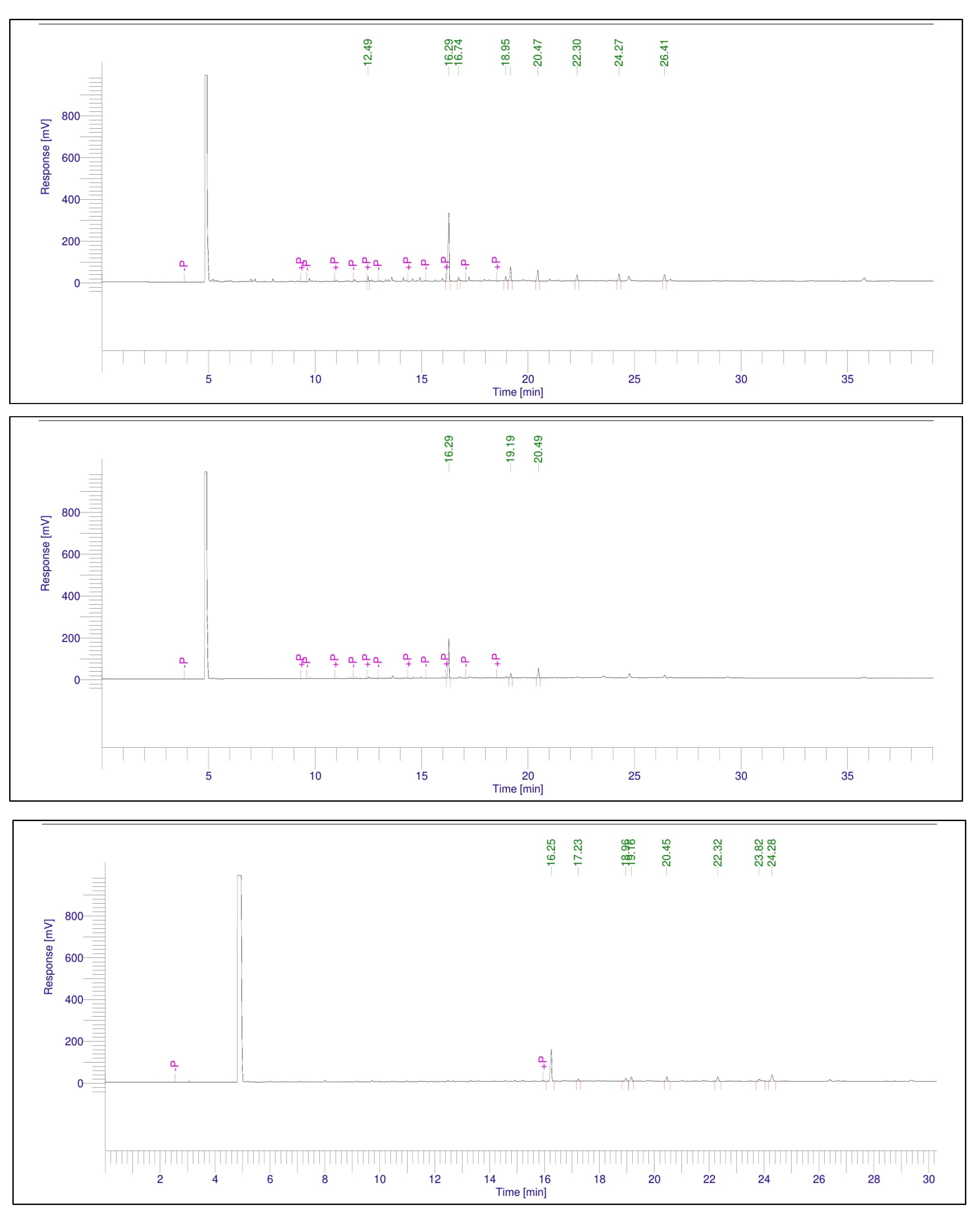
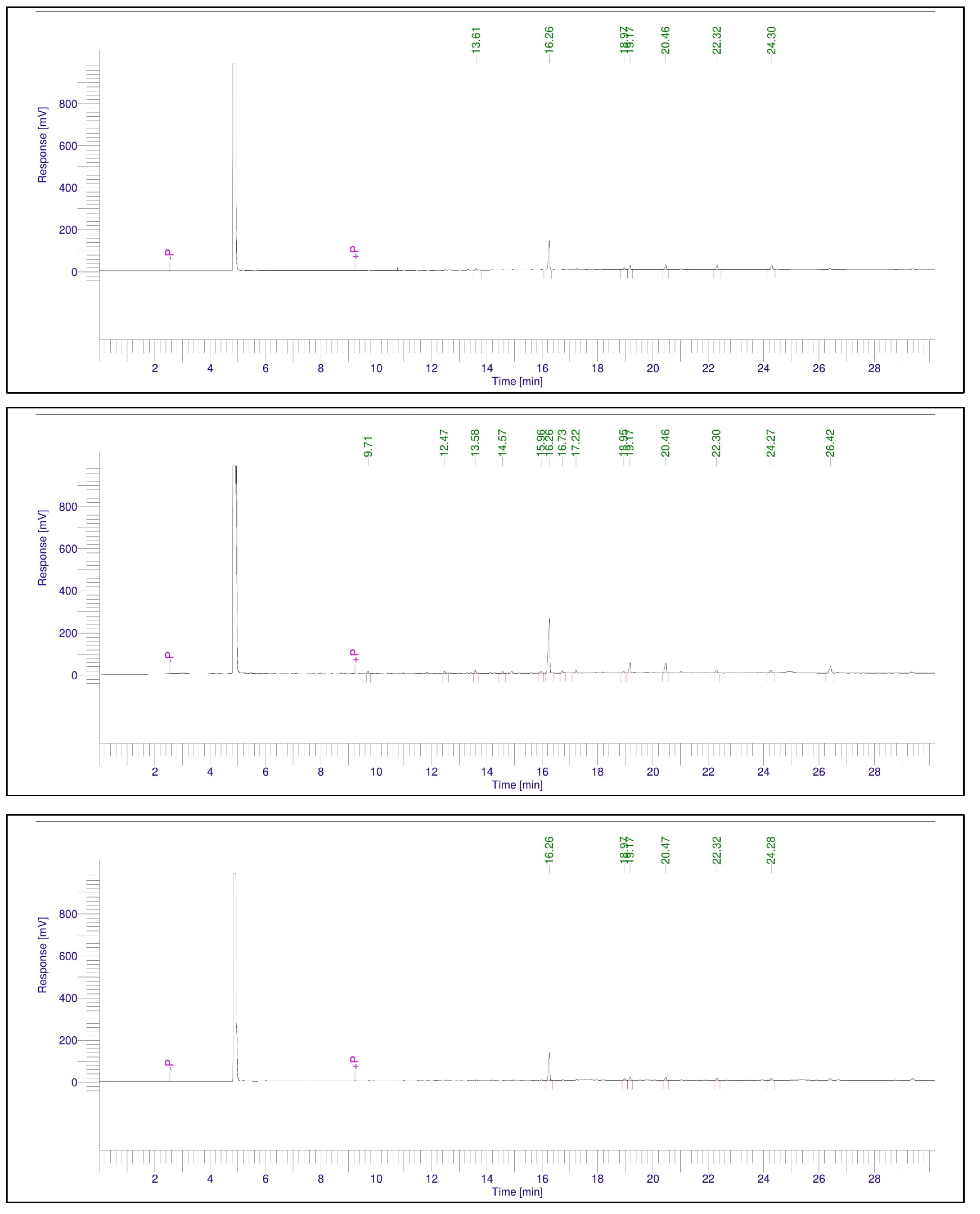
The fatty acid profile of the tomato leaves’ extracted oil was determined, as shown in Table 2 (Figure A1 and Figure A2). Nine fatty acids were expressed. One of those detected fatty acids is medium chain (lauric acid), and the other eight fatty acids were long chain. The lauric fatty acid was not detected in the investigated tomato leaves’ extracts, except in the leaves from the fifth treatment plants (T5) with a percentage of 1.81%. It was noticed that the highest obtained fatty acid was the palmitic (C16:0), and it was observed in all the leaves from different treatments with 67.24% in T6, 66.21% in T2, 58.04% in T1, 56.5% in T3, 56.44% in T5, and 56.33% in T4. The lowest fatty acid was lauric acid, with 1.81% in T5, and it was not detected in the other treatments. The lauric acid was ascendingly followed by the palmitoleic acid (C16:1n-7) with 2.9% in T3 and T4, 2.43% in T6, 2.31% in T1, 2.24% in T5, and 2.08% in T2. Myristic acid (C14:0) was not detected in T2, T3, and T4. Also, arachidic acid (C20:0) was not detected in T3 and T4.
Table 3 illustrates the variations in DM, CP, ash, ADF, and NDF levels in tomato leaves. The non-salinity-stressed treatments (T1–T3) exhibited higher values than the salinity-stressed treatments (T4–T6) in terms of dry matter. The fourth treatment, subjected to 150 mM NaCl and not treated with ZnONPs, had the lowest crude protein content. The third treatment, succeeded by the fifth and sixth treatments (T5 and T6), exhibited the highest crude protein content. No significant differences were seen between the treatments for the quantities of ash and neutral detergent fiber. The second treatment, which was not emphasized but administered with 0.075 g/L ZnONPs, exhibited the highest acid detergent fiber across all treatments. The results indicated that the application of foliar spray containing synthesized zinc oxide nanoparticles had a positive effect, while salt stress adversely impacted several parameters.

3.3. Antimicrobial Activity of Tomato Leaves’ Crude Aqueous and Diethyl Ether Extracts

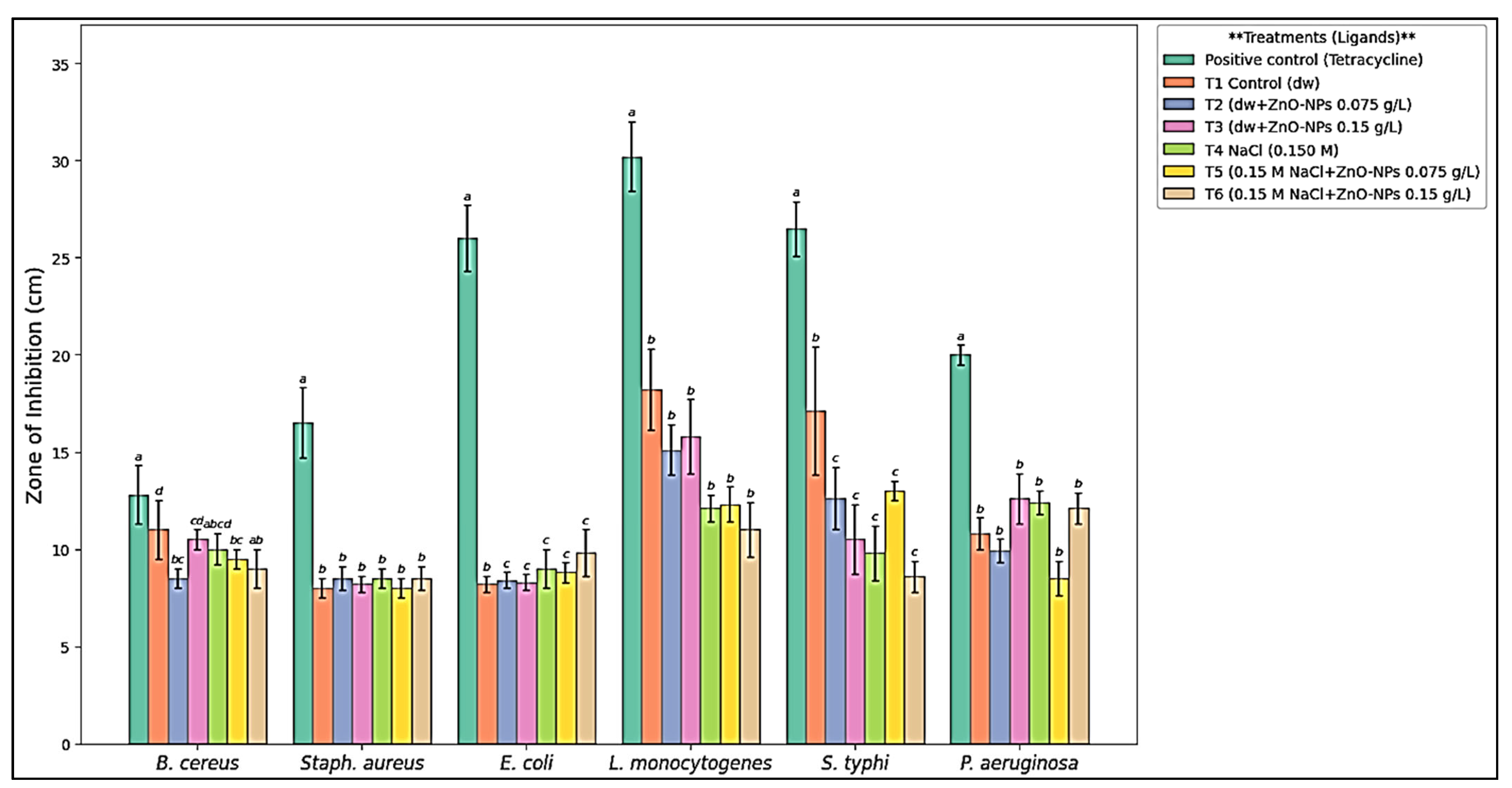
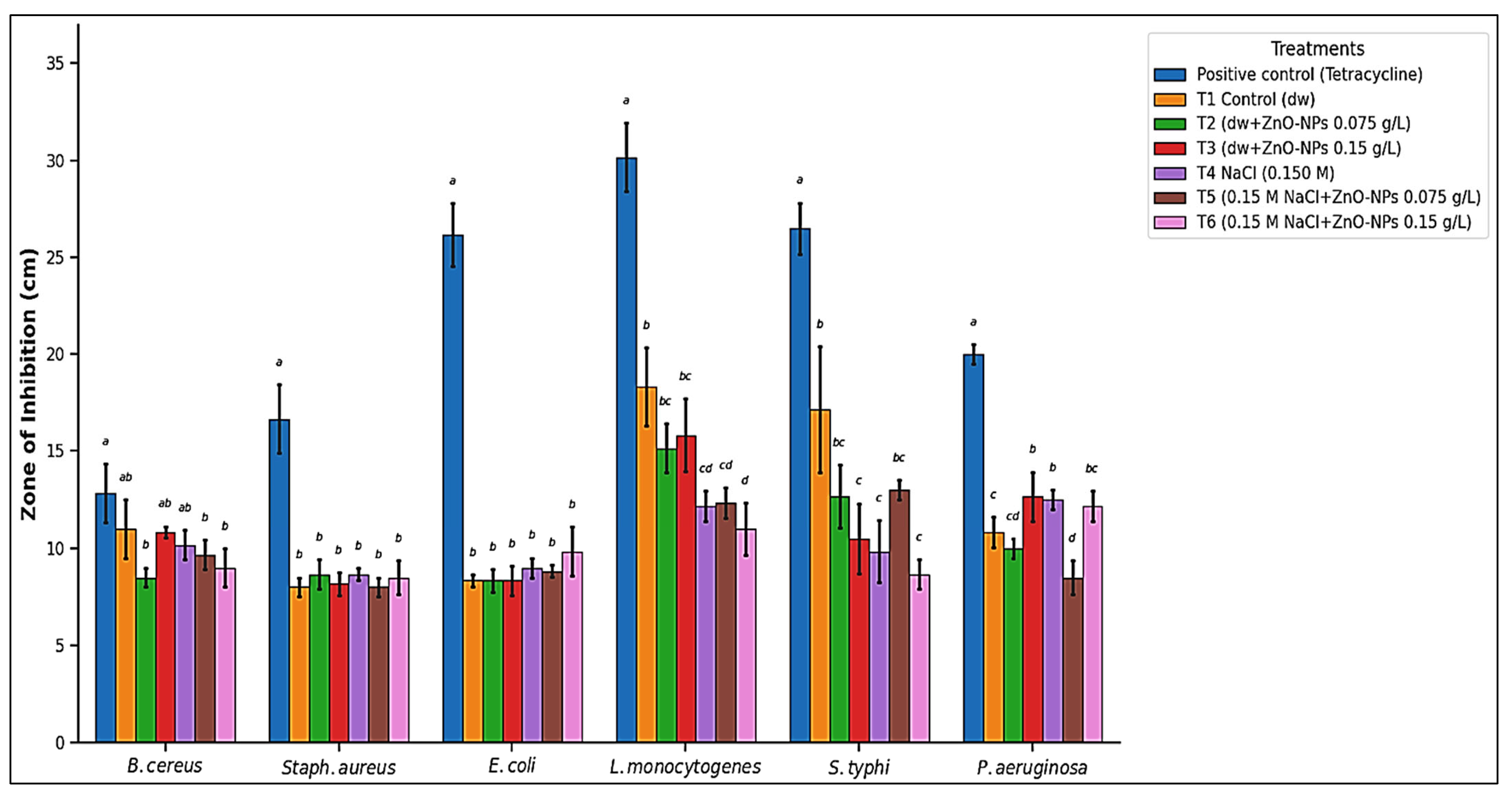
The antimicrobial activity was determined against pathogenic bacteria (antibacterial) and mycotoxigenic fungi (antifungal activity). According to the aqueous extract from tomato leaves, it was reported in Figure 1 that T1, T3, and T4 extracts had the highest antibacterial activity against B. cereus, followed by T2, T5, and T6 extracts. For E. coli and Staph. aureus, there was no significant difference between the six treatments’ extracts (T1–T6) except the positive control (tetracycline), which had the highest effect. For L. monocytogenes, S. typhi, and P. aeruginosa, the T1 extract had the highest activity. On the other hand, the diethyl ether extract from tomato leaves showed no significant differences between the six different treatment extracts (T1–T6) with L. monocytogenes and P. aeruginosa (Figure 2). However, the T1 extract had the highest activity against E. coli and S. typhi. The T6 extract had the highest activity against B. cereus, and the T2 extract had the highest activity against Staph. aureus.
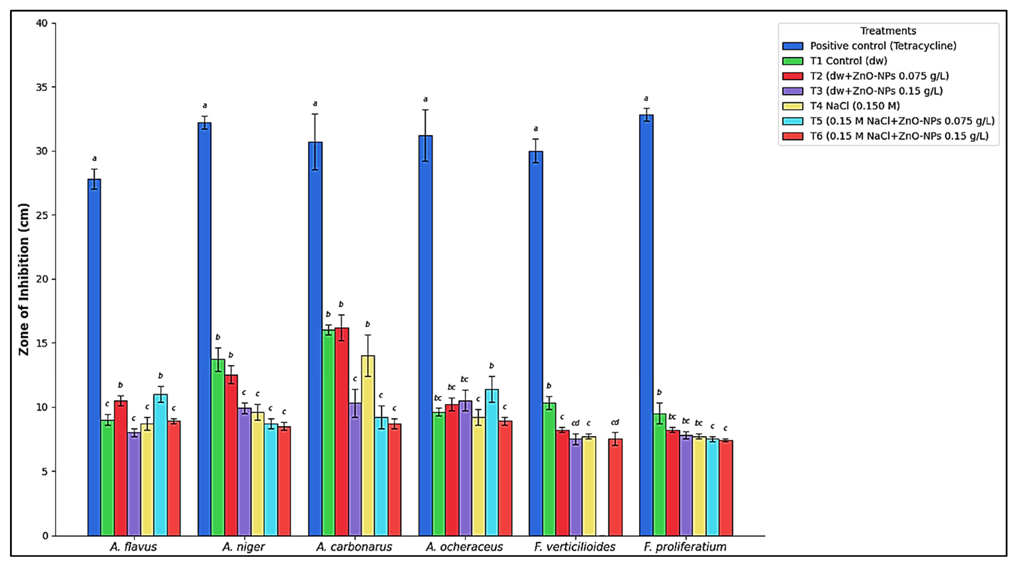
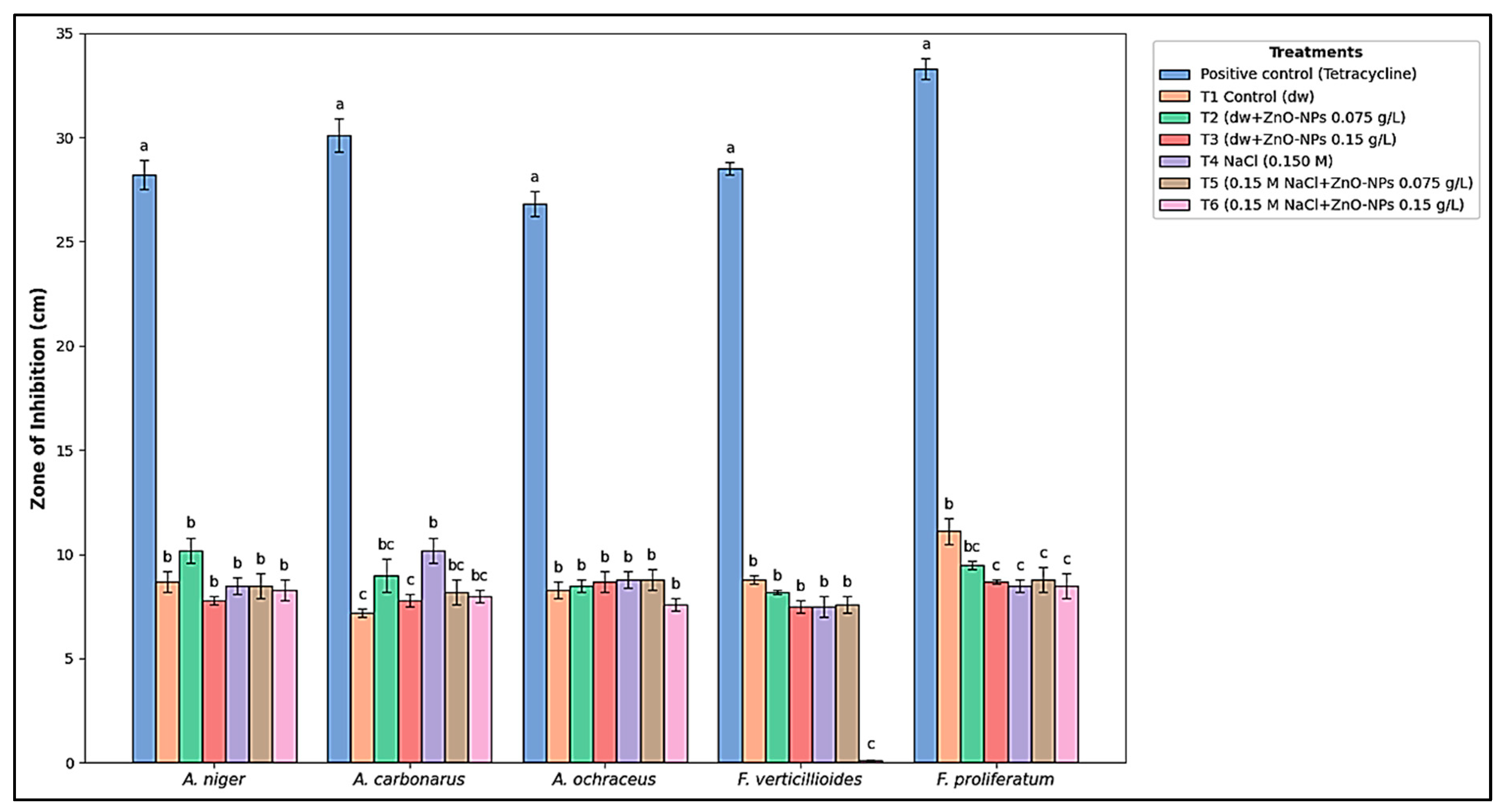
Following the antifungal activity, the aqueous extract from the tomato leaves showed varying results against the fungi (Figure 3), depending on the examined treatment extracts. There was a trend that the aqueous extract from T1 plants showed the highest activity against all the tested mycotoxigenic fungi; A. flavus, A. niger, A. carbonarius, A. ocheraceus, F. verticilioides, and F. proliferatium, followed by the extract from T2 plants that were not stressed but sprayed with 0.075 g/L ZnONPs. For the diethyl ether extract, there was no significant difference between the different extracts from all the treatments against the fungi (Figure 4); A. carbonarius, A. ocheraceus, and A. flavus. However, the T1 extract showed the highest activity against F. verticilioides and F. proliferatium. The T2 and T4 extracts showed the highest activity against A. niger.
In the dataset of the aqueous extracts (Table 4), the extracts generally exhibited high potency across most tested pathogens. All treatments (T1–T6) demonstrated strong inhibitory activity against B. cereus (0.15–0.42 mg/mL) and L. monocytogenes (0.10–0.37 mg/mL). Treatments T1, T4, and T5 showed strong activity against E. coli (0.33–0.42 mg/mL), while T1, T3, T4, and T6 were highly effective against P. aeruginosa (0.33–0.50 mg/mL). T1 displayed the highest overall potency in this set, notably achieving an MIC of 0.10 mg/mL against L. monocytogenes. Activity against Staph. aureus was primarily moderate, particularly for T3 (1.08 mg/mL) and T5 (0.83 mg/mL). Moderate inhibition was also observed for T2 against E. coli (0.83 mg/mL) and P. aeruginosa (0.58 mg/mL).
The dataset of Table 5 (diethyl ether extracts) showed a more varied distribution of potency, with several treatments shifting toward the “Moderate” classification compared to Table 4. T1 and T2 exhibited strong inhibition against S. typhi with MIC values of 0.15 mg/mL and 0.42 mg/mL, respectively. T6 showed strong activity against B. cereus (0.42 mg/mL), while T1 and T2 remained strong against Staph. aureus (0.20–0.33 mg/mL). T1 also showed strong activity against E. coli (0.20 mg/mL). The majority of treatments against P. aeruginosa and L. monocytogenes in this dataset were classified as moderate, with values ranging from 0.58 to 1.08 mg/mL. Notably, T3, T4, and T5 showed a decrease in potency against S. typhi (0.92–1.08 mg/mL) compared to their profiles in the first dataset, moving from strong/borderline to moderate.
The antifungal results indicate a broader range of efficacy, varying from strong to weak depending on the specific fungal strain and treatment conditions. In the first antifungal set for the aqueous extracts (Table 6), T2 exhibited a strong inhibitory effect against A. flavus (0.33 mg/mL), and T1 was highly effective against F. proliferatium (0.33 mg/mL). The second set for the diethyl ether extracts (Table 7) showed more widespread strong activity; T1 and T2 were highly potent against A. niger (0.20–0.42 mg/mL) and A. carbonarius (0.15 mg/mL). T5 also showed strong efficacy against A. ochraceus (0.28 mg/mL). The majority of treatments across both sets showed moderate activity against A. ochraceus and A. carbonarius, with MIC values typically between 0.58 and 1.17 mg/mL. F. proliferatium generally showed moderate susceptibility to most treatments in both sets. A notable limitation in efficacy was observed against F. verticilioides in some instances. T6 in the first set (2.33 mg/mL) and T5 in the second set (2.17 mg/mL) were categorized as having weak antifungal activity.

3.4. Results of Molecular Docking

Quantitative HPLC analysis [32] indicated that the accumulation of secondary metabolites was markedly affected by both salinity levels and ZnONP dosage, with unique metabolic signatures emerging across the various treatment groups. The application of 0.15 M salt stress (T4) served as a potent primary stimulus, resulting in a marked quantitative enhancement in the phenolic and flavonoid profiles relative to the untreated control (T1). Catechin concentrations had an approximate four-fold increase, escalating from 25.14 to 95.52 µg/g, while chlorogenic acid levels amplified from 154.08 to 359.51 µg/g. The data indicate that elevated salinity significantly redirects carbon resources into the biosynthesis pathways of defense-related compounds.
In the absence of abiotic stress, ZnONPs functioned as independent exogenous elicitors, provoking dose-dependent elevations in certain bioactive molecules. Concentrations of apigenin-7-glucoside showed a significant increase, rising from 5.69 µg/g in the control (T1) to 15.31 µg/g with 0.075 g/L ZnONPs treatment (T2), and further escalating to 36.98 µg/g at the 0.15 g/L concentration (T3). This trend indicates that ZnONPs can influence the plant’s metabolic production even in non-stress settings. The simultaneous application of salinity stress and ZnONPs led to an increased concentration of metabolites beyond those generated by salt stress alone, indicating a synergistic or additive metabolic response.
In saline conditions alone (T4), chlorogenic acid attained a concentration of 359.51 µg/g. The incorporation of 0.075 g/L ZnONPs (T5) virtually doubled the concentration to 617.96 µg/g, whereas levels stabilized at 603.38 µg/g with the increased ZnONP dosage of 0.15 g/L (T6). Sinapic acid concentrations increased from 27.67 µg/g under salt stress alone (T4) to 60.21 µg/g when coupled with 0.15 g/L ZnONPs (T6). In apigenin-7-glucoside, the concentration peaked at 69.89 µg/g with the combined application of 0.15 M salt and 0.15 g/L ZnONPs (T6), in contrast to 52.80 µg/g with salt alone (T4). The quantitative data provide a definitive framework indicating that the application of ZnONPs, especially at a concentration of 0.15 g/L, fundamentally alters the plant’s biosynthetic production. The systematic elevation of essential phenolic acids and flavonoids establishes a physiological foundation for the observed alterations in biological activity in both optimum and high-salinity conditions.
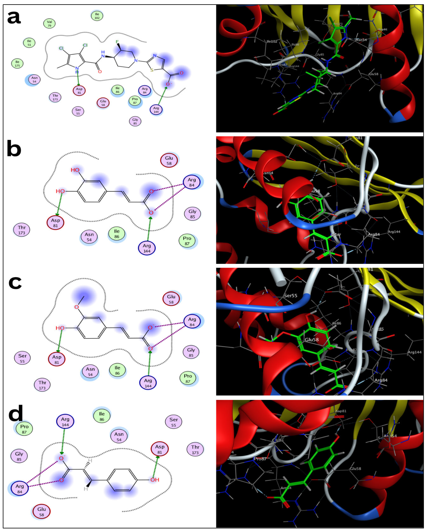
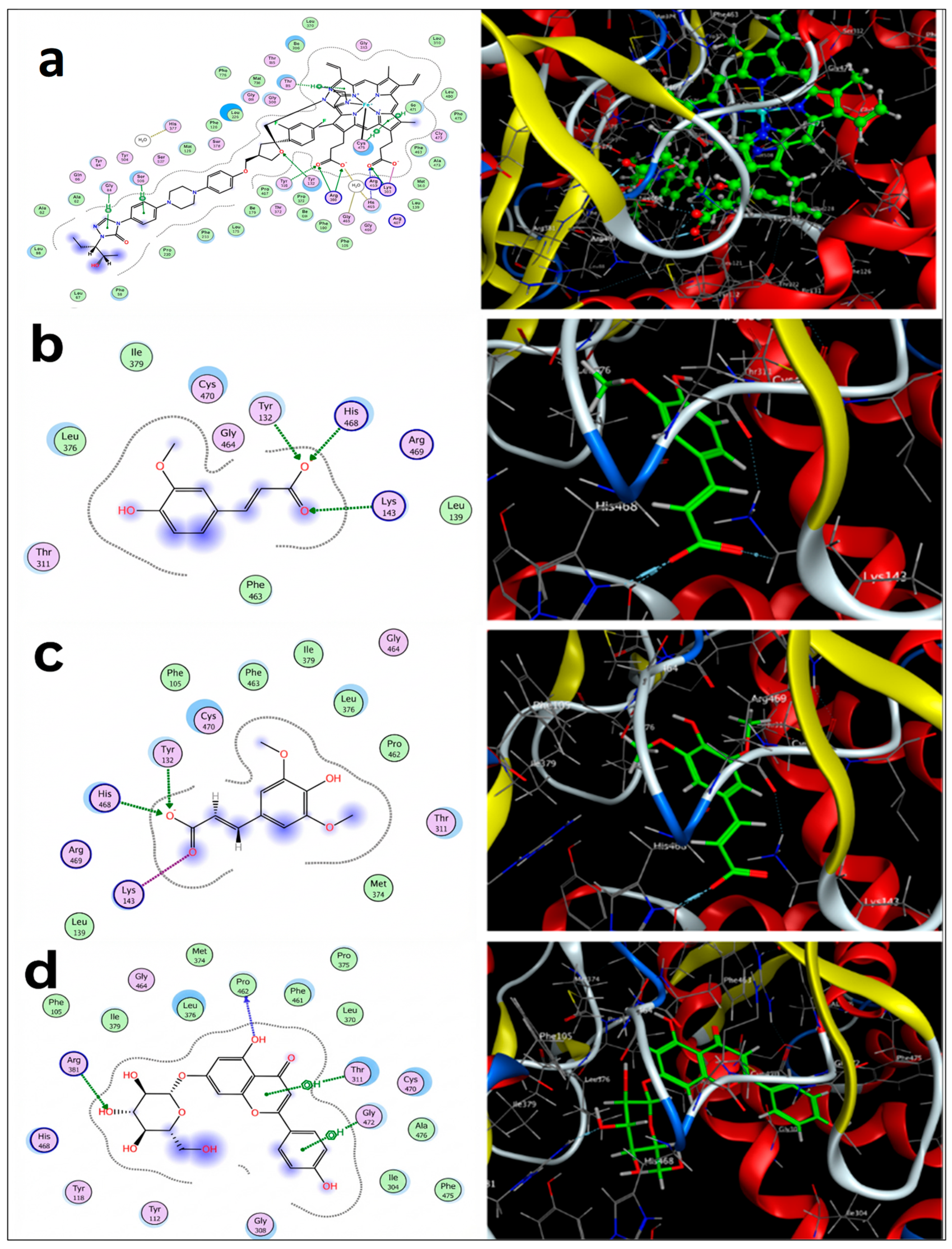
Table 8 demonstrates docking energy scores (kcal/mol) and predicted interactions with key active site residues and their distances (Å) for molecules under investigation against DNA Gyrase inhibitor as an antibacterial target (PDB ID: 3TTZ). Similarly, Table 9 presents the results of the tested compounds against the sterol 14-alpha demethylase (CYP51) enzyme (PDB ID: 5FSA), serving as an antifungal target. Accordingly, most of the compounds exhibited high affinity (docking scores) and comparable interactions to the original inhibitor (07N) within the antibacterial target’s active site. With the antifungal target attaining some similar interactions within the active site to the native ligand (posaconazole), they did, however, demonstrate relatively high scores. The 2D and 3D visualizations of these predicted interactions are shown in Figure 5 and Figure 6 for DNA Gyrase and CYP51, respectively.
The interaction of 07N original ligand 07N, caffeic acid, ferulic acid, and p-coumaric acid within DNA Gyrase’s active site (as antibacterial targets) has been investigated and illustrated in both two and three dimensions, as shown in Figure 5a, Figure 5b and Figure 5c and Figure 5d, respectively. The native ligand coordinated two hydrogen bond interactions to link with the key residues Asp81 and Arg144 at a distance of 2.80 and 3.51 Å, respectively, and a docking score value of −7.4 kcal/mol. When compared to tested compounds, rutin had the highest docking score for the antibacterial activity (−7.3 kcal/mol), followed by quercetin, sinapic, and apigenin-7-glucoside (−5.7, −5.6, and −5.6 kcal/mol, respectively). However, when considering their interactions, they showed only one hydrogen bond interaction with either ASP 81 or ARG 144. Three of the tested compounds (ferulic acid, caffeic acid, and p-coumaric acid) scored lower binding affinities (−5.2, −5.1, and −4.6 kcal/mol, respectively), but attained the same mode of interaction like 07N, mediating two hydrogen bond interactions to bind with ASP 81 and ARG 144. All other compounds, apart from apigenin, which did not exhibit any interactions within the active site, were able to form at least one hydrogen bond with either ASP 81 or ARG 144, regardless of their scores.
Similarly, the interaction between the original ligand (X2N), ferulic acid, sinapic acid, and apigenin-7-glucoside within the active site of CYP51 (as antifungal targets) has been studied and visualized (2D and 3D) in Figure 6a, Figure 6b and Figure 6c and Figure 6d, respectively. X2N showed high docking scores (−15.2 kcal/mol), mediating seven key residues, ARG 381, LYS 143, HIS 468, TYR 132, ARG 381, LYS 143, and HIS 468, to form hydrogen and ionic interactions. Although docking energy scores for the tested compounds ranged between −4.3 and 7.9 kcal/mol, they exhibited interesting binding interactions in closed proximity to the active site pocket of CYP51, as listed in Table 9.
To illustrate, apigenin-7-glucoside showed the highest score (−7.9), while protocatechuic acid showed the lowest score (−4.3), but both have one hydrogen bond interaction within the active site pocket in conjunction with ARG 381 at a similar distance of 3.29 Å. Interestingly, ferulic acid (−5.1 kcal/mol) and sinapic acid (−5.6 kcal/mol) mediated three interactions with TYR 132 (H-bond interaction), HIS 468 (H-bond interaction), and LYS 143 (ionic interaction) in the active site pocket. On the other hand, although rutin, apigenin, quercetin, and chrysin followed apigenin-7-glucoside in docking score values (−7.3, −6.1, −5.5, and −5.1 kcal/mol), they presented interactions in the active site with residues that were different from that of the original ligand. To clarify, rutin had a single hydrogen bond with MET 508, apigenin had no interaction in the active site, and quercetin and chrysin both had a single hydrogen bond with PRO 462. However, all the other tested compounds had at least one hydrogen bond interaction with ARG 381 or more, as shown in Table 9.

4. Discussion

While ZnONPs provide an effective means to mitigate salt stress, it is imperative to evaluate their safety and longevity prior to their use on edible crops, such as tomatoes. This study utilized dosages of 50 mg/L and 100 mg/L, which are within the eustressic range for vegetable crops [56]. These levels are well below the 500 mg/L threshold often associated with acute phytotoxicity, which may result in DNA damage or pronounced oxidative inhibition.
The primary food safety hazard is the bioaccumulation of zinc in the edible portions of food. Our findings demonstrate that foliar treatment increases tissue Zn concentrations, potentially serving as a strategy for agronomic biofortification to address Zn deficiency in human diets [57]. To ensure that the detected antibacterial activity was not due to external contamination, all leaf samples were subjected to three rigorous washes with deionized water before extraction. This approach eliminates NPs adhered to the surface, ensuring that any Zn present is either absorbed by the plant or utilized in its defense mechanism.
Under saline-alkaline circumstances, foliar application is more environmentally advantageous than soil application. In soils with elevated pH, zinc frequently forms insoluble complexes such as zinc phosphate, hindering plant access to the mineral and resulting in its accumulation within the soil profile [58]. Direct application of ZnONPs to the canopy enhances absorption efficiency and reduces the overall mineral discharge into the environment. This reduces the likelihood of persistent soil toxicity or leaking into groundwater.
In tomato fruits, salt stress can increase lycopene concentration by two to three times, facilitating the accumulation of carbohydrates, sugars, and amino acids [59,60]. T3 is the superior treatment, yielding the most quantity of fruits (22.50) and the heaviest fruits (1304.60). Conversely, T4 performed the poorest in all domains, exhibiting values significantly inferior to those of all other treatments. T1, T2, T3, T4, and T6 for moist leaves exhibited no significant differences among them. Treatment T5 resulted in a drastically reduced weight of 216. Most treatments yielded comparable results for dried biomass; however, T2 exhibited the highest weight (40.36), significantly surpassing T4 and T5. Quddus et al. (2024) [61] showed that using foliar ZnONPs has significantly enhanced the growth and development, yield, quality, and absorption of the nutrients by tomato plants. Their experiment revealed that the application of 0.01 ppt of ZnONPs yielded optimal results for critical growing and productivity parameters [61].
Sun et al. (2024) [59] proved that by applying variant levels of salinity stress to the potted tomatoes, the plant growth was adversely affected. Plants subjected to salinity stress experienced a reduction of 25.23% and 51.39% in their aboveground fresh weight relative to the control group, which received fresh water. Under 300 mg salt/1000 g and 600 mg salt/1000 g soil conditions, the weight of an individual tomato fruit was reduced by 14.28% and 38.17%, respectively, compared to the non-salinated soil treatment.
Trienoic fatty acids constitute a significant portion of the membrane lipids in higher plants. Some believe that specific fatty acids, particularly linolenic acid, are essential since they facilitate the production of a signaling molecule known as jasmonate, which aids the human body in combating sickness [62]. Dombrowski (2003) [63] proved that salinity stress in tomatoes may increase the accumulation of C18:3 (linolenic acid), the precursor to jasmonic acid biosynthesis, as the increase in salt-induced signals in jasmonic acid levels may occur due to changes in the membrane composition and structure, or the activation of fatty acid desaturases. In another study, the microsomal fraction from soybean was purified. It exhibited the most significant modifications in lipids compared to the plasma membrane fraction. The concentration of phospholipids and sterols decreased by 50%, although the concentration of saturated fatty acids (C16:0 and C18:0) elevated, which represent 49% of the total fatty acids in the selective permeable membrane, they increased by 56% under the stressed conditions [64].
Fatty acid profiling was conducted to evaluate the integrity and adaptive response of the plant’s lipidome under oxidative stress induced by salinity. Fatty acids are essential components of the plasma membrane. Their saturation levels influence the fluidity of the membrane and the efficacy of membrane-bound proteins such as H+-ATPase. It is crucial for maintaining ionic equilibrium during salt stress [65]. Moreover, fatty acids such as linolenic acid (C18:3) serve as the primary precursors for the biosynthesis of jasmonic acid (JA), a crucial signaling molecule that initiates the production of secondary metabolites and defense proteins [66].
The antibacterial efficacy of the extracts is likely attributable to the presence of certain fatty acids. Long-chain unsaturated fatty acids, such as linoleic acid (C18:2) and oleic acid (C18:1), inhibit bacterial growth by compromising the bacterial cell membrane, obstructing oxidative phosphorylation, and diminishing nutritional absorption [67]. The alteration in the FA profile induced by ZnONP is not just a mechanism for the plant’s survival. The elevated phenolic compounds are likely the primary antibacterial agents in the ZnO-treated plants. The fatty acid profile, conversely, provides a metabolic foundation that is beneficial. The stabilization of the lipidome highlights the systemic nature of ZnO-mediated stress relief, ensuring the functional integrity of cellular mechanisms for the production of complex secondary metabolites.
These alterations caused by salinity stress in plants lead to the accumulation or degradation of specific metabolic products, potentially misbalancing a restricted set of cellular proteins. Following salt treatment, these proteins may exhibit increased or decreased abundance, appear, or vanish [68]. In their study on tomatoes, Amini et al. (2007) showed that salinity inhibited the formation of -at least- four leaf proteins [69]. In the current study, it was found that the lowest value of protein content was observed in T4 which was salinity-stressed without any spraying with ZnONPs.
Treatments T1, T2, and T3 maintained elevated DM levels (70–71%), but treatments T4, T5, and T6 experienced a significant reduction to approximately 64–65%. There exists a significant disparity here. T3 exhibited the highest protein content (12.33 ± 0.23), with T5 and T6 closely following. T4 exhibited the lowest protein content (3.61 ± 0.50). This was statistically stable across all treatments, averaging approximately 13–14%. This indicates that the mineral composition remained mostly unaltered by the various treatments. The maximum ADF value was recorded in T2 (19.46 ± 1.19), while the minimum was observed in T1. NDF did not demonstrate any statistically significant changes among the treatments, remaining between 13.6% and 14.9%. In a study by De Lima et al. (2014), the authors found that salt stress on Coffea arabica L. leaves led to alterations to the polysaccharides in the cell walls, an increase in monolignol levels, and damage to the mesophyll cells [70].
Although tomato leaves are considered agricultural waste, they can be used as high-value fodder. So, the fodder quality significantly affects producing some animal products. Furthermore, animals will encounter increased difficulty in digesting feed. Animals can consume inferior forages and digest them more rapidly to compensate for the diminished quality. Conversely, ruminants are unable to adjust their food intake to compensate for inferior-quality nourishment. The lower the quality of the fodder, the longer it remains in the ruminant’s digestive tract. This renders the animal less productive [71,72,73]. The ADF and NDF metrics indicate the digestibility of fodder, its nutrient content, energy value, and feeding costs. These factors influence animal feed consumption [72,74]. Grinding or pelleting forages may mitigate the adverse effects of low-quality forage, characterized by elevated neutral detergent fiber concentration, on dry matter intake. The present investigation revealed no substantial differences in NDF among the treatments. This suggests that tomato leaves are suitable for use as animal feed. The activation of secondary metabolism is a hallmark of environmental stressors [75].
Attia et al. (2021) [76] examined the antifungal activity against some fungal strains such as Aspergillus niger and Rhizopus stolonifer. The authors indicated that the constituents of safflower essential oils varied, with the compounds γ-cadinene, myrtenal, and β-caryophyllene in roots, stems, and leaves, respectively, exhibiting a decline, while other compounds were increased in roots, stems, and leaves (β-thujone and 1-pentadecene) under salinity stress [76]. The reduced concentrations of the aforementioned compounds in various areas of salinity-stressed plants may be attributed to NaCl, which induces cellular dehydration. Their research indicated that safflower essential oils had a strong antifungal effect against Aspergillus niger. When salt was present, the antifungal effect was seen at the level of the leaves, and when salt was not present, it was shown at the level of the stems.
In a study on P. aeruginosa, E. coli, Staph. aureus, B. subtilis, and Klebsiella pneumonia, the antimicrobial activity was estimated [1]. That study examined the antibacterial mechanisms of Salicornia persica under salt stress to further understanding of its bacterial resistance. The authors demonstrated that S. persica exerted a markedly distinct effect on E. coli and S. aureus compared to other bacteria examined. They discovered that S. persica exhibited the greatest efficacy and possessed the highest antibacterial activity. Upon increasing the salt concentration in the medium, S. persica accumulated photosynthetic pigments, carotenoids, soluble carbohydrates, phenolic compounds, and anthocyanins. Plants cultivated in a medium with 400 mM NaCl exhibited the highest lipid peroxidation, which was 46% and 39% more than that of the control and plants grown in a medium with 0.2 M sodium chloride, respectively.
This was associated with an enhancement in total antioxidant capacity and the activity of antioxidant enzymes. The augmented antibacterial activity appears to be associated with elevated concentrations of phenol, anthocyanin, carotenoids, and antioxidants in S. persica under salt stress. In another study, the broth microdilution technique was employed to assess the efficacy of the Spirulina platensis extracts in inhibiting the growth of E. coli, Yersinia ruckeri, Salmonella sp., and Vibrio cholerae bacteria [77]. The study’s findings indicated that as salt stress increased, the antibacterial efficacy of methanolic (Me.OH) extracts from Spirulina against the tested bacteria also intensified.
Previous studies have shown that salt stress might facilitate bacterial infection in plants [13]. Upon the addition of salt, the researchers observed that the Pseudomonas brassicacearum strain inhibited plant development. Numerous Gram-negative bacteria induce illness in humans by directly injecting pathogenic proteins into the cytoplasm of host cells. Additionally, numerous pathogenic bacteria proliferate excessively on their plant hosts and employ various strategies to undermine the plant’s immune system.
In the current study, the lowest MIC values against S. typhi resulted from T1, T2, and T5 aqueous extracts. Meanwhile, the T3 aqueous extract had the highest activity (lowest MIC) against P. aeruginosa. It showed that spraying the ZnONPs enhanced the antibacterial activity against the aforementioned strains. It was also noticed that the fourth treatment (T4) that was completely stressed without any spraying with ZnONPs had high values of MIC (low activity) compared to the other aqueous extracts from the rest of the treatments. Those findings were with the aqueous and diethyl ether extracts, and they may be explained by the lowest concentrations of the protein content and the antioxidative enzymes. The salinity stress may have affected the abilities of the plant extracts against the different pathogenic strains.
The same trend was also observed in the case of the antifungal activity, as the plants that were sprayed with ZnONPs—especially T2 plants—had the lowest MIC values, sharing the highest activities with the control (T1) plants compared to the other treatments. The plants that were stressed with/without spraying ZnONPs were not as good as the T1 and T2 aqueous and diethyl ether extracts in inhibiting the mycotoxigenic fungi.
The Food and Drug Administration [78] states that ZnONPs are inorganic substances and are “generally recognized as safe” (GRAS) materials [79]. They serve as a novel class of antibacterial agents against many pathogenic microorganisms. Numerous studies have shown that ZnONPs eradicate P. aeruginosa, E. coli, and Staph. aureus. They are effective against both Gram-positive and Gram-negative bacteria [80]. The antibacterial efficacy of ZnO against bacterial infections is correlated with its surface area. ZnO nanoparticles having a substantial specific surface area have been shown to be highly effective against E. coli and S. aureus [81]. Consequently, ZnONPs can be securely utilized as pharmaceuticals, preservatives in packaging, and antibacterial agents [82]. It disseminates effortlessly throughout the meal, eradicates pathogens, and prevents illness. Therefore, it is frequently utilized as a preservative and incorporated into plastic packaging to avoid the microbial contamination of food [83].
A significant discovery in this study was that the untreated control plants had antibacterial levels comparable to or above those of the ZnO-treated salt-stressed plants. In a stress-free environment, plants allocate their carbon resources to primary growth and the continuous synthesis of secondary metabolites [84,85]. Even in the presence of ZnONPs, the plant undergoes a “metabolic pivot” when subjected to salt stress [86,87]. This indicates that it produces primary osmolytes, such as proline, and antioxidant enzymes to sustain life. Although ZnONPs mitigated the detrimental effects of NaCl and aligned the antimicrobial profile with the control, they did not function as hyper-stimulants [57]. This indicates that the utilization of nanoparticles aids in preserving bioactivity during stress, rather than enhancing the metabolic state beyond ideal developmental settings.
The increased antibacterial efficacy of extracts from ZnO-treated plants is apparent, although the possible influence of residual zinc should be acknowledged. Zinc ions are recognized for their bioaccumulation in leaf tissues after foliar application [88]. Despite the mitigatory concentrations employed in this work (75 and 150 mg/L), it is conceivable that trace quantities of Zn2+ were sequestered in the extracts. The observed antibacterial activity should be viewed as a synergistic effect: a confluence of nanoparticle-induced metabolic flux (elevated phenolics and flavonoids) and the presence of trace bioactive zinc.
The significant enhancement in antibacterial activity following the application of ZnONPs cannot be attributed to a single factor. Our observations indicated a significant increase in phenolic and flavonoid concentrations; nevertheless, it is essential to recognize that bioaccumulated Zn2+ or nanoparticles absorbed by cells may be responsible for this phenomenon. Zinc is an acknowledged antibacterial agent; however, its role in our extracts likely functions as a potentiator rather than the sole active ingredient. We propose a synergistic mechanism wherein ZnO-induced metabolites (e.g., quercetin, gallic acid) undermine the structural integrity of the bacterial cell wall, hence increasing the pathogen’s susceptibility to trace Zn2+ ions. The results should be interpreted as an enhanced plant-defense response that produces a more potent biocomposite extract.
Molecular docking is commonly used to discover the optimal orientation of a ligand (a single compound or a group) to a target receptor as well as to thoroughly examine the mode of interaction and binding energy for this ligand/s [30]. In this investigation, docking was employed as a reliable and effective method to evaluate the antibacterial activity of tomato leaves extracts in silico and validate the in vitro findings. Despite the identified metabolites demonstrating significant binding affinities for target proteins, these in silico predictions should not be considered conclusive evidence of biological activity. Crude plant extracts are complex mixtures; hence, the antimicrobial capabilities found may be affected by synergistic or antagonistic interactions that cannot only be clarified via the docking of an individual molecule. Furthermore, these data provide a theoretical basis for the proposed mechanism of action. Further study employing bio-guided separation and purification of these metabolites is essential to validate these computational predictions.
DNA Gyrase is a Type II topoisomerase present in bacteria, essential for cellular viability. It regulates DNA conformation by introducing negative supercoils, which are essential for DNA replication and transcription. All bacteria possess it, however humans utilize a distinct variant of Type II topoisomerase, rendering it particularly hazardous to some organisms. The configuration 3TTZ is the ATPase domain of the GyrB subunit from Staphylococcus aureus, conjugated to a pyrrolamide inhibitor. Inhibiting the ATPase site prevents the enzyme from using ATP to initiate supercoiling. Sherer et al. (2011) investigated the optimization of pyrrolamide inhibitors based on the 3TTZ structure to achieve substantial antibacterial activity against Gram-positive bacteria, including MRSA [89].
Sterol 14α-demethylase (CYP51) is a cytochrome P450 enzyme that facilitates the biosynthesis of ergosterol, the principal sterol in fungal cell membranes. Inhibiting CYP51 results in a decrease in ergosterol levels and an accumulation of deleterious methylated sterol precursors. This disrupts the membrane, resulting in the death or inhibition of fungal cell development. The 5FSA structure is the crystal structure of CYP51 from the pathogenic yeast Candida albicans in complex with posaconazole, a potent triazole therapeutic. Hargrove et al. (2017) were the pioneers in demonstrating [90], with excellent resolution, the binding of modern azoles to the fungal enzyme. This discovery is crucial for understanding how specific mutations in the binding site lead to clinical treatment resistance.
Additionally, to comprehend how the molecules they contain will interact with CYP51 and DNA Gyrase, there were two possible antimicrobial targets. According to our previous HPLC analysis [32], tomato leaves included 17 polyphenols and flavonoids, which are naturally occurring substances that may be found in a variety of plant sources and have demonstrated promising antimicrobial action. Phenolic compounds may have an antibacterial effect because they can permeabilize and destabilize the cytoplasmic membrane, block essential enzymes, and affect DNA synthesis [91]. DNA Gyrase enzyme is a necessary component of bacterial cells and plays important roles in transcription, replication, and repair processes, all of which are critical to cell life and reproduction [92]. Lanosterol 14α-demethylase (CYP51), an enzyme essential for preserving the integrity of fungal cell membranes, is inhibited by conventional antifungal drugs like azoles, which target ergosterol production [93].
For the antibacterial action, rutin had the strongest affinity at −7.3 kcal/mol, which is in line with the findings of Shaker et al. (2022) [51]. The researchers found that rutin has the highest binding energy with the 3TTZ active site, at −7.29 kcal/mol. Additionally, rutin showed a hydrogen bond interaction with ASP 81 in both investigations. Likewise, in both studies, ferulic acid interacted with ASP 81 and ARG 144 through two hydrogen bonds, with a docking score value of −5.2 kcal/mol. Interestingly, caffeic acid (5.1 kcal/mol) and p-coumaric (−4.6 kcal/mol) acid both had two H-bond interactions with the key residues ASP 81 and ARG 144.
According to Merlani et al. (2019), caffeic acid derivatives exhibit strong antibacterial action against a variety of bacteria, possibly by inhibiting DNA Gyrase [94]. In their study, they assessed the biological activity of caffeic acid derivatives against eight Gram-positive and Gram-negative bacteria, and in silico against E. coli DNA GyrB (DNA Gyrase) (PDB ID: 1KZN), MurB (PDB ID: 2Q85), and Thymidylate Kinase (PDB ID: 4QGG). Their research indicated that all tested caffeic acid derivatives were effective against the studied bacteria, suggesting that DNA Gyrase inhibition is the key mechanism of antibacterial effect. Another study used both in vitro and in silico (molecular docking) methods to evaluate the antibacterial efficiency of the phytochemical quercetin compared to ciprofloxacin as a reference antibiotic. Several bacterial targets, including Sortase B, Toxic Shock Syndrome Toxin-1, Multidrug Efflux Pump AdeJ, and LasR, were employed by the researchers as antibacterial targets. Ciprofloxacin showed binding energies of −7.3 to −7.4 kcal/mol against the target proteins, but quercetin showed exceptionally high binding energies of −6.9 to −10.3 kcal/mol.
Furthermore, quercetin showed antibacterial activity against Gram-positive (Staph. aureus, Strept. pneumonia) and Gram-negative (Acinetobacter baumannii, E. coli, Ps. aeruginosa) bacteria. It also showed a synergistic effect with piperacillin and cefotaxime against most of the tested bacterial strains [95]. Notably, quercetin interacted with ASP 81 and GLY 85 through hydrogen bonds, achieving the second-highest energy scores in our study (−5.7 kcal/mol). Additionally, all tested compounds, except apigenin, had at least one interaction like the original DNA Gyrase within the active site. These similar and near the key residues in the active site may explain the tomato extracts’ antibacterial activity. Moreover, ferulic acid, caffeic acid, and p-coumaric acid may have similar modes of action to that of the original inhibitor as they mediated similar interactions with the same residues.
Regarding the antifungal effect, potential sterol 14α-demethylase (CYP51) inhibitors have been found using in silico techniques such as pharmacophore-based virtual screening and molecular docking [96]. Phytochemicals have been studied as possible 14α-demethylase inhibitors; several plant compounds have shown good binding affinities and comparable binding residues to well-known antifungal agents such as ketoconazole [97]. To assess phytochemicals’ potential as antifungal drugs, a molecular docking study by Jadhav et al. (2020) was conducted on 60 plant-based compounds against CYP51 as an antifungal target [98]. The results showed that the majority of the compounds were able to establish hydrogen bond interactions with the CYP51 active site pocket, while 14 compounds had binding residues that were comparable to those of the antifungal drug ketoconazole.
For instance, several phytochemicals showed repeated hydrogen bond interactions with HIS468, TYR132, TYR 118, THR311, LYS143, and TYR132 amino acids, while ketoconazole interacted with LYS143 by a single H-bond. Similar interactions were also seen in some of the molecules in our investigation, including p-coumaric acid (HIS468, TYR 118, ARG381), protocatechuic acid (TYR 118, ARG 381), syringic acid (TYR 118, ARG 381), and ferulic acid (HIS468, TYR 132, LYS 143).
Another study [94] revealed that caffeic acid derivatives exhibit a strong antifungal effect, possibly by inhibiting 14α-demethylase enzyme. The research team assessed the antifungal activity of caffeic acid derivatives in vitro against eight strains of fungi. They also assessed the activity in silico by performing molecular docking against Dihydrofolate reductase enzyme (PDB ID: 4HOF) and C. albicans lanosterol 14-αdemethylase CYP51 (PDB ID: 5V5Z). According to their results, seven compounds of caffeic acid derivatives seemed to be more active than bifonazole, whereas all compounds were more active than ketoconazole. Moreover, they highlighted that the primary cause of the antifungal effect seems to be due to the inhibition of 14α-demethylase rather than the Dihydrofolate reductase enzyme. Like the original ligand interaction (posaconazole), caffeic acid exhibited two interactions with the active site residues in our docking study.
A study by Sama-ae et al. (2023) sought to identify novel natural-based antifungal agents that might successfully block CYP51’s enzymatic activity (PDB ID: 5TZ1) while also having favorable pharmacokinetics [97]. They studied forty-six molecules originating from algae, bacteria, plants, sponges, and fungi. Didymellamide compounds showed the highest binding energy scores against CYP51 out of the 15 candidate molecules with strong binding affinity that were studied using molecular docking. Through hydrophobic interactions with the HEM601 molecule and hydrogen bonding to Tyr132, Ser378, Met508, His377, and Ser507, didymellamide molecules attach to similar active pocket poses of antifungal ketoconazole and itraconazole drugs.
In our study, ferulic acid and sinapic acid mediated H-bonds with TYR132 (together with HIS468 and LYS143), while rutin formed a single H-bond with MET 508 in the active site of CYP51 (PDB ID: 5FSA). However, it is well established that a molecule and protein/enzyme (antimicrobial target) must engage by some interaction, such as a H-bond interaction, to have an impact [99]. The antifungal activity of the tomato leaf extract may be explained by the fact that all the studied compounds have at least one interaction in the active site pocket similar to the original ligand. Additionally, as an extract, a group of compounds may synergize to demonstrate different biological activities [100].
In the current study, only apigenin did not interact with the tested targets (DNA Gyrase and CYP51), despite some research indicating that it possesses antibacterial and antifungal properties [101]. This, however, might be because it interacts with other targets or has a different mode of action. Furthermore, the foliar application of zinc oxide nanoparticles (ZnONPs) to tomato plants can not only enhance tomato yields and fruit quality but also increase the antibacterial activity of leaf extracts due to their higher concentrations of phenolic compounds.

5. Conclusions

ZnONPs can be utilized to enhance the antibacterial and antifungal efficacy of leaf extracts from salt-stressed tomato plants. The study’s findings indicate that extracts of improved tomato leaves with/without the foliar spray of ZnONPs possess microbiological and functional qualities that enable their utilization in various culinary and therapeutic applications. Despite the advantageous features and therapeutic effects of these produced nanoparticles, a comprehensive toxicity evaluation across a broader spectrum is necessary to establish their safety for clinical application. Moreover, tomato leaves are beneficial for ruminants due to their high nutritional content. This suggests that farmers may utilize them as animal feed, which is a by-product of agriculture. Further research is required to determine the effects of increased dosages of ZnONPs on tomato fruit yield to identify the optimal dosage for maximizing production in an open field trial.

Author Contributions

Conceptualization, M.A. and K.D.; data curation, M.A., K.D., S.I.A.-E.F., A.S.S. and Z.T.; formal analysis, M.A., S.I.A.-E.F., A.S.S. and K.D.; funding acquisition, Z.T.; investigation, M.A. and K.D.; methodology, M.A., S.I.A.-E.F., A.S.S. and K.D.; project administration, Z.T.; resources, Z.T.; software, M.A., A.S.S. and S.I.A.-E.F.; supervision, Z.T. and K.D.; validation, M.A. and K.D.; visualization, M.A.; writing—original draft, M.A., S.I.A.-E.F., A.S.S., Z.T. and K.D.; writing—review and editing, M.A., S.I.A.-E.F., A.S.S., Z.T. and K.D. All authors have read and agreed to the published version of the manuscript.

Funding

This work was supported by the Hungarian University of Agriculture and Life Sciences Research Excellence Programme and the Flagship Research Groups Programme.

Data Availability Statement

All data are available within the manuscript.

Conflicts of Interest

The authors declare no conflicts of interest.

Appendix A

Figure A1. Gas chromatograms of T1, T2, and T3: non-salinity-stressed treatments, with 0, 75, and 0.15 g/L spraying of zinc oxide nanoparticles.
Oxygen 06 00002 g0a1
Figure A2. Gas chromatograms of T4, T5, and T6: salinity-stressed treatments, with 0, 75, and 0.15 g/L spraying of zinc oxide nanoparticles.
Oxygen 06 00002 g0a2

References

  1. Forghani, A.H.; Mohebatinejad, H.; Fazilati, M. The Effect of Salt Stress on Antimicrobial Activity and Potential Production of Anthocyanin and Total Phenolic of Salicornia in Hydroponic Culture. Proc. Natl. Acad. Sci. USA India Sect. B Biol. Sci. 2024, 94, 793–801. [Google Scholar] [CrossRef] [Scilit]
  2. Peck, S.; Mittler, R. Plant Signaling in Biotic and Abioticstress. J. Exp. Bot. 2020, 71, 1649–1651. [Google Scholar] [CrossRef] [Scilit]
  3. Rizk, R.; Ahmed, M.; Abdul-Hamid, D.; Zedan, M.; Tóth, Z.; Decsi, K. Resulting Key Physiological Changes in Triticum aestivum L. Plants Under Drought Conditions After Priming the Seeds with Conventional Fertilizer and Greenly Synthesized Zinc Oxide Nanoparticles from Corn Wastes. Agronomy 2025, 15, 211. [Google Scholar] [CrossRef] [Scilit]
  4. Ahmed, M.; Decsi, K.; Tóth, Z. Different Tactics of Synthesized Zinc Oxide Nanoparticles, Homeostasis Ions, and Phytohormones as Regulators and Adaptatively Parameters to Alleviate the Adverse Effects of Salinity Stress on Plants. Life 2023, 13, 73. [Google Scholar] [CrossRef] [Scilit]
  5. Ahmed, M.; Tóth, Z.; Decsi, K. The Impact of Salinity on Crop Yields and the Confrontational Behavior of Transcriptional Regulators, Nanoparticles, and Antioxidant Defensive Mechanisms under Stressful Conditions: A Review. Int. J. Mol. Sci. 2024, 25, 2654. [Google Scholar] [CrossRef] [Scilit]
  6. Dresselhaus, T.; Hückelhoven, R. Biotic and Abiotic Stress Responses in Crop Plants. Agronomy 2018, 8, 267. [Google Scholar] [CrossRef] [Scilit]
  7. Ahmed, M.; Tóth, Z.; Rizk, R.; Nasir, M.W.; Decsi, K. Ecofriendly Application of Synthetic Zinc Oxide Nanoparticles as Stress Regulator Bio-Fertilizer for Zea mays. Agronomy 2025, 15, 2875. [Google Scholar] [CrossRef] [Scilit]
  8. Friedman, M. Tomato Glycoalkaloids: Role in the Plant and in the Diet. J. Agric. Food Chem. 2002, 50, 5751 5780. [Google Scholar] [CrossRef] [Scilit]
  9. Medina, J.M.; Rodrigues, J.C.; Moreira, O.C.; Atella, G.; Souza, W.; Barrabin, H. Mechanisms of growth inhibition of Phytomonas serpens by the alkaloids tomatine and tomatidine. Mem. Inst. Oswaldo Cruz 2015, 110, 48–55. [Google Scholar] [CrossRef] [Scilit] [PubMed]
  10. Pungin, A.; Lartseva, L.; Loskutnikova, V.; Shakhov, V.; Popova, E.; Skrypnik, L.; Krol, O. Effect of Salinity Stress on Phenolic Compounds and Antioxidant Activity in Halophytes Spergularia marina (L.) Griseb. and Glaux maritima L. Cultured In Vitro. Plants 2023, 12, 1905. [Google Scholar] [CrossRef] [Scilit] [PubMed]
  11. Valifard, M.; Mohsenzadeh, S.; Kholdebarin, B.; Rowshan, V. Effects of Salt Stress on Volatile Compounds, Total Phenolic Content and Antioxidant Activities of Salvia mirzayanii. S. Afr. J. Bot. 2014, 93, 92–97. [Google Scholar] [CrossRef] [Scilit]
  12. Birhanie, Z.M.; Yang, D.; Luan, M.; Xiao, A.; Liu, L.; Zhang, C.; Biswas, A.; Dey, S.; Deng, Y.; Li, D. Salt Stress Induces Changes in Physiological Characteristics, Bioactive Constituents, and Antioxidants in Kenaf (Hibiscus cannabinus L.). Antioxidants 2022, 11, 2005. [Google Scholar] [CrossRef] [Scilit]
  13. Getzke, F.; Wang, L.; Chesneau, G.; Böhringer, N.; Mesny, F.; Denissen, N.; Wesseler, H.; Adisa, P.T.; Marner, M.; Schulze-Lefert, P.; et al. Physiochemical Interaction between Osmotic Stress and a Bacterial Exometabolite Promotes Plant Disease. Nat. Commun. 2024, 15, 4438. [Google Scholar] [CrossRef] [Scilit]
  14. Kelly, S.A.; Havrilla, C.M.; Brady, T.C.; Abramo, K.H.; Levin, E.D. Oxidative Stress in Toxicology: Established Mammalian and Emerging Piscine Model Systems. Environ. Health Perspect. 1998, 106, 375–384. [Google Scholar] [CrossRef] [PubMed]
  15. Elkelish, A.A.; Soliman, M.H.; Alhaithloul, H.A.; El-Esawi, M.A. Selenium Protects Wheat Seedlings against Salt Stress-Mediated Oxidative Damage by up-Regulating Antioxidants and Osmolytes Metabolism. Plant Physiol. Biochem. 2019, 137, 144–153. [Google Scholar] [CrossRef] [Scilit] [PubMed]
  16. Khan, I.; Saeed, K.; Khan, I. Nanoparticles: Properties, Applications and Toxicities. Arab. J. Chem. 2019, 12, 908–931. [Google Scholar] [CrossRef] [Scilit]
  17. Borm, P.J.; Robbins, D.; Haubold, S.; Kuhlbusch, T.; Fissan, H.; Donaldson, K.; Schins, R.; Stone, V.; Kreyling, W.; Lademann, J.; et al. The Potential Risks of Nanomaterials: A Review Carried out for ECETOC. Part. Fibre Toxicol. 2006, 3, 11. [Google Scholar] [CrossRef] [Scilit]
  18. Gupta, R.K.; Guha, P.; Srivastav, P.P. Investigating the Toxicological Effects of Nanomaterials in Food Packaging Associated with Human Health and the Environment. J. Hazard. Mater. Lett. 2024, 5, 100125. [Google Scholar] [CrossRef] [Scilit]
  19. Tam, C.C.; Nguyen, K.; Nguyen, D.; Hamada, S.; Kwon, O.; Kuang, I.; Gong, S.; Escobar, S.; Liu, M.; Kim, J.; et al. Antimicrobial Properties of Tomato Leaves, Stems, and Fruit and Their Relationship to Chemical Composition. BMC Complement. Med. Ther. 2021, 21, 229. [Google Scholar] [CrossRef] [Scilit]
  20. Kozukue, N.; Han, J.-S.; Lee, K.-R.; Friedman, M. Dehydrotomatine and Alpha-Tomatine Content in Tomato Fruits and Vegetative Plant Tissues. J Agric. Food Chem. 2004, 52, 2079–2083. [Google Scholar] [CrossRef] [Scilit]
  21. Friedman, M. Anticarcinogenic, Antiglycation, and Protective Effects of Potato and Tomato Alkaloids and Glycoalkaloids in Health and Disease. J. Agric. Food Chem. 2013, 61, 1052–1067. [Google Scholar]
  22. Serratì, S.; Guida, M.; Fonte, R.; Summa, S.; Argentiero, A.; Iacobuzio, S.; Porcelli, L.; Ramagnano, S.; Silvestris, N.; Azzariti, A. α-Tomatine Inhibits Angiogenesis and Reduces the Viability of Human Metastatic Melanoma Cells In Vitro. Int. J. Mol. Sci 2020, 21, 4584. [Google Scholar]
  23. Friedman, M.; Fitch, T.E.; Yokoyama, W.E. Lowering of Plasma LDL Cholesterol in Hamsters by the Tomato Glycoalkaloid Tomatine. Food Chem. Toxicol. 2000, 38, 549–553. [Google Scholar] [CrossRef] [Scilit]
  24. Diosa-Toro, M.; Troost, B.; Pol, D.; Heberle, A.M.; Urcuqui-Inchima, S.; Thedieck, K.; Smit, J.M. Tomatidine, a Natural Steroidal Alkaloid, Inhibits Zika Virus Infection by Targeting the Late Stage of the Viral Replication Cycle. Antivir. Res 2019, 169, 104543. [Google Scholar]
  25. Dyle, M.C.; Ebert, S.M.; Cook, D.P.; Kunkel, S.D.; Fox, D.K.; Bongers, K.S.; Bullard, S.A.; Dierdorff, J.M.; Adams, C.M. Tomatidine Enhances Skeletal Muscle Growth and Attenuates Protein Degradation in Mice. J. Biol. Chem 2014, 289, 14913–14924. [Google Scholar] [CrossRef] [Scilit]
  26. Orfei, B.; Scian, A.; Del Buono, D.; Paglialunga, M.; Tolisano, C.; Priolo, D.; Moretti, C.; Buonaurio, R. Biogenic Zinc Oxide Nanoparticles Protect Tomato Plants Against Pseudomonas syringae Pv. Tomato. Horticulturae 2025, 11, 431. [Google Scholar] [CrossRef] [Scilit]
  27. ur Rehman, F.; Paker, N.P.; ur Rehman, S.; Javed, M.T.; Farooq Hussain Munis, M.; Chaudhary, H.J. Zinc Oxide Nanoparticles: Biogenesis and Applications against Phytopathogens. J. Plant Pathol. 2024, 106, 45–65. [Google Scholar] [CrossRef] [Scilit]
  28. Siddiqi, K.S.; ur Rahman, A.; Tajuddin, N.; Husen, A. Properties of Zinc Oxide Nanoparticles and Their Activity Against Microbes. Nanoscale Res. Lett. 2018, 13, 141. [Google Scholar] [CrossRef] [Scilit]
  29. Sofy, A.R.; Sofy, M.R.; Hmed, A.A.; Dawoud, R.A.; Alnaggar, A.E.-A.M.; Soliman, A.M.; El-Dougdoug, N.K. Ameliorating the Adverse Effects of Tomato Mosaic Tobamovirus Infecting Tomato Plants in Egypt by Boosting Immunity in Tomato Plants Using Zinc Oxide Nanoparticles. Molecules 2021, 26, 1337. [Google Scholar] [CrossRef] [Scilit]
  30. Mostafa, H.S.; Shaker, A.S.; El-Shaboury, G.A. Inhibitory Effect of Green Coffee Extracts on Rhizopus Stolonifer: In-Silico and in-Situ Evidence. Eur. Food Res. Technol. 2025, 251, 1587–1601. [Google Scholar] [CrossRef] [Scilit]
  31. Mosallaie, F.; Pirnia, M.; Dehghan, Z.; Falah, F.; Sabbaghzadeh, R.; Behbahani, B.A.; Arab, F.L.; Yazdi, F.T.; Vasiee, A. Unveiling the Chemical Composition, Antioxidant and Antibacterial Properties, and Mechanistic Insights of Convolvulus arvensis Extract through Molecular Docking Simulations. Appl. Food Res. 2024, 4, 100580. [Google Scholar] [CrossRef] [Scilit]
  32. Ahmed, M.; Marrez, D.A.; Rizk, R.; Zedan, M.; Abdul-Hamid, D.; Decsi, K.; Kovács, G.P.; Tóth, Z. The Influence of Zinc Oxide Nanoparticles and Salt Stress on the Morphological and Some Biochemical Characteristics of Solanum lycopersicum L. Plants. Plants 2024, 13, 1418. [Google Scholar] [CrossRef] [Scilit]
  33. Ahmed, M.; Tóth, Z.; Rizk, R.; Abdul-Hamid, D.; Decsi, K. Investigation of Antioxidative Enzymes and Transcriptomic Analysis in Response to Foliar Application of Zinc Oxide Nanoparticles and Salinity Stress in Solanum lycopersicum. Agronomy 2025, 15, 1715. [Google Scholar] [CrossRef] [Scilit]
  34. Cuartero, J.; Fernández-Munoz, R. Tomato and Salinity. Sci. Hortic. 1999, 78, 83–125. [Google Scholar] [CrossRef] [Scilit]
  35. Faizan, M.; Bhat, J.A.; Chen, C.; Alyemeni, M.N.; Wijaya, L.; Ahmad, P.; Yu, F. Zinc Oxide Nanoparticles (ZnO-NPs) Induce Salt Tolerance by Improving the Antioxidant System and Photosynthetic Machinery in Tomato. Plant Physiol. Biochem. 2021, 161, 122–130. [Google Scholar] [CrossRef] [Scilit]
  36. Rajput, V.D. Effects of High Concentrations of ZnO Nanoparticles on Growth and Antioxidant System of Fagus sylvatica L. Seedlings. Appl. Geochem. 2018, 97, 177–184. [Google Scholar]
  37. Venzhik, Y. Zinc Oxide Nanoparticles: Impact on Plants, Phytotoxicity and Alleviation of Abiotic Stress. Plants 2024, 13, 263. [Google Scholar]
  38. Christie, W. Preparation of Ester Derivatives of Fatty Acids for Chromatographic Analysis. In Advances in Lipid Methodology—Two; Christie, W., Ed.; Oily Press: Dundee, UK, 1993; pp. 69–111. [Google Scholar]
  39. Soest, P.J.; Robertson, J.B.; Lewis, B.A. Methods for Dietary Fiber, Neutral Detergent Fiber, and Nonstarch Polysaccharides in Relation to Animal Nutrition. J. Dairy Sci. 1991, 74, 3583–3597. [Google Scholar] [CrossRef] [Scilit]
  40. AOAC. Official Methods of Analysis of the Association of Official Analytical Chemists: Official Methods of Analysis of AOAC International; AOCO: Rockville, MD, USA, 2019. [Google Scholar]
  41. ATCC. American Type Culture Collection, 13th ed.; ATCC: Washington, DC, USA, 1984. [Google Scholar]
  42. Tsubouchi, H.; Yamamoto, K.; Hisada, K.; Sakabe, Y.; Udagawa, S. Effect of Roasting on Ochratoxin A Level in Green Coffee Beans Inoculated with Aspergillus ochraceus. Mycopathologia 1987, 97, 111–115. [Google Scholar] [CrossRef] [Scilit]
  43. Ahmed, M.; Marrez, D.A.; Abdelmoeen, N.M.; Mahmoud, E.A.; Abdel-Shakur Ali, M.; Decsi, K.; Tóth, Z. Proximate Analysis of Moringa Oleifera Leaves and the Antimicrobial Activities of Successive Leaf Ethanolic and Aqueous Extracts Compared with Green Chemically Synthesized Ag-NPs and Crude Aqueous Extract against Some Pathogens. Int. J. Mol. Sci. 2023, 24, 3529. [Google Scholar] [CrossRef] [Scilit]
  44. Ahmed, M.; Marrez, D.A.; Mohamed Abdelmoeen, N.; Abdelmoneem Mahmoud, E.; Ali, M.A.-S.; Decsi, K.; Tóth, Z. Studying the Antioxidant and the Antimicrobial Activities of Leaf Successive Extracts Compared to the Green-Chemically Synthesized Silver Nanoparticles and the Crude Aqueous Extract from Azadirachta Indica. Processes 2023, 11, 1644. [Google Scholar] [CrossRef] [Scilit]
  45. Bauer, A.; Kirby, W.; Sheriss, J.; Turck, M. Antibiotic Susceptibility Testing by Standardized Single Method. Am. J. Clin. Pathol. 1996, 45, 493–496. [Google Scholar] [CrossRef] [Scilit]
  46. Medeiros, F.H.V.; Martins, S.J.; Zucchi, T.D.; Melo, I.S.; Batista, L.R.; Machado, J.C. Biological Control of Mycotoxin-Producing Molds. Cienc. Agrotecnol. 2011, 36, 483–497. [Google Scholar] [CrossRef] [Scilit]
  47. Perrucci, A.; Okmen, A.; Gulluce, M.; Akpulat, H.; Dafera, D. The in Vitro Antimicrobial and Antioxidant Activities of the Essential Oils and Methanol Extracts of Endemic Thymus spathulifolius. Food Control 2004, 15, 627–634. [Google Scholar] [CrossRef] [Scilit]
  48. Wiegand, I.; Hilpert, K.; Hancock, R.E.W. Agar and Broth Dilution Methods to Determine the Minimal Inhibitory Concentration (MIC) of Antimicrobial Substances. Nat. Protoc. 2008, 3, 163–175. [Google Scholar] [CrossRef] [Scilit]
  49. Maurya, V.K.; Kachhwaha, D.; Bora, A.; Khatri, P.K.; Rathore, L. Determination of Antifungal Minimum Inhibitory Concentration and Its Clinical Correlation among Treatment Failure Cases of Dermatophytosis. J. Fam. Med. Prim. Care 2019, 8, 2577–2581. [Google Scholar] [CrossRef] [Scilit]
  50. Sharma, S.; Singh, S. Synthetic Aspect of Quinoline & Indole Analogues Having Antibacterial & Antifungal Properties: Molecular Docking Study of 2-Chloro-3-Formylquinoline & 2-Chloro-3-Formylindole. Recent Adv. Eng.-Proc. ICRAE 2023, 2022, 52. [Google Scholar]
  51. Shaker, A.S.; Marrez, D.A.; Ali, M.A.; Fathy, H.M. Potential Synergistic Effect of Alhagi Graecorum Ethanolic Extract with Two Conventional Food Preservatives against Some Foodborne Pathogens. Arch. Microbiol. 2022, 204, 686. [Google Scholar] [CrossRef] [Scilit]
  52. Mashat, K.H.; Babgi, B.A.; Hussien, M.A.; Arshad, M.N.; Abdellattif, M.H. Synthesis, Structures, DNA-Binding and Anticancer Activities of Some Copper (I)-Phosphine Complexes. Polyhedron 2019, 158, 164–172. [Google Scholar] [CrossRef] [Scilit]
  53. Marrez, D.A.; El-Ssayad, M.F.; Shaker, A.S.; Elaaser, M.; Badr, A.N. Antimicrobial Synergy Interaction of Microalgae and Nisin to Improve Ice Cream Shelf Life and Retaining Quality. Food Biosci. 2024, 63, 105638. [Google Scholar] [CrossRef] [Scilit]
  54. Berkhout, S.W.; Haaf, J.M.; Gronau, Q.F.; Heck, D.W.; Wagenmakers, E.-J. A Tutorial on Bayesian Model-Averaged Meta-Analysis in JASP. Behav. Res. 2024, 56, 1260–1282. [Google Scholar] [CrossRef] [Scilit]
  55. Hoffmann, T.; Hofman, A.; Wagenmakers, E.-J. Bayesian Tests of Two Proportions: A Tutorial With R and JASP. Methodology 2022, 18, 239–277. [Google Scholar] [CrossRef] [Scilit]
  56. Faizan, M. Zinc Oxide Nanoparticles (ZnO-NPs) Enhance Growth, Antioxidant System and Photosynthesis in Tomato (Lycopersicon esculentum Mill.) under Salt Stress. Medicina 2020, 56, 186. [Google Scholar]
  57. Zulfiqar, F.; Ashraf, M. Nanoparticles Potentially Mediate Salt Stress Tolerance in Plants. Plant Physiol. Biochem. 2021, 160, 257–268. [Google Scholar] [CrossRef] [Scilit]
  58. Hossain, M.S. Roles of Secondary Metabolites and Fatty Acids in the Responses of Plants to Nanomaterials. Front. Plant Sci. 2021, 12, 631333. [Google Scholar]
  59. Sun, Y.; Ma, R.; Yang, X.; Zhang, G. A Metabolomic Analysis of Tomato Fruits in Response to Salt Stress. Horticulturae 2024, 10, 1303. [Google Scholar] [CrossRef] [Scilit]
  60. Tang, H.; Zhang, X.; Gong, B.; Yan, Y.; Shi, Q. Proteomics and Metabolomics Analysis of Tomato Fruit at Different Maturity Stages and under Salt Treatment. Food Chem. 2020, 311, 126009. [Google Scholar] [CrossRef] [Scilit] [PubMed]
  61. Quddus, M.; Uddin, M.; Kasim, S.; MohdYusoff, K.; Hossain, M.A.; Solaiman, Z.; Haque, A.N.A. Influence of Zinc Oxide Nanoparticles on the Productivity, Mineral Element Accumulation, and Fruit Quality of Tomato (Solanum lycopersicum L.). J. Exp. Biol. Agric. Sci. 2024, 12, 887–904. [Google Scholar] [CrossRef] [Scilit]
  62. Nishiuchi, T.; Iba, K. Roles of Plastid ε-3 Fatty Acid Desaturases in Defense Response of Higher Plants. J. Plant Res. 1998, 111, 481–486. [Google Scholar] [CrossRef] [Scilit]
  63. Dombrowski, J.E. Salt Stress Activation of Wound-Related Genes in Tomato Plants. Plant Physiol. 2003, 132, 2098–2107. [Google Scholar] [CrossRef] [Scilit]
  64. Surjus, A.; Durand, M. Lipid Changes in Soybean Root Membranes in Response to Salt Treatment. J. Exp. Bot. 1996, 47, 17–23. [Google Scholar] [CrossRef] [Scilit]
  65. Guo, Q.; Liu, L.; Uppalapati, S.R. Membrane Lipid Metabolism and Its Adaptation to Environmental Abiotic Stresses in Plants. Int. J. Mol. Sci. 2019, 20, 3930. [Google Scholar]
  66. Wasternack, C.; Feussner, I. The Oxylipin Pathways: Biochemistry and Function. Annu. Rev. Plant Biol. 2018, 69, 363–386. [Google Scholar] [CrossRef] [Scilit]
  67. Casillas-Vargas, G. Antimicrobial Products Are Everywhere: A Review of Their Promising Properties and Their Main Targets in the Bacterium. Molecules 2021, 26, 2270. [Google Scholar]
  68. Kong-Ngern, K.; Daduang, S.; Wongkham, C.H.; Bunnag, S.; Kosittrakun, M.; Theerakulpisut, P. Protein Profiles in Response to Salt Stress in Leaf Sheaths of Rice Seedlings. Sci. Asia 2005, 31, 403–408. [Google Scholar] [CrossRef]
  69. Amini, F.; Ehsanpour, A.A.; Hoang, Q.T.; Shin, J.S. Protein Pattern Changes in Tomato under In Vitro Salt Stress. Russ. J. Plant Physiol. 2007, 54, 464–471. [Google Scholar] [CrossRef] [Scilit]
  70. de Lima, R.B.; dos Santos, T.B.; Vieira, L.G.E.; de Lourdes Lúcio Ferrarese, M.; Ferrarese-Filho, O.; Donatti, L.; Boeger, M.R.T.; de Oliveira Petkowicz, C.L. Salt Stress Alters the Cell Wall Polysaccharides and Anatomy of Coffee (Coffea arabica L.) Leaf Cells. Carbohydr. Polym. 2014, 112, 686–694. [Google Scholar] [CrossRef] [Scilit]
  71. Ahmed, M.; Marrez, D.A.; Rizk, R.; Abdul-Hamid, D.; Tóth, Z.; Decsi, K. Interventional Effect of Zinc Oxide Nanoparticles with Zea mays L. Plants When Compensating Irrigation Using Saline Water. Nanomaterials 2024, 14, 1341. [Google Scholar] [CrossRef] [Scilit] [PubMed]
  72. Hoppe, K.; Carlson, Z. Quality Forage Series: Interpreting Composition and Determining Market Value; North Dakota State University: Fargo, ND, USA, 2023. [Google Scholar]
  73. Vago, M.E.; Jaurena, G.; Estevez, J.M.; Castro, M.A.; Zavala, J.A.; Ciancia, M. Salt Stress on Lotus Tenuis Triggers Cell Wall Polysaccharide Changes Affecting Their Digestibility by Ruminants. Plant Physiol. Biochem. 2021, 166, 405–415. [Google Scholar] [CrossRef] [Scilit] [PubMed]
  74. Beauchemin, K.A. Using ADF and NDF in Dairy Cattle Diet Formulation—A Western Canadian Perspective. Anim. Feed Sci. Technol. 1996, 58, 101–111. [Google Scholar] [CrossRef] [Scilit]
  75. Pirbalouti, A.G. Phytochemical and Bioactivity Diversity in the Extracts from Bulbs and Leaves of Different Populations of Allium Jesdianum, a Valuable Underutilized Vegetable. Acta Sci. Pol. Hortorum Cultus 2019, 18, 115–122. [Google Scholar] [CrossRef] [Scilit]
  76. Attia, H.; Harrathi, J.; Alamer, K.H.; Alsalmi, F.A.; Magné, C.; Khalil, M. Effects of NaCl on Antioxidant, Antifungal, and Antibacterial Activities in Safflower Essential Oils. Plants 2021, 10, 2809. [Google Scholar] [CrossRef] [Scilit]
  77. Sanchooli, N.; Rahdari, A. The Effect of Salinity Stress on the Antibacterial Activity of Spirulina platensis Algae. Gene Cell Tissue 2024, 11, e148559. [Google Scholar] [CrossRef] [Scilit]
  78. FDA. CFR—Code of Federal Regulations Title 21 2023; FDA: Silver Spring, MD, USA.
  79. Sripo-ngam, S.; Khaengraeng, C.; Kasem, S.; Chatnaparat, T. Antibacterial Activity of High Surface Area Zinc Oxide Nanoparticles for Controlling Black Rot Disease of Chinese Kale. Plant Pathol. 2024, 73, 1957–1968. [Google Scholar] [CrossRef] [Scilit]
  80. Wang, Y.; Hu, Q.; Wu, Z.; Wang, H.; Han, S.; Jin, Y.; Yang, W. HISTONE DEACETYLASE 6 Represses Pathogen Defence Responses in Arabidopsis Thaliana. Plant Cell Environ. 2017, 40, 2972–2986. [Google Scholar] [CrossRef] [Scilit]
  81. Babayevska, N.; Przysiecka, Ł.; Iatsunskyi, I.; Nowaczyk, G.; Jarek, M.; Janiszewska, E.; Jurga, S. ZnO Size and Shape Effect on Antibacterial Activity and Cytotoxicity Profile. Sci. Rep. 2022, 12, 8148. [Google Scholar] [CrossRef] [Scilit] [PubMed]
  82. Baum, M.K.; Shor-Posner, G.; Campa, A. Zinc Status in Human Immunodeficiency Virus Infection. J. Nutr. 2000, 130, 1421S–1423S. [Google Scholar] [CrossRef] [Scilit]
  83. Espitia, P.J.P.; de Soares, N.F.F.; dos Coimbra, J.S.R.; de Andrade, N.J.; Cruz, R.S.; Medeiros, E.A.A. Zinc Oxide Nanoparticles: Synthesis, Antimicrobial Activity and Food Packaging Applications. Food Bioprocess Technol. 2012, 5, 1447–1464. [Google Scholar] [CrossRef] [Scilit]
  84. Herms, D.A.; Mattson, W.J. The Dilemma of Plants: To Grow or Defend. Q. Rev. Biol. 1992, 67, 283–335. [Google Scholar] [CrossRef] [Scilit]
  85. Huot, B.; Yao, J.; Montgomery, B.L.; He, S.Y. Growth–Defense Tradeoffs in Plants: A Balancing Act to Optimize Fitness. Mol. Plant 2014, 7, 1267–1287. [Google Scholar] [CrossRef] [Scilit]
  86. Munns, R.; Tester, M. Mechanisms of Salinity Tolerance. Annu. Rev. Plant Biol. 2008, 59, 651–681. [Google Scholar] [CrossRef] [Scilit]
  87. Isah, T. Stress Responses of Secondary Metabolites in Plants: A Review. Front. Plant Sci. 2019, 10, 21. [Google Scholar]
  88. Rossi, L.; Fedenia, L.N.; Sharifan, H.; Ma, X.; Lombardini, L. Effects of Foliar Application of Zinc Sulfate and Zinc Nanoparticles in Coffee (Coffea arabica L.) Plants. Plant Physiol. Biochem. 2019, 135, 160–166. [Google Scholar] [CrossRef] [Scilit]
  89. Sherer, B.A.; Hull, K.; Green, O. Pyrrolamide DNA Gyrase Inhibitors: Optimization of Antibacterial Activity and Efficacy. Bioorganic Med. Chem. Lett. 2011, 21, 7416–7420. [Google Scholar] [CrossRef] [Scilit] [PubMed]
  90. Hargrove, T.Y.; Friggeri, L.; Wawrzak, Z. Structural Analyses of Candida Albicans Sterol 14α-Demethylase Complexed with Azole Drugs Address the Molecular Basis of Azole-Mediated Inhibition of Fungal Sterol Biosynthesis. J. Biol. Chem. 2017, 292, 6728–6743. [Google Scholar] [CrossRef] [Scilit]
  91. Borges, A.; Ferreira, C.; Saavedra, M.J.; Simões, M. Antibacterial Activity and Mode of Action of Ferulic and Gallic Acids against Pathogenic Bacteria. Microb. Drug Resist. 2013, 19, 256–265. [Google Scholar] [CrossRef] [Scilit] [PubMed]
  92. Andrade-Pavón, D.; Gómez-García, O.; Villa-Tanaca, L. Review and Current Perspectives on DNA Topoisomerase I and II Enzymes of Fungi as Study Models for the Development of New Antifungal Drugs. J. Fungi 2024, 10, 629. [Google Scholar] [CrossRef] [Scilit]
  93. Zhang, C.W.; Huang, D.Y.; Rajoka, M.S.R.; Wu, Y.; He, Z.D.; Ye, L.; Song, X. The Antifungal Effects of Berberine and Its Proposed Mechanism of Action through CYP51 Inhibition, as Predicted by Molecular Docking and Binding Analysis. Molecules 2024, 29, 5079. [Google Scholar] [CrossRef] [Scilit]
  94. Merlani, M.; Barbakadze, V.; Amiranashvili, L.; Gogilashvili, L.; Poroikov, V.; Petrou, A.; Sokovic, M. New Caffeic Acid Derivatives as Antimicrobial Agents: Design, Synthesis, Evaluation and Docking. Curr. Top. Med. Chem. 2019, 19, 292–304. [Google Scholar] [CrossRef] [Scilit]
  95. Majumdar, G.; Mandal, S. Evaluation of Broad-Spectrum Antibacterial Efficacy of Quercetin by Molecular Docking, Molecular Dynamics Simulation and in Vitro Studies. Chem. Phys. Impact 2024, 8, 100501. [Google Scholar] [CrossRef] [Scilit]
  96. Yuan, Y.; Han, R.; Cao, Q.; Yu, J.; Mao, J.; Zhang, T.; Liu, D. Pharmacophore-Based Virtual Screening of Novel Inhibitors and Docking Analysis for CYP51A from Penicillium Italicum. Mar. Drugs 2017, 15, 107. [Google Scholar] [CrossRef] [Scilit]
  97. Sama-ae, I.; Pattaranggoon, N.C.; Tedasen, A. In Silico Prediction of Antifungal Compounds from Natural Sources towards Lanosterol 14-Alpha Demethylase (CYP51) Using Molecular Docking and Molecular Dynamic Simulation. J. Mol. Graph. Model. 2023, 121, 108435. [Google Scholar] [CrossRef] [Scilit] [PubMed]
  98. Jadhav, A.K.; Khan, P.K.; Karuppayil, S.M. Phytochemicals as Potential Inhibitors of Lanosterol 14 A-Demethylase (Cyp51) Enzyme: An In Silico Study on Sixty Molecules. Int. J. Appl. Pharm. 2020, 12, 18–30. [Google Scholar] [CrossRef] [Scilit]
  99. Ruge, E.; Korting, H.C.; Borelli, C. Current State of Three-Dimensional Characterisation of Antifungal Targets and Its Use for Molecular Modelling in Drug Design. Int. J. Antimicrob. Agents 2005, 26, 427–441. [Google Scholar] [CrossRef] [Scilit] [PubMed]
  100. Silva-Beltrán, N.P.; Ruiz-Cruz, S.; Cira-Chávez, L.A.; Estrada-Alvarado, M.I.; de Ornelas-Paz, J.J.; López-Mata, M.A.; Del-Toro-Sánchez, C.L.; Ayala-Zavala, J.F.; Márquez-Ríos, E. Total Phenolic, Flavonoid, Tomatine, and Tomatidine Contents and Antioxidant and Antimicrobial Activities of Extracts of Tomato Plant. Int. J. Anal. Chem. 2015, 2015, 284071. [Google Scholar] [CrossRef] [Scilit]
  101. Wang, M.; Firrman, J.; Liu, L.; Yam, K. A Review on Flavonoid Apigenin: Dietary Intake, ADME, Antimicrobial Effects, and Interactions with Human Gut Microbiota. BioMed Res. Int. 2019, 2019, 7010467. [Google Scholar] [CrossRef] [Scilit]
Figure 1. Antibacterial activity of tomato aqueous extract against different bacterial strains by disc diffusion method. +Ve Ctrl: tetracycline. T1, T2, and T3: control, and non-salinity-stressed treatments, with 0, 0.075, and 0.15 g/L spraying of zinc oxide nanoparticles. T4, T5, and T6: salinity-stressed treatment, and salinity-stressed treatments with 0, 0.075, and 0.15 g/L spraying of zinc oxide nanoparticles. Each value represents the mean ± SE. The significance of the numbers is shown by the lowercase letters that appear above them. Two values are not substantially different (p < 0.05) if they share a letter. All samples were statistically compared to each item (confidence intervals corrected using Tukey method).
Oxygen 06 00002 g001
Figure 2. Antibacterial activity of tomato diethyl ether extract against different bacterial strains by disc diffusion method. +Ve Ctrl: tetracycline. T1, T2, and T3: control, and non-salinity-stressed treatments, with 0, 0.075, and 0.15 g/L spraying of zinc oxide nanoparticles. T4, T5, and T6: salinity-stressed treatment, and salinity-stressed treatments with 0, 0.075, and 0.15 g/L spraying of zinc oxide nanoparticles. Each value represents the mean ± SE. The significance of the numbers is shown by the lowercase letters that appear above them. Two values are not substantially different (p < 0.05) if they share a letter. All samples were statistically compared to each item (confidence intervals corrected using Tukey method).
Oxygen 06 00002 g002
Figure 3. Antifungal activity of tomato aqueous extract against different fungal strains by disc diffusion method. +Ve Ctrl: nystatin. T1, T2, and T3: control, and non-salinity-stressed treatments, with 0, 0.075, and 0.15 g/L spraying of zinc oxide nanoparticles. T4, T5, and T6: salinity-stressed treatment, and salinity-stressed treatments with 0, 0.075, and 0.15 g/L spraying of zinc oxide nanoparticles. Each value represents the mean ± SE. The significance of the numbers is shown by the lowercase letters that appear above them. Two values are not substantially different (p < 0.05) if they share a letter. All samples were statistically compared to each item (confidence intervals corrected using Tukey method).
Oxygen 06 00002 g003
Figure 4. Antifungal activity of tomato diethyl ether extract against different fungal strains by disc diffusion method. +Ve Ctrl: nystatin. T1, T2, and T3: control, and non-salinity-stressed treatments, with 0, 0.075, and 0.15 g/L spraying of zinc oxide nanoparticles. T4, T5, and T6: salinity-stressed treatment, and salinity-stressed treatments with 0, 0.075, and 0.15 g/L spraying of zinc oxide nanoparticles. Each value represents the mean ± SE. The significance of the numbers is shown by the lowercase letters that appear above them. Two values are not substantially different (p < 0.05) if they share a letter. All samples were statistically compared to each item (confidence intervals corrected using Tukey method).
Oxygen 06 00002 g004
Figure 5. Selected 2D and 3D visualizations of the projected interaction modes and mechanisms of binding between studied compounds and DNA Gyrase enzyme’s active site. (a) Original ligand 07N, (b) caffeic acid, (c) ferulic acid, (d) p-coumaric acid.
Oxygen 06 00002 g005
Figure 6. Selected 2D and 3D illustrations of the projected interaction modes and mechanisms of binding between the studied compounds and the sterol 14-alpha demethylase (CYP51) enzyme active site. (a) Original ligand, (b) ferulic acid, (c) sinapic acid, (d) apigenin-7-glucoside.
Oxygen 06 00002 g006
Table 1. Measurements taken after harvest from various tomato treatments.
TreatmentsNo. of the FruitsWeight of the Fruits (g)Weight of the Moist Leaves (g)Weight of the Dried Leaves (g)
T117.50 ± 0.65 b963.13 ± 33.83 b280.71 ± 19.78 a37.33 ± 3.15 ab
T220.50 ± 0.65 ab1042.31 ± 36.72 b293.24 ± 16.14 a40.36 ± 2.68 a
T322.50 ± 0.65 a1304.60 ± 62.83 a274.32 ± 26.54 a37.35 ± 4.46 ab
T45.25 ± 0.25 d283.29 ± 28.72 e261.91 ± 12.86 a32.48 ± 2.28 b
T512.00 ± 0.91 c489.94 ± 25.26 d216.00 ± 12.61 b28.31 ± 1.25 b
T617.25 ± 0.48 bc508.78 ± 55.48 d246.65 ± 41.13 a32.11 ± 5.20 ab
Each value represents the mean ± SE. The significance of the numbers is shown by the lowercase letters that appear above them. Two values are not substantially different (p < 0.05) if they share a letter. All samples were statistically compared to each item (confidence intervals corrected using Tukey method). T1, T2, and T3: control, and non-salinity-stressed treatments, with 0, 0.075, and 0.15 g/L spraying of zinc oxide nanoparticles. T4, T5, and T6: salinity-stressed treatment, and salinity-stressed treatments with 0, 0.075, and 0.15 g/L spraying of zinc oxide nanoparticles.
Table 2. Resulting fatty acids in tomato leaves from different treatments.
Fatty AcidsConcentration (%)
T1T2T3T4T5T6
Lauric acid (C12:0)00001.810
Myristic acid (C14:0)2.540002.320
Palmitic acid (C16:0)58.0466.2156.556.3356.4467.24
Palmitoleic acid (C16:1n-7)2.312.082.92.912.242.43
Margaric acid (C17:0)11.658.378.087.899.219.09
Stearic acid (C18:0)8.186.227.439.019.818.31
Linoleic acid (C18:2)5.14.1110.249.584.426.67
γ- Linolenic acid (C18:3)6.196.8614.8514.286.876.26
Arachidic acid (C20:0)5.996.15006.880
T1, T2, and T3: control, and non-salinity-stressed treatments, with 0, 0.075, and 0.15 g/L spraying of zinc oxide nanoparticles. T4, T5, and T6: salinity-stressed treatment, and salinity-stressed treatments with 0, 0.075, and 0.15 g/L spraying of zinc oxide nanoparticles.
Table 3. Alteration in DM, CP, ash, and fibers values in tomato leaves.
TreatmentsConcentrations (g/100 g Dry Weight)
DMCPAsh ADF NDF
T170.61 ± 0.49 a4.56 ± 0.87 d13.91 ± 0.23 a15.99 ± 0.89 b13.87 ± 0.09 ab
T271.14 ± 0.32 a6.54 ± 0.70 c13.03 ± 0.49 a19.46 ± 1.15 a13.63 ± 0.19 b
T370.45 ± 0.78 a12.33 ± 0.23 a13.83 ± 0.50 a18.75 ± 0.60 ab13.63 ± 0.10 b
T464.73 ± 0.79 b3.61 ± 0.51 d13.98 ± 0.15 a16.05 ± 0.36 b14.98 ± 0.65 a
T565.07 ± 0.90 b10.20 ± 0.25 b13.83 ± 0.26 a16.80 ± 0.77 b13.74 ± 0.25 b
T665.56 ± 0.40 b10.80 ± 0.45 b13.72 ± 0.35 a17.27 ± 0.47 ab13.79 ± 0.28 b
Each value represents the mean ± SE. The significance of the numbers is shown by the lowercase letters that appear above them. Two values are not substantially different (p < 0.05) if they share a letter. All samples were statistically compared to each item (confidence intervals corrected using Tukey method). T1, T2, and T3: control, and non-salinity-stressed treatments, with 0, 0.075, and 0.15 g/L spraying of zinc oxide nanoparticles. T4, T5, and T6: salinity-stressed treatment, and salinity-stressed treatments with 0, 0.075, and 0.15 g/L spraying of zinc oxide nanoparticles.
Table 4. The minimal concentration (mg/mL) inhibiting the different bacterial strains using the tomato leaf aqueous extracts.
TreatmentsB. cereusStaph. aureusE. coliL. monocytogenesS. typhiP. aeruginosa
T10.15 ± 0.05 a0.67 ± 0.08 ab0.58 ± 0.08 ab0.10 ± 0.00 a0.15 ± 0.05 a0.42 ± 0.08 a
T20.33 ± 0.08 ab0.75 ± 0.14 ab0.83 ± 0.08 b0.15 ± 0.05 a0.20 ± 0.05 a0.58 ± 0.08 ab
T30.20 ± 0.05 a1.08 ± 0.08 b0.58 ± 0.22 ab0.20 ± 0.05 a0.33 ± 0.08 a0.33 ± 0.08 a
T40.28 ± 0.12 ab0.67 ± 0.08 ab0.42 ± 0.08 a0.28 ± 0.12 ab0.50 ± 0.14 ab0.42 ± 0.08 a
T50.42 ± 0.08 b0.83 ± 0.17 ab0.33 ± 0.08 a0.37 ± 0.13 b0.20 ± 0.05 a0.83 ± 0.08 b
T60.33 ± 0.08 ab0.42 ± 0.17 a0.42 ± 0.08 a0.20 ± 0.05 a0.67 ± 0.08 b0.50 ± 0.14 ab
Each value represents the mean ± SE. The significance of the numbers is shown by the lowercase letters that appear above them. Two values are not substantially different (p < 0.05) if they share a letter. All samples were statistically compared to each item (confidence intervals corrected using Tukey method). T1, T2, and T3: control, and non-salinity-stressed treatments, with 0, 0.075, and 0.15 g/L spraying of zinc oxide nanoparticles. T4, T5, and T6: salinity-stressed treatment, and salinity-stressed treatments with 0, 0.075, and 0.15 g/L spraying of zinc oxide nanoparticles.
Table 5. The minimal concentration (mg/mL) inhibiting the different bacterial strains using the tomato leaf diethyl ether extracts.
TreatmentsB. cereusStaph. aureusE. coliL. monocytogenesS. typhiP. aeruginosa
T11.00 ± 0.14 a0.33 ± 0.08 bc0.20 ± 0.05 d0.83 ± 0.08 ab0.15 ± 0.05 c0.58 ± 0.08 b
T20.92 ± 0.08 ab0.20 ± 0.05 c0.67 ± 0.17 bc0.58 ± 0.08 bc0.42 ± 0.08 bc0.83 ± 0.08 ab
T30.83 ± 0.08 abc0.92 ± 0.08 a0.92 ± 0.17 ab0.33 ± 0.08 c1.08 ± 0.08 a0.67 ± 0.08 b
T40.67 ± 0.08 bcd0.58 ± 0.17 ab1.08 ± 0.17 a1.08 ± 0.17 a1.00 ± 0.14 a0.58 ± 0.08 b
T50.58 ± 0.08 cd0.50 ± 0.14 b0.67 ± 0.08 bc0.58 ± 0.08 bc1.08 ± 0.17 a1.08 ± 0.08 a
T60.42 ± 0.17 d0.58 ± 0.08 ab0.58 ± 0.08 c0.67 ± 0.17 b0.92 ± 0.22 ab0.92 ± 0.08 ab
Each value represents the mean ± SE. Two values are not substantially different (p < 0.05) if they share a letter. All samples were statistically compared to each item (confidence intervals corrected using Tukey method). T1, T2, and T3: control, and non-salinity-stressed treatments, with 0, 0.075, and 0.15 g/L spraying of zinc oxide nanoparticles. T4, T5, and T6: salinity-stressed treatment, and salinity-stressed treatments with 0, 0.075, and 0.15 g/L spraying of zinc oxide nanoparticles.
Table 6. The minimal concentration (mg/mL) inhibiting the different fungal strains using the tomato leaf aqueous extracts.
TreatmentsA. flavusA. nigerA. carbonariusA. ocheraceusF. verticilioidesF. proliferatium
T10.58 ± 0.08 b0.20 ± 0.05 a0.15 ± 0.05 a0.67 ± 0.08 b0.58 ± 0.08 a0.58 ± 0.17 a
T20.33 ± 0.08 a0.42 ± 0.08 b0.15 ± 0.05 a0.58 ± 0.08 b1.08 ± 0.08 b0.83 ± 0.08 b
T31.08 ± 0.08 d0.67 ± 0.08 c0.67 ± 0.17 b0.67 ± 0.17 b1.33 ± 0.17 bc1.17 ± 0.08 c
T40.67 ± 0.08 bc0.58 ± 0.08 bc0.28 ± 0.12 a0.83 ± 0.08 b1.42 ± 0.08 bc1.42 ± 0.08 d
T50.42 ± 0.08 ab0.92 ± 0.08 d0.83 ± 0.17 b0.28 ± 0.08 a2.17 ± 0.17 d1.17 ± 0.08 c
T60.83 ± 0.08 c0.83 ± 0.17 cd1.00 ± 0.14 c0.83 ± 0.14 b1.25 ± 0.12 bc1.33 ± 0.08 cd
Each value represents the mean ± SE. Two values are not substantially different (p < 0.05) if they share a letter. All samples were statistically compared to each item (confidence intervals corrected using Tukey method). T1, T2, and T3: control, and non-salinity-stressed treatments, with 0, 0.075, and 0.15 g/L spraying of zinc oxide nanoparticles. T4, T5, and T6: salinity-stressed treatment, and salinity-stressed treatments with 0, 0.075, and 0.15 g/L spraying of zinc oxide nanoparticles.
Table 7. The minimal concentration (mg/mL) inhibiting the different fungal strains using the tomato leaf diethyl ether extracts.
TreatmentsA. flavusA. nigerA. carbonariusA. ocheraceusF. verticilioidesF. proliferatium
T10.67 ± 0.08 ab1.33 ± 0.08 a0.58 ± 0.08 b0.67 ± 0.08 b0.58 ± 0.08 d0.33 ± 0.08 b
T20.33 ± 0.08 b0.58 ± 0.08 c0.67 ± 0.08 ab0.75 ± 0.14 ab0.67 ± 0.08 cd0.58 ± 0.08 ab
T31.08 ± 0.08 a0.83 ± 0.08 bc1.08 ± 0.08 a0.67 ± 0.08 b1.17 ± 0.08 bc0.75 ± 0.14 ab
T40.58 ± 0.08 ab0.67 ± 0.08 c0.67 ± 0.08 ab0.58 ± 0.08 b1.33 ± 0.17 b0.67 ± 0.08 ab
T50.75 ± 0.14 ab1.08 ± 0.08 ab0.83 ± 0.08 ab0.67 ± 0.08 b1.33 ± 0.08 b0.83 ± 0.08 ab
T60.92 ± 0.22 ab0.92 ± 0.08 bc1.00 ± 0.14 ab1.17 ± 0.08 a2.33 ± 0.17 a1.00 ± 0.14 a
Each value represents the mean ± SE. The significance of the numbers is shown by the lowercase letters that appear above them. Two values are not substantially different (p < 0.05) if they share a letter. All samples were statistically compared to each item (confidence intervals corrected using Tukey method). T1, T2, and T3: control, and non-salinity-stressed treatments, with 0, 0.075, and 0.15 g/L spraying of zinc oxide nanoparticles. T4, T5, and T6: salinity-stressed treatment, and salinity-stressed treatments with 0, 0.075, and 0.15 g/L spraying of zinc oxide nanoparticles.
Table 8. Docking energy scores (S), interactions, and distances for the investigated molecules against DNA Gyrase inhibitor as an antibacterial target (PDB Id: 3TTZ).
Studied Compounds3TTZ Docking Site
S (kcal/mol)InteractionDistance (Å)
Original ligand, 07N−7.4ASP 81 (H-donor)
ARG 144 (H-acceptor)
2.80
3.51
Gallic acid−4.2ASP 81 (H-donor)3.18
Protocatechuic acid−4.4ASP 81 (H-donor)3.04
p-hydroxybenzoic acid−4.2ASP 81 (H-donor)2.91
Caffeic acid−5.1ASP 81 (H-donor)
ARG 144 (H-acceptor)
3.33
3.02
Syringic acid−4.3ARG 144 (acceptor)
ARG 84 (ionic)
3.07
3.33
Vanillic acid−4.4ARG 144 (acceptor)3.02
ARG 84 (ionic)3.53
Ferulic acid−5.2ASP 81 (H-donor)3.38
ARG 144 (H-acceptor)2.99
ARG 84 (ionic)3.30
Sinapic acid−5.6ARG 144 (H-acceptor)2.91
ARG 84 (ionic)2.91
Rutin−7.3ASP 81 (H-donor)
ARG 84 (acceptor)
3.03
3.12
p-coumaric acid−4.9ASP 81 (H-donor)3.13
ARG 144 (H-acceptor)3.03
ARG 84 (ionic)3.65
Apigenin-7-glucoside−5.6ASP 81 (H-donor)2.97
Cinnamic acid−4.5ARG 144 (H-acceptor)
ARG 84 (ionic)
2.92
3.71
Quercetin−5.7ASP 81 (H-donor)
GLY 85 (H-donor)
3.23
3.02
Apigenin---
Chrysin−5.0ASP 81 (H-donor)3.14
Catechin−5.7ASP 81 (H-donor) 3.46 and 3.37
Chlorogenic acid−4.9ARG 144 (H-acceptor)2.86
Dashes (-) refer to compounds that have no interactions within the active site.
Table 9. Docking energy scores (S), interactions, and distances for the investigated molecules against sterol 14-alpha demethylase (CYP51) as an antifungal target (PDB Id: 5FSA).
Studied Compounds5FSA Docking Site
S (kcal/mol)Interaction (Å)Distance (Å)
Original ligand, X2N−15.2ARG 381 (H-acceptor)
LYS 143 (H-acceptor)
HIS 468 (H-acceptor)
TYR 132 (H-acceptor)
ARG 381 (ionic)
LYS 143 (ionic)
HIS 468 (ionic)
3.17
2.92
3.01
2.20
3.17
2.92
3.01
Gallic acid−4.8ARG 381 (H-acceptor)
ARG 381 (ionic)
2.80
2.80
Protocatechuic acid−4.3TYR 118 (H-acceptor)
ARG 381 (H-acceptor)
3.19
3.29
p-hydroxybenzoic acid−4.7ARG 381 (H-acceptor)3.17
Caffeic acid−5.0TYR 118 (H-acceptor)
ARG 381 (H-acceptor)
3.16
3.25 and 3.23
Syringic acid−5.0ARG 381 (H-acceptor)
TYR 118 (H-acceptor)
2.98
3.17
Vanillic acid−4.8ARG 381 (H-acceptor)
ARG 381 (ionic)
2.79
3.86
Ferulic acid−5.1TYR 132 (H-acceptor)
HIS 468 (H-acceptor)
LYS 143 (H-acceptor)
2.97
2.88
3.54
Sinapic acid−5.6TYR 132 (H-acceptor)
HIS 468 (H-acceptor)
LYS 143 (ionic)
2.94
2.96
3.97
Rutin−7.3MET 508 (H-donor)3.34
p-coumaric acid−4.6TYR 118 (H-acceptor)
ARG 381 (H-acceptor)
HIS 468 (ionic)
3.26
2.97
3.94
Apigenin-7-glucoside−7.9PRO 462 (H-donor)
ARG 381 (H-acceptor)
3.09
3.29
Cinnamic acid−5.3ARG 381 (H-acceptor)
ARG 381 (ionic)
2.90
3.51
Quercetin−5.5PRO 462 (H-donor)3.07
Apigenin−6.1--
Chrysin−5.1PRO 462 (H-donor)3.06
Catechin−5.5MET 508 (H-donor)
GLY 307 (H-donor)
3.50
3.05
Chlorogenic acid−5.5PRO 462 (H-donor)
TYR 132 (H-acceptor)
HIS 468 (H-acceptor)
LYS 143 (H-acceptor)
2.76
2.77
3.04
2.91
Dashes (-) refer to compounds that have no interactions within the active site.
Disclaimer/Publisher’s Note: The statements, opinions and data contained in all publications are solely those of the individual author(s) and contributor(s) and not of MDPI and/or the editor(s). MDPI and/or the editor(s) disclaim responsibility for any injury to people or property resulting from any ideas, methods, instructions or products referred to in the content.