
Article

Vitamin K1 Can Effectively Promote the Photosynthesis and Lipid Production of Chlorella pyrenoidosa
1
National Engineering Research Center for Marine Aquaculture, Zhoushan 316000, China
2
School of Petrochemical and Environment, Zhejiang Ocean University, Zhoushan 316000, China
3
Marine Science and Technology College, Zhejiang Ocean University, Zhoushan 316000, China
4
Zhejiang Marine Fisheries Research Institute, Zhejiang Ocean University, Zhoushan 316021, China
*
Authors to whom correspondence should be addressed.
Phycology 2026, 6(2), 62; https://doi.org/10.3390/phycology6020062
Submission received: 30 March 2026 / Revised: 28 May 2026 / Accepted: 2 June 2026 / Published: 3 June 2026
(This article belongs to the Special Issue Microbial Interactions in the Phycosphere)

Abstract

Whether exogenous vitamin K1 (VK1) promotes microalgal growth is unknown. This study reports for the first time that Bacillus megaterium can promote the growth of Chlorella pyrenoidosa, an effect possibly mediated by VK1. To confirm this finding and clarify the corresponding mechanism, the effects of exogenous VK1 on the growth and lipid production of C. pyrenoidosa were studied. The results showed that the microalgal cell density, chlorophyll a content, lipid content, and lipid productivity increased by 117%, 90%, 291%, and 247%, respectively, following the addition of 0.3 g/L exogenous VK1. Additionally, the microalgal Fv/Fo, Fv/Fm, and Fm/Fo were also improved by the VK1, indicating that VK1 promoted microalgae growth by increasing microalgal photosynthesis activities. The microalgal genes of lipid synthesis, including the acetyl-CoA carboxylase gene, malonyl-transferase gene, 3-oxoacylsynthase I gene, and 3-oxoacyl-[acyl-carrier-protein] synthase II gene, were all up-regulated by VK1 at the transcript level, revealing the promotion mechanism of VK1 on microalgal lipid production. Moreover, the total phosphorus utilization of the C. pyrenoidosa reached nearly 100%, indicating its excellent phosphorus utilization ability. The results are beneficial for exploring a more effective technology of utilizing C. pyrenoidosa for biofuel production and provide a new perspective on understanding the interaction mechanism between bacteria and microalgae.

1. Introduction

Utilizing a microalgal–bacterial system to treat wastewater while producing biofuel has enormous development prospects and has received widespread attention in recent years [1,2,3]. Nutrition exchange [4], oxygen metabolism [5], and chemical signaling [6] constitute the basic relationship between microalgae and bacteria in most microalgal–bacterial systems. Vitamins are among the most important signaling chemicals underpinning such relationships [7], because vitamins are very important for microalgal growth and metabolism [8]. However, it is worth noting that most previously reported vitamins with such increasing function are mainly focused on B vitamins [7]. For example, Ghafari et al. [9] found that VB7 could increase the lipid accumulation of Chlorella sorokiniana and T. suecica by 39% and 26%, respectively. This is because VB7 is an important co-enzyme of acetyl-CoA carboxylase (ACCase), which catalyzes the carboxylation of acetyl-CoA to form malonyl-CoA, a key step of microalgal lipid synthesis [10,11,12].
Besides B vitamins, K vitamins (VKs) are also very important for microalgae growth and metabolism [13], but the effect of exogenous VKs from bacteria and other sources on microalgae is rarely reported. VKs refer to a range of compounds, including phylloquinone (vitamin K1, VK1) and menaquinones (vitamin K2, VK2). VK1 is one of the most important electron carriers in microalgal photosystem I (PSI), in which VK1 is responsible for transferring photosynthetic electrons from chlorophyll a (Chl-a) to the iron–sulfur protein complex (FeSx) [14,15]. In addition, VK1 plays a role in managing redox balance and overall cellular health under fluctuating environmental conditions. Significantly, electrons transferred by VK1 play a very important role in enabling fatty acid synthesis in microalgae [15]. Therefore, in theory, the supplementation of exogenous VK1 could help microalgae transfer more electrons to synthesize fatty acids, thereby increasing microalgae lipid production.
Beyond the fundamental role of microalgal VK1, recent research has expanded into sustainable nutrition and high-value compound production [14]. A major research focus is the biofortification and enhanced production of VK1 from microalgae. Species like Euglena gracilis [16] and Chlorella sp. [17] are being studied as efficient, scalable, and sustainable “cell factories” for VK1 production. Scientists are optimizing growth conditions (light, nutrients) and employing metabolic engineering strategies to upregulate microalgal biosynthetic pathways, aiming to boost VK1 yields for commercial applications in nutraceuticals and food supplements [18,19]. In summary, currently, most research on microalgae VK1 mainly focuses on the biotechnology production of VK1 using microalgae.
C. pyrenoidosa is a very important microalgae bioresource that has been widely utilized in many fields, including aquaculture, wastewater treatment, biofuels, medicine, and so on [10,20]. To the author’s knowledge, the effects of exogenous VK1, especially from bacteria, on the photosynthesis and lipid production of C. pyrenoidosa have not been reported. Therefore, studying the effects of VK1 on C. pyrenoidosa has important significance. On the other hand, early studies showed that Bacillus megaterium could synthesize VK1 [21]. Based on the important role of VK1 in microalgae, the authors speculated that a synergistic interaction between B. megaterium and C. pyrenoidosa mediated by VK1 would appear. In addition, though the understanding of microalgal–bacterial interaction has greatly progressed in the last few decades, related studies on the issue of how such interactions promote microalgal photosynthesis and lipid accumulation are less elucidated. Hence, more research should be carried out in the future [22].
This study first aimed at constructing a co-culture system of B. megaterium and C. pyrenoidosa to promote microalgal growth, an effect possibly mediated by VK1. Subsequently, the impacts of exogenous VK1 on the microalgal cell density, lipid content, lipid productivity, pigment content, photosynthesis activities, and transcript level of lipid synthesis genes were determined to understand the possible mechanism of how VK1 impacted the microalgal growth and lipid production. The results are not only beneficial for microalgae-based biofuel production and wastewater treatment, but also can provide a new perspective on understanding the interaction mechanism between bacteria and microalgae.

2. Materials and Methods

2.1. Microbe Strains and Chemicals

The C. pyrenoidosa ZJOUC6 strain used in the experiment was stored at the Water Pollution Control and Ecological Restoration Laboratory of Zhejiang Ocean University. A modified BG11 culture medium was used to cultivate the C. pyrenoidosa ZJOUC6 strain [23]. After sterilizing the BG11 culture medium by autoclaving at 121 °C for 30 min, the microalgae strain was incubated at room temperature. Subsequently, the microalgal strain was cultivated in flasks, which were placed in a constant-temperature incubator (HNY-111A, Gao De, Changzhou, China) under continuous illumination (40 μmol photons/m2/s) at 25 ± 2 °C, with a shaking speed of 120 rpm.
The bacterial strain of B. megaterium CGMCC1.217 was purchased from the China General Microbial Collection and Management Center. After activation, as a propagation procedure, the bacterial strain was incubated in sterilized (121 °C, 30 min) nutrient broth (NB) medium in flasks, which were placed in a constant temperature incubator at 37 ± 1 °C without illumination, and with a shaking speed of 120 rpm.
All chemical agents (analytical grade) used herein were purchased from Chinese Sinopharm Chemical Reagent Co., Ltd., Shanghai, China, without extra purification.

2.2. Experiment Design

To determine the impacts of B. megaterium CGMCC1.217 on the growth of C. pyrenoidosa ZJOUC6, C. pyrenoidosa ZJOUC6 cells in the logarithmic growth stage were collected by centrifugation (8000 rpm, 5 min, 4 °C). The pellet was washed three times with ultrapure water. Subsequently, the collected microalgal pellets were transferred into 500 mL flasks, which contained 350 mL sterilized (121 °C, 30 min) NB culture medium. The initial microalgae cell density was set as 0.3 × 10⁷ cells/mL. In the treatment of B. megaterium CGMCC1.217, an appropriate amount of B. megaterium CGMCC1.217 culture was added to the microalgal culture with a final cell density of 0.1 × 10⁷ cells/mL based on our pre-experiment result. Set three parallel samples for the B. megaterium CGMCC1.217 treatment and the control (without B. megaterium CGMCC1.217 incubation). All flasks were randomly placed in a constant-temperature incubator (HNY-111A, Gao De, Changzhou, China) under continuous illumination (40 μmol photons/m2/s) at 25 ± 2 °C, with a shaking speed of 120 rpm. Every day, an appropriate aliquot of microalgal culture was collected to determine the microalgal cell density. In addition, appropriate microalgal cells in the control and the treatment were collected through centrifugation (8000 rpm, 5 min, 4 °C) for transcriptome analysis at the end of the experiment.
To determine the effects of the supernatant of B. megaterium CGMCC1.217 culture medium on C. pyrenoidosa ZJOUC6, B. megaterium CGMCC1.217 was incubated in sterilized (121 °C, 30 min) NB medium in flasks under the same conditions of the propagation cultivation. The initial bacterial cell density of the B. megaterium CGMCC1.217 culture medium was 0.1 × 10⁷ cells/mL. After 72 h, the supernatant of the culture medium was collected through centrifugation (8000 rpm, 5 min, 4 °C). Then, 50, 100, and 200 µL of the supernatant were added to the C. pyrenoidosa ZJOUC6 culture. The C. pyrenoidosa ZJOUC6 cultivation system was the same as the study on the impacts of B. megaterium CGMCC1.217 on C. pyrenoidosa ZJOUC6, while no B. megaterium CGMCC1.217 was added. Every day, an appropriate aliquot of microalgal culture was collected to determine the microalgal cell density.
The cultivation results using the supernatant of B. megaterium CGMCC1.217 indicated that there should be a chemical that has promoted the growth of C. pyrenoidosa ZJOUC6. To analyze the possible chemical, the transcriptomic characteristics of the C. pyrenoidosa ZJOUC6 cultured separately and cultured with B. megaterium CGMCC1.217 were compared. The detailed sampling methods were elucidated in Section 2.3.
The transcriptomic analysis results indicated that the microalgal VK1 metabolism was significantly up-regulated. Therefore, the effects of exogenous VK1 on the growth, nutrient uptake, and lipid production of C. pyrenoidosa ZJOUC6 were systematically studied in the following experiment. To determine the effects of VK1 on C. pyrenoidosa ZJOUC6, a C. pyrenoidosa ZJOUC6 cultivation system was constructed with the same procedure as the experiment on the impacts of B. megaterium CGMCC1.217 on the growth of C. pyrenoidosa ZJOUC6, while no B. megaterium was added. In addition, VK1 with different concentrations of 0.0 g/L, 0.07 g/L, 0.15 g/L, 0.3 g/L, and 0.6 g/L, respectively, was supplemented to the mixed culture medium based on pre-experiment results. Set three parallel samples for the VK1 treatment and the control (without VK1 addition). Every day, microalgae culture was collected to detect the microalgae cell density. In addition, 0.3 g/L VK1 treatment was used to determine the microalgal pigments content, photosynthetic parameters, lipid content, lipid productivity, the solution’s total phosphate (TP) concentration, and total nitrogen (TN) concentration. The microalgae cells were collected by centrifugation (8000 rpm, 5 min, 4 °C) to determine their lipid content at the end of the experiment. Simultaneously, the solution TP and TN concentrations were determined to analyze the phosphate and nitrogen utilization of the C. pyrenoidosa ZJOUC6 strain. In addition, the transcriptomic characteristics of C. pyrenoidosa ZJOUC6 cultivated with exogenous VK1 were analyzed to further analyze the mechanism of VK1 in promoting the microalgal growth, nutrient uptake, and lipid production. The detailed sampling methods were elucidated in Section 2.3. All experiment groups, treatments, and measured parameters are shown in Table 1.

2.3. Transcriptome Sequencing of C. pyrenoidosa ZJOUC6

For transcriptome analysis, appropriate C. pyrenoidosa ZJOUC6 cells were collected from the control, B. megaterium CGMCC1.217 treatment, and the 0.3 g/L VK1 by centrifugation (8000 rpm, 5 min, 4 °C). Specifically, due to the lower density of B. megaterium CGMCC1.217 than that of C. pyrenoidosa ZJOUC6, the bacterial cells are distributed on top and outside of the algal cells after centrifugation. After washing the residual with ultrapure water 5 times, all bacterial cells are removed, leaving only green algal cells. All microalgal cells were frozen immediately with liquid nitrogen (−196 °C, 15 min) for the following transcriptome analysis according to previous reports [24,25]. In brief, the total RNA was first purified by RNase-free DNase I (Takara Biotechnology, Shanghai, China). Then, an Agilent 2100 Bioanalyzer RNA 6000 NANO Chip (Agilent, Wilmington, DE, USA) was used to detect the RNA quality and quantity. A paired-end library was obtained with a DNA Sample Prep Kit (HONGRONG MICRO RE, Shanghai, China), and the desired length fragments were purified with a QIAquick PCR Extraction Kit (TIANGEN, Beijing, China). Illumina HiSeq2000A was used to sequence the cDNA library on a flow cell. Trinity software was used to assemble the clean reads, and then mapping with Bowtie and RSEM packages. Only the genes with expression levels exhibiting FDR ≤ 0.001 and log2 ratio ≥ 1 in the control and B. megaterium CGMCC1.217 treatment were taken for differentially expressed genes (DEGs). All uni-genes were annotated by GO analysis. In addition, a KEGG mapping was used to characterize the metabolic pathways.

2.4. Determination Methods

The microalgal cell density was measured using a hemocytometer.
A method of determining VK1 was used according to a previous report [26]. In brief, the LC system with an infinity (Agilent, 1290, Santa Clara, CA, USA) was used at a flow rate of 0.3 mL/min. The separation column was ZORBAX Eclipse plus C18 (2.1 × 50 mm, 1.8 mm, Agilent). The column temperature was set to 30 °C. The sample injection volume was 5 mL, and isocratic elution was applied with 1% water (including 0.025% formic acid and 2.5 mM ammonium formate) in methanol. An LC-MS/MS equipment with an electrospray ionization (ESI) source (Agilent, 6460, controlled by Agilent MassHunter software) was used in MRM mode. Optimum positive ESI conditions and fragmentation parameters were set as follows: capillary voltage, 4500 V; gas temperature, 375 °C; gas flow rate, 12 L/min; nebulizer, 25 psi; fragment, 500 V; collision energy, 23 V; dwell time, 50 ms.
An ethanol extraction method was used to determine microalgal pigment content. In brief, 2 mL microalgae solution was collected for centrifugation (8000 rpm, 5 min, 4 °C). Remove the supernatant, then use 2 mL 95% ethanol/water solution to resuspend the microalgae residual, and subsequently crush the microalgae residual (20 KHz, 300 W, 30 min) in an ultrasonic crusher (Xinzhi, JY-92-IIN, Ningbo, China). Next, place the mixture in a refrigerator at 4 °C for 24 h. After that, the mixture was centrifuged (8000 rpm, 10 min, 4 °C). The OD values of the supernatant were measured using a UV spectrophotometer (Mapada, UV-5800, Shanghai, China) at the wavelengths of 664 nm (OD664), 648 nm (OD648), and 470 nm (OD470), respectively. The calculation formula for different pigments is as follows:
Chlorophyll a (Chla, mg/L) = 13.36 × A664 − 5.19 × A648
Chlorophyll b (Chlb, mg/L) = 27.43 × A648 − 8.12 × A664
Carotenoids (Car, mg/L) = (1000 × A470 − 2.13 × Chla − 104.96 × Chlb)/209
To determine the chlorophyll fluorescence parameters of C. pyrenoidosa ZJOUC6, every 24 h, 4 mL microalgae solution was directly measured for the photosynthetic parameters using a Fluorescence & P700 Measuring System (WALZ, PAM-2500, Effeltrich, Germany). Before measurement, the acclimated microalgae solution was incubated for 20 min, and then the values of initial fluorescence (Fo), maximum fluorescence (Fm), a ratio of variable fluorescence yield to initial fluorescence yield (Fv/Fo), maximum photochemical quantum yield (Fv/Fm), and maximum photochemical efficiency (Fm/Fo) of photosystem II (PSII) were calculated.
A method in previous reports was used to determine microalgal pigment content [10]. The dried algal powder was weighed and transformed into a clean, 10 mL screw-top glass bottle, to which 7.5 mL fresh solution of a mixture of methane and chloroform (v/v = 1:2) was pre-added. Then, the bottle was shaken for 5 min, placed in an air bath thermostat (THZ-92B, Boxun, Shanghai, China) for 12 h at 37 °C, and then centrifuged (8000 rpm, 10 min, 20 °C). The supernatant was transformed into a clean, 40 mL screw-top glass bottle. The residue was then re-extracted for another 2 h, and the supernatant was re-collected twice. A proper amount of chloroform and 1% NaCl (w/v) of 5 mL was added to the organic phase, with the proportions of the volume of methanol, chloroform, and 1% NaCl being 2:2:1.8 (v/v/v). After centrifuging for 10 min (8000 rpm, 20 °C), the rock-bottom organic phase was transferred into a weighted clean screw-top glass bottle and successively evaporated and dried using a microtrap sample concentrator (N-EVAP24, Organomation, Berlin, MA, USA) and a vacuum oven (DZF-6051, Yiheng, Shanghai, China). Finally, the remaining was gravimetrically measured as total lipid. The lipid content (LC, mg/L) was defined as the lipid per liter culture, while the lipid percentage (LP, %) was defined as the percentage of the algal lipid to the algal dry cell weight. Moreover, the lipid productivity (mg/L/d) was defined as the following equation:
Lipid productivity (mg/L/d) = (LCt2 − LCt1)/(t2 − t1)
where LCt2 and LCt1 were the LP at day t2 and day t1 (t2 > t1), respectively.
The determination of total nitrogen and total phosphorus in water quality indicators adopted the standard methods issued by the State Environmental Protection Administration. Solution TN concentration and TP concentration were determined according to GB11894-89. The formula for calculating the utilization rate (r) of TP or TN was as follows:
r = S 0 S t S 0 × 100 %
Among them, S0 (mg/L) was the initial TN or TP concentration (mg/L), and St (mg/L) was the concentration after t days of cultivation.
The formula conversion efficiency of TN or TP was as follows:
L = C 0 C t S t S 0
where Ct was the dry weight of C. pyrenoidosa ZJOUC6. Ct was the TN or TP concentration after t days of cultivation, and C0 was the initial TN or TP concentration. S0 was the initial dry weight of C. pyrenoidosa ZJOUC6. And St was the dry weight of C. pyrenoidosa ZJOUC6 after t days.

2.5. Data and Statistical Analysis

All data in the article are three parallel averages. Using one-way analysis of variance (ANOVA) to analyze differences, p < 0.05 is considered a statistical difference. Transcriptome analysis is based on the KEGG database for bioinformatics analysis. The name and time of change of metabolites are introduced into the bioinformatics analysis software Meiji Cloud Platform (Shanghai Meiji Biomedical Technology Co., Ltd., Shanghai, China) for metabolic pathway analysis.

3. Results and Discussion

3.1. B. megaterium Promotes the Growth of C. pyrenoidosa

As shown in Figure 1a, C. pyrenoidosa ZJOUC6 maintained a sustained growth regardless of whether there was B. megaterium CGMCC1.217 in the culture medium. At the end of the experiment, the maximum cell density of C. pyrenoidosa ZJOUC6 culture medium reached 0.63 × 10⁷ cells/mL in the control (no B. megaterium CGMCC1.217 was added in the culture medium). As a comparison, the maximum cell density of the C. pyrenoidosa ZJOUC6 culture medium reached 0.87 × 10⁷ cells/mL in the treatment of B. megaterium CGMCC1.217, 38% higher than that in the control. This indicates that B. megaterium CGMCC1.217 promoted the growth of C. pyrenoidosa ZJOUC6 in the treatment. Previous studies reported that some Bacillus species could promote the growth of Chlorella species. For example, it has been reported that Bacillus sp. AK3 could promote the growth of green microalgae of Chlorella sp., and simultaneously, this bacterial strain could significantly inhibit the growth of two cyanobacterial species of Microcystis and Pseudanabaena. The authors proposed that the promotion effect of Bacillus sp. AK3 on the Chlorella sp. was due to the nutrients as well as vitamins [27]. In addition, Yu et al. [28] found that the dry weight and specific growth rate of the microalgal–bacterial consortia significantly increased with the addition of B. subtilis in the C. salina culture medium compared with C. salina alone. On the other hand, some studies showed that Bacillus species could inhibit the growth of Chlorella species. For example, Huo et al. [29] found that B. firmus slightly inhibited the growth of Chlorella sp. in vinegar production wastewater, though the microalgal nutrients utilization, pigment content, and lipid accumulation were all improved. Simultaneously, some studies indicated that some Bacillus species (B. licheniformis and B. subtilis) had no effects on the growth of Chlorella species [30]. In sum, the effects of different Bacillus-Chlorella combinations on the growth of Chlorella species are different. The present study indicated that B. megaterium CGMCC1.217 could promote the growth of C. pyrenoidosa ZJOUC6. As far as the authors know, this is the first report on the promotion effect of B. megaterium on Chlorella species.
The authors speculated that certain metabolites from B. megaterium CGMCC1.217 itself, rather than the bacteria directly, promoted the growth of C. pyrenoidosa ZJOUC6, inspired by Boonbangkeng et al. [27]. Therefore, the effect of the supernatant from B. megaterium CGMCC1.217 culture on the growth of C. pyrenoidosa ZJOUC6 was studied. As shown in Figure 1b, adding the supernatant increased the microalgal cell density. The larger the amount of supernatant added, the higher the algae cell density increased. The algae cell density in the treatment of adding 50, 100, and 200 μL supernatant increased by 62%, 73%, and 96%, respectively, compared with that in the control. This result indicated that it was some metabolites contained in the supernatant of the B. megaterium culture, instead of the bacterial itself, that had promoted the microalgal growth. Previous studies on integrated microalgal–bacterial interactions have revealed a significant impact of mutualistic or parasitic relationships on microalgal growth. Microalgal growth, for instance, is enhanced by growth-promoting factors produced by bacteria, such as plant hormones (indole-3-acetic acid: IAA, auxin), Vibrio ferritin, antibiotics, vitamins, iron carriers, and so on [5]. To reveal which substance or substances had played a crucial role in the interaction between B. megaterium CGMCC1.217 and C. pyrenoidosa ZJOUC6, the authors then studied the transcriptomic differences between the microalgae cultured alone and co-cultured with the bacteria.

3.2. Transcriptomic and LC-MS Evidence of VK1 from B. megaterium Promoting the Growth of C. pyrenoidosa

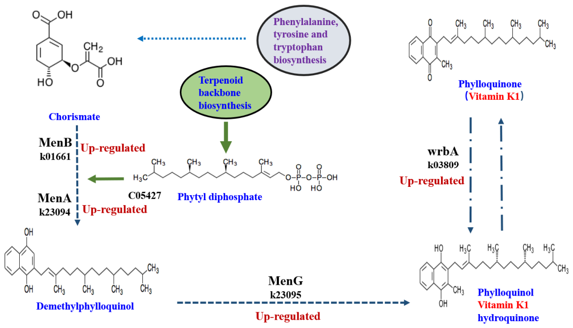
There were VB1, VB5, and VB6 in the standard NB culture medium, and simultaneously, a previous study indicated that Bacillus sp. could promote the growth of Chlorella sp. due to vitamins [8,27]. Therefore, the microalgal transcriptomic characteristics of vitamin metabolism genes were especially focused on in this study. Surprisingly and interestingly, the expected transcription levels of B vitamin metabolism genes were not up-regulated, but rather a significant up-regulation of VK1 metabolism genes was observed. In the transcriptomic sequencing results, the transcript level of the 2-carboxyl-1,4-naphthoquinone phytase (MenA) gene, naphthalate synthase (MenB) gene, demethylphylloquinol methyltransferase (MenG) gene, and wrb A gene were all up-regulated when B. megaterium CGMCC1.217 was added to the C. pyrenoidosa ZJOU6 culture. Previous studies showed that MenA, MenB, MenG, and wrb were the key enzymes of VK synthesis [14,31]. The relationship of these enzymes with VK1 is shown in Figure 2. The transcriptomic sequencing results proposed that the promotion of B. megaterium CGMCC1.217 on the growth of C. pyrenoidosa ZJOU6 might be mediated by VK1. Therefore, the authors of this study hereupon detected whether VK1 was synthesized in the co-cultured system of B. megaterium CGMCC1.217 and C. pyrenoidosa ZJOU6.
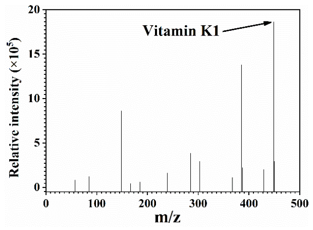
As expected, and as shown in Figure 3, a VK1 peak (m/z = 451.5) in the LC-MS spectrum of the co-culture solution of B. megaterium CGMCC1.217 and C. pyrenoidosa ZJOU6 appeared [26]. Although the presence of VK1 was detected in the co-culture solution of B. megaterium CGMCC1.217 and C. pyrenoidosa ZJOU6, it does not necessarily indicate that VK1 can promote the growth of C. pyrenoidosa ZJOU6. Therefore, the effects of VK1 on the growth and metabolism of C. pyrenoidosa ZJOU6 were further investigated.

3.3. Exogenous VK1 Promotes the Growth and Lipid Production of C. pyrenoidosa

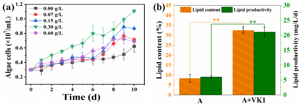
As shown in Figure 4a, VK1 at different concentrations had positive effects on the growth of C. pyrenoidosa ZJOU6. The highest final algae cell density was obtained in the treatment of VK1 with 0.3 g/L. At the end of the experiment, the algae cell density reached 1.11 × 10⁷ cells/mL in the treatment of VK1 at 0.3 g/L. At the same time, the algae cell density in the control was only 0.51 × 10⁷ cells/mL. The algae cell density increased by 117% with the addition of VK1. The present result indicates that VK1 is a very effective chemical that can promote the growth of C. pyrenoidosa ZJOU6. Though studies have shown that other vitamins, including exogenous B1, B6, B7, and B12, are favorites for microalgal growth [6,7], as far as the authors know, this is the first report on the promotion effects of exogenous VK1 on Chlorella species. Since Chlorella species have been widely utilized in wastewater treatment, bioenergy production, antibiotic removal, CO2 fixation, etc. [32,33,34], the present results will be beneficial for providing an effective method for better utilization of Chlorella species in a wide range of fields.
As shown in Figure 4b, both the microalgal lipid content and lipid productivity were significantly (p < 0.01) improved by VK1. At the end of the experiment, the lipid content of the C. pyrenoidosa ZJOU6 in the control was only 8%, while it reached 32% in the VK1 treatment, increased by 291%. On the other side, the average lipid productivity of C. pyrenoidosa ZJOU6 in the control was only 6.08 mg/L/d, while it reached 21.11 mg/L/d, increased by 247%. Previous studies showed that the lipid productivity of Chlorella sp. could be greatly improved in heterotrophic and mixotrophic culture modes [28,35]. Nevertheless, the lipid productivity of Chlorella sp. in phototrophic mode is normally only about 10 mg/L/d or even lower [36]. Present results indicated that supplementing exogenous VK1 could remarkably improve the lipid content and lipid productivity of C. pyrenoidosa in mixotrophic culture mode. This finding is beneficial for the development and utilization of microalgae-based technology in wastewater treatment and high-value product production. Further, to discuss the possible mechanism of VK1 promoting the lipid production of C. pyrenoidosa ZJOU6, the transcriptomic difference between the control and VK1 treatment was determined.
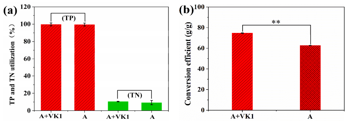
Since microalgae need to uptake nitrogen and phosphorus from the environment to meet their growth needs, and simultaneously, this kind of nutrient uptake is beneficial for wastewater treatment and reuse [5,10], the microalgal utilization efficiency and transformation efficiency of nitrogen and phosphorus were determined in this research. As shown in Figure 5a, the TP utilization rates of C. pyrenoidosa ZJOU6 with and without VK1 supplement were both close to 100%. There is no significant statistical difference between the control and VK1 treatment. On the 10th day after inoculation, the final TP concentration in the control and in VK1 treatment decreased from 4.5 mg/L to lower than 0.02 mg/L, which meets the Class I (<0.02 mg/L) water quality standard for surface water environment (GB 3838-2002) of China [37]. Many studies have indicated that microalgae possess excellent utilization characteristics of phosphorus from wastewaters [10,23,38,39]. The present result of TP utilization is consistent with these studies. Notably, since the initial TP concentration of the used BG11 culture medium was relatively low (only 4.5 mg/L), the changes in the TP concentration in the control and VK1 treatment were not displayed. The characteristics of VK1 in improving the efficiency of microalgae phosphorus utilization may only be reflected in the treatment of wastewater containing high concentrations of TP, such as concentrated municipal wastewater that contained over 200 mg/L of phosphorus [40]. In addition, as shown in Figure 5a, the utilization of C. pyrenoidosa ZJOU6 for nitrogen, both in the control and VK1 treatment, was relatively low. This was possibly due to the high content of TN in the BG11 medium, which is more than enough compared to the nitrogen required for microalgae growth [23].
Significantly, as a concern in traditional agriculture production, phosphate utilization efficiency is very important considering a higher input–output ratio [41]. Undoubtedly, a higher input–output ratio in microalgae production should also be considered, especially when an artificial culture medium is used [36]. Therefore, the ratio of TP reduction to the microalgal biomass increase (TP conversion efficiency) was calculated to evaluate the input–output ratio of phosphate utilization. As shown in Figure 5b, the phosphorus conversion efficiency of C. pyrenoidosa ZJOU6 was only 62.74 g/g in the control, while it reached 74.73 g/g in the VK1 treatment with an increase of 19%. It is worth noting that the phosphate in the present culture medium was completely utilized. Like the TP utilization, the characteristics of VK1 in improving microalgal efficiency of TP conversion may only be reflected in the treatment of wastewater containing high concentrations of TP.

3.4. Transcriptomic and Photosynthetic Evidence of Exogenous VK1 Promoting the Growth and Lipid Production of C. pyrenoidosa

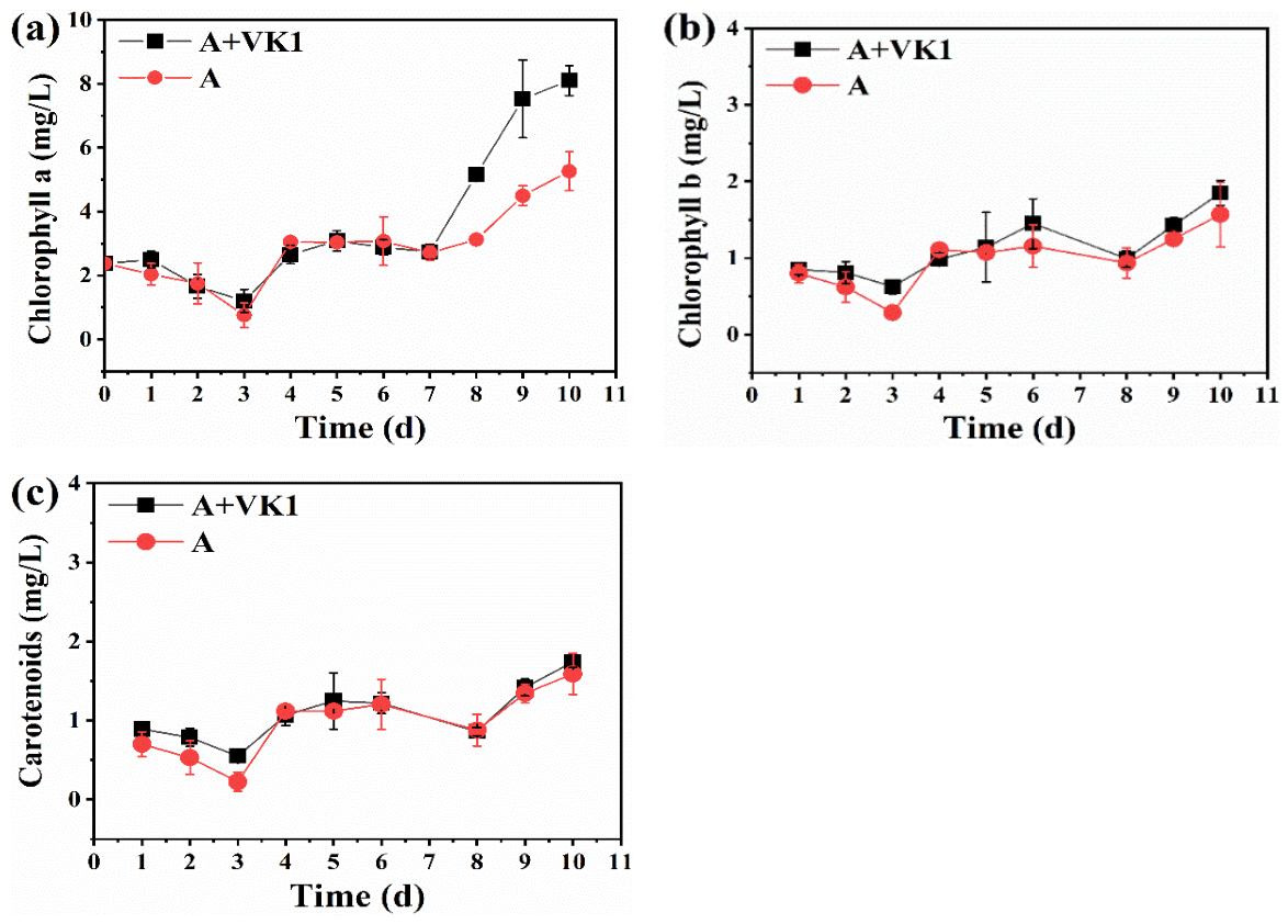
Previous studies showed that VK1 is responsible for transferring photosynthetic electrons from Chl-a to the iron–sulfur protein complex of photosynthetic organisms [13,14]. Therefore, the effects of VK1 on the microalgal pigment content and photosynthetic parameters were determined. As shown in Figure 6, among the three detected pigments of Chl-a (Figure 6a), Chl-b (Figure 6b), and carotenoids (Figure 6c), only the content of Chl-a in the C. pyrenoidosa ZJOU6 increased under the stimulation of VK1, while the contents of Chl-b and carotenoids in VK1 treatment showed no observable change compared with the control. The maximum concentration of Chl-a in the C. pyrenoidosa ZJOU6 system with added vitamin K was 8.10 mg/L on the 10th day, which was 90% higher compared with the control. Chl-a is an important pigment in phytoplankton, algae, and some bacteria, which has the function of converting light energy into chemical energy [42]. Therefore, theoretically, C. pyrenoidosa ZJOU6 might exhibit changes in photosynthetic characteristics corresponding to the increase in Chl-a. In the following experiment, the photosynthetic parameters, including Fv/Fo, Fv/Fm, and Fm/Fo, were determined to evaluate the impacts of VK1 treatment on microalgal photosynthesis. On the other hand, carotenoids and Chl-b are important mediators for microalgae responding to various environmental stresses by dissipation of oversaturated energy [43]. The lack of change in these pigments might be due to the suitable culture conditions in the VK1 treatment and control. The C. pyrenoidosa ZJOU6 did not need extra Chl-b and carotenoids to respond to environmental stresses.
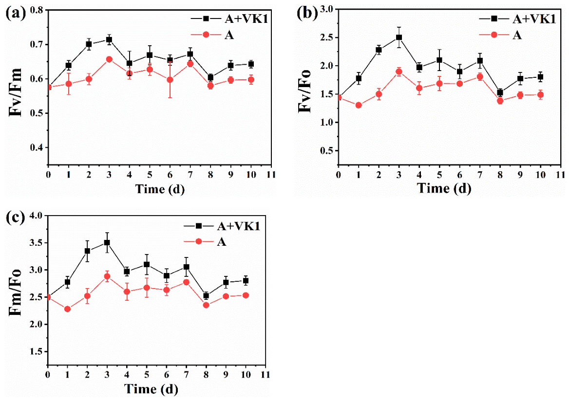
After 3 days of cultivation, the photosynthesis parameters of microalgae in the treatment and the control reached their maximum values. The maximum values of Fv/Fm (Figure 7a), Fv/Fo (Figure 7b), and Fm/Fo (Figure 7c) were 2.50, 0.71, and 3.50, respectively, which were 70%, 132%, and 161% higher than those in the control, respectively. The maximal quantum yield of PSII (Fv/Fm) is an indicator of the efficiency of PSII photochemistry. The higher Fv/Fm in the VK1 treatment indicated the increased activity of the reaction centers attributable to the conserved cysteines in the VKOR enzyme domain [44]. In addition, the higher Fv/Fo in the VK1 treatment than that in the control indicated that the maximum light energy conversion potential of the PSII reaction of the C. pyrenoidosa ZJOU6 strain was increased by the VK1 [45]. Further, Fm/Fo is the quantum yield of PSII electron transport, which can reflect the overall efficiency of photosynthesis and the efficiency of light energy transport [22]. The higher the Fm/Fo, the higher the photosynthetic energy transport efficiency of C. pyrenoidosa ZJOU6. In sum, the VK1 supplement improved the photosynthesis characteristics of C. pyrenoidosa ZJOU6. This should be the main reason for promoting the microalgal growth, which is consistent with the results of transcriptomic analysis.
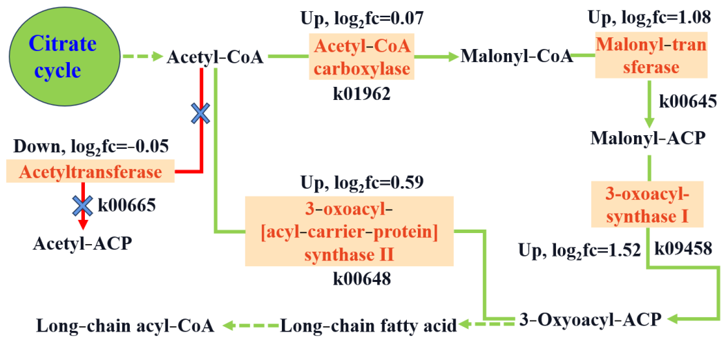
As shown in Figure 8, the up-regulated genes at the transcript level due to VK1 addition included Acetyl-CoA carboxylase (ACCase) gene, Malonyl-transferase gene, 3-oxoacylsynthase I gene, and 3-oxoacyl-[acyl-carrier-protein] synthase II gene. All these genes and their corresponding pathways are directly related to microalgal lipid biosynthesis [11]. The up-regulation of these genes at the transcriptomic level indicated that the function of VK1 in promoting the lipid accumulation of C. pyrenoidosa ZJOU6 could be achieved by regulating these genes, although conclusive proof of this speculation still requires more molecular biology evidence. Significantly, previous studies found that exogenous VB7, as an important co-enzyme of ACCase in microalgal lipid biosynthesis, could improve microalgal lipid productivity [12]. Present results indicated that VK1 should possess a similar function to VB7.
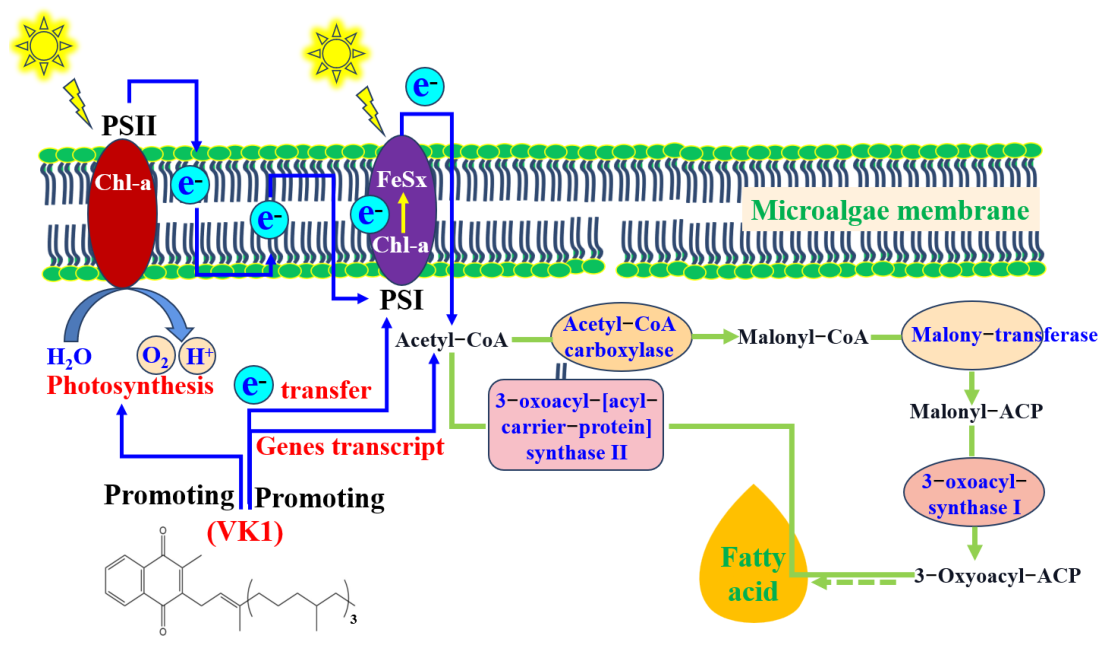
As a sum based on the above results, a promotion mechanism of exogenous VK1 on the growth and lipid production of C. pyrenoidosa ZJOU6 was proposed, as shown in Figure 9. In brief, VK1 promoted the microalgal Chl-a synthesis and PSII activities. As a result, more photosynthetic electrons were transferred through Chl-a and FeSx of PSI to the microalgal lipid biosynthesis system. In this system, more fatty acid (the main component of microalgal lipid) was synthesized under the catalysis of a series of enzymes, including ACCase, Malonyl-transferase, 3-oxoacylsynthase I, and 3-oxoacyl-[acyl-carrier-protein] synthase II.

4. Conclusions

This study elucidated the effects and mechanisms of VK1 in promoting microalgal growth and lipid production. The key conclusions are summarized as follows:
(1)
B. megaterium CGMCC1.217 effectively promotes the growth of C. pyrenoidosa ZJOUC6.
(2)
Transcriptomic analysis showed that co-cultivation with B. megaterium upregulates the expression of VK1 biosynthesis-related genes in C. pyrenoidosa, and LC-MS detection confirmed the presence of VK1 in the co-culture system.
(3)
Exogenous VK1 supplementation exerts a positive effect on the growth and lipid production of C. pyrenoidosa. The optimal concentration of VK1 is 0.3 g/L, which maximizes microalgal cell density, lipid content, and lipid productivity.
(4)
The mechanism by which VK1 promotes the growth and lipid production of C. pyrenoidosa may involve two key pathways: (I) enhancing photosynthetic efficiency by increasing Chl-a content and improving PSII activity (as reflected by the elevated values of Fv/Fm, Fv/Fo, and Fm/Fo), thereby facilitating the transfer of photosynthetic electrons; (II) upregulating the expression of lipid synthesis-related genes, which accelerates fatty acid synthesis and lipid accumulation.

Author Contributions

Conceptualization, Y.G. and J.L.; methodology, W.X. and Y.C.; software, Y.Y.; validation, X.Z., Y.G. and J.L.; formal analysis, Y.Y. and J.L.; investigation, Y.Y. and Y.G.; resources, Y.G.; data curation, Y.Y. and W.X.; writing—original draft preparation, Y.Y. and W.X.; writing—review and editing, Y.G.; visualization, J.L.; supervision, X.Z.; project administration, Y.G.; funding acquisition, J.L., X.Z. and Y.C. All authors have read and agreed to the published version of the manuscript.

Funding

This research was funded by the National Natural Science Foundation of China, grant number 32571903; the Natural Science Foundation of Zhejiang Province, grant number MS26B060033, LY23D060005; and Zhoushan Science and Technology Project, grant number 2025C31022.

Data Availability Statement

The original contributions presented in this study are included in the article. Further inquiries can be directed to the corresponding authors.

Acknowledgments

The National Natural Science Foundation of China, the Natural Science Foundation of Zhejiang Province, and Zhoushan Science and Technology Project.

Conflicts of Interest

The authors declare no conflicts of interest.

References

  1. Minhas, A.; Gaur, S.; Sunny, S.; Paladugu, C.; Ravishankar, G.; Pereira, L.; Ambati, R. Microalgae-Based Wastewater Treatment Processes for the Bioremediation and Valorization of Biomass: A Review. Phycology 2026, 6, 18. [Google Scholar] [CrossRef] [Scilit]
  2. Xu, T.; Liu, W.; Liu, X.; Li, X.; Xian, Q.; Huo, S.; Zhao, C.; Guo, B.; Li, Q. The Symbiotic System between Tribonema sp. and Aerobic Denitrifying Phosphorus Accumulating Bacteria: A Promising Method for Wastewater Treatment. Algal Res. 2025, 85, 103824. [Google Scholar] [CrossRef] [Scilit]
  3. Liu, J.; Qin, W.; Sun, S.; Liang, W. Nutrient Supplementation Strategies Regulate Metabolic Pathways in Microalgae–Bacteria Symbiotic Systems. Algal Res. 2026, 95, 104674. [Google Scholar] [CrossRef] [Scilit]
  4. Yang, Q.; Ge, Y.; Iqbal, N.; Yang, X.; Zhang, X. Sulfitobacter alexandrii sp. nov., A New Microalgae Growth-promoting Bacterium with Exopolysaccharides Bioflocculanting Potential Isolated from Marine Phycosphere. Antonie Van Leeuwenhoek 2021, 114, 1091–1106. [Google Scholar] [CrossRef] [Scilit]
  5. Qi, H.; Lv, J.; Liao, J.; Jin, J.; Ren, Y.; Tao, Y.; Wang, D.; Alvarez, P.; Yu, P. Metagenomic Insights into Microalgae-bacterium-virus Interactions and Viral Functions in Phycosphere Facing Environmental Fluctuations. Water Res. 2025, 268, 122676. [Google Scholar] [CrossRef] [Scilit]
  6. Ali, S.; Waqas, W.; Bakky, M.; Zada, S.; Saif, U.; Hasan, M.; Shafiq, M.; Hui, W. Implications of Microalgal–Bacterial Interactions in Modern Aquaculture Practices: A Review of the Current Knowledge. Rev. Aquacult. 2025, 17, e12980. [Google Scholar] [CrossRef] [Scilit]
  7. Astafyeva, Y.; Gurschke, M.; Qi, M.; Bergmann, L.; Indenbirken, D.; de Grahl, I.; Katzowitsch, E.; Reumann, S.; Hanelt, D.; Alawi, M.; et al. Microalgae and Bacteria Interaction—Evidence for Division of Diligence in the Alga Microbiota. Microbiol. Spectr. 2022, 10, e00633-22. [Google Scholar] [CrossRef] [Scilit]
  8. Tong, C.; Honda, K.; Derek, C. Enhanced Microalgal Growth and Metabolites Through Co-cultivation with Escherichia coli in Algal Organic Matter Solution. Algal Res. 2024, 77, 103351. [Google Scholar] [CrossRef] [Scilit]
  9. Ghafari, M.; Rashidi, B.; Haznedaroglu, B. Effects of Macro and Micronutrients on Neutral Lipid Accumulation in Oleaginous Microalgae. Biofuels 2018, 9, 147–156. [Google Scholar] [CrossRef] [Scilit]
  10. Liu, J.; Yin, J.; Han, H.; Ge, Y.; Wang, Z.; Bao, X.; Gao, F. Enhancements of Lipid Productivity and Phosphorus Utilization Efficiency of Chlorella pyrenoidosa by Iron and Acetate Supplements in Actual Municipal Wastewater. Renew. Energy 2021, 170, 927–935. [Google Scholar] [CrossRef] [Scilit]
  11. Li-Beisson, Y.; Thelen, J.; Fedosejevs, E.; Harwood, J. The Lipid Biochemistry of Eukaryotic Algae. Prog. Lipid Res. 2019, 74, 31–68. [Google Scholar] [CrossRef] [Scilit]
  12. Tandon, P.; Jin, Q.; Huang, L. A Promising Approach to Enhance Microalgae Productivity by Exogenous Supply of Vitamins. Microb. Cell Factories 2017, 16, 219. [Google Scholar] [CrossRef] [Scilit]
  13. Tarento, T.; McClure, D.; Vasiljevski, E.; Schindeler, A.; Dehghani, F.; Kavanagh, J. Microalgae as A Source of Vitamin K1. Algal Res. 2018, 36, 77–87. [Google Scholar] [CrossRef] [Scilit]
  14. Basset, G.; Latimer, S.; Fatihi, A.; Soubeyrand, E.; Block, A. Phylloquinone (vitamin K1): Occurrence, Biosynthesis and Functions. Mini-Rev. Med. Chem. 2017, 17, 1028–1038. [Google Scholar] [CrossRef] [Scilit]
  15. Tarento, T.; McClure, D.; Talbot, A.; Regtop, H.; Biffin, J.; Valtchev, P.; Dehghani, F.; Kavanagh, J. A Potential Biotechnological Process for the Sustainable Production of Vitamin K1. Crit. Rev. Biotechnol. 2019, 39, 1–19. [Google Scholar] [CrossRef] [Scilit] [PubMed]
  16. Arora, N.; Philippidis, G. The prospects of algae-derived vitamins and their precursors for sustainable cosmesceuticals. Processes 2023, 11, 587. [Google Scholar] [CrossRef] [Scilit]
  17. Prates, J. Applications of Bioactive Compounds from Marine Microalgae in Health, Cosmetics, and Functional Foods. Appl. Sci. 2025, 15, 6144. [Google Scholar] [CrossRef] [Scilit]
  18. Mladěnka, P.; Macáková, K.; Kujovská Krčmová, L.; Javorská, L.; Mrštná, K.; Carazo, A.; Protti, M.; Remião, F.; Nováková, L.; the OEMONOM researchers and collaborators. Vitamin K–Sources, Physiological Role, Kinetics, Deficiency, Detection, Therapeutic Use, and Toxicity. Nutr. Rev. 2021, 80, 677–698. [Google Scholar] [CrossRef] [Scilit]
  19. Jesús Alberto, C.; González-Hernández, J. Vitamins from Microalgae. In Handbook of Food and Feed from Microalgae; Academic Press: Cambridge, MA, USA, 2023; pp. 111–115. [Google Scholar]
  20. Zhang, X.; Pan, Z.; Zhang, J.; Liu, B.; Yang, Q. Phycobacteria Biodiversity, Selected Isolation, and Bioactivity Elucidation of New Bacterial Species of Highly Toxic Marine Dinoflagellate Alexandrium minutum amtk4. Microorganisms 2025, 13, 1198. [Google Scholar] [CrossRef] [Scilit]
  21. Bentley, R.; Meganathan, R. Biosynthesis of Vitamin K (menaquinone) in Bacteria. Microbiol. Rev. 1982, 46, 241–280. [Google Scholar] [CrossRef]
  22. Ge, Y.; Xing, W.; Lu, X.; Hu, S.; Liu, J.; Xu, W.; Cheng, H.; Feng, G.; Chen, Q. Growth, Nutrient Removal, and Lipid Productivity Promotion of Chlorella Sorokiniana by Phosphate Solubilizing Bacteria Bacillus megatherium in Swine Wastewater: Performances and Mechanisms. Bioresour. Technol. 2024, 400, 130697. [Google Scholar] [CrossRef] [Scilit]
  23. Ge, Y.; Liu, J.; Tian, G. Growth Characteristics of Botryococcus braunii 765 under High CO2 Concentration in Photobioreactor. Bioresour. Technol. 2011, 102, 130–134. [Google Scholar] [CrossRef] [Scilit]
  24. Wu, D.; Hou, Y.; Cheng, J.; Han, T.; Hao, N.; Zhang, B.; Fan, X.; Ji, X.; Chen, F.; Gong, D.; et al. Transcriptome Analysis of Lipid Metabolism in Response to Cerium Stress in the Oleaginous Microalga Nannochloropsis oculata. Sci. Total Environ. 2022, 838, 156420. [Google Scholar] [CrossRef] [Scilit] [PubMed]
  25. Zhao, Y.; Hou, Y.; Chai, W.; Liu, Z.; Wang, X.; He, C.; Hu, Z.; Chen, S.; Wang, W. Transcriptome Analysis of Haematococcus pluvialis of Multiple Defensive Systems Against Nitrogen Starvation. Enzym. Microb. Technol. 2020, 134, 109487. [Google Scholar] [CrossRef] [Scilit] [PubMed]
  26. Huang, B.; Wang, Z.; Yao, J.; Ke, X.; Xu, J.; Pan, X.; Xu, X.; Lu, M.; Ren, Y. Quantitative Analysis of Vitamin K 1 in Fruits and Vegetables by Isotope Dilution LC-MS/MS. Anal. Methods 2016, 8, 5707–5711. [Google Scholar] [CrossRef] [Scilit]
  27. Boonbangkeng, D.; Treesubsuntorn, C.; Krobthong, S.; Yingchutrakul, Y.; Pekkoh, J.; Thiravetyan, P. Using Cell-free Supernatant of Bacillus sp. AK3 in Combination with Chlorella to Remove Harmful Algal Bloom Species, TP, TN, and COD from Water. J. Environ. Chem. Eng. 2022, 10, 108645. [Google Scholar] [CrossRef] [Scilit]
  28. Yu, L.; Li, T.; Ma, J.; Zhao, Q.; Wensel, P.; Lian, J.; Chen, S. A kinetic Model of Heterotrophic and Mixotrophic Cultivation of the Potential Biofuel Organism Microalgae Chlorella sorokiniana. Algal Res. 2022, 64, 102701. [Google Scholar] [CrossRef] [Scilit]
  29. Huo, S.; Kong, M.; Zhu, F.; Qian, J.; Huang, D.; Chen, P.; Ruan, R. Co-culture of Chlorella and Wastewater-borne Bacteria in Vinegar Production Wastewater: Enhancement of Nutrients Removal and Influence of Algal Biomass Generation. Algal Res. 2020, 45, 101744. [Google Scholar] [CrossRef] [Scilit]
  30. Luo, X.; Zhang, H.; Zhang, J. The Influence of a Static Magnetic Field on a Chlorella vulgaris-Bacillus licheniformis Consortium and its Sewage Treatment Effect. J. Environ. Manag. 2021, 295, 112969. [Google Scholar] [CrossRef] [Scilit]
  31. Ferland, G. The Discovery of Vitamin K and Its Clinical Applications. Ann. Nutr. Metab. 2012, 61, 213–218. [Google Scholar] [CrossRef] [Scilit]
  32. Gao, F.; Zhou, J.; Zhang, Y.; Vadiveloo, A.; Chen, Q.; Liu, J.; Yang, Q.; Ge, Y. Efficient Coupling of Sulfadiazine Removal with Microalgae Lipid Production in a Membrane Photobioreactor. Chemosphere 2023, 316, 137880. [Google Scholar] [CrossRef] [Scilit]
  33. Kotoula, D.; Iliopoulou, A.; Irakleous-Palaiologou, E.; Gatidou, G.; Aloupi, M.; Antonopoulou, P.; Fountoulakis, M.; Stasinakis, A. Municipal Wastewater Treatment by Combining in Series Microalgae Chlorella sorokiniana and Macrophyte Lemna minor: Preliminary Results. J. Clean. Prod. 2020, 271, 122704. [Google Scholar] [CrossRef] [Scilit]
  34. Qiu, R.; Gao, S.; Lopez, P.; Ogden, K. Effects of pH on Cell Growth, Lipid Production and CO2 Addition of Microalgae Chlorella sorokiniana. Algal Res. 2017, 28, 192–199. [Google Scholar] [CrossRef] [Scilit]
  35. Li, T.; Zheng, Y.; Yu, L.; Chen, S. Mixotrophic Cultivation of a Chlorella Sorokiniana strain for Enhanced Biomass and Lipid Production. Biomass Bioenergy 2014, 66, 204–213. [Google Scholar] [CrossRef] [Scilit]
  36. Sun, Y.; Huang, Y. Effect of Trace Elements on Biomass, Lipid Productivity and Fatty Acid Composition in Chlorella sorokiniana. Braz. J. Bot. 2017, 40, 871–881. [Google Scholar] [CrossRef] [Scilit]
  37. Li, Y.; Zhang, C.; Li, P.; Wang, J.; Zhou, W.; Zhang, Y. Study on Phosphorus Releasing and Releasing Kinetics of Sediments in Dongping Lake. J. Hazard. Mater. Adv. 2023, 9, 100234. [Google Scholar] [CrossRef] [Scilit]
  38. Liu, J.; Ge, Y.; Cheng, H.; Wu, L.; Tian, G. Aerated Swine Lagoon Wastewater: A Promising Alternative Medium For Botryococcus Braunii Cultivation in Open System. Bioresour. Technol. 2013, 139, 190–194. [Google Scholar] [CrossRef] [Scilit]
  39. Liu, J.; Yin, J.; Ge, Y.; Han, H.; Liu, M.; Gao, F. Improved Lipid Productivity of Scenedesmus Obliquus with High Nutrient Removal Efficiency by Mixotrophic Cultivation in Actual Municipal Wastewater. Chemosphere 2021, 285, 131475. [Google Scholar] [CrossRef] [Scilit] [PubMed]
  40. Zhou, W.; Min, M.; Li, Y.; Hu, B.; Ma, X.; Cheng, Y.; Liu, Y.; Chen, P.; Ruan, R. A Hetero-photoautotrophic Two-stage Cultivation Process to Improve Wastewater Nutrient Removal and Enhance Algal Lipid Accumulation. Bioresour. Technol. 2012, 110, 448–455. [Google Scholar] [CrossRef] [Scilit] [PubMed]
  41. López-Arredondo, D.; Leyva-González, M.; González-Morales, S.; López-Bucio, J.; Herrera-Estrella, L. Phosphate Nutrition: Improving Low-phosphate Tolerance in Crops. Annu. Rev. Plant Biol. 2014, 65, 95–123. [Google Scholar] [CrossRef] [Scilit]
  42. Björn, L.; Papageorgiou, G.; Blankenship, R.; Govindjee. A Viewpoint: Why Chlorophyll a? Photosynth. Res. 2009, 99, 85–98. [Google Scholar] [CrossRef] [Scilit] [PubMed]
  43. Strzałka, K.; Kostecka-Gugała, A.; Latowski, D. Carotenoids and Environmental Stress in Plants: Significance of Carotenoid-mediated Modulation of Membrane Physical Properties. Russ. J. Plant Physiol. 2003, 50, 168–173. [Google Scholar] [CrossRef] [Scilit]
  44. Du, J.; Zhan, C.; Lu, Y.; Cui, H.; Wang, X. The Conservative Cysteines in Transmembrane Domain of at VKOR/LTO1 are Critical for Photosynthetic Growth and Photosystem II Activity in Arabidopsis. Front. Plant Sci. 2015, 6, 238. [Google Scholar] [CrossRef] [Scilit] [PubMed]
  45. Tan, Z.; Wu, C.; Xuan, Z.; Cheng, Y.; Xiong, R.; Su, Z.; Wang, D. Lead Exposure Dose-dependently Affects Oxidative Stress, AsA-GSH, Photosynthesis, and Mineral Content in Pakchoi (Brassica chinensis L.). Front. Plant Sci. 2022, 13, 1007276. [Google Scholar] [CrossRef] [Scilit] [PubMed]
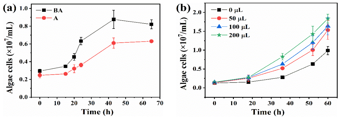
Figure 1. Cell density changes of Chlorella pyrenoidosa ZJOU6 alone (abbreviation as “A”) and together with Bacillus megaterium CGMCC1.217 (abbreviation as “BA”) (a), and supplemented with different doses of sterilized supernatant from B. megaterium culture medium (b). (Notes: The numbers in the legend of (b) are the added volumes of the bacterial suspension).
Phycology 06 00062 g001
Figure 2. Up-regulated genes related to vitamin K1 biosynthesis at the transcript level, and their transformation relationship in Chlorella pyrenoidosa ZJOU6.
Phycology 06 00062 g002
Figure 3. The vitamin K1 peak in the LC-MS spectrum of the co-culture of Bacillus megaterium CGMCC1.217 and Chlorella pyrenoidosa ZJOU6.
Phycology 06 00062 g003
Figure 4. Effects of vitamin K1 at different concentrations on the growth (a) of Chlorella pyrenoidosa ZJOU6, and the effects of 0.3 g/L vitamin K1 on the microalgal lipid content and productivity (b). (Note: The microalgal culture with and without adding VK1 is presented by “A” and “A + VK1”, respectively.). “**” indicates significant differences between the compared treatments (p < 0.01).
Phycology 06 00062 g004
Figure 5. Effects of vitamin K1 on total phosphorus and total nitrogen utilization efficiency (a), and total phosphate conversion efficiency (b) of Chlorella pyrenoidosa ZJOU6. (Note: The microalgal culture with and without adding VK1 is presented by “A” and “A + VK1”, respectively. TP: the total phosphorus; TN: the total nitrogen.). “**” indicates significant differences between the compared treatments (p < 0.05).
Phycology 06 00062 g005
Figure 6. Promoting effects of vitamin K on microalgal chlorophyll a content (a), chlorophyll b content (b), and carotenoid content (c). (Note: The microalgal culture with and without adding VK1 is presented by “A” and “A + VK1”, respectively.).
Phycology 06 00062 g006
Figure 7. Promoting effects of vitamin K1 on the photosynthesis parameters of Fv/Fm (a), Fv/Fo (b), and Fm/Fo (c) of Chlorella pyrenoidosa ZJOU6. (Note: The microalgal culture with and without adding VK1 is presented by “A” and “A + VK1”, respectively.).
Phycology 06 00062 g007
Figure 8. Up-regulated genes related to lipid biosynthesis at the transcript level due to vitamin K1 addition, and their enzymatic relationship in Chlorella pyrenoidosa ZJOU6.
Phycology 06 00062 g008
Figure 9. Proposed mechanism by which vitamin K1 (VK1) promotes the growth and lipid production of Chlorella pyrenoidosa ZJOU6. (Note: PSI, PSII, CoA, FeSx, and ACP are the abbreviations of photosystem I, photosystem II, coenzyme A, iron–sulfur protein complex, and acylcarnitine profile, respectively).
Phycology 06 00062 g009
Table 1. All experiment groups, treatments, and measured parameters.
GroupsTreatmentParameters
Group 1AAlgae density----
A + BAlgae density----
Group 2A + 50 µL SAlgae density----
A + 100 µL SAlgae density----
A + 200 µL SAlgae density----
Group 3A + 0.07 g/L VK1Algae density----
A + 0.15 g/L VK1Algae density----
A + 0.30 g/L VK1Algae density----
A + 0.60 g/L VK1Algae density----
Group 4A + 0.30 g/L VK1TPTNLCLPChl a
Chl bCarFv/FmFv/FoFm/Fo
Notes: A, B, S, TP, TN, LC, LP, Chl a, Chl b, Car, means C. pyrenoidosa ZJOUC6, B. megaterium CGMCC1.217, supernatant of B. megaterium CGMCC1.217 culture medium, solution total phosphate concentration, solution total nitrogen concentration, algae lipid content, algae lipid productivity, algae chlorophyll a content, algae chlorophyll b content, carotenoid content, respectively.
Disclaimer/Publisher’s Note: The statements, opinions and data contained in all publications are solely those of the individual author(s) and contributor(s) and not of MDPI and/or the editor(s). MDPI and/or the editor(s) disclaim responsibility for any injury to people or property resulting from any ideas, methods, instructions or products referred to in the content.