Review

Regulatory Landscapes of Bacterial DNA Methylation: Mechanism, Dynamics, and Detection

1
Department of Biotechnology, Amity School of Biological Sciences, Amity University, Mohali 140306, Punjab, India
2
Department of Human Genetics and Molecular Medicine, Amity School of Biological Sciences, Amity University, Mohali 140306, Punjab, India
3
Department of Cell Biology, Cancer Dormancy Institute, Albert Einstein College of Medicine, Bronx, NY 10461, USA
*
Authors to whom correspondence should be addressed.
Submission received: 26 December 2025 / Revised: 1 March 2026 / Accepted: 2 April 2026 / Published: 4 May 2026

Abstract

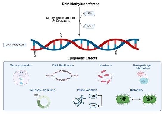
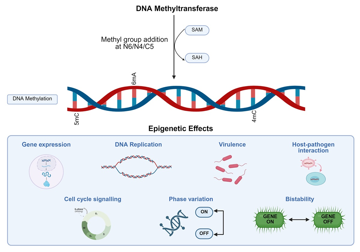
Epigenetics is a widely present mechanism for the modulation of gene expression without alterations in the underlying genetic sequence. Epigenetic signatures are significantly present in bacteria, with DNA methylation playing a key role in the modulation of bacterial physiology and pathogenesis. DNA methyltransferases (MTases) are the enzymes catalyzing the transfer of methyl groups to adenine or cytosine residues in the DNA using the methyl donor S-adenosyl-L-methionine (SAM). This process generates modified bases, N6-methyladenine (m6A), 5-methylcytosine (5mC), or N4-methyl cytosine (4mC) in the DNA, which influence fundamental cellular processes such as DNA transactions, DNA replication, transcription, and DNA repair. These MTases, earlier thought to be a part of primitive bacterial immune system, are now considered to be active players in gene regulation. They regulate bacterial adaptability to stress by virtue of phase variation and bistability. In pathogenic species such as Mycobacterium tuberculosis (Mtb), DNA methylation driven epigenetic reprogramming influences the expression of virulence factors, antibiotic tolerance, and persistence genes. This review gives a detailed account of role of DNA methyltransferases in bacterial epigenomics influencing various cellular processes. With the development of long-read high-throughput sequencing technologies, single-base mapping of bacterial methylomes has become possible. In the latter part of the review, we talk about these advances and the integration of synthetic biology to expand the potential of methylation systems for developing biosensors and switchable gene expression platforms. These strategies can be translated into future vaccine design and precision drugs for disease control. Deciphering bacterial DNA methylation can help gain insights into microbial evolution and design innovative therapeutics for various diseases.

Graphical Abstract

1. Introduction

Epigenetics encompasses mechanisms that convey heritable regulatory information independently of alterations in the DNA sequence. These epigenetic marks can transmit through mitotic or meiotic cell divisions, thereby contributing to stable yet reversible changes in gene activity. It involves chemical modifications—such as DNA methylation—and histone modifications, which play key roles in regulating gene expression [1]. In bacteria, this regulatory mechanism influences bacterial physiology, stress response, environmental adaptation, and host–pathogen interactions. DNA methylation is the process through which a methyl group (−CH3) is covalently added to specific nucleotide bases, primarily adenine and/or cytosine. This reaction is catalyzed by DNA methyltransferases (MTases) using S-adenosyl-L-methionine (SAM) as a methyl donor [2,3,4]. This leads to a change in chemical and structural properties of DNA, resulting in altered DNA–protein interactions, chromosomal organization, repair, and transcription [3,5].
5-methylcytosine was first discovered in tuberculinic acid of Mtb in 1925 [6]. In 1960s, it was found that methylation is not only a defensive mechanism against foreign DNA but also acts as a tool for regulating gene expression and maintaining genome integrity [4,5]. Bacterial DNA methyltransferases are classified according to their structural and functional role, the type of nucleotide base they modify, and the nature of methylation reaction they catalyze. These MTases recognize and methylate specific sequences, and modify them. Most species have more than one type of methyltransferase, each recognizing different motifs [7,8]. These MTases not only influence chromatin organization, but they also play wider roles in the regulation of cell cycle progression, DNA replication, and mismatch repair [7]. In clinically important pathogens, distinct methylation signatures have been linked to altered gene expression, tolerance to antibiotics, hypoxia survival, and virulence, making them important for studying antimicrobial resistance [9,10,11,12,13,14]. One important consequence of methylation-dependent gene expression is the generation of phenotypic heterogeneity within bacterial populations. This heterogeneity is often achieved through phase variation, a reversible and heritable ON/OFF switching of gene expression. This aids in bacterial persistence, immune evasion, and rapid adaptation to fluctuating host environments without any permanent genetic changes [15,16].
Understanding these epigenetically driven switches therefore requires methods capable of capturing methylation patterns across bacterial genomes. With the development of newer sequencing technologies, research on bacterial methylation has progressed substantially. Earlier biochemical approaches like restriction digestion, Southern blotting, and bisulfite conversion are now replaced by long-read methods like Pacific Biosciences Single Molecule Real Time (SMRT) and Oxford Nanopore sequencing, which directly detect modified bases in native DNA [10,14,17]. Coupled with bioinformatic pipelines for motif discovery, MTase assignment, and muti-omics integration with transcriptomics and proteomic data, these sequencing technologies have greatly accelerated functional methylome analysis. When paired with computational tools for mapping and when combined with transcriptomic data, these methods make methylation studies more precise and informative [18,19,20].
This review compiles recent advances in understanding bacterial DNA methylation, emphasizing the importance of methyltransferases, regulatory pathways, and emerging methylome profiling technologies. It explores how DNA methylation serves as a key mechanism for many cellular processes ranging from phase variability, adaptation, cell cycle control, virulence, and stress responses. In the end, we discuss advanced methylation detection techniques and comprehensive methylome mapping approaches to harness bacterial epigenetic regulation for applications in disease diagnosis and synthetic biology engineering.

2. DNA Methylation

DNA methylation represents one of the earliest recognized epigenetic mechanisms in prokaryotes [2]. It was previously believed that genetic information resided solely in the DNA sequence, but, with the emergence of epigenetic mechanisms such as methylation, it was revealed that chemical modifications in DNA also have a regulatory role [21]. In 1960s, it was observed that certain bacterial strains could resist bacteriophage infections by degrading foreign DNA while protecting their own genomes via methylation. Several groups also showed that, apart from genome protection, DNA methylation has various regulatory roles in bacteria [22]. Although DNA methylation does not disrupt Watson–Crick base pairing or change the primary DNA sequence, the incorporation of a methyl group (−CH3) alters the electrostatic surface potential, increases local hydrophobicity, and induces subtle conformational changes in the structure of the DNA helix [3]. Genes encoding methyltransferases are found in nearly all bacterial genomes. Many species harbor multiple methyltransferase genes that target different sequences and motifs, suggesting that DNA methylation is a conserved regulatory mechanism refined through bacterial evolution [7,23]. A distinctive feature of bacterial methylation is its dynamic nature during DNA replication. Following DNA replication, only parental DNA strand contains the methylation mark leading to a hemi-methylated or semi-methylated DNA. This strand is then subsequently methylated to generate a fully methylated DNA strand. For instance, the Escherichia coli DNA adenine methyltransferase (Dam) rapidly methylates most of the GATC sites on the daughter strand so that the hemi-methylated sites become fully methylated again, within seconds [24]. The timing of this post-replicative methylation is important as transient hemi-methylated state is not a merely short-lived intermediate but also has a regulatory role. Delayed remethylation at the origin of replication (ori) allows SeqA to bind to hemi-methylated DNA and temporarily sequester the origin, preventing premature replication initiation. Prolonged SeqA binding has been shown to delay nucleoid segregation and cell division, highlighting the role of timed methylation in coordinating DNA replication with cell cycle [25].
Broadly, the biological significance of DNA methylation encompasses gene regulation, chromosome replication, DNA repair, transcriptional control, and the maintenance of genome stability. DNA MTases do not merely mark DNA for replication or repair but act as regulators of gene networks that affect virulence, stress response, antibiotic tolerance, biofilm formation, and host adaptation. For example, in Mycobacterium bovis, the absence of orphan MTase HsdM, increases trcR expression, reducing susceptibility to isoniazid, and in E. coli, dam and recA knockouts, make the cells more sensitive to β-lactams and quinolones [13,26].
The differences in methylation at promoter regions can modulate the binding of transcription factors and nucleoid-associated proteins, leading to turnover between active and repressed transcriptional states that affect stress and metabolic pathways [27]. Methylation, together with modified nucleoid-associated proteins, also influences transcription through bridging, bending, and wrapping of DNA [28]. Both the DNA methylation systems and nucleoid-associated proteins influence gene expression and are more amenable to manipulation and therapeutic targeting [29]. The methylation-dependent regulatory mechanisms are often reversible and sensitive to environmental cues, which provide a way for bacteria to flexibly fine tune gene expression in response to changing conditions that integrate epigenetic signals with physiological outcomes [27].

2.1. DNA Methyltransferases

Bacterial DNA MTases represent a diverse family of specialized enzymes that function as sequence-specific DNA-binding proteins that can modify the chemical properties of the target nucleotides. These are universally present in all bacterial species [7]. Based on the type of DNA bases they methylate and their sequence specificity, they are classified into adenine methyltransferases, cytosine-5 methyltransferases, and cytosine N4 methyltransferases, respectively [30]. These can be further categorized according to mode of transfer as exocyclic or endocyclic methyltransferases. Exocyclic methyltransferases catalyze the transfer of SAM to an exocyclic amino group located outside the nucleobase ring, at the N4 position of cytosine, generating 4mC, or at the N6 position of adenine, generating m6A. In contrast, endocyclic methyltransferases transfer methyl groups from SAM to the carbon atom within the nucleobase ring, at the C5 position of cytosine, resulting in the formation of 5mC [31]. Based on the structural and functional organization, they are broadly classified into two categories: one is associated with the restriction-modification (R-M) system as part of the bacteria’s defense mechanism, and the other is associated with ‘orphan’ or solitary methyltransferases that are not paired with restriction enzymes [7,31]. This will be discussed in detail in Section 2.3.
Methyltransferases operate via the ‘base-flipping’ strategy, where the enzyme first binds to its specific recognition sequence and induces a distortion in the DNA helix. The amino acid side chains are inserted between the base pairs to open the helix. This causes target adenine or cytosine to flip out approximately 180° outwards from their normal positions into the enzyme’s active-site pocket [32]. The base’s reactive site now becomes accessible to methyl group donors that catalyze transfer of methyl group to the atom. After methylation, the base with its normal structure is flipped back into the DNA helix [19]. The types of DNA methyltransferases are discussed in the following sections.

2.2. Types of DNA Methyltransferases Based on Chemical Modification

2.2.1. Cytosine Methyltransferases

Although adenine methylation is the most abundant methylation type in prokaryotes, certain bacterial species exhibit robust cytosine methylation at the C5 or N4 positions. Therefore, cytosine methylation is now gaining attention as an important epigenetic mechanism, influencing diverse processes such as gene regulation, stress adaptation, genome defense, and pathogenesis [30].
5-Methyl Cytosine (5mC)
C5-DNA MTases, such as Dcm, methylate internal cytosine at CCWGG’ in target recognition motif. Cytosine methylation in bacteria helps us link epigenetic marks with genome evolution [33]. Within the broad spectrum of cytosine methylation, one motif of particular interest is CpG dinucleotide, a cytosine immediately followed by guanine in the DNA sequence. In eukaryotes, methylation at CpG sites is quite common, and it is involved in transcriptional control, genomic stability, and imprinting [34]. However, in bacteria, reporting CpG methylation is rare because of presence of restriction enzymes that target CpG DNA [35]. CpG DNA is also highly mutable, as it is prone to spontaneous deamination. Over evolutionary timescales, this mutability has driven CpG underrepresentation, which is depletion of CpG motifs in the genome [36]. As a result, CpG-specific MTases are uncommon, and those that exist often reflect unusual evolutionary pressures and, in some cases, distinctive pathogenic strategies. Cytosine methyltransferases have special roles, e.g., in Gram-negative bacteria like Caulobacter crescentus, the solitary cytosine methyltransferase, ScmA, plays a crucial role in maintaining genome stability by preventing DNA damage through SOS response activation [37].
Other examples are Mycoplasma hyorhinis and M. penetrans, which encode for CpG specific MTases capable of methylating their own genomes, as well as host genomic DNA during infection. CpG methylation decreases significantly when M. hyorhinis enters host cells, implying that the bacterium modulates its methylation patterns in response to environmental cues to enhance its survival in the host [38]. Various studies using next-generation sequencing techniques suggested that CpG hypomethylation during intracellular stages may help bacterium evade immune detection or mitigate deleterious methylation effects [38]. Contrastingly, M. penetrans expresses a constitutively active CpG specific methyltransferase, M.MpeI, which fully methylates CpG sites across the genome in both in vivo and in vitro settings. The enzyme’s activity creates widespread 5mC marks, but the genome of M. penetrans is exceptionally CpG-depleted, which is a consequence of long-term methylation-induced mutagenesis [20]. In H. pylori, the JHP1050 methyltransferase, also known as M/Hpy99III, methylates GCGC sites and controls gene expression by affecting transcription factor binding. This remodels the transcriptome and generates phenotypic heterogeneity within clonal populations [39].
The methylation landscape in Gram-positive bacteria like Mycobacterium tuberculosis (Mtb) is more complex. While adenine methylation via MamA dominates, some strains exhibit detectable 5mC modifications. The Mtb genome encodes multiple putative MTases, whose expression patterns differ in drug-sensitive and drug-resistant strains [9]. These MTases have been associated with antibiotic resistance, immune evasion, and adaptive responses. Cytosine methylation marks have been detected in the genome of Mtb residing within infected macrophages, although specific genes remain unidentified. A major understanding of the regulatory role of cytosine methylation comes from the genome-wide analysis of 5mC modifications in Streptomyces coelicolor. Using the dot-blot analysis, the authors demonstrated that cytosine methylation levels change during growth in a defined maltose-glutamate medium, supporting the idea that 5mC is dynamically maintained in response to physiological state. Functional relevance was established by hypomethylating genomic DNA with 5-aza-2-deoxycytidine, which led to impaired growth and reduced antibiotic production. Genome-wide mapping using bisulfite sequencing identified two conserved motifs, ‘GGCCGG’ and ‘GCCCG’, located upstream of 321 genes. Many of these genes are involved in development, metabolism, transcriptional regulation, and secondary metabolite biosynthesis. This study provides the first comprehensive description of the cytosine methylome of S. coelicolor and offers direct evidence that 5-mC functions as a regulatory epigenetic mark influencing growth, differentiation, and gene expression in bacteria [40].
N4-Methyl Cytosine (4mC)
4mC modification is exclusively present in bacteria and archaea. This modification involves exocyclic methylation at N4 cytosine and accounts for 20% of prokaryotic DNA methylation. These methyltransferases are integral part of restriction–modification systems. The M2.Hpy.AII MTase in H. pylori is an excellent example of 4mC methyltransferase in Gram-negative bacteria. The enzyme methylates cytosine at the ‘TCTTC’ motif in the upstream regions of 102 genes and regulates their expression. This influences bacterial adherence to host cells and inflammatory response of the host [41].
In Gram-positive bacteria, such as Streptomyces roseosporus L30, the deletion of sroLm3 gene coding for SroLm3 methyltransferase led to a pronounced reduction in global 4mC levels without affecting adenine methylation, confirming the specificity of this methyltransferase for cytosine modifications. Notably, the loss of sroLm3 resulted in enhanced production of secondary metabolites, including increased yields of daptomycin, accompanied by changes in some biosynthetic gene clusters. This suggests that, in S. roseosporus, 4mC methylation controls the secondary metabolism and provides a link between 4mC modification and antibiotic synthesis [42]. In a separate study, TagR, a TetR-family transcription regulator was identified, whose activity was influenced by 4mC modification in its promoter region. A genetic disruption of tagR resulted in increased daptomycin production. A mechanistic analysis showed that TagR represses genes involved in cell envelope functions, including teichoic acid transport, indicating that 4mC methylation enhanced TagR-dependent transcriptional control [43]. All these studies suggest that 4mC modifications play global transcription regulatory roles in bacteria [42,43].

2.2.2. Adenine Methyltransferases

Adenine methylation is the most prevalent and significant epigenetic modification in prokaryotes [44]. It alters the DNA structure dynamics, which directly impacts DNA–protein interaction. In Gram-negative bacteria like E. coli, m6A maintains replication fidelity by marking the parental DNA strands and regulating the replication timing by binding to SeqA protein [45]. In Gram-positive bacteria, Lacticaseibacillus paracasei, an industrially important strain, 78% of m6A sites have been found in coding regions of carbohydrate metabolism genes acting as a master regulator controlling carbohydrate utilization [46]. The m6A methylation contributes to a metabolic break that restricts metabolic activity and promotes efficient substrate utilization, which is important under nutrient limiting conditions of industrial fermentation. Mutation in the pglX gene, coding for m6A methyltransferase, led to faster growth in the exponential phase and to the upregulation of carbohydrate metabolism at transcriptomic, proteomic, and metabolic levels [46]. DNA methylation reflects an evolutionary trade-off between bacterial survival and industrial needs, paving the way for targeted epigenetic manipulation for the optimization of strains for flavor and food production.
In Mycobacterium bovis BCG, MamA catalyzes m6A modifications at the target DNA sequence, ‘CTCCAG’. It was found that methylated DNA was more efficiently recognized by the host immune receptor TLR9. This enhanced recognition leads to the activation of macrophages and increases the production of pro-inflammatory cytokines such as IL-2, IL-6, and IFN-β. However, when this target sequence is absent or not methylated, cytokine production is reduced and macrophage activation does not occur. This property of methylation was used to enhance adjuvant potential of CpG DNA through the ligation with oligodeoxynucleiotides harboring N6-methylated adenine. This combination led to a strong Th1 and type I interferon responses through TLR9 activation. This could help in designing improved TB vaccines or more effective DNA-based adjuvants [47,48,49].

2.3. Types of DNA Methyltransferases Based on Functional Organization

2.3.1. Restriction–Modification Enzymes

Restriction–modification (R-M) systems represent an important category of DNA methyltransferases that are essential for bacterial defense [48]. Each system comprises a restriction endonuclease (REase) and a cognate DNA methyltransferase that modifies adenine or cytosine residues, enabling the discrimination between self and foreign DNA. Bacterial genomic DNA is protected through sequence-specific methylation, whereas unmethylated foreign DNA is cleaved, limiting viral infection and horizontal gene transfer (HGT) [49]. Biological significance of these systems is underscored by the identification of more than 2000 R-M systems in various bacterial species, which are cataloged in the REBASE database [50]. Table 1 summarizes the classification and characteristics of R-M systems.
Beyond host defense, R-M systems influence genome evolution and gene transfer dynamics. Plasmids under strong selective pressure preferentially mutate short palindromic sequences commonly targeted by the R-M systems, with smaller plasmids eliminating these sites and larger plasmids relying on orphan methyltransferases for protection, affecting the distribution of antibiotic-resistant genes [63]. In addition, R-M systems can function as selfish genetic elements, particularly Type II systems, where the loss of methylation leads to the cleavage of host bacterial DNA. This enforces the retention of the R-M pair by the bacterium, ensuring genome maintenance [64]. At the population level, certain systems, including Type IV R-M systems introduced via mobile genetic elements, target DNA with atypical methylation patterns and induce the death of infected cells, thereby limiting the spread of invasive genetic material and highlighting the dual role of R-M systems in selfish maintenance and community-level defense [65,66]. R-M systems are classified into distinct types based on mode of action and organization.

2.3.2. Orphan Methyltransferases

Unlike the R-M system, where DNA MTases work together with restriction enzymes to defend against foreign DNA, orphan methyltransferases are a distinct class of solitary methyltransferases unaccompanied by cognate restriction enzymes. They act independently and are conserved across many bacterial and archaeal lineages [67].
Comparative genomics reveal that orphan methyltransferases are far more prevalent and evolutionarily stable than R-M system-associated MTases [8]. Many of these originated from the decaying R-M systems, where the restriction counterpart was lost, but the methylase was retained due to essential cellular functions. This is evident from the example of H. pylori, where less than 30% of predicted type II R-M systems are fully functional. They contain a functional methyltransferase, while the neighboring restriction enzyme is disrupted by frameshifts, truncations, or complete deletion [68]. Comparative genomics across ~1000 bacterial genomes show that a substantial fraction of orphan MTases are found adjacent to the ‘remnants’ of restriction enzyme genes, suggesting that these R-M systems have gradually fallen apart [8]. This evidence supports a consistent evolutionary mechanism, where the restriction component is preferentially lost, while methyltransferase component is maintained, as a beneficial strategy for the host.
The methylation patterns influence DNA replication, gene expression, and other regulatory functions; therefore, changes in DNA recognition sequence could alter the outcomes. This functional constraint helps explain why orphan MTases do not consistently exhibit high sequence divergence, as seen in many R-M systems. Conserved orphan MTases such as Dam and CcrM retain stable recognition motifs because their methylation sites are found in genes that are part of essential regulatory pathways. Altering these sites would affect the genome-wide gene expression and cellular control mechanisms [53]. However, it has been seen that the degree of divergence depends on the biological role and evolutionary history of orphan MTases, as MTases linked to phase variation or those derived from mobile R-M systems can tolerate changes in their DNA recognition domain and therefore show variability [69].
In addition to their effects on gene regulation, they also influence adaptability and antibiotic susceptibility by modulating stress response pathways. A well-characterized example is VchM, a 5mC orphan MTase in Vibrio cholerae. VchM methylates specific ‘RCCGGY’ (R = A/G, Y = T/C) motifs across the genome. In the absence of VchM-mediated methylation, the expression of the groESL-2 chaperonin genes is upregulated, enabling the bacterium to resist the antibiotic-induced protein damage. This demonstrates that DNA methylation can fine-tune stress-adaptive transcriptional programs, thereby increasing survival under antibiotic pressure [70].
DNA Adenine Methyltransferase (Dam)
Dam represents the most extensively studied orphan methyltransferase, which was first identified in E. coli [71,72]. Dam methyltransferases are commonly found in Gammaproteobacteria. They catalyze the methylation of adenine residues in the ‘GATC’ target sequence and serve as epigenetic regulators, influencing numerous cellular processes [73]. With an abundance of 130 molecules per E. coli cell, Dam possesses the ability to methylate 55 ‘GATC’ sites while moving along the DNA [44]. Interestingly, researchers have also found that some genes lacking the GATC sequence are also responsive to Dam [74]. Through X-ray crystallography, they found that E. coli Dam (EcoDam) also identified a five-base non-cognate sequence ‘GTYTA/TARAC’ (Y = C/T and R = A/G), which challenged traditional views and introduced a new layer of epigenetic control. Thus, Dam has emerged as a dual function protein capable of both methylation-dependent and methylation-independent gene regulation [74].
Dam methylation has also been linked with bacterial persistence and stress tolerance mechanisms [75,76]. The following examples illustrate how DNA methylome maintained by Dam buffers bacteria against diverse stresses. In many E. coli strains, the absence of Dam increases the susceptibility to β-lactams and fluoroquinolones. The antibiotic exposure leads to an increase in activity of error-prone polymerase, PolIV, and the absence of Dam leads to the failure of methyl-directed mismatch repair system, leading to lethal dsDNA breaks. This indicated that Dam methylation provides structural support during antibiotic stress [75,77]. Another study demonstrated that a dam knockout mutant shows decreased persister cell formation on exposure to antibiotics such as β-lactams and aminoglycosides. Additionally, the mutants were also hypersensitive to environmental stressors, suggesting Dam’s role in stress regulation. Complementation of the mutant reversed the phenotype, suggesting the same [78]. In Salmonella typhimurium, Dam enhances fitness against oxidative stress from hydrogen peroxide in intracellular niches like the Salmonella-containing vacoule (SCV). It also establishes epigenetic memory by methylating the intA promoter of the ST64B prophage during stress, sustaining integrase expression for prophage reintegration and genomic stability [76].
DNA Cytosine Methyltransferases (Dcm)
Dcm methylases, first discovered in E. coli, are encoded by the dcm gene that methylates at the C5 position of the internal cytosine in the sequence ‘CCWGG’ (W = A/T). Dcm-methylated cytosines are hypermutable and have higher C-T transition rate in natural populations [79]. Dcm methylation also influences a very short patch (VSP) DNA repair pathway by defining targets, and it may regulate the transcription of stress-responsive and ribosomal protein genes. Although Dcm is an orphan MTase, DNA methylation by Dcm may protect against restriction enzymes such as EcoRII [80].
Cell Cycle Regulated Methyltransferase (CcrM)
CcrM is a specific DNA adenine methyltransferase that is commonly found in Alphaproteobacteria such as Caulobacter crescentus, Agrobacterium tumefaciens, Brucella abortus, and Rhizobium meliloti. It is classified as a β-class N6 adenine MTase, which forms a dimer and methylates adenine at the target recognition sequence, ‘GANTC’. β-class N6 adenine MTases are enzymes characterized by Rossmann-like fold structure and a conserved catalytic motif essential for methyltransferase activity [81]. For enhancing the interaction surface, CcrM opens up a ‘bubble’ at the recognition site, bends and unwinds the DNA, and flips out target bases for methylation [82,83]. Its expression is tightly linked to the cell cycle, where the ccrM gene is transcribed only at the pre-divisional phase (S phase), activated by the CtrA response regulator and rapidly degraded by the Lon protease after division [84]. CcrM has been utilized to generate long-term memory circuits in E. coli (discussed in detail in Section 4).

2.4. Role of DNA Methyltransferases in Regulation of Gene Expression

2.4.1. Phase Variation

DNA methylation plays a central role in gene regulation by directly influencing promoter accessibility, changing affinity of DNA-binding proteins, including sigma factors and transcriptional repressors, and local changes in DNA topology [16]. Notably, many bacterial methyltransferases have switchable ON/OFF expression, producing phase-variable regulons or phasevarions. Phase variation is a well-known epigenetic mechanism in bacteria. It is defined as high-frequency switching ON/OFF of gene expression, which is reversible. However, the extent and symmetry of reversibility depends on the underlying molecular mechanism. It occurs at high frequencies exceeding 1× 10⁻⁵ variants per total number of cells [85]. Phase-variable MTases generate transcriptional heterogeneity within a population, providing a powerful strategy that improves survival under diverse host or environmental conditions [15].
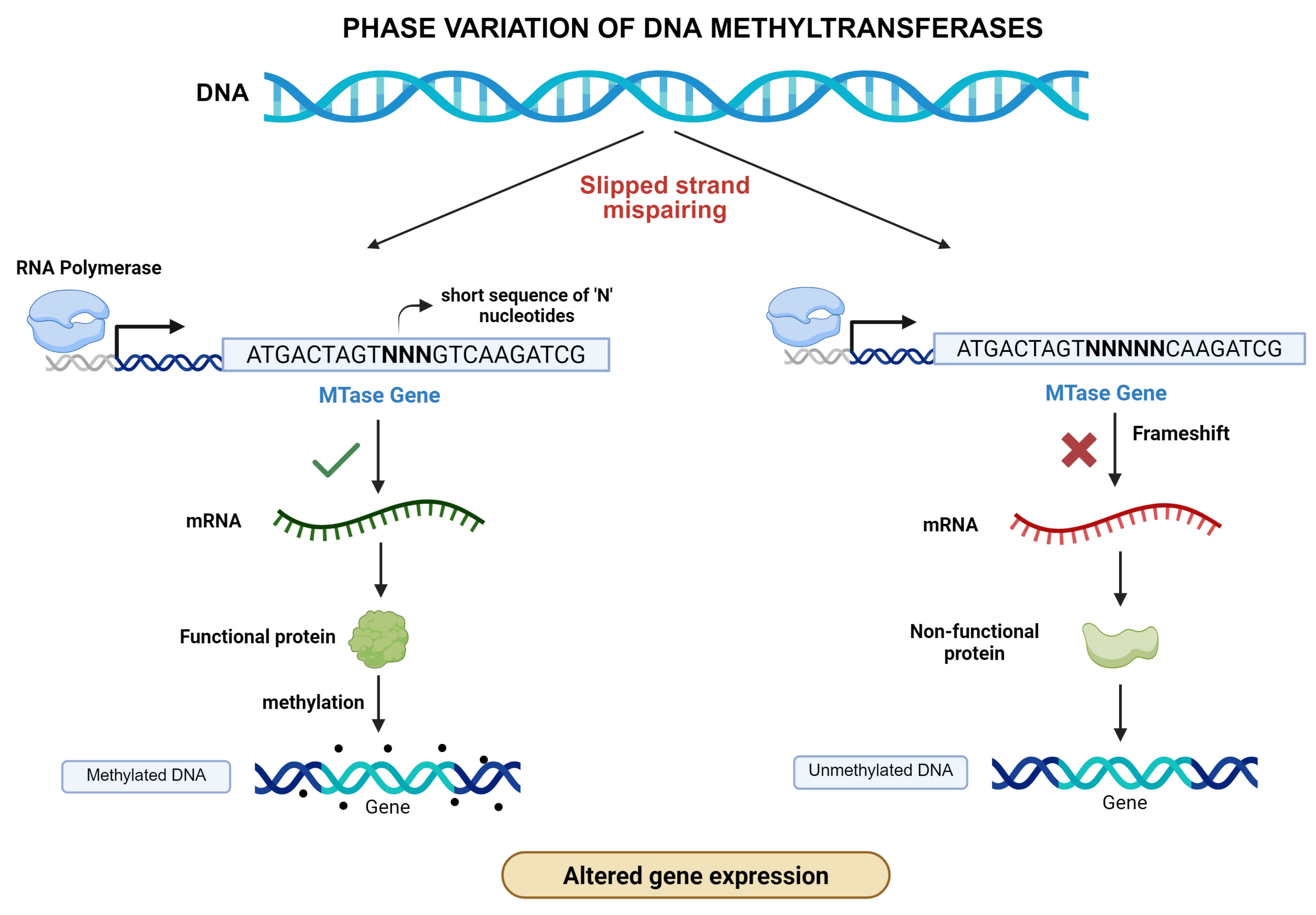
There are two main mechanisms underlying phase variation: these are slipped-strand mispairing (SSR) of simple sequence repeats and site-specific recombination [86]. Variation in the number of SSRs arises from replication slippage within repetitive DNA tracts. When an SSR is located within the open reading frame (ORF), repeat length determines whether the gene remains in-frame and expressed (ON) or undergoes a frameshift that introduces a premature stop codon, producing truncated protein (OFF). Therefore, bacterial populations contain mixed ON and OFF variants, as shown in Figure 1. If SSRs are located in the promoter region, changes in repeat length modulate transcriptional efficiency, generating graded protein expression levels, with long tracts typically associated with reduced expression [87]. SSRs exhibit a biased behavior toward OFF switching, as the frameshift-induced changes occur more frequently than the precise restoration of original repeat length. However, the reversal to the ON state can occur through subsequent less frequent slippage events, making SSR potentially reversible [88]. In contrast, site-specific recombinase generates phase variation between inverted repeats (IRs), resulting in the shuffling of expressed and silent loci. Genes encoding proteins with variable domains are recognized, allowing alternative combinations of N and C-terminal domains to be expressed. In Streptococcus pneumoniae, SpnIII phase-variable type I restriction–modification system, six alternative epigenetic states (SpnIII A-F) are generated through TRD shuffling. This is a partially recombinase-independent and temperature-dependent phenomenon that produces specialized subpopulations optimized for colonization or invasion [89]. This is an example of true bidirectional and reversible switching, not merely ON and OFF states, but between alternative functional variants. It has remarkable implications in vaccine and antimicrobial design, where effective interventions should consider population heterogeneity in the pathogenic species [88].
Phase-variable MTases have been identified across multiple unrelated bacterial genera. In Haemophilus influenzae, the genes associated with immune evasion, nutrient acquisition, and colonization, are regulated by stochastic ON/OFF switching of the ModA MTase. The switching of ModA was shown to modify the transcription of surface-exposed proteins and factors associated with nasopharynx colonization. This allows H. influenzae to quickly evolve in response to immune pressures and to various host niches, thus increasing persistence and transmission [90].
In Helicobacter pylori, the methyltransferase M.HpyAVIB C5Mtase undergoes slipped-strand mispairing that generates subpopulations in which the enzyme is expressed, providing survival advantage in gastric niche [91,92]. The ModH methyltransferase further regulates the expression of flagellin and adhesion genes essential for gastric colonization and motility within the mucus layer. The presence of multiple ModH allelic variants among H. pylori strains provides a mechanism for the diversification of gene expression and phenotypic variation at the population level. These heritable ON/OFF methylation states influence the expression of virulence-associated genes, such as flaA and hopG, contributing to strain-specific differences in disease outcomes that range from asymptomatic colonization to gastritis, peptic ulcer disease, and gastric cancer [90,92].
Similarly, in pathogenic Neisseria species, like N. meningitidis and N. gonorrhoeae, phase-variable type III R-M system MTases encoded by Mod genes switched ON and OFF by SSR produce distinct DNA methylation patterns that alter expression of genes involved in iron acquisition, biofilm formation, and antimicrobial resistance [88]. Phase variation in Campylobacter jejuni modulates motility, and the invasion in a methyltransferase-mediated manner Type IIG methyltransferase leads to site-specific changes in methylation patterns across the genome. This system causes broad transcriptional shifts affecting motility, chemotaxis, and metabolism [93]. These shifts affected the methylation of genes required to colonize hosts and for survival, highlighting methylation as an indicator of pathogenic success [92]. In Salmonella enterica, DNA methylation patterns in pathogenic islands are involved in the regulation of the expression of type III secretion system effectors necessary to induce invasion and intracellular survival [25]. All these examples indicate that phase variation is a widespread and a conserved adaptive strategy for epigenetic regulation.

2.4.2. Bistability

In contrast to phase variation, which is driven by changs in DNA, bistability arises from gene regulatory networks and epigenetic changes rather than alterations in the genome [94]. In bistability, noise or fluctuations in gene expression are amplified by positive feedback loops allowing genetically identical cells to adopt and maintain distinct phenotypic states that are heritable over several generations, yet fully reversible [94]. This mechanism allows subsets of cells to survive under stresses such as starvation or antibiotic exposure. A well-studied example is the opvAB system in Salmonella enterica, where bistability alters O-antigen chain length in lipopolysaccharides [95]. The OpvAB ON state produces short O-antigens, conferring phage resistance, while the OFF-state yields longer O-antigens that increase susceptibility [95,96]. Under fluctuating phase pressures, both states are maintained in constant conditions; genetic adaptation favors the OFF state, reducing bistability [96]. In Salmonella, additional bistable regulatory circuits include the flagellar fliBA–fliC phase variation system, the pef, stf, and gtr operons, and the transcriptional regulator csgD, all of which play key roles in controlling surface structures and biofilm formation [97].
In Bacillus subtilis, the competence regulator, ComK, forms a positive feedback loop that generates bistable expression in only a fraction of cells that enter competence, while the rest remain vegetative [98]. Bistability is also linked with inoculum effect, where bacterial survival against antimicrobials depends on the initial population size [99]. Species with larger inoculum, including Methicillin-resistant Staphylococcus aureus (MRSA), Klebsiella pneumoniae and Pseudomonas aeruginosa, may be able to endure higher concentrations than smaller ones, indicating bistable growth processes and undermining conventional MIC-based antibiotic susceptibility measurements [99]. In Pseudomomas aeruginosa, epigenetically stabilized heterogeneity is maintained in the glpD gene expression. This gene codes for glycerol-3-phosphate dehydrogenase and is involved in glycerol metabolism. Using promoter-reporter fusions, the populations were shown to partition into high and low glpD expression states that are maintained across several generations and can spontaneously switch, reflecting regulatory bistability. Cells with high glpD expression exhibited increased toxin production and motility, whereas low expression cells behaved conservatively, supporting survival under variable host environments [100]. Bistability is thus a widespread strategy for phenotypic diversification in several bacterial populations.

2.4.3. DNA Replication and Cell Cycle Control

DNA methylation is closely connected with the regulation of DNA replication and progression of cell cycle. Adenine methylation at GATC positions through Dam methyltransferase has long been known to be an indicator of replication origin activity and mismatch repair in E. coli. There is new evidence indicating that cytosine methylation may also play a role in replication dynamics of certain bacterial lineages [101].
Cytosine methylation is also dynamically changed near the oriC region in cyanobacteria in various cell cycle phases [102]. The timing of initiation events is correlated with the methylation of replication origins, and it changes in the state of methylation destabilized replication synchrony. This implies that cytosine methylation may also play a similar role to that of Dam methylation in the regulation of replication initiation, particularly as a timing cue in helicase loading and replisome assembly [102]. The strand discrimination during mismatch repair is also contributed by hemi-methylation, which is instantaneously done after the DNA replication, and only the parental strand is targeted by the methylation [103]. Cytosine methylation can be used to ensure genomic fidelity in bacteria that do not have Dam methylation to provide the signal that repair enzymes need to be targeted to the newly synthesized strand. This underscores a neglected contribution of cytosine methyltransferases in maintaining DNA replication stability [101]. In addition to its immediate impact on replication foci, there is evidence of methylation leading to change in the overall structure of the nucleoid. The binding of nucleoid-associated proteins, including HNS, Fis, and HU, can be changed by DNA methylation, which can regulate the compaction and accessibility of chromosomes. These structural changes in chromatin may control the progression of replication forks, the segregation of chromosomes, and the cell cycle transition [101].
Combined, these observations suggest that DNA methyltransferases are constitutive controllers of bacterial replication and cell cycle. Combining the dynamics of methylation with events of replication and genome maintenance, bacteria regulate their growth in response to the environment and guarantee accurate inheritance of epigenetic memory [101].

2.4.4. Stress Response and Drug Resistance

Stress response genes are epigenetically controlled to give bacteria a means of survival in hostile conditions on a reversible basis. In contrast to stable genetic mutations, regulation by means of methylation enables quick adaptation and maintenance of genomic integrity, which makes it an essential survival mechanism in dynamic environments [25]. Bacteria face numerous environmental and host-related stresses, such as oxidative stresses, nutrient deprivation, hypoxia, and antibiotic pressures [85]. DNA methyltransferases dynamically reorganize transcriptional networks and assist bacteria with adapting to such pressures [25]. MamA DNA methyltransferase in Mycobacterium tuberculosis is important for the survival under hypoxic stress inside the macrophages. Many MamA recognition motifs are located in promoter regions, overlapping the −10 sigma factor elements. This strategic positioning allows MamA to modulate sigma factor-dependent transcription by influencing RNA polymerase binding and transcriptional initiation. The loss of MamA decreased bacterial hypoxic survival due to the loss of promoter methylation, leading to altered or reduced expression of genes involved in metabolic adaptation, stress response, and maintenance of cellular homeostasis during oxygen limitation. Through this epigenetic regulation, MamA fine-tunes the expression of genes required for the adaptation to hypoxic conditions [104]. MamA, along with MamB and HsdM, is also involved in development of multidrug resistance in Mtb [13].
Epigenetic regulation also modulates the expression of extracellular polysaccharide biosynthesis and adhesion genes that play a role in biofilm formation [105]. Biofilm-associated genes in Vibrio cholerae are strongly modulated by epigenetic regulation. Biofilms offer physical and physiological resistance to antibiotics and host defense, and the methylation-mediated transcriptional control of biofilm formation, together with the ability to switch between free-living and sessile forms, is seen in V. cholerae [105].

2.4.5. Epigenetic Inheritance and Cellular Memory

Another characteristic of bacterial DNA methyltransferases is that they can relay epigenetic information through generations, thus creating cellular memory. Maintenance methyltransferases preserve the methylation state of newly synthesized strands during replication, so that the daughter cells can take over the transcriptional condition of their parents [101]. The same has been observed in H. influenzae, in which ModA switching produces two epigenetically distinguishable subpopulations. They are stably inherited during cell division, enabling populations to trade-off between immune evasion and the efficiency of colonization [90].
The hypoxic survival-related methylation signatures are spread and maintained in Mtb in response to infection, supporting persistence-requiring transcriptional programs [106]. It is possible that these consistent patterns of methylation may contribute to the development of latent infection and treatment failure in case of chronic diseases. On a population scale, these systems of inheritance produce non-DNA sequence-dependent epigenetic heterogeneity. The combination of stability and reversibility that epigenetic inheritance offers bacteria introduces a memory system that mediates short-term environmental adaptation and long-term evolutionary fitness [101].

3. Advances in DNA Methylation Detection

3.1. Illumina-Based Indirect Detection

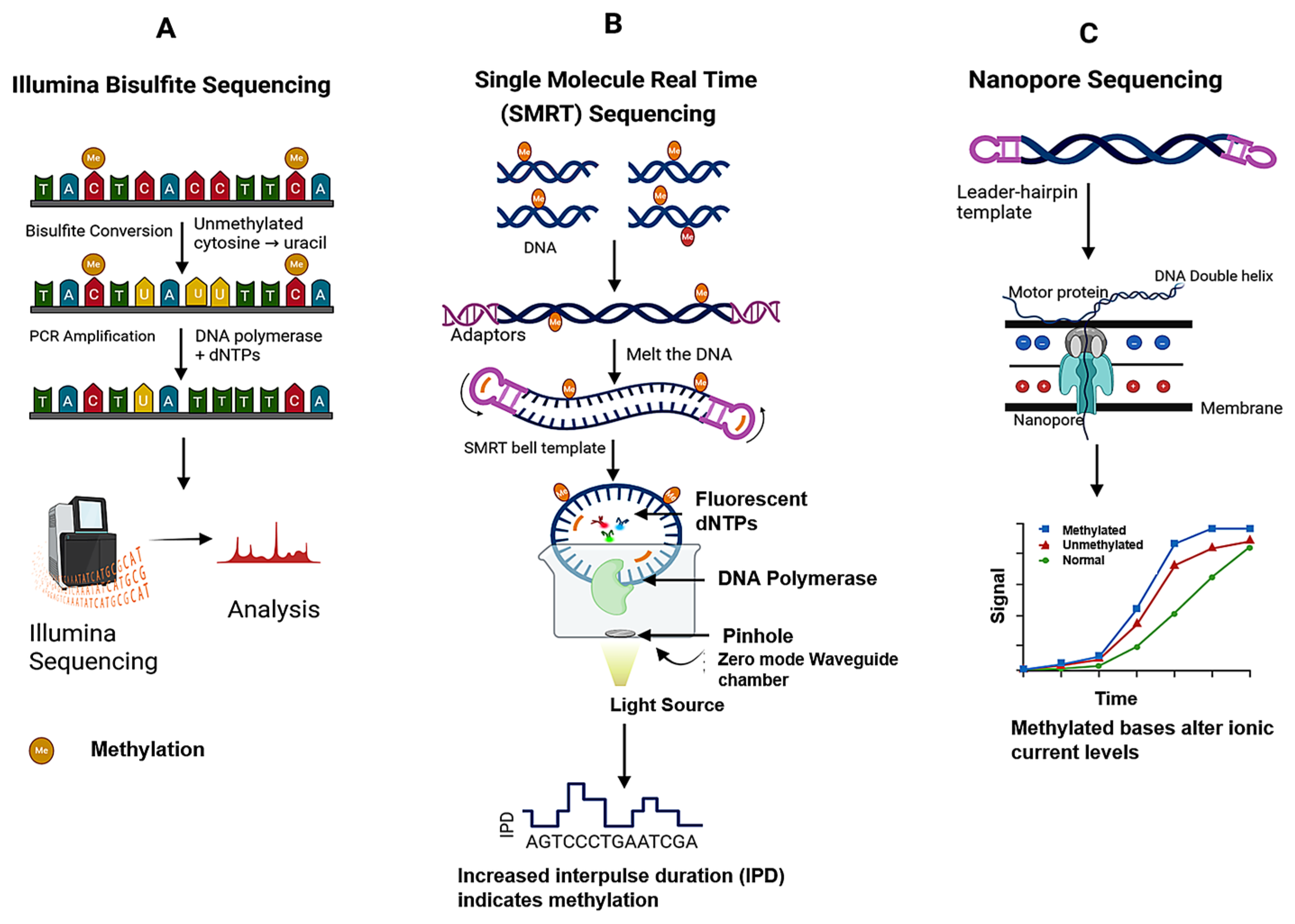
Illumina-based sequencing is a gold standard technique for indirect detection of 5-methylcytosine (5mC) in the DNA [17]. It is a short-read sequencing approach that does not directly recognize modified bases, but sequencing is achieved via chemical or enzymatic methods. The most common approach is bisulfite sequencing, involving treatment of DNA with sodium bisulfite, which converts unmethylated cytosine into uracil, which is read as thymine during sequencing, while methylated cytosine remains unchanged. The common Illumina-based variations for the methylome analysis are Reduced Representation Bisulfite Sequencing (RRBS) and Whole-Genome Bisulfite Sequencing (WGBS) [107]. RRBS utilizes restriction enzyme digestion and size fractionation to allow single-base resolution of functionally important regions, such as promoters and CpG islands. On the other hand, WGBS enables an unbiased and comprehensive description of cytosine methylation of the entire genome.

3.2. PacBio Single-Molecule Real-Time (SMRT) Sequencing

SMRT sequencing has revolutionized bacterial epigenetics research, as it can directly observe modifications on native DNA templates [10,11]. The method relies on viewing a DNA polymerase and on the incorporation of fluorescently labeled nucleotides under microscopic zero-mode waveguide, where the enzyme’s kinetic properties shift upon detecting a modified base [14]. By comparing these kinetic profiles systematically to unmanipulated models, SMRT sequencing not only detects unique methylated positions but also infers recurring sequence motifs that are linked to specific methyltransferases [17]. With this technique, it is possible to map methylation patterns at single-base resolution with chemical treatment, as opposed to traditional methods. In Mycobacterium tuberculosis, SMRT sequencing of various clinical isolates has identified lineage-specific patterns of methylation, with enzymes like MamA and HsdM targeting different motifs. These marks were found to be associated with transcriptional heterogeneity, changed responses to oxidative stress, dormancy, and virulence-related gene repression [108].

3.3. Oxford Nanopore Sequencing

Oxford Nanopore sequencing is an advanced and complementary technique for bacterial epigenomics, capable of directly detecting DNA modifications as native DNA molecules transit through protein nanopores [14,107]. A k-mer represents a defined stretch of k-nucleotides that produces a distinct ionic signature as it translocates through the nanopore. Because each k-mer environment produces a distinct electrical signal, the methylation adds a methyl group, which results in subtle shifts in expected current pattern. This enables real-time, label-free detection of DNA methylation without the usage of chemical or amplification steps. Chemical advances and algorithmic base calling improvements have progressively refined the sensitivity of methylation detection, whereas specialized software like Guppy, Nanopolish, Tombo, and, recently, deep learning algorithms like DeepMod2 and Megalodon have improved modification call precision and resolution [109]. Oxford Nanopore sequencing is increasingly being valued for its worth in portability, cost, flexibility of throughput, and the ability to resolve epigenetic heterogeneity within bacterial communities It enables simultaneous profiling of genetic variation, as well as epigenetic changes presentation of lineage-specific methylation characteristics and regulatory signatures for persistence, virulence, and drug resistance [107,110]. The schematic representation of all the three sequencing techniques is given in Figure 2.

3.4. Methylome Mapping

Full-scale, high-resolution mapping of bacterial epigenome has become feasible with the development of single-molecule, long-read sequencing (as discussed in previous section) and the parallel emergence of analytical pipelines [2]. Contemporary bacterial epigenomics adopts a reproducible protocol, starting with the isolation of native, genomic DNA and extending to non-amplified library preparation, production of modification-sensing reads (mainly through PacBio SMRT or Oxford Nanopore platforms), signal-level processing to detect modified bases and enriched sequence motifs, and integrative interpretation that associates methylation maps with transcriptomes, proteomes, and phenotypes [3]. The strength of this pipeline lies in its two unique abilities: single-base resolution and phasing (the capability to read patterns of modifications along long contiguous molecules). These characteristics combined with direct correlation of methylation marks with operon organization, mobile genetic elements, and allele-specific control, in organisms like Mycobacterium tuberculosis, are of biological significance [11,111].
REBASE is the standard, highly curated database for restriction–modification (RM) systems, which is used when finding motifs of methyltransferase genes. It organizes experimentally characterized and predicted methyltransferases, their recognition sequences, and pointers to primary sequence and structure data [18]. Apart from it, community methylome databases and organism-specific resources (such as MethBank) consolidate processed methylation maps and correlate metadata, facilitating cross-study comparisons and meta-analysis [112].
The computational analysis includes two interrelated tasks: (i) per-site modification calling and (ii) motif discovery and enzyme assignment. For PacBio data, kinetic measurements like interpulse duration (IPD) are read out and aggregated per position; statistical tests or model-based tools then detect sites whose kinetics differ significantly from expectation and group these sites into sequence motifs [113]. For Nanopore data, signal-level tools align raw ionic current to reference sequence and employ hidden Markov models or deep learning to generate per-site modification probabilities. Public software that utilizes these methods are the SMRT Analysis suite and IPD-based programs for kinetic analysis (PacBio), and Nanopolish, Tombo, and various deep-learning pipelines (e.g., DeepSignal/DeepMod variants and ONT’s model-based callers) for nanopore signal processing [114].
In Mycobacterium tuberculosis, SMRT-based methylome analyses across multiple isolates showed lineage-specific methylation motifs and enabled unambiguous assignment of numerous motifs to methyltransferases like MamA and HsdM [10]. These studies are representative of contemporary bacterial epigenomics, from discovery through single-molecule signal, annotation using REBASE and homology, orthogonal validation, and integrative interpretation with transcriptome and phenotype data, which ultimately facilitates mechanistic hypotheses connecting methylation to persistence, virulence, and drug response [18].

4. Applications of Epigenetic Advances in Disease Control

Bacterial DNA methylation and other epigenetic processes are now emerging as key determinants of pathogen behavior, with direct relevance to epidemiology, diagnostics, vaccine development, and new therapeutic approaches. Orphan DNA methyltransferases like Dam in Gammaproteobacteria and CcrM in Alphaproteobacteria (discussed previously in Section 2.3.2) can serve as conserved therapeutic targets for pathogenic species of these classes. Similarly, Mtb lineage specific methyltransferases like MamA and HsdM can also be targeted for precision medicine [13,102].
Structure-guided screens have found small-molecule inhibitors for various bacterial methyltransferases, illustrating the biochemical traceability of these enzymes as drug targets; however, the conversion of inhibitors to whole-cell antibacterial drugs is still problematic due to challenges such as bacterial permeability and compensatory pathways. Therefore, the inhibition of methyltransferases is an exciting but still nascent antibacterial strategy that will need to be pursued with precision in target selection and medicinal chemistry optimization [115]. A second, less lethal antimicrobial strategy is virulence modulation: by modifying methylation patterns or inhibiting phasevarion switching. Experimental and modeling studies indicate that the disruption of methylation networks can reprogram regulatory cascades for adhesion, immune evasion, and biofilm formation, which are directly relevant to virulence [27].

Synthetic Biology and Epigenetic Engineering

Synthetic biology leverages DNA methylation and other epigenetic processes as programmable and heritable control systems in engineered microorganisms [116]. Pioneering research has demonstrated that methylation systems can be diverted to build synthetic epigenetic memory circuits that record methylation marks in response to transitory inputs and thus retain information from one cell division to another cell division in a stable but reversible way [117].
This methylation memory has been constructed with cognate methyltransferase systems and methylation-sensitive DNA-binding proteins to produce bistable switches and responsive reporters, illustrating the viability of epigenetic encoding in a synthetic biology setting. Maier et al., in 2017, designed synthetic epigenetic circuits for establishing long-term cellular memory in E. coli [117]. In this circuit, CcrM from Caulobacter crescentus was used to methylate the promoter at the ‘GANTC’ site, which helps in regulating a reporter maintenance operon encoding EGFP and CcrM. In the OFF state, an engineered zinc-finger repressor binds to the operator and inhibits transcription. Upon transient stimuli such as heat stress, nutrient signals, UV radiation, or DNA damaging agents, CcrM expression is initiated. Subsequent methylation of the promoter prevents repressor binding, establishing positive feedback that stabilizes the ON state. This epigenetic configuration enables bacteria to remember exposure over 30–40 generations without modifying the DNA sequence. In reversible system, targeted degradation of CcrM leads to the passive loss of methylation and restoration of the OFF state. This work provides a compelling proof-of-concept that bacterial DNA methylation can be harnessed as a programmable, reversible, and heritable memory module [117].
Targeted epigenetic editing is the ordered deposition or removal of methylation marks at specified loci. It is rapidly developing in eukaryotic systems and is being applied to bacterial systems as well [118]. Experimental strategies employed are: (a) attaching catalytic methyltransferase domains to DNA-binding scaffolds like dCas9, TALENs, or zinc fingers in such a way that methylation is delivered at a programmable genomic site and (b) employing dCas9 binding as a steric inhibitor of endogenous maintenance methylation. The proof-of-principle experiments showed targeted CpG methylation in bacteria and enhanced targeting specificity via linker and enzyme engineering; these strategies present a toolkit for locus-specific epigenetic regulation of gene expression in microbial systems. Although most of the conceptual and technical background is derived from mammalian epigenome engineering (dCas9–DNMT/TET fusions), bacterial versions (e.g., split-M.SssI or dCas9-directed methyltransferases validated in E. coli) have proved that targeted methylation is possible and adjustable. Split-enzyme architectures and judicious optimization of DNA-binding linkers enhance locus specificity and decrease off-target methylation [115].

5. Challenges and Future Directions

Although the investigation of bacterial epigenetics has revealed significant molecular signatures of control, important hurdles persist before these findings can be precisely translated into clinical or biotechnological applications. Methylome maps for many pathogens are available, but connections between methylation events and phenotypic outcomes will only be established through integrative studies combining epigenomics with transcriptomics, proteomics, and host–pathogen interaction models [17,119].
The second challenge is technical standardization in existing sequencing technologies (SMRT and Nanopore) and bioinformatic pipelines, which vary in sensitivity, error rates, and motif-calling approaches. This complicates cross-study comparisons. Community standards for data quality, analysis, deposition, raw signal files, and validated motif annotations will be critical in creating dependable, comparable methylation data sets. Synthetic epigenetic engineering faces challenges in attaining locus specificity and long-term stability in the presence of endogenous methyltransferases [120].
In the future, some exciting avenues are opening, such as technical improvements in AI-based methylation calling and multi-omics integration, which will speed up functional annotation of epigenomes. The creation of hybrid systems, integrating genetic editing (CRISPR) and programmable methylation systems, may create new, efficient tools for precise control of bacterial physiology. On the disease side, its extension to longitudinal patient cohorts will elucidate how epigenetic variation affects disease course and response to treatment. In biotechnology, engineered methylation circuits can become adaptable platforms for biosensing, memory storage, and programmable medicines [110]. Overall, advances in data interpretation, standardization, and synthetic biology engineering will be determinant for realizing the maximum potential of bacterial epigenetics in controlling diseases and biotechnology [24].

Author Contributions

J.K.L.: conceptualization, investigation/data collection, and writing—original draft; R.G.: conceptualization, investigation/data collection, writing—original draft, supervision, and writing—review and editing; T.K. and R.J.: investigation/data collection, and writing—review and editing; R.K.: writing—original draft, supervision, and writing—review and editing. All authors have read and agreed to the published version of the manuscript.

Funding

The authors would like to acknowledge ANRF PM Early Career Grant, project file no. ANRF/ECRG/2024/004798LS.

Institutional Review Board Statement

Not applicable.

Informed Consent Statement

Not applicable.

Data Availability Statement

No new data were created or analyzed in this study. Data sharing is not applicable to this article.

Acknowledgments

R.G. would like to acknowledge ANRF PM Early Career Grant for financial support. T.K. would like to acknowledge ANRF PM Early Career Grant for Junior Research Fellowship. The Biorender software was used for the generation of figures in this review article.

Conflicts of Interest

The authors declare no conflicts of interest.

Abbreviations

MTase, methyltransferase; TB, tuberculosis; m6A, N6-methyladenine; 5mC, 5-methyl cytosine; 4mC, 4-methyl cytosine.

References

  1. Deichmann, U. Epigenetics: The Origins and Evolution of a Fashionable Topic. Dev. Biol. 2016, 416, 249–254. [Google Scholar] [CrossRef]
  2. Beaulaurier, J.; Schadt, E.E.; Fang, G. Deciphering Bacterial Epigenomes Using Modern Sequencing Technologies. Nat. Rev. Genet. 2019, 20, 157–172. [Google Scholar] [CrossRef]
  3. Moore, L.D.; Le, T.; Fan, G. DNA Methylation and Its Basic Function. Neuropsychopharmacology 2013, 38, 23–38. [Google Scholar] [CrossRef]
  4. Mattei, A.L.; Bailly, N.; Meissner, A. DNA Methylation: A Historical Perspective. Trends Genet. 2022, 38, 676–707. [Google Scholar] [CrossRef]
  5. Oliveira, P.H. Bacterial Epigenomics: Coming of Age. mSystems 2021, 6, e0107521. [Google Scholar] [CrossRef] [PubMed]
  6. Wyatt, G.R. Occurrence of 5-Methyl-Cytosine in Nucleic Acids. Nature 1950, 166, 237–238. [Google Scholar] [CrossRef]
  7. Gao, Q.; Lu, S.; Wang, Y.; He, L.; Wang, M.; Jia, R.; Chen, S.; Zhu, D.; Liu, M.; Zhao, X.; et al. Bacterial DNA Methyltransferase: A Key to the Epigenetic World with Lessons Learned from Proteobacteria. Front. Microbiol. 2023, 14, 1129437. [Google Scholar] [CrossRef] [PubMed]
  8. Seshasayee, A.S.N.; Singh, P.; Krishna, S. Context-Dependent Conservation of DNA Methyltransferases in Bacteria. Nucleic Acids Res. 2012, 40, 7066–7073. [Google Scholar] [CrossRef] [PubMed]
  9. Tarashi, S.; Zamani, M.S.; Bahramali, G.; Fuso, A.; Vaziri, F.; Karimipoor, M.; Siadat, S.D. RNA Expression Analysis of Mycobacterial Methyltransferases Genes in Different Resistant Strains of Mycobacterium tuberculosis. Iran. Biomed. J. 2022, 26, 240–251. [Google Scholar] [CrossRef]
  10. Zhu, L.; Zhong, J.; Jia, X.; Liu, G.; Kang, Y.; Dong, M.; Zhang, X.; Li, Q.; Yue, L.; Li, C.; et al. Precision Methylome Characterization of Mycobacterium tuberculosis Complex (MTBC) Using PacBio Single-Molecule Real-Time (SMRT) Technology. Nucleic Acids Res. 2016, 44, 730–743. [Google Scholar] [CrossRef]
  11. Phelan, J.; De Sessions, P.F.; Tientcheu, L.; Perdigao, J.; Machado, D.; Hasan, R.; Hasan, Z.; Bergval, I.L.; Anthony, R.; McNerney, R.; et al. Methylation in Mycobacterium tuberculosis Is Lineage Specific with Associated Mutations Present Globally. Sci. Rep. 2018, 8, 160. [Google Scholar] [CrossRef]
  12. Modlin, S.J.; Conkle-Gutierrez, D.; Kim, C.; Mitchell, S.N.; Morrissey, C.; Weinrick, B.C.; Jacobs, W.R.; Ramirez-Busby, S.M.; Hoffner, S.E.; Valafar, F. Drivers and Sites of Diversity in the DNA Adenine Methylomes of 93 Mycobacterium tuberculosis Complex Clinical Isolates. eLife 2020, 9, e58542. [Google Scholar] [CrossRef]
  13. Hu, X.; Zhou, X.; Yin, T.; Chen, K.; Hu, Y.; Zhu, B.; Mi, K. The Mycobacterial DNA Methyltransferase HsdM Decreases Intrinsic Isoniazid Susceptibility. Antibiotics 2021, 10, 1323. [Google Scholar] [CrossRef]
  14. Tourancheau, A.; Mead, E.A.; Zhang, X.-S.; Fang, G. Discovering Multiple Types of DNA Methylation from Bacteria and Microbiome Using Nanopore Sequencing. Nat. Methods 2021, 18, 491–498. [Google Scholar] [CrossRef]
  15. Phillips, Z.N.; Tram, G.; Seib, K.L.; Atack, J.M. Phase-Variable Bacterial Loci: How Bacteria Gamble to Maximise Fitness in Changing Environments. Biochem. Soc. Trans. 2019, 47, 1131–1141. [Google Scholar] [CrossRef] [PubMed]
  16. De Ste Croix, M.; Vacca, I.; Kwun, M.J.; Ralph, J.D.; Bentley, S.D.; Haigh, R.; Croucher, N.J.; Oggioni, M.R. Phase-Variable Methylation and Epigenetic Regulation by Type I Restriction–Modification Systems. FEMS Microbiol. Rev. 2017, 41, S3–S15. [Google Scholar] [CrossRef]
  17. Chen, X.; Xu, H.; Shu, X.; Song, C.-X. Mapping Epigenetic Modifications by Sequencing Technologies. Cell. Death Differ. 2025, 32, 56–65. [Google Scholar] [CrossRef]
  18. Roberts, R.J.; Vincze, T.; Posfai, J.; Macelis, D. REBASE—A Database for DNA Restriction and Modification: Enzymes, Genes and Genomes. Nucleic Acids Res. 2015, 43, D298–D299. [Google Scholar] [CrossRef] [PubMed]
  19. Abdelraheem, E.; Thair, B.; Varela, R.F.; Jockmann, E.; Popadić, D.; Hailes, H.C.; Ward, J.M.; Iribarren, A.M.; Lewkowicz, E.S.; Andexer, J.N.; et al. Methyltransferases: Functions and Applications. ChemBioChem 2022, 23, e202200212. [Google Scholar] [CrossRef] [PubMed]
  20. Wojciechowski, M.; Czapinska, H.; Bochtler, M. CpG Underrepresentation and the Bacterial CpG-Specific DNA Methyltransferase M.MpeI. Proc. Natl. Acad. Sci. USA 2013, 110, 105–110. [Google Scholar] [CrossRef]
  21. Felsenfeld, G. A Brief History of Epigenetics. Cold Spring Harb. Perspect. Biol. 2014, 6, a018200. [Google Scholar] [CrossRef]
  22. Arber, W.; Hattman, S.; Dussoix, D. On the Host-Controlled Modification of Bacteriophage λ. Virology 1963, 21, 30–35. [Google Scholar] [CrossRef] [PubMed]
  23. Payelleville, A.; Brillard, J. Novel Identification of Bacterial Epigenetic Regulations Would Benefit From a Better Exploitation of Methylomic Data. Front. Microbiol. 2021, 12, 685670. [Google Scholar] [CrossRef]
  24. Urig, S.; Gowher, H.; Hermann, A.; Beck, C.; Fatemi, M.; Humeny, A.; Jeltsch, A. The Escherichia coli Dam DNA Methyltransferase Modifies DNA in a Highly Processive Reaction. J. Mol. Biol. 2002, 319, 1085–1096. [Google Scholar] [CrossRef]
  25. Bach, T. Excess SeqA Prolongs Sequestration of oriC and Delays Nucleoid Segregation and Cell Division. EMBO J. 2003, 22, 315–323. [Google Scholar] [CrossRef]
  26. Diaz-Diaz, S.; Recacha, E.; Pulido, M.R.; Romero-Muñoz, M.; De Gregorio-Iaria, B.; Docobo-Pérez, F.; Pascual, A.; Rodríguez-Martínez, J.M. Synergistic Effect of SOS Response and GATC Methylome Suppression on Antibiotic Stress Survival in Escherichia coli. Antimicrob. Agents Chemother. 2023, 67, e0139222. [Google Scholar] [CrossRef] [PubMed]
  27. Ma, Y.; Wang, X.; Li, X. The Emerging Role of DNA Methylation in the Pathogenicity of Bacterial Pathogens. J. Bacteriol. 2025, 207, e00108–e00125. [Google Scholar] [CrossRef]
  28. Dorman, C.J. Function of Nucleoid-Associated Proteins in Chromosome Structuring and Transcriptional Regulation. Microb. Physiol. 2014, 24, 316–331. [Google Scholar] [CrossRef] [PubMed]
  29. Blow, M.J.; Clark, T.A.; Daum, C.G.; Deutschbauer, A.M.; Fomenkov, A.; Fries, R.; Froula, J.; Kang, D.D.; Malmstrom, R.R.; Morgan, R.D.; et al. The Epigenomic Landscape of Prokaryotes. PLoS Genet. 2016, 12, e1005854. [Google Scholar] [CrossRef]
  30. Kumar, S.; Chinnusamy, V.; Mohapatra, T. Epigenetics of Modified DNA Bases: 5-Methylcytosine and Beyond. Front. Genet. 2018, 9, 640. [Google Scholar] [CrossRef]
  31. Adhikari, S.; Curtis, P.D. DNA Methyltransferases and Epigenetic Regulation in Bacteria. FEMS Microbiol. Rev. 2016, 40, 575–591. [Google Scholar] [CrossRef]
  32. Ren, R.; Horton, J.R.; Hong, S.; Cheng, X. Recent Advances on DNA Base Flipping: A General Mechanism for Writing, Reading, and Erasing DNA Modifications. In DNA Methyltransferases—Role and Function; Jeltsch, A., Jurkowska, R.Z., Eds.; Advances in Experimental Medicine and Biology; Springer International Publishing: Cham, Switzerland, 2022; Volume 1389, pp. 295–315. ISBN 978-3-031-11453-3. [Google Scholar]
  33. Baduel, P.; Sammarco, I.; Barrett, R.; Coronado-Zamora, M.; Crespel, A.; Díez-Rodríguez, B.; Fox, J.; Galanti, D.; González, J.; Jueterbock, A.; et al. The Evolutionary Consequences of Interactions Between the Epigenome, the Genome and the Environment. Evol. Appl. 2024, 17, e13730. [Google Scholar] [CrossRef]
  34. Li, D.; Da, L.; Tang, H.; Li, T.; Zhao, M. CpG Methylation Plays a Vital Role in Determining Tissue- and Cell-Specific Expression of the Human Cell-Death-Inducing DFF45-like Effector A Gene through the Regulation of Sp1/Sp3 Binding. Nucleic Acids Res. 2007, 36, 330–341. [Google Scholar] [CrossRef]
  35. Nichol, K.; Pearson, A.C.E. CpG Methylation Modifies the Genetic Stability of Cloned Repeat Sequences. Genome Res. 2002, 12, 1246–1256. [Google Scholar] [CrossRef] [PubMed]
  36. Shen, J.-C.; Rideout, W.M.; Jones, P.A. High Frequency Mutagenesis by a DNA Methyltransferase. Cell 1992, 71, 1073–1080. [Google Scholar] [CrossRef]
  37. Matthey, N.; Wennubst, G.; Bojkowska, K.; Marquis, J.; Collier, J. Cytosine Methylation by ScmA Contributes to the Fitness of Caulobacter crescentus Cells Naturally Expressing a Vsr-like Protein. iScience 2026, 29, 114749. [Google Scholar] [CrossRef] [PubMed] [PubMed Central]
  38. Chernov, A.V.; Reyes, L.; Peterson, S.; Strongin, A.Y. Depletion of CG-Specific Methylation in Mycoplasma Hyorhinis Genomic DNA after Host Cell Invasion. PLoS ONE 2015, 10, e0142529. [Google Scholar] [CrossRef]
  39. Estibariz, I.; Overmann, A.; Ailloud, F.; Krebes, J.; Josenhans, C.; Suerbaum, S. The Core Genome m5C Methyltransferase JHP1050 (M.Hpy99III) Plays an Important Role in Orchestrating Gene Expression in Helicobacter pylori. Nucleic Acids Res. 2019, 47, 2336–2348. [Google Scholar] [CrossRef] [PubMed]
  40. Pisciotta, A.; Sampino, A.M.; Presentato, A.; Galardini, M.; Manteca, A.; Alduina, R. The DNA Cytosine Methylome Revealed Two Methylation Motifs in the Upstream Regions of Genes Related to Morphological and Physiological Differentiation in Streptomyces Coelicolor A(3)2 M145. Sci. Rep. 2023, 13, 7038. [Google Scholar] [CrossRef]
  41. Prasad, Y.; Kumar, R.; Chaudhary, A.K.; Dhanaraju, R.; Majumdar, S.; Rao, D.N. Kinetic and Catalytic Properties of M.HpyAXVII, a Phase-Variable DNA Methyltransferase from Helicobacter pylori. J. Biol. Chem. 2019, 294, 1019–1034. [Google Scholar] [CrossRef]
  42. Fang, J.-L.; Gao, W.-L.; Xu, W.-F.; Lyu, Z.-Y.; Ma, L.; Luo, S.; Chen, X.-A.; Mao, X.-M.; Li, Y.-Q. m4C DNA Methylation Regulates Biosynthesis of Daptomycin in Streptomyces roseosporus L30. Synth. Syst. Biotechnol. 2022, 7, 1013–1023. [Google Scholar] [CrossRef]
  43. Gao, W.-L.; Fang, J.-L.; Zhu, C.-Y.; Xu, W.-F.; Lyu, Z.-Y.; Chan, X.-A.; Zhao, Q.-W.; Li, Y.-Q. Identification and Characterization of a New Regulator, TagR, for Environmental Stress Resistance Based on the DNA Methylome of Streptomyces roseosporus. Microbiol. Spectr. 2023, 11, e0038023. [Google Scholar] [CrossRef]
  44. Shen, C.; Wang, K.; Deng, X.; Chen, J. DNA N6-Methyldeoxyadenosine in Mammals and Human Disease. Trends Genet. 2022, 38, 454–467. [Google Scholar] [CrossRef]
  45. Boulias, K.; Greer, E.L. Means, Mechanisms and Consequences of Adenine Methylation in DNA. Nat. Rev. Genet. 2022, 23, 411–428. [Google Scholar] [CrossRef] [PubMed]
  46. Zhao, J.; Zhang, M.; Hui, W.; Zhang, Y.; Wang, J.; Wang, S.; Kwok, L.-Y.; Kong, J.; Zhang, H.; Zhang, W. Roles of Adenine Methylation in the Physiology of Lacticaseibacillus paracasei. Nat. Commun. 2023, 14, 2635. [Google Scholar] [CrossRef]
  47. Tsukamoto, Y.; Tamura, T.; Maeda, Y.; Miyake, K.; Ato, M. N6-Methylated Adenine on the Target Sites of mamA from Mycobacterium Bovis BCG Enhances Macrophage Activation by CpG DNA in Mice. Tuberculosis 2020, 121, 101890. [Google Scholar] [CrossRef] [PubMed]
  48. Takahashi, M.; Hiraoka, S.; Matsumoto, Y.; Shibagaki, R.; Ujihara, T.; Maeda, H.; Seo, S.; Nagasaki, K.; Takeuchi, H.; Matsuzaki, S. Host-Encoded DNA Methyltransferases Modify the Epigenome and Host Tropism of Invading Phages. iScience 2025, 28, 112264. [Google Scholar] [CrossRef] [PubMed]
  49. Sánchez-Romero, M.A.; Cota, I.; Casadesús, J. DNA Methylation in Bacteria: From the Methyl Group to the Methylome. Curr. Opin. Microbiol. 2015, 25, 9–16. [Google Scholar] [CrossRef]
  50. Huang, X.; Wang, J.; Li, J.; Liu, Y.; Liu, X.; Li, Z.; Kurniyati, K.; Deng, Y.; Wang, G.; Ralph, J.D.; et al. Prevalence of Phase Variable Epigenetic Invertons among Host-Associated Bacteria. Nucleic Acids Res. 2020, 48, 11468–11485. [Google Scholar] [CrossRef]
  51. Murray, N.E. Type I Restriction Systems: Sophisticated Molecular Machines (a Legacy of Bertani and Weigle). Microbiol. Mol. Biol. Rev. 2000, 64, 412–434. [Google Scholar] [CrossRef]
  52. DebRoy, S.; Shropshire, W.C.; Tran, C.N.; Hao, H.; Gohel, M.; Galloway-Peña, J.; Hanson, B.; Flores, A.R.; Shelburne, S.A. Characterization of the Type I Restriction Modification System Broadly Conserved among Group A Streptococci. mSphere 2021, 6, e0079921. [Google Scholar] [CrossRef]
  53. Oliveira, P.H.; Fang, G. Conserved DNA Methyltransferases: A Window into Fundamental Mechanisms of Epigenetic Regulation in Bacteria. Trends Microbiol. 2021, 29, 28–40. [Google Scholar] [CrossRef]
  54. Morgan, R.D.; Bhatia, T.K.; Lovasco, L.; Davis, T.B. MmeI: A Minimal Type II Restriction-Modification System That Only Modifies One DNA Strand for Host Protection. Nucleic Acids Res. 2008, 36, 6558–6570. [Google Scholar] [CrossRef]
  55. Shen, B.W.; Quispe, J.D.; Luyten, Y.; McGough, B.E.; Morgan, R.D.; Stoddard, B.L. Coordination of Phage Genome Degradation versus Host Genome Protection by a Bifunctional Restriction-Modification Enzyme Visualized by CryoEM. Structure 2021, 29, 521–530.e5. [Google Scholar] [CrossRef] [PubMed]
  56. Xu, Q.; Morgan, R.D.; Roberts, R.J.; Blaser, M.J. Identification of Type II Restriction and Modification Systems in Helicobacter pylori Reveals Their Substantial Diversity among Strains. Proc. Natl. Acad. Sci. USA 2000, 97, 9671–9676. [Google Scholar] [CrossRef] [PubMed]
  57. Van Aelst, K.; Tóth, J.; Ramanathan, S.P.; Schwarz, F.W.; Seidel, R.; Szczelkun, M.D. Type III Restriction Enzymes Cleave DNA by Long-Range Interaction between Sites in Both Head-to-Head and Tail-to-Tail Inverted Repeat. Proc. Natl. Acad. Sci. USA 2010, 107, 9123–9128. [Google Scholar] [CrossRef]
  58. Wyszomirski, K.H.; Curth, U.; Alves, J.; Mackeldanz, P.; Möncke-Buchner, E.; Schutkowski, M.; Krüger, D.H.; Reuter, M. Type III Restriction Endonuclease EcoP15I Is a Heterotrimeric Complex Containing One Res Subunit with Several DNA-Binding Regions and ATPase Activity. Nucleic Acids Res. 2012, 40, 3610–3622. [Google Scholar] [CrossRef]
  59. Rao, D.N.; Dryden, D.T.F.; Bheemanaik, S. Type III Restriction-Modification Enzymes: A Historical Perspective. Nucleic Acids Res. 2014, 42, 45–55. [Google Scholar] [CrossRef] [PubMed]
  60. Sutherland, E.; Coe, L.; Raleigh, E.A. McrBC: A Multisubunit GTP-Dependent Restriction Endonuclease. J. Mol. Biol. 1992, 225, 327–348. [Google Scholar] [CrossRef]
  61. Anton, B.P.; Blumenthal, R.; Eaglesham, J.B.; Mruk, I.; Roberts, R.J.; Xu, S.; Weigele, P.R.; Raleigh, E.A. Biology of Host-Dependent Restriction-Modification in Prokaryotes. EcoSal Plus 2025, 13, eesp-0014. [Google Scholar] [CrossRef]
  62. Bell, R.T.; Sahakyan, H.; Makarova, K.S.; Wolf, Y.I.; Koonin, E.V. CoCoNuTs Are a Diverse Subclass of Type IV Restriction Systems Predicted to Target RNA. eLife 2024, 13, RP94800. [Google Scholar] [CrossRef]
  63. Shaw, L.P.; Rocha, E.P.C.; MacLean, R.C. Restriction-Modification Systems Have Shaped the Evolution and Distribution of Plasmids across Bacteria. Nucleic Acids Res. 2023, 51, 6806–6818. [Google Scholar] [CrossRef] [PubMed]
  64. Loenen, W.A.M.; Raleigh, E.A. The Other Face of Restriction: Modification-Dependent Enzymes. Nucleic Acids Res. 2014, 42, 56–69. [Google Scholar] [CrossRef] [PubMed]
  65. Fukuyo, M.; Sasaki, A.; Kobayashi, I. Success of a Suicidal Defense Strategy against Infection in a Structured Habitat. Sci. Rep. 2012, 2, 238. [Google Scholar] [CrossRef] [PubMed]
  66. Ishikawa, K.; Fukuda, E.; Kobayashi, I. Conflicts Targeting Epigenetic Systems and Their Resolution by Cell Death: Novel Concepts for Methyl-Specific and Other Restriction Systems. DNA Res. 2010, 17, 325–342. [Google Scholar] [CrossRef]
  67. Papaleo, S.; Alvaro, A.; Nodari, R.; Panelli, S.; Bitar, I.; Comandatore, F. The Red Thread between Methylation and Mutation in Bacterial Antibiotic Resistance: How Third-Generation Sequencing Can Help to Unravel This Relationship. Front. Microbiol. 2022, 13, 957901. [Google Scholar] [CrossRef]
  68. Lin, L.F.; Posfai, J.; Roberts, R.J.; Kong, H. Comparative Genomics of the Restriction-Modification Systems in Helicobacter pylori. Proc. Natl. Acad. Sci. USA 2001, 98, 2740–2745. [Google Scholar] [CrossRef]
  69. Atack, J.M.; Tan, A.; Bakaletz, L.O.; Jennings, M.P.; Seib, K.L. Phasevarions of Bacterial Pathogens: Methylomics Sheds New Light on Old Enemies. Trends Microbiol. 2018, 26, 715–726. [Google Scholar] [CrossRef]
  70. Carvalho, A.; Mazel, D.; Baharoglu, Z. Deficiency in Cytosine DNA Methylation Leads to High Chaperonin Expression and Tolerance to Aminoglycosides in Vibrio Cholerae. PLoS Genet. 2021, 17, e1009748. [Google Scholar] [CrossRef]
  71. Brooks, J.E.; Blumenthal, R.M.; Gingeras, T.R. The Isolation and Characterization of the Esherichia coli DNA Adenine Methylase (Dam) Gene. Nucleic Acids Res. 1983, 11, 837–851. [Google Scholar] [CrossRef]
  72. Marinus, M.G.; Morris, N.R. Isolation of Deoxyribonucleic Acid Methylase Mutants of Escherichia coli K-12. J. Bacteriol. 1973, 114, 1143–1150. [Google Scholar] [CrossRef] [PubMed]
  73. Marinus, M.G.; Casadesus, J. Roles of DNA Adenine Methylation in Host–Pathogen Interactions: Mismatch Repair, Transcriptional Regulation, and More. FEMS Microbiol. Rev. 2009, 33, 488–503. [Google Scholar] [CrossRef] [PubMed]
  74. Horton, J.R.; Zhang, X.; Blumenthal, R.M.; Cheng, X. Structures of Escherichia coli DNA Adenine Methyltransferase (Dam) in Complex with a Non-GATC Sequence: Potential Implications for Methylation-Independent Transcriptional Repression. Nucleic Acids Res. 2015, 43, 4296–4308. [Google Scholar] [CrossRef] [PubMed]
  75. Cohen, N.R.; Ross, C.A.; Jain, S.; Shapiro, R.S.; Gutierrez, A.; Belenky, P.; Li, H.; Collins, J.J. A Role for the Bacterial GATC Methylome in Antibiotic Stress Survival. Nat. Genet. 2016, 48, 581–586. [Google Scholar] [CrossRef]
  76. Yang, J.; Son, Y.; Park, J.; Park, W. Dam-Dependent Epigenetic Memory Regulates Prophage Reintegration in Salmonella. Nucleic Acids Res. 2025, 53, gkaf951. [Google Scholar] [CrossRef]
  77. Wyrzykowski, J.; Volkert, M.R. The Escherichia coli Methyl-Directed Mismatch Repair System Repairs Base Pairs Containing Oxidative Lesions. J. Bacteriol. 2003, 185, 1701–1704. [Google Scholar] [CrossRef]
  78. Xu, Y.; Liu, S.; Zhang, Y.; Zhang, W. DNA Adenine Methylation Is Involved in Persister Formation in E. coli. Microbiol. Res. 2021, 246, 126709. [Google Scholar] [CrossRef]
  79. Cherry, J.L. Methylation-Induced Hypermutation in Natural Populations of Bacteria. J. Bacteriol. 2018, 200, 10–1128. [Google Scholar] [CrossRef]
  80. Militello, K.T.; Simon, R.D.; Qureshi, M.; Maines, R.; Horne, M.L.; Hennick, S.M.; Jayakar, S.K.; Pounder, S. Conservation of Dcm-Mediated Cytosine DNA Methylation in Escherichia coli. FEMS Microbiol. Lett. 2012, 328, 78–85. [Google Scholar] [CrossRef]
  81. Woodcock, C.B.; Horton, J.R.; Zhang, X.; Blumenthal, R.M.; Cheng, X. Beta Class Amino Methyltransferases from Bacteria to Humans: Evolution and Structural Consequences. Nucleic Acids Res. 2020, 48, 10034–10044. [Google Scholar] [CrossRef]
  82. Horton, J.R.; Woodcock, C.B.; Opot, S.B.; Reich, N.O.; Zhang, X.; Cheng, X. The Cell Cycle-Regulated DNA Adenine Methyltransferase CcrM Opens a Bubble at Its DNA Recognition Site. Nat. Commun. 2019, 10, 4600. [Google Scholar] [CrossRef]
  83. Reich, N.O.; Dang, E.; Kurnik, M.; Pathuri, S.; Woodcock, C.B. The Highly Specific, Cell Cycle–Regulated Methyltransferase from Caulobacter crescentus Relies on a Novel DNA Recognition Mechanism. J. Biol. Chem. 2018, 293, 19038–19046. [Google Scholar] [CrossRef]
  84. Kahng, L.S.; Shapiro, L. The CcrM DNA Methyltransferase of Agrobacterium tumefaciens Is Essential, and Its Activity Is Cell Cycle Regulated. J. Bacteriol. 2001, 183, 3065–3075. [Google Scholar] [CrossRef] [PubMed]
  85. Gor, V.; Ohniwa, R.L.; Morikawa, K. No Change, No Life? What We Know about Phase Variation in Staphylococcus aureus. Microorganisms 2021, 9, 244. [Google Scholar] [CrossRef] [PubMed]
  86. Bayliss, C.D.; Clark, J.L.; Van Der Woude, M.W. 100+ Years of Phase Variation: The Premier Bacterial Bet-Hedging Phenomenon. Microbiology 2025, 171, 001537. [Google Scholar] [CrossRef]
  87. Fraser, A.J.; McMahon, F.E.; Atack, J.M. Microbial Primer: Phase Variation—Survival and Adaptability by Generation of a Diverse Population: This Article Is Part of the Microbial Primers Collection. Microbiology 2024, 170, 001492. [Google Scholar] [CrossRef]
  88. Seib, K.L.; Jen, F.E.-C.; Scott, A.L.; Tan, A.; Jennings, M.P. Phase Variation of DNA Methyltransferases and the Regulation of Virulence and Immune Evasion in the Pathogenic Neisseria. Pathog. Dis. 2017, 75, ftx080. [Google Scholar] [CrossRef]
  89. De Ste Croix, M.; Chen, K.Y.; Vacca, I.; Manso, A.S.; Johnston, C.; Polard, P.; Kwun, M.J.; Bentley, S.D.; Croucher, N.J.; Bayliss, C.D.; et al. Recombination of the Phase-Variable spnIII Locus Is Independent of All Known Pneumococcal Site-Specific Recombinases. J. Bacteriol. 2019, 201, 10–1128. [Google Scholar] [CrossRef]
  90. Atack, J.M.; Srikhanta, Y.N.; Fox, K.L.; Jurcisek, J.A.; Brockman, K.L.; Clark, T.A.; Boitano, M.; Power, P.M.; Jen, F.E.-C.; McEwan, A.G.; et al. A Biphasic Epigenetic Switch Controls Immunoevasion, Virulence and Niche Adaptation in Non-Typeable Haemophilus Influenzae. Nat. Commun. 2015, 6, 7828. [Google Scholar] [CrossRef] [PubMed]
  91. Kumar, R.; Sabareesh, V.; Mukhopadhyay, A.K.; Rao, D.N. Mutations in hpyAVIBM, C5 Cytosine DNA Methyltransferase from Helicobacter pylori Result in Relaxed Specificity. FEBS J. 2012, 279, 1080–1092. [Google Scholar] [CrossRef]
  92. Srikhanta, Y.N.; Gorrell, R.J.; Steen, J.A.; Gawthorne, J.A.; Kwok, T.; Grimmond, S.M.; Robins-Browne, R.M.; Jennings, M.P. Phasevarion Mediated Epigenetic Gene Regulation in Helicobacter pylori. PLoS ONE 2011, 6, e27569. [Google Scholar] [CrossRef]
  93. Anjum, A.; Brathwaite, K.J.; Aidley, J.; Connerton, P.L.; Cummings, N.J.; Parkhill, J.; Connerton, I.; Bayliss, C.D. Phase Variation of a Type IIG Restriction-Modification Enzyme Alters Site-Specific Methylation Patterns and Gene Expression in Campylobacter jejuni Strain NCTC11168. Nucleic Acids Res. 2016, 44, 4581–4594. [Google Scholar] [CrossRef] [PubMed]
  94. Dubnau, D.; Losick, R. Bistability in Bacteria. Mol. Microbiol. 2006, 61, 564–572. [Google Scholar] [CrossRef] [PubMed]
  95. Cota, I.; Sánchez-Romero, M.A.; Hernández, S.B.; Pucciarelli, M.G.; García-del Portillo, F.; Casadesús, J. Epigenetic Control of Salmonella Enterica O-Antigen Chain Length: A Tradeoff between Virulence and Bacteriophage Resistance. PLoS Genet. 2015, 11, e1005667. [Google Scholar] [CrossRef] [PubMed]
  96. Fernández-Fernández, R.; Olivenza, D.R.; Weyer, E.; Singh, A.; Casadesús, J.; Antonia Sánchez-Romero, M. Evolution of a Bistable Genetic System in Fluctuating and Nonfluctuating Environments. Proc. Natl. Acad. Sci. USA 2024, 121, e2322371121. [Google Scholar] [CrossRef]
  97. García-Pastor, L.; Puerta-Fernández, E.; Casadesús, J. Bistability and Phase Variation in Salmonella Enterica. Biochim. Biophys. Acta (BBA)-Gene Regul. Mech. 2019, 1862, 752–758. [Google Scholar] [CrossRef]
  98. Veening, J.-W.; Smits, W.K.; Kuipers, O.P. Bistability, Epigenetics, and Bet-Hedging in Bacteria. Annu. Rev. Microbiol. 2008, 62, 193–210. [Google Scholar] [CrossRef]
  99. Frenkel, N.; Saar Dover, R.; Titon, E.; Shai, Y.; Rom-Kedar, V. Bistable Bacterial Growth Dynamics in the Presence of Antimicrobial Agents. Antibiotics 2021, 10, 87. [Google Scholar] [CrossRef]
  100. Vatareck, E.; Rick, T.; Oswaldo Gomez, N.; Bandyopadhyay, A.; Kramer, J.; Strunin, D.; Erdmann, J.; Hartmann, O.; Alpers, K.; Boedeker, C.; et al. Epigenetic Cellular Memory in Pseudomonas aeruginosa Generates Phenotypic Variation in Response to Host Environments. Proc. Natl. Acad. Sci. USA 2025, 122, e2415345122. [Google Scholar] [CrossRef]
  101. Sánchez-Romero, M.A.; Casadesús, J. The Bacterial Epigenome. Nat. Rev. Microbiol. 2020, 18, 7–20. [Google Scholar] [CrossRef]
  102. Bakker, A.; Smith, D.W. Methylation of GATC Sites Is Required for Precise Timing between Rounds of DNA Replication in Escherichia coli. J. Bacteriol. 1989, 171, 5738–5742. [Google Scholar] [CrossRef]
  103. Putnam, C.D. Strand Discrimination in DNA Mismatch Repair. DNA Repair 2021, 105, 103161. [Google Scholar] [CrossRef] [PubMed]
  104. Shell, S.S.; Prestwich, E.G.; Baek, S.-H.; Shah, R.R.; Sassetti, C.M.; Dedon, P.C.; Fortune, S.M. DNA Methylation Impacts Gene Expression and Ensures Hypoxic Survival of Mycobacterium Tuberculosis. PLoS Pathog. 2013, 9, e1003419. [Google Scholar] [CrossRef]
  105. Hammer, B.K.; Bassler, B.L. Quorum Sensing Controls Biofilm Formation in Vibrio cholerae. Mol. Microbiol. 2003, 50, 101–104. [Google Scholar] [CrossRef]
  106. Thomas, S.S.; Abhinand, K.; Menon, A.M.; Nair, B.G.; Kumar, G.B.; Arun, K.B.; Edison, L.K.; Madhavan, A. Epigenetic Mechanisms Induced by Mycobacterium Tuberculosis to Promote Its Survival in the Host. Int. J. Mol. Sci. 2024, 25, 11801. [Google Scholar] [CrossRef]
  107. Galeone, V.; Dabernig-Heinz, J.; Lohde, M.; Brandt, C.; Kohler, C.; Wagner, G.E.; Hölzer, M. Decoding Bacterial Methylomes in Four Public Health-Relevant Microbial Species: Nanopore Sequencing Enables Reproducible Analysis of DNA Modifications. BMC Genom. 2025, 26, 394. [Google Scholar] [CrossRef]
  108. DiNardo, A.R.; Rajapakshe, K.; Nishiguchi, T.; Grimm, S.L.; Mtetwa, G.; Dlamini, Q.; Kahari, J.; Mahapatra, S.; Kay, A.; Maphalala, G.; et al. DNA Hypermethylation during Tuberculosis Dampens Host Immune Responsiveness. J. Clin. Investig. 2020, 130, 3113–3123. [Google Scholar] [CrossRef] [PubMed]
  109. Halliwell, D.O.; Honig, F.; Bagby, S.; Roy, S.; Murrell, A. Double and Single Stranded Detection of 5-Methylcytosine and 5-Hydroxymethylcytosine with Nanopore Sequencing. Commun. Biol. 2025, 8, 243. [Google Scholar] [CrossRef]
  110. Ahsan, M.U.; Gouru, A.; Chan, J.; Zhou, W.; Wang, K. A Signal Processing and Deep Learning Framework for Methylation Detection Using Oxford Nanopore Sequencing. Nat. Commun. 2024, 15, 1448. [Google Scholar] [CrossRef] [PubMed]
  111. Milos, P.M. Emergence of Single-Molecule Sequencing and Potential for Molecular Diagnostic Applications. Expert. Rev. Mol. Diagn. 2009, 9, 659–666. [Google Scholar] [CrossRef]
  112. Zhang, M.; Zong, W.; Zou, D.; Wang, G.; Zhao, W.; Yang, F.; Wu, S.; Zhang, X.; Guo, X.; Ma, Y.; et al. MethBank 4.0: An Updated Database of DNA Methylation across a Variety of Species. Nucleic Acids Res. 2023, 51, D208–D216. [Google Scholar] [CrossRef]
  113. Sawaya, S.; Boocock, J.; Black, M.A.; Gemmell, N.J. Exploring Possible DNA Structures in Real-Time Polymerase Kinetics Using Pacific Biosciences Sequencer Data. BMC Bioinform. 2015, 16, 21. [Google Scholar] [CrossRef]
  114. Liu, Q.; Fang, L.; Yu, G.; Wang, D.; Xiao, C.-L.; Wang, K. Detection of DNA Base Modifications by Deep Recurrent Neural Network on Oxford Nanopore Sequencing Data. Nat. Commun. 2019, 10, 2449. [Google Scholar] [CrossRef] [PubMed]
  115. Won, C.; Yim, S.S. Emerging Methylation-Based Approaches in Microbiome Engineering. Biotechnol. Biofuels 2024, 17, 96. [Google Scholar] [CrossRef] [PubMed]
  116. Wang, Y.-H.; Wei, K.Y.; Smolke, C.D. Synthetic Biology: Advancing the Design of Diverse Genetic Systems. Annu. Rev. Chem. Biomol. Eng. 2013, 4, 69–102. [Google Scholar] [CrossRef]
  117. Maier, J.A.H.; Möhrle, R.; Jeltsch, A. Design of Synthetic Epigenetic Circuits Featuring Memory Effects and Reversible Switching Based on DNA Methylation. Nat. Commun. 2017, 8, 15336. [Google Scholar] [CrossRef]
  118. Cano-Rodriguez, D.; Rots, M.G. Epigenetic Editing: On the Verge of Reprogramming Gene Expression at Will. Curr. Genet. Med. Rep. 2016, 4, 170–179. [Google Scholar] [CrossRef] [PubMed]
  119. Passeri, I.; Vaccaro, F.; Mengoni, A.; Fagorzi, C. Moving toward the Inclusion of Epigenomics in Bacterial Genome Evolution: Perspectives and Challenges. Int. J. Mol. Sci. 2024, 25, 4425. [Google Scholar] [CrossRef]
  120. Amarasinghe, S.L.; Su, S.; Dong, X.; Zappia, L.; Ritchie, M.E.; Gouil, Q. Opportunities and Challenges in Long-Read Sequencing Data Analysis. Genome Biol. 2020, 21, 30. [Google Scholar] [CrossRef]
Figure 1. Phase variation in DNA methyltransferases through slipped strand mispairing, leading to ON/OFF switching of MTase expression and resulting in differential DNA methylation and altered gene expression.
Dna 06 00022 g001
Figure 2. Sequencing technologies for detecting DNA methylation. (A) Illumina bisulfite sequencing: genomic DNA undergoes bisulfite conversion through deamination of unmethylated cytosines to uracils (U), and methylated cytosines remain unchanged. After PCR amplification, the uracils are amplified as thymines (T), allowing the discrimination between methylated and unmethylated cytosines based on base substitution during sequencing. (B) Single-molecule real-time (SMRT) sequencing: DNA molecules are ligated with adapters and denatured to make SMRTbell templates. New strands are synthesized through DNA polymerase in zero-mode waveguide (ZMW) chambers using fluorescently labeled dNTPs. The detection of fluorescent pulses and interpulse duration (IPD) changes is used to identify methylated bases from polymerase kinetics. (C) Nanopore sequencing: Two strands of DNA are attached to a leader hairpin adapter complex with an embedded motor protein. When the DNA is threaded through a Nanopore in a membrane, the duplex is unwound by the motor protein, and one strand moves through the pore. Interruptions in ionic current correspond to nucleotides.
Dna 06 00022 g002
Table 1. Classification and key features of restriction–modification (R-M) systems.
TypeSubunitsModificationRecognition SequenceCleavage Position and Energy RequirementsExamplesReferences
Type 1Single enzyme with 3 subunits: M (MTase), R(REase), S (specificity)m6AAsymmetric, bipartiteCleavage far from site; ATP-dependent DNA translocationEcoKI, EcoBI[51,52]
Type IISeparate MTase and REasem6A or 5mC (one strand)Palindromic, 4–8 bpCleaves at or near site; ATP-independent; Mg2+ requiredEcoRI, HindIII, MmeI[53,54,55,56]
Type IIIMod (MTase), Res (REase)m6A, 4mC (rarely); one strandNon-palindromic, 4–6 bpCleaves 25–57 bp downstream; ATP-dependant; requires two inversely oriented sitesEcoP1I, EcoP15I[57,58,59]
Type IVRestrcition enzyme onlyTargets modified (methylated/hemi-methylated/glycosylated) DNAModification dependentCleaves methylated/glycosylated DNA outside recognition sequence; GTP-dependent McrBC, Mrr, CoCoNuTs[60,61,62]
Disclaimer/Publisher’s Note: The statements, opinions and data contained in all publications are solely those of the individual author(s) and contributor(s) and not of MDPI and/or the editor(s). MDPI and/or the editor(s) disclaim responsibility for any injury to people or property resulting from any ideas, methods, instructions or products referred to in the content.