Article

Molecular Identification of Kava-Kava (Piper methysticum G. Forst.) Using the Internal Transcribed Spacer (ITS2) Region


1
National Center for Natural Products Research, School of Pharmacy, University of Mississippi, University, MS 38677, USA
2
Human Foods Program, Office of Laboratory Operations and Applied Science, Office of Applied Microbiology and Technology, U.S. Food and Drug Administration, College Park, MD 20740, USA
3
Botanical Review Team, Office of Pharmaceutical Quality, Center for Drug Evaluation and Research, Food and Drug Administration, Silver Spring, MD 20993, USA
4
Division of Pharmacognosy, Department of BioMolecular Sciences, School of Pharmacy, University of Mississippi, University, MS 38677, USA
*
Author to whom correspondence should be addressed.
Submission received: 7 August 2025 / Revised: 2 March 2026 / Accepted: 3 April 2026 / Published: 28 April 2026

Abstract

Background: Piper is one of the largest genera in the family Piperaceae, with approximately 2100 species. Most Piper species are used as spices or as medicinal plants. Piper methysticum G. Forst., popularly known as kava-kava (or kava), is widely used to treat anxiety disorders. Due to similar morphological features, P. auritum Kunth (known as “false kava”) is sometimes mistakenly or intentionally used as an alternative botanical source for “kava” extracts. The false kava extracts do not contain active kavalactones but contain safrole, which is hepatotoxic. It is important to verify the component botanical materials in order to evaluate the quality and safety attributes of a potential botanical drug. Some studies have evaluated genetic variation in Piper sp. using the chloroplast regions matK, rbcL, rpoC1 and trnH-psbA and the nuclear ITS2 markers. However, none has focused on the identification of P. methysticum using DNA barcodes. In the present investigation, the ITS2 DNA barcode region from the nuclear genome was tested to confirm the identification and authentication of kava-kava samples. Methods: Seven P. methysticum samples were collected from three different geographic lo-cations and two P. auritum samples were collected and the ITS2 region from the nuclear genome, was amplified, sequenced and aligned to determine their genetic distances. Results: The ITS2 locus showed high amplification and sequence output with a discriminating barcode gap. A distance-based phylogenetic tree and BLAST confirmation (using blastn) revealed the ITS2 locus as a diagnostic DNA barcode for the accurate identification of kava-kava species. Discussion: In conclusion, the ITS2 region proves to be an effective and reliable DNA barcode for distinguishing P. methysticum from closely related species such as P. auritum. Its application can significantly improve the safety, quality, and traceability of kava-containing products, addressing a critical need in the standardization of botanical drugs.

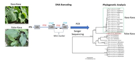
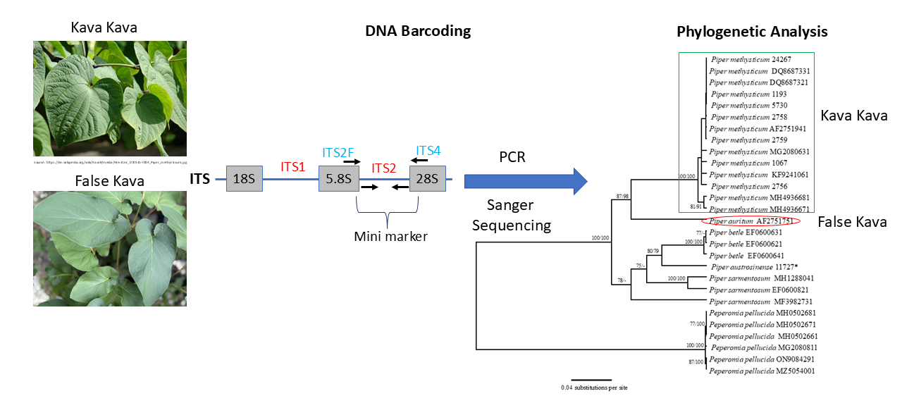
Graphical Abstract

1. Introduction

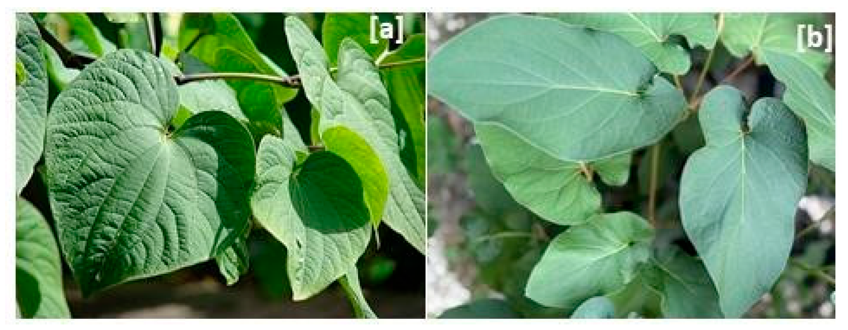
Piperaceae is one of the largest families of angiosperms, with approximately 3600 species distributed in tropical and subtropical regions of the world. The family contains five genera, with Piper L. and Peperomia Ruiz and Pav. as the two with the highest number of species—ca. 2171 and 1000 species, respectively [1]. Most Piper species are used as spices, as medicine, or grown as ornamentals. Piper nigrum L., commonly known as black pepper, is one of the most popular spices worldwide, and P. methysticum G. Forst. (kava-kava) is used as a medicinal plant to treat anxiety disorders [2,3,4,5,6,7]. P. methysticum rhizome/root is used in traditional medicine in the Pacific Islands for its psychotropic effects. Other traditional uses of kava-kava are to treat fever, respiratory problems and urinogenital conditions. Some modern herbal medicines use kava-containing preparations to treat anxiety disorders and/or depression. The kavapyrones and kavalactones in its water extracts are believed to contribute to the sedative, anxiolytic (anxiety-reducing), and muscle-relaxing properties associated with kava consumption. Water extracts of its active compounds, when consumed in limited quantities, have been indicated to have positive effects on anxiety and anxiety disorders [8], although further research is required to confirm these observations. However, some cases of liver toxicity have been associated with kava-kava preparations, especially in extracts and products marketed as supplements [9]. It has been found that kava-kava (P. methysticum) extracts in organic solvents at higher concentrations or in other kava-kava containing Piper species, including P. auritum Kunth, are hepatotoxic. Piper auritum is often referred to as false kava due to its morphological similarity with true kava-kava. The leaves of both plants have similar shapes, but the lobes of the P. methysticum leaf are equal and have palmate venation (the arrangement of veins in the leaf), while the leaf lobes of P. auritum are unequal, and the venation pattern is pinnate [Figure 1]. False kava does not contain kavalactones but has a high concentration of safrole, which is highly hepatotoxic [10]. The morphological similarity of false kava species to P. methysticum often leads to adverse effects in adulterated kava-kava beverages and dietary supplements. Hence, the correct identification of true kava-kava from false kava species is important to establish the safety and efficacy of their beverage and herbal products.
DNA barcoding has become an effective technique to identify many species and to discriminate among close relatives, provided that the correct regions are used [11,12,13,14]. DNA barcodes are short, highly variable sequences of DNA from either the nuclear or organellar genome used for the identification of biological species. The COXI (cytochrome c oxidase) region from the mitochondrial genome is frequently employed for animal species identification [11,12]. However, in plants, the standardized DNA barcode from a single gene region has not been readily agreed upon since different taxa have more variable levels of species identification resolution [15]. Therefore, multiple loci, including rbcL, matK, rpoB, rpoC1, trnH-trnL, trnH-psbA, ndf, and atpB, from the chloroplast genome and the Internal Transcribed Spacer (ITS) region from the nuclear genomes have been utilized as suitable DNA barcodes to discriminate among different plant species [16,17,18]. The barcode identification approach usually includes the comparison of unidentified sequences with published reference sequences in NCBI GenBank (or other standard databases) using BLAST (Basic Local Alignment Search Tool version 2.17.0) and the identification of species-specific sequences.
There are some scientific publications on the identification of Piper species, which have included the chloroplast regions of matK, rbcL, rpoC1 and trnH-psbA, as well as nuclear ITS2 markers [19,20,21,22]. Although specific studies focused solely on DNA barcoding of P. methysticum are limited, the application of different DNA markers has been explored within the genus. For example, a comparative analysis of three different barcode regions for identifying 36 species of Piper found that the multi-locus combination of matK, rbcL, and psbA-trnH was effective for distinguishing among them [21]. Brandao et al. [22] evaluated DNA barcoding markers to differentiate among three Piper species: P. gaudichaudianum, P. malacophyllum, and P. regnellii. Three DNA regions (ITS2, trnH-psbA, and rbcL) were analyzed, along with three microsatellite (SSR) markers (Psol 3, Psol 6, and Psol 10). ITS2 was found to be the most effective marker, accurately identifying all species with strong phylogenetic clustering of the respective species. The other two loci from the chloroplast genome, trnH-psbA and rbcL, had lower discriminatory power [22]. Combining markers did not significantly improve species resolution. SSR markers show potential for species identification, but further research is needed for broader application [22]. These two studies provided valuable genetic tools for the accurate identification and conservation of economically and medicinally important Piper species. It was concluded that ITS2 is appropriate for DNA barcoding to determine the identities of Piper species. The genetic analysis by Shi et al. [23] using AFLPs (Amplified Fragment Length Polymorphisms) also showed that all tested samples of P. methysticum clustered together as one clade that was distantly related to other species of Piper, including P. betle, P. sarmentosum and P. nigrum; the study discerned that the Peperomia species was instead of another genus in the Piperaceae family. In these studies, most Piper species can be accurately identified and resolved using the DNA barcodes discussed above. Additionally, the Medicinal Materials DNA Barcode Database [24] provides a platform for the storage, retrieval, comparison, and analysis of DNA sequences to distinguish medicinal materials, including those species in the Piper genus. The database contains eight sequences of P. methysticum from different DNA barcode regions—one from the 18S ribosomal region, two each from the chloroplast ribosomal protein L16 gene and trnS-rps4 intergenic spacer, and three from ITS [24]. While direct studies on kava-kava’s DNA barcoding are scarce, the research to date suggests that the regions ITS2, matK, and rbcL provide effective identification of some species in the genus Piper. These markers can potentially be applied to P. methysticum for increased accurate identification and authentication purposes. Hence, in the present investigation, the ITS2 region from the nuclear genome was tested to confirm the species-level identification and authentication of kava-kava samples.

2. Materials and Methods

2.1. Plant Material

P. methysticum G. Forst. samples were collected at the Maynard W. Quimby Medicinal Plant Garden (MPG) at the University of Mississippi, from Alden Lane Nursery, CA, and the New York Botanical Garden (Bronx, NY) and one sample each from Pohnpei, Micronesia, and Ecuador (South America) (Table 1). The two P. auritum Kunth samples were collected from MPG (Table 1).

2.2. Primer Design

The universal primers—ITS-S2F from the 5.8S region and ITS4 from the 28S region—were used for amplification of ITS2 [25]. The M13-F and M13-R adapters were added to the ITS2 primer set for sequencing (Table 2).

2.3. DNA Extraction and PCR Amplifications

The dried plant materials (100 mg) were weighed, and 10–20 mg of polyvinylpolypyrrolidone (PVPP) was added to a 2 mL Eppendorf tube with two 2 mm metal balls (Retsch USA, Newtown, PA, USA). The plant material was ground in a Retsch MM301 Mixer Mill (Retsch USA, Newtown, PA, USA). DNA was extracted from the plant material using the DNeasy Plant Mini Kit (Qiagen, Hilden, Germany), and the quality of DNA was assessed using a 0.8% agarose gel. The first PCR amplification was carried out in 25 or 50 µL of reaction mixture containing 10–20 ng of genomic DNA, 1× PCR reaction buffer, 0.2 mM dNTP mixture, 0.2 µM of each forward and reverse primer, 1.5 mM MgCl2, and 2 U of Platinum Taq DNA Polymerase (Invitrogen, Thermo Fisher Scientific, Carlsbad, CA, USA). The ITS2 genomic region was amplified using touchdown annealing temperatures of 50–60 °C for the first 6 cycles, and second PCRs were carried out at 94 °C for 30 s, 50 °C annealing temperature for 30 s and 72 °C for 1 m for 30 cycles, with a final extension at 72 °C for 2 m. PCRs were run in a SimpliAmp Thermal Cycler (Life Technologies Thermo Fisher Scientific, Carlsbad, CA, USA). After amplification, a 10 µL aliquot was analyzed by electrophoresis on a 1.5% TAE agarose gel stained with ethidium bromide, visualized under UV light. The PCR product aliquots with single bands were purified with the Wizard® SV Gel and PCR Clean-Up System (Promega Corporation, Fitchburg, WI, USA), and the concentrations were measured using a NanoDrop™ 2000/2000c Spectrophotometer (Thermo Fisher Scientific, Waltham, MA, USA). The PCR amplicons were then sent to an external sequencing service provider—GENEWIZ (from Azenta Life Sciences, South Plainfield, NJ, USA) for Sanger sequencing in both directions.

2.4. Sanger Sequencing

Purified PCR products were directly sequenced in both directions at GENEWIZ (from Azenta Life Sciences, South Plainfield, NJ, USA). Bidirectional sequences were assembled and trimmed using DNAStar Lasergene 10 (DNASTAR Inc., Madison, WI, USA) to remove the low-quality bases at the end of sequences. The identity of 500 bp (approx.) ITS2 contigs was then confirmed using BLAST (v2.17.0) on NCBI GenBank. The Piper ITS2 sequences obtained in this study have been deposited in NCBI GenBank as accession numbers PZ000070-PZ000077 (Table 3).

2.5. Species Identification Using Distance Method

The trimmed ITS2 sequences were aligned in MUSCLE v5.1 (https://www.drive5.com/muscle5/), using the standard iterative refinement strategy and otherwise default gap penalties. Pairwise genetic distances were calculated in MEGAX [26] using the Jukes–Cantor (JC69) substitution model [27]. Model adequacy was evaluated by comparing alternative nucleotide substitution models in MEGAX, which indicated that JC69 provided an appropriate fit for this ITS2 dataset. For barcode-gap analysis, the pairwise genetic distances were computed in MEGAX under the Jukes–Cantor (JC69) model. For each species with two or more sequences, we computed all pairwise intraspecific distances and recorded the minimum, maximum and mean intraspecific distance per species (File S1).

2.6. Species Identification Using a Phylogenetic Tree-Based Approach

ITS sequences of closely related species were downloaded from NCBI GenBank (File S2), and sequence data was aligned with the MUSCLE 5.1 plugin for Geneious Prime version 2023.2.1, Build 2023-07-20 11:29 (www.geneious.com) (File S3). A phylogenetic tree-based approach was used for species identification and inferred in two ways, with Peperonia pellucida included as an outgroup. All inferences were made using Geneious Prime version 2026.0.2. These included a UPGMA analysis with a Jukes–Cantor genetic distance model (JC69) with 1000 times bootstrap resampling and a neighbor-joining analysis with a Jukes–Cantor genetic distance method (JC69) with 1000 times bootstrap resampling and default consensus tree parameters. An initial clustering analysis was performed using UPGMA under the JC69 distance model. Because UPGMA assumes a constant molecular rate, we also reconstructed a neighbor-joining (NJ) tree to assess topological stability.

3. Results

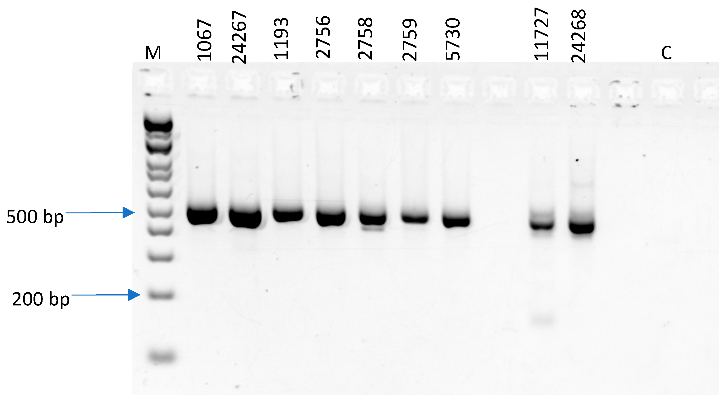
Genomic DNA was extracted from nine dried samples (Table 1). High-quality DNA was obtained from most of the samples with a single, high-molecular-weight band visible on agarose gel, except samples #2758 and 2759, where a smear was observed. The ITS2 genomic region was successfully amplified from all the analyzed samples (Figure 2). The universal primers used for ITS2 were ITS-S2F/ITS4 [17,25,28] (Table 2). The primers include a 5′M13-F/M13-R adapter to enable direct sequencing with standard primers. The ITS2 exhibited a 100% amplification success, of which 90% were able to be sequenced. The PCR amplification resulted in single bands of 500 bp, which were then directly sequenced in both directions after purification. Eight good-quality sequences were obtained out of nine amplicons. The ITS2 locus with universal primers worked best for the amplification and sequencing of Piper sp. The ITS2 sequences from the nuclear genome were analyzed, and the interspecific distances were estimated as 0.165–0.177. Intraspecific ITS2 distances were calculated for species with ≥2 sequences. For P. methysticum, the intra-specific distance ranged from 0 to 0.056 with a mean of 0.011; for P. betle, the range was from 0.022 to 0.005, with a mean of 0.003; for P. sarmetosum, the range was 0.015–0.122, with a mean of 0.085. The reported interspecific distance range of 0.165–0.177 therefore contrasts with a lower intraspecific variation for our samples. Using the species-level barcode-gap criterion (min interspecific [0.165] > max intraspecific [0.122]), all three of the Piper species (100%) tested here that had more than two sequences displayed a clear barcode gap. These results support the notion that the maximum intraspecific distances for ITS2 were lower than the minimum interspecific distances, and hence, the barcode gap for the tested locus is appropriate for diagnosing among these species.
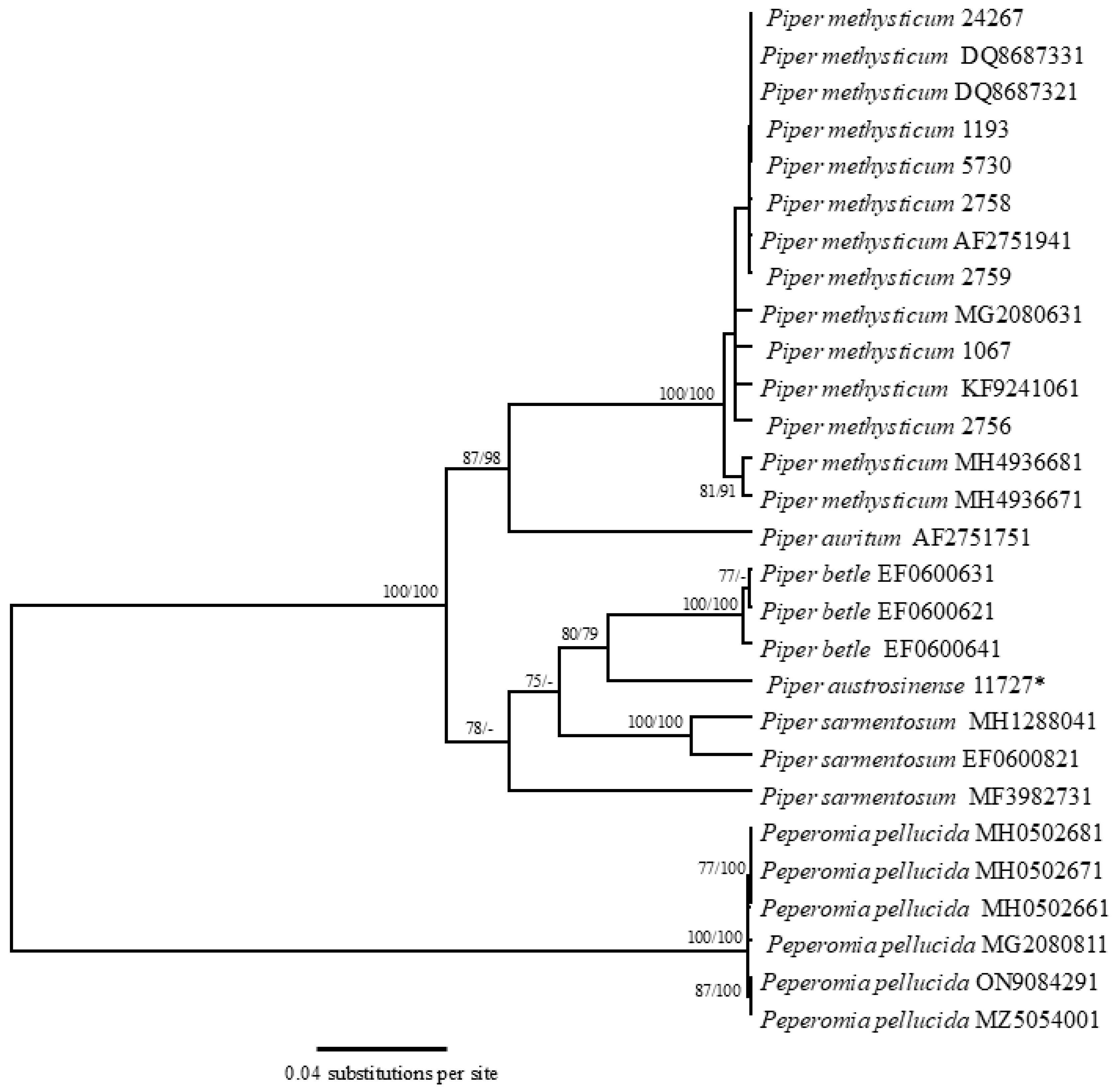
Distance-based phylogenetic analysis methods, including UPGMA and neighbor-joining, were used to evaluate the species identification success using the selected ITS2 DNA barcodes. The published ITS sequences of P. methysticum, P. auritum, P. betle L., P. sarmentosum Roxb., and Peperomia pellucida Kunth were downloaded from NCBI GenBank and were also included in the analyses to increase species resolution. The analyses showed that all tested species were resolved as unique clades with statistical support, using the ITS2 sequences. Two major clades were identified: one comprising Piper and the other with Peperomia species. Within the Piper clade, all the kava-kava samples formed a distinct sub-clade that was separate from the remaining Piper species (Figure 3). Also, the different individuals of the same species of P. methysticum formed monophyletic clades, having bootstrap values of 100% with both methods (Figure 3). The single tested sample labeled as P. auritum (#11727) formed a separate clade different from the downloaded sequence of the same species (Figure 3). Basic Local Alignment Search tool (BLAST) from NCBI GenBank was used to confirm the identity of the generated DNA barcodes for kava-kava. The BLAST analysis for ITS2 sequences showed that seven ITS2 sequences from P. methysticum matched with the correct species with 99–100% homology (Table 3). The P. auritum sequence (#11727) matched with P. austrosinense Y.C. Tseng with 99.5% homology, and the other sequence was not obtained (#24268).

4. Discussion

Extracts of P. methysticum (kava-kava) from roots are being widely marketed for their sedative and anxiolytic properties. However, the false kava species (P. auritum) is its prevalent substitute, whose addition then reduces the efficacy of kava-kava. The current study demonstrated that the ITS2 DNA marker from the nuclear genome serves as a robust and suitable DNA barcode for the authentication of P. methysticum and for distinguishing it from other closely related and/or morphologically similar species of Piper. Although the ITS/ITS2 region has been extensively discussed in fungal barcoding frameworks [25], its applicability to plants is also well established. Numerous plant-focused studies have demonstrated that ITS/ITS2 provides high species-level resolution across angiosperms and is an effective complement to plastid barcodes [17,28]. The conceptual basis for using ribosomal spacers in fungi is transferable to plants since the evolutionary dynamics of the nuclear ribosomal cistron—concerted evolution, multicopy structure, and relatively rapid substitution rates—are shared among eukaryotes [29]. Accordingly, ITS/ITS2 is one of the most informative nuclear markers for species discrimination in plants [15,24]. The high amplification and sequencing success obtained with the ITS2 locus in the current investigation further affirmed its utility for analyzing dried and processed botanical materials, which is important given the commercial status of kava-kava in herbal products and dietary supplements.
In the present study, the maximum intraspecific distance was lower than the minimum interspecific distance among the different Piper species, implying that the barcode gap is well defined. This clear barcode gap indicates that ITS2 provides sufficient sequence variability to differentiate P. methysticum from other species of Piper. This is consistent with earlier published reports demonstrating the discriminatory power of ITS2 for medicinal plant identification and for delimitation among Piper species [15,17,18,19,20]. The low intraspecific divergence among P. methysticum samples—even those collected from geographically distinct regions such as Micronesia and Ecuador—further supports the high-fidelity genetic delineation of the species at the ITS2 locus. This is particularly relevant for kava-kava, which has a long history of clonal propagation in the Pacific Islands; many cultivars are vegetatively maintained, leading to reduced genetic variation.
The distance-based phylogenetic tree showed two major clades containing Piper and Peperomia species, respectively. The P. methysticum sequences were grouped in a single monophyletic clade that was separated from other species of Piper. The genetic analysis by Shi et al. [23] using AFLPs (Amplified Fragment Length Polymorphisms) also showed that all the tested samples of P. methysticum were clustered together into one single clade distantly related to other species of Piper, such as P. betle, P. sarmentosum and P. nigrum, and that the Peperomia species is another genus in the Piperaceae family. The strong clustering of P. methysticum sequences in a monophyletic clade with high bootstrap support in both the UPGMA and neighbor-joining analyses reinforces the reliability of ITS2 as a species-level identifier.
A notable finding in this study is the misidentification of a sample labeled as P. auritum (#11727). The P. auritum sequence downloaded from NCBI GenBank grouped closely with the monophyletic clade of kava-kava sequences, hence exhibiting their close genetic similarity, while the other tested sample, labeled as P. auritum (#11727), formed a different clade from the other two clades. BLAST analysis identified this labeled sample as P. austrosinense. This highlights one of the major challenges in the trade and regulation of botanical materials: morphological similarity among Piper species can lead to incorrect labeling, intentional substitution, or inadvertent adulteration. Because false kava Piper species such as P. auritum may contain hepatotoxic compounds, including safrole, their presence in kava-associated products presents a significant safety risk. DNA barcoding therefore provides an essential tool for quality control, ensuring that consumers and manufacturers can verify the authenticity of raw plant materials and avoid adulteration.
Although ITS2 performed well in this study, some limitations warrant consideration. One P. auritum sample did not yield a sequence, which may have been due to DNA degradation, PCR inhibitors in the plant matrix, or primer–template mismatches. While ITS2 is widely recognized for its universality and discriminatory power, supplementary barcode loci—such as matK, rbcL, or trnH-psbA—could enhance identification success in challenging samples or in studies aiming to resolve deeper phylogenetic relationships within Piperaceae. Future work, including multi-locus approaches or high-throughput sequencing methods and more closely related species, may further increase confidence in species-level identifications, especially for complex or highly processed botanical products.
In summary, although studies on kava-kava’s DNA barcoding are scarce, existing research within the Piper genus and the current investigation suggest that regions including ITS2, matK, and rbcL are effective for Piper species identification and authentication. Given the growing global demand for kava-kava and ongoing concerns regarding product adulteration and hepatotoxicity, implementing DNA barcoding as a routine authentication tool can significantly improve safety standards and regulatory compliance. By ensuring that herbal products contain the correct botanical species, DNA barcoding strengthens both consumer trust and the scientific foundation of medicinal plant quality assurance. Future studies incorporating additional barcode loci or multi-locus barcoding strategies may further refine species discrimination within Piperaceae, particularly for highly processed commercial samples.

Supplementary Materials

The following supporting information can be downloaded at https://www.mdpi.com/article/10.3390/dna6020021/s1, File S1: Distance matrix for ITS2 sequences for interspecific and intraspecific analysis [26,30]; File S2: List of sequences along with their accession numbers downloaded from NCBI GenBank; File S3: Muscle alignment for UPGMA and neighbor-joining analyses.

Author Contributions

N.T. and I.P. developed the primers, and I.P. performed all the experiments. I.P. wrote the manuscript. All authors discussed the results and contributed to the final manuscript. I.A.K. and A.G.C. helped supervise the project. Design of the study: I.P., N.T., S.M.H., J.L., C.W., A.G.C. and I.A.K.; data collection and experiments: I.P.; data analysis and interpretation: I.P. and S.M.H.; drafting the manuscript: I.P.; critical revision of the manuscript: N.T., S.M.H., J.L., C.W., A.G.C. and I.A.K. All authors have read and agreed to the published version of the manuscript.

Funding

This research was supported by “Holistic Approach for Potential Drug Interactions with Botanical Drugs—Importance of Chemical Fingerprinting and Biosimilarity”, funded by the U.S. Food and Drug Administration, grant HHSF223201810175C.

Institutional Review Board Statement

Not applicable.

Informed Consent Statement

Not applicable.

Data Availability Statement

NCBI GenBank as accession numbers PZ000070-PZ000077.

Acknowledgments

The authors thank Jeff Solomon for his editorial support and manuscript proofreading.

Conflicts of Interest

The authors declare no conflicts of interest.

References

  1. Stevens Peter, F. Home. Angiosperm Phylogeny Website, Version 9. 2001. Missouri Botanical Garden. Available online: https://en.wikipedia.org/wiki/Missouri_Botanical_Garden (accessed on 1 June 2025).
  2. Savage, K.M.; Stough, C.K.; Byrne, G.J.; Scholey, A.; Bousman, C.; Murphy, J.; Macdonald, P.; Suo, C.; Hughes, M.; Thomas, S.; et al. Kava for the treatment of generalized anxiety disorder (K-GAD): Study protocol for a randomized controlled trial. Trials 2015, 16, 493. [Google Scholar] [CrossRef]
  3. Barić, H.; Đorđević, V.; Cerovečki, I.; Trkulja, V. Complementary and alternative medicine treatments for generalized anxiety disorder: Systematic review and meta-analysis of randomized controlled trials. Adv. Ther. 2018, 35, 261–288. [Google Scholar] [CrossRef]
  4. Smith, K.; Leiras, C. The effectiveness and safety of Kava-kava for treating anxiety symptoms: A systematic review and analysis of randomized clinical trials. Complement. Ther. Clin. 2018, 33, 107–117. [Google Scholar] [CrossRef] [PubMed]
  5. White, C.M. The pharmacology, pharmacokinetics, efficacy, and adverse events associated with kava-kava. J. Clin. Pharmacol. 2018, 58, 1396–1405. [Google Scholar] [CrossRef]
  6. Sarris, J.; Byrne, G.J.; Bousman, C.A.; Lachlan, C.; Savage, K.M.; Holmes, O.; Murphy, J.; Macdonald, P.; Short, A.; Nazareth, S.; et al. Kava for generalized anxiety disorder: A 16-week double-354 blind, randomized, placebo-controlled study. Aust. N. Z. J. Psychiat. 2020, 54, 288–297. [Google Scholar] [CrossRef] [PubMed]
  7. Burton, N.; Sneed, K.B.; Pathak, Y. Systemic review of the use of Kava Kava for the reduction of anxiety disorder. Medicon Med. Sci. 2023, 5, 37–46. [Google Scholar]
  8. LiverTox: Clinical and Research Information on Drug-Induced Liver Injury; Kava Kava; National Institute of Diabetes and Digestive and Kidney Diseases: Bethesda, MD, USA, 2012. Available online: https://www.ncbi.nlm.nih.gov/books/NBK548637/ (accessed on 10 April 2025).
  9. Ernst, E. A re-evaluation of kava-kava (Piper methysticum). Br. J. Clin. Pharmacol. 2007, 64, 415–417. [Google Scholar] [CrossRef][Green Version]
  10. Yang, A.H.; Zhang, L.; Zhi, D.X.; Liu, W.L.; Gao, X.; He, X. Identification and analysis of the reactive metabolites related to the hepatotoxicity of safrole. Xenobiotica 2017, 48, 1164–1172. [Google Scholar] [CrossRef] [PubMed]
  11. Hebert, P.D.N.; Cywinska, A.; Ball, S.L. Biological identifications through DNA barcodes. Proc. R. Soc. London. Ser. B Biol. Sci. 2003, 270, 313–321. [Google Scholar] [CrossRef]
  12. Hebert, P.D.N.; Ratnasingham, S.; deWaard, J.R. Barcoding animal life: Cytochrome c oxidase subunit 1 divergences among closely related species. Biol. Lett. 2003, 270, S96–S99. [Google Scholar] [CrossRef]
  13. Kress, W.J.; Wurdack, K.J.; Zimmer, E.A.; Weigt, L.A.; Janzen, D.H. Use of DNA barcodes to identify flowering plants. Proc. Natl. Acad. Sci. USA 2005, 102, 8369–8374. [Google Scholar] [CrossRef] [PubMed]
  14. CBOL Plant Working Group. A DNA barcode for land plants. Proc. Natl. Acad. Sci. USA 2009, 106, 12794–12797. [Google Scholar] [CrossRef]
  15. Li, X.; Yang, Y.; Henry, R.J.; Rossetto, M.; Wang, Y.; Chen, S. Plant DNA barcoding: From gene to genome. Biol. Rev. Camb. Philos. Soc. 2015, 90, 157–166. [Google Scholar] [CrossRef] [PubMed]
  16. Kress, W.J.; Erickson, D.L. A two-locus global DNA barcode for land plants: The coding rbcL gene complements the non-coding trnH-psbA spacer region. PLoS ONE 2007, 2, e508. [Google Scholar] [CrossRef] [PubMed]
  17. Chen, S.; Yao, H.; Han, J.; Liu, C.; Song, J.; Shi, L.; Zhu, Y.; Ma, X.; Gao, T.; Pang, X.; et al. Validation of the ITS2 region as a novel DNA barcode for identifying medicinal plant species. PLoS ONE 2010, 5, e8613. [Google Scholar] [CrossRef]
  18. Parveen, I.; Singh, H.K.; Malik, S.; Raghuvanshi, S.; Babbar, S.B. Evaluating five different loci (rbcL, rpoB, rpoC1, matK, and ITS) for DNA barcoding of Indian orchids. Genome 2017, 60, 665–671. [Google Scholar] [CrossRef]
  19. Yu, N.; Gu, H.; Wei, Y.; Zhu, N.; Wang, Y.; Zhang, H.; Zhu, Y.; Zhang, X.; Ma, C.; Sun, A. Suitable DNA barcoding for identification and supervision of Piper kadsura in Chinese medicine markets. Molecules 2016, 21, 1221. [Google Scholar] [CrossRef]
  20. Parvathy, V.A.; Swetha, V.P.; Sheeja, T.E.; Sasikumar, B. A two-locus barcode for discriminating Piper nigrum from its related adulterant species. Indian J. Biotechnol. 2018, 17, 346–350. [Google Scholar]
  21. Chaveerach, A.; Tanee, T.; Sanubol, A.; Monkheang, P.; Sudmoon, R. Efficient DNA barcode regions for classifying Piper species (Piperaceae). PhytoKeys 2016, 70, 1–10. [Google Scholar] [CrossRef]
  22. Egydio Brandão, A.P.M.; Yamaguchi, L.F.; Tepe, E.J.; Salatino, A.; Kato, M.J. Evaluation of DNA markers for molecular identi-379 fication of three Piper species from Brazilian Atlantic Rainforest. PLoS ONE 2020, 15, e0239056. [Google Scholar] [CrossRef] [PubMed]
  23. Shi, J.; Xin, L.; Yang, Y.; Zheng, X.; Kong, X.; Li, Z. Analysis of the genetic relationship between Piper methysticum and Pepper 381 by AFLP. Pak. J. Bot. 2009, 41, 1163–1171. [Google Scholar]
  24. Materials DNA Barcode Database. Available online: https://rdccm.cuhk.edu.hk/mherbsdb/ (accessed on 3 November 2022).
  25. White, T.J.; Bruns, T.; Lee, S.; Taylor, J. Amplification and direct sequencing of fungal ribosomal RNA genes for phylo-384 genetics. In PCR Protocols: A Guide to Methods and Applications; Innis, M.A., Gelfand, D.H., Sninsky, J.J., Whit, T.J., Eds.; Academic Press, Inc.: New York, NY, USA, 1990; pp. 315–322. [Google Scholar]
  26. Kumar, S.; Stecher, G.; Li, M.; Knyaz, C.; Tamura, K. Molecular Evolutionary Genetics Analysis across computing platforms. Mol. Biol. Evol. 2018, 35, 1547–1549. [Google Scholar] [CrossRef] [PubMed]
  27. Jukes, T.H.; Cantor, C.R. Evolution of protein molecules. In Mammalian Protein Metabolism; Munro, H.N., Ed.; Academic Press: New York, NY, USA, 1969; Volume 3, pp. 21–132. [Google Scholar]
  28. China Plant BOL Group. Comparative analysis of a large dataset indicates that internal transcribed spacer (ITS) should be in-corporated into the core barcode for seed plants. Proc. Natl. Acad. Sci. USA 2011, 108, 19641–19646. [Google Scholar] [CrossRef] [PubMed] [PubMed Central]
  29. Álvarez, I.; Wendel, J.F. Ribosomal ITS sequences and plant phylogenetic inference. Mol. Phylogenetics Evol. 2003, 29, 417–434. [Google Scholar] [CrossRef] [PubMed]
  30. Kimura, M. A simple method for estimating evolutionary rate of base substitutions through comparative studies of nucleotide sequences. J. Mol. Evol. 1980, 16, 111–120. [Google Scholar]
Figure 1. Morphologically similar leaves of (a) Piper methysticum (source: https://en.wikipe-109dia.org/wiki/Kava#/media/File:Starr_070515-7054_Piper_methysticum.jpg, accessed on 12 June 2025) and (b) P. auritum. The P. methysticum leaf has equal symmetrical lobes and palmate venation (a), while the P. auritum leaf lobes are unequal and the venation pattern is pinnate (b).
Dna 06 00021 g001
Figure 2. Agarose gel image showing the single band of amplicons obtained using ITS2 universal primers. Abbreviations: M—Standard 1 Kb Plus DNA ladder (5 µL, Invitrogen, Waltham, MA, USA), C—control.
Dna 06 00021 g002
Figure 3. UPGMA consensus tree for ITS2 with support values from an analysis with 1000 bootstrap replications and a neighbor-joining analysis with 1000 bootstrap replications. Nodes are listed with support values as follows: UPGMA bootstrap support over 75/neighbor-joining bootstrap support over 75. The different individuals of a particular species formed a single clade, but sample #11727, labeled as P. auritum, was identified as P. austrosinense (*) and thus did not group with the sequences of the former, indicating misidentification. The scale shows substitutions per site.
Dna 06 00021 g003
Table 1. The list of Piper samples analyzed, along with their NCNPR (National Center for Natural Products Research) voucher numbers, plant part used and the place of collection.
NCNPR Voucher NumberBotanical Name Plant Part Source Latitude/Longitude
1067Piper methysticumRootPohnpei, Micronesia No information
24267Piper methysticumRootMPG34.358, −89.554
1193Piper methysticumRootAlden Lane Nursery, CANo information
2756Piper methysticumStemNew York Botanical Garden 40.864, −73.881
2758Piper methysticumStemNew York Botanical Garden 40.864, −73.881
2759Piper methysticumStemNew York Botanical Garden 40.864, −73.881
5730Piper methysticumRootEcuador, South AmericaNo information
11727Piper auritumAerial Parts (Leaves and Stem) MPG34.358, −89.554
24268Piper auritumRootMPG34.358, −89.554
Table 2. List of primers used for the amplification and sequencing of ITS2. The primers consist of a 5′ adapter sequence, either M13-F or M13-R, to facilitate direct sequencing of PCR products using standard primers.
Primer Primer Sequence Annealing Temperature
ITS-S2F/M13TGTAAAACGACGGCCAGT-ATGCGATACTT—51.9 °C (no adapter), 65.4 °C (with GGTGTGAAT adapter)
ITS4/M13AGGAAACAGCTATGAC—52.1 °C (no adapter), 63.3 °C (with TCCTCCGCTTATTGATATGC adapter)
Table 3. BLAST analysis of ITS2 sequences. The numbers in parentheses are the accession number of the published sequences at NCBI GenBank (13 February 2026).
NCNPR Voucher
Number
SpeciesBLAST Analysis ResultBLAST-e Values
1067Piper methysticum
(PZ000070)
100% match with P. methysticum (MG208063.1)0.0
1193Piper methysticum
(PZ000071)
100% match with P. methysticum (MG208063.1)0.0
2756Piper methysticum
(PZ000072)
100% match with P. methysticum (MG208063.1)0.0
2758Piper methysticum
(PZ000073)
100% match with P. methysticum (MG208063.1)0.0
2759Piper methysticum
(PZ000074)
100% match with P. methysticum (MG208063.1)0.0
5730Piper methysticum
(PZ000075)
100% match with P. methysticum (MG208063.1)0.0
24267Piper methysticum
(PZ0000776)
99.5% match with P. methysticum (MG208063.1)0.0
11727Piper auritum
(PZ000077)
99.5% match with P. austrosinense (MG730434.1)0.0
24268Piper auritumNon-specific sequence-
Disclaimer/Publisher’s Note: The statements, opinions and data contained in all publications are solely those of the individual author(s) and contributor(s) and not of MDPI and/or the editor(s). MDPI and/or the editor(s) disclaim responsibility for any injury to people or property resulting from any ideas, methods, instructions or products referred to in the content.