Communication

Electrospun DegraPol® Meshes with Incorporated Bakuchiol: Characterization and Tissue Integration In Ovo—A Pilot Study


by
Julia Rieber
1,
Tiziano A. Schweizer
2,
Gabor Kadler
2,
Gabriella Meier Bürgisser
1,
Pietro Giovanoli
1 and
Johanna Buschmann
1,*
1
Division of Plastic Surgery and Hand Surgery, University Hospital Zurich, Sternwartstrasse 14, 8091 Zurich, Switzerland
2
Department of Cranio-Maxillo-Facial and Oral Surgery, University Hospital Zurich, University of Zurich, 8091 Zurich, Switzerland
*
Author to whom correspondence should be addressed.
Micro 2026, 6(1), 18; https://doi.org/10.3390/micro6010018
Submission received: 19 November 2025 / Revised: 3 February 2026 / Accepted: 24 February 2026 / Published: 5 March 2026
(This article belongs to the Section Microscale Biology and Medicines)

Abstract

Background: Surgical tendon rupture repair suffers from scar formation, leading to tendons with inferior mechanics and consequently to re-ruptures, as well as from adhesion formation to the surrounding tissue, reducing the range of motion. In an approach of re-purposing the phytochemical Bakuchiol to be incorporated in the polymer DegraPol® (DP), we fabricated a novel implant material by emulsion electrospinning. Methods: To characterize the emulsion electrospun novel materials, we used Scanning Electron Microscopy (SEM) to determine the fiber diameter and pore size. In addition, we used Fourier Transformed Infrared Spectroscopy (FTIR). Finally, we planted the materials onto the chorioallantoic membrane of the chicken embryo (CAM assay) to assess tissue integration and collagen expression. Results: While the pure DP meshes were very well integrated in the CAM assay and showed a significantly higher collagen deposition within the scaffold, the DP + Bakuchiol meshes exhibited poor tissue integration, showing rather the beginning of a fibrous encapsulation. Conclusions: The novel electrospun material DP + Bakuchiol could be used as an anti-adhesion barrier to prevent tendon adhesion.

1. Introduction

Bakuchiol is a natural compound that has gained increasing attention lately [1]. Pharmacological benefits include antibacterial and antiviral properties, anti-inflammatory characteristics and anti-aging effects [2]. Commonly, it is harvested from the seeds and leaves of Psoralea corylifolia (Babchi plant) in India, and it exists in the natural (S) enantiomer as well as in the naturally not occurring (R) configuration [2]. A series of Bakuchiol derivatives have been synthesized and their anti-inflammatory effects have been studied and verified [3]. Furthermore, phytochemicals including Bakuchiol have been reviewed and reported to be cardio-protective [4] by blocking the NFκB signaling pathway and attenuating pressure overload-induced cardiac hypertrophy, fibrosis and inflammatory response [5]. Finally, nano emulsions of Bakuchiol applied on the skin of patients aged older than 30, 40 or 50 years were applied for one month, and a trend towards less wrinkles, discoloration and vessels was reported for all three age groups [6].
So far, however, Bakuchiol has not been applied as a therapeutic in degenerative chronic tendinopathies nor for acute tendon injuries, where the antibacterial, antiviral and anti-inflammatory properties of an implant are also welcome, opening a window for the development of novel implant materials that release Bakuchiol in a sustained way. Based on the promising reported characteristics of Bakuchiol, we therefore fabricated a tubular electrospun DegraPol® (DP) fiber mesh with incorporated Bakuchiol, utilizing an emulsion electrospinning approach, where Bakuchiol was dissolved in ethanol and the ethanol-in-oil emulsion was utilized for electrospinning, following a protocol that has been established previously in our group [7,8,9]. The new implant material was characterized by Scanning Electron Microscopy (SEM) to assess the fiber thickness and the pore size of the random fiber meshes. In addition, we used Fourier Transformed Infrared Spectroscopy (FTIR) to verify the chemical bonds of the polymer. As the envisioned application is to place the Bakuchiol releasing tubes around freshly sutured tendons to promote healing, the architecture of the electrospun mesh had the format of a tube, as previously realized for growth factors [9] or stem cell-derived secretome [10].
The chorioallantoic membrane (CAM) of the chicken embryo is a model situated between in vitro cell cultures and animal in vivo models, such as mice, rats or rabbits [11]. It can be used as an easy, fast and cost-effective model to test if novel biomaterials are biocompatible and whether tissue integration is promoted within a time window of, at maximum, 7 days. Thus, it is an optimal assay for the purposes of pre-screening new materials before using more complex, longer and more expensive small and large animal models.
Hence, circular pieces of these newly fabricated electrospun tubes were planted onto the CAM assay to assess biocompatibility and cellular response towards DP + Bakuchiol compared to pure DP meshes [11]. After one week of incubation, the CAM tissue integration in the fiber meshes as well as the cell density at the margin of the Bakuchiol tubes were compared to pure DP fiber meshes to determine the impact of Bakuchiol on the biological response in ovo—in the living chicken embryo.
Our hypothesis regarding these novel materials was that Bakuchiol facilitates tissue integration and promotes and supports microvascularization in ovo, enabling the envisioned proper and fast initial tissue integration of a ruptured tendon after surgical implantation of a two-layered tube. For that purpose, the orientation is planned with the DP + Bakuchiol layer facing the transected and conventionally sutured Achilles tendon [12], analogously to previously reported implant materials that released a bioactive factor to the injured tendon longitudinally during healing [13], which resulted in significantly stronger tendons at an earlier time point post-operation. The novelty of this approach is the combination of the biocompatible phytochemical Bakuchiol with the biocompatible, biodegradable and highly elastic DegraPol® polymer using an emulsion electrospinning technique.

2. Materials and Methods

2.1. The Polymer DegraPol® (DP)

The polymer DegraPol® was synthesized, using 25 wt% of poly (3-(R-hydroxybutyrate)-co-(ε-caprolactone)-diol (Mn = 2824 g mol−1) and 75 wt% of poly(ε-caprolactone)-diol-co-glycolide (15 mol% glycolide, 85 mol% ε-caprolactone) (Mn = 1000 g mol−1) that were dissolved in 1,4-dioxane and dried until water content was less than 20 ppm. Subsequently, the solution was cooled, and a stoichiometric amount of 2,2,4-trimethylhexane-diisocyanate (TMDI) was added. On the next day, dibutyltin dilaurate (20 ppm) was added three times during the day to reach a molecular weight of 100–110 kDa. The polymer was precipitated in cooled hexane and purified with chloroform and silicagel 60 column (Fluka), followed by precipitation in cooled ethanol.

2.2. Incorporation of Bakuchiol

The solutions were prepared one to three days before electrospinning. For each scaffold, a polyethylene glycol (PEG) (35 kDa, Sigma-Aldrich, Berlin, Germany #81310) solution was prepared by adding 1.5 g of PEG and 3.5 g of chloroform (Sigma–Aldrich, Germany, #132950). The DP solution was produced by adding 0.6 g of DP powder, 3.52 g of chloroform, and 0.88 g of 1,1,1,3,3,3-Hexafluoro-2-propanol (HFP, Sigma-Aldrich, Germany, #105228) into a glass with a screw cap.
For the incorporation of Bakuchiol (Sigma-Aldrich, Darmstadt, Germany, #SMB00604), 400 μL of a 100 μg/mL Bakuchiol solution in 80% ethanol was added dropwise to the DP solution, while stirring for five minutes on the magnetic stirrer at 500 rpm. The emulsion was filled in a 5 mL glass syringe (Huberlab, Aesch, Switzerland, # 3.7102.33) and used immediately for emulsion electrospinning. The amount of Bakuchiol per unsegmented tubular electrospun mesh, having a diameter of 6 mm and a length of 15 cm, was estimated to be 1/3 of total amount used for electrospinning. Thus, 1/3 × 0.4 mL × 100 μg/mL yielding ~13.3 μg. As the tube was segmented into 3 mm long pieces, there was approximately 0.3 μg of Bakuchiol per piece of scaffold to be onplanted in the CAM assay. This amount seemed more than sufficient to allow a first in ovo trial, because previous studies with substances having much higher molecular weights and with smaller amounts of them (proteins IGF-1 (7.8 kDa; and 45 ng IGF-1 per 1 cm piece) or PDGF-BB (24.5 kDa; and 59 ng per 1 cm piece)) exhibited high-enough released amounts to provoke a bioactive response [9].

2.3. Electrospinning

An in-house assembly was used for electrospinning, consisting of a DC high-voltage supply (Glassman High Voltage Inc., High Bridge, NJ, USA), a needle holder, transporter, and syringe pump (SP210cZ, WPI, Berlin, Germany). The spinning head with a blunt end included a needle (1 mm inner diameter and 0.3 mm wall thickness) made of stainless steel (Angst and Pfister AG, Zürich, Switzerland). A metal rod with a length of 55 cm was mounted to the rotary motor (the Euro Star B rotary motor, IKA Labortechnik, Staufen im Breisgau, Germany) and served as collector. The electrospinning conditions for tube production were 1 mL/h as a flow rate, with 9.5 cm working distance between the spinning needle and the metal rod and 12.5 kV voltage applied. The collector rotated at 500 rpm. The room temperature was constant (22–23 °C), and the humidity varied between 35 and 45%. The needle that was transporting the DP solution was moving constantly sideways in both directions in a total range of 20 cm. A first layer of PEG was deposited on the metal rod to facilitate the detachment of the DP tube at the end of the electrospinning process. Subsequently, the DP or DP + Bakuchiol layers were electrospun on top of the PEG layer. After detachment with 50% ethanol, the tubes were stored in a desiccator.

2.4. Scanning Electron Microscopy (SEM) for Fiber and Pore Size Analysis

Small sections from each scaffold tube, including both inner and outer surfaces, were prepared for imaging. The samples were fixed to SEM stubs utilizing conductive double-sided adhesive tapes. All coating and imaging procedures were conducted using equipment maintained by the Center for Microscopy and Image Analysis at the University of Zurich (UZH). Prior to imaging, the samples were coated with a 10 nm layer of platinum using a Safematic CCU-010 sputter coater. SEM was then performed with a Zeiss Gemini SEM 450, operating at an accelerating voltage of 5 kV. Images were captured at 500× magnification using the secondary electron detector, with a brightness setting of 49%. Fiber diameters and tube wall thicknesses were quantified using ImageJ software (version 1.53 e/Java 1.8.0_172, 64-bit). Length measurements were assessed by comparison with the scale bars provided in the SEM images. For each analysis, a diagonal reference line was drawn across each image, and all fibers or pores intersecting this line were measured.

2.5. Fourier Transformed Infrared Spectroscopy (FTIR)

FTIR spectroscopy was carried out utilizing a Varian 640 FTIR spectrometer equipped with a Golden Gate diamond ATR unit featuring temperature control. Spectra were collected over a wavenumber range of 600 to 4000 cm−1, at a resolution of 4 cm−1. Each spectrum represented an average of 64 individual scans to enhance signal quality. For comparative evaluation, the intensity ratio of the C=O absorption peak at 1720 cm−1 to the C–O peak at 1175 cm−1 was determined. All spectra were normalized relative to the C=O peak at 1720 cm−1. The analysis included pure DP tubes and DP tubes with incorporated Bakuchiol.

2.6. Chorioallantoic Membrane of the Chicken Embryo (CAM) Assay

In accordance with Swiss animal care guidelines (TSchV, Art. 112), experiments involving chicken embryos up to embryonic day 14 do not require IACUC approval. Fertilized Lowman White LSL chicken eggs (Animalco AG Geflügelzucht, Schongau, Switzerland) were incubated at 37 °C with 65% relative humidity for 3.5 days. On embryonic day 4, 3 mL of albumin was removed to facilitate the detachment of the developing chorioallantoic membrane (CAM) from the eggshell. A circular window was then carefully excised from the eggshell; and the as-prepared eggs were incubated for another 3 days until embryonic day 7. Tube pieces with a diameter of 6 mm and a height of 3 mm were gently placed within a silicone ring of 1 cm in diameter on the CAM (n = 4 scaffolds per experimental group). The eggs were again incubated for 1 more week. On day 14, fixation using paraformaldehyde (4%) was conducted overnight. The tube pieces and silicone rings were excised and cut in half before they were embedded in paraffin. The 3 μm thick tissue sections were stained with hematoxylin and eosin (H&E) for the quantitative histological evaluation of the cell density at the interface (3 fields of view (FOVs) per sample) and the semi-quantitative analysis of the tissue integration (scores low, medium and high tissue integration), with 3 FOVs per sample. The semi-quantitative analysis of the collagen intensity was accomplished using Masson Goldner Trichrome (MGT) staining (scores low, medium and high collagen intensity), with 3 FOVs per sample.

2.7. Statistics

For the statistical analysis, IBM SPSS software was used (Version 26). After checking normal distribution of data (Shapiro–Wilk) and variance homogeneity (Levene) of the data sets, either parametric 1-way ANOVA or non-parametric Kruskal–Wallis test was utilized to compare more than two groups. For a 2-group-only comparison, either unpaired t-test (parametric) or Mann–Whitney U test (non-parametric) was applied as well as Cohen’s d effect size. Data are presented as mean ± standard deviation, with 95% Confidence Intervals (CI). For semi-quantitative analysis, cross tables were used, and contingency coefficients were calculated as well as p values. Significance was considered if p < 0.05 (*), p < 0.01 (**), p < 0.001 (***) or p < 0.0001 (****).

3. Results

3.1. SEM

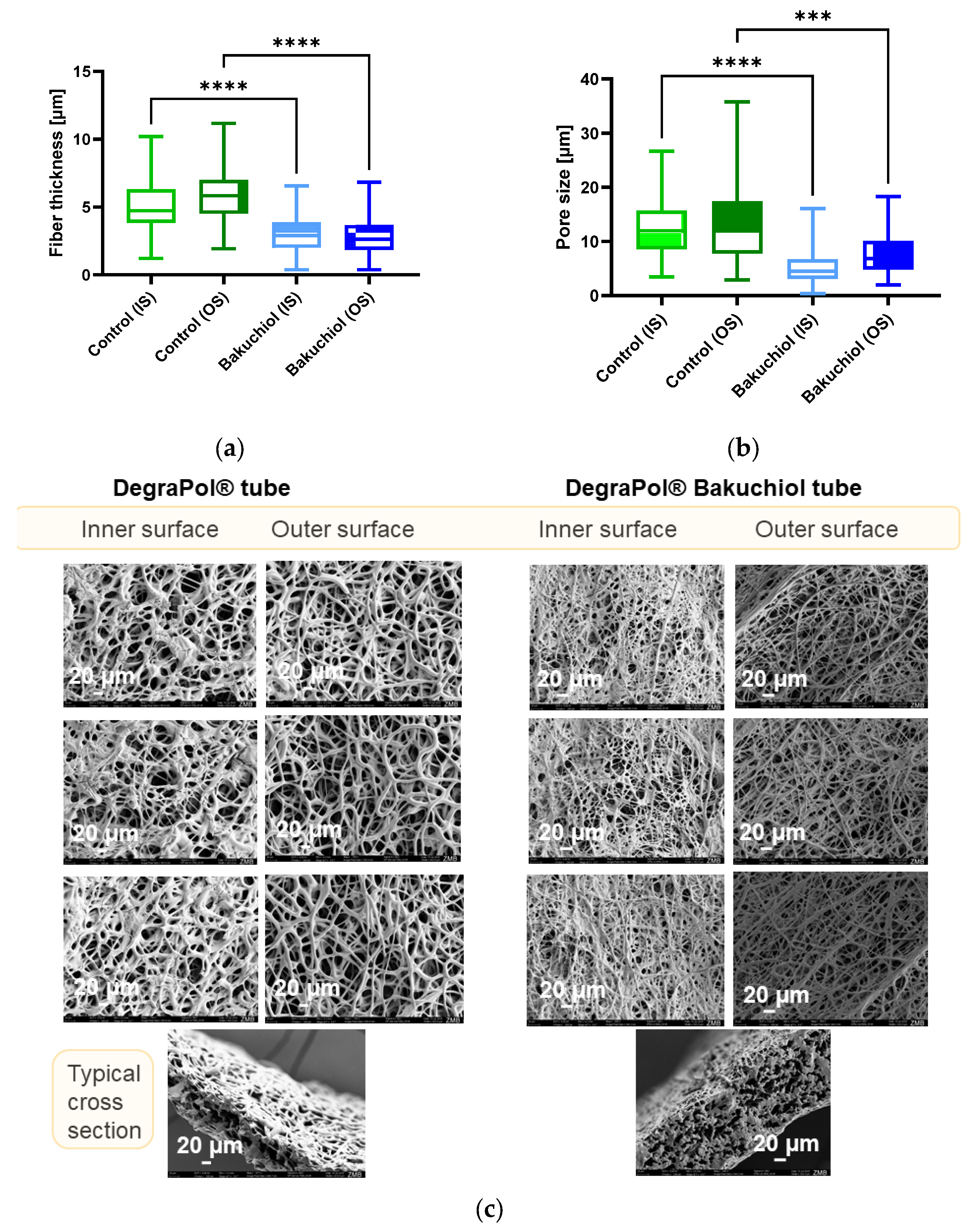
After successful emulsion electrospinning, the novel implant materials were scanned by SEM and the fiber thickness as well as pore size were assessed. SEM images were assessed in pure DP (control) and DP + Bakuchiol electrospun meshes (Figure 1). As can be seen, the fiber diameter was significantly smaller for DP + Bakuchiol fibers compared to pure DP fibers for both the inner surface (IS) as well as the outer surface (OS) (Figure 1a). Specifically, on the inner surface, the pure DP fibers exhibited a thickness of 5.15 ± 2.09 µm, while the Bakuchiol-containing DP meshes had thicknesses of 2.96 ± 1.24 µm. For the outer surface, the pure DP fibers were determined to have a thickness of 5.90 ± 1.95 µm, while it was 2.97 ± 1.31 µm for the DP + Bakuchiol fibers. In addition, the pore size was significantly smaller on both sides for the DP + Bakuchiol meshes compared to the pure DP meshes that acted as a control material (Figure 1b). Specifically, on the inner surface, the pure DP fibers exhibited a pore size of 12.67 ± 5.49 µm, while the Bakuchiol-containing DP meshes had pore sizes of 5.15 ± 3.09 µm. On the outer surface, we found 13.46 ± 7.38 µm for pure DP meshes and a pore size of 7.78 ± 3.86 µm for DP + Bakuchiol meshes.

3.2. FTIR Spectra

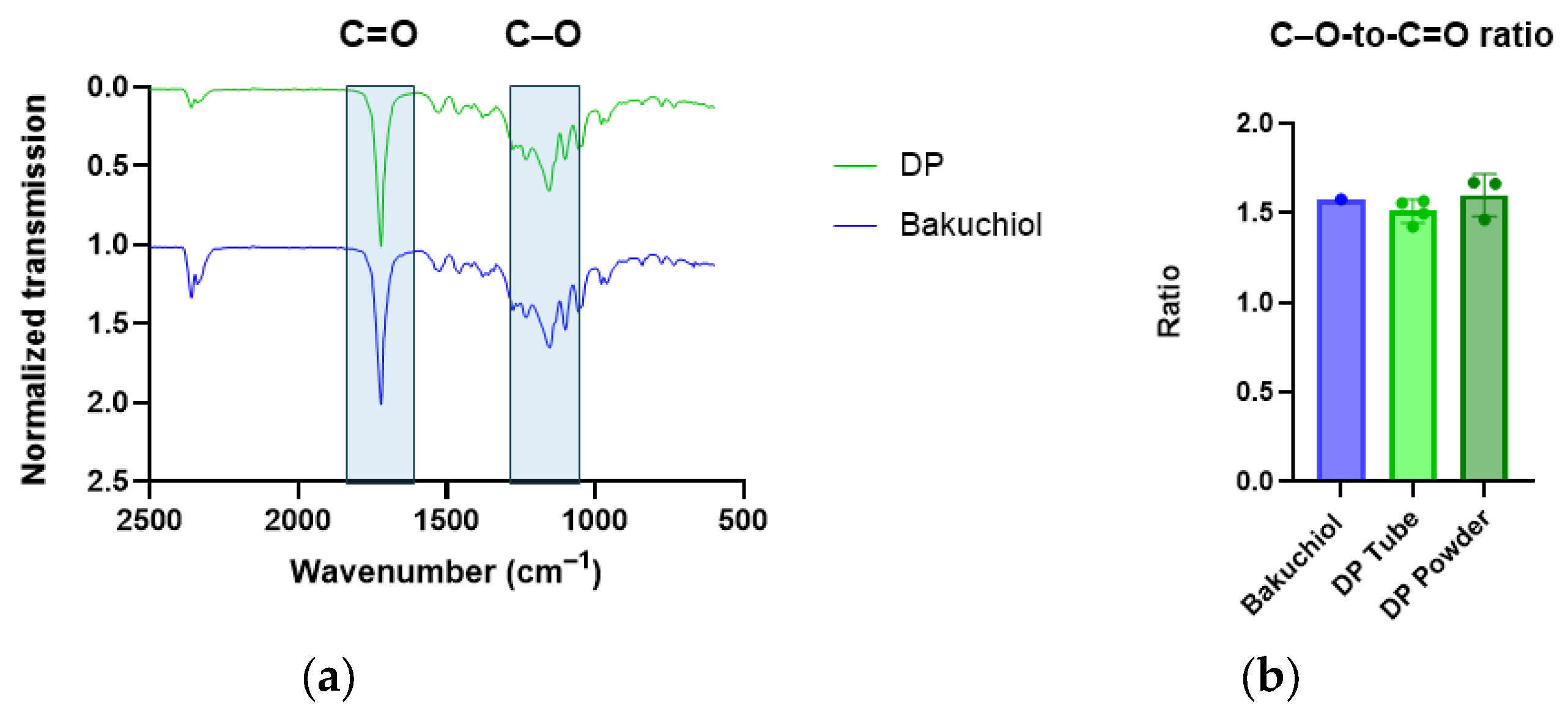
Furthermore, FTIR spectra were assessed for the pure DegraPol® tubes and the tubes with incorporated Bakuchiol. Figure 2 indicates that there was no obvious difference in these spectra. There were the typical and prominent peaks for C=O double bonds at 1720 cm−1 and for the C-O single bond at 1150 cm−1, occurring in the co-block polymer DegraPol®, a polyester urethane. The small double peaks at 2330 and 2365 cm−1 are attributed to CO2 and are not part of the analyzed material. The typical C=C double bond of Bakuchiol is expected to occur at 1610–1640 cm−1; however, the peak found in this range of wavenumbers can also be seen in pure DP and is therefore difficult to attribute only to the DP + Bakuchiol sample. Moreover, when the C–O to C=O ratio was quantitatively assessed (Figure 2b), there was no significant difference, even when compared to pure DP powder.

3.3. CAM Assay

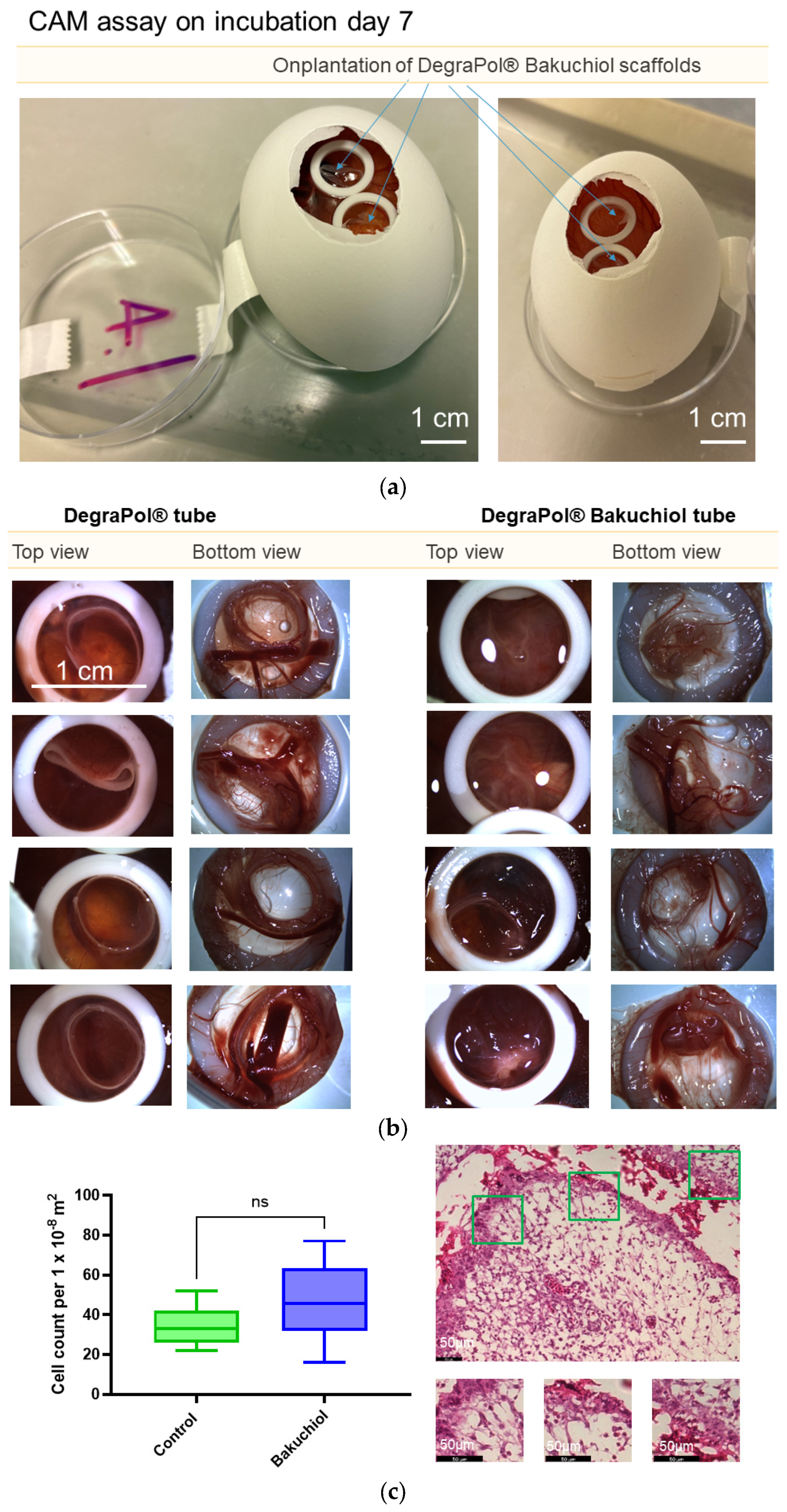
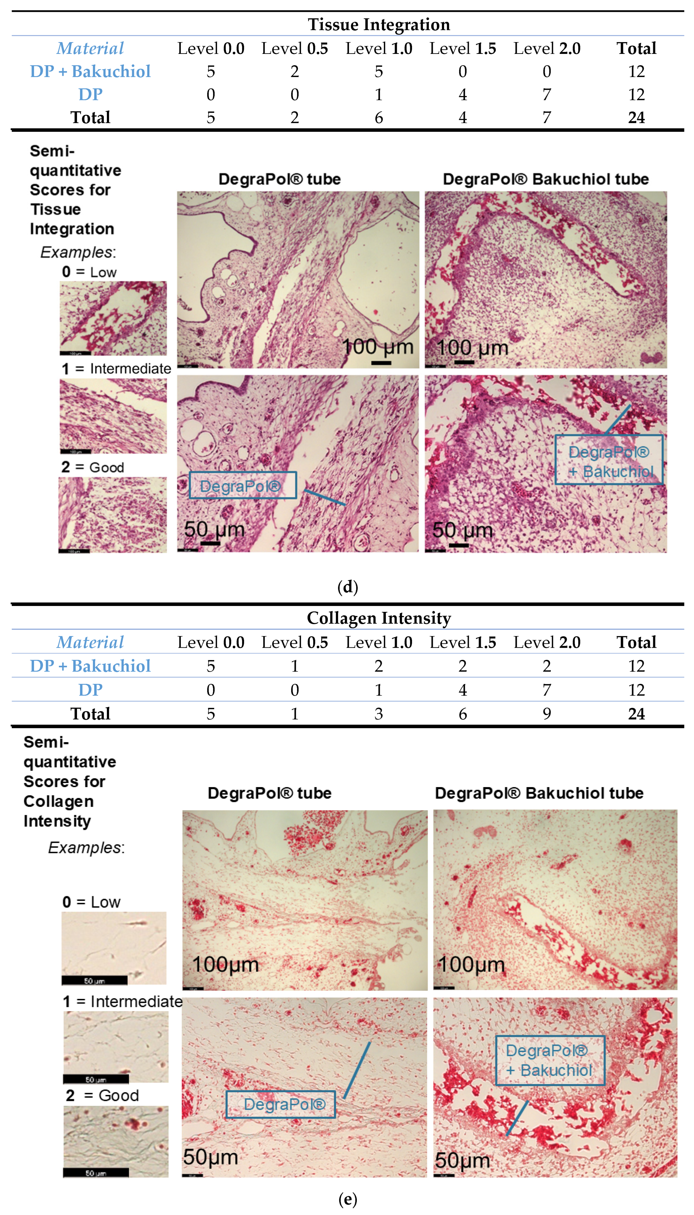
In order to determine the in vivo performance of the new implant material and to assess the biocompatibility, we planted pure DP meshes as well as DP + Bakuchiol meshes onto the surface of the chorioallantoic membrane (CAM) of the chicken embryo for 1 week (n = 4 per experimental group) (Figure 3). We found no significant difference in cell density at the interface between the experimental groups (p = 0.057); however, the cell density under the Bakuchiol treatment exhibited a non-significant upward trend and higher variability. Tissue integration was assessed using a semi-quantitative scoring system with a contingency table, revealing a significant difference between the two experimental groups (p < 0.001). The pure DegraPol® (DP) fiber mesh showed superior tissue integration, evidenced by cell migration through the fiber pores and an even population of the mesh. Conversely, the DP + Bakuchiol fiber mesh showed limited cell invasion and consequently low tissue integration. Furthermore, a semi-quantitative assessment of the collagen intensity within the fibers showed that the pure DP mesh exhibited significantly higher collagen intensity compared to the DP + Bakuchiol fiber mesh (p = 0.044).

4. Discussion

The current study presents results on the fabrication and characterization of a novel electrospun material, where the co-block polymer DegraPol® [14] was combined with the natural drug Bakuchiol [2] by emulsion electrospinning. Against our hypothesis that Bakuchiol would promote the tissue integration utilizing the in ovo CAM assay, we found that Bakuchiol was hindering cell invasion and migration into the pores of the mesh. Nevertheless, this novel material that was initially envisioned to be applied as a two-layer tubular implant material placed around a ruptured and sutured Achilles tendon [12], with the Bakuchiol layer facing the lacerated tendon, can still be of valuable use in tendon repair—by incorporating Bakuchiol in the outer layer of such an implant—facing the surrounding tissue instead, where anti-adhesion properties are needed and cell invasion should be avoided [15].
As tendon rupture repair is confronted with two basic problems, (i) the fibrotic adhesion formation to the surrounding tissue that may lead to a reduced range of motion [16] and (ii) the mechanically inferior scar tissue that may result in re-ruptures [17,18], we screened the phytochemical literature [19,20] for drugs in a re-purposing mode and found the plant-derived Bakuchiol molecule to be a very promising agent. Widely used as a cosmetic additive against photoaging [21], it exhibits antioxidant properties [22], potentially also reducing the overexpression of ROS during tendon healing [23]; it has anti-inflammatory characteristics, supporting the proper resolution of the inflammatory phase after tendon injury [24]; and it shows distinct antibacterial properties that may be needed if our previously fabricated implant materials for tendon repair should fulfill the criteria to be translated into clinical practice [25], which would be facilitated by modifying them with an additional layer of Bakuchiol.
Therefore, Bakuchiol was electrospun into DegraPol® fiber meshes that were characterized. We found that the fibers of such Bakuchiol-containing DegraPol® meshes were significantly smaller than fibers of pure DegraPol® (Figure 1a). This stands in accordance with other emulsion electrospun fibers compared to pure DP fibers, independent of the incorporated drug, as exemplified by PDGF-BB incorporation [7], IGF-1 [8], TIMP-1 [26] or a co-culture derived secretome [10]. Whenever an emulsion of a hydrophilic solvent (here ethanol) in a hydrophobic solution (here DegraPol in CHCl3/HFP) is electrospun, the increased solution conductivity caused by the presence of tiny ethanol droplets within the more hydrophobic polymer promotes an increased charge build-up, leading to a more distinct formation of the Taylor cone and an improved stability of the jet during electrospinning. This therefore reduces Rayleigh instabilities and improves the whipping, which results in smaller and more uniform fibers [27].
Furthermore, we found significantly smaller pores in the DP + Bakuchiol fiber meshes compared to the pure DP meshes (Figure 1b), on both sides of the mesh. This may be at least one of the confounding factors contributing to the significantly lower tissue infiltration of the Bakuchiol-containing mesh observed in the CAM assay (Figure 3d). Although the CAM exhibits different fibroblasts than the tendon fibroblasts (tenocytes), the fibroblast size is much larger than the pore sizes of the presented meshes. Even though typical tenocyte sizes differ among species [28], the pore sizes of the DP and the DP + Bakuchiol meshes were small compared to the cell size of tenocytes which have a reported length of approximately 160–190 µm and a width of around 10–40 µm [28]. Nevertheless, as the DP fibers are highly elastic [29], tendon cells (and immune cells) may find their way into electrospun DP fibers as has been shown previously [30]. This was also observed here for the pure DP mesh in the CAM assay, with complete tissue integration within a week on the chorioallantoic membrane (Figure 3d).
Besides morphological features, the DP and DP + Bakuchiol meshes were analyzed by Fourier Transformed Infrared Spectroscopy (FTIR). Because the amount of Bakuchiol was very small compared to the amount of the polymer, the FTIR spectra did not show any obvious differences between the two materials (Figure 2) in accordance with previous findings, where small protein amounts incorporated as an ethanol-in-DegraPol® emulsion also did not change the FTIR spectra [8,9,13,31]. A closer look at the C–O-to-C=O intensity ratio revealed no significant differences either. Hence, as expected, the overall chemical composition remained unchanged despite the incorporation of Bakuchiol into the DP fiber mesh. While FTIR lacks the sensitivity required to detect such minor concentrations of Bakuchiol within the fiber mesh, NMR spectroscopy would provide a more suitable analytical approach for future quantitative verification.
Screening the biocompatibility of novel materials can be perfectly performed with the CAM assay of the chicken embryo [32]. Originally, this assay was developed by Prof. Domenico Ribatti to assess aspects of angiogenesis, utilizing pro- and anti-angiogenic stimuli [33]. However, since then, it has been further developed and extended for many more aspects in research, such as for cancer biology and metastasis [34,35,36], for organoids [11], biomaterials [37], and scaffolds [11]; this is because the chicken embryo starts to develop an immune system quite late, with immune cells from the yolk sac visible in the thymus only at day 11 [38]—enabling the early grafting of dissimilar materials or cells from species other than birds without the risk of an immune rejection [39]. A further benefit of the delayed immune response in the CAM assay is the potential to decouple the reaction of resident cells toward the onplanted tissue-engineered construct from the systemic immune response. At the interface of the onplanted DP + Bakuchiol mesh and the membrane, we observed an absence of neutrophils and macrophages, finding only fibroblasts. This accumulation of resident cells suggests that the interfacial response occurred independently of the typical fibroblast–macrophage interactions. Specifically, this indicates a bypass of the cell-to-cell communication pathways—such as those involving CS1, TGF-β, and PDGF-AA—that characterize fibrotic development in other tissues, such as the lungs [40].
Therefore, we chose the CAM assay to test slices of the two different tubular implant materials and grafted them onto the membrane for 1 week (Figure 3). Egg survival was 100%, indicating no obvious toxic effects for the embryo development during this time frame [41]. The cell density at the interface between the materials and the membrane was not significantly different for the two groups; however, a trend towards higher cell density in the Bakuchiol-containing DP meshes was observed. Also, the variability in cell density at the interface was higher in the Bakuchiol group compared to pure DP group (Figure 3c). Examination of the tissue integration, however, revealed a significantly better integration for the DP material without Bakuchiol (Figure 3d). While the pure DP meshes were invaded by the CAM cells that proliferated and migrated through the whole width of the tube wall evenly, the tissue integration of the Bakuchiol-containing meshes was rather low; instead, there was an encapsulation of the scaffold material with a clear border of cell accumulation at the interface, suggesting a “frustrated” attempt by cells to invade the DP + Bakuchiol material. In contrast to our hypothesis that Bakuchiol would favor tissue integration in the mesh, we found the opposite—Bakuchiol clearly impedes and hampers proper integration. It rather acts as a barrier, which can be recognized in the corresponding H&E-stained sections where the scaffold area of the Bakuchiol scaffolds could be delineated precisely (Figure 3d).
Although the immune system is not fully developed at day 14 (the endpoint of our CAM assay experiments), the appearance of a fibrotic encapsulated DP + Bakuchiol mesh recalls the foreign body reaction sometimes encountered after biomaterial implantation [42]. It is well-known that macrophages releasing ROS and acid to degrade foreign materials fuse to foreign body giant cells (FBGCs) that accumulate around non-degradable implant materials together with fibroblasts depositing abundant extracellular matrix (ECM) [43]. Even though we did not observe FBGCs nor macrophages in the histological sections, the process of building up a dense ECM around the DP + Bakuchiol fiber mesh, with an increased accumulation of CAM fibroblasts at the interface, can be interpreted as a starting encapsulation process. In addition, the semi-quantitatively assessed collagen intensity within the scaffolds was significantly higher for pure DP meshes compared to the Bakuchiol-containing meshes (Figure 3e), corroborating the significantly higher tissue integration into pure DP meshes (Figure 3d). In other words, a DP mesh without Bakuchiol supported cell invasion and migration into the material, while DP meshes with Bakuchiol turned out to exhibit a barrier function and led to the beginning of a fibrous encapsulation [44].
There are studies confirming Bakuchiol’s favorable cell compatibility with keratinocytes, fibroblasts, and endothelial cells, supporting its application in skincare [45]. Protective effects against pathological cardiac hypertrophy have also been demonstrated [5]. Conversely, Bakuchiol exhibits selective toxicity towards cancer cells (e.g., gastric [46], HKC-8 [47], skin cancer [48]), a point that needs highlighting [46,49,50]. Because the chicken embryo is highly sensitive towards toxic substances and has been reported to be a valuable first screening tool to assess the biocompatibility of tissue-engineered new materials [32], the otherwise positive anti-aging and skin-protecting effects may not be reflected when the novel material is tested in the CAM assay. Moreover, the released amounts of Bakuchiol affecting the cells in the chorioallantoic membrane may not have been adequate, because potentially high concentrations, as assumed to be found at the interface between the material and the CAM, could elicit adverse effects, overpowering Bakuchiol’s beneficial actions.
One limitation of the present study is the absence of dose–response experiments. To evaluate the preliminary effects of Bakuchiol, the novel implants were tested using only a single, but estimated to be high, concentration for emulsion electrospinning. This estimation was based on the low molecular weight (256.4 g/mol) and the high amount loaded during electrospinning (300 μg per 0.3 cm piece), both compared to previous findings on IGF-1 loading (7.8 kDa protein; and 45 ng IGF-1 per 1 cm piece was bioactive) [8] or simultaneously with PDGF-BB loading (24.5 kDa; and 59 ng per 1 cm piece was bioactive) [9]. Furthermore, the observation period was restricted to the 7-day timeframe of the CAM assay. Future research should therefore consider scaffold fabrication with different Bakuchiol concentrations as well as (adult) rodent models with extended observation periods to more comprehensively evaluate long-term outcomes. Finally, the novel combined formulation of DegraPol® and Bakuchiol in emulsion electrospun meshes should be characterized further. Although SEM images gave insights into the morphology of the fibrous meshes and FTIR on the chemical composition, more in-depth studies should additionally include water contact angles and differential scanning calorimetry (DSCs). Also, the biomechanical properties of the novel DP/Bakuchiol meshes should be assessed, although similar properties are expected to be found as with other water-in-oil DegraPol® fiber meshes, as reported for DP meshes with incorporated PDGF-BB [7] or IGF-1 [8], respectively, where, despite different bioactive molecule incorporation, the mechanical properties of these emulsion electrospun meshes were found to be very similar.

5. Conclusions

The study presents the fabrication and characterization of novel DegraPol® fiber meshes with incorporated Bakuchiol and compares them to pure DegraPol® fibers. Morphologically, DP + Bakuchiol meshes had significantly smaller fiber thicknesses and pore sizes compared to pure DP meshes, while having very similar FTIR spectra, indicating and verifying low relative amounts of Bakuchiol compared to the amount of DP. Grafting the novel material onto the CAM assay revealed a hindered cell infiltration into the Bakuchiol fiber mesh, while tissue integration was significantly higher for the pure DP fiber meshes. Furthermore, the DP + Bakuchiol scaffolds showed the start of an encapsulation process, which should be studied further in animal models to allow a longer period of observation than the CAM assay with a limitation of 7 days. Thus, while DP and Bakuchiol have been reported to show no adverse effects as individual components, their combination within an emulsion electrospun fiber mesh appears to function as a physical barrier rather than facilitating tissue infiltration. Given our objective of applying the mesh in future tendon repair, this combination could be utilized in the outer layer of a dual-layer implant, where both cell exclusion and barrier properties are desired to avoid adhesion formation. The efficacy of such applications should, however, be evaluated in rodent models for tendon repair in the future.

Author Contributions

Conceptualization, J.R., T.A.S. and J.B.; methodology, J.R., G.M.B., G.K. and J.B.; software, J.R.; validation, J.R.; formal analysis, J.R.; investigation, J.R.; resources, J.B.; data curation, J.R. and G.M.B.; writing—original draft preparation, J.R. and J.B.; writing—review and editing, J.R., T.A.S., G.M.B., P.G. and J.B.; visualization, J.R.; supervision, G.M.B., P.G. and J.B.; project administration, J.B.; funding acquisition, J.B. All authors have read and agreed to the published version of the manuscript.

Funding

This research was funded by the Swiss National Science Foundation, grant number 310030_197578.

Institutional Review Board Statement

Not applicable, because in accordance with Swiss animal care guidelines (TSchV, Art. 112), experiments involving chicken embryos up to embryonic day 14 do not require IACUC approval. We performed experiments with chicken embryos only until day 14.

Informed Consent Statement

Not applicable.

Data Availability Statement

Data are contained within the article.

Acknowledgments

We thank Ab Medica, Italy, for providing the DegraPol® polymer powder free of charge for research purposes. We thank Ines Kleiber-Schaaf for starting the Histokinette machine.

Conflicts of Interest

The authors declare no conflicts of interest.

Abbreviations

The following abbreviations are used in this manuscript:
CAMChorioallantoic membrane
FOVField of view
H&EHematoxylin and Eosin
MGTMasson Goldner Trichrome

References

  1. Puyana, C.; Chandan, N.; Tsoukas, M. Applications of bakuchiol in dermatology: Systematic review of the literature. J. Cosmet. Dermatol. 2022, 21, 6636–6643. [Google Scholar] [CrossRef] [PubMed]
  2. Nizam, N.N.; Mahmud, S.; Ark, S.M.A.; Kamruzzaman, M.; Hasan, M.K. Bakuchiol, a natural constituent and its pharmacological benefits. F1000Research 2023, 12, 29. [Google Scholar] [CrossRef] [PubMed]
  3. Ma, Q.; Bian, M.; Gong, G.; Bai, C.; Liu, C.; Wei, C.; Quan, Z.S.; Du, H.H. Synthesis and Evaluation of Bakuchiol Derivatives as Potent Anti-inflammatory Agents in Vitro and in Vivo. J. Nat. Prod. 2022, 85, 15–24. [Google Scholar] [CrossRef]
  4. Treviño-Saldaña, N.; García-Rivas, G. Regulation of Sirtuin-Mediated Protein Deacetylation by Cardioprotective Phytochemicals. Oxidative Med. Cell. Longev. 2017, 2017, 1750306. [Google Scholar] [CrossRef]
  5. Wang, Z.; Gao, L.; Xiao, L.; Kong, L.; Shi, H.; Tian, X.; Zhao, L. Bakuchiol protects against pathological cardiac hypertrophy by blocking NF-κB signaling pathway. Biosci. Rep. 2018, 38, BSR20181043. [Google Scholar] [CrossRef] [PubMed]
  6. Lewińska, A.; Domżał-Kędzia, M.; Maciejczyk, E.; Łukaszewicz, M.; Bazylińska, U. Design and Engineering of “Green” Nanoemulsions for Enhanced Topical Delivery of Bakuchiol Achieved in a Sustainable Manner: A Novel Eco-Friendly Approach to Bioretinol. Int. J. Mol. Sci. 2021, 22, 10091. [Google Scholar] [CrossRef]
  7. Evrova, O.; Houska, J.; Welti, M.; Bonavoglia, E.; Calcagni, M.; Giovanoli, P.; Vogel, V.; Buschmann, J. Bioactive, elastic, and biodegradable emulsion electrospun DegraPol tube delivering PDGF-BB for tendon rupture repair. Macromol. Biosci. 2016, 16, 1048–1063. [Google Scholar] [CrossRef]
  8. Rieber, J.; Meier-Bürgisser, G.; Miescher, I.; Weber, F.E.; Wolint, P.; Yao, Y.; Ongini, E.; Milionis, A.; Snedeker, J.G.; Calcagni, M.; et al. Bioactive and elastic emulsion electrospun DegraPol tubes Delivering IGF-1 for tendon rupture repair. Int. J. Mol. Sci. 2023, 24, 10272. [Google Scholar] [CrossRef]
  9. Rieber, J.; Wolint, P.; Meier-Bürgisser, G.; Ongini, E.; Giovanoli, P.; Calcagni, M.; Snedeker, J.G.; Buschmann, J. Synergistic Effects of Insulin-like Growth Factor-1 and Platelet-Derived Growth Factor-BB in Tendon Healing. Int. J. Mol. Sci. 2025, 26, 4039. [Google Scholar] [CrossRef]
  10. Rieber, J.; Miescher, I.; Wolint, P.; Bürgisser, G.M.; Grigioni, J.; Snedeker, J.G.; Vogel, V.; Giovanoli, P.; Calcagni, M.; Buschmann, J. Electrospun DegraPol Tube Delivering Stem Cell/Tenocyte Co-Culture-Derived Secretome to Transected Rabbit Achilles Tendon—In Vitro and In Vivo Evaluation. Int. J. Mol. Sci. 2025, 26, 5457. [Google Scholar] [CrossRef]
  11. Ribatti, D. Two new applications in the study of angiogenesis the CAM assay: Acellular scaffolds and organoids. Microvasc. Res. 2022, 140, 104304. [Google Scholar] [CrossRef] [PubMed]
  12. Meier Buergisser, G.M.; Calcagni, M.; Bachmann, E.; Fessel, G.; Snedeker, J.G.; Giovanoli, P.; Buschmann, J. Rabbit Achilles tendon full transection model—Wound healing, adhesion formation and biomechanics at 3, 6 and 12 weeks post-surgery. Biol. Open 2016, 5, 1324–1333. [Google Scholar] [CrossRef] [PubMed]
  13. Evrova, O.; Bürgisser, G.M.; Ebnöther, C.; Adathala, A.; Calcagni, M.; Bachmann, E.; Snedeker, J.G.; Scalera, C.; Giovanoli, P.; Vogel, V.; et al. Elastic and surgeon friendly electrospun tubes delivering PDGF-BB positively impact tendon rupture healing in a rabbit Achilles tendon model. Biomaterials 2020, 232, 119722. [Google Scholar] [CrossRef] [PubMed]
  14. Saad, B.; Hirt, T.D.; Welti, M.; Uhlschmid, G.K.; Neuenschwander, P.; Suter, U.W. Development of degradable polyesterurethanes for medical applications: In vitro and in vivo evaluations. J. Biomed. Mater. Res. 1997, 36, 65–74. [Google Scholar] [CrossRef]
  15. Meier Buergisser, G.; Calcagni, M.; Muller, A.; Bonavoglia, E.; Fessel, G.; Snedeker, J.G.; Giovanoli, P.; Buschmann, J. Prevention of peritendinous adhesions using an electrospun DegraPol polymer tube: A histological, ultrasonographic, and biomechanical study in rabbits. BioMed Res. Int. 2014, 2014, 656240. [Google Scholar] [CrossRef]
  16. Meier Buergisser, G.M.; Buschmann, J. History and performance of implant materials applied as peritendinous antiadhesives. J. Biomed. Mater. Res. Part B-Appl. Biomater. 2015, 103, 212–228. [Google Scholar] [CrossRef]
  17. Biggs, A.; Scott, G.; Solan, M.C.; Williamson, M. Achilles tendon rupture: What you need to know. Br. J. Hosp. Med. 2021, 82, 1–7. [Google Scholar] [CrossRef]
  18. Chen, C.; Hunt, K.J. Open Reconstructive Strategies for Chronic Achilles Tendon Ruptures. Foot Ankle Clin. 2019, 24, 425–437. [Google Scholar] [CrossRef]
  19. Shin, S.A.; Joo, B.J.; Lee, J.S.; Ryu, G.; Han, M.; Kim, W.Y.; Park, H.H.; Lee, J.H.; Lee, C.S. Phytochemicals as Anti-Inflammatory Agents in Animal Models of Prevalent Inflammatory Diseases. Molecules 2020, 25, 5932. [Google Scholar] [CrossRef]
  20. Kumar, A.; P, N.; Kumar, M.; Jose, A.; Tomer, V.; Oz, E.; Proestos, C.; Zeng, M.; Elobeid, T.; K, S.; et al. Major Phytochemicals: Recent Advances in Health Benefits and Extraction Method. Molecules 2023, 28, 887. [Google Scholar] [CrossRef]
  21. Dhaliwal, S.; Rybak, I.; Ellis, S.R.; Notay, M.; Trivedi, M.; Burney, W.; Vaughn, A.R.; Nguyen, M.; Reiter, P.; Bosanac, S.; et al. Prospective, randomized, double-blind assessment of topical bakuchiol and retinol for facial photoageing. Br. J. Dermatol. 2019, 180, 289–296. [Google Scholar] [CrossRef] [PubMed]
  22. Cariola, A.; El Chami, M.; Granatieri, J.; Valgimigli, L. Anti-tyrosinase and antioxidant activity of meroterpene bakuchiol from Psoralea corylifolia (L.). Food Chem. 2023, 405, 134953. [Google Scholar] [CrossRef] [PubMed]
  23. Kračun, D.; Görlach, A.; Snedeker, J.G.; Buschmann, J. Reactive oxygen species in tendon injury and repair. Redox Biol. 2025, 81, 103568. [Google Scholar] [CrossRef]
  24. Dakin, S.G.; Martinez, F.O.; Yapp, C.; Wells, G.; Oppermann, U.; Dean, B.J.F.; Smith, R.D.J.; Wheway, K.; Watkins, B.; Roche, L.; et al. Inflammation activation and resolution in human tendon disease. Sci. Transl. Med. 2015, 7, 311ra173. [Google Scholar] [CrossRef]
  25. Miescher, I.; Rieber, J.; Schweizer, T.A.; Orlietti, M.; Tarnutzer, A.; Andreoni, F.; Meier Buergisser, G.; Giovanoli, P.; Calcagni, M.; Snedeker, J.G.; et al. In Vitro Assessment of Bacterial Adhesion and Biofilm Formation on Novel Bioactive, Biodegradable Electrospun Fiber Meshes Intended to Support Tendon Rupture Repair. ACS Appl. Mater. Interfaces 2024, 6, 6348–6355. [Google Scholar] [CrossRef] [PubMed]
  26. Rieber, J.; Niederhauser, R.K.; Giovanoli, P.; Buschmann, J. Fabrication and Characterization of Electrospun DegraPol® Tubes Releasing TIMP-1 Protein to Modulate Tendon Healing. Materials 2025, 18, 665. [Google Scholar] [CrossRef]
  27. Abdulhussain, R.; Adebisi, A.; Conway, B.R.; Asare-Addo, K. Electrospun nanofibers: Exploring process parameters, polymer selection, and recent applications in pharmaceuticals and drug delivery. J. Drug Deliv. Sci. Technol. 2023, 90, 105156. [Google Scholar] [CrossRef]
  28. Oreff, G.L.; Fenu, M.; Vogl, C.; Ribitsch, I.; Jenner, F. Species variations in tenocytes’ response to inflammation require careful selection of animal models for tendon research. Sci. Rep. 2021, 11, 12451. [Google Scholar] [CrossRef]
  29. Buschmann, J.; Calcagni, M.; Meier Buergisser, G.; Bonavoglia, E.; Neuenschwander, P.; Milleret, V.; Giovanoli, P. Synthesis, characterization and histomorphometric analysis of cellular response to a new elastic DegraPol (R) polymer for rabbit Achilles tendon rupture repair. J. Tissue Eng. Regen. Med. 2015, 9, 584–594. [Google Scholar] [CrossRef]
  30. Buschmann, J.; Meier Buergisser, G.; Bonavoglia, E.; Neuenschwander, P.; Milleret, V.; Giovanoli, P.; Calcagni, M. Cellular response of healing tissue to DegraPol tube implantation in rabbit Achilles tendon rupture repair: An in vivo histomorphometric study. J. Tissue Eng. Regen. Med. 2013, 7, 413–420. [Google Scholar] [CrossRef]
  31. Rieber, J.; Niederhauser, R.K.; Giovanoli, P.; Buschmann, J. Release kinetics, static and dynamic water contact angles and FTIR data for tissue inhibitor of matrix metalloprotease-1 (TIMP-1) incorporated in electrospun random DegraPol® fibers and TIMP-1 impact on tenocytes and adipose-derived stem cells proliferation and gene expression data. Data Brief 2025, 60, 111468. [Google Scholar] [CrossRef] [PubMed]
  32. Baiguera, S.; Macchiarini, P.; Ribatti, D. Chorioallantoic membrane for in vivo investigation of tissue-engineered construct biocompatibility. J. Biomed. Mater. Res. Part B-Appl. Biomater. 2012, 100B, 1425–1434. [Google Scholar] [CrossRef] [PubMed]
  33. Ribatti, D.; Vacca, A.; Roncali, L.; Dammacco, F. The chick embryo chorioallantoic membrane as a model for in vivo research on angiogenesis. Int. J. Dev. Biol. 1996, 40, 1189–1197. [Google Scholar] [CrossRef] [PubMed]
  34. Ribatti, D. The chick embryo chorioallantoic membrane as a model for tumor biology. Exp. Cell Res. 2014, 328, 314–324. [Google Scholar] [CrossRef]
  35. Waschkies, C.F.; Pfiffner, F.K.; Heuberger, D.M.; Schneider, M.A.; Tian, Y.; Wolint, P.; Calcagni, M.; Giovanoli, P.; Buschmann, J. Tumor grafts grown on the chicken chorioallantoic membrane are distinctively characterized by MRI under functional gas challenge. Sci. Rep. 2020, 10, 7505. [Google Scholar] [CrossRef]
  36. Ribatti, D. The CAM assay in the study of the metastatic process. Exp. Cell Res. 2021, 400, 112510. [Google Scholar] [CrossRef]
  37. Ribatti, D.; Annese, T.; Tamma, R. The use of the chick embryo CAM assay in the study of angiogenic activiy of biomaterials. Microvasc. Res. 2020, 131, 104026. [Google Scholar] [CrossRef]
  38. Leene, W.; Duyzings, M.J.; van Steeg, C. Lymphoid stem cell identification in the developing thymus and bursa of Fabricius of the chick. Z. Zellforsch. Mikrosk. Anat. 1973, 136, 521–533. [Google Scholar] [CrossRef]
  39. Ribatti, D.; Nico, B.; Vacca, A.; Presta, M. The gelatin sponge-chorioallantoic membrane assay. Nat. Protoc. 2006, 1, 85–91. [Google Scholar] [CrossRef]
  40. Buechler, M.B.; Fu, W.; Turley, S.J. Fibroblast-macrophage reciprocal interactions in health, fibrosis, and cancer. Immunity 2021, 54, 903–915. [Google Scholar] [CrossRef]
  41. Ghimire, S.; Zhang, X.; Zhang, J.; Wu, C. Use of Chicken Embryo Model in Toxicity Studies of Endocrine-Disrupting Chemicals and Nanoparticles. Chem. Res. Toxicol. 2022, 35, 550–568. [Google Scholar] [CrossRef] [PubMed]
  42. Anderson, J.M.; Rodriguez, A.; Chang, D.T. Foreign body reaction to biomaterials. Semin. Immunol. 2008, 20, 86–100. [Google Scholar] [CrossRef] [PubMed]
  43. Witherel, C.E.; Abebayehu, D.; Barker, T.H.; Spiller, K.L. Macrophage and Fibroblast Interactions in Biomaterial-Mediated Fibrosis. Adv. Healthc. Mater. 2019, 8, 1801451. [Google Scholar] [CrossRef] [PubMed]
  44. Davidson, M.D.; Burdick, J.A.; Wells, R.G. Engineered Biomaterial Platforms to Study Fibrosis. Adv. Healthc. Mater. 2020, 9, 1901682. [Google Scholar] [CrossRef]
  45. Chaudhuri, R.K.; Bojanowski, K. Bakuchiol: A retinol-like functional compound revealed by gene expression profiling and clinically proven to have anti-aging effects. Int. J. Cosmet. Sci. 2014, 36, 221–230. [Google Scholar] [CrossRef]
  46. Lv, L.; Liu, B. Anti-tumor effects of bakuchiol on human gastric carcinoma cell lines are mediated through PI3K/AKT and MAPK signaling pathways. Mol. Med. Rep. 2017, 16, 8977–8982. [Google Scholar] [CrossRef]
  47. Wang, P.L.; Zhang, F.X.; Qiu, Z.C.; Yao, Z.H.; Wong, M.S.; Yao, X.S.; Dai, Y. Isolation and identification of metabolites of bakuchiol in rats. Fitoterapia 2016, 109, 31–38. [Google Scholar] [CrossRef]
  48. Kim, J.E.; Kim, J.H.; Lee, Y.; Yang, H.; Heo, Y.S.; Bode, A.M.; Lee, K.W.; Dong, Z. Bakuchiol suppresses proliferation of skin cancer cells by directly targeting Hck, Blk, and p38 MAP kinase. Oncotarget 2016, 7, 14616–14627. [Google Scholar] [CrossRef]
  49. Lin, J.; Yao, H.J.; Li, R.Y. Bakuchiol inhibits cell proliferation and induces apoptosis and cell cycle arrest in SGC-7901 human gastric cancer cells. J. BUON 2016, 21, 889–894. [Google Scholar]
  50. Xin, Z.; Wu, X.; Ji, T.; Xu, B.; Han, Y.; Sun, M.; Jiang, S.; Li, T.; Hu, W.; Deng, C.; et al. Bakuchiol: A newly discovered warrior against organ damage. Pharmacol. Res. 2019, 141, 208–213. [Google Scholar] [CrossRef]
Figure 1. Fiber thickness (a) and pore size (b) of pure DegraPol® fiber mesh (control) and of DP with incorporated Bakuchiol (Bakuchiol) fibers on the inner surface (IS) and on the outer surface (OS); assessed in typical SEM images (c), obtained from three electrospun pure DP tubes (rows 1–3; left) and from three electrospun DP + Bakuchiol tubes (rows 1–3; right), depicting inner and outer surfaces as well as a typical cross section (bottom row), respectively. Statistical significance was attributed if p-values were <0.05. Only significances for the pairwise comparison of OS or IS are indicated, not for OS-IS comparisons for clarity reasons. Cohen’s d effect size for IS was 1.18; for OS it was 1.44. Key p < 0.001 (***) and p < 0.0001 (****).
Micro 06 00018 g001
Figure 2. FTIR spectra of pure DegraPol® (DP) and of DP with incorporated Bakuchiol (a). As the amount of Bakuchiol was quite small compared to the bulk DP, no obvious differences were found between the two FTIR spectra (averaged from three spectra taken of individual tubes per condition). This was confirmed by the intensity ratios obtained for the intensity of the CO single-to-double bond that was not significantly different (b). For comparison, DegraPol® powder (DP powder) was also assessed and neither showed any difference in the C–O-to-C=O ratio, implicating no change in DegraPol® polymer during the electrospinning process.
Micro 06 00018 g002
Figure 3. CAM assay performed with DegraPol® (DP) and of DP with incorporated Bakuchiol; example of DP Bakuchiol during planting onto the surface of the CAM assay at day 7 after incubation (a). Top (in the egg) and bottom view (after cutting it out) at day 14 after incubation, 7 days after onplantation (b). Both scaffold types exhibited a similar cell density in the reaction zone at the interface between CAM and scaffold with a Cohen’s d effect size of 0.80; p = 0.057; and 95% CI [−0.4290, 25.43], and all scale bars indicating 50 μm ((c), left) and with a typical histological cross section of a DP + Bakuchiol scaffold stained with H&E and the three fields of view (FOVs; green squares and excised below main image) ((c), right). Tissue integration semi-quantitatively assessed in H&E-stained sections and presented as a cross table, significantly different between the two groups (p < 0.001); typical images of H&E-stained sections; blue line indicates scaffold; all scale bars are 100 μm in the representative images for scores (d). Collagen intensity semi-quantitatively assessed in MGT-stained sections and presented as a cross table, significantly different between the two groups (p = 0.044); typical images of MGT-stained sections; blue line indicates scaffold; all scale bars are 50 μm in the representative images for scores (e).
Micro 06 00018 g003aMicro 06 00018 g003b
Disclaimer/Publisher’s Note: The statements, opinions and data contained in all publications are solely those of the individual author(s) and contributor(s) and not of MDPI and/or the editor(s). MDPI and/or the editor(s) disclaim responsibility for any injury to people or property resulting from any ideas, methods, instructions or products referred to in the content.