Review

Artificial Intelligence in Cardiovascular Pathology: Toward a Diagnostic Revolution

1
Pathology Unit, Department of Precision and Regenerative Medicine and Ionian Area, University of Bari “Aldo Moro”, 70124 Bari, Italy
2
Ph.D Course in Public Health, Department of Experimental Medicine, University of Campania “Luigi Vanvitelli”, 80138 Naples, Italy
*
Author to whom correspondence should be addressed.
BioMedInformatics 2026, 6(2), 18; https://doi.org/10.3390/biomedinformatics6020018
Submission received: 7 February 2026 / Revised: 28 March 2026 / Accepted: 30 March 2026 / Published: 1 April 2026

Abstract

Artificial intelligence (AI) in cardiovascular pathology involves the use of computational models, including machine learning and deep learning (DL), to analyse complex and heterogeneous data. These data include histopathological whole-slide images, cardiovascular imaging techniques such as cardiac magnetic resonance, echocardiography, computed tomography (CT), clinical parameters, and molecular information. The integration of these multimodal data sources allows AI to overcome the limitations of single-modality analysis, improving diagnostic accuracy, prognostic stratification, and personalised clinical decision-making while reducing inter-observer variability. Cardiovascular disease remains the leading cause of mortality worldwide, highlighting the need for more precise and timely diagnostic tools. AI has shown significant promise, particularly in digital pathology, where the digitisation of histological slides combined with advanced algorithms enables improved diagnosis, prognostic assessment, and translational research. This review summarises current AI applications in cardiovascular pathology, focusing on heart transplant rejection, cardiomyopathies, myocarditis, and atherosclerotic and valvular diseases. Automated methods offer important advantages, including diagnostic standardisation, quantitative histological analysis, and improved reproducibility. However, several challenges remain, such as the need for large, well-annotated shared datasets, limited interpretability of AI models, and ethical and legal issues related to clinical implementation. AI represents a promising tool for advancing cardiovascular pathology and personalised medicine, although robust multicentre validation is required before routine clinical adoption.

Graphical Abstract

1. Introduction

Cardiovascular diseases remain the leading cause of mortality worldwide, with profound health, social, and economic consequences [1,2,3]. Although non-invasive diagnosis methods, such as echocardiography, cardiac magnetic resonance imaging (MRI), and coronary angiography, have markedly advanced the detection and longitudinal monitoring of cardiac disorders, histopathological examination continues to play an indispensable role in clarifying pathogenic mechanisms, evaluating therapeutic responses, and informing prognostic assessment [4,5,6].
Despite its central role, cardiovascular pathology is still affected by inter-observer variability and structural limitations, which have motivated the progressive adoption of digital pathology and artificial intelligence-based approaches [7,8,9,10,11,12].
In parallel, artificial intelligence (AI) has found important applications in cardiovascular diagnostics not only at the histopathological level but also in the automated interpretation of electrocardiograms (ECGs), in the early detection of acute myocardial infarction, with reported accuracies of up to 99% and a substantial reduction in diagnostic times, and in remote monitoring of high-risk patients, where its use has been associated with reductions in short-term mortality of up to 31% [13,14,15]. Recent technological innovations include AI-enabled digital stethoscopes, which can detect valvular heart disease with sensitivities exceeding 94%, significantly outperforming conventional auscultation [16].
Moreover, AI is increasingly driving precision medicine approaches by integrating multimodal clinical, genetic, demographic, and imaging data, with potential applications spanning from therapy individualisation to outcome prediction, domains that have traditionally been the prerogative of histopathological evaluation [17,18].
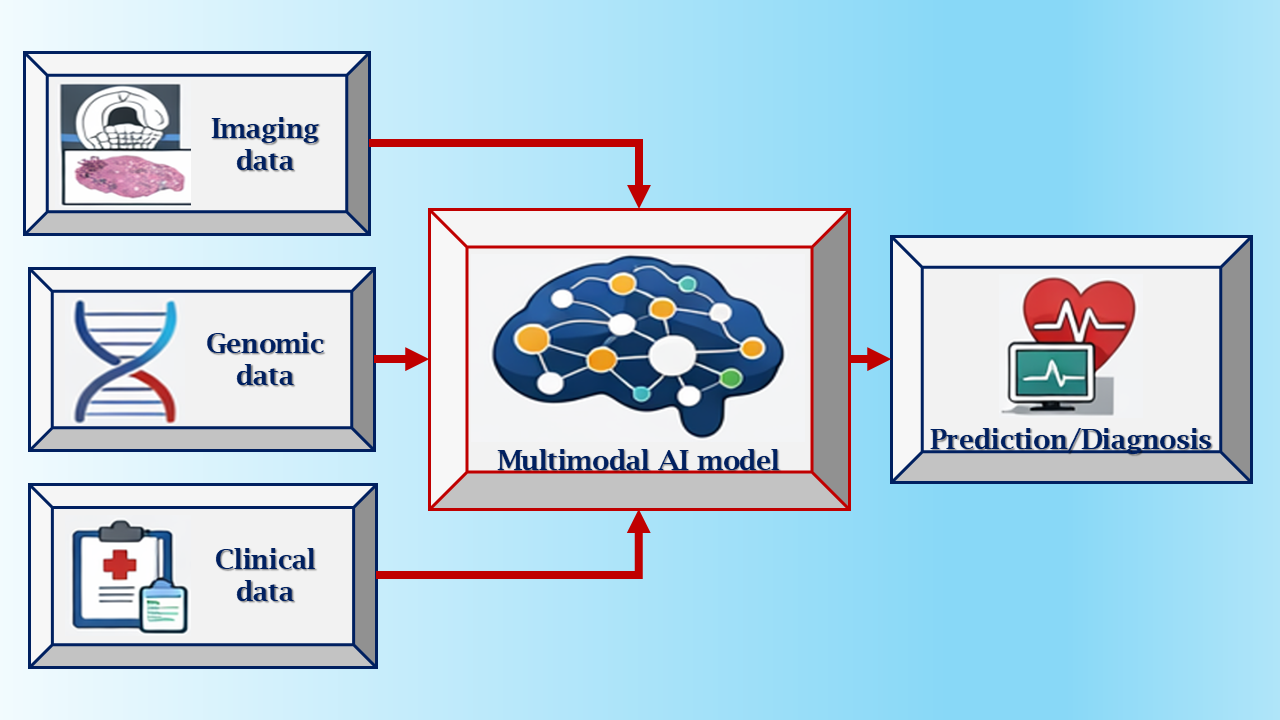
In this context, the concept of multimodal AI deserves explicit clarification. Multimodal AI refers to computational frameworks capable of jointly analysing and integrating heterogeneous data types, including histopathological features, imaging-derived biomarkers, clinical parameters, and molecular or genetic information. Rather than treating these data streams in isolation, multimodal models aim to capture their complex interactions, thereby improving robustness, interpretability, and clinical relevance. In cardiovascular pathology, this approach is particularly valuable, as disease phenotypes often emerge from the interplay between structural, functional, and molecular alterations.
The aim of this review is therefore to critically examine the current state of AI in cardiovascular pathology, from histological applications to broader clinical and technological perspectives, highlighting the benefits, limitations, and future directions for the integration of digital pathology, intelligent algorithms, and clinical diagnostic practice.
Within this evolving landscape, the transition from traditional light microscopy to digital pathology represents a critical step that has enabled the practical implementation of AI in cardiovascular pathology.
The following sections will first outline the technological foundations of AI, followed by its clinical applications and future perspectives.

2. Materials and Methods

This review is a critically oriented narrative review.
The literature search was conducted using PubMed/MEDLINE (U.S. National Library of Medicine, Bethesda, MD, USA), Scopus (Elsevier, Amsterdam, The Netherlands), and Web of Science (Clarivate, Philadelphia, PA, USA) databases. The main keywords used, in different combinations, included: “artificial intelligence”, “machine learning”, “deep learning”, “digital pathology”, “whole slide imaging”, “cardiovascular pathology”, “heart transplant rejection”, “endomyocardial biopsy”, “myocarditis”, “cardiomyopathy”, “atherosclerosis”, “valvular disease”, “cardiac magnetic resonance”, “echocardiography”.
Studies meeting the following general criteria were included: original articles published in peer-reviewed journals, AI applications to cardiovascular pathology or closely related cardiovascular diagnostic domains, use of structured datasets (histological, imaging, electrocardiographic, or clinical), and quantitative reporting of model performance (accuracy, Area Under the Curve (AUC), sensitivity, specificity).
Editorials, commentaries, or opinion pieces lacking original data, case reports, studies with insufficient methodological description, and studies lacking internal validation or a clearly defined test set were excluded.
The final selection of studies was guided not only by eligibility criteria, but also by methodological relevance, the innovativeness of the computational approach, and the potential clinical impact. Therefore, this review presents representative examples of the main research directions, without claiming to be exhaustive.

3. From Traditional Microscopy to Digital Pathology

For more than a century, histopathological diagnosis has relied on the direct visual inspection of glass slides under a light microscope, a well-established approach yet inherently limited by interobserver variability and the predominance of qualitative rather than quantitative assessment. The advent of digital pathology, enabled by Whole Slide Images (WSIs), has fundamentally transformed this paradigm. Histological slides can now be digitised at high resolution, enabling archiving, sharing, and remote consultation [19,20,21,22].
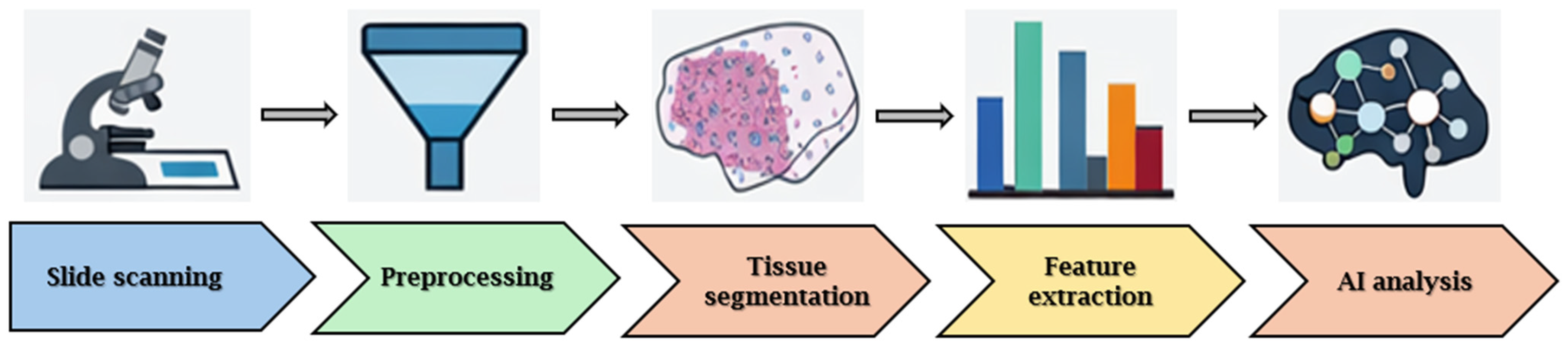
The main workflow of digital pathology and the integration of AI techniques are illustrated in Figure 1.
The integration of WSIs with machine learning and DL algorithms has significantly advanced histopathological analysis, rendering it more quantitative, rapid, and reproducible. These technologies demonstrate sophisticated capabilities in image segmentation, automated detection of regions of interest, and recognition of histological patterns that extend beyond the limits of human visual perception [23,24,25]. However, the intrinsic nature of WSIs presents substantial technological challenges: the extremely large image sizes, often comprising hundreds of thousands of pixels, the variability in magnification, differences in staining protocols, and the presence of multiple focal planes (z-stacks) all complicate the development of robust machine learning models [23,26].
In this new era, digital pathology provides a fundamental infrastructure for the application of AI, supporting not only diagnostic efficiency but also advancements in education, such as the use of digital repositories for training, and in quality assurance, including second-opinion consultations and assessments of diagnostic consistency [27,28,29,30].
In the cardiovascular domain, the digitalisation of tissue specimens represents an essential prerequisite for the implementation of AI. It is only through WSI that algorithms capable of recognising patterns associated with cardiac transplant rejection or cardiomyopathies in experimental models have been developed [12,31].

3.1. Methodological Considerations and Data Requirements

From a methodological perspective, the development and implementation of AI models in cardiovascular pathology critically depend on the quality, quantity and structure of the available data, as well as on the computational frameworks adopted. WSIs represent extremely high-dimensional data and often reach the gigapixel scale. They require dedicated computational pipelines, including image tiling, feature extraction, and classification [19,20,21,22,23,26].
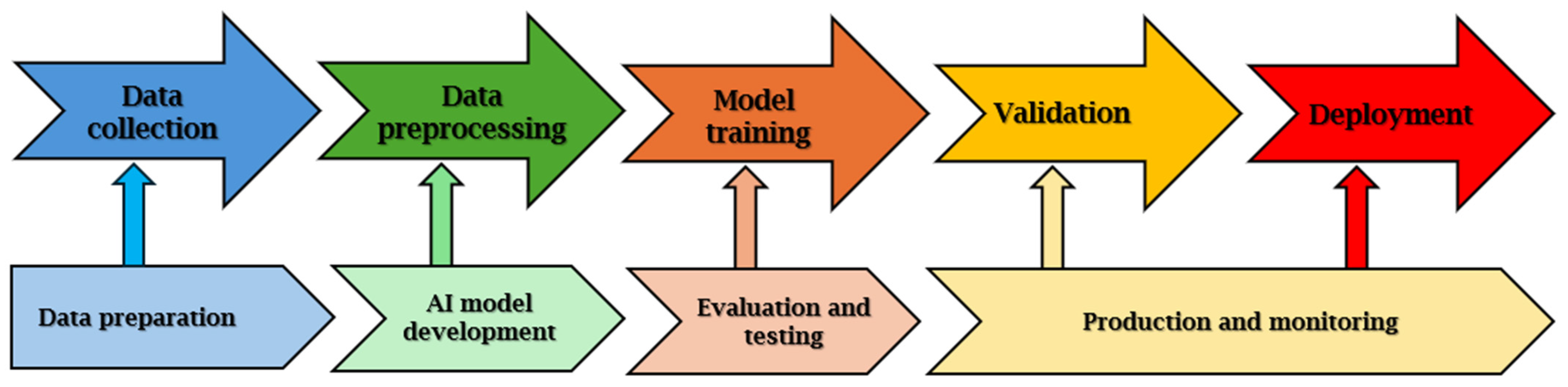
As shown in Figure 2, AI-based analysis pipelines typically involve dividing images into patches, extracting features, and applying classification or prediction models.
Another fundamental requirement concerns data curation. Inter-laboratory variability in tissue fixation, staining procedures and slide digitisation can introduce significant bias into machine learning models, thereby compromising robustness and reproducibility. For this reason, digital pathology pipelines commonly incorporate colour normalisation techniques, image quality control and artefact detection or removal steps in order to improve the generalisability of algorithms across different centres and acquisition systems [23,24,25,26].
Annotation strategies also play a crucial role in determining model performance. Traditional supervised learning approaches rely on detailed region-level or pixel-level annotations, which enhance interpretability and localisation accuracy but are labour-intensive, time-consuming and difficult to scale. In response to these limitations, several studies have explored weakly supervised learning approaches based on slide-level labels, enabling the development of high-performing models without the need for exhaustive manual annotation and facilitating the use of larger datasets [21,23].
From a computational standpoint, the most widely adopted models in cardiovascular pathology are based on convolutional neural networks and DL architectures, which have demonstrated strong performance in pattern recognition tasks applied to histopathological images [11,12]. However, the complexity of these models gives rise to limited interpretability, commonly referred to as the “black box problem”, which remains a relevant barrier to clinical adoption. Addressing this issue is essential to foster trust among clinicians and pathologists and to support the safe integration of AI-based tools into routine diagnostic practice [12,27,28,29,30,31].

3.2. Dataset Characteristics, Annotation Strategies and Computational Pipelines

Studies in cardiovascular pathology adopt heterogeneous datasets and annotation strategies, ranging from pixel-level to slide-level labels. While detailed annotations improve interpretability, they are time-consuming and difficult to scale [11,19,20,21,22,23].
Dataset sizes vary considerably, and external validation remains essential to ensure generalizability [19,20,21,22].
Data curation processes, including colour normalization, quality control, and artefact removal, are critical but inconsistently reported, limiting reproducibility [23,24,25,26]. Finally, feature engineering techniques, either explicitly designed or implicitly learned through DL models, play a central role in extracting diagnostically relevant information from histological tissue. Nevertheless, such techniques are often insufficiently detailed in published studies, further hindering reproducibility and the objective comparison of different methodological approaches [11,12,27,28,29,30,31].
These methodological and technical advances have paved the way for the application of artificial intelligence across a broad spectrum of cardiovascular diseases, where AI-based tools are increasingly being explored for diagnostic, prognostic, and translational purposes.

4. Artificial Intelligence Applications in Cardiovascular Pathology

The applications of AI in cardiovascular pathology described in the following subsections are not intended to represent an exhaustive review of all the studies available in the literature, but rather a selection of representative examples of the main current research directions. The selection of the cited studies was guided by their methodological relevance, potential clinical impact, and the availability of structured histopathological or imaging data. For each pathology, an overview of emerging evidence and main limitations is also provided, to contextualize the state of the art and prospects.

4.1. Heart Transplant Rejection

Rejection remains one of the most critical complications following heart transplantation, with endomyocardial biopsies (EMBs) continuing to represent the diagnostic gold standard. Nonetheless, manual interpretation is subject to considerable inter- and intra-observer variability. Recently, a DL-based system applied to WSIs of EMBs demonstrated excellent diagnostic performance, achieving an AUC of approximately 0.962. Notably, it was able to differentiate cellular from antibody-mediated rejection and to stratify various grades of rejection, thereby significantly reducing variability among human readers [32].
In another study, an algorithm was developed to identify myocyte injury characteristic of acute cellular rejection (ACR), achieving validation accuracies exceeding 90% in distinguishing myocyte damage from benign conditions on digitised slides [33].
A further non-invasive strategy exploits ECG signals in heart transplant recipients. An AI-based ECG model demonstrated the ability to detect moderate-to-severe rejection with an AUC of approximately 0.84 and a sensitivity of 95% [34].
Overall, several studies have demonstrated how AI can improve diagnostic accuracy and reduce interobserver variability in the assessment of EMBs, particularly for the recognition of different forms of rejection. In addition to the examples cited, the literature includes further approaches based on convolutional neural networks and weakly supervised learning, which aim to support the pathologist in classifying according to the criteria of the International Society for Heart and Lung Transplantation. However, most studies remain limited to retrospective and single-center cohorts, necessitating further multicenter validation before large-scale clinical adoption [32,33,34].
Although the reported performance metrics are particularly promising (AUC up to 0.962 and accuracy above 90%), most of the available studies are retrospective and based on single-center cohorts. External validation on independent, multicenter datasets remains limited. Furthermore, variability in slide fixation, staining, and digitization protocols can significantly impact model performance when applied in different institutional settings. Therefore, although AI-assisted analysis of EMB shows high diagnostic potential, its full clinical maturity requires prospective multicenter studies and standardized benchmarks.

4.2. Myocarditis

In the context of myocarditis, AI applied to cardiac magnetic resonance (CMR) imaging has demonstrated excellent diagnostic performance. In a cohort of 269 subjects, of whom 231 had biopsy-confirmed myocarditis, a DL model achieved an accuracy of 96.9% in distinguishing myocarditis from normal cases, particularly when leveraging selected views such as late gadolinium enhancement (LGE) in the two-chamber projection [35].
A recent systematic review evaluated 12 studies comprising 141 machine learning (ML) model outputs for the diagnosis of myocarditis using CMR. The review highlighted that ML generally outperforms human interpretation in cases with clear pathological features, but its performance remains suboptimal in borderline cases or in the presence of imaging artefacts [36].
In myocarditis, AI applications have focused primarily on cardiovascular imaging, while histopathological analysis using DL models remains less explored. Recent studies suggest that AI can contribute to the automatic identification of inflammatory infiltrates and areas of myocyte damage, but the paucity of large, well-annotated datasets represents a significant limitation. Therefore, although the preliminary results are promising, the available evidence is still limited and requires consolidation through prospective studies and multicenter collaborations [35,36].
Although high levels of accuracy have been reported in selected cohorts, many studies rely on relatively homogeneous datasets and internal validation strategies. Performance may be impaired in borderline cases or in the presence of imaging artifacts, as highlighted in systematic reviews. Furthermore, histopathologically based AI applications in myocarditis remain limited, mainly due to the scarcity of large and adequately annotated biopsy datasets. These factors limit immediate generalizability to routine clinical practice.

4.3. Cardiomyopathies

Cardiomyopathies constitute a heterogeneous group of myocardial disorders characterised by structural and functional alterations, which can lead to heart failure, malignant arrhythmias, and sudden cardiac death. AI is increasingly emerging as a crucial support tool for the diagnosis, classification, and prognostication of these conditions.
At the histopathological level, algorithms based on convolutional neural networks (CNNs) have been developed to automatically identify and quantify characteristic alterations in cardiomyopathies, including fibrosis, necrosis, myocyte disarray, and inflammatory infiltrates. In experimental models, such approaches have demonstrated superior sensitivity and reproducibility compared with manual analysis [37].
In the domain of imaging, AI has demonstrated a significant role in the interpretation of CMR. DL models applied to cine CMR images have enabled the differentiation of ischaemic and non-ischaemic cardiomyopathies, achieving an AUC of approximately 0.82 using exclusively functional parameters derived from left ventricular motion [38]. Similarly, machine learning algorithms have been employed to distinguish hypertrophic cardiomyopathy (HCM) from other infiltrative cardiomyopathies, such as amyloidosis, demonstrating good discriminatory performance even in complex cases [39].
AI applied to echocardiography has shown promise in distinguishing pathological left ventricular hypertrophy from physiological exercise-induced hypertrophy, reducing interobserver variability and facilitating early diagnosis [40]. Additionally, AI-assisted point-of-care ultrasound (POCUS) systems have proven effective in the early identification of underdiagnosed cardiomyopathies in non-specialist clinical settings [41].
Finally, integrated predictive models combining clinical, imaging, and genetic data are providing new perspectives for precision medicine. For instance, the MAARS model was recently validated to predict sudden arrhythmic death in patients with HCM, achieving an internal AUC of 0.89 and external validation AUC of 0.81, outperforming conventional clinical risk stratification guidelines [42].
Overall, AI in cardiomyopathies offers tools to enhance differential diagnosis, improve reporting reproducibility, and deliver clinically relevant prognostic information, thereby supporting a truly integrated precision medicine approach [43].
Although several studies report encouraging AUCs (approximately between 0.80 and 0.89 in specific diagnostic or prognostic tasks), the size of the datasets, the number of centers involved, and the external validation strategies vary considerably. Many predictive models, including multimodal ones, rely on retrospective datasets and internal training-test splits, with limited validation on independent populations. Consequently, reproducibility across heterogeneous clinical settings remains to be definitively demonstrated.

4.4. Atherosclerosis

In the context of coronary atherosclerosis, AI-enabled tools applied to coronary computed tomography angiography (CCTA) have facilitated the automatic quantification of plaque volume, demonstrating performance comparable to invasive reference standards such as intravascular ultrasound (IVUS). A recent study reported a high correlation between plaque measurements obtained automatically from CCTA and those derived from IVUS, indicating that AI has the potential to enhance the non-invasive assessment and characterisation of atherosclerotic plaques [44].
In the field of atherosclerosis, AI has been widely applied to the analysis of intravascular imaging and atherosclerotic plaques, enabling quantitative characterization of plaque components and improved risk stratification. Although the role of histopathology in clinical practice is more limited, experimental studies suggest that AI can provide useful insights into underlying pathological mechanisms. Even in this context, however, methodological variability and lack of standardization represent significant challenges [44].
Although automated plaque quantification shows a strong correlation with invasive reference standards, most validation studies have been conducted in highly specialized centers with controlled acquisition protocols. Broader validation, including different scanners, acquisition settings, and heterogeneous populations, is needed to confirm its robustness and real-world applicability.

4.5. Valvular Disease

In the context of valvular heart disease, AI has demonstrated utility in echocardiography for diagnosis, patient phenotyping, and prediction of disease progression. Advanced algorithms have been developed to segment valvular structures and model clinical scenarios, particularly in cases of aortic stenosis and mitral regurgitation, thereby supporting more precise assessment and management planning [45].
In valvular heart disease, AI applications have mainly focused on echocardiographic and tomographic imaging, with the aim of supporting severity assessment and therapeutic planning. AI-assisted histopathological analysis remains less developed, but preliminary studies indicate a potential role in characterizing degenerative and calcific processes. The integration of histological, imaging and clinical data represents a promising perspective for future applications in this field [45].
However, AI models are frequently trained and validated on selected datasets from tertiary care centers. Prospective studies evaluating integration into clinical workflow and external validation remain limited. Therefore, translation into daily clinical practice should be considered with caution.

4.6. Translational Research

Beyond individual clinical cases, AI is increasingly applied in translational research to integrate histological findings with genomic, molecular, and clinical data. For instance, recent reviews in Transplant Pathology have highlighted how AI can be employed to analyse biopsies across various organs, including the heart, to enhance the diagnosis of rejection and optimise immunosuppressive management [46].
Moreover, emerging approaches such as self-supervised learning applied to EMB histological images have facilitated the development of interpretable models capable of detecting cellular rejection patterns even in heterogeneous datasets, thereby offering significant potential for multicentre validation studies [9,47].
Table 1 summarises the main applications of AI in cardiovascular pathology.

4.7. Critical Evaluation of Available Evidence

Despite promising results reported in various fields of cardiovascular pathology, the current literature presents some significant methodological limitations. Many studies are based on relatively small datasets and retrospective designs, factors that may lead to an overestimation of the performance of models when evaluated on internal or single-center cohorts [23,24,25,26]. Furthermore, the availability of adequately annotated digital images remains limited and requires significant effort from expert pathologists, with possible variations in annotation strategies and in the quality of the ground truth used for training the algorithms [27,28,29,30].
Further critical issues arise from the heterogeneity of image acquisition protocols, differences between digital scanners and variations in histological staining protocols, elements that can influence the generalizability of the models [32,33,34]. Furthermore, external validation on independent multicenter cohorts remains limited in several application areas [35,36,37].
Another critical aspect concerns the potential bias of the datasets used for training and validating AI models. Many available studies are based on single-center cohorts, often from highly specialized academic centers, characterized by relatively homogeneous diagnostic protocols, patient populations, and clinical flows. While such datasets may be adequate for initial algorithm development, they can introduce selection and representativeness bias, with the risk that models learn specific features of the local context rather than generalizable disease patterns. Furthermore, the uneven distribution of clinical and demographic variables, such as age, gender, comorbidities, or geographic origin, can lead to reduced performance when algorithms are applied to different populations.
Single-center cohorts also pose additional challenges related to technical variability, including tissue fixation protocols, histological staining methods, digital scanner settings, and annotation strategies used by pathologists. These factors can generate systematic biases in the input data, affecting the ability of models to maintain stable performance across different clinical settings. Consequently, models trained on limited or poorly heterogeneous datasets risk overfitting the source domain, with reduced transferability to other centers.
To address these challenges, multicenter datasets, standardized acquisition protocols, and independent external validation are essential, as well as the adoption of emerging strategies such as federated learning, which allows models to be trained on distributed data without the need to directly share sensitive information. Integrating these strategies represents a fundamental step to ensure the development of more robust, equitable, and clinically generalizable AI systems.
Overall, these findings highlight the need for larger collaborative datasets, prospective studies, and standardized validation procedures to foster more robust clinical implementation of AI techniques in cardiovascular disease.

5. Clinical and Economic Implications

Beyond diagnostic applications, AI also has important clinical and economic implications. First, the automation of analytical processes and the standardisation of histopathological and imaging reports enable a reduction in diagnostic turnaround times, thereby improving workflow efficiency and promoting a more rational use of available healthcare resources. This aspect is particularly relevant in healthcare settings characterised by increasing clinical demand and a growing shortage of specialised personnel.
From an economic perspective, the introduction of AI-based tools has the potential to reduce indirect costs associated with avoidable hospitalisations, unnecessary invasive procedures and diagnostic delays. At the same time, however, the implementation of these technologies requires substantial initial investments, including the acquisition of digital infrastructure, whole-slide imaging systems, maintenance of information technology platforms and rigorous clinical validation of algorithms. Consequently, comprehensive cost-effectiveness evaluations are essential to ensure long-term sustainability.
Another crucial consideration relates to the training of healthcare professionals. The integration of AI into routine clinical practice necessitates a redefinition of educational curricula, as pathologists and clinicians must acquire digital competencies and develop familiarity with the principles underlying algorithmic decision-making and interpretation. This transition promotes a multidisciplinary model in which clinical expertise is augmented, rather than replaced, by computational tools.
Finally, the impact of AI on patient management and clinical outcomes is potentially substantial. The application of predictive models and clinical decision-support systems can enhance risk stratification, enable more precise personalisation of therapeutic strategies and support more effective disease monitoring. Overall, these developments have the potential to improve survival and quality of life for patients with cardiovascular disease while simultaneously strengthening the efficiency and sustainability of healthcare systems.

6. Main Advantages and Limitations

Based on the previous sections, the main advantages and limitations of AI can be summarized. as follows:
One of the main advantages of AI is diagnostic standardisation. In cardiovascular pathology, especially in the evaluation of endomyocardial biopsies for transplant rejection, inter- and intra-observer variability has long represented a major limitation [8,48]. DL models applied to whole-slide images have demonstrated the ability to reduce this variability and to achieve diagnostic performance comparable to, or in some cases exceeding, that of expert pathologists [10,32,49,50]. These tools can support the identification of histological patterns of myocardial injury, inflammation, and rejection with high accuracy, contributing to more consistent reporting and reduced turnaround times.
Another relevant strength of AI lies in its capacity to analyse large volumes of data efficiently. Automated image analysis enables the rapid assessment of extensive histological material and imaging datasets, which would be impractical to evaluate manually. This capability is particularly valuable in high-throughput clinical settings and supports the increasing demand for timely and precise diagnoses.
A further advantage is the possibility of multimodal data integration. AI systems can combine histopathological information with imaging data (such as cardiac magnetic resonance, echocardiography, and CT), as well as clinical, genetic, and demographic variables, to develop predictive models of disease progression and adverse outcomes [51]. Importantly, multimodal AI does not simply aggregate data. Instead, it enables the generation of unified predictive representations that more closely reflect the biological and clinical complexity of cardiovascular disease. This approach is central to the development of precision medicine strategies.
Despite these advantages, several limitations currently restrict the widespread clinical adoption of AI. A major challenge is the need for large, well-annotated, and heterogeneous datasets for model training and validation. Such datasets are difficult to obtain because of privacy regulations, data fragmentation across institutions, and the lack of shared repositories [23,52]. As a result, many AI models are developed using single-centre cohorts, which limits their generalisability.
Limited robustness across different centres represents another important issue. Variations in tissue processing, staining protocols, image acquisition systems, and patient populations can significantly affect model performance when algorithms are applied outside the original development setting [26,53]. Multicentre validation is therefore essential but remains insufficiently addressed in many published studies.
The interpretability of AI models also remains a key limitation. Many DL approaches function as “black boxes”, providing accurate predictions without transparent explanations of the underlying decision-making process [18,54]. This lack of interpretability may reduce clinician trust and hinder integration into routine practice. To address this concern, increasing attention is being directed toward explainable AI techniques, which aim to highlight relevant image regions or features driving algorithmic outputs [26,55].
Finally, ethical and medico-legal considerations must be carefully addressed. Issues related to professional liability in case of diagnostic errors, data protection, and equitable access to AI technologies remain unresolved [43,56]. Without appropriate governance and regulatory frameworks, there is a risk that AI could exacerbate existing healthcare disparities rather than reduce them.
Table 2 summarizes the advantages and limitations of AI in cardiovascular pathology.

Considerations on Bias and Equity

Algorithmic bias represents a major challenge in AI applications to cardiovascular pathology. Models trained on limited or unbalanced datasets may perform poorly in underrepresented populations, potentially exacerbating healthcare inequities.
To mitigate these risks, strategies such as multicentre and demographically balanced datasets, data augmentation, and federated learning are increasingly adopted to improve fairness and generalisability. Ensuring transparency and robustness is essential for the safe clinical implementation of AI.
Another key limitation is the “black box” nature of many deep learning models, which can reduce clinician trust. Explainable AI approaches, including saliency maps and attention mechanisms, aim to improve interpretability by highlighting features that drive model predictions, thereby supporting clinical acceptance and accountability.

7. A Look at the Future

The evolution of AI in cardiovascular pathology is anticipated to accelerate in the coming years, driven by the expansion of digital pathology, advances in computational capacity, and the increasing availability of multimodal data. Future developments are expected to centre on three key pillars: multicentre validation, clinical integration, and progression towards precision medicine.
From a methodological perspective, the priority will be to ensure the robustness and generalisability of AI models. International data-sharing initiatives and the establishment of common repositories represent an essential step towards overcoming current data fragmentation. The implementation of approaches such as federated learning will enable the training of complex models while safeguarding sensitive data, thereby fostering collaboration across centres [52,57,58].
From a clinical standpoint, AI will need to transition gradually from the experimental stage to routine implementation. This will not only require its integration into daily diagnostic workflows but also the design of hybrid systems in which algorithmic outputs support, rather than supplant, the expertise of pathologists and cardiologists. Such an approach to ‘augmented intelligence’ aims to enhance human capabilities rather than replace them, thereby reducing the risk of cultural resistance and increasing clinical acceptability [54,59,60]. Preliminary studies on conversational AI, such as ChatGPT (version 5 mini), have already highlighted both opportunities and limitations in diagnostic pathology, suggesting that these tools may represent a complementary resource to image-based AI in future clinical practice [61].
From an application perspective, the most promising prospects lie in precision medicine. The integration of histological, genetic, clinical, and imaging data will enable the development of predictive models capable of anticipating the clinical course, individualising therapeutic protocols, and more accurately stratifying risk in patients with cardiomyopathies, post-transplant rejection, or valvular disease [54].
Nonetheless, several crucial challenges remain, including the need for a clear and internationally harmonised regulatory framework, the establishment of shared guidelines for model validation, and the management of ethical and medico-legal issues associated with the use of automated systems in high-impact clinical decision-making [62,63].

Practical and Data-Driven Considerations for Robust AI Development

Data fragmentation and heterogeneity across centres represent major barriers to the clinical translation of AI models, limiting their generalisability and reliability in different settings [19,20,21,22,23,26,32,33,34].
The development of robust AI systems requires not only large datasets, but also standardised processes for data curation, including image normalisation, quality control, and annotation [23,24,25,26,35,36,37,38]. In this context, multicentre collaborations and federated learning approaches offer promising solutions for training models on distributed data while preserving privacy [27,28,29,39,40,41,42].
Another critical aspect is the integration of AI into clinical workflows. Models that lack interpretability or are difficult to implement risk remaining confined to experimental settings. Therefore, future developments should prioritise transparency, robustness, and ease of integration, supporting an “augmented intelligence” approach in which AI complements clinical expertise [11,12,30,31,43,44,45,46].
Overall, addressing these challenges is essential to ensure the development of reliable and clinically applicable AI tools in cardiovascular pathology.

8. Regulatory Perspectives

The clinical adoption of AI in cardiovascular pathology is closely linked to the development of clear and robust regulatory frameworks for AI-based medical technologies. In the United States, the Food and Drug Administration (FDA) has introduced regulatory pathways for artificial intelligence and machine learning systems classified as software as a medical device (SaMD). Recent guidance documents emphasize the importance of lifecycle management, continuous performance monitoring, and appropriate validation strategies for AI-enabled medical software [64,65,66].
In Europe, the European Medicines Agency (EMA) has also addressed the use of AI in the lifecycle of medical products, highlighting key principles such as transparency, data quality, risk-based evaluation, and post-market monitoring [67]. These regulatory initiatives aim to ensure that AI systems used in clinical practice meet adequate standards of safety, reliability, and interpretability.
For AI applications in cardiovascular pathology, a particularly relevant regulatory aspect concerns the need for robust validation on large and heterogeneous datasets. Multicenter studies and independent external validation are essential to demonstrate the generalizability and reliability of AI-based diagnostic tools across different clinical environments. Addressing issues such as dataset bias, demographic representativeness, and algorithm transparency will therefore be critical to facilitate both regulatory approval and safe clinical implementation.
Table 3 summarises the key regulatory documents governing the use of AI in medicine.

9. Conclusions

AI represents a potentially transformative tool in cardiovascular pathology, with the ability to enhance diagnostic accuracy, reproducibility, and prognostic assessment. By enabling the integration of histopathological, imaging, clinical, and molecular data, AI supports a shift toward a more predictive and personalised approach to cardiovascular disease.
The future impact of these technologies will depend on their rigorous clinical validation, seamless integration into diagnostic workflows, and alignment with regulatory and professional standards. If appropriately implemented, AI has the potential to complement pathological expertise and to contribute meaningfully to the evolution of cardiovascular precision medicine.
Future progress will depend on rigorous validation, interdisciplinary collaboration, and responsible integration into clinical workflows.

Author Contributions

Conceptualization, A.M. and C.S.; methodology, A.M. and A.Q.; investigation, C.S. and G.C.; resources, A.M. and G.C.; data curation, C.S. and A.Q.; writing—original draft preparation, C.S. and G.C.; writing—review and editing, C.S. and G.C.; supervision, A.M. All authors have read and agreed to the published version of the manuscript.

Funding

This research received no external funding.

Institutional Review Board Statement

Not applicable.

Informed Consent Statement

Not applicable.

Data Availability Statement

No new data were created or analyzed in this study. Data sharing is not applicable to this article.

Acknowledgments

During the preparation of this manuscript, the AI-based language assistance tool ChatGPT (version 5 mini) was used to assist with grammatical and stylistic revision of the text. The authors critically reviewed all generated content and take full responsibility for the final content of the manuscript.

Conflicts of Interest

The authors declare no conflicts of interest.

Abbreviations

ACRAcute Cellular Rejection
AIArtificial Intelligence
AUCArea Under the Curve
CCTACoronary Computed Tomography Angiography
CMRCardiac Magnetic Resonance
CTComputed Tomography
DLDeep Learning
ECGElectrocardiogram
EMAEuropean Medicines Agency
EMBEndomyocardial Biopsy
FDAFood and Drug Administration
HCMHypertrophic Cardiomyopathy
IVUSIntravascular Ultrasound
LGELate Gadolinium Enhancement
MLMachine Learning
MRIMagnetic Resonance Imaging
POCUSPoint-of-Care Ultrasound
SaMDSoftware as a Medical Device
WSIWhole Slide Imaging
XAIExplainable Artificial Intelligence

References

  1. Roth, G.A.; Mensah, G.A.; Johnson, C.O.; Addolorato, G.; Ammirati, E.; Baddour, L.M.; Barengo, N.C.; Beaton, A.Z.; Benjamin, E.J.; Benziger, C.P.; et al. GBD-NHLBI-JACC Global Burden of Cardiovascular Diseases Writing Group. Global Burden of Cardiovascular Diseases and Risk Factors, 1990–2019: Update from the GBD 2019 Study. J. Am. Coll. Cardiol. 2020, 76, 2982–3021. [Google Scholar] [CrossRef] [Scilit]
  2. Salzillo, C.; Ciccone, M.M.; Introna, F.; Santobuono, V.E.; Solarino, B.; Marzullo, A. Sudden cardiac death in the young: Single-center study of Bari autopsy cases. Front. Cardiovasc. Med. 2025, 12, 1630511. [Google Scholar] [CrossRef] [Scilit]
  3. Salzillo, C.; Sansone, V.; Napolitano, F. Sudden Cardiac Death in the Young: State-of-the-Art Review in Molecular Autopsy. Curr. Issues Mol. Biol. 2024, 46, 3313–3327. [Google Scholar] [CrossRef] [Scilit]
  4. Barison, A.; Aimo, A.; Todiere, G.; Grigoratos, C.; Aquaro, G.D.; Emdin, M. Cardiovascular magnetic resonance for the diagnosis and management of heart failure with preserved ejection fraction. Heart Fail. Rev. 2022, 27, 191–205. [Google Scholar] [CrossRef] [Scilit] [PubMed]
  5. Nagueh, S.F.; Smiseth, O.A.; Appleton, C.P.; Byrd, B.F., 3rd; Dokainish, H.; Edvardsen, T.; Flachskampf, F.A.; Gillebert, T.C.; Klein, A.L.; Lancellotti, P.; et al. Recommendations for the Evaluation of Left Ventricular Diastolic Function by Echocardiography: An Update from the American Society of Echocardiography and the European Association of Cardiovascular Imaging. Eur. Heart J. Cardiovasc. Imaging 2016, 17, 1321–1360. [Google Scholar] [CrossRef] [PubMed]
  6. Cooper, L.T.; Baughman, K.L.; Feldman, A.M.; Frustaci, A.; Jessup, M.; Kuhl, U.; Levine, G.N.; Narula, J.; Starling, R.C.; Towbin, J.; et al. The role of endomyocardial biopsy in the management of cardiovascular disease: A scientific statement from the American Heart Association, the American College of Cardiology, and the European Society of Cardiology. Circulation 2007, 116, 2216–2233. [Google Scholar] [CrossRef] [Scilit]
  7. Marijon, E.; Uy-Evanado, A.; Dumas, F.; Karam, N.; Reinier, K.; Teodorescu, C.; Narayanan, K.; Gunson, K.; Jui, J.; Jouven, X.; et al. Warning Symptoms Are Associated with Survival from Sudden Cardiac Arrest. Ann. Intern. Med. 2016, 164, 23–29. [Google Scholar] [CrossRef] [Scilit] [PubMed]
  8. Shanes, J.G.; Ghali, J.; Billingham, M.E.; Ferrans, V.J.; Fenoglio, J.J.; Edwards, W.D.; Tsai, C.C.; Saffitz, J.E.; Isner, J.; Furner, S. Interobserver variability in the pathologic interpretation of endomyocardial biopsy results. Circulation 1987, 75, 401–405. [Google Scholar] [CrossRef] [Scilit]
  9. Seraphin, T.P.; Luedde, M.; Roderburg, C.; van Treeck, M.; Scheider, P.; Buelow, R.D.; Boor, P.; Loosen, S.H.; Provaznik, Z.; Mendelsohn, D.; et al. Prediction of heart transplant rejection from routine pathology slides with self-supervised deep learning. Eur. Heart J. Digit. Health 2023, 4, 265–274. [Google Scholar] [CrossRef] [Scilit]
  10. Jiao, Y.; Yuan, J.; Sodimu, O.M.; Qiang, Y.; Ding, Y. Deep Neural Network-Aided Histopathological Analysis of Myocardial Injury. Front. Cardiovasc. Med. 2022, 8, 724183. [Google Scholar] [CrossRef] [Scilit]
  11. Baxi, V.; Edwards, R.; Montalto, M.; Saha, S. Digital pathology and artificial intelligence in translational medicine and clinical practice. Mod. Pathol. 2022, 35, 23–32. [Google Scholar] [CrossRef] [Scilit]
  12. Glass, C.; Lafata, K.J.; Jeck, W.; Horstmeyer, R.; Cooke, C.; Everitt, J.; Glass, M.; Dov, D.; Seidman, M.A. The Role of Machine Learning in Cardiovascular Pathology. Can. J. Cardiol. 2022, 38, 234–245. [Google Scholar] [CrossRef] [Scilit]
  13. Attia, Z.I.; Noseworthy, P.A.; Lopez-Jimenez, F.; Asirvatham, S.J.; Deshmukh, A.J.; Gersh, B.J.; Carter, R.E.; Yao, X.; Rabinstein, A.A.; Erickson, B.J.; et al. An artificial intelligence-enabled ECG algorithm for the identification of patients with atrial fibrillation during sinus rhythm: A retrospective analysis of outcome prediction. Lancet 2019, 394, 861–867. [Google Scholar] [CrossRef] [Scilit]
  14. Cho, Y.; Kwon, J.M.; Kim, K.H.; Medina-Inojosa, J.R.; Jeon, K.H.; Cho, S.; Lee, S.Y.; Park, J.; Oh, B.H. Artificial intelligence algorithm for detecting myocardial infarction using six-lead electrocardiography. Sci. Rep. 2020, 10, 20495. [Google Scholar] [CrossRef] [Scilit]
  15. Stehlik, J.; Schmalfuss, C.; Bozkurt, B.; Nativi-Nicolau, J.; Wohlfahrt, P.; Wegerich, S.; Rose, K.; Ray, R.; Schofield, R.; Deswal, A.; et al. Continuous Wearable Monitoring Analytics Predict Heart Failure Hospitalization: The LINK-HF Multicenter Study. Circ. Heart Fail. 2020, 13, e006513. [Google Scholar] [CrossRef] [Scilit]
  16. Thompson, W.R.; Reinisch, A.J.; Unterberger, M.J.; Schriefl, A.J. Artificial Intelligence-Assisted Auscultation of Heart Murmurs: Validation by Virtual Clinical Trial. Pediatr. Cardiol. 2019, 40, 623–629. [Google Scholar] [CrossRef] [Scilit]
  17. Johnson, K.W.; Torres Soto, J.; Glicksberg, B.S.; Shameer, K.; Miotto, R.; Ali, M.; Ashley, E.; Dudley, J.T. Artificial Intelligence in Cardiology. J. Am. Coll. Cardiol. 2018, 71, 2668–2679. [Google Scholar] [CrossRef] [Scilit] [PubMed]
  18. Topol, E.J. High-performance medicine: The convergence of human and artificial intelligence. Nat. Med. 2019, 25, 44–56. [Google Scholar] [CrossRef] [Scilit] [PubMed]
  19. Pantanowitz, L.; Sharma, A.; Carter, A.B.; Kurc, T.; Sussman, A.; Saltz, J. Twenty Years of Digital Pathology: An Overview of the Road Travelled, What is on the Horizon, and the Emergence of Vendor-Neutral Archives. J. Pathol. Inform. 2018, 9, 40. [Google Scholar] [CrossRef] [Scilit]
  20. Mukhopadhyay, S.; Feldman, M.D.; Abels, E.; Ashfaq, R.; Beltaifa, S.; Cacciabeve, N.G.; Cathro, H.P.; Cheng, L.; Cooper, K.; Dickey, G.E.; et al. Whole Slide Imaging Versus Microscopy for Primary Diagnosis in Surgical Pathology: A Multicenter Blinded Randomized Noninferiority Study of 1992 Cases (Pivotal Study). Am. J. Surg. Pathol. 2018, 42, 39–52. [Google Scholar] [CrossRef] [Scilit] [PubMed]
  21. Campanella, G.; Hanna, M.G.; Geneslaw, L.; Miraflor, A.; Werneck Krauss Silva, V.; Busam, K.J.; Brogi, E.; Reuter, V.E.; Klimstra, D.S.; Fuchs, T.J. Clinical-grade computational pathology using weakly supervised deep learning on whole slide images. Nat. Med. 2019, 25, 1301–1309. [Google Scholar] [CrossRef] [Scilit]
  22. Hijazi, A.; Bifulco, C.; Baldin, P.; Galon, J. Digital Pathology for Better Clinical Practice. Cancers 2024, 16, 1686. [Google Scholar] [CrossRef] [Scilit] [PubMed]
  23. Niazi, M.K.K.; Parwani, A.V.; Gurcan, M.N. Digital pathology and artificial intelligence. Lancet. Oncol. 2019, 20, e253–e261. [Google Scholar]
  24. Go, H. Digital Pathology and Artificial Intelligence Applications in Pathology. Brain Tumor Res. Treat. 2022, 10, 76–82. [Google Scholar] [CrossRef] [Scilit]
  25. Shafi, S.; Parwani, A.V. Artificial intelligence in diagnostic pathology. Diagn. Pathol. 2023, 18, 109. [Google Scholar] [CrossRef] [Scilit]
  26. Basak, K.; Ozyoruk, K.B.; Demir, D. Whole Slide Images in Artificial Intelligence Applications in Digital Pathology: Challenges and Pitfalls. Whole Slide Images in Artificial Intelligence Applications in Digital Pathology: Challenges and Pitfalls. Turk Patoloji Derg. 2023, 39, 101–108. [Google Scholar]
  27. Fraggetta, F.; Garozzo, S.; Zannoni, G.F.; Pantanowitz, L.; Rossi, E.D. Routine Digital Pathology Workflow: The Catania Experience. J. Pathol. Inform. 2017, 8, 51. [Google Scholar] [CrossRef] [Scilit] [PubMed]
  28. Kumar, N.; Gupta, R.; Gupta, S. Whole Slide Imaging (WSI) in Pathology: Current Perspectives and Future Directions. J. Digit. Imaging 2020, 33, 1034–1040. [Google Scholar] [CrossRef] [Scilit]
  29. Vodovnik, A. Diagnostic time in digital pathology: A comparative study on 400 cases. J. Pathol. Inform. 2016, 7, 4. [Google Scholar] [CrossRef] [Scilit] [PubMed]
  30. Hosseini, M.S.; Bejnordi, B.E.; Trinh, V.Q.; Chan, L.; Hasan, D.; Li, X.; Yang, S.; Kim, T.; Zhang, H.; Wu, T.; et al. Computational pathology: A survey review and the way forward. J. Pathol. Inform. 2024, 15, 100357. [Google Scholar] [CrossRef] [Scilit]
  31. Tong, L.; Hoffman, R.; Deshpande, S.R.; Wang, M.D. Predicting Heart Rejection Using Histopathological Whole-Slide Imaging and Deep Neural Network with Dropout. In Proceedings of the 2017 IEEE EMBS International Conference on Biomedical & Health Informatics (BHI), Orlando, FL, USA, 16–19 February 2017. [Google Scholar]
  32. Lipkova, J.; Chen, T.Y.; Lu, M.Y.; Chen, R.J.; Shady, M.; Williams, M.; Wang, J.; Noor, Z.; Mitchell, R.N.; Turan, M.; et al. Deep learning-enabled assessment of cardiac allograft rejection from endomyocardial biopsies. Nat. Med. 2022, 28, 575–582. [Google Scholar] [CrossRef] [Scilit] [PubMed]
  33. Glass, C.; Davis, R.; Xiong, B.; Dov, D.; Glass, M. The Use of Artificial Intelligence (AI) Machine Learning to Determine Myocyte Damage in Cardiac Transplant Acute Cellular Rejection. J. Heart Lung Transplant. 2020, 39, S59. [Google Scholar] [CrossRef] [Scilit]
  34. Adedinsewo, D.; Hardway, H.D.; Morales-Lara, A.C.; Wieczorek, M.A.; Johnson, P.W.; Douglass, E.J.; Dangott, B.J.; Nakhleh, R.E.; Narula, T.; Patel, P.C.; et al. Non-invasive detection of cardiac allograft rejection among heart transplant recipients using an electrocardiogram based deep learning model. Eur. Heart J. Digit. Health 2023, 4, 71–80. [Google Scholar] [CrossRef] [Scilit]
  35. Hatfaludi, C.A.; Roșca, A.; Popescu, A.B.; Chitiboi, T.; Sharma, P.; Benedek, T.; Itu, L.M. Deep learning automatically distinguishes myocarditis patients from normal subjects based on MRI. Int. J. Cardiovasc. Imaging 2024, 40, 2617–2629. [Google Scholar] [CrossRef] [Scilit]
  36. Łajczak, P.; Sahin, O.K.; Matyja, J.; Puglla Sanchez, L.R.; Sayudo, I.F.; Ayesha, A.; Lopes, V.; Majeed, M.W.; Krishna, M.M.; Joseph, M.; et al. Machine learning for myocarditis diagnosis using cardiovascular magnetic resonance: A systematic review, diagnostic test accuracy meta-analysis, and comparison with human physicians. Int. J. Cardiovasc. Imaging 2025, 41, 1921–1947. [Google Scholar] [CrossRef] [Scilit]
  37. Tokarz, D.A.; Steinbach, T.J.; Lokhande, A.; Srivastava, G.; Ugalmugle, R.; Co, C.A.; Shockley, K.R.; Singletary, E.; Cesta, M.F.; Thomas, H.C.; et al. Using Artificial Intelligence to Detect, Classify, and Objectively Score Severity of Rodent Cardiomyopathy. Toxicol. Pathol. 2021, 49, 888–896. [Google Scholar] [CrossRef] [Scilit]
  38. Cau, R.; Pisu, F.; Pintus, A.; Palmisano, V.; Montisci, R.; Suri, J.S.; Salgado, R.; Saba, L. Cine-cardiac magnetic resonance to distinguish between ischemic and non-ischemic cardiomyopathies: A machine learning approach. Eur. Radiol. 2024, 34, 5691–5704. [Google Scholar] [CrossRef] [Scilit]
  39. Weberling, L.D.; Ochs, A.; Benovoy, M.; Aus dem Siepen, F.; Salatzki, J.; Giannitsis, E.; Duan, C.; Maresca, K.; Zhang, Y.; Möller, J.; et al. Machine Learning to Automatically Differentiate Hypertrophic Cardiomyopathy, Cardiac Light Chain, and Cardiac Transthyretin Amyloidosis: A Multicenter CMR Study. Circ. Cardiovasc. Imaging 2025, 18, e017761. [Google Scholar] [CrossRef] [Scilit] [PubMed]
  40. Cirillo, C.; Matarrese, M.A.G.; Monda, E.; Pagnano, M.E.; Vitale, J.; Verrillo, F.; Palmiero, G.; Bassolino, S.; Buono, P.; Caiazza, M.; et al. Artificial intelligence for left ventricular hypertrophy detection and differentiation on echocardiography, cardiac magnetic resonance and cardiac computed tomography: A systematic review. Int. J. Cardiol. 2025, 422, 132979. [Google Scholar] [CrossRef] [Scilit] [PubMed]
  41. Oikonomou, E.K.; Vaid, A.; Holste, G.; Coppi, A.; McNamara, R.L.; Baloescu, C.; Krumholz, H.M.; Wang, Z.; Apakama, D.J.; Nadkarni, G.N.; et al. Artificial intelligence-guided detection of under-recognised cardiomyopathies on point-of-care cardiac ultrasonography: A multicentre study. Lancet Digit. Health 2025, 7, e113–e123. [Google Scholar] [CrossRef] [Scilit]
  42. Lai, C.; Yin, M.; Kholmovski, E.G.; Popescu, D.M.; Lu, D.Y.; Scherer, E.; Binka, E.; Zimmerman, S.L.; Chrispin, J.; Hays, A.G.; et al. Multimodal AI to forecast arrhythmic death in hypertrophic cardiomyopathy. Nat. Cardiovasc. Res. 2025, 4, 891–903. [Google Scholar] [CrossRef] [Scilit]
  43. Khan, M.S.; Arshad, M.S.; Greene, S.J.; Van Spall, H.G.C.; Pandey, A.; Vemulapalli, S.; Perakslis, E.; Butler, J. Artificial intelligence and heart failure: A state-of-the-art review. Eur. J. Heart Fail. 2023, 25, 1507–1525. [Google Scholar] [CrossRef] [Scilit]
  44. Ihdayhid, A.R.; Tzimas, G.; Peterson, K.; Ng, N.; Mirza, S.; Maehara, A.; Safian, R.D. Diagnostic Performance of AI-enabled Plaque Quantification from Coronary CT Angiography Compared with Intravascular Ultrasound. Radiol. Cardiothorac. Imaging 2024, 6, e230312. [Google Scholar] [CrossRef] [Scilit] [PubMed]
  45. Nedadur, R.; Wang, B.; Tsang, W. Artificial intelligence for the echocardiographic assessment of valvular heart disease. Heart 2022, 108, 1592–1599. [Google Scholar] [CrossRef] [Scilit] [PubMed]
  46. Rahman, M.A.; Yilmaz, I.; Albadri, S.T.; Salem, F.E.; Dangott, B.J.; Taner, C.B.; Nassar, A.; Akkus, Z. Artificial Intelligence Advances in Transplant Pathology. Bioengineering 2023, 10, 1041. [Google Scholar] [CrossRef] [Scilit]
  47. Shafique, A.; Dy, A.; Qin, X.; Alshamlan, N.; Androutsos, D.; Done, S.J.; Khademi, A. Lightweight self supervised learning framework for domain generalization in histopathology. Sci. Rep. 2025, 15, 36631. [Google Scholar] [CrossRef] [Scilit]
  48. Kveton, M.; Hudec, L.; Vykopal, I.; Halinkovic, M.; Laco, M.; Felsoova, A.; Benesova, W.; Fabian, O. Digital pathology in cardiac transplant diagnostics: From biopsies to algorithms. Cardiovasc. Pathol. Off. J. Soc. Cardiovasc. Pathol. 2024, 68, 107587. [Google Scholar] [CrossRef] [Scilit]
  49. Nan, T.; Zheng, S.; Qiao, S.; Quan, H.; Gao, X.; Niu, J.; Zheng, B.; Guo, C.; Zhang, Y.; Wang, X.; et al. Deep learning quantifies pathologists’ visual patterns for whole slide image diagnosis. Nat. Commun. 2025, 16, 5493. [Google Scholar] [CrossRef] [Scilit]
  50. McGenity, C.; Clarke, E.L.; Jennings, C.; Matthews, G.; Cartlidge, C.; Freduah-Agyemang, H.; Stocken, D.D.; Treanor, D. Artificial intelligence in digital pathology: A systematic review and meta-analysis of diagnostic test accuracy. NPJ Digit. Med. 2024, 7, 114. [Google Scholar] [CrossRef] [Scilit] [PubMed]
  51. Corianò, M.; Lanera, C.; De Michieli, L.; Perazzolo Marra, M.; Iliceto, S.; Gregori, D.; Tona, F. Deep learning-based prediction of major arrhythmic events in dilated cardiomyopathy: A proof of concept study. PLoS ONE 2024, 19, e0297793. [Google Scholar] [CrossRef] [Scilit] [PubMed]
  52. Pereira, T.; Morgado, J.; Silva, F.; Pelter, M.M.; Dias, V.R.; Barros, R.; Freitas, C.; Negrão, E.; Flor de Lima, B.; Correia da Silva, M.; et al. Sharing Biomedical Data: Strengthening AI Development in Healthcare. Healthcare 2021, 9, 827. [Google Scholar] [CrossRef] [Scilit] [PubMed]
  53. Asadi-Aghbolaghi, M.; Darbandsari, A.; Zhang, A.; Contreras-Sanz, A.; Boschman, J.; Ahmadvand, P.; Köbel, M.; Farnell, D.; Huntsman, D.G.; Churg, A.; et al. Learning generalizable AI models for multi-center histopathology image classification. NPJ Precis. Oncol. 2024, 8, 151. [Google Scholar] [CrossRef] [Scilit]
  54. Salahuddin, Z.; Woodruff, H.C.; Chatterjee, A.; Lambin, P. Transparency of deep neural networks for medical image analysis: A review of interpretability methods. Comput. Biol. Med. 2022, 140, 105111. [Google Scholar] [CrossRef] [Scilit]
  55. Borys, K.; Schmitt, Y.A.; Nauta, M.; Seifert, C.; Krämer, N.; Friedrich, C.M.; Nensa, F. Explainable AI in medical imaging: An overview for clinical practitioners-Saliency-based XAI approaches. Eur. J. Radiol. 2023, 162, 110787. [Google Scholar] [CrossRef] [Scilit]
  56. Pham, T. Ethical and legal considerations in healthcare AI: Innovation and policy for safe and fair use. R. Soc. Open Sci. 2025, 12, 241873. [Google Scholar] [CrossRef] [Scilit]
  57. Moscalu, M.; Moscalu, R.; Dascălu, C.G.; Țarcă, V.; Cojocaru, E.; Costin, I.M.; Țarcă, E.; Șerban, I.L. Histopathological Images Analysis and Predictive Modeling Implemented in Digital Pathology-Current Affairs and Perspectives. Diagnostics 2023, 13, 2379. [Google Scholar] [CrossRef] [Scilit]
  58. Kang, Y.; Kim, Y.J.; Park, S.; Ro, G.; Hong, C.; Jang, H.; Cho, S.; Hong, W.J.; Kang, D.U.; Chun, J.; et al. Development and operation of a digital platform for sharing pathology image data. BMC Med. Inform. Decis. Mak. 2021, 21, 114. [Google Scholar] [CrossRef] [Scilit] [PubMed]
  59. Williams, M.C.; MacArthur, J.A.L.; Forsyth, R.; Petersen, S.E. New ways to use imaging data in cardiovascular research: Survey of opinions on federated learning and synthetic data. Eur. Heart J. Imaging Methods Pract. 2025, 3, qyaf012. [Google Scholar] [CrossRef] [Scilit] [PubMed]
  60. Brancato, V.; Esposito, G.; Coppola, L.; Cavaliere, C.; Mirabelli, P.; Scapicchio, C.; Borgheresi, R.; Neri, E.; Salvatore, M.; Aiello, M. Standardizing digital biobanks: Integrating imaging, genomic, and clinical data for precision medicine. J. Transl. Med. 2024, 22, 136. [Google Scholar] [CrossRef] [Scilit]
  61. Cazzato, G.; Capuzzolo, M.; Parente, P.; Arezzo, F.; Loizzi, V.; Macorano, E.; Marzullo, A.; Cormio, G.; Ingravallo, G. Chat GPT in Diagnostic Human Pathology: Will It Be Useful to Pathologists? A Preliminary Review with ‘Query Session’ and Future Perspectives. AI 2023, 4, 1010–1022. [Google Scholar] [CrossRef] [Scilit]
  62. Linardos, A.; Kushibar, K.; Walsh, S.; Gkontra, P.; Lekadir, K. Federated learning for multi-center imaging diagnostics: A simulation study in cardiovascular disease. Sci. Rep. 2022, 12, 3551. [Google Scholar] [CrossRef] [Scilit] [PubMed]
  63. Choi, G.; Cha, W.C.; Lee, S.U.; Shin, S.Y. Survey of Medical Applications of Federated Learning. Healthc. Inform. Res. 2024, 30, 3–15. [Google Scholar] [CrossRef] [Scilit] [PubMed]
  64. U.S. Food and Drug Administration. Artificial Intelligence-Enabled Device Software Functions: Lifecycle Management and Marketing Submission Recommendations. Draft Guidance, 7 January 2025. Available online: https://www.fda.gov/media/184856/download (accessed on 1 February 2026).
  65. U.S. Food and Drug Administration. Marketing Submission Recommendations for a Predetermined Change Control Plan for Artificial Intelligence-Enabled Device Software Functions. Guidance. Available online: https://www.fda.gov/regulatory-information/search-fda-guidance-documents/marketing-submission-recommendations-predetermined-change-control-plan-artificial-intelligence (accessed on 1 February 2026).
  66. U.S. Food and Drug Administration. Guidances with Digital Health Content. Available online: https://www.fda.gov/medical-devices/digital-health-center-excellence/guidances-digital-health-content (accessed on 1 February 2026).
  67. European Medicines Agency (EMA). Reflection Paper on the Use of Artificial Intelligence (AI) in the Medicinal Product Lifecycle. Final Version, 9 September 2024. Available online: https://www.ema.europa.eu/en/documents/scientific-guideline/reflection-paper-use-artificial-intelligence-ai-medicinal-product-lifecycle_en.pdf (accessed on 1 February 2026).
Figure 1. Workflow WSI-AI.
Biomedinformatics 06 00018 g001
Figure 2. Pipeline AI.
Biomedinformatics 06 00018 g002
Table 1. Representative applications of artificial intelligence in cardiovascular pathology.
Clinical DomainData TypeTask TypeCohort and
Validation
Key
Findings
Heart transplant
Rejection
[32,33,34]
WSI of EMBs; electrocardiogram data.Classification; myocardial injury detection.Predominantly single-center retrospective cohorts; limited external validation.AUC up to 0.962; diagnostic accuracy > 90%; potential reduction in inter-observer variability.
Myocarditis
[35,36]
CMR imaging.Classification.Cohorts up to 269 subjects; mainly internal validation.Accuracy up to 96.9%; AI models outperform human readers in typical cases.
Cardiomyopathies
[37,38,39,40,41,42,43]
WSI; cine-CMR; echocardiography; POCUS.Classification; segmentation; risk prediction.Mixed mono- and multicenter datasets; limited external validation.AUC ≈ 0.82 for ischemic vs. non-ischemic cardiomyopathy; multimodal models up to 0.89.
Atherosclerosis
[44]
CCTA.Plaque quantification and characterization.Imaging cohorts from specialized centers; comparison with IVUS.High agreement with invasive reference standards.
Valvular disease
[45]
Echocardiography; CT.Segmentation; severity assessment.Datasets from tertiary referral centers.Improved phenotyping and support for treatment planning.
Translational
research and digital pathology
[9,46,47]
WSI; multimodal datasets.Feature extraction; domain generalization.Emerging multicenter and self-supervised learning approaches.Improved pattern recognition and scalability for large datasets.
Table 2. Advantages and limitations of artificial intelligence in cardiovascular pathology.
AdvantagesLimitations
Diagnostic standardisation:
reduction in inter- and intra-observer variability.
The need for extensive annotated datasets:
difficulties in data collection, privacy concerns, and data fragmentation.
Improved accuracy:
performance comparable to or exceeding that of human experts in selected applications.
Limited generalisability:
models often developed in single-centre settings, with reduced transferability to other contexts.
Speed and automation:
rapid analysis of large volumes of histological and clinical data.
“Black box” problem:
lack of interpretability reduces clinical trust and hinders adoption.
Multimodal integration:
combination of histological, clinical, genetic, and imaging data to enable precision medicine.
Costs and resources:
implementation requires digital infrastructures and specialised training.
Clinical decision support:
reduced diagnostic turnaround times, improved prognostic stratification, and therapeutic personalisation.
Bias and disparities:
unbalanced datasets may lead to unfair algorithms and exacerbate health inequalities.
Telepathology and collaboration:
remote sharing and digital consultations.
Ethical and medico-legal challenges:
liability in case of error, protection of sensitive data, and equitable access to technology.
Table 3. Key regulatory documents on the use of AI in medicine.
Regulatory BodyDocument/GuidanceYearFocus
FDA
(USA)
Artificial Intelligence-Enabled Device Software Functions: Lifecycle Management and Marketing Submission Recommendations (Draft Guidance)2025Lifecycle management of AI-based devices; requirements for market submission; continuous performance monitoring.
FDA
(USA)
Marketing Submission Recommendations for a Predetermined Change Control Plan for AI-Enabled Device Software Functions (Guidance)2021Framework for predetermined change control plans: managing algorithm updates and modifications after regulatory clearance.
FDA
(USA)
Guidances with Digital Health ContentOngoing updatesComprehensive list of FDA guidance documents related to digital health, including software as a medical device (SaMD).
EMA
(EU)
Reflection Paper on the Use of Artificial Intelligence (AI) in the Medicinal Product Lifecycle (Final Version)2024Use of AI across the medicinal product lifecycle: data quality, transparency, post-authorisation monitoring, and risk-based approaches.
Disclaimer/Publisher’s Note: The statements, opinions and data contained in all publications are solely those of the individual author(s) and contributor(s) and not of MDPI and/or the editor(s). MDPI and/or the editor(s) disclaim responsibility for any injury to people or property resulting from any ideas, methods, instructions or products referred to in the content.