Article

DoE-Assisted Green Synthesis of Silver Nanoparticles Using Peel Extract from Nephelium lappaceum: Systematic Size Optimization Enabling Antibacterial and Antioxidant Activity


by
Luis Castillo-Henríquez
1,2,*,
Pablo Agüero-Hidalgo
2,†,
Juan Miguel Zúñiga-Umaña
2,†,
Gabriela Montes de Oca-Vásquez
3,
Fátima Arce-Vásquez
1,
Zacarías Pereira-Vega
1,
Badr Bahloul
4,
Yohann Corvis
5 and
José Roberto Vega-Baudrit
2
1
Laboratory of Pharmaceutical Physical Chemistry, Faculty of Pharmacy, University of Costa Rica (UCR), San José 11501-2060, Costa Rica
2
National Nanotechnology Laboratory (LANOTEC), National Center for High Technology (CeNAT-CONARE), San José 1174-1200, Costa Rica
3
Center for Studies on Sustainable Development, Universidad Técnica Nacional (UTN), Alajuela 1902-4050, Costa Rica
4
Drug Development Laboratory LR12ES09, Faculty of Pharmacy, Monastir University, Monastir 5060, Tunisia
5
Chemical and Biological Technologies for Health Group (UTCBS), CNRS-INSERM, Université Paris Cité, 75006 Paris, France
*
Author to whom correspondence should be addressed.
These authors contributed equally to this work.
Physchem 2026, 6(2), 20; https://doi.org/10.3390/physchem6020020

Submission received: 28 November 2025 / Revised: 13 March 2026 / Accepted: 24 March 2026 / Published: 1 April 2026
(This article belongs to the Section Nanoscience)

Abstract

Green-synthesized silver nanoparticles (AgNPs) exhibit outstanding antibacterial and antioxidant potential for designing and developing nanomedicines and medical devices. Nephelium lappaceum or rambutan contains polyphenol-based phytochemicals, which suggests its suitability for the green synthesis of NPs. However, the lack of a systematic approach directly impacts the robustness and reproducibility of the process. Design of experiments can address these challenges in obtaining NPs with the desired quality profile. In this work, we demonstrated the advantages of a Plackett–Burman model in the semi-automated green synthesis of AgNPs using N. lappaceum peel extract. The extract concentration was the only significant factor affecting the particle size. The optimized NPs exhibited triangular and hexagonal morphologies and a hydrodynamic diameter of 80 nm after 24 h without a stabilizing agent, representing 1.2% prediction error according to the model’s equation. The in vitro antioxidant capacity was confirmed through the ABTS radical scavenging assay. The AgNPs displayed a minimum inhibitory concentration of 23.5 µg mL−1 against Escherichia coli and Staphylococcus aureus. Overall, this work highlights the synergistic role between a DoE-assisted green synthesis, the phytochemicals from N. lappaceum peel extract, and the formed AgNPs, positioning this systematic approach as a sustainable and efficient process for novel antibacterial and antioxidant agents.

1. Introduction

Nanomedicine applies materials with sizes in the nanometer range to provide novel diagnostic tools, treatments, and theragnostic applications in healthcare [1]. Among these, silver nanoparticles (AgNPs) are an innovative approach in this field, enabling industrial-scale production. Additionally, a straightforward characterization is achieved through techniques such as UV-Vis spectroscopy, dynamic light scattering (DLS), transmission electron microscopy (TEM), Fourier-transform infrared spectroscopy (FTIR), and X-ray diffraction (XRD), among others [2]. Notably, many synthetic strategies have been developed, including chemical reduction, electrochemical methods, photochemical synthesis, and green approaches [3].
In particular, the green synthesis of AgNPs involves plant-based extracts, which serve as natural reducing agents, while water is an eco-friendly solvent [4]. The outstanding therapeutic potential of these plant-derived agents comes from a wide diversity of bioactive metabolites, including polyphenols, ketones, aldehydes, tannins, alkaloids, sugars, terpenes, proteins, enzymes, and nucleic acids [5]. Therefore, green-synthesized AgNPs can exhibit outstanding antibacterial activity [6], potential anticancer properties (or used as carriers for anticancer drugs) [7], and thrombolytic activity combined with blood compatibility [8], and have applications as coatings for medical devices [9]. Beyond these, environmental remediation, catalysis, diagnostics, and sensing applications have also been reported [10].
Moreover, synthesis methods are known to significantly influence the size, polydispersity index (PDI), morphology, surface charge, and stability of the resulting NPs. However, the literature often overlooks these critical quality attributes (CQAs), thereby reporting processes with limited reproducibility and scalability. A Quality-by-Design approach using Design of Experiments (DoE) can address these challenges for the green synthesis of AgNPs [11]. This statistical method allows for the rational evaluation of multiple variables with fewer experimental runs, helping to identify and optimize critical material attributes (CMAs) (e.g., extract concentration, extract-to-silver nitrate (AgNO3) solution ratio, the addition of a stabilizing polymer) and critical process parameters (CPPs) (e.g., temperature, pH, stirring speed, reaction rate) [12]. In particular, the Plackett–Burmann model stands out as a powerful screening tool in preliminary optimization stages. The latter becomes a convenient and cost-effective approach to developing eco-friendly processes with improved control over NP characteristics [13]. Overall, DoE models allow precise control over CQAs to achieve prospective efficacy and long-term stability [14].
While several plant extracts have been explored for the green synthesis of AgNPs, most studies rely on one-factor-at-a-time (OFAT) approaches. These protocols, which have poor statistical power for decision-making, are incapable of fulfilling the defined quality profile [12]. Contrary to these empirical green synthesis approaches, a DoE-assisted strategy extends beyond parameter screening by providing a statistically robust framework to rationalize how formulation and process variables influence nanoparticle CQAs.
In this work, we aim to implement a systematic and advanced data analysis-driven strategy based on DoE, for the robust semi-automated green synthesis of AgNPs using the peel extract of Nephelium lappaceum L. as the source of reducing and capping agents. This methodology enables the identification of the CMAs and CPPs specific to this case, thereby improving the mechanistic interpretability, efficiency, and reproducibility of the synthesis protocol.
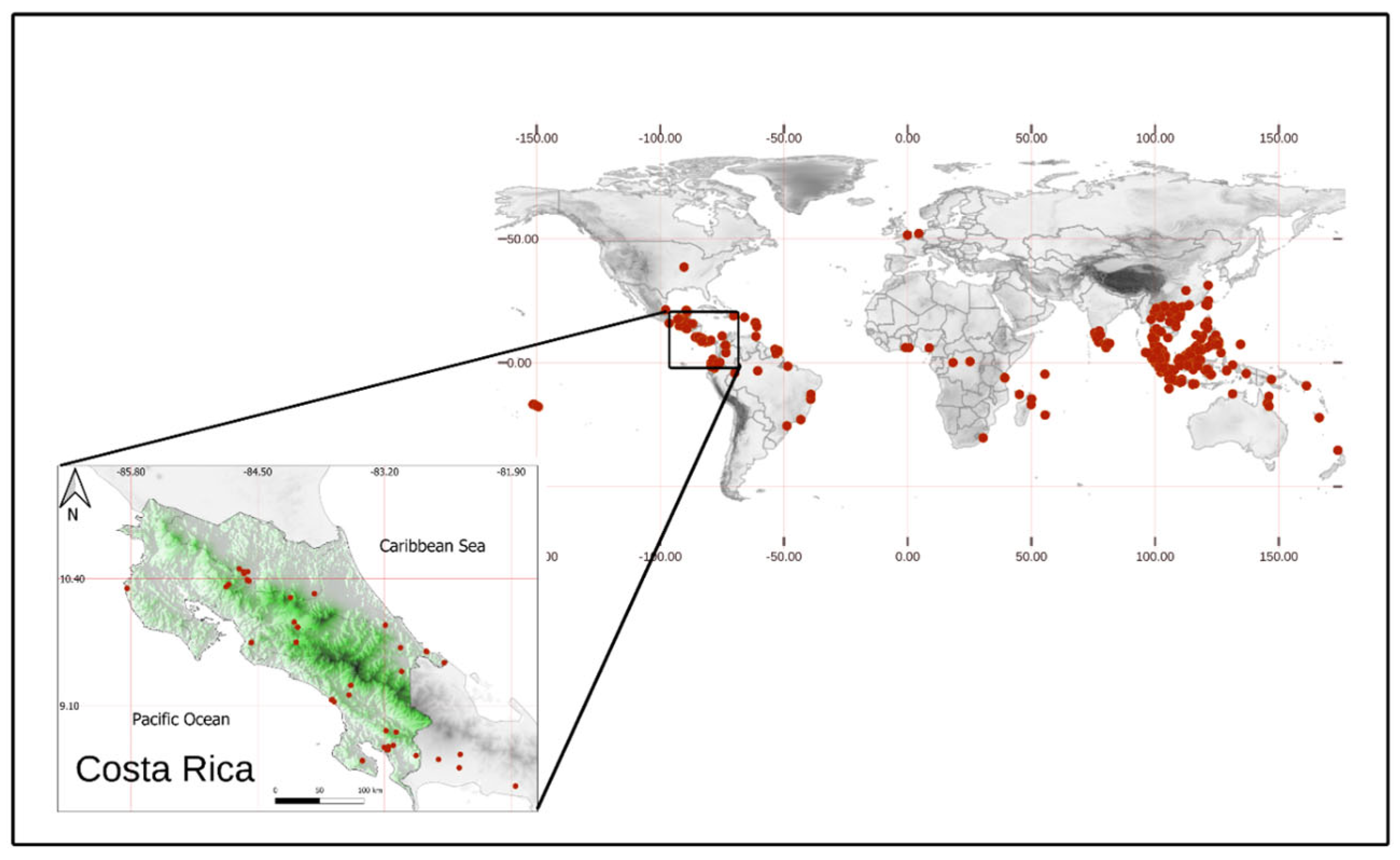
N. lappaceum, also known as rambutan, is rich in antioxidants and widely consumed and valued in tropical regions, including Costa Rica (Figure 1) [15,16]. The main phytochemicals from the peel—with strong reducing capacity owing to their high polyphenolic content—are ellagitannins, ellagic acid, corilagin, and geraniin [17]. However, its use in green synthesis processes has been empirical and scarcely explored. Kumar et al. [18] have previously reported an OFAT approach for the synthesis of AgNPs using N. lappaceum extract, resulting in a hydrodynamic diameter above 100 nm and a polydisperse population. Additionally, other studies have utilized the extract for synthesizing ZnO nanocrystals [19], AgNPs as photocatalysts and fluorescence quenchers [20], and for encapsulating it into polycaprolactone nanofibers for antibacterial applications [21].
Therefore, in this study, we developed a green synthesis method for AgNPs using an aqueous extract from the waste peels of N. lappaceum, assisted by the Plackett–Burman experimental design model for size control and optimization. The optimized NPs exhibited a hydrodynamic diameter of 80 ± 4 nm and a PDI of 0.260 ± 0.029 after 24 h without a stabilizing agent. TEM revealed predominantly triangular and hexagonal morphologies. The in vitro antioxidant capacity was confirmed through the ABTS radical scavenging assay. Additionally, the NPs displayed a minimum inhibitory concentration (MIC) of 23.5 µg mL−1 against Escherichia coli ATCC 25922 and Staphylococcus aureus ATCC 25923. These results encourage searching for new antimicrobial and antioxidant alternatives while promoting the valorization of plant waste.

2. Materials and Methods

2.1. Materials

N. lappaceum was collected in San Carlos, Alajuela, Costa Rica. All other materials employed were of reagent grade and required no further purification. AgNO3 (≥99.7% purity) was provided by J.T. Baker (Phillipsburg, NJ, USA). Potassium persulfate, sodium chloride (NaCl), sodium carbonate, tannic acid (CAS No. 1401-55-4), Folin–Ciocalteu reagent (FCR, CAS No. 12111-13-6), ABTS reagent (2,2′-azino-bis(3-ethylbenzothiazoline-6-sulfonic acid), CAS No. 30931-67-0), and Muller-Hinton (MH) liquid medium were purchased from Sigma Aldrich (St. Louis, MO, USA). Poloxamer 188 (P188, CAS 9003-11-6), also known as Pluronic F-68, was provided by Basf (Ludwigshafen, Germany). E. coli (ATCC 25922) and S. aureus (ATCC 25923) were obtained from the American Type Culture Collection (Manassas, VA, USA). Ultrapure water was obtained by distilled water filtration through two ion-exchange membranes (resistivity = 18.2 MΩ cm, 25 °C) and final filtration through a 0.2 μm Q-Gard®1 Merck KGaA membrane (Darmstadt, Germany).

2.2. Exploratory Study

2.2.1. Preparation of N. lappaceum Aqueous Extract

The fruit peel was washed exhaustively to remove any trace of chemical agents and dirt. The spintern-free peel was finely chopped after drying at room temperature for 3 h. Amounts of 5, 10, or 15 g of the vegetal material were directly weighed in a flask, mixed with 100.0 mL of distilled water (resulting in three different systems of 0.05, 0.10, and 0.15 g mL−1, respectively), and heated at 70 °C under stirring for one hour. The mixtures were initially filtered by gravity, followed by filtration through 0.22 µm membranes. Finally, the obtained extracts were stored at 4 °C under lucifugal conditions.

2.2.2. Total Phenolic Content Quantification

The assay was conducted using a slightly modified Folin–Ciocalteu method reported by Everette et al. [22]. A plastic cuvette of 3 mL was filled with 250 µL of previously diluted extract and AgNPs (1:10 with deionized water). Subsequently, 312.5 µL of the diluted FCR (1:10) and 375 µL of deionized water were added. The reaction was allowed to proceed for 5 min before adding 1562.5 µL of a sodium carbonate solution at 20% w/v. Each mixture was incubated in the dark at room temperature for 40 min to ensure complete color development. Finally, absorbance was measured at 725 nm using a UV-1800 double beam UV-Vis spectrophotometer (Shimadzu, Kyoto, Japan). Total phenolic quantification was done through a calibration curve ( A = 0.0014396 ± 0.0000051 C n µ g   m L 1 0.0124 ± 0.0017 , r2 = 1) obtained by plotting the absorbance of tannic acid working standards (100 to 500 µg mL−1) against their concentration. The results were expressed as mg of tannic acid equivalents (TAE) per mL of sample. Deionized water was used as a negative control. All measurements were done in triplicate.

2.2.3. Proof-of-Concept for the Green Synthesis of AgNPs

The synthesis followed the green process proposed by Arreche et al. [23] with modifications to evaluate the feasibility of obtaining AgNPs using the N. lappaceum peel extract. A 10.00 mL aliquot of AgNO3 1 mM aqueous solution was added to a 50 mL beaker protected from light. Then, 1 mL of the extract at a given concentration was added dropwise manually using a micropipette (Biohit, Helsinki, Finland), lasting approximately 10 min. The injection was done under magnetic stirring at 500 rpm and maintained for 24 h. Rapid and low-resource characterization was performed for this purpose using UV-Vis spectroscopy and DLS, as described in Section 2.4.1 and Section 2.4.2.

2.3. DoE-Assisted Synthesis and Optimization

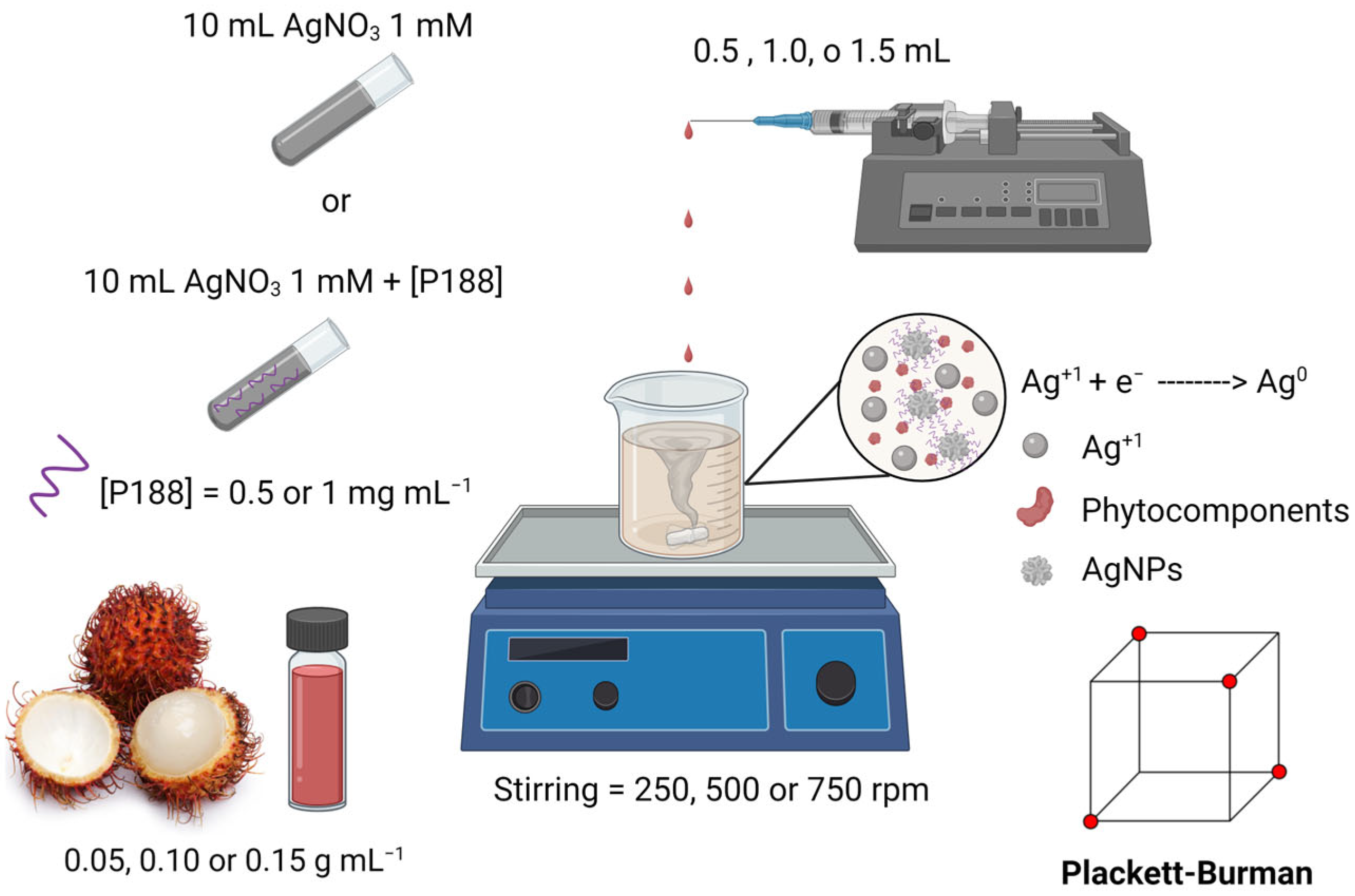
A Plackett–Burman design was executed to determine the CMAs and CPPs affecting the hydrodynamic diameter of the synthesized AgNPs. The required volume of the corresponding concentration was added using a 3 mL syringe (BD plastic, Franklin Lakes, NJ, USA) and a 30 G × 1/2″ needle (NIPRO, Osaka, Japan). In order to provide a more controlled and scalable process, the extract was injected at a flow rate of 400 µL min−1, as described by Castillo et al. [24], using a syringe pump (Darwin microfluidics, Paris, France). A scheme of this semi-automated and systematic green synthesis process is presented in Figure 2. Purification involved repeated centrifugation at 6000 rpm for 15 min and washing, followed by resuspension of the obtained pellet in 10 mL of Milli-Q distilled water, as reported previously in available protocols [25].
Table 1 shows the assessed factors with their denotation from X1 to X4 for statistical computation and their respective levels. A total of 12 experiments were randomly performed at low (−1) and high (+1) values of the input variables (cf. Table S2). The DLS procedure for hydrodynamic diameter measurement was followed as described in Section 2.4.2.
Multivariate regression analysis was done to obtain a linear equation relating Y1 (hydrodynamic diameter) to the factors under study (Equation (1)), where b0 is the intercept, b1b4 are the regression coefficients for each term, and e accounts for the standard error of the model. The best conditions for process optimization, based on the composite desirability (i.e., where one represents the most desirable settings and zero the least), were also used to validate the model. The latter experiment was done in triplicate, and the predicted outcome considered only the significant terms in the calculation. The optimized AgNPs were stored at 4 °C under lucifugal conditions and subjected to a more exhaustive characterization, including an orthogonal approach for size evaluation using DLS and TEM.
Y 1 = b 0 + b 1   X 1 + b 2 X 2 + b 3 X 3 + b 4 X 4 + e  

2.4. Physicochemical Characterization

2.4.1. Synthesis Monitoring by UV-Spectroscopy

To monitor the synthesis process, 2 mL samples were taken at 0.5, 1, and 24 h and analyzed by UV-Vis spectroscopy to identify the presence of the surface plasmon resonance (SPR) band associated with nanosized silver. The optimized NPs were monitored at 0.5, 1, 2, 3, 4, and 24 h.

2.4.2. Dynamic Light Scattering and Zeta Potential

One mL samples were evaluated by DLS at 24 h using a NanoPartica SZ-100V2 nanoparticle size analyzer (Horiba, Kyoto, Japan) based on their diffusion coefficient at 25 °C using the Stokes–Einstein formula. A backscattering (173°) was performed for the measurement. The zeta potential (ZP) was assessed by mixing 10 µL of sodium chloride 1.01 M with 1 mL of the preparation. The suspension’s electrophoretic mobility was measured at 25 °C (NanoPartica SZ-100V2, Horiba) using the Smoluchowski equation. All measurements were done in triplicate.

2.4.3. Transmission Electron Microscopy

AgNPs were drop-casted over carbon-coated copper grids and dried for 24 h in a desiccator with silica. TEM (TEM 2011, Jeol, Tokyo, Japan) measured the size and morphology coupled with energy-dispersive X-ray fluorescence. The analyses were performed in triplicate, using an accelerating voltage of 120 kV. Particle sizes were determined using ImageJ (NIH, USA, v.1.54p). Images were calibrated with the microscope scale bar (200 nm) and analyzed by manually outlining at least 200 particles.

2.4.4. Fourier-Transform Infrared Spectroscopy

FTIR analyses were conducted at room temperature and 30% relative humidity to identify the functional groups from the N. lappaceum extract involved in the synthesis, capping, and stabilization of the AgNPs. The extract and the AgNPs were analyzed using the transmission technique (Nicolet 6700, Thermo Scientific, Waltham, MA, USA) in the 4000 to 500 cm−1 range, with 200 scans per sample.

2.5. Antioxidant Activity

The antioxidant activity of green-synthesized AgNPs was evaluated using the ABTS radical scavenging assay with potassium persulfate as the oxidative agent [26]. The reduction in the ABTS radical (ABTS·+) via neutralization by the electron donor (AH) (ABTS·+ + AH → ABTS + A· + H+) leads to a decrease in absorbance measured by UV-Vis spectroscopy, which is quantified using Equation (2) to determine the antioxidant activity:
I n h i b i t i o n   % = A 0 A f A 0 100
where A0 is the absorbance of the ABTS·+ solution, and Af corresponds to the absorbance after adding 50 µL of the sample, measured at 414 nm. Results obtained for the AgNPs were compared to the inhibition caused by the N. lappaceum aqueous extract at 0.05 g mL−1 and an aqueous solution of AgNO3, 1 mM. All measurements were done in triplicate.

2.6. Antimicrobial Activity

Two bacterial species, E. coli (Gram−) ATCC 25922 and S. aureus (Gram+) ATCC 25923, were cultured in MH liquid medium at 37 °C for 24 h. The cultures were then transferred to a fresh medium and incubated for four hours at 37 °C. After incubation, cultures were centrifuged at 3500 rpm for 7 min, the supernatant was discarded, and the pellet was resuspended in 0.85% NaCl. Optical density (OD) at 600 nm was measured (Genesys 150, Thermo Scientific, Waltham, MA, USA) and adjusted to 0.05 (E. coli) and 0.037 (S. aureus), corresponding to an estimated 1 × 10⁵ CFU mL−1. Antimicrobial activity was assessed in 96-well microplates using serial dilutions (five levels) of N. lappaceum extract and AgNPs. A 1 mM AgNO3 solution was used as a positive control (four levels), and culture medium without bacteria or antimicrobial agents was used as the negative control. Each experiment was performed in triplicate, with 200 µL per well. OD readings were taken over 20–22 h using an EPOCH2 Plate Reader (BioTek Agilent, Santa Clara, CA, USA), with shaking before each reading. Absorbance values were normalized using the preprocess function (“range” method) and analyzed with the predict function of the Caret package (v.7.0-1) [27] in R (v4.4.1) and R studio (2024.12.0) (R Core Team, Austria). Bacterial growth trends were visualized with ggplot2 (v3.5.1.) [28], applying the geom_smooth function using a local regression with the Locally Estimated Scatterplot Smoothing (LOESS) method.

2.7. Statistical Analysis

Results are presented as mean ± Standard Deviation (SD). The DoE was created and analyzed using Minitab 21 (Minitab Inc., State College, PA, USA). Through residual plots, assumptions for the analysis of variance (ANOVA)—including normality, homogeneity of variance, and independence of the observations—were verified. Probability tests were conducted to evaluate the data’s distribution fit (α = 0.05). The goodness of fit was assessed using R2 and adjusted R2 values. Model predictability was assessed using the predicted R2, calculated from the predicted residual error sum of squares statistic using cross-validation.

3. Results and Discussion

3.1. Exploratory Study

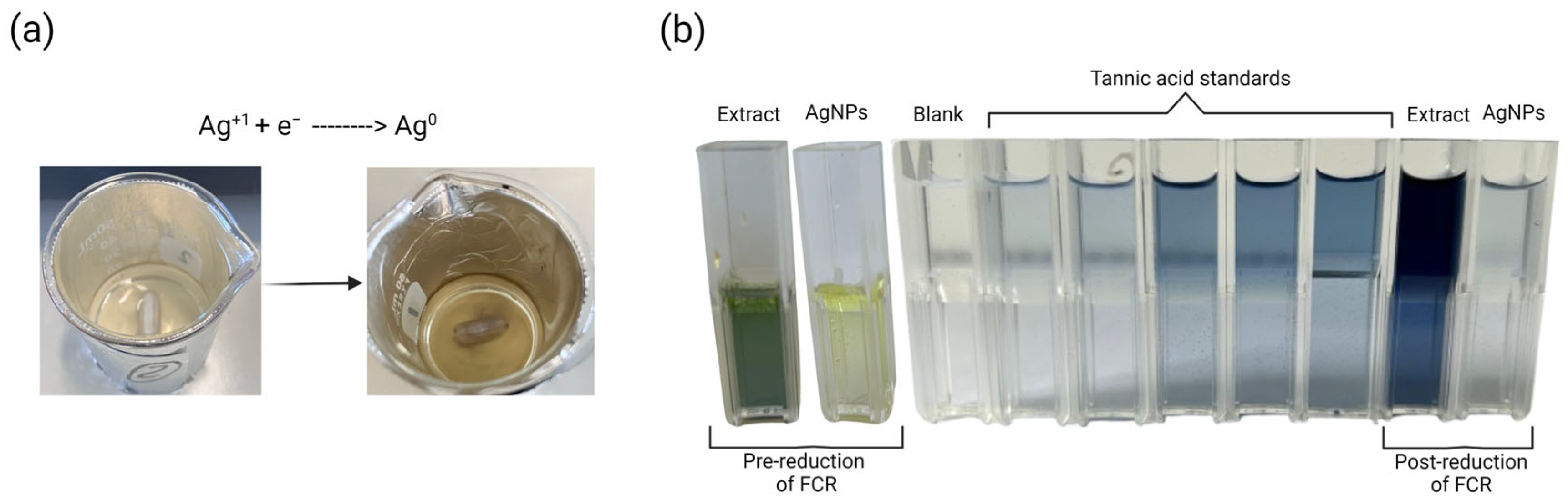
Green chemistry approaches for NP synthesis enable avoiding toxic agents, such as sodium borohydride (NaBH4), promoting more environmentally friendly and biocompatible processes [29]. In the present work, injecting N. lappaceum extract into the AgNO3 solution caused almost an immediate change to a greenish-gray color, which intensified over time (Figure 3a).
The latter is attributed to the continuous formation of AgNPs, which comprises two important stages: nucleation and particle growth. In the former, the bioactive compounds from the extract—mainly polyphenols and flavonoids as determined by the Folin–Ciocalteu assay (13.977 ± 0.051 mg TAE mL−1, Figure 3b)—act as electron donors. The redox activity of these phytochemicals is primarily associated with hydroxylated aromatic structures, which undergo oxidation (e.g., conversion of -OH groups to quinone-like species). Similar reduction pathways have been widely reported for polyphenol- and flavonoid-rich extracts in AgNPs synthesis [30]. This facilitates the reduction of silver ions (Ag+1) to metallic silver atoms (Ag0), which aggregate to form clusters that become stable nuclei upon reaching a critical size [31].
These nanosized structures are the foundation for particle growth by continuously adding Ag0. Remarkably, polyphenols and flavonoids also adsorb onto the surface of the newly formed AgNPs through hydrogen bonding and coordination interactions involving hydroxyl and carbonyl groups (cf. FTIR analysis). The dual functionality—reduction and capping—limits uncontrolled aggregation and contributes to nanoparticle stability [32]. However, other growth mechanisms, such as Ostwald ripening, may also influence this stage, contributing to the growth of more energetically favored larger particles [33].
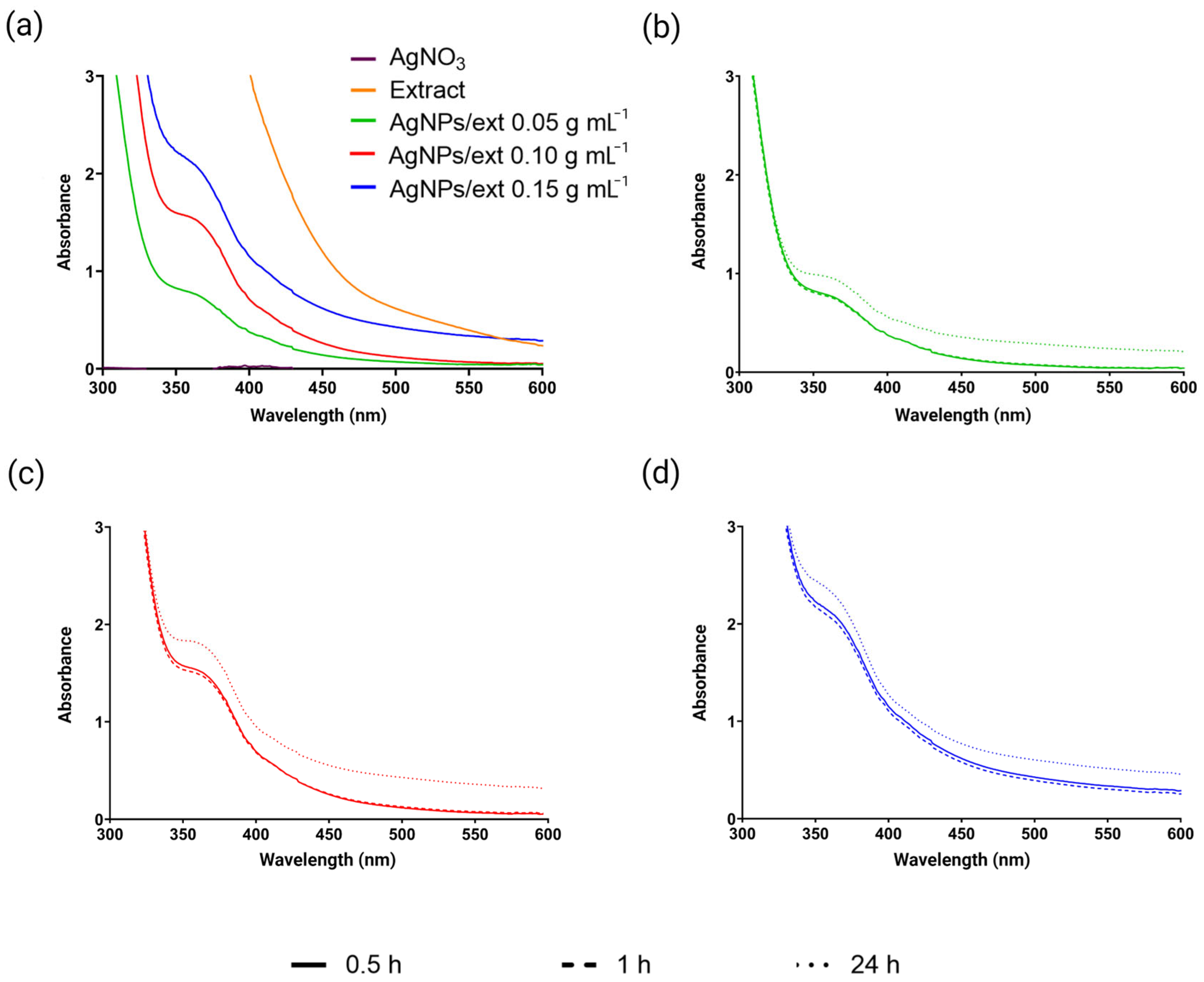
Moreover, UV spectroscopy evaluation at 30 min showed the localized SPR (LSPR) within the 350–400 nm wavelength range for all tested extract concentrations (Figure 4a). The characteristic LSPR peak is reported in the literature to be around 350–500 nm, but this is conditioned by the morphology of the NPs [34]. The same behavior was noticed at different times (Figure 4b–d), where no difference was seen in the synthesis extension during the first hour in each case. This observation confirms the fast reduction in Ag+1 ions, which continues more slowly until 24 h post-injection. The latter is common in the green synthesis-based processes, characterized by an initial fast reduction stage, followed by a longer time for stabilizing and maturing the metallic NPs [35].
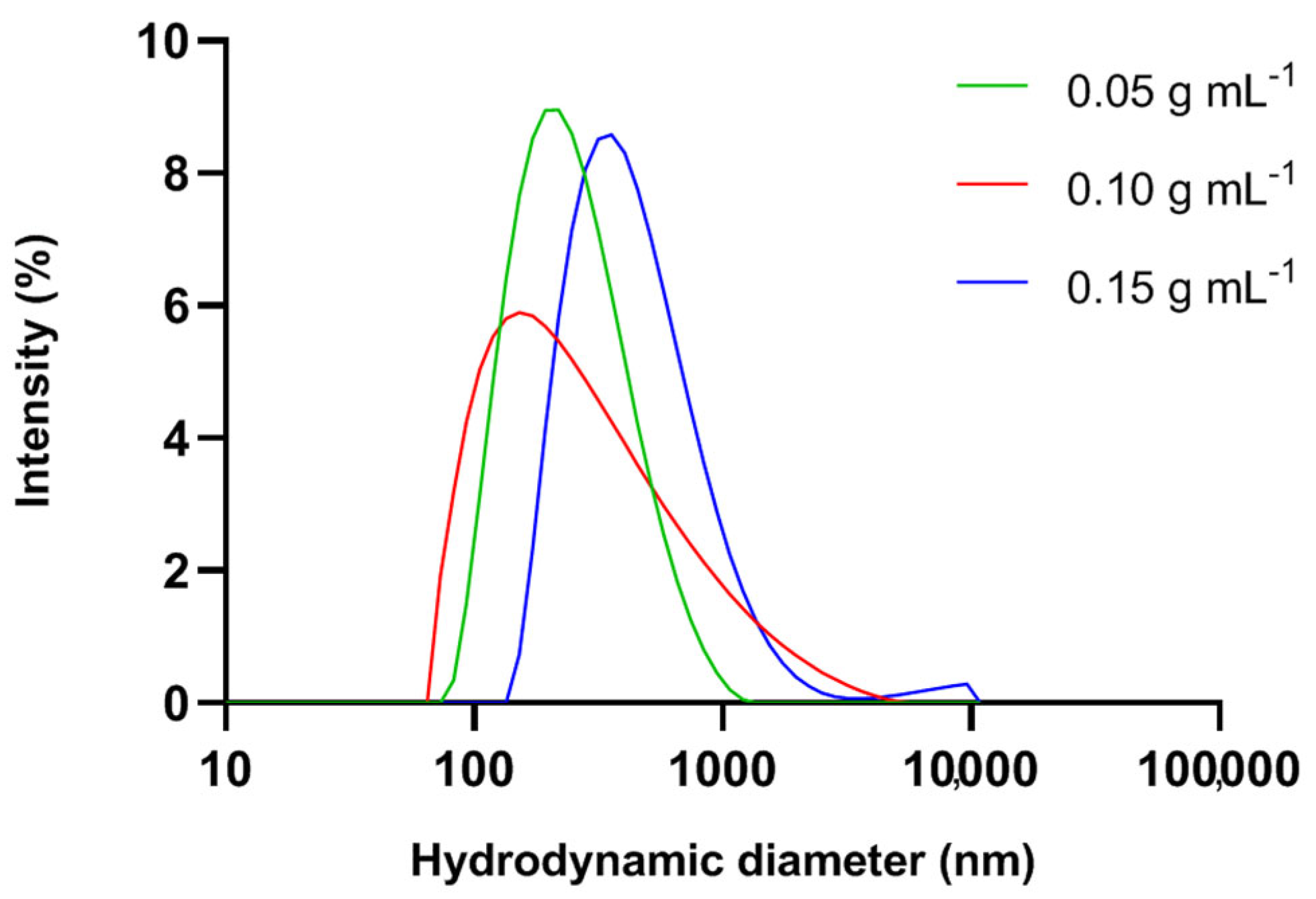
The increased absorbance observed with the higher extract concentration indicates an enhanced nucleation rate, resulting in a greater synthesis yield. However, the hydrodynamic diameters for the AgNPs prepared using 0.05, 0.10, and 0.15 g mL−1 extract were 261 ± 13 nm, 338 ± 40 nm, and 466 ± 133 nm, respectively (Figure 5). Additionally, PDI was higher than 0.5 in the three cases. The abundant presence of bioactive agents in the reaction medium accelerates the initial reduction and enhances the formed NPs’ early stability, which facilitates the Ostwald ripening process [36]. Overall, higher extract concentrations provided an environment that favored particle growth.
Remarkably, the LSPR can also suffer shifts towards shorter wavelengths (i.e., blue shift) or larger wavelengths (i.e., red shift) due to a decrease or increase in the NP size, respectively [37]. Nevertheless, no change was observed during the UV monitoring. This situation can be explained by the intricate interplay between the size, shape, and composition of the AgNPs and the particles’ interaction and dielectric environment, which define the absorption spectra [38].

3.2. DoE-Assisted Synthesis and Optimization

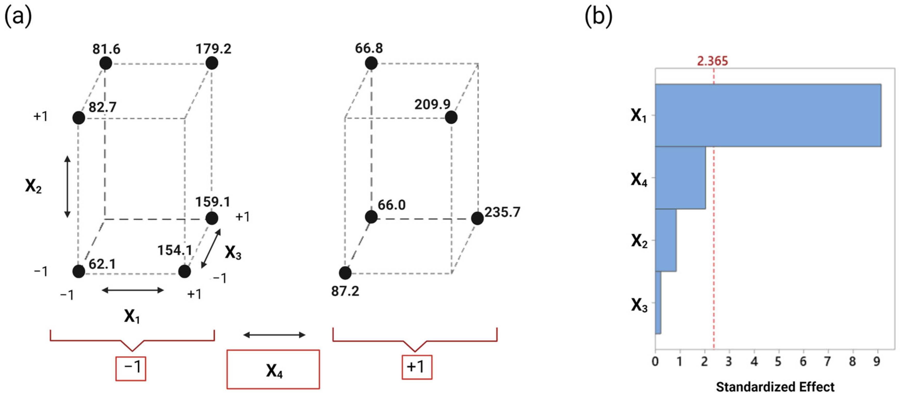
Because of the large size of the NPs and the high polydispersity, a trade-off between the different CQAs demands resorting to DoE. As shown in Figure 6a, the hydrodynamic diameters obtained ranged from 62 to 236 nm. This variation is attributed to the only critical factor identified by the regression analysis (cf. Table S3): the extract concentration (X1), as shown in the Pareto Chart from Figure 6b. All the experiments performed at a low concentration resulted in a hydrodynamic diameter below 90 nm. In contrast, the ones at high concentration showed responses above 150 nm for the reasons explained earlier.
The Plackett–Burman is a cost-effective screening model widely employed in bench-top investigations. Although screening designs are often followed by response surface methodologies for optimization as a default pipeline, this step was not necessary in this approach. Since the analysis revealed only one significant term, a response surface model would not provide additional mechanistic or predictive value [39]. Moreover, given the intrinsic variability of the PDI, DoE strategies usually fail to establish a model to predict and optimize said attribute. Therefore, the performed DoE aimed to identify suitable conditions to obtain the smallest particle size, which has been related to a simultaneous improvement of the PDI [24].
The defined levels for the selected factors considered not only operational suitability and stability issues but also potential applications (cf. Table S1). For instance, AgNPs intended to be administered parenterally require surface decoration with PEG or a PEG copolymer, such as P188, to confer a stealth effect (i.e., prevention of premature uptake by the mononuclear phagocyte system) [40]. Additionally, automation of the injection step was also introduced to provide a more controlled and uniform environment during nucleation, removing errors caused by the operator. This approach has been reported to facilitate future scale-up endeavors of the synthesis process [41].
In this study, the goodness of fit enabled the multilinear regression model to explain 92.7% of the response according to the R2 value. In comparison, the R2 adjusted is slightly lower (88.5%) due to the inclusion of irrelevant predictors. Despite the latter, these statistical metrics differ within a 5–10% accepted threshold. Additionally, the R2 predicted was 78.5%, which is above the minimum required (70%). Interestingly, if the confidence level is lowered from 95% to 90%, P188 concentration (X4) would also become significant, indicating a weak but non-negligible secondary effect. In other words, the factor was not identified by the Plackett–Burman model as a dominant term governing the hydrodynamic diameter, but may still play a role in application-specific formulation, especially in parenteral administration. This particular case highlights a trade-off between statistical significance and practical relevance that should be considered during formulation development.
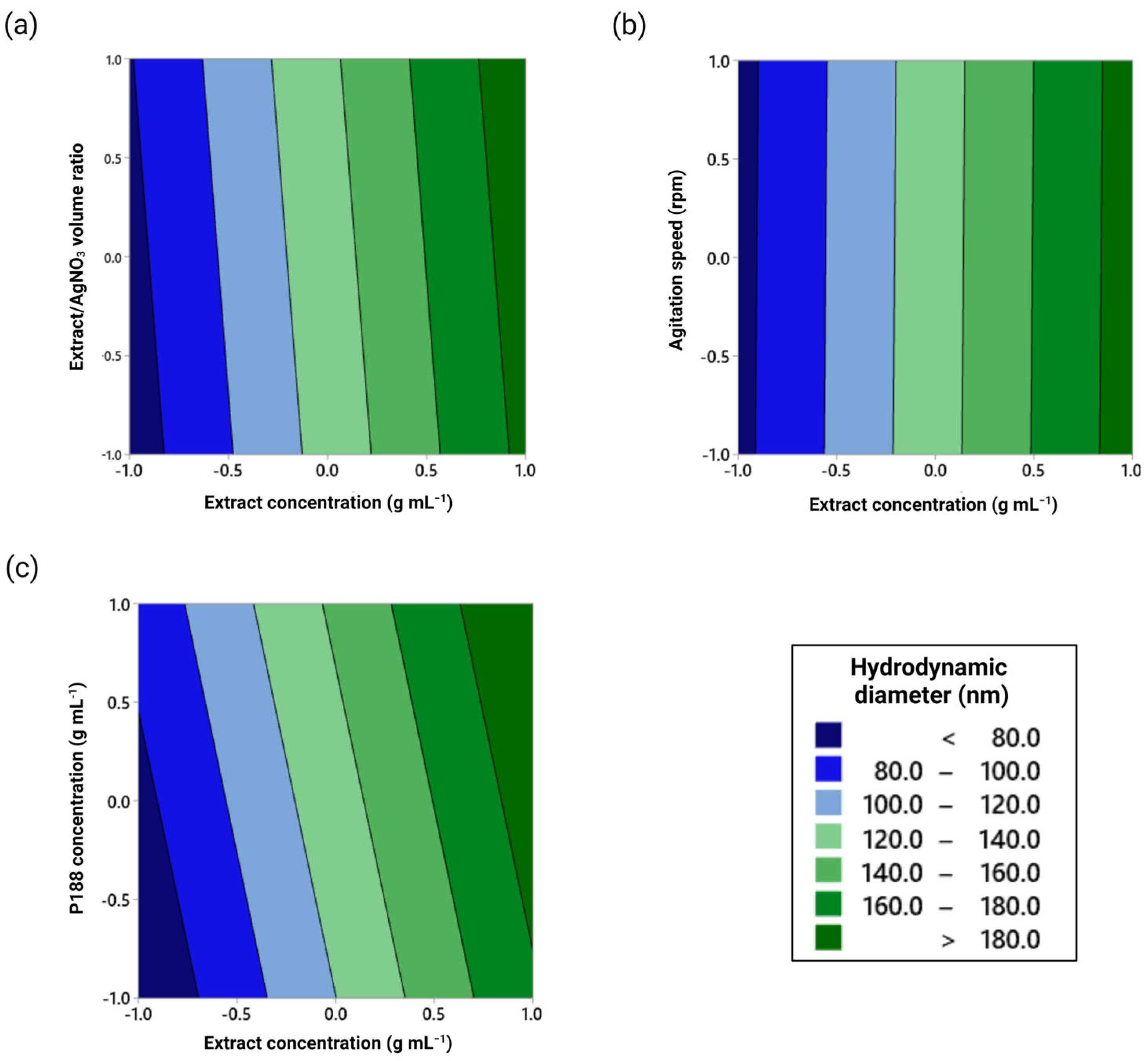
Furthermore, considering the target goal of minimizing the hydrodynamic diameter of the synthesized AgNPs, Figure 7 shows the influence of the extract concentration when evaluated in parallel with the non-significant terms. Regardless of the volume ratio and agitation speed, the hydrodynamic diameter should remain the same for a given extract concentration. On the contrary, the latter cannot be claimed when considering P188 concentration (Figure 7c) since the lowest predicted size (~80 nm) could only be achieved when the polymer is not used during the synthesis.
Usually, stabilizing agents are used for rehydration of excipient-free dried NPs or in-process operations, where mixing is done under laminar flow (e.g., microfluidics) or by cavitation bubbles, as in ultrasound [42]. The presence of the polymer under high shear stress due to the agitation could have led to the aggregation of the AgNPs. However, a former study by Shkodra et al. reported the size increase in PLGA-NPs due to the formation of a thicker coating layer when using P188 in comparison to poloxamer 407 (Pluronic F-127), tween 80, and polyvinyl alcohol [40].
Therefore, to optimize the synthesis process targeting a hydrodynamic diameter of 80 nm, the control strategy comprised using a low extract concentration (0.05 g mL−1) in combination with (i) a high extract/AgNO3 volume ratio (1.5 mL injected into 10.00 mL of AgNO3) in order to increase the extension of the redox reaction and the potential antimicrobial and antioxidant activity, (ii) a high agitation speed (750 rpm) to provide a faster dispersion of the injected extract, and (iii) no P188 in the bulk of the reaction to avoid the aggregation phenomena previously discussed. The predicted response using the model equation under the proposed optimum settings (−1, +1, +1, −1; composite desirability: 0.93) was 81 ± 10 nm.

3.3. Physicochemical Characterization of the Optimized AgNPs

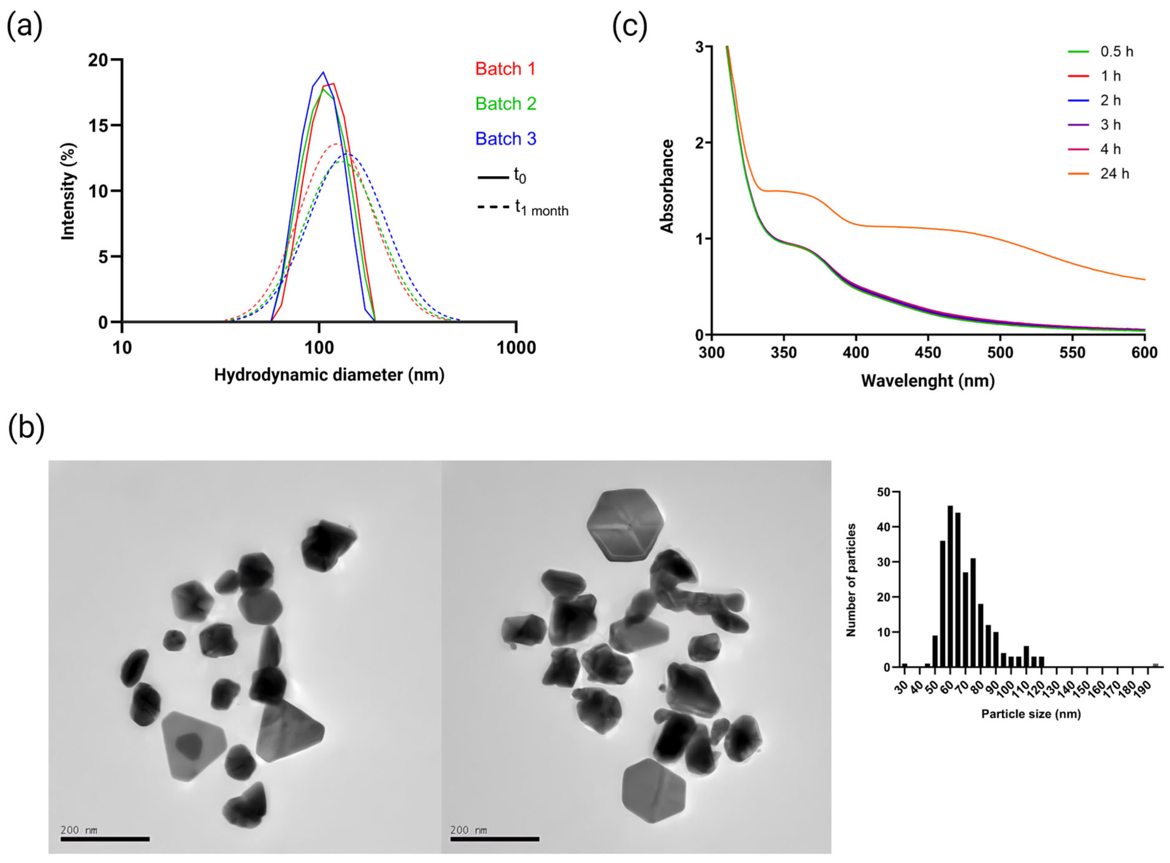
A combination of spectroscopic and size-measuring techniques was employed to confirm the successful DoE-assisted synthesis optimization and evaluate the physicochemical properties of the green-synthesized AgNPs. Figure 8a shows the DLS analysis of three batches from the control strategy. The average hydrodynamic diameter 24 h post-synthesis was 80 ± 4 nm (cf. Table S4), representing a relative prediction error close to 1%. The standardized process was reproducible and led to a monodisperse system (PDI: 0.260 ± 0.016) since PDI values between 0.2 and 0.3 are generally considered as such for NPs [43]. On the other hand, the hydrodynamic diameter and PDI increased to 96 ± 2 nm and 0.360 ± 0.018, respectively, one-month post-synthesis under storage conditions (4 °C). This slight increase suggests the system remained well-dispersed, indicating minimal aggregation, and confirming both a suitable short- to medium-term colloidal stability under storage conditions and the robustness of the standardized synthesis protocol.
Likewise, TEM was performed to orthogonally characterize (i.e., aiming to accurately assess the actual value of an attribute) the particle size (Figure 8b), showing AgNPs of approximately 69 ± 17 nm (n = 267 individual particles) with triangular and hexagonal morphologies (relative standard deviation = 24%). Differences in the reported values for both techniques rely on the physical principle on which they are based. In the case of DLS, the hydrodynamic diameter of NPs is estimated based on their diffusion coefficient and assuming a hypothetical hard sphere shape [44]. On the other hand, TEM allows NP size and morphology evaluation but involves a drying step that can reduce the layer’s thickness, resulting in smaller sizes than those determined by DLS [45].
Moreover, the presence of the identified triangular and hexagonal morphologies justifies the two SPR peaks observed in Figure 8c. The former type has been linked to the LSPR band in the range of 350–400 nm [46]. On the other hand, the second peak was found at 24 h in the range of 450–500 nm, corresponding to the hexagonal NPs [47]. Their late formation suggests that the initially formed triangular NPs underwent truncation over time (i.e., modification of a particle’s shape, where sharp edges transition to a more faceted appearance). The latter reveals that morphology is dynamic and time-dependent [48].
Alteration in the surface facets is often due to changes in the synthesis conditions, which can influence the particle’s stability and growth kinetics [49]. For instance, the process could have been affected by the increased agitation rate (750 rpm) compared to the exploratory study (500 rpm), which was assumed to be free of hexagonal NPs because of the single LSPR peak shown at 24 h. The time-dependent interactions between capping agents and the facets, as well as the bioactive compounds depletion and/or desorption, can also expose passivated facets. These promote transformations to more thermodynamically stable structural configurations and thus influence the proportion of fully developed versus truncated geometries [50].
Kumar et al. [18] have previously reported the synthesis of triangular and hexagonal Ag0 nanoplates using N. lappaceum extract, with SPR peaks near 370 and 470 nm, respectively. Nevertheless, the lack of a systematic approach for the green synthesis resulted in a hydrodynamic diameter of 133 ± 42 nm and a polydisperse population of NPs. These facts highlight the superiority of our DoE-assisted and semi-automated green synthesis in obtaining smaller and monodisperse AgNPs reproducibly.
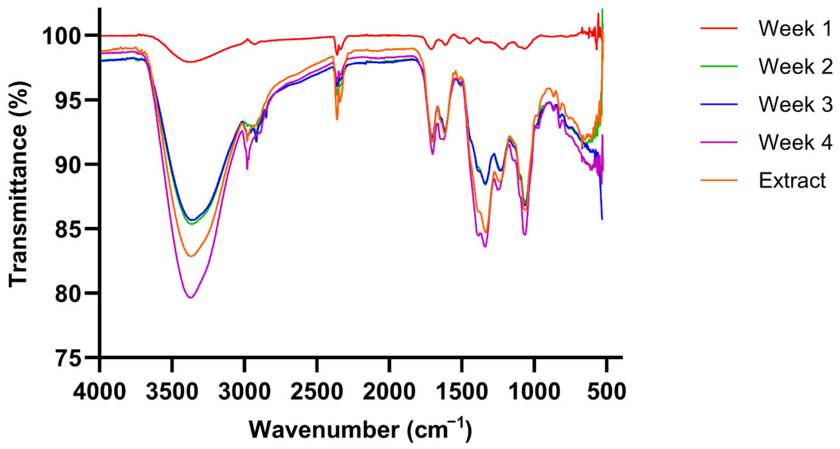
Furthermore, the FTIR analysis (Figure 9) showed broad absorption bands in the 3600–3200 cm−1 region, indicative of O-H stretching vibrations associated with hydroxyl groups. This is consistent with phenolic compounds previously identified in N. lappaceum extracts, such as ellagic acid and geraniin [17]. The peaks at ~2920 cm−1 and ~2850 cm−1 are attributed to C-H stretching vibrations of aliphatic hydrocarbons found in the fatty acids and lipids from the peel [51]. Fatty acids are also associated with C=O stretching at ~1740 cm−1 [17]. Additionally, in the fingerprint region (1500–400 cm−1), the IR spectrum also shows peaks around 1300–1200 cm−1, characteristic of C-O stretching vibrations from phenolic compounds [52].
These bands were also identified in the AgNP samples, indicating the involvement of polyphenolic compounds in NP capping to prevent aggregation and ensure colloidal stability [53]. The rationale behind the capping mechanism involves coordination chemistry and interfacial electron transfer due to the following: (i) main polyphenols, such as ellagic acid and geraniin, possess multiple catechol and galloyl moieties that can chelate Ag0 surface given the involvement of adjacent hydroxyl groups via bidentate coordination [30]; (ii) these molecules acquire carbonyl functionalities upon their oxidation to quinone-like structures, enhancing their binding affinity toward Ag0 via π-metal interactions [54]; and ultimately, (iii) the latter leads to the formation of an organic–inorganic interface with the presence of a phytochemical corona that stabilizes surface facets [55].
Notably, early stability can be due to the conferred moderate electrostatic repulsion given the ZP (−14.5 ± 0.7 mV). However, said value is not high enough to ensure long-term electrostatic stability [44]. This condition is consistent with the slight increase in hydrodynamic diameter observed during storage. Therefore, the slow aggregation over time is likely to be controlled by the joint effect of electrostatic and steric effects provided by surface-bound phytochemicals.
Remarkably, while minor variations in band transmittance are observed over time, FTIR spectra recorded in transmittance mode are not quantitative unless performed under strict control of sample amount and data normalization. Therefore, these variations are attributed to differences in detected material or possible reorganization of the capping layer on the NPs surface rather than polyphenol degradation [56].

3.4. Antioxidant Activity

The presence of polyphenols in both the extract and the optimized green-synthesized AgNPs and their role as reducing agents suggested a potential application of these NPs as an antioxidant alternative. AgNO3 is known to have almost no antioxidant activity, as confirmed experimentally (˂1%). On the contrary, the N. lappaceum extract at 0.05 g mL−1 exhibited a radical scavenging activity of 77.3 ± 2.6%. Notably, the optimized NP-based preparation—containing approximately 0.0065 g mL−1 of the extract—demonstrated a higher activity upon stabilization (~24 h) and reached values above 90% four weeks post-synthesis, as seen in Table 2. This is significantly higher than the reported value for the green-synthesized AgNPs using Azadirachta indica (40%) and Catharantus roseus (32%) at similar concentrations [57]. The high value for the AgNPs suggests a synergistic antioxidant activity arising from the nanosized Ag0 and the phenolic residues remaining at the organic–inorganic interface mentioned before. These can participate in the electron transfer cycling, and therefore, promote controlled release of Ag+ in oxidative environments [58].
The ABTS assay was chosen for the antioxidant activity because it is widely used to evaluate aqueous systems in vitro [59]. The assay is based on the single electron transfer mechanism, which involves the donation of one electron from the antioxidant molecule (AH) to a free radical (ABTS·+). This neutralization pathway reduces the free radical’s reactivity and prevents oxidative damage. Consequently, the antioxidant becomes a less reactive radical (A⋅) than the former, eventually transforming into a fully oxidized non-radical form of the reducing compound. In the case of polyphenols, the stability shown by the formed radical species can be due to resonance structures that distribute the unpaired electron [60].

3.5. Antimicrobial Activity

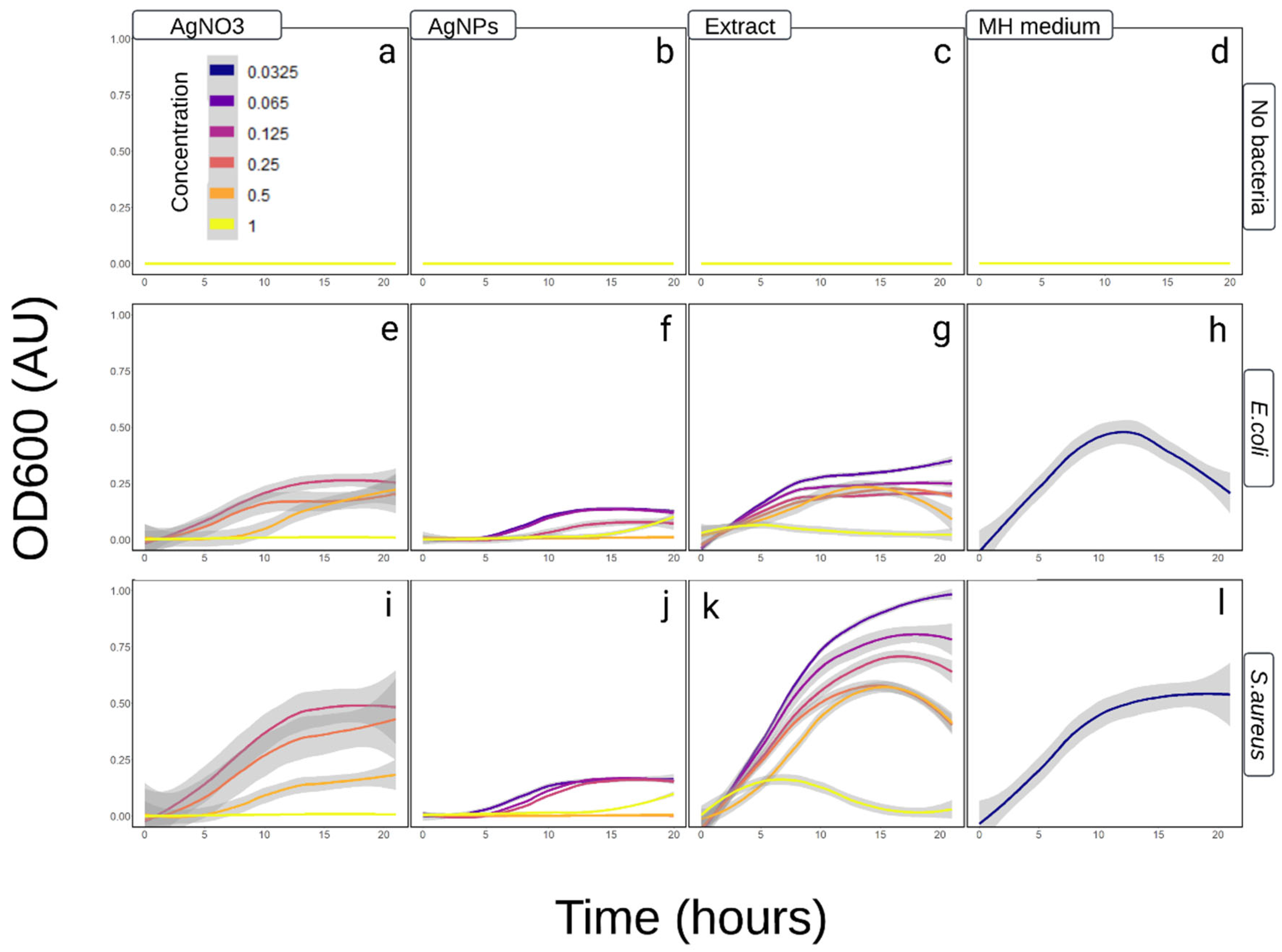
The antimicrobial activity of N. lappaceum extract and AgNPs was evaluated against E. coli and S. aureus (Figure 10). Compared to the negative control, the extract exhibited a more significant inhibitory effect on E. coli, with reduced growth throughout the incubation period at all tested concentrations. However, similar to S. aureus, only the 1× concentration inhibited bacterial growth in terms of OD600, starting at approximately 7 h, with the value approaching zero at 15 h of incubation. In the case of S. aureus, growth was approximately twice that of E. coli for the other extract concentrations, surpassing the negative control (with optimal culture medium for bacterial growth).
Regarding the antimicrobial activity of AgNPs, a similar behavior was observed in both species, with a typical logistic growth [61,62] reduced in S. aureus (Figure 10j). E. coli showed more significant inhibition at the concentration of 0.125× than at the two lowest concentrations tested (Figure 10f), whereas in S. aureus, the three lowest concentrations showed no difference. The former bacteria revealed the most significant susceptibility, with a total inhibition observed at concentrations of 0.5× and 0.25× (MIC = 23.5 µg mL−1) throughout the incubation period. Similarly, for S. aureus, concentrations of 0.5× and 0.25× wholly inhibited growth. Moreover, at the highest concentration tested, both bacterial species showed an OD600 value approaching zero within the first five hours of incubation, followed by an exponential increase in CFU around 15 h. This biocidal effect is dose-dependent, as observed, and has been related to the electrostatic interaction between Ag0 from the AgNPs and the sulfhydryl and phosphorous groups on the bacterial cell wall. The latter causes membrane disruption, leading to a loss of permeability, osmoregulation, electron transport, and ultimately, cellular death [29].
Notably, the identified MIC is comparable to the reported value for green-synthesized AgNPs of significantly smaller diameter (25 nm) using Eucalyptus globulus extract, which ranges from 27 to 36 µg mL−1 [63]. The latter highlights that biological performance (e.g., antimicrobial and antioxidant activity) is governed by multiple interdependent parameters rather than size alone (e.g., morphology, surface chemistry, accessible reactive sites). On the one hand, the hydrodynamic diameter dictates nanoparticle diffusion. On the other hand, non-spherical morphologies, such as triangular and hexagonal nanoplates, are known to enhance biological activity [64]. The latter is due to the exposure of high-energy (111) crystallographic facets, which exhibit higher atomic density and surface energy compared to the dominant ones in spherical nanoparticles [65]. Subsequently, a larger membrane disruption is promoted owing to a more sustained and localized Ag+ release at the nano-bio interface [66].
Moreover, triangular morphologies also present an edge-mediated mechanism of antimicrobial action, resulting from locally intensified electric fields and mechanical stress upon contact with bacterial membranes. The latter has been shown to promote direct physical disruption of the membranes and enhanced interactions with negatively charged phospholipid groups [67]. This morphology-dependent behavior, together with the contribution of the phytochemicals involved in surface capping, may explain the compensation in antimicrobial activity for larger particle sizes, and therefore, lead to a comparable biological activity to that reported for smaller AgNPs in the literature [68].
The positive control used in this study follows the same pattern reported by other authors [69], where 1 mM of AgNO3 inhibits the growth of E. coli and S. aureus (Figure 10e,i). On the other hand, pulverized peel extract of N. lappaceum was reported to inhibit the growth of both species after 10 h of incubation [70]. The presence of compounds such as eplerenone, Oritin-4-beta-ol, and Catechin in the peel extract can exert competitive inhibition in the DNA binding domains in some microorganisms [71]. The latter directly affects fission events and gene expression mechanisms. Nevertheless, under certain conditions, plant extracts can contribute to microbial growth, with antimicrobial efficiency depending on the extraction method, concentration, and the plant species used [72].
The enhanced antimicrobial activity shown by the AgNPs compared to AgNO3, the extract, and other green-synthesized AgNPs of smaller size using different extracts is another proof of the synergic interaction between N. lappaceum’s phytochemicals and nanosized Ag0. In this case, bacterial growth kinetics are subjected to the stochasticity provided by the dispersivity of the synthesis.
Considering the inverse relationship of the inhibitory activity as a function of the size of the AgNPs [73], the hydrodynamic diameter—focused on the main population determined by DLS—allowed the observation of CFU growth trends according to the assessed concentrations. Nevertheless, the absence of stabilizing agents has been linked to NP aggregation in culture media [74]. Under in vitro conditions, interaction with biomolecules such as glucose or glutamine can alter the hydrodynamic diameter of AgNPs, forming a “soft-corona” with a greater diameter and ultimately contributing to their precipitation [75]. Monte Carlo analysis performed in this study using hydrodynamic diameter data of AgNPs after incubation (cf. Figure S1) confirms a statistically significant shift toward larger values, providing experimental support for aggregation under biologically relevant conditions.
Notably, AgNP antimicrobial evaluation can also be influenced by the formation of an extracellular polymeric substance due to bacteria’s preference to live in colonies rather than individually in a planktonic state. The latter acts as a protective matrix that promotes AgNPs aggregation in the biofilm, which has been described before for E. coli and S. aureus [71]. In this scenario, the effective concentration of AgNPs falls, the direct interaction with cell membranes is reduced, especially in highly concentrated treatments, and thus, the antimicrobial activity decreases. This phenomenon, observed convergently in E. coli and S. aureus, poses the need to identify the optimal particle size and concentration of novel NP-based antimicrobial agents.

4. Conclusions

This work has presented a systematic approach for the semi-automated green synthesis of AgNPs, enabling size control, where N. lappaceum peel-derived phytochemicals play a dual role as reducing and stabilizing agents. The DoE strategy successfully identified optimal settings to control the nucleation and subsequent growth of the NPs, leading to small sizes and monodisperse populations with suitable stability over time under storage conditions. Notably, the Plackett–Burman model has been confirmed as a simple method to help identify CMAs and CPPs without considerable time and resource investment. Furthermore, the synergistic interaction between the phytochemicals from the peel extract and the synthesized AgNPs with a well-defined size, highlights the potential of this systematic approach as a sustainable and efficient strategy for the green synthesis of novel antibacterial and antioxidant agents with better performance than other green-synthesized NPs reported. Comprehensive biological safety assessments, including cytotoxicity and hemocompatibility studies, should be performed in future works aiming to use green-synthesized AgNPs, depending on the application-specific requirements.

Supplementary Materials

The following supporting information can be downloaded at: https://www.mdpi.com/article/10.3390/physchem6020020/s1. Figure S1. DLS analysis of rambutan AgNPs samples in MH medium; Table S1: Special considerations for the factors under study; Table S2. Experimental uncoded plan for the Plackett-Burman design and collected data; Table S3. Factorial regression for hydrodynamic diameter at t24h; Table S4. Optimized green-synthesized AgNPs data and validation of the predictive model at (−1, +1, +1, −1).

Author Contributions

Conceptualization: L.C.-H. and G.M.d.O.-V.; methodology: L.C.-H., G.M.d.O.-V., P.A.-H. and J.M.Z.-U.; investigation: L.C.-H., P.A.-H., J.M.Z.-U., G.M.d.O.-V. and F.A.-V.; data curation: L.C.-H., J.M.Z.-U. and P.A.-H.; Formal analysis: L.C.-H., J.M.Z.-U., P.A.-H. and G.M.d.O.-V.; visualization: L.C.-H., J.M.Z.-U. and P.A.-H.; validation: P.A.-H., J.M.Z.-U. and B.B.; writing—original draft: L.C.-H., J.M.Z.-U., P.A.-H. and Z.P.-V.; writing—review & editing: L.C.-H., B.B., G.M.d.O.-V., Y.C. and J.R.V.-B.; supervision: L.C.-H., G.M.d.O.-V.; resources: J.R.V.-B. and Z.P.-V.; project administration: L.C.-H. and J.R.V.-B. All authors have read and agreed to the published version of the manuscript.

Funding

This research received no external funding.

Data Availability Statement

The data that support the findings of this study are available from the corresponding author upon reasonable request.

Acknowledgments

This work represents an initiative from the Faculty of Pharmacy of the University of Costa Rica. The authors would like to express their gratitude to the Dean of Pharmacy for supporting the implementation of practical work in the Laboratory of Pharmaceutical Physical Chemistry I and to the laboratory instructors and undergraduate students who participated in the preliminary study (data not used in this work). Also, the authors appreciate LANOTEC’s support in developing it into a formal research project at its facilities. L.C.-H. is also grateful to the International Affairs and Foreign Cooperation Office (OAICE) of the University of Costa Rica for the scholarship granted (contract No. OAICE-13-2022). The authors also express their gratitude to Reynaldo Pereira for the assistance with the TEM experiments. During the preparation of this manuscript, the authors used Biorender for the purpose of graphical design of Figure 2. The authors have reviewed and edited the output and take full responsibility for the content of this publication.

Conflicts of Interest

The authors declare no conflicts of interest.

References

  1. Vijayaram, S.; Razafindralambo, H.; Sun, Y.-Z.; Vasantharaj, S.; Ghafarifarsani, H.; Hoseinifar, S.H.; Raeeszadeh, M. Applications of Green Synthesized Metal Nanoparticles—A Review. Biol. Trace Elem. Res. 2024, 202, 360–386. [Google Scholar] [CrossRef] [PubMed]
  2. Giang, T.T.; Duc, C.K.T.; Ha, P.T.M.; Hone, N.; Tram, D.T.N.; Nguyen, N.Y.; Luong, H.V.T.; Tran, Q.T.B.; Nguyen, M.Q.; Pham, D.T. Green-Engineered PEI/PVA-Functionalized Silk Fibroin Nanoparticles for Heat-Stable Delivery of Clitoria ternatea L. Anthocyanins: Formulation, Characterization, and Sustainability Assessment. ACS Omega 2025, 10, 38703–38718. [Google Scholar] [CrossRef]
  3. Elattar, R.H.; Magdy, G.; Abdel Salam, R.A.; Hadad, G.M.; El-Deen, A.K. Cutting-Edge Progress in Carbon Nanomaterial-Functionalized Silver Nanoparticles for Enhanced Optical and Electrochemical Sensing Applications. TrAC Trends Anal. Chem. 2024, 181, 117995. [Google Scholar] [CrossRef]
  4. Ghasemi, S.; Dabirian, S.; Kariminejad, F.; Koohi, D.E.; Nemattalab, M.; Majidimoghadam, S.; Zamani, E.; Yousefbeyk, F. Process Optimization for Green Synthesis of Silver Nanoparticles Using Rubus Discolor Leaves Extract and Its Biological Activities against Multi-Drug Resistant Bacteria and Cancer Cells. Sci. Rep. 2024, 14, 4130. [Google Scholar] [CrossRef] [PubMed]
  5. Marslin, G.; Siram, K.; Maqbool, Q.; Selvakesavan, R.K.; Kruszka, D.; Kachlicki, P.; Franklin, G. Secondary Metabolites in the Green Synthesis of Metallic Nanoparticles. Materials 2018, 11, 940. [Google Scholar] [CrossRef]
  6. Shabulal, N.B.; Thiyagarajan, P.; Albeshr, M.F.; Nagaraj, K.; Velmurugan, G.; Maity, P.; Mathivanan, I.; Abhijith, S.M.; Lad, N.H. Green Synthesis of Silver Nanoparticles: Understanding Biological Activities—Anticancer, Antimicrobial, and Plasmid DNA Cleavage Properties through Reactive Oxygen Species Release. New J. Chem. 2024, 48, 13764–13775. [Google Scholar] [CrossRef]
  7. Tunç, T. Synthesis and Characterization of Silver Nanoparticles Loaded with Carboplatin as a Potential Antimicrobial and Cancer Therapy. Cancer Nanotechnol. 2024, 15, 2. [Google Scholar] [CrossRef]
  8. Barabadi, H.; Jounaki, K.; Karami, K.; Mobaraki, K.; Noqani, H.; Ashouri, F.; Jahani, R.; Hussain, C.M. Laboratory Evidence Supports Thrombolytic Performance and Blood Compatibility of Green-Synthesized Silver and Gold Nanomaterials. Results Surf. Interfaces 2024, 17, 100309. [Google Scholar] [CrossRef]
  9. Burdușel, A.-C.; Gherasim, O.; Grumezescu, A.M.; Mogoantă, L.; Ficai, A.; Andronescu, E. Biomedical Applications of Silver Nanoparticles: An Up-to-Date Overview. Nanomaterials 2018, 8, 681. [Google Scholar] [CrossRef]
  10. El Hamd, M.A.; El-Maghrabey, M.; Almawash, S.; El-Shaheny, R.; Magdy, G. Self-Ratiometric Fluorescence Approach Based on Plant Extract-Assisted Synthesized Silver Nanoparticles for the Determination of Vanillin. Microchim. Acta 2023, 191, 16. [Google Scholar] [CrossRef] [PubMed]
  11. Fukuda, I.M.; Pinto, C.F.F.; Moreira, C.d.S.; Saviano, A.M.; Lourenço, F.R. Design of Experiments (DoE) Applied to Pharmaceutical and Analytical Quality by Design (QbD). Braz. J. Pharm. Sci. 2018, 54, e01006. [Google Scholar] [CrossRef]
  12. Rampado, R.; Peer, D. Design of Experiments in the Optimization of Nanoparticle-Based Drug Delivery Systems. J. Control. Release 2023, 358, 398–419. [Google Scholar] [CrossRef] [PubMed]
  13. Fazil, M.M.; Gul, A.; Jawed, H. Optimization of Silver Nanoparticles Synthesis via Plackett–Burman Experimental Design: In Vitro Assessment of Their Efficacy Against Oxidative Stress-Induced Disorders. RSC Adv. 2024, 14, 20809–20823. [Google Scholar] [CrossRef] [PubMed]
  14. Rai, M.; Ingle, A.P.; Trzcińska-Wencel, J.; Wypij, M.; Bonde, S.; Yadav, A.; Kratošová, G.; Golińska, P. Biogenic Silver Nanoparticles: What We Know and What Do We Need to Know? Nanomaterials 2021, 11, 2901. [Google Scholar] [CrossRef]
  15. Albuquerque, B.R.; Pinela, J.; Dias, M.I.; Pereira, C.; Petrović, J.; Soković, M.; Calhelha, R.C.; Oliveira, M.B.P.P.; Ferreira, I.C.F.R.; Barros, L. Valorization of Rambutan (Nephelium lappaceum L.) Peel: Chemical Composition, Biological Activity, and Optimized Recovery of Anthocyanins. Food Res. Int. 2023, 165, 112574. [Google Scholar] [CrossRef]
  16. Zizka, A.; Silvestro, D.; Andermann, T.; Azevedo, J.; Ritter, C.D.; Edler, D.; Farooq, H.; Herdean, A.; Ariza, M.; Scharn, R.; et al. CoordinateCleaner: Standardized cleaning of occurrence records from biological collection databases. Methods Ecol. Evol. 2019, 10, 744–751. [Google Scholar] [CrossRef]
  17. Monrroy, M.; Araúz, O.; García, J.R. Active Compound Identification in Extracts of N. Lappaceum Peel and Evaluation of Antioxidant Capacity. J. Chem. 2020, 2020, 4301891. [Google Scholar] [CrossRef]
  18. Kumar, B.; Smita, K.; Cumbal, L.; Angulo, Y. Fabrication of Silver Nanoplates Using Nephelium lappaceum (Rambutan) Peel: A Sustainable Approach. J. Mol. Liq. 2015, 211, 476–480. [Google Scholar] [CrossRef]
  19. Yuvakkumar, R.; Suresh, J.; Nathanael, A.J.; Sundrarajan, M.; Hong, S.I. Novel Green Synthetic Strategy to Prepare ZnO Nanocrystals Using Rambutan (Nephelium lappaceum L.) Peel Extract and Its Antibacterial Applications. Mater. Sci. Eng. C 2014, 41, 17–27. [Google Scholar] [CrossRef]
  20. Perera, K.M.K.G.; Kuruppu, K.A.S.S.; Chamara, A.M.R.; Thiripuranathar, G. Characterization of Spherical Ag Nanoparticles Synthesized from the Agricultural Wastes of Garcinia mangostana and Nephelium lappaceum and Their Applications as a Photo Catalyzer and Fluorescence Quencher. SN Appl. Sci. 2020, 2, 1974. [Google Scholar] [CrossRef]
  21. Urena-Saborio, H.; Rodríguez, G.; Madrigal-Carballo, S.; Gunasekaran, S. Characterization and Applications of Silver Nanoparticles-Decorated Electrospun Nanofibers Loaded with Polyphenolic Extract from Rambutan (Nepelium lappaceum). Materialia 2020, 11, 100687. [Google Scholar] [CrossRef]
  22. Everette, J.D.; Bryant, Q.M.; Green, A.M.; Abbey, Y.A.; Wangila, G.W.; Walker, R.B. A Thorough Study of Reactivity of Various Compound Classes Towards the Folin-Ciocalteu Reagent. J. Agric. Food Chem. 2010, 58, 8139–8144. [Google Scholar] [CrossRef]
  23. Arreche, R.A.; Montes de Oca-Vásquez, G.; Vega-Baudrit, J.R.; Vázquez, P.G. Synthesis of Silver Nanoparticles Using Extracts from Yerba Mate (Ilex paraguariensis) Wastes. Waste Biomass Valor. 2020, 11, 245–253. [Google Scholar] [CrossRef]
  24. Castillo Henríquez, L.; Bahloul, B.; Alhareth, K.; Oyoun, F.; Frejková, M.; Kostka, L.; Etrych, T.; Kalshoven, L.; Guillaume, A.; Mignet, N.; et al. Step-By-Step Standardization of the Bottom-Up Semi-Automated Nanocrystallization of Pharmaceuticals: A Quality By Design and Design of Experiments Joint Approach. Small 2024, 20, e2306054. [Google Scholar] [CrossRef]
  25. Ahmed, S.; Saifullah; Ahmad, M.; Swami, B.L.; Ikram, S. Green Synthesis of Silver Nanoparticles Using Azadirachta indica Aqueous Leaf Extract. J. Radiat. Res. Appl. Sci. 2016, 9, 1–7. [Google Scholar] [CrossRef]
  26. Fogliano, V.; Verde, V.; Randazzo, G.; Ritieni, A. Method for Measuring Antioxidant Activity and Its Application to Monitoring the Antioxidant Capacity of Wines. J. Agric. Food Chem. 1999, 47, 1035–1040. [Google Scholar] [CrossRef]
  27. Kuhn, M. Building Predictive Models in R Using the Caret Package. J. Stat. Softw. 2008, 28, 1–26. [Google Scholar] [CrossRef]
  28. Wickham, H. Getting Started with Ggplot2. In ggplot2: Elegant Graphics for Data Analysis; Wickham, H., Ed.; Springer International Publishing: Cham, Switzerland, 2016; pp. 11–31. ISBN 978-3-319-24277-4. [Google Scholar]
  29. Castillo-Henríquez, L.; Alfaro-Aguilar, K.; Ugalde-Álvarez, J.; Vega-Fernández, L.; Montes de Oca-Vásquez, G.; Vega-Baudrit, J.R. Green Synthesis of Gold and Silver Nanoparticles from Plant Extracts and Their Possible Applications as Antimicrobial Agents in the Agricultural Area. Nanomaterials 2020, 10, 1763. [Google Scholar] [CrossRef] [PubMed]
  30. Mittal, A.K.; Chisti, Y.; Banerjee, U.C. Synthesis of Metallic Nanoparticles Using Plant Extracts. Biotechnol. Adv. 2013, 31, 346–356. [Google Scholar] [CrossRef] [PubMed]
  31. Chaikali, C.; Stola, N.D.; Lampropoulou, P.; Papoulis, D.; Lamari, F.N.; Orkoula, M.; Lykouras, M.; Avgoustakis, K.; Hatziantoniou, S. Green Synthesis and Comparative Analysis of Silver, Copper Oxide, and Bimetallic Ag/CuO Nanoparticles Using Cistus creticus L. Extract: Physicochemical Properties, Stability, and Antioxidant Potential. Int. J. Mol. Sci. 2025, 26, 2518. [Google Scholar] [CrossRef] [PubMed]
  32. Gwada, C.A.; Ndivhuwo, P.S.; Matshetshe, K.; Aradi, E.; Mdluli, P.; Moloto, N.; Otieno, F.; Airo, M. Phytochemical-Assisted Synthesis, Optimization, and Characterization of Silver Nanoparticles for Antimicrobial Activity. RSC Adv. 2025, 15, 14170–14181. [Google Scholar] [CrossRef]
  33. Thanh, N.T.K.; Maclean, N.; Mahiddine, S. Mechanisms of Nucleation and Growth of Nanoparticles in Solution. Chem. Rev. 2014, 114, 7610–7630. [Google Scholar] [CrossRef]
  34. Abbas, R.; Luo, J.; Qi, X.; Naz, A.; Khan, I.A.; Liu, H.; Yu, S.; Wei, J. Silver Nanoparticles: Synthesis, Structure, Properties and Applications. Nanomaterials 2024, 14, 1425. [Google Scholar] [CrossRef]
  35. Gharari, Z.; Hanachi, P.; Sadeghinia, H.; Walker, T.R. Eco-Friendly Green Synthesis and Characterization of Silver Nanoparticles by Scutellaria Multicaulis Leaf Extract and Its Biological Activities. Pharmaceuticals 2023, 16, 992. [Google Scholar] [CrossRef] [PubMed]
  36. Alarcon, R.; Walter, M.; Paez, M.; Azócar, M.I. Ostwald Ripening and Antibacterial Activity of Silver Nanoparticles Capped by Anti-Inflammatory Ligands. Nanomaterials 2023, 13, 428. [Google Scholar] [CrossRef]
  37. Lee, K.-C.; Lin, S.-J.; Lin, C.-H.; Tsai, C.-S.; Lu, Y.-J. Size Effect of Ag Nanoparticles on Surface Plasmon Resonance. Surf. Coat. Technol. 2008, 202, 5339–5342. [Google Scholar] [CrossRef]
  38. Chicea, D.; Nicolae-Maranciuc, A.; Doroshkevich, A.S.; Chicea, L.M.; Ozkendir, O.M. Comparative Synthesis of Silver Nanoparticles: Evaluation of Chemical Reduction Procedures, AFM and DLS Size Analysis. Materials 2023, 16, 5244. [Google Scholar] [CrossRef]
  39. Pulido, H.G.; Salazar, R.d.l.V. Análisis y Diseño de Experimentos; McGraw-Hill: Columbus, OH, USA, 2004; ISBN 978-970-10-4017-1. [Google Scholar]
  40. Shkodra, B.; Grune, C.; Traeger, A.; Vollrath, A.; Schubert, S.; Fischer, D.; Schubert, U.S. Effect of Surfactant on the Size and Stability of PLGA Nanoparticles Encapsulating a Protein Kinase C Inhibitor. Int. J. Pharm. 2019, 566, 756–764. [Google Scholar] [CrossRef]
  41. Fan, Y.; Yen, C.-W.; Lin, H.-C.; Hou, W.; Estevez, A.; Sarode, A.; Goyon, A.; Bian, J.; Lin, J.; Koenig, S.G.; et al. Automated High-Throughput Preparation and Characterization of Oligonucleotide-Loaded Lipid Nanoparticles. Int. J. Pharm. 2021, 599, 120392. [Google Scholar] [CrossRef] [PubMed]
  42. Ullah, S.; Baloch, M.K.; Nawaz, M.; Ur Rehman, Z.; Rehman, W.; Uddin, I.; Khan, Q.U. Investigation of Stability and Rheological Properties of Silver Nanoparticles Stabilized by Polyethylene Glycol. J. Mater. Sci. Mater. Electron. 2020, 31, 10470–10477. [Google Scholar] [CrossRef]
  43. Alshaer, W.; Nsairat, H.; Lafi, Z.; Hourani, O.M.; Al-Kadash, A.; Esawi, E.; Alkilany, A.M. Quality by Design Approach in Liposomal Formulations: Robust Product Development. Molecules 2022, 28, 10. [Google Scholar] [CrossRef]
  44. Bhattacharjee, S. DLS and Zeta Potential—What They Are and What They Are Not? J. Control. Release 2016, 235, 337–351. [Google Scholar] [CrossRef] [PubMed]
  45. Nimesh, S. 3-Tools and Techniques for Physico-Chemical Characterization of Nanoparticles. In Gene Therapy; Nimesh, S., Ed.; Woodhead Publishing Series in Biomedicine; Woodhead Publishing: Sawston, UK, 2013; pp. 43–63. ISBN 978-1-907568-40-4. [Google Scholar]
  46. Alzoubi, F.Y.; Ahmad, A.A.; Aljarrah, I.A.; Migdadi, A.B.; Al-Bataineh, Q.M. Localize Surface Plasmon Resonance of Silver Nanoparticles Using Mie Theory. J. Mater. Sci. Mater. Electron. 2023, 34, 2128. [Google Scholar] [CrossRef]
  47. Zheng, X.; Peng, Y.; Cui, X.; Zheng, W. Modulation of the Shape and Localized Surface Plasmon Resonance of Silver Nanoparticles via Halide Ion Etching and Photochemical Regrowth. Mater. Lett. 2016, 173, 88–90. [Google Scholar] [CrossRef]
  48. Chen, S.; Carroll, D.L. Synthesis and Characterization of Truncated Triangular Silver Nanoplates. Nano Lett. 2002, 2, 1003–1007. [Google Scholar] [CrossRef]
  49. Zhong, Y.; Liang, G.; Jin, W.; Jian, Z.; Wu, Z.; Chen, Q.; Cai, Y.; Zhang, W. Preparation of Triangular Silver Nanoplates by Silver Seeds Capped with Citrate-CTA +. RSC Adv. 2018, 8, 28934–28943. [Google Scholar] [CrossRef]
  50. Safo, I.A.; Dosche, C.; Özaslan, M. Effects of Capping Agents on the Oxygen Reduction Reaction Activity and Shape Stability of Pt Nanocubes. ChemPhysChem 2019, 20, 3010–3023. [Google Scholar] [CrossRef] [PubMed]
  51. Chai, K.F.; Mohd Adzahan, N.; Karim, R.; Rukayadi, Y.; Ghazali, H.M. Characteristics of Fat, and Saponin and Tannin Contents of 11 Varieties of Rambutan (Nephelium lappaceum L.) Seed. Int. J. Food Prop. 2018, 21, 1091–1106. [Google Scholar] [CrossRef]
  52. Thitilertdecha, N.; Teerawutgulrag, A.; Kilburn, J.D.; Rakariyatham, N. Identification of Major Phenolic Compounds from Nephelium lappaceum L. and Their Antioxidant Activities. Molecules 2010, 15, 1453–1465. [Google Scholar] [CrossRef]
  53. Pasieczna-Patkowska, S.; Cichy, M.; Flieger, J. Application of Fourier Transform Infrared (FTIR) Spectroscopy in Characterization of Green Synthesized Nanoparticles. Molecules 2025, 30, 684. [Google Scholar] [CrossRef]
  54. Ahmed, S.; Ahmad, M.; Swami, B.L.; Ikram, S. A Review on Plants Extract Mediated Synthesis of Silver Nanoparticles for Antimicrobial Applications: A Green Expertise. J. Adv. Res. 2016, 7, 17–28. [Google Scholar] [CrossRef]
  55. Yan, M.; Yin, S.; Meng, F.; Qi, J.; Li, X.; Cui, P.; Wang, Y.; Wang, L. Metal Nanoparticles Capped with Plant Polyphenol for Oxygen Reduction Electrocatalysis. J. Colloid. Interface Sci. 2023, 641, 359–365. [Google Scholar] [CrossRef]
  56. Johnson, I.; Prabu, H.J. Green Synthesis and Characterization of Silver Nanoparticles by Leaf Extracts of Cycas circinalis, Ficus amplissima, Commelina benghalensis and Lippia nodiflora. Int. Nano Lett. 2015, 5, 43–51. [Google Scholar] [CrossRef]
  57. Lakkim, V.; Reddy, M.C.; Lekkala, V.V.V.; Lebaka, V.R.; Korivi, M.; Lomada, D. Antioxidant Efficacy of Green-Synthesized Silver Nanoparticles Promotes Wound Healing in Mice. Pharmaceutics 2023, 15, 1517. [Google Scholar] [CrossRef]
  58. Qubtia, M.; Ghumman, S.A.; Noreen, S.; Hameed, H.; Noureen, S.; Kausar, R.; Irfan, A.; Akhtar Shah, P.; Afzal, H.; Hameed, M.; et al. Evaluation of Plant-Based Silver Nanoparticles for Antioxidant Activity and Promising Wound-Healing Applications. ACS Omega 2024, 9, 12146–12157. [Google Scholar] [CrossRef]
  59. Kut, K.; Cieniek, B.; Stefaniuk, I.; Bartosz, G.; Sadowska-Bartosz, I. A Modification of the ABTS Decolorization Method and an Insight into Its Mechanism. Processes 2022, 10, 1288. [Google Scholar] [CrossRef]
  60. Andrés, C.M.C.; Pérez de la Lastra, J.M.; Juan, C.A.; Plou, F.J.; Pérez-Lebeña, E. Polyphenols as Antioxidant/Pro-Oxidant Compounds and Donors of Reducing Species: Relationship with Human Antioxidant Metabolism. Processes 2023, 11, 2771. [Google Scholar] [CrossRef]
  61. Wang, J.; Koseki, S.; Chung, M.-J.; Oh, D.-H. A Novel Approach to Predict the Growth of Staphylococcus aureus on Rice Cake. Front. Microbiol. 2017, 8, 1140. [Google Scholar] [CrossRef] [PubMed]
  62. Xie, Z.; Peng, Y.; Li, C.; Luo, X.; Wei, Z.; Li, X.; Yao, Y.; Fang, T.; Huang, L. Growth Kinetics of Staphylococcus aureus and Background Microorganisms in Camel Milk. J. Dairy Sci. 2020, 103, 9958–9968. [Google Scholar] [CrossRef]
  63. Roy, A.; Bulut, O.; Some, S.; Mandal, A.K.; Yilmaz, M.D. Green Synthesis of Silver Nanoparticles: Biomolecule-Nanoparticle Organizations Targeting Antimicrobial Activity. RSC Adv. 2019, 9, 2673–2702. [Google Scholar] [CrossRef]
  64. Pal, S.; Tak, Y.K.; Song, J.M. Does the Antibacterial Activity of Silver Nanoparticles Depend on the Shape of the Nanoparticle? A Study of the Gram-Negative Bacterium Escherichia coli. Appl. Environ. Microbiol. 2007, 73, 1712–1720. [Google Scholar] [CrossRef] [PubMed]
  65. Silva-Holguín, P.N.; Garibay-Alvarado, J.A.; Reyes-López, S.Y. Silver Nanoparticles: Multifunctional Tool in Environmental Water Remediation. Materials 2024, 17, 1939. [Google Scholar] [CrossRef]
  66. Karataş, H.; Eker, F.; Akdaşçi, E.; Bechelany, M.; Karav, S. Silver Nanoparticles in Antibacterial Research: Mechanisms, Applications, and Emerging Perspectives. Int. J. Mol. Sci. 2026, 27, 927. [Google Scholar] [CrossRef]
  67. Wang, S.; Zhang, Y.; Chen, X.; Mourdikoudis, S.; Fan, S.; Li, H.; Gómez-Graña, S.; Ren, S.; Zheng, G. Disentangling the “Tip-Effects” Enhanced Antibacterial Mechanism of Ag Nanoparticles. Dalton Trans. 2024, 53, 12281–12290. [Google Scholar] [CrossRef]
  68. Nel, A.E.; Mädler, L.; Velegol, D.; Xia, T.; Hoek, E.M.V.; Somasundaran, P.; Klaessig, F.; Castranova, V.; Thompson, M. Understanding Biophysicochemical Interactions at the Nano–Bio Interface. Nat. Mater. 2009, 8, 543–557. [Google Scholar] [CrossRef]
  69. Mohamed, D.S.; Abd El-Baky, R.M.; Sandle, T.; Mandour, S.A.; Ahmed, E.F. Antimicrobial Activity of Silver-Treated Bacteria against Other Multi-Drug Resistant Pathogens in Their Environment. Antibiotics 2020, 9, 181. [Google Scholar] [CrossRef]
  70. Phuong, N.N.M.; Le, T.T.; Van Camp, J.; Raes, K. Evaluation of Antimicrobial Activity of Rambutan (Nephelium lappaceum L.) Peel Extracts. Int. J. Food Microbiol. 2020, 321, 108539. [Google Scholar] [CrossRef] [PubMed]
  71. Asghar, A.; Tan, Y.C.; Zahoor, M.; Zainal Abidin, S.A.; Yow, Y.-Y.; Khan, E.; Lahiri, C. A Scaffolded Approach to Unearth Potential Antibacterial Components from Epicarp of Malaysian Nephelium lappaceum L. Sci. Rep. 2021, 11, 13859. [Google Scholar] [CrossRef]
  72. Wu, X.; Wang, H.; Xiong, J.; Yang, G.-X.; Hu, J.-F.; Zhu, Q.; Chen, Z. Staphylococcus aureus Biofilm: Formulation, Regulatory, and Emerging Natural Products-Derived Therapeutics. Biofilm 2024, 7, 100175. [Google Scholar] [CrossRef]
  73. Ershov, V.A.; Ershov, B.G. Effect of Silver Nanoparticle Size on Antibacterial Activity. Toxics 2024, 12, 801. [Google Scholar] [CrossRef] [PubMed]
  74. Gimenez-Ingalaturre, A.C.; Rubio, E.; Chueca, P.; Laborda, F.; Goñi, P. Contribution to Optimization and Standardization of Antibacterial Assays with Silver Nanoparticles: The Culture Medium and Their Aggregation. J. Microbiol. Methods 2022, 203, 106618. [Google Scholar] [CrossRef] [PubMed]
  75. Bélteky, P.; Rónavári, A.; Igaz, N.; Szerencsés, B.; Tóth, I.Y.; Pfeiffer, I.; Kiricsi, M.; Kónya, Z. Silver Nanoparticles: Aggregation Behavior in Biorelevant Conditions and Its Impact on Biological Activity. Int. J. Nanomed. 2019, 14, 667–687. [Google Scholar] [CrossRef] [PubMed]
Figure 1. Geographical distribution of N. lappaceum. The inset highlights regions in Costa Rica (red dots) where the fruit is native or cultivated. Adapted with permission from https://www.gbif.org/occurrence/search?taxon_key=5421126 (accessed on 14 march 2025) [16].
Physchem 06 00020 g001
Figure 2. Schematic overview of the semi-automated and systematic green synthesis of AgNPs (created in BioRender. LANOTEC-CeNAT-CONARE. (2026) https://BioRender.com/qood79r).
Physchem 06 00020 g002
Figure 3. Visual indicators of the green synthesis of AgNPs: (a) color evolution during the synthesis process; (b) Folin–Ciocalteu assay for total phenolic content of the N. lappaceum peel extract.
Physchem 06 00020 g003
Figure 4. Green synthesis of AgNPs monitored by UV-Vis spectroscopy: (a) UV-Vis spectra of the aqueous extract (orange), AgNO3 1 mM (purple), and AgNPs synthesized using 1 mL of extract at concentrations of 0.05 g mL−1 (green), 0.10 g mL−1 (red), and 0.15 g mL−1 (blue) after 0.5 h (solid lines). Time-dependent spectral evolution was assessed at 1 h (dashed lines) and 24 h (dotted lines) post-injection for extract concentrations of (b) 0.05 g mL−1, (c) 0.10 g mL−1, and (d) 0.15 g mL−1.
Physchem 06 00020 g004
Figure 5. DLS-derived hydrodynamic size distributions of green-synthesized AgNPs at the proof-of-concept stage. Nanoparticles were synthesized using 1 mL of extract at concentrations of 0.05 g mL−1 (green), 0.10 g mL−1 (red), and 0.15 g mL−1 (blue).
Physchem 06 00020 g005
Figure 6. Design of Experiments (DoE) approach for the green synthesis of AgNPs: (a) cube plot showing the response distribution across the experimental levels of X1 (extract concentration), X2 (extract/AgNO3 volume ratio), X3 (agitation speed), and X4 (P188 concentration); (b) Pareto Chart identifying X1 as the only significant factor influencing the synthesis outcome.
Physchem 06 00020 g006
Figure 7. Contour plots illustrating the effect of extract concentration on the hydrodynamic diameter of AgNPs when analyzed in combination with: (a) extract/AgNO3 volume ratio; (b) agitation speed; (c) P188 concentration.
Physchem 06 00020 g007
Figure 8. Physicochemical characterization of the optimized AgNPs: (a) DLS-derived hydrodynamic size distributions of three independent batches (red: batch 1, green: batch 2, blue: batch 3), with solid lines corresponding to measurements at 24 h post-synthesis and dashed lines t1 month at 4 °C; (b) size and morphology assessment by TEM, showing triangular and hexagonal NPs; (c) UV-Vis spectral monitoring of the synthesis from 0.5 to 24 h (green: 0.5 h, red: 1 h, blue: 2 h, purple: 3 h, violet: 4 h, orange: 24 h). A single localized surface plasmon resonance (LSPR) band appears between 350 and 400 nm at 4 h, while a second band emerges between 450 and 500 nm at later times.
Physchem 06 00020 g008
Figure 9. FTIR spectra (transmittance mode) of AgNPs over time (red: week 1; green: week 2; blue: week 3; purple: week 4) compared with the N. lappaceum extract (orange).
Physchem 06 00020 g009
Figure 10. Normalized optical density at 600 nm (OD600), used as an estimator of bacterial growth (colony-forming units, CFU) for E. coli (panels (eh)) and S. aureus (panels (il)) cultures. Treatments include serial dilutions of the original AgNPs formulation (panels (b,f,j)) and N. lappaceum extract (panels (c,g,k)). Positive control: AgNO3 at 1 mM (panels (a,e,i)). Medium blank: culture medium without bacteria or antimicrobial agent (panel (d)). All treatments were performed in triplicate for each bacterial species.
Physchem 06 00020 g010
Table 1. Plackett–Burman experimental design for hydrodynamic diameter.
Factor−1+1
X1: Extract concentration (g mL−1)0.050.15
X2: Extract/AgNO3 volume ratio0.050.15
X3: Agitation speed (rpm)250750
X4: P188 concentration (g mL−1)00.001
Table 2. ABTS radical scavenging activity of green-synthesized AgNPs.
SampleTime
4 h24 h1 Week4 Weeks
16.586.788.192.8
23.180.486.192.9
36.983.487.892.2
Average (%)5.5 ± 2.183.5 ± 3.287.3 ± 1.192.6 ± 0.4
AgNO3: ˂1%; N. lappaceum extract (0.05 g mL−1): 77.3 ± 2.6%.
Disclaimer/Publisher’s Note: The statements, opinions and data contained in all publications are solely those of the individual author(s) and contributor(s) and not of MDPI and/or the editor(s). MDPI and/or the editor(s) disclaim responsibility for any injury to people or property resulting from any ideas, methods, instructions or products referred to in the content.