


Abstract
Background/Objectives: This paper investigates the application of radial basis function (RBF) interpolation to adapt the Toyota Human Model for Safety (THUMS) version 6 finite element (FE) models to diverse anthropometric profiles using ANSUR II data. The research focuses on generating personalized human body models (HBMs) across 50th, 80th, and 98th percentiles for both sexes in standing and seated postures, evaluating mesh quality with quantitative metrics, and assessing posture-dependent transformations. Methods: The geometric accuracy for the standing configuration was quantified using DICE similarity coefficients and the 95th percentile Hausdorff distance (HD95). Results: While global whole-body DICE similarity averaged approximately 0.40 due to an inherent variability in distal limb positioning, regional analysis demonstrated strong volumetric overlap in the critical chest and torso regions with DICE values ranging from 0.80 to 0.88. Regional HD95 values were within 20–30 mm across most of the surface area. Surfaces distance analyses showed that more than 95% of the nodes were within mm of the target surfaces with the distribution centered near zero across all the percentiles. The mesh quality for both standing and seated morphs demonstrated low violation rates with the aspect ratio being 28% to 30%, while warpage, skewness and, Jacobian determinants were less than 15%. The seated morphs preserved anatomical alignment and posture despite mesh density differences between the postures. Conclusions: These findings indicate that the morphing process preserves anatomical fidelity while highlighting the need for further optimization to mitigate localized distortions in dynamic simulations.
1. Introduction
Finite element (FE) human body models (HBMs) have emerged as indispensable tools for simulating human response and injury mechanisms. Over the past two decades, computational HBMs have increasingly complemented or replaced anthropomorphic test devices by enabling tissue-level stress, strain, and injury metric evaluation that cannot be directly measured experimentally. This capability has supported both injury prediction and the optimization of analytical injury criteria across a range of impact scenarios [1,2]. These models capture detailed representations of skeletal geometry, soft tissue, and organ systems with constitutive material laws that reproduce tissue level deformation and stress distribution [3,4,5,6]. Among the most widely used are the Total Human Model for Safety (THUMS) [7], developed by Toyota Central R&D Laboratories, and the Global Human Body Models Consortium (GHBMC) [8,9] model, which was jointly created by a consortium of research institutions and industry partners. Both have been validated extensively for automotive and aerospace applications [3,10]. These HBMs provide anatomical representations of the human body and have been used to explore a wide range of loading environments including vehicle crashes [11,12,13], ejection seat impacts [14], and blast exposures [15]. These models have been applied to a wide range of loading environments including vehicle crashes, ejection seat impacts, and blast exposures, where FE simulations enable detailed assessments of organ and brain injury mechanisms [2,16].
However, current baseline models typically represent only a few demographic configurations—most notably the 50th percentile male and the 5th percentile female [17], which limits their ability to account for population variability in body shape, mass distribution, and posture. As a result, their predictive accuracy for individuals outside these percentiles remains uncertain [18].
To address this limitation, researchers have developed a range of morphing and scaling techniques that transform existing HBMs to new anthropometric targets derived from statistical or imaging data. Reviews of patient-specific biomechanical model generation highlight mesh morphing as a computationally efficient alternative to full remeshing, preserving validated topology while enabling geometric personalization [19]. Morphing enables the efficient adaptation of a validated baseline model without remeshing complex anatomical geometries from scratch, thereby accelerating model generation and reducing cost. Approaches include affine scaling [20], radial basis function (RBF) interpolation [3], and kriging based deformation [21]. More recently, advanced methodologies have been introduced to improve geometric fidelity: Yuan et al. [22] combined Coherent Point Drift (CPD) with RBF to automate skeletal feature mapping, while Andreassen et al. [23] utilized generalized regression neural networks to automate element overclosure adjustments. Despite these advances, RBF interpolation has gained widespread use because it produces smooth, continuous displacement fields that preserve local topology while accurately mapping features between the source and target geometries. Using a set of similar landmarks, RBF morphing defines a spatial transformation governed by basis functions such as thin-plate splines, multi-quadrics, or Gaussian kernels, ensuring a continuity of derivatives and mesh regularity across the entire domain [24,25]. In comparative studies, kriging and RBF yielded comparable global accuracy, but RBF interpolation demonstrated greater numerical stability and ease of parameterization for whole body-morphing applications [21].
The success of any morphing procedure depends heavily on the definition and placement of landmarks of corresponding anatomical or geometric points on both the reference and target surfaces that guide the transformation. In this study, we used landmarks that are manually selected based on bony or surface features [17]. This approach remains widely used and provides precise anatomical control, but it can introduce operator-dependent variability. Manual anatomical landmarks remain standard practice in biomechanics because palpable bony features provide repeatable inter-subject correspondence, which is consistent with International Society of Biomechanics (ISB) recommendations for joint and segment definitions [26]. Anthropometric studies further demonstrate that anatomically consistent segment definitions are critical for accurate mass distribution and inertial properties in human body modeling [27]. Statistical shape models (SSMs) [28,29] derived from population datasets such as the Anthropometric Survey of U.S. Army Personnel II (ANSUR II) [30] offer an additional foundation for defining correspondence and synthesizing new geometries that reflect realistic human variability. Statistical shape modeling and model-based segmentation techniques have been widely applied in medical image analysis to capture anatomical variability and generate anatomically plausible population-specific geometries [31]. ANSUR II provides detailed body measurements from more than 7000 male and female service members, capturing a broad distribution of statures, body proportions, and segment dimensions, and it is widely used as a benchmark for generating percentile-based anthropometric targets in biomechanical modeling. By integrating SSMs with morphing algorithms, researchers can generate anatomically coherent whole-body geometries spanning a full range of stature, weight, sex, and posture.
Recent work has demonstrated the potential of parametric and population-based HBMs for representing human variability. Hwang et al. and Reed et al. [10,17] developed the Parametric THUMS (P-THUMS) framework, which uses RBF morphing combined with statistical geometry models of the skeleton and external body shape to rapidly create models across a range of ages, BMIs, and statures. Each model can be generated within minutes while maintaining element quality comparable to the original THUMS. Similarly, Li et al. [32] introduced an image registration-based morphing framework that eliminates manual land marking by using intensity-based registration to map baseline models to target images, achieving high geometric fidelity and reduced processing time. The PIPER scalable framework [18] extended this concept to pediatric populations, providing a continuously scalable and posable HBM for ages 1.5–6 years that allows both anthropometric scaling and postural adjustment within a unified mesh. However, a critical methodological gap remains in the efficient generation of personalized adult models in seated configurations suitable for high G aerospace applications. Current scaling methods often struggle to transition standing baselines to seated postures without inducing element collapse in the pelvic and abdominal regions, or they require computationally expensive re-meshing workflows that sever the topological link to the validated baseline.
The present study introduces a streamlined pipeline that couples RBF morphing with a novel displacement-seeded posture transfer technique. Unlike standard geometric morphing, this approach leverages the nodal displacement fields inherent in the baseline THUMS models to drive the standing-to-seated transformation, preserving mesh quality in critical high-curvature regions. This capability critically sets the stage for avatar-based morphing workflows, where 2D images or 3D body scans are most easily acquired in a standing orientation; the ability to robustly transform these readily acquired standing avatars into simulation-ready seated models is therefore a prerequisite for operationalizing personalized safety analysis. This paper has five objectives:
- 1.
- Generate target skin surface models for male and female subjects in the standing posture using ANSUR-based anthropometric parameters.
- 2.
- Apply RBF morphing to map landmarks between the baseline THUMS and the generated target geometries, producing population-specific HBMs.
- 3.
- Evaluate the geometric accuracy of the morphed models using quantitative similarity metrics, including DICE and the 95th percentile Hausdorff distance (HD95).
- 4.
- Develop and validate a displacement-seeded transformation method to enable efficient morphing from standing to seated configurations while maintaining element integrity.
- 5.
- Assess the mesh quality and numerical stability of the morphed HBMs using criteria such as aspect ratio, element Jacobians, and skewness.
By integrating landmark extraction, RBF morphing, and quantitative mesh evaluation, this research contributes to the development of a scalable, population-representative, and posture-dependent human modeling framework. Such a framework enables the simulation of diverse body types and seated configurations encountered in aerospace and defense environments, facilitating an improved understanding of spinal loading, tissue injury mechanisms, and protective system design under high G and impact conditions where occupant posture is a governing factor [33].
2. Methods
In this study, we developed a streamlined pipeline for creating anatomically personalized HBMs by morphing the baseline THUMS toward a statistically derived target geometry using RBF interpolation. The methodology was formulated to produce HBMs tailored to specific percentiles and sexes across standing and seated postures, leveraging anthropometric data from the ANSUR II database. The pipeline integrated baseline model preparation, posture articulation, landmark identification, RBF-based mesh deformation, and posture transfer.
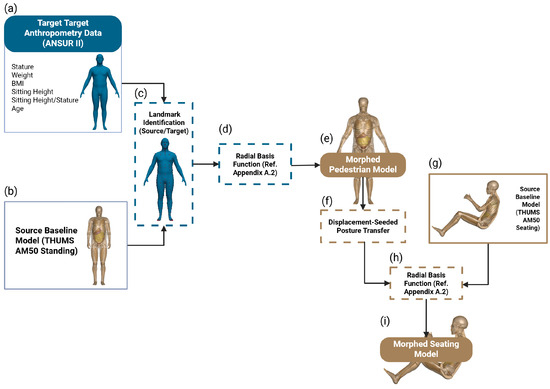
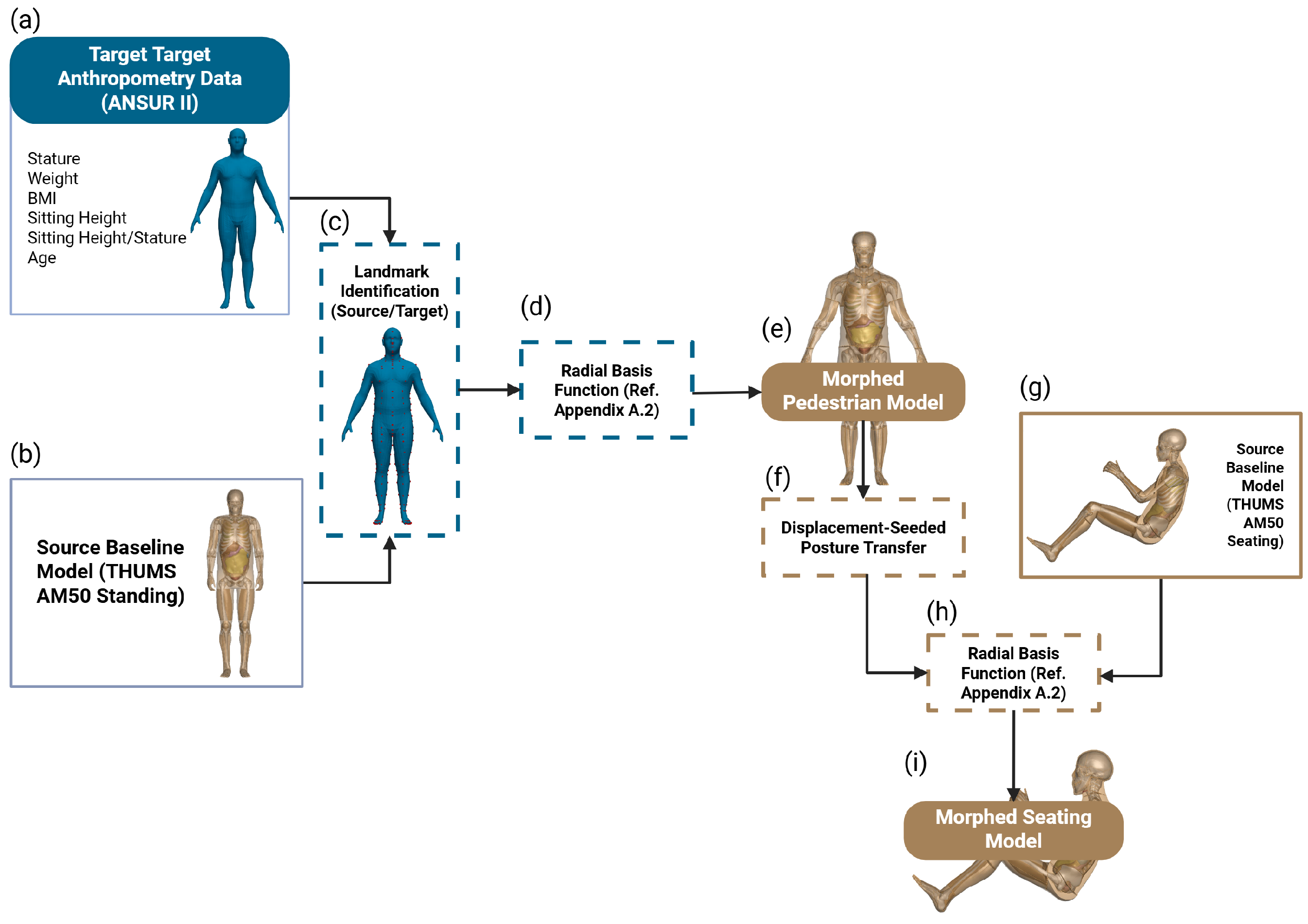
The pipeline’s structure is detailed in Figure 1, which serves as a blueprint for transforming the THUMS models into personalized HBMs. The pipeline begins with the baseline THUMS geometry and generates a target body geometry derived from ANSUR II anthropometric data. The THUMS model is then aligned by adjusting the arms and legs using the LS-DYNA (R12) BOUNDARY_PRESCRIBED_MOTION_SET command to ensure geometric consistency between the baseline and target models. RBF interpolation is then applied to morph the THUMS mesh toward the target models to produce anatomically accurate, percentile-specific models. Finally, the pipeline incorporates a displacement-seeded posture transfer technique that converts morphed standing models into seated configurations by applying regional displacements derived from the original THUMS seated and standing models. The workflow concluded with a comprehensive mesh quality evaluation, which confirmed that the morphed models preserve structural integrity and remain suitable for simulation.
Figure 1.
Comprehensive workflow of the RBF morphing process, delineating a multi-step procedure for personalizing THUMS finite element models. The diagram sequentially illustrates the following: (a) Target anthropometric parameters derived from ANSUR II define subject-specific body dimensions. (b) The baseline THUMS AM50 standing model serves as the source geometry. (c) Corresponding anatomical landmarks are identified on both source and target shapes. (d) Radial basis function (RBF) interpolation generates a smooth deformation field to morph the source mesh Appendix A.2. (e) The resulting morphed pedestrian model matches the desired anthropometry. (f) A displacement-seeded posture transfer introduces additional control points to guide large articulated changes from standing to seated posture. (g) The reference seated THUMS configuration provides the target posture. (h) A second RBF transformation applies the seeded displacements to the full mesh Appendix A.2. (i) The final morphed seating model preserves anatomical fidelity and element quality while achieving the desired seated configuration.
2.1. Models
2.1.1. THUMS Models
The THUMS version 6 served as the baseline finite element model for this study. This model provides a detailed anatomical representation of the human body that includes the skeletal system, soft tissue, and internal organs. Additionally, this model was extensively validated for impact simulation. Each model contains approximately 2.0 to 2.6 million elements with high resolution in regions of high mechanical interest, such as the spine, pelvis, and head, while it has a coarser resolution in other regions. All computational analyses, including morphing and mesh evaluations, relied exclusively on THUMS and were sourced from its official dataset.
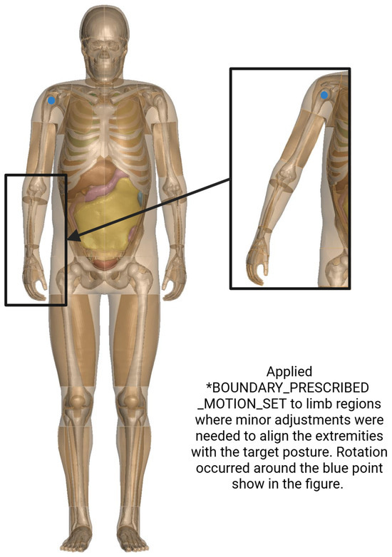
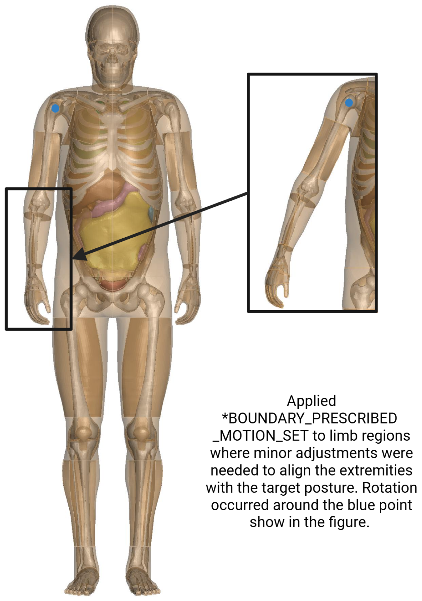
Figure 2 presents the baseline THUMS geometry used as the anatomical and finite element foundation for the morphing process. The model is shown in the standing (pedestrian) posture with semi-transparent soft tissue and skeletal structures to illustrate the underlying anatomical detail relevant for biomechanical simulation. Prior to morphing, small discrepancies existed between the baseline limb posture and the target geometries, particularly in the arms and legs. To improve geometric correspondence, the *BOUNDARY_PRESCRIBED_MOTION_SET keyword was applied to perform minor kinematic adjustments that repositioned the limbs into alignment. These steps were limited to articulations and were used solely to reduce mesh distortion and improve landmark correspondence before applying the RBF-based deformation.
Figure 2.
Baseline THUMS standing configuration used for morphing. Insets highlight limb regions where small kinematic adjustments were applied using *BOUNDARY_PRESCRIBED_MOTION_SET to align the arms and legs with the target posture. This pre-positioning step improves correspondence prior to RBF-based mesh deformation.
2.1.2. Target Model
Using the HumanShape model [34], six target HBMs were generated to represent male and female landmarking the 50th, 80th, and 98th percentiles in standing. HumanShape requires anthropometric inputs including stature, weight, body mass, sitting height, and age. Target anthropometric dimensions were derived from military-specific distributions in the ANSUR II database. As shown in Table 1, these targets span a wide range of stature (1455–1905 mm) and weight (66.8–118.9 kg), ensuring robust demographic coverage for biomechanical modeling.
Table 1.
Anthropometricparameters defining the six target models (M1–M3 male, F1–F3 female) derived from the ANSUR II database, representing the 50th, 80th, and 98th percentiles. Values were computed from measurements of 4082 males and 2946 females in the U.S. Army population [30].
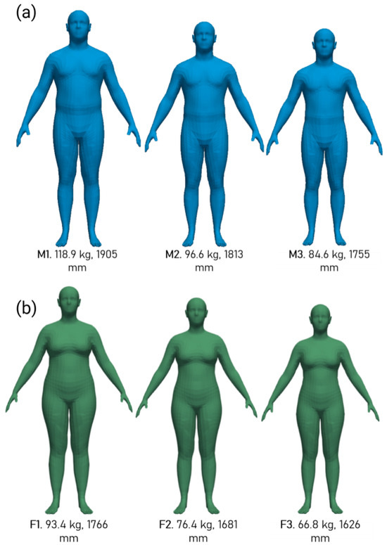
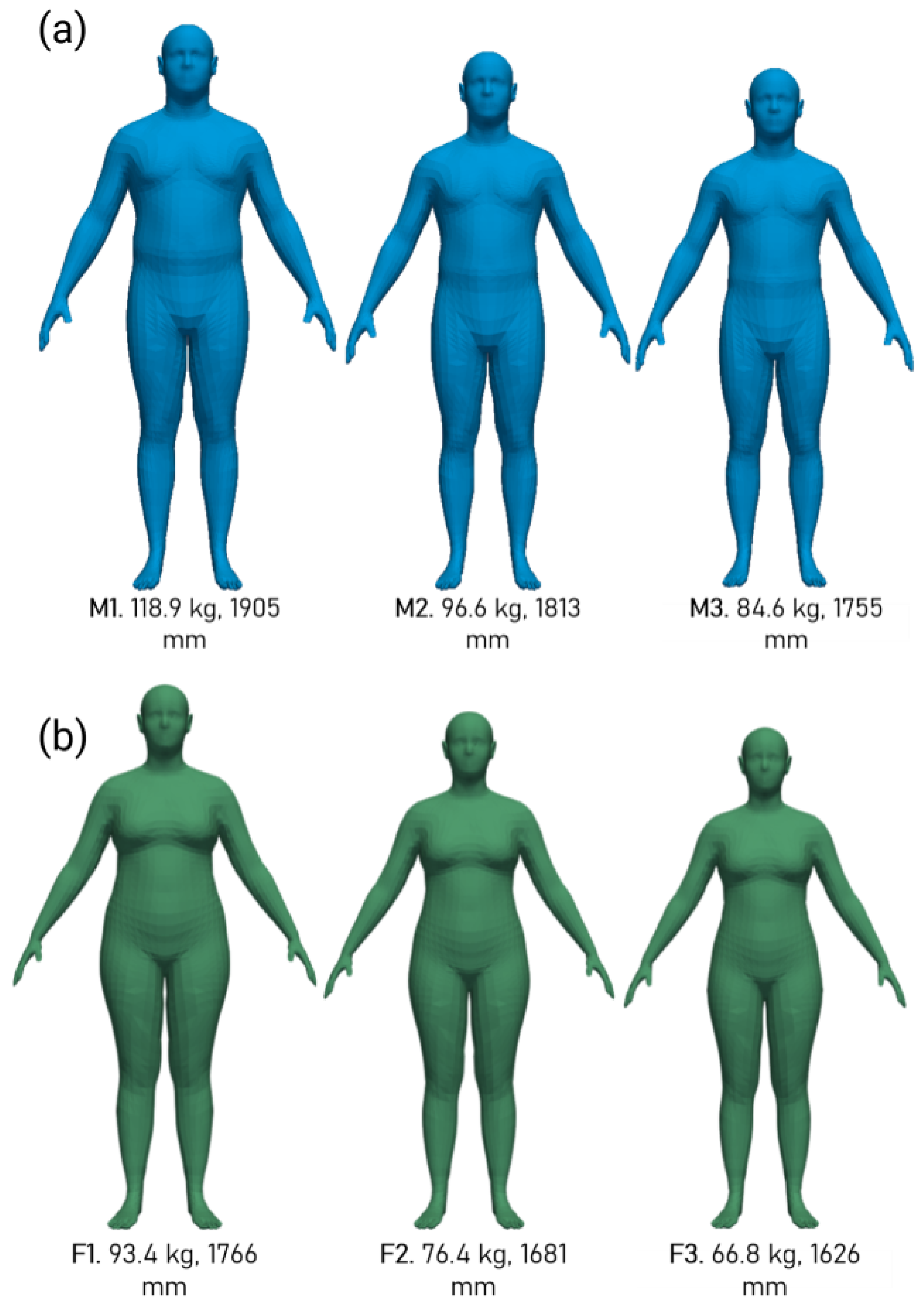
Figure 3 offers a consolidated view of target models. In Figure 3a, the progression from the 98th percentile male (M1: 118.9 kg, stature 1905 mm) to the 80th (M2: 96.6 kg, stature 1813 mm) and the 50th (M3: 84.6 kg, stature 1755 mm) percentiles illustrates decreasing body volume and limb proportions, which are directly informed by ANSUR II statistical distributions to ensure realistic scaling for biomechanical loading scenarios. Similarly, Figure 3b depicts the female counterparts, from the 98th percentile (F1: 93.4 kg, stature 1766 mm) to the 80th (F2: 76.4 kg, stature 1681 mm) and 50th (F3: 66.8 kg, stature 1626 mm), highlighting sex-specific morphological differences such as narrower shoulders and wider hips relative to males, which are critical for accurate representation in finite element simulations. This integration supports the pipeline’s validation across sex-specific profiles by enabling a direct comparison of morphed geometries against these baselines, thereby enhancing the precision of injury analysis in high G environments through an improved prediction of stress distributions and failure thresholds in diverse anthropometric cohorts. The depicted variations underscore the necessity of percentile-based customization, as deviations in stature and mass can alter inertial properties and joint loading by up to 30% under acceleration forces, as demonstrated in analogous scaling studies [35].
Figure 3.
Consolidated visualization of the target models, integrating male and female percentile variations. (a) Models for male percentiles (98th, 80th, 50th), shown in blue show the variations in body mass and stature, highlighting differences in torso width and limb length from ANSUR II data. (b) Models for female percentiles (98th, 80th, 50th), shown in green show the specific differences in stature, hip width, and torso proportions. This figure is key, providing a unified baseline for validating the pipeline’s adaptability to diverse anthropometric profiles under high G loading conditions, which are essential for sex-differentiated injury analysis.
2.2. Radial Basis Function
The RBF morphing technique was implemented using the PyGeM library (v2.0.4) [36], specifically employing the multi-quadratic biharmonic spline function to deform the baseline THUMS mesh toward each target skin surface. This approach leverages a sparse set of anatomical landmarks that are automatically extracted from both the THUMS baseline and target meshes. The multi-quadratic biharmonic spline is defined below:
where r is the Euclidean distance between a given node and landmark points, and c is a shape parameter controlling the smoothness of interpolation (typically set to average landmark spacing), which provides compact support and ensures smooth deformation fields suitable for finite element mesh morphing. The displacement field at any point is then interpolated as shown below:
where N is the number of landmarks, represents coefficients solved via a linear system minimizing the error between target and interpolated displacements, and represents landmark positions. This formulation, implemented through PyGeM’s RBF class, preserves mesh connectivity while enabling efficient transformation of the entire THUMS model comprising millions of nodes and elements.
Mesh quality post morphing was evaluated using the Jacobian determinant, which was calculated as shown below:
where J is the Jacobian determinant, represents the physical coordinates, and denotes the parametric coordinates in the mesh. A value of indicates acceptable element quality with no inverted elements (negative Jacobian), as per established finite element guidelines [37]. Additional metrics including the warpage (<50°), aspect ratio (<5), and skewness (<60°) were assessed to ensure the morphed meshes remained suitable for explicit dynamic simulations in LS-DYNA (R12). This comprehensive quality control, integrated into the pipeline illustrated in Figure 1, guaranteed smooth interpolation gradients from the multi-quadratic RBF while maintaining topological integrity essential for accurate biomechanical response prediction under high G loading conditions.
In addition to ensuring mesh integrity, the geometric accuracy of the morphed models was assessed by comparing them to the ANSUR II target geometries using the DICE similarity coefficient and the HD95. These metrics quantified volumetric overlap and surface deviation, respectively, providing objective measures of morphing precision. Distance field maps were also generated to visualize regional differences between the morphed and target surfaces. A detailed discussion of these results and their regional variations is presented in the following section.
2.3. Displacement Seeded Posture Transfer
In addition to percentile-based morphing, a displacement seeded posture transfer method was developed to transform the standing morphed THUMS models into seated configurations. This approach allowed for posture adaptation while maintaining anatomical accuracy and mesh consistency in regions with equivalent mesh topology between the standing and seated baselines. The posture transfer process began by identifying corresponding regions between the original standing and seated THUMS models that shared the same number of elements and a comparable mesh structure. These included relatively rigid or nonbending anatomical regions such as the thighs, calves, biceps, chest, and head. For each of these regions, nodal displacements were computed as the positional difference between the standing and seated THUMS meshes.
The calculated regional displacement fields were then applied to the newly morphed standing models to generate a new set of coordinates representing an approximate seated posture. This step effectively embedded posture information from the validated THUMS seating configuration into the morphed percentile-specific geometries. Because the seated and standing models differ in mesh density in highly articulated areas, direct one-to-one mapping was not possible in those regions. Therefore, only the compatible portions of the model were displacement seeded. The resulting partially transferred posture model was subsequently refined using RBF interpolation to propagate smooth deformations across the remaining regions, ensuring anatomical continuity and maintaining element integrity.
This combined displacement and RBF-based method provided an efficient way to adapt morphed standing models into seated postures while minimizing mesh distortion. The final posture transferred geometries preserved correct joint alignment and body proportions, forming the foundation for seated high G and occupant simulation studies.
3. Results
The morphing process was evaluated through comprehensive assessments of mesh quality and geometric accuracy for both standing and seated postures across male and female models at the 50th, 80th, and 98th percentiles. These evaluations ensure that the personalized human body models maintain structural integrity and anatomical fidelity, which are critical for reliable biomechanical simulations in high G environments.
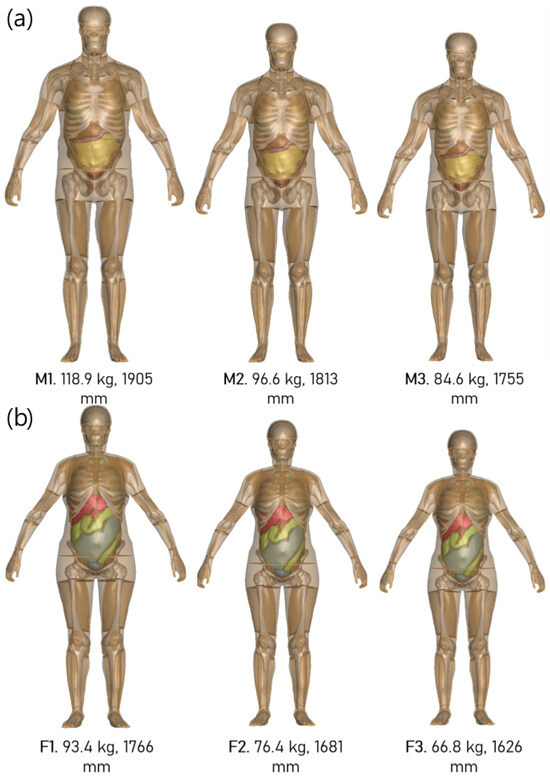
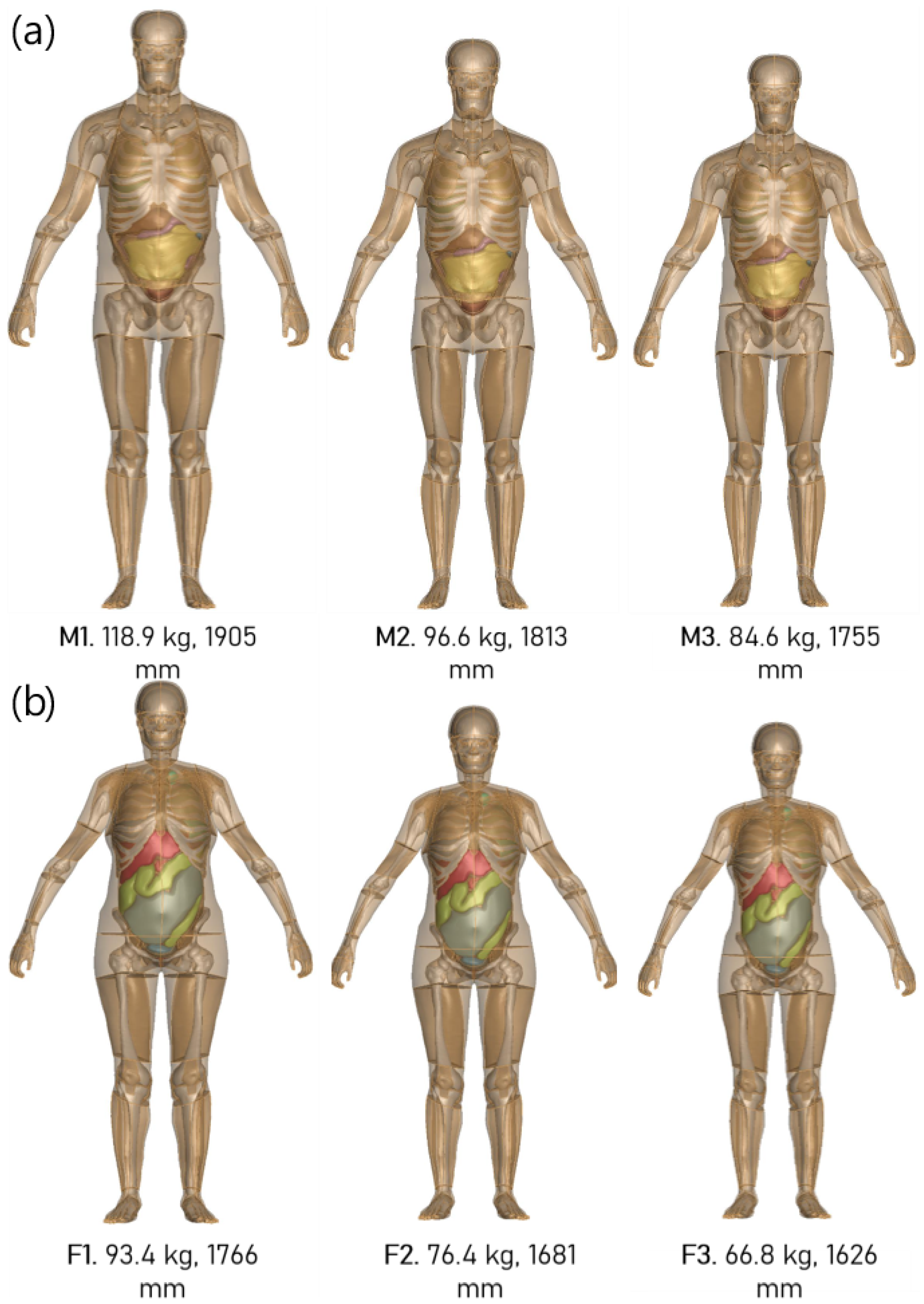
Figure 4 illustrates the morphed male and female models generated at the 50th, 80th, and 98th percentiles (denoted as M3/F3, M2/F2, and M1/F1). The RBF-based morphing method successfully captured the expected anthropometric variations across percentiles while preserving anatomical proportions and smooth surface continuity. The 98th percentile models (M1/F1) exhibited visibly larger torso breadth and limb volume, while the 50th percentile models (M3/F3) were more slender and consistent with population averages. The 80th percentile models (M2/F2) provided intermediate geometries, demonstrating the method’s ability to scale continuously between percentile extremes. Importantly, the morphing preserved mesh quality in high curvature regions such as the shoulders, abdomen, and thighs, avoiding self intersections or excessive distortion.
Figure 4.
Morphed standing male (a) and female (b) THUMS models at the 98th (M1/F1), 80th (M2/F2), and 50th (M3/F3) percentiles, demonstrating anthropometric variation across the population range while preserving anatomical structure and surface smoothness.
3.1. Standing
3.1.1. Mesh Quality
Mesh quality metrics, including the warpage, aspect ratio, skewness, and Jacobian, were analyzed to quantify the impact of the RBF morphing on element integrity. Ideal thresholds were defined as warpage <50°, aspect ratio <5, skewness <60, and Jacobian >0.3 with no negative values. Deviations from these thresholds indicate potential issues in simulation stability, such as numerical inaccuracies or convergence problems in finite element analyses. These indicators were analyzed for all male and female models across the 50th, 80th, and 98th percentiles and compared against the original THUMS baseline to determine whether the morphing process preserved element integrity across varying anthropometries.
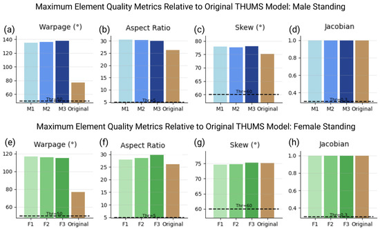
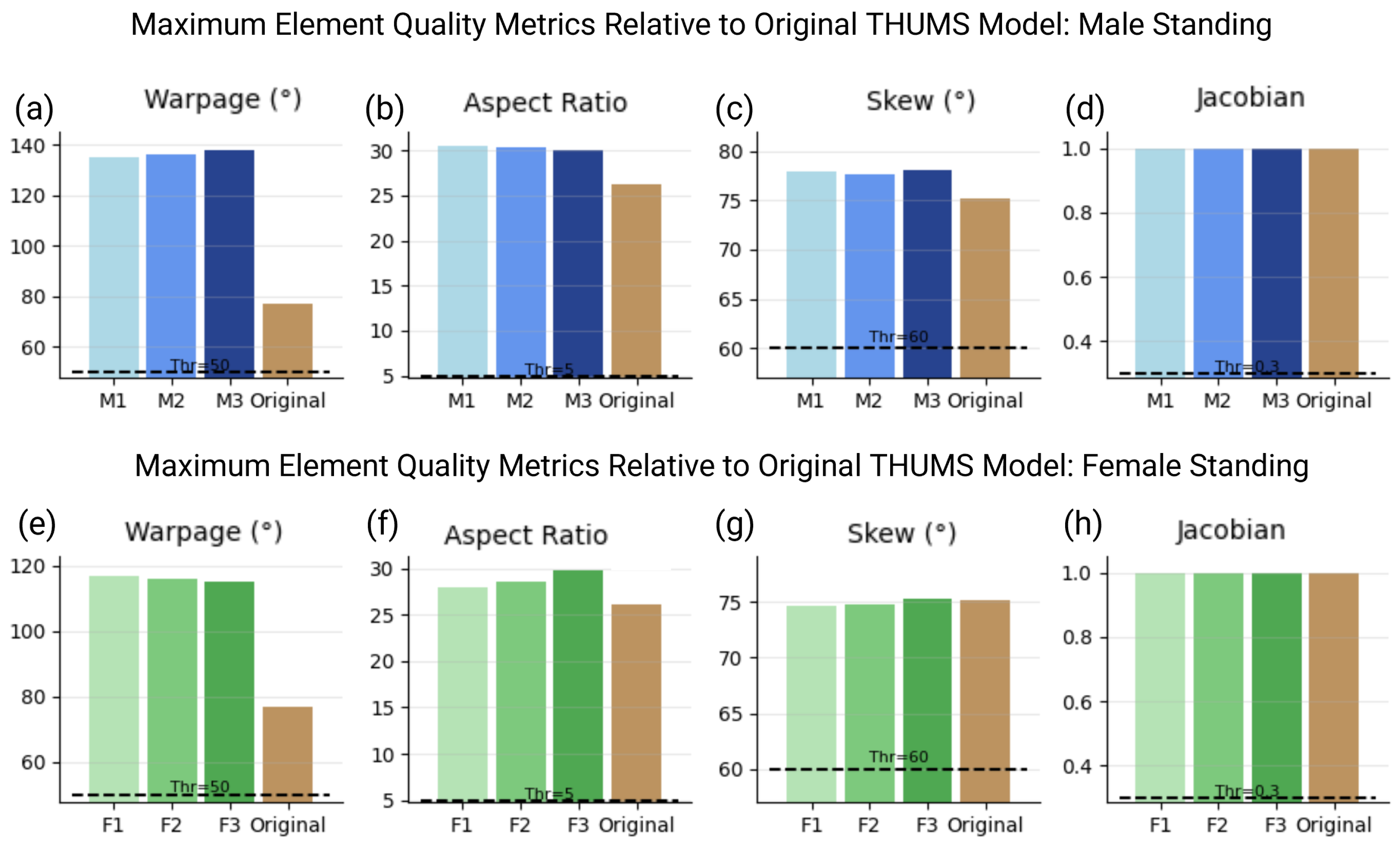
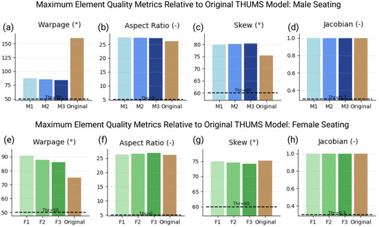
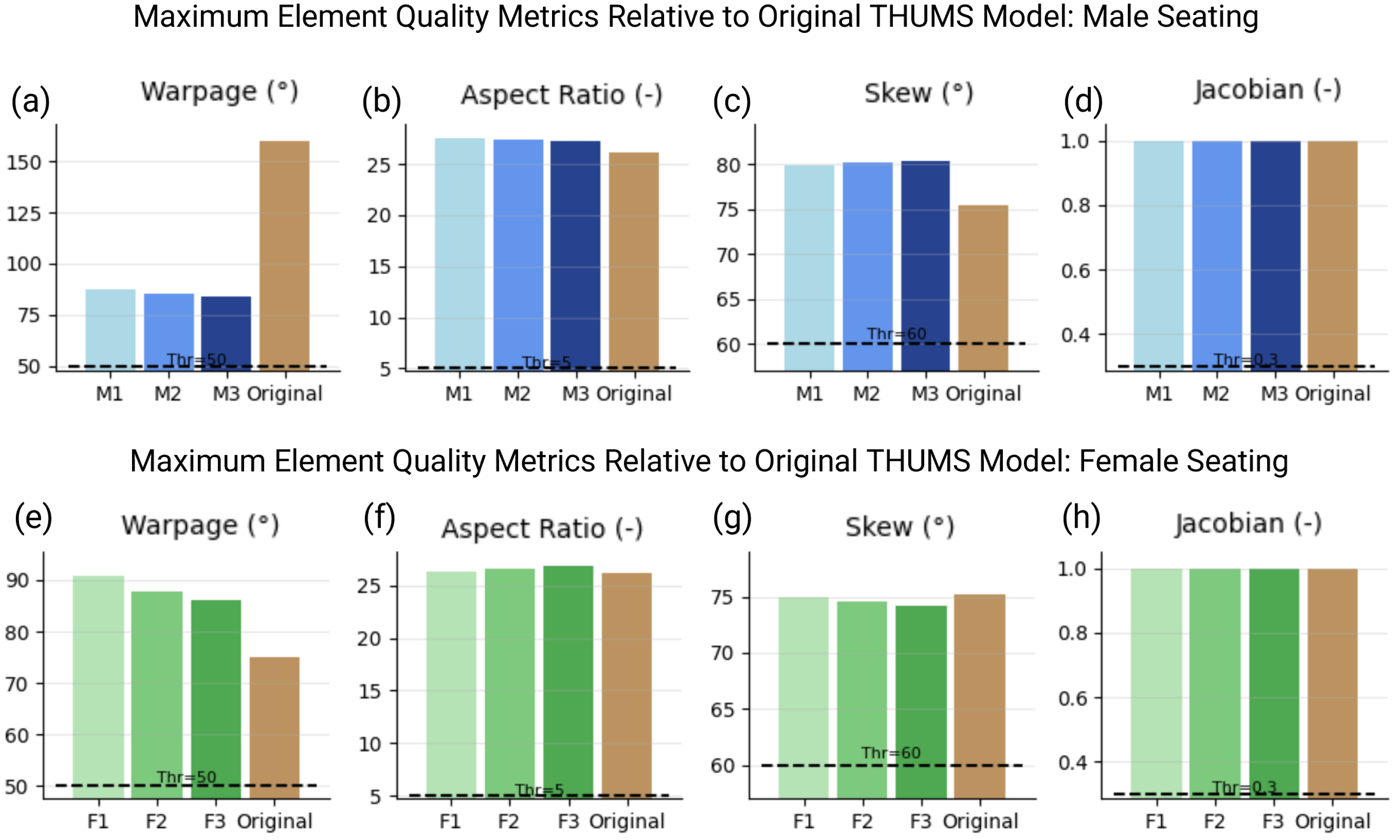
Figure 5 summarizes the maximum mesh quality values for each model. The standing mesh-quality results show that the morphed skin surfaces remain closely aligned with the original THUMS standing model across all male and female percentiles. Maximum element metrics indicate that warpage values increase relative to the baseline, reaching approximately 130–140° in the male morphs and 115–120° in the female morphs, while the original THUMS mesh remains lower. While maximum warpage values in the morphed models occasionally exceeded the nominal 50° threshold, a topological analysis reveals that these violations are strictly localized to non-critical anatomy, specifically the external ear cartilage and distal phalanges. As these regions are removed from the primary vertical load path, they do not influence spinal kinematics. Furthermore, a preliminary dynamic validation to be reported in a forthcoming study on cockpit seat configurations by Reyes et al. [38] has confirmed that these models execute to normal termination in LS-DYNA (R12) under high G loading without instability. Despite this increase in peak warpage, the overall mesh structure remains consistent with the baseline with aspect ratios remaining below roughly 30, skewness below 80°, and all Jacobians above 0.3, indicating that no elements inverted or collapsed during the morphing.
Figure 5.
Maximum element quality metrics for morphed standing THUMS models across male (a–d) and female (e–h) percentiles. Plots show peak values for warpage, aspect ratio, skewness, and Jacobian compared against the original baseline model.
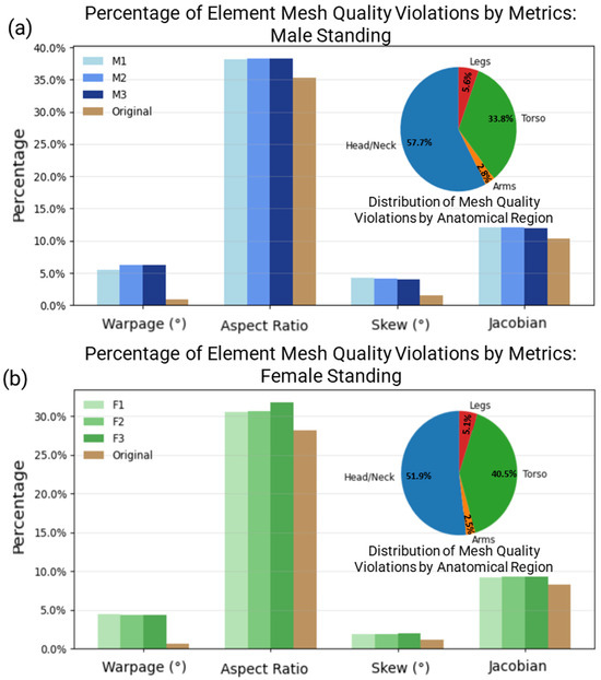
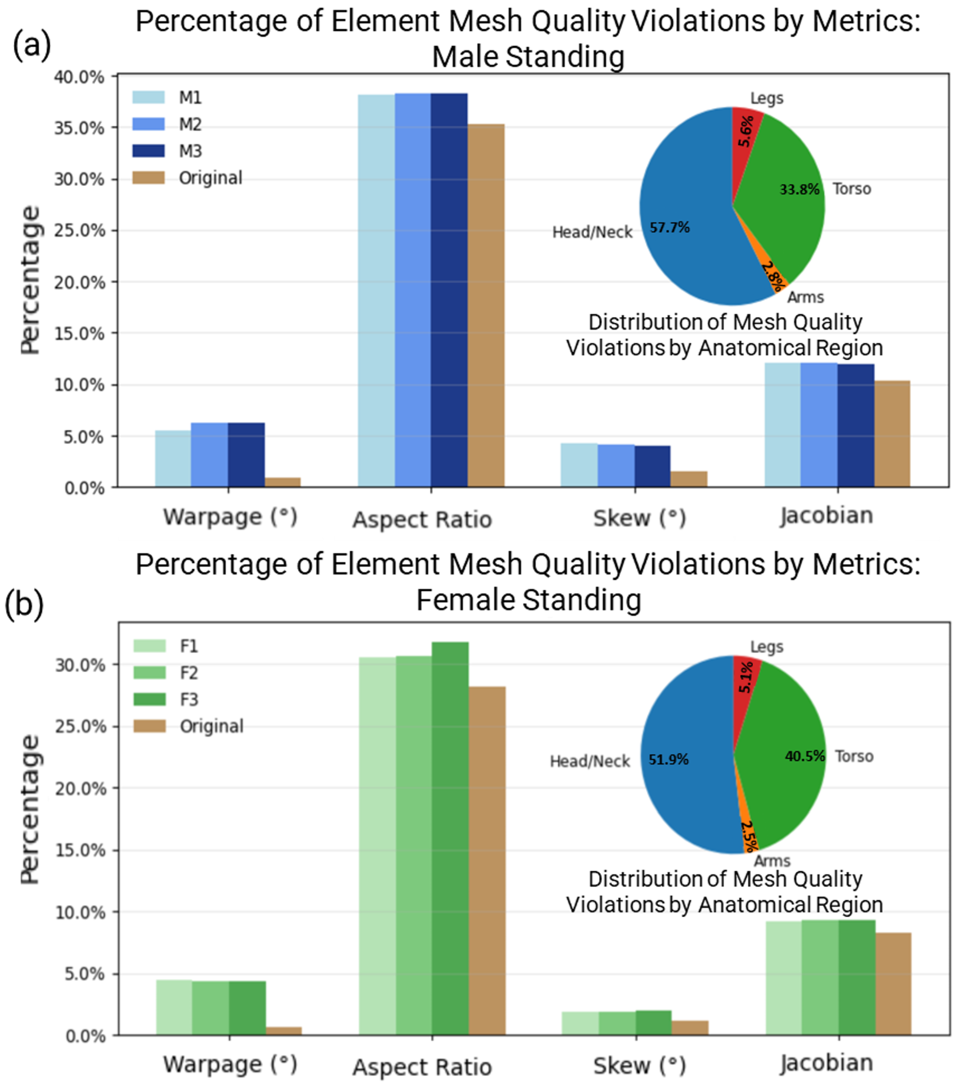
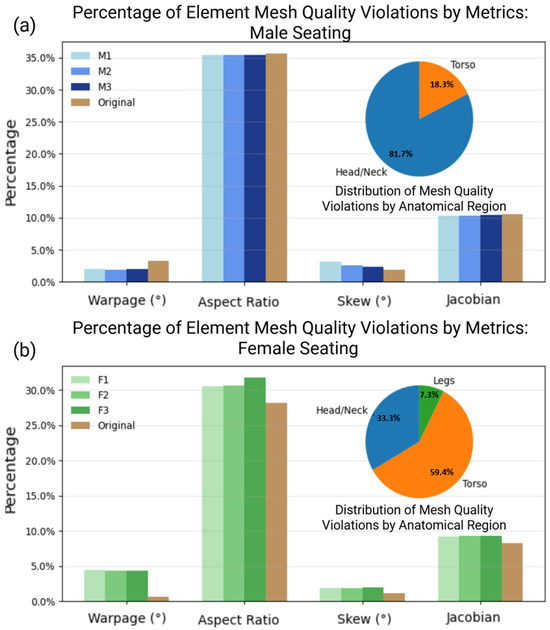
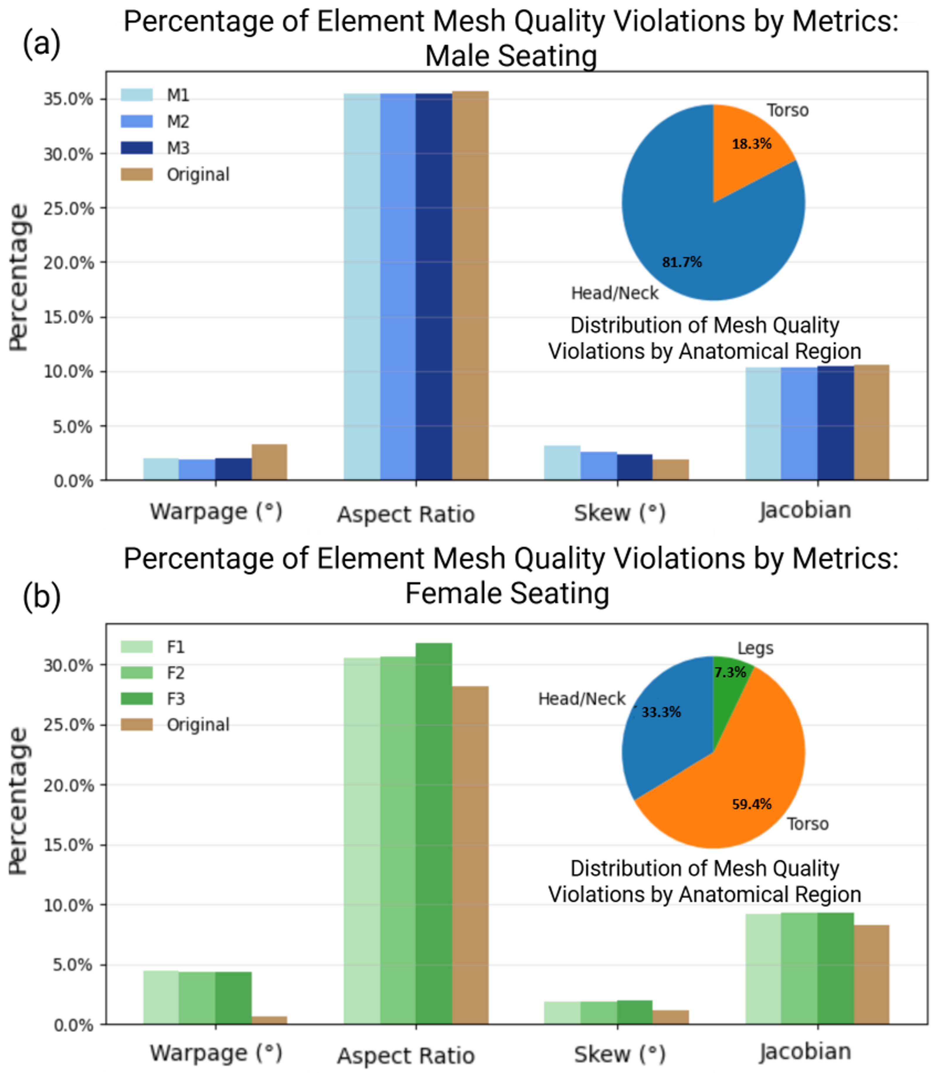
The violation percentages shown in Figure 6 further support this interpretation. Aspect-ratio violations remain below 40% for the male morphs and below 32% for female morphs, which is only slightly higher than the original THUMS model and reflects the increased geometric variability introduced by large-scale morphing. Warpage, skew, and Jacobian violations also appear higher in the morphs but remain low overall, staying within approximately 2–12% across percentiles and sexes. These values are still acceptable for explicit dynamic analyses, particularly because the violations are concentrated in the same anatomical regions as the original mesh. Male violations are primarily located in the head and neck (58%) and torso (34%), and female violation patterns are similarly distributed (52% and 41%), indicating that the morphing process preserves the underlying structural behavior of the THUMS standing mesh.
Figure 6.
Percentage of element violations for morphed standing male (a) and female (b) THUMS models. Aspect ratio violations were the most frequent, occurring in fewer than 40% of elements, while warpage, skewness, and Jacobian violations remained below 15%. Pie charts highlight the anatomical distribution of violations, which were concentrated primarily in the head/neck and torso regions.
These findings demonstrate that the standing morphs successfully retained high element quality and anatomical consistency across all percentile variations, establishing the reliability of the RBF pipeline.
3.1.2. DICE Similarity Coefficient (DICE) and 95th Percentile Hausdorff Distance (HD95)
Geometric accuracy was quantified using the DICE similarity coefficient [39] and HD95 [32] to compare the morphed meshes against target avatars. These metrics evaluate volumetric overlap and surface deviations, respectively, providing a robust measure of how well the morphing preserves target anthropometry. These measures quantify the agreement between the morphed THUMS models and their corresponding ANSUR II targets. Higher DICE values indicate greater volumetric overlap, while lower HD95 values represent smaller localized deviations along the surface.
The DICE measures the volumetric overlap between two geometries:
where and represent the volumes (or voxel counts) of the morphed and target models, respectively, and denotes their intersection volume. DICE values range from 0 to 1 with higher values indicating greater overlap and thus improved morphing accuracy. In general, values above 0.90 are considered indicative of high geometric fidelity for biomechanical model registration.
The Hausdorff distance (HD) quantifies the largest surface-to-surface deviation between two meshes. To reduce the influence of outliers caused by local mesh irregularities, HD95 was employed, which is defined as
where denotes the 95th percentile, and are surface points on the morphed and target meshes, and is the Euclidean distance. HD95 thus represents the 95th percentile of the bidirectional point-to-surface distances between M and T, capturing typical surface discrepancies while mitigating sensitivity to extreme outliers.
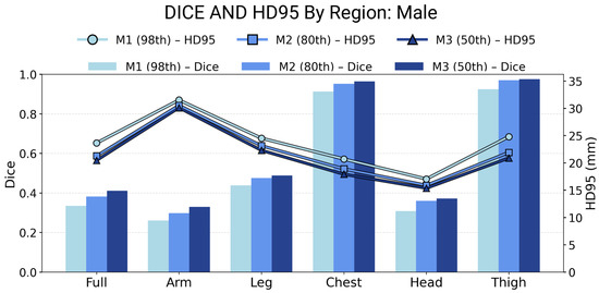
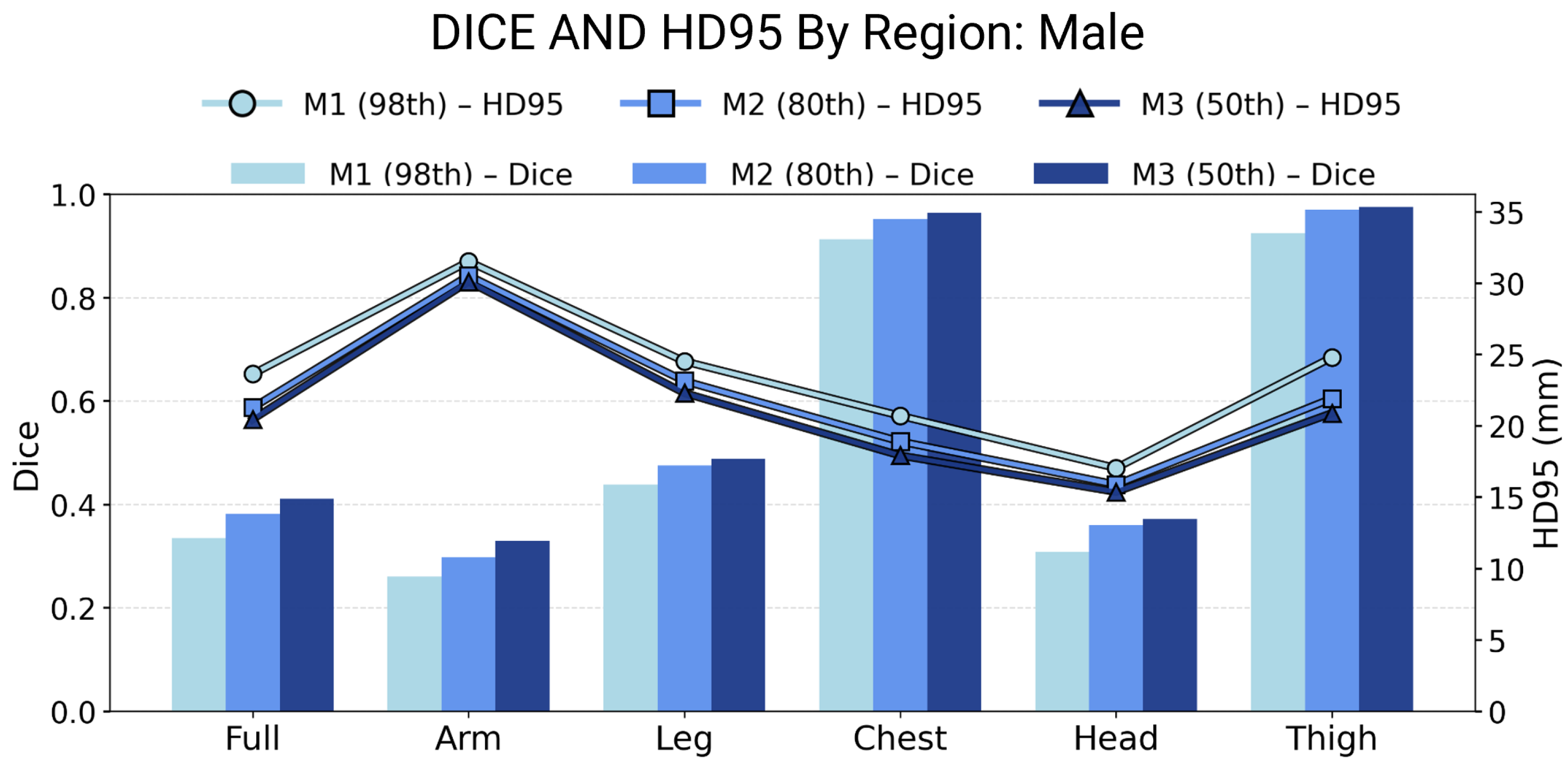
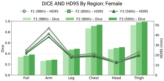
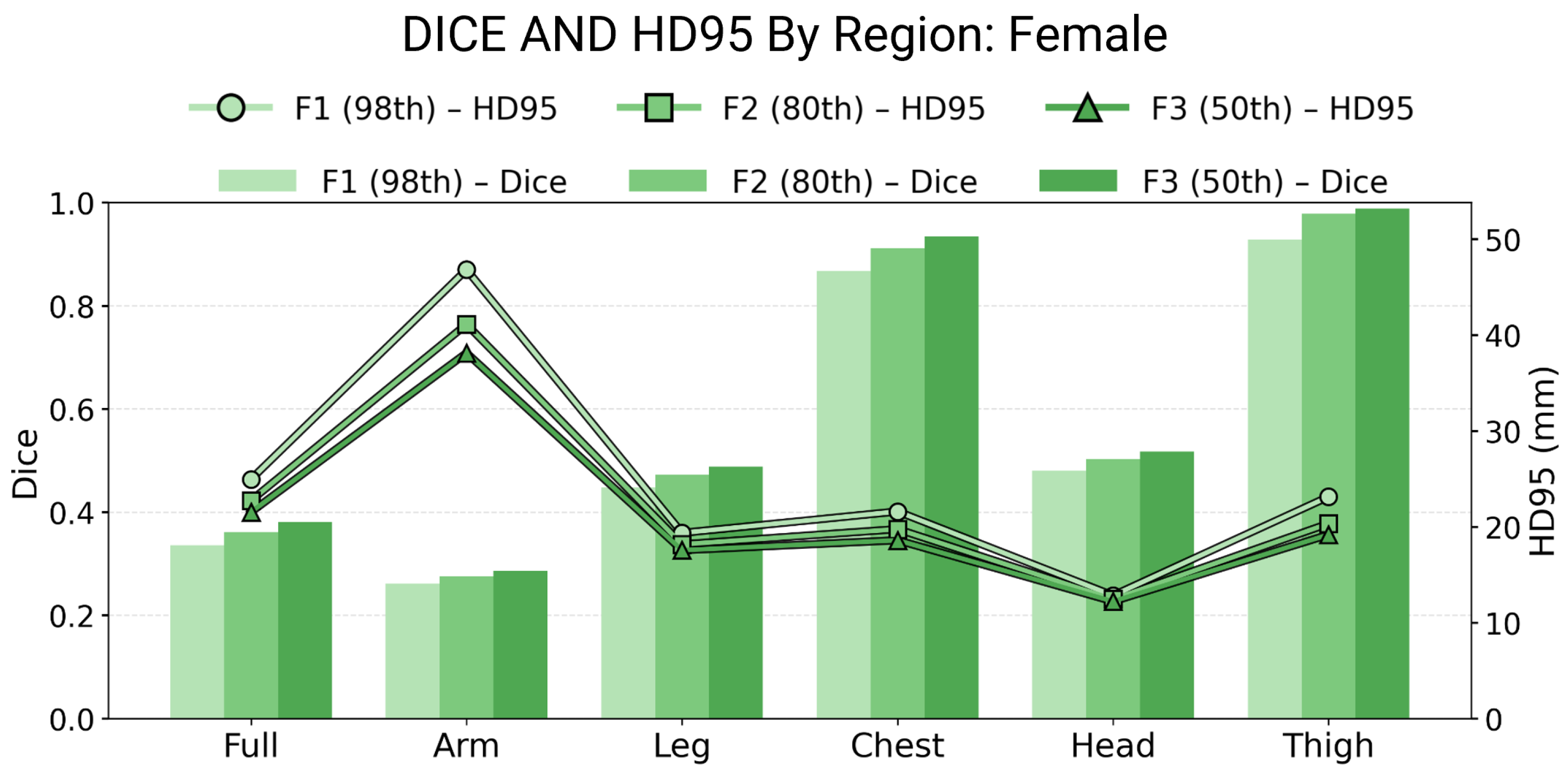
Together, these metrics provide a robust evaluation framework: DICE assesses global volumetric similarity, while HD95 characterizes localized deviations. Across all percentile variations, the morphing process produced consistent and accurate geometries, as shown in Figure 7 and Figure 8. The DICE values exceeded 0.85 in the chest region, demonstrating strong correspondence between the morphed and target geometries. The head and thigh regions showed moderate DICE values, averaging between 0.35 and 0.45, while the arms and legs displayed slightly lower scores due to their higher local variability and reduced landmark density, resulting in a global DICE average of approximately 0.40.
Figure 7.
Regional DICE and HD95 results for male morphed standing models (50th, 80th, and 98th percentiles). DICE values demonstrate strong agreement in the torso and moderate accuracy in peripheral regions.
Figure 8.
Regional DICE and HD95 results for female morphed standing models (50th, 80th, and 98th percentiles). Similar regional accuracy patterns are observed, confirming consistent morphing performance across percentiles and sexes.
For surface-based evaluation, the HD95 values remained within 20–30 mm across most anatomical regions, confirming that the morphing achieved smooth and spatially consistent deformations. The chest region again exhibited the lowest HD95 distances, reflecting the stability of RBF interpolation around high-density landmark zones.
3.1.3. Methodology for Surface Comparison Using Normal-Based Ray Casting
While global metrics between the morphed and target models provide a valuable measure of volumetric overlap, it does not indicate where and in what direction the surfaces differs. To quantify geometric differences between the target surface and the morphed subject-specific surface, we employ a normal-based ray intersection method. The process begins by sampling a subset of triangular surface elements on the reference mesh. For each selected triangle with centroid and unit outward normal vector , a ray is cast along the direction of . The intersection of this ray with the morphed surface defines the corresponding point on the morphed geometry. The surface-to-surface deviation is then measured as the signed distance between the centroid and intersection point:
Here, the sign convention is determined by the relative orientation of the intersection vector with respect to the reference surface normal. Positive values indicate that the morphed surface lies along the outward normal direction (i.e., external to the reference), whereas negative values indicate an inward offset.
By repeating this procedure for a statistically representative subset of surface elements (e.g., 20% of the total triangles), a sparse spatial sampling of the distance field is obtained. These distances are then mapped back onto the corresponding intersected triangles of the morphed mesh to create a per-triangle deviation field:
where denotes the morph triangle hit by the ray.
The resulting scalar field visually encodes geometric fidelity: small magnitudes (blue regions) represent a close match between the reference and morphed geometries, while large positive or negative values (red regions) indicate localized outward or inward deviations, respectively. This approach provides a computationally efficient and interpretable measure of surface conformance between digital human body models without requiring a one-to-one vertex correspondence.
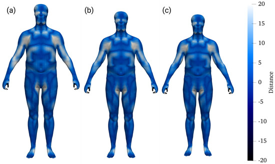
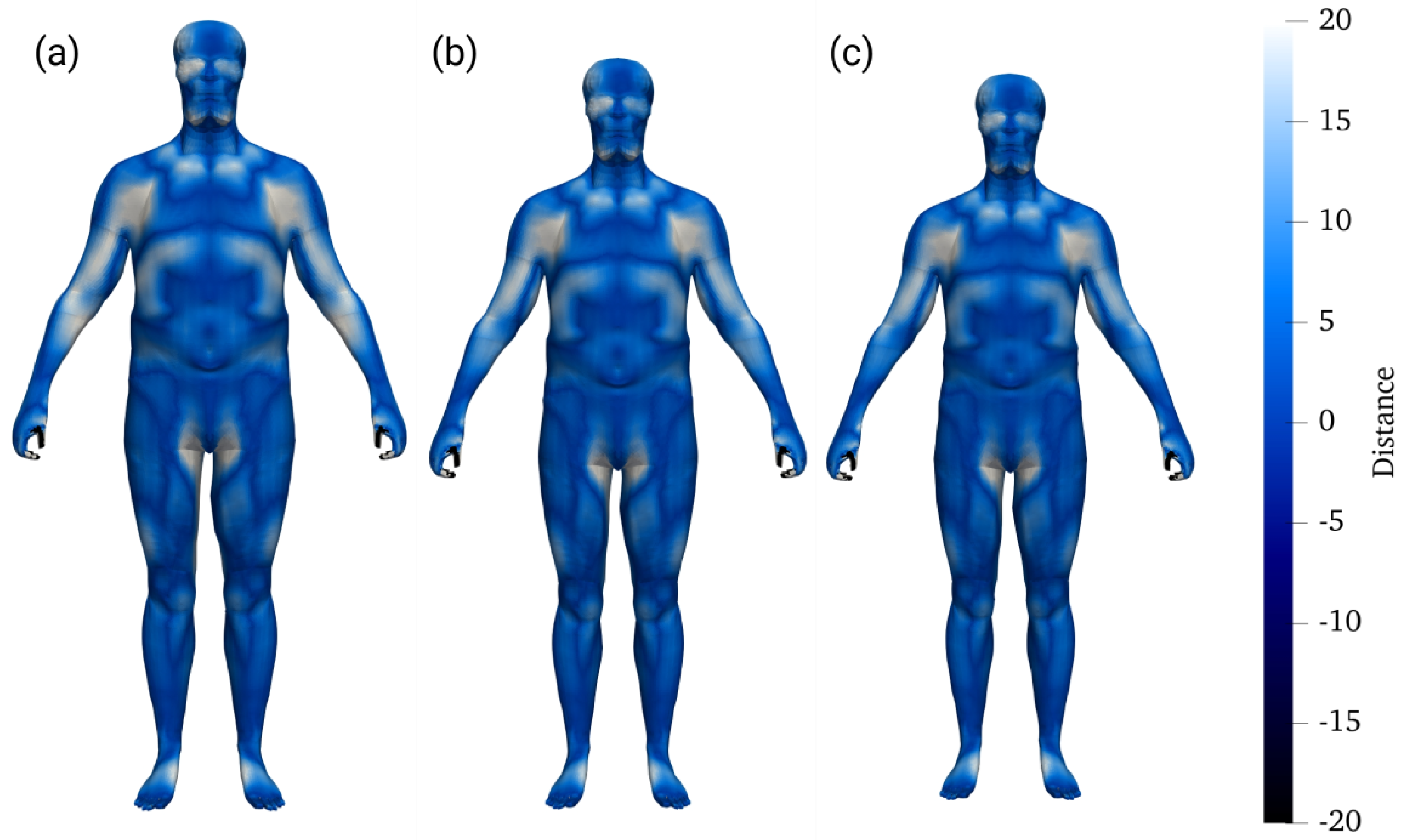
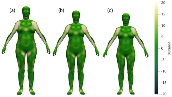
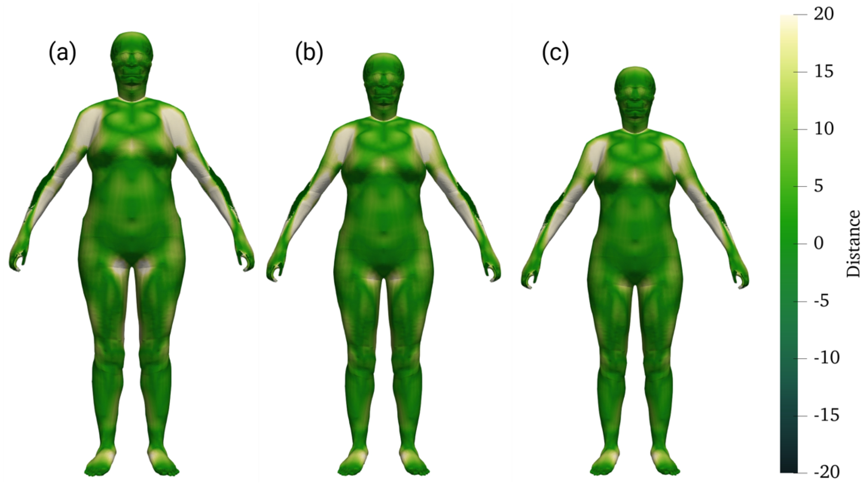
The signed surface distance fields provided a visual and quantitative assessment of geometric deviations between the reference and morphed standing models. Figure 9 and Figure 10 illustrate the distance distributions for male and female morphs across the 50th, 80th, and 98th percentiles. The color maps represent localized surface deviations with positive values indicating outward displacement and negative values indicating inward deviation relative to the reference surface.
Figure 9.
Signed distance maps for the morphed male standing models at the 98th (a), 80th (b), and 50th (c) percentiles, showing deviations relative to the reference surface.
Figure 10.
Signed distance maps for the morphed female standing models at the 98th (a), 80th (b), and 50th (c) percentiles.
Across all percentile variations, the distance fields demonstrated close geometric alignment with most areas falling within ±20 mm of the reference. Deviations were primarily concentrated in high-curvature regions such as the shoulders, abdomen, and hips, where local shape differences between the THUMS baseline and the ANSUR II target meshes are greatest. These localized offsets remained small and spatially consistent, confirming that the RBF-based morphing maintained surface smoothness and avoided topological distortion.The male morphs displayed highly uniform surface conformity, particularly in the torso and upper limb regions, reflecting the effectiveness of the morphing process in preserving anatomical proportion across large body masses. The female morphs exhibited similarly consistent results, although slightly larger deviations were observed in the chest and thigh areas, corresponding to regions with greater anthropometric variation between the reference and target geometries.
To complement the surface distance visualization, statistical analyses were performed on the signed distance data for all percentile models. Table 2 shows the mean and standard deviation values were evaluated for the whole body and for key anatomical regions, including the torso and extremities. These metrics quantify the average geometric error between the morphed and target surfaces and complement the visual deviation maps and cumulative distributions.
Table 2.
Summary of surface distance deviations between the morphed HBM and target ANSUR II geometries.
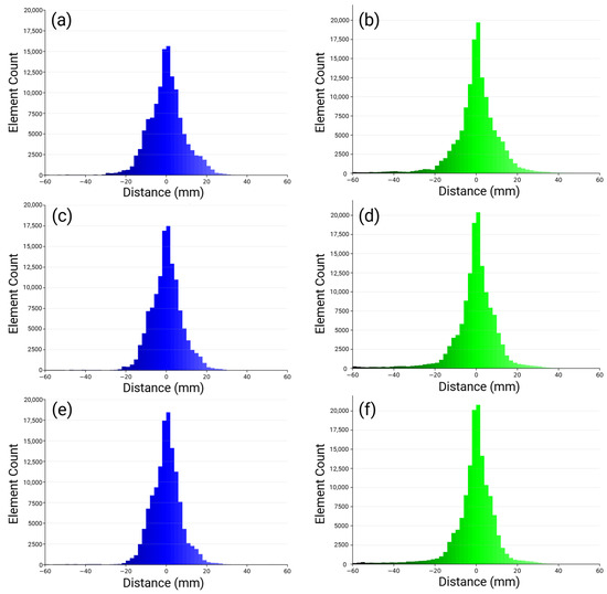
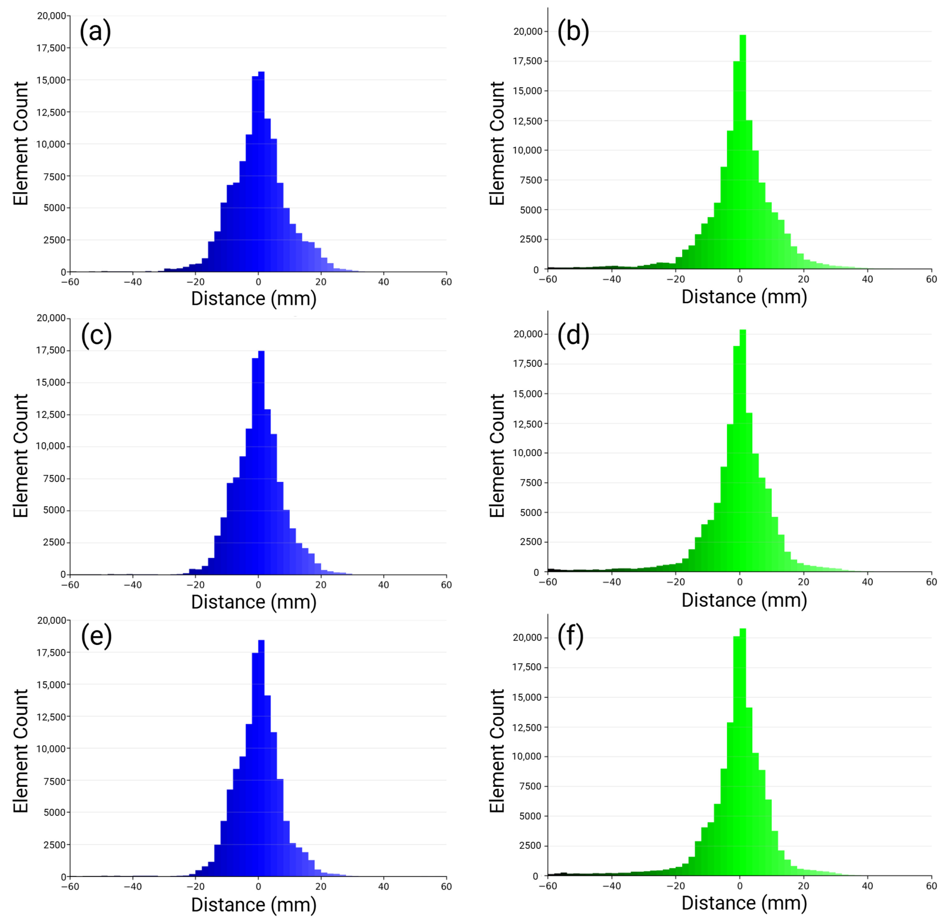
Histograms were computed to quantify the spatial distribution and magnitude of deviations across the entire body surface. The histograms as seen in Figure 11 show narrow, symmetric distributions centered around zero, confirming that the morphing process produced balanced deviations without directional bias. For both male (Figure 11a,c,e) and female (Figure 11b,d,f) models, over 80% of the nodes fell within ±10 mm of the reference surface, and fewer than 5 % exceeded ±20 mm. These results demonstrate that large deformations were effectively avoided, maintaining the global body proportions across all percentile morphs.
Figure 11.
Histogram of signed surface distances for (a) male 50th percentile; (b) female 50th percentile; (c) male 80th percentile; (d) female 80th percentile; (e) male 98th percentile; (f) female 98th percentile. The blue histograms correspond to male models and the green histograms correspond to female models. Distributions are symmetric and centered near zero, confirming minimal surface bias.
It was essential to evaluate whether this mesh integrity could be preserved after applying posture transformations. The following section extends the analysis to the seated models, examining mesh quality and deformation behavior following the displacement seeded posture transfer process.
3.2. Seating
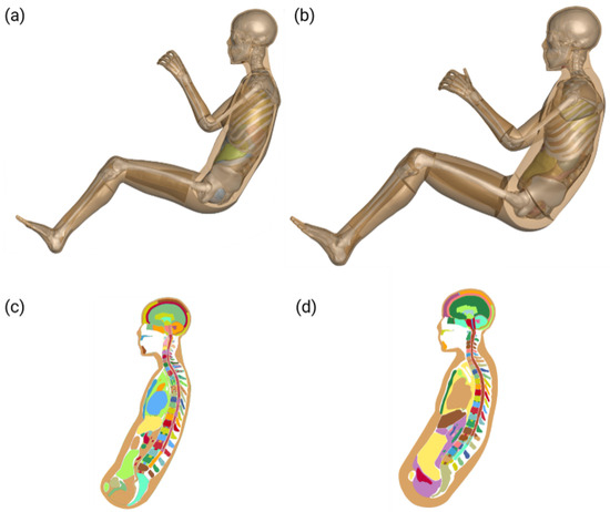
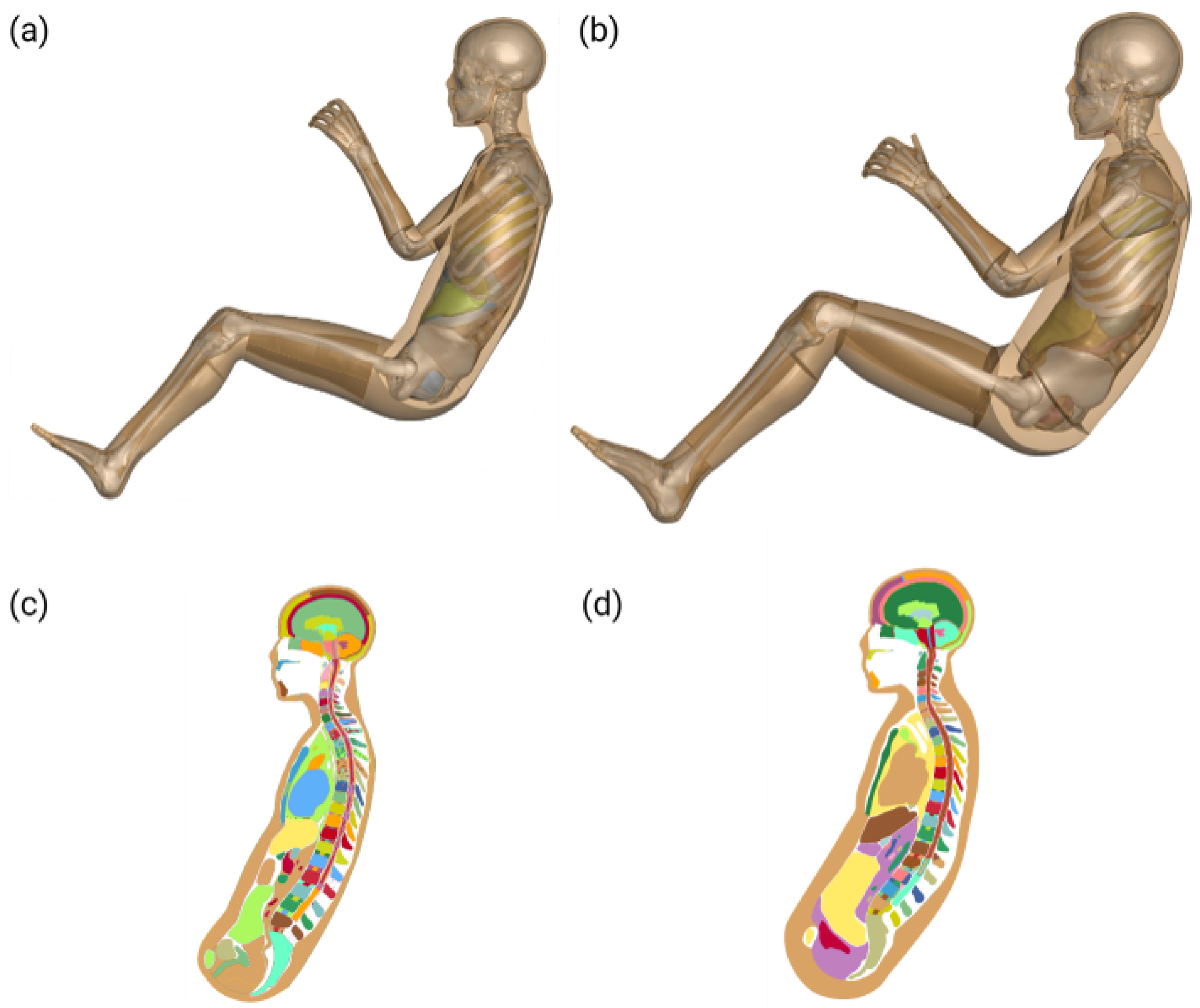
To extend the morphing pipeline beyond static postures, the displacement seeded posture transfer method was applied to generate anatomically accurate seated configurations from morphed THUMS standing models. This approach simulates realistic seated postures representative of aerospace and automotive environments, maintaining both internal and external anatomical coherence. The baseline seated THUMS models were first introduced as geometric references for posture transfer. These original configurations represent anatomically accurate seated postures derived directly from the THUMS database, serving as the foundation for evaluating the displacement seeded transformation.
Figure 12 shows the unmodified seated THUMS models for both male and female configurations. The internal cutaway views highlight the baseline alignment of the spine, pelvis, and internal organs, which define the target anatomical relationships for posture transformation. These models were not subjected to displacement seeded morphing and therefore serve as reference geometries for validating the accuracy of the newly generated seated morphs.
Figure 12.
Overview of the baseline seated THUMS configurations for (a,c) female and (b,d) male models. These baselines define the target anatomical geometry for subsequent displacement seeded posture transfer.
After establishing the reference postures, the displacement seeded posture transfer method was applied to convert the morphed standing models into anatomically consistent seated configurations. This process transferred regional displacements from the original THUMS seated and standing models to the newly morphed standing meshes. This would ensure anatomically consistent seating without introducing element distortion.
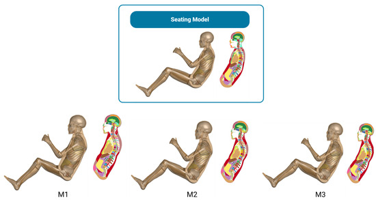
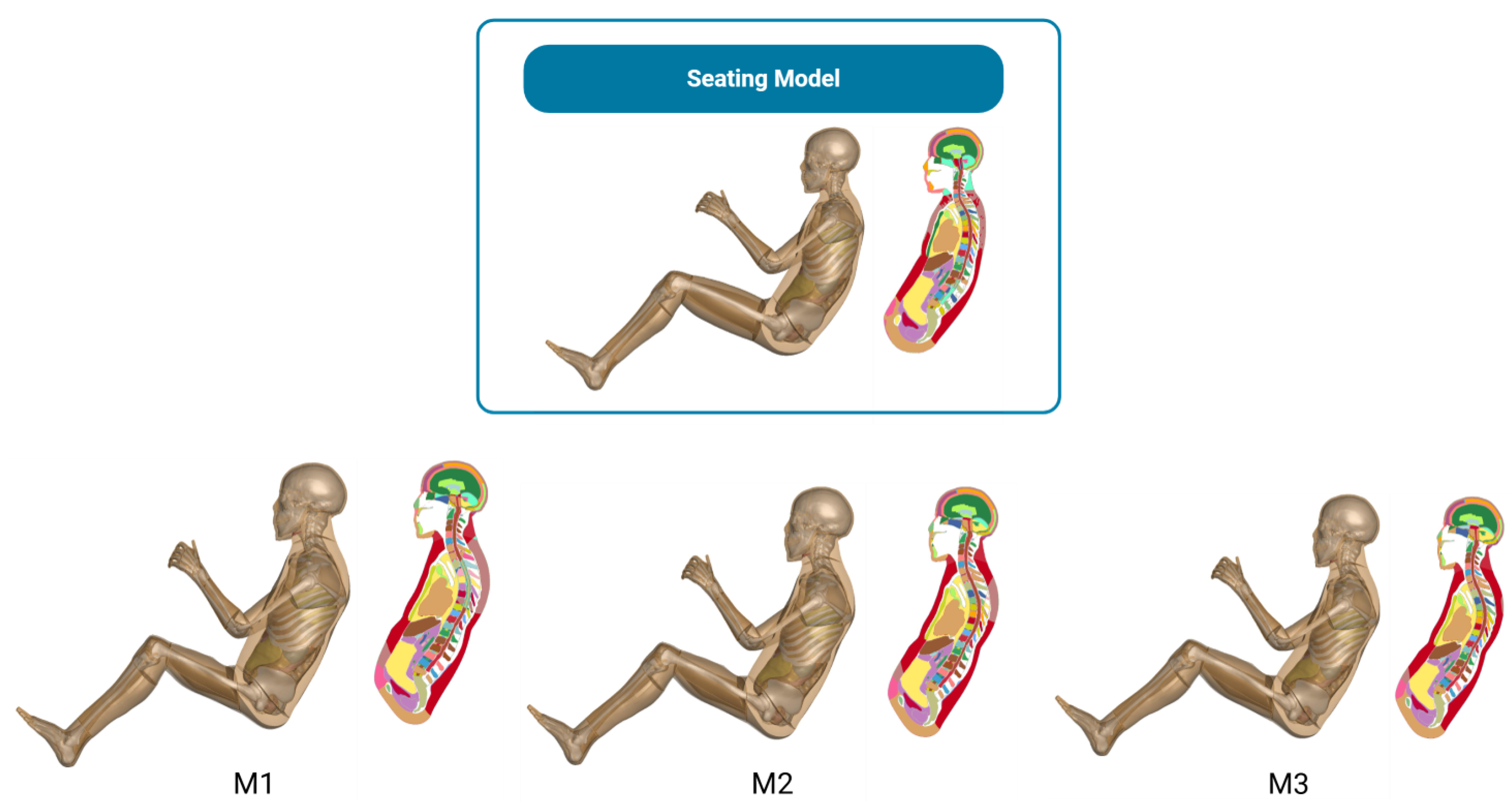
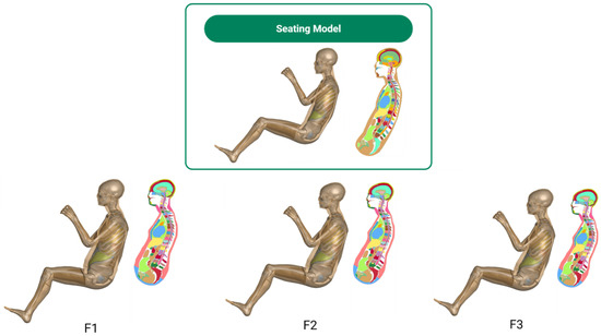
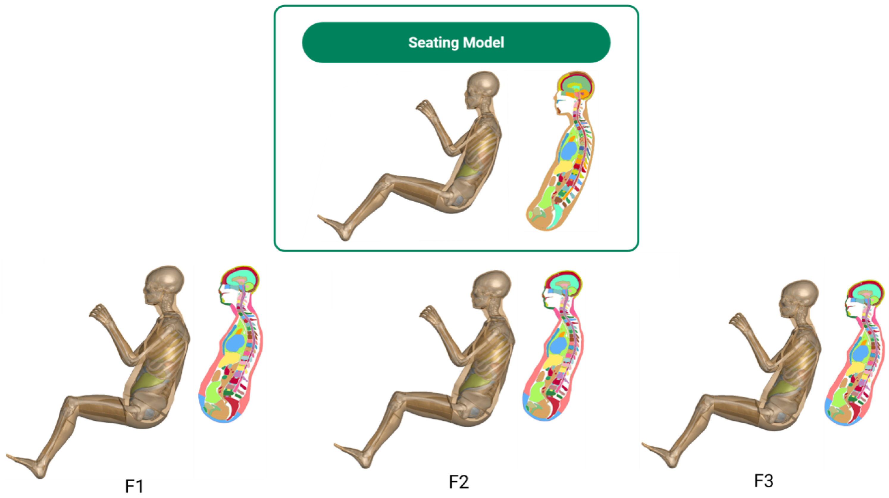
Figure 13 presents the resulting seated configurations for male models (M1–M3) and Figure 14 present female (F1–F3) models across percentile variations. The transformed meshes successfully captured posture curvature and skeletal articulation consistent with the original seated baselines while maintaining internal anatomical fidelity. Cutaway views reveal well-aligned organs, spinal curvature, and soft tissue contours, indicating an accurate transfer of internal geometry through the displacement seeded method.
Figure 13.
Seated male THUMS morphs generated through displacement seeded posture transfer. (Top): reference seated THUMS baseline; (bottom row): percentile-specific morphs (M1—98th, M2—80th, M3—50th). Each configuration preserves spinal curvature, pelvic rotation, and internal organ alignment.
Figure 14.
Seated female THUMS morphs generated through displacement seeded posture transfer. (Top): baseline seated reference; (bottom row): percentile-specific morphs (F1—98th, F2—80th, F3—50th). Anatomical alignment and curvature continuity are maintained across percentile variations.
A quantitative evaluation of element quality confirmed that mesh integrity in the seated morphs closely matched that of the standing configurations. As shown in Figure 15, the maximum values for warpage, aspect ratio, skewness, and Jacobian remained within acceptable thresholds for all percentile variations. Observed values included warpage values below 100° for males and 90° for females, aspect ratios under 30, skewness values below 80°, and no inverted elements. These results confirm that the displacement seeded transformation maintained numerical stability suitable for high-fidelity finite element simulations.
Figure 15.
Maximum element quality metrics (warpage, aspect ratio, skewness, and Jacobian) for seated male (a–d) and female (e–h) models. All percentile morphs remained within established thresholds, indicating strong element stability across both sexes.
The distribution of element quality violations is summarized in Figure 16. Aspect ratio remained the most affected metric but accounted for less than 0.36% of all elements, while warpage, skewness, and Jacobian violations were negligible (<0.05%). For the male seated morphs, violations were primarily concentrated in the head, neck, and upper torso areas. In contrast, the female models exhibited a slightly different distribution pattern. The overall violation magnitudes were lower; additional concentrations appeared in the upper leg and pelvic regions, reflecting localized mesh stretching along the thigh surface where contact curvature is highest. This difference highlights subtle geometric and anatomical variations in seated posture between male and female body shapes, particularly in lower body articulation.
Figure 16.
Percentage of element quality violations for seated male (a) and female (b) morphs. Aspect ratio violations dominate but remain around 0.36% with negligible warpage, skewness, and Jacobian issues. Pie charts indicate that male violations are concentrated in the head and torso, while female models also include localized violations in the leg regions due to curvature-induced stretching during posture transfer.
The displacement seeded posture transfer method successfully generated anatomically accurate seated morphs across all percentile variations while maintaining high mesh integrity. The overall mesh quality remained consistent with the standing configurations, and most element violations were minor and spatially localized. Increased violations in the head and neck regions were primarily attributed to differences in element resolution between the original THUMS meshes and the morphed configurations, where finer discretizations around cranial and cervical structures amplified local metric sensitivity. In contrast, the female models exhibited additional minor violations in the upper legs and pelvic regions, reflecting anatomical curvature and contact induced stretching during seated adaptation. Despite these localized discrepancies, the seated morphs preserved structural continuity and numerical stability, confirming their suitability for high G and occupant safety simulations where posture fidelity and element quality are critical.
4. Discussion
The RBF morphing pipeline developed in this study personalizes THUMS full body finite element models across 50th, 80th, and 98th percentiles using ANSUR II anthropometry and skin surface target meshes. Geometric accuracy in the standing posture shows an overall DICE similarity of about 0.40 with substantially higher agreement in the chest region at approximately 0.85. The corresponding HD95 values are in the order of 20–30 mm overall and 15–20 mm in the chest region. This pattern indicates that while the global correspondence over the entire body surface is moderate, the regions that are most relevant for chest and spinal loading exhibit strong agreement with the target geometry.
Compared with approaches that combine coherent point drift (CPD) and RBF interpolation, such as the method reported by Yuan et al. [22], our pipeline demonstrates comparable performance in key anatomical regions. Yuan et al. achieved average skeletal errors below 3 mm but also reported maximum discrepancies of up to 15 mm in areas like the ribcage and pelvis. In contrast, our method emphasizes full-body personalization and still achieves chest-region DICE values of approximately 0.85 and HD95 values of 15–20 mm for the male standing morphs. These findings indicate that the pipeline performs reliably in regions that are most critical for biomechanical assessment. The overall body-surface DICE value of about 0.40 is lower because it reflects differences across the entire external surface, particularly in distal areas where small positional offsets lead to proportionally greater reductions in overlap.
Mesh quality evaluations for both standing and seated postures show that the morphed models maintain surface integrity comparable to the original THUMS meshes. In the standing configuration, male models exhibit aspect ratio violation rates below 40% and female models remain below 32%, closely matching the baseline distributions. Warpage violations stay low at approximately 4–5% for both sexes, and skew and Jacobian violations remain under 5% and 12%, respectively. Similar trends appear in the seated posture, where male aspect ratio violations remain below 35% and female violations stay below 32%. Warpage remains low at 2–5%, and skew and Jacobian violations stay within 3% and 10% across all percentiles. The regional distribution of violations also aligns with the original THUMS model with most issues occurring in the head and neck for males and shifting toward the torso for females. These consistent patterns across both postures indicate that neither the morphing process nor the standing-to-seated transformation introduces substantial degradation to the surface mesh, and the resulting models remain suitable for dynamic simulation and downstream biomechanical analysis.
Posture-dependent transformations further demonstrate that standing morphs can be repositioned into seated configurations while retaining the mesh quality trends observed in the standing posture. Slight geometric degradations in highly flexed regions are consistent with observations from posture adaptation and image registration studies, where complex joint articulations remain challenging to represent accurately with purely geometric methods. Nevertheless, the overall behavior of the morphed seated models suggests that the pipeline can support simulations in seated occupant scenarios once boundary conditions and loading environments are specified appropriately. The geometric fidelity achieved in the chest region and the robust preservation of element quality during the standing-to-seated transformation directly translate to improved simulation reliability in high G aerospace environments. In ejection seat scenarios (typically 12–14 Gz) or carrier landings, accurate spinal alignment and ribcage circumference are paramount for predicting spinal compression and restraint interaction. By aligning the HBM morphology to specific aviator anthropometry, this framework minimizes the initial positioning errors that often propagate into non-physical energy modes during dynamic events. Specifically, this improved geometric fidelity, particularly in the cervical and torso regions, is expected to reduce the variability of predicted cervical strains and contact forces by approximately 10 to 20% under high G loading compared to the baseline. This foundational geometric validation enables the subsequent fatigue damage modeling and injury risk assessment presented in our forthcoming companion study [38], where these personalized models are subjected to realistic cockpit acceleration profiles.
5. Limitations
While the current pipeline successfully generates personalized HBMs in both standing and seated postures, it remains inherently dependent on the articulation capabilities of the baseline model. THUMS provides posture-specific variants with increased mesh complexity in the seated configuration, including additional elements that support joint flexion and anatomical deformation. This reliance limits its applicability to alternative human body models, such as those from the Global Human Body Models Consortium (GHBMC), where comparable posture variants or consistent mesh topologies may be absent. Extending the pipeline to such models could introduce element mismatches potentially compromising mesh integrity.
A further limitation is the absence of tissue-specific material property adjustments with the pipeline defaulting to THUMS’s generic constitutive models. This may inadequately reflect inter-individual variability in soft tissue stiffness or viscoelasticity under high G loading, which is a critical factor given the prevalence of cervical spine degeneration among fighter pilots [40]. The lack of empirical validation through centrifuge experiments or in vivo kinematic data further restricts confidence in predicting injury mechanisms, such as spinal strain. Computational demands also pose a constraint; transforming meshes with approximately 1.5–1.6 million elements via PyGeM, as outlined in the workflow of Figure 1, requires significant resources, potentially hindering real-time applications in clinical or safety engineering contexts.
6. Conclusions
This paper demonstrates that an RBF-based morphing pipeline can effectively personalize THUMS models across a wide anthropometric range while achieving high geometric fidelity in the standing posture. Overall, the DICE coefficient was approximately 0.40 with substantially higher performance in the chest region (0.85). Meanwhile, HD95 values ranged 20–30 mm overall and 15–20 mm in the chest region. The pipeline also successfully extends these standing morphs into seated configurations using displacement-based posture morphing, maintaining anatomical continuity and mesh quality even under large joint flexion. By combining automated landmark extraction with external skin-surface targets, the framework enables scalable and anatomically coherent morphs without manual segmentation or cadaveric data, and it avoids inverted elements throughout the mesh. These results provide a strong foundation for personalized simulations in aerospace and defense applications particularly for evaluating spinal loading and injury mechanisms in high G environments. Future work should incorporate alternative skin sources, tissue-specific material properties, and hybrid morphing strategies to further minimize localized distortions and expand applicability across broader demographic profiles.
Author Contributions
Conceptualization, A.N.R., T.R.D. and R.H.K.; methodology, A.N.R. and R.H.K.; software, A.N.R.; validation, A.N.R. and T.R.D.; formal analysis, A.N.R.; investigation, A.N.R.; resources, R.H.K.; data curation, A.N.R.; writing, original draft preparation, A.N.R.; writing, review and editing, A.N.R., T.R.D. and R.H.K.; visualization, A.N.R. and T.R.D.; supervision, R.H.K.; project administration, R.H.K.; funding acquisition, R.H.K. All authors have read and agreed to the published version of the manuscript.
Funding
This work was supported by the U.S. Air Force Research Laboratory (AFRL), 711th Human Performance Wing, Wright-Patterson Air Force Base, Ohio, under Contract No. FA8650-22-C-6462.
Institutional Review Board Statement
Not applicable.
Informed Consent Statement
Not applicable.
Data Availability Statement
All data supporting the findings of this study are contained within the manuscript and Appendix A.
Acknowledgments
The authors gratefully acknowledge Peter Le, Chris Dooley, Tara Crowl, and 2Lt Brandon K. Brown, USAF, of the 711th Human Performance Wing for their valuable programmatic oversight, technical guidance, and continued support throughout the project. The authors also thank Jingwen Hu of the University of Michigan Transportation Research Institute for insightful discussions and generous assistance with the HumanShape tool. The Penn State Institute for Computational and Data Sciences (ICDS) is acknowledged for providing high-performance computing resources and LS-DYNA (R12) software support. Additional institutional resources provided by The Pennsylvania State University are sincerely appreciated.
Conflicts of Interest
The authors declare no conflicts of interest.
Appendix A. Reproducibility Details
Appendix A.1. Code Availability and Implementation
To facilitate complete reconstruction of the work, the source code and configuration files have been released in a public repository at https://github.com/PSUCompBio/AF_Morphing_Project. The morphing pipeline relies on the PyGeM library (https://github.com/mathLab/PyGeM, accessed on 1 June 2024) for geometric deformation and is implemented as follows:
- 1.
- Standing Posture Morphing:
- creating_RBF_standing.py: generates the RBF parameter file (.prm) based on manually selected anatomical landmarks on the source (THUMS) and target geometries.
- RBF_Standing.py: uses PyGem to compute the RBF-based deformation field from the (.prm) inputs, applies the mapping to the full THUMS mesh and exporting the morphed LS-DYNA (R12) keyword file (.k).
- 2.
- Seating Posture Morphing:
- creating_RBF_seating.py: constructs control points specific to the seated configuration.
- RBF_Seating.py: applies the displacement field to transform the mesh into the final seated posture.
- 3.
- Configuration: Target anthropometry for each execution is controlled via the percentile variable (e.g., 50, 80, 98) defined within the scripts, ensuring that input/output paths align with the correct target model folder.
Appendix A.2. RBF Kernel Parameters
The radial basis function interpolation utilized a multi-quadric biharmonic spline compact support kernel to ensure matrix sparsity and numerical stability. The support radius was set to 0.5, which acts as a scaling parameter controlling the spatial spread and smoothness of the interpolation.
Appendix A.3. Boundary Prescribed Motion Implementation
The standing-to-seated posture transition was implemented using a displacement-seeded morphing strategy rather than LS-DYNA’s (R12) *BOUNDARY_PRESCRIBED_MOTION_SET. Prescribed kinematic motion was not suitable because the original standing THUMS mesh contained fewer nodes and geometric detail than the seated configuration, resulting in mismatched topology between the source and target models. Consequently, a simple rotational boundary condition could not reliably reproduce the seated posture or capture the additional anatomical features required in the seated mesh. To address this, supplemental control points and seeded displacements were introduced to guide the deformation, enabling the standing model to be smoothly morphed into the seated configuration while maintaining mesh quality and anatomical consistency.
Appendix A.4. Landmark Selection Criteria
Landmarks were manually selected based on palpable bony prominences and distinct surface features. Key locations included the following:
- Head: Sellion, Bilateral Tragion, Vertex.
- Torso: Suprasternale, Xyphoid process, Bilateral Acromion.
- Pelvis: ASIS (left/right), PSIS (left/right).
- Extremities: Lateral/Medial epicondyles (humerus and femur), Lateral/Medial malleoli.
The anatomical landmark locations and nodal indices used for morphing are provided in the repository creating_RBF_seating.py and creating_RBF_standing.py to allow an exact replication of the deformation procedure.
References
- Sanchez-Molina, D.; Velazquez-Ameijide, J.; Arregui-Dalmases, C.; Crandall, J.R.; Untaroiu, C.D. Minimization of Analytical Injury Metrics for Head Impact Injuries. Traffic Inj. Prev. 2012, 13, 278–285. [Google Scholar] [CrossRef] [Scilit] [PubMed]
- Shigeta, K.; Kitagawa, Y.; Yasuki, T. Development of Next Generation Human FE Model Capable of Organ Injury Prediction. In Proceedings of the 21st Enhanced Safety of Vehicles (ESV) Conference, Stuttgart, Germany, 15–18 June 2009; pp. 15–18. [Google Scholar]
- Vavalle, N.A.; Schoell, S.L.; Weaver, A.A.; Stitzel, J.D.; Gayzik, F.S. Application of radial basis function methods in the development of a 95th percentile male seated FEA model. Stapp Car Crash J. 2014, 58, 361–384. [Google Scholar] [CrossRef] [Scilit] [PubMed]
- Mokhtar, A.A.; Hyncik, L. A comprehensive review of human body model in different crash scenarios: Active and passive models. Int. J. Crashworthiness 2025, 30, 1–13. [Google Scholar] [CrossRef] [Scilit]
- Meng, Y.; Buckland, E.; Untaroiu, C. Numerical investigation of driver injury risks in car-to-end terminal crashes using a human finite element model. Comput. Methods Biomech. Biomed. Eng. 2024, 29, 245–255. [Google Scholar] [CrossRef] [Scilit] [PubMed]
- Frazer, L.; Kote, V.; Hostetler, Z.; Davis, M.; Nicolella, D.P. A comparative analysis of dimensionality reduction surrogate modeling techniques for full human body finite element impact simulations. Comput. Methods Biomech. Biomed. Eng. 2024, 27, 1250–1263. [Google Scholar] [CrossRef] [Scilit] [PubMed]
- Toyota Motor Corporation. Total HUman Model for Safety (THUMS). Available online: https://www.toyota.co.jp/thums/ (accessed on 15 October 2025).
- Global Human Body Models Consortium. GHBMC. Available online: https://www.ghbmc.com/ (accessed on 15 October 2025).
- Davis, M.L.; Koya, B.; Schap, J.M.; Gayzik, F.S. Development and Full Body Validation of a 5th Percentile Female Finite Element Model. Stapp Car Crash J. 2016, 60, 509–544. [Google Scholar] [CrossRef] [Scilit]
- Hwang, E.; Hallman, J.; Klein, K.; Rupp, J.; Reed, M.; Hu, J. Rapid Development of Diverse Human Body Models for Crash Simulations Through Mesh Morphing; Technical Report 2016-01-1491; SAE International: Warrendale, PA, USA, 2016. [Google Scholar] [CrossRef] [Scilit]
- Gayzik, F.S.; Moreno, D.P.; Vavalle, N.A.; Rhyne, A.C.; Stitzel, J.D. Development of the global human body models consortium mid-sized male full body model. In Proceedings of the International Workshop on Human Subjects for Biomechanical Research; National Highway Traffic Safety Administration: Washington, DC, USA, 2011; Volume 39. [Google Scholar]
- Iwamoto, M.; Nakahira, Y.; Kimpara, H. Development and Validation of the Total HUman Model for Safety (THUMS) Toward Further Understanding of Occupant Injury Mechanisms in Precrash and During Crash. Traffic Inj. Prev. 2015, 16, S36–S48. [Google Scholar] [CrossRef] [Scilit]
- Larsson, K.J.; Pipkorn, B.; Iraeus, J.; Forman, J.; Hu, J. Evaluation of a diverse population of morphed human body models for prediction of vehicle occupant crash kinematics. Comput. Methods Biomech. Biomed. Eng. 2021, 25, 1125–1155. [Google Scholar] [CrossRef] [Scilit]
- Iwamoto, M.; Kisanuki, Y.; Watanabe, I.; Furusu, K.; Miki, K.; Hasegawa, J. Development of a Finite Element Model of the Total HUman Model for Safety (THUMS) and Application to Injury Reconstruction. In Proceedings of the 20th International Technical Conference on the Enhanced Safety of Vehicles, IRCOBI, Munich, Germany, 18–20 September 2002. [Google Scholar]
- Beillas, P.; Berthet, F. An investigation of human body model morphing for the assessment of abdomen responses to impact against a population of test subjects. Traffic Inj. Prev. 2017, 18, S142–S147. [Google Scholar] [CrossRef] [Scilit]
- Panzer, M.B.; Myers, B.S.; Capehart, B.P.; Bass, C.R. Development of a Finite Element Model for Blast Brain Injury and the Effects of CSF Cavitation. Ann. Biomed. Eng. 2012, 40, 1530–1544. [Google Scholar] [CrossRef] [Scilit]
- Wang, Y.; Cao, L.; Bai, Z.; Reed, M.P.; Rupp, J.D.; Hoff, C.N.; Hu, J. A parametric ribcage geometry model accounting for variations among the adult population. J. Biomech. 2016, 49, 2791–2798. [Google Scholar] [CrossRef] [Scilit] [PubMed]
- Giordano, C.; Li, X.; Kleiven, S. Performances of the PIPER scalable child human body model in accident reconstruction. PLoS ONE 2017, 12, e0187916. [Google Scholar] [CrossRef] [Scilit] [PubMed]
- Wittek, A.; Grosland, N.M.; Joldes, G.R.; Magnenat-Thalmann, N.; Miller, K. From Finite Element Meshes to Clouds of Points: A Review of Methods for Generation of Computational Biomechanics Models for Patient-Specific Applications. Ann. Biomed. Eng. 2016, 44, 3–15. [Google Scholar] [CrossRef] [Scilit] [PubMed]
- Kimpara, H.; Lee, J.B.; Yang, K.H.; King, A.I.; Iwamoto, M.; Watanabe, I.; Miki, K. Development of a Three-Dimensional Finite Element Chest Model for the 5th Percentile Female. In Proceedings of the 49th Stapp Car Crash Conference; SAE Technical Paper 2005-22-0012; SAE International: Washington, DC, USA, 2005. [Google Scholar] [CrossRef] [Scilit]
- Jolivet, E.; Lafon, Y.; Petit, P.; Beillas, P. Comparison of Kriging and Moving Least Square Methods to Change the Geometry of Human Body Models. In Proceedings of the 59th Stapp Car Crash Conference; SAE Technical Paper 2015-22-0013; SAE International: New Orleans, LA, USA, 2015. [Google Scholar] [CrossRef] [Scilit]
- Yuan, Q.; Jiang, B.; Zhu, X.; Hu, J.; Wang, Y.; Chou, C.C.; Xu, S. A fast methodology for generating skeletal FEM with detailed human geometric features based on CPD and RBF algorithms. Sci. Rep. 2023, 13, 8864. [Google Scholar] [CrossRef] [Scilit]
- Andreassen, T.E.; Hume, D.R.; Hamilton, L.D.; Higinbotham, S.E.; Shelburne, K.B. Automated 2D and 3D finite element overclosure adjustment and mesh morphing using generalized regression neural networks. Med Eng. Phys. 2024, 126, 104136. [Google Scholar] [CrossRef] [Scilit]
- Bookstein, F.L. Principal warps: Thin-plate splines and the decomposition of deformations. IEEE Trans. Pattern Anal. Mach. Intell. 1989, 11, 567–585. [Google Scholar] [CrossRef] [Scilit]
- Trochu, F. A contouring program based on dual kriging interpolation. Eng. Comput. 1993, 9, 160–177. [Google Scholar] [CrossRef] [Scilit]
- Wu, G.; Siegler, S.; Allard, P.; Kirtley, C.; Leardini, A.; Rosenbaum, D.; Whittle, M.; D’Lima, D.D.; Cristofolini, L.; Witte, H.; et al. ISB Recommendation on Definitions of Joint Coordinate System of Various Joints for the Reporting of Human Joint Motion—Part I: Ankle, Hip, and Spine. J. Biomech. 2002, 35, 543–548. [Google Scholar] [CrossRef] [Scilit]
- Dumas, R.; Chèze, L.; Verriest, J.P. Adjustments to McConville et al. and Young et al. Body Segment Inertial Parameters. J. Biomech. 2007, 40, 543–553. [Google Scholar] [CrossRef] [Scilit]
- Reed, M.P.; Parkinson, M.B. Modeling anthropometric variation in the human torso for design automation. J. Biomech. Eng. 2008, 130, 061008. [Google Scholar] [CrossRef] [Scilit]
- Zhang, K.; Cao, L.; Fanta, A.; Reed, M.P.; Neal, M.O.; Wang, J.T.; Hu, J. An automated method to morph finite element whole-body human models with a wide range of stature and body shape. J. Biomech. 2017, 60, 253–260. [Google Scholar] [CrossRef] [Scilit]
- Gordon, C.C.; Blackwell, C.L.; Bradtmiller, B.; Parham, J.L.; Barrientos, P.; Paquette, S.P.; Corner, B.D.; Carson, J.M.; Venezia, J.C.; Rockwell, B.M.; et al. 2012 Anthropometric Survey of U.S. Army Personnel: Methods and Summary Statistics; Technical Report NATICK/TR-15/007; U.S. Army Natick Soldier Research, Development and Engineering Center: Natick, MA, USA, 2014; Available online: https://apps.dtic.mil/sti/citations/ADA611869 (accessed on 17 November 2025).
- Heimann, T.; Delingette, H. Model-based Segmentation. In Biomedical Image Processing; Springer: Berlin/Heidelberg, Germany, 2010; pp. 279–303. [Google Scholar]
- Li, X.; Yuan, Q.; Lindgren, N.; Huang, Q.; Fahlstedt, M.; Östh, J.; Pipkorn, B.; Jakobsson, L.; Kleiven, S. Personalization of human body models and beyond via image registration. Front. Bioeng. Biotechnol. 2023, 11, 1169365. [Google Scholar] [CrossRef] [Scilit]
- Reyes Kadozono, A.; DeWitt, T.; Kraft, R.H. Seat Angle Effects on Disc Degeneration for Pilots in High-G Environments. In Proceedings of the ASME International Mechanical Engineering Congress and Exposition (IMECE), Portland, OR, USA, 17–21 November 2024. [Google Scholar]
- Reed, M.P.; Raschke, U.; Tirumali, R.; Parkinson, M. Developing and Implementing Parametric Human Body Shape Models in Ergonomics Software. In Proceedings of the 3rd International Digital Human Modeling Conference, Tokyo, Japan, 20–22 May 2014. [Google Scholar]
- Lindgren, N.; Yuan, Q.; Pipkorn, B.; Kleiven, S.; Li, X. Development of personalizable female and male pedestrian SAFER human body models. Traffic Inj. Prev. 2024, 25, 182–193. [Google Scholar] [CrossRef] [Scilit]
- Istrate, A.; Tezzele, M.; Dedà, A.; Rozza, G. PyGeM: Python Geometrical Morphing. Softw. Impacts 2021, 7, 100047. [Google Scholar] [CrossRef] [Scilit]
- Cook, R.D.; Malkus, D.S.; Plesha, M.E.; Witt, R.J. Concepts and Applications of Finite Element Analysis, 4th ed.; John Wiley & Sons: Hoboken, NJ, USA, 2007. [Google Scholar]
- Reyes, A.N.; DeWitt, T.R.; Kraft, R.H. Simulation of Personalized Human Body Models in High-G Aircraft Cockpit Environments for Predicting Disc Degeneration in Pilots. Biomechanics, 2026; manuscript in preparation.
- Ou, Y.; Akbari, H.; Bilello, M.; Da, X.; Davatzikos, C. Comparative evaluation of registration algorithms in different brain databases with varying difficulty: Results and insights. IEEE Trans. Med Imaging 2014, 33, 2039–2062. [Google Scholar] [CrossRef] [Scilit]
- Mastalerz, A.; Maruszyńska, I.; Kowalczuk, K.; Garbacz, A.; Maculewicz, E. Pain in the Cervical and Lumbar Spine as a Result of High G-Force Values in Military Pilots—A Systematic Review and Meta-Analysis. Int. J. Environ. Res. Public Health 2022, 19, 13413. [Google Scholar] [CrossRef] [Scilit]

Disclaimer/Publisher’s Note: The statements, opinions and data contained in all publications are solely those of the individual author(s) and contributor(s) and not of MDPI and/or the editor(s). MDPI and/or the editor(s) disclaim responsibility for any injury to people or property resulting from any ideas, methods, instructions or products referred to in the content. |
© 2026 by the authors. Licensee MDPI, Basel, Switzerland. This article is an open access article distributed under the terms and conditions of the Creative Commons Attribution (CC BY) license.