Article

Evolution of Gait Biomechanics During a Nine-Month Exercise Program for Parkinson’s Disease: An Interventional Cohort Study

by
Dielise Debona Iucksch
1,
Elisangela Ferretti Manffra
2,* and
Vera Lucia Israel
3
1
Postgraduate Program in Physical Education, Federal University of Paraná, Curitiba 81531-980, Brazil
2
Health Technology Graduate Program, Pontifícia Universidade Católica do Paraná, Curitiba 80215-901, Brazil
3
Department of Physical Therapy Prevention and Rehabilitation, Federal University of Paraná, Curitiba 81531-980, Brazil
*
Author to whom correspondence should be addressed.
Biomechanics 2025, 5(3), 53; https://doi.org/10.3390/biomechanics5030053 (registering DOI)
Submission received: 8 May 2025 / Revised: 19 July 2025 / Accepted: 27 July 2025 / Published: 1 August 2025
(This article belongs to the Special Issue Gait and Balance Control in Typical and Special Individuals)

Abstract

It is well established that combining exercise with medication may benefit functionality in individuals with PD (Parkinson’s disease). However, the long-term evolution of gait biomechanics under this combination remains poorly understood. Objectives: This study aims to analyze the evolution of spatiotemporal gait parameters, kinetics, and kinematics throughout a long-term exercise program conducted in water and on dry land. Methods: We have compared the trajectories of biomechanical variables across the treatment phases using statistical parametric mapping (SPM). A cohort of fourteen individuals with PD (mean age: 65.6 ± 12.1 years) participated in 24 sessions of aquatic exercises over three months, followed by a three-month retention phase, and then 24 additional sessions of land-based exercises. Three-dimensional gait data and spatiotemporal parameters were collected before and after each phase. Two-way ANOVA with repeated measures was used to compare spatiotemporal parameters. Results: The walking speed increased while the duration of the double support phase decreased. Additionally, the knee extensor moment consistently increased in the entire interval from midstance to midswing (20% to 70% of the stride period), approaching normal gait patterns. Regarding kinematics, significant increases were observed in both hip and knee flexion angles. Furthermore, the abnormal ankle dorsiflexion observed at the foot strike disappeared. Conclusions: These findings collectively suggest positive adaptations in gait biomechanics during the observation period.

1. Introduction

Parkinson’s disease (PD) is a multifaceted and intricate neurodegenerative condition [1] characterized by a spectrum of motor and non-motor symptoms that significantly impact independence and quality of life [2]. Among these symptoms, gait abnormalities stand out as one of the most prevalent and disabling motor impairments [3].
In PD, specific gait dysfunctions manifest as reductions in speed, step and stride lengths, swing time, and range of motion [3,4]. Additionally, alterations in the timing and amplitude of leg muscle activation are observed [5,6]. Ankle angle kinematics are often notably affected, alongside reductions in joint moments and power generation during pre-swing [7,8,9,10]. These gait impairments significantly contribute to the functional limitations experienced by individuals with PD.
The primary treatment for motor dysfunction in individuals with PD is medication. However, compelling evidence suggests that combining it with physical exercise may significantly enhance quality of life and motor outcomes, particularly those related to gait [11,12,13]. Consequently, physical therapy guidelines have recommended physical exercises for PD patients [12,14,15].
Several studies on gait function have indicated improvements in functional and spatiotemporal gait outcomes following physical exercise interventions [12]. For example, Rafferty et al. [16] compared the effects of progressive resistance exercise and a multimodal exercise program on spatiotemporal and stability-related gait outcomes, noting enhanced off-medication gait velocity and cadence in both groups. Shen et al. [17] demonstrated that balance and gait training with augmented feedback enhanced gait velocity and stride length, whereas the active control group showed improvement in gait velocity only. Additionally, individuals with PD who participated in aquatic gait training significantly improved spatiotemporal parameters [6].
In a specific guideline for exercise protocols targeting gait function, Ni et al. [12] recommended a comprehensive approach that includes multidimensional physical therapy (balance and gait training), treadmill gait training with body weight support, cycling, aquatic exercises, resistance training, and complementary treatments such as tai chi, yoga, and tango. The authors endorsed various modalities because no single approach was deemed superior to the others when considering overall gait function outcomes.
More recently, systematic reviews with meta-analyses have highlighted the superiority of aquatic exercises (AEs) for enhancing functional mobility and balance [11,13,18,19]. Hvingelby et al. [13] concluded that aquatic therapy with dual-task training exhibited the most significant effect on dynamic gait outcomes, such as scores in Timed Up and Go, Dynamic Gait, and other gait functional scales, compared to other exercise protocols. Conversely, Ernst et al. [11] concluded that aqua-based exercises were superior to other forms of exercise for improving quality of life and were equivalent to them regarding motor signs.
In summary, it is well established that both water-based and land-based physical exercises can improve functional gait outcomes in PD. However, studies have primarily focused on spatiotemporal parameters or functional mobility scales [20], overlooking the biomechanics of gait. One study has described changes in gait kinematics and muscle recruitment patterns following aquatic training protocols [6,21], but, to our knowledge, none have explored gait kinetics. Therefore, a gap remains in understanding how gait kinetics evolve in individuals with PD undergoing interventions, especially aquatic ones, over the long term.
While studies analyzing gait kinetics in PD after AE training are lacking, it has been demonstrated that healthy individuals exhibit different joint moments when walking underwater compared to on land [22,23]. On the other hand, a similar set of muscle synergies has been observed in both land and water walking [24]. Thus, training gait underwater may induce force production and muscle activation patterns that could influence gait on land.
More recently, a study analyzed the gait kinetics of individuals with PD in comparison with healthy individuals during walking and obstacle crossing, both with and without the use of virtual reality [25]. They observed a difference between groups regarding the maximum joint moments, especially in the sagittal plane, and also that virtual reality influenced the kinetics of both groups. Therefore, joint moments in PD might bring valuable information.
Given this scenario, this study aims to analyze the evolution of gait biomechanics throughout a long-term exercise program conducted in water and on dry land. We hypothesize that angular kinematics and kinetics will exhibit changes across time, related to a more functional gait.
Usually, studies on gait biomechanics in PD investigate the behavior of discrete parameters such as maximum, minimum, or average values during the step, stride, or some gait phase [26]. To provide a more comprehensive view of the phenomenon, we decided not to compare discretized values, but rather the entire trajectories of kinematic and kinetic variables along the stride. To do so, we employed the statistical parametric mapping (SPM) analysis [27], which also reduces the risk of bias introduced by the choice of specific discrete parameters.

2. Materials and Methods

This is a longitudinal interventional cohort study conducted over nine months during which individuals with Parkinson’s disease participated in a physical exercise program.
This study spanned over nine months and included assessment and intervention sessions, as depicted in Figure 1.
To ensure assessments and interventions were conducted during the “on” phase of medication, all sessions took place 1 to 2 h after taking dopaminergic medication. The assessment sessions were scheduled to match each participant’s usual medication routine. Since the exercise sessions were conducted in groups, some participants had to make minor adjustments to their medication schedules, with the counseling of their physicians.

2.1. Participants

Nineteen individuals diagnosed with Parkinson’s disease were initially recruited from the local association, the Parkinson Paraná Association. Four subjects withdrew from this study during the intervention phase, and one subject was excluded from the statistical analysis due to corrupted biomechanical data. We have conducted a complete-case analysis, i.e., only data from 14 participants with Parkinson’s disease (9 men) were analyzed.
The participants’ average age, height, and weight were 65.6 ± 12.1 years, 165.4 ± 9.6 cm, and 70.5 ± 10.9 kg, respectively. The average score on the modified Hoehn and Yahr (H&Y) scale was 2.8 ± 1.1, with 8 participants classified in stages I–II and 6 in stages III–IV. The average score on the Unified Parkinson’s Disease Rating Scale (UPDRS) part III was 14.4 ± 6.1 points, and on the Mini-Mental State Examination, it was 27.1.
This study’s inclusion criteria were a confirmed diagnosis of idiopathic Parkinson’s disease, an age between 40 and 90, the ability to walk unassisted, a Mini-Mental State Examination score equal to or above 20, and medical clearance to engage in physical exercise both on land and in a heated pool. Additionally, participants were required to maintain their regular physical activity unchanged throughout the study duration.
Exclusion criteria included the presence of other neurological conditions that affect gait or cognitive function, adjustments in the dosage of Parkinson’s disease medications during the study period, and failure to attend assessment sessions or participate in more than 10% of the exercise program sessions.

2.2. Assessment Sessions—Data Collection

Assessment sessions included the administration of clinical scales and the collection of biomechanical data.
Clinical scales included the Mini-Mental State Examination, part III of the Unified Parkinson’s Disease Rating Scale (UPDRS) [28], and the modified Hoehn and Yahr (H&Y) scale [29].
Biomechanical data were collected using a 3D motion tracking system equipped with eleven infrared cameras (Vicon Motion System Ltd., Oxford, UK) and a force plate (Advanced Mechanical Technology Inc., Watertown, USA). Fifteen reflective markers were positioned on anatomical landmarks in accordance with the plug-in gait lower body model [30].
Participants were instructed to walk along a 9 m pathway at a self-selected speed. Each session consisted of one trial of walking back and forth along the pathway for familiarization, followed by at least ten recording trials. Participants could take breaks during the session if needed to prevent fatigue.
The markers were sampled at 100 Hz, while the ground reaction forces (GRFs) were sampled at 1000 Hz. The Vicon Nexus® 2.5 software was used for raw data acquisition, as well as for the calculation of joint angles and moments. At least three gait cycles were analyzed for each participant and limb. After conducting consistency analyses, data from one stride of the most compromised lower limb and one stride of the least compromised lower limb for each subject were selected for further analysis of gait variables. We defined the most compromised limb as the one that first exhibited symptoms of the disease.
Customized signal processing scripts were developed in MATLAB R2018a (The Mathworks Inc., Natick, MA, USA) for filtering using a fourth-order zero-lag Butterworth low-pass filter with a 6 Hz cut-off frequency and time normalization to 100% gait cycle. Joint moments (measured in Nm) were normalized to the participant’s body mass (measured in kilograms).

2.3. Exercise Programs

Before starting the 12-week aquatic exercise (AE) program, the volunteers participated in two sessions to familiarize themselves with the environment. The water temperature was kept at approximately 33 °C. Following a 12-week retention period, participants began the 12-week land environment (LE) program. The sessions took place twice a week in both environments and lasted about 50 min.
The choice of 12-week interventions was based on the findings of Carroll et al. [31], and the idea of including both water and land exercises stemmed from clinical practice, where exercises are seldom performed solely in the AE. They typically co-occur in both environments or sequentially in alternating environments, as in this study.
The duration of the retention period was chosen to match that of the exercise programs. During that time, participants were instructed not to enroll in any other exercise programs or physical activities, except those they were already enrolled in during the intervention period. They were also instructed to maintain their habitual medication regimen. This was monitored through self-report and confirmed during follow-up contacts.
The exercise program was designed in accordance with GRADE recommendations for the core domains of gait and balance, as outlined in the European PD Guidelines [14]. Its structure and content were aligned with multidimensional physical therapy recommendations for improving gait and balance in individuals with Parkinson’s disease [12].
It encompassed multimodal exercises to maximize intervention effects. Each session included a warm-up, strength and power training exercises for the lower limbs, balance training, and a cool-down (Ai Chi or Tai Chi Chuan). We provide a brief overview of the program in the following paragraphs, and more details can be found in [32,33].
The warm-up in AE and LE included walking combined with coordinated movements of the upper limbs, trunk, and head. These were performed in various directions and at different speeds, incorporating turns, squats, skipping, and running. It involved walking forward, backward, and sideways at a comfortable pace, as well as performing hip and knee flexion with alternating lower limbs while rotating the trunk, using a kickboard (in AE) or a stick (in AE). Participants also ran or walked as fast as possible. Different instructions were provided in each environment to ensure proper movement execution. The warm-up lasted about 10 min.
The strength and power training was based on the protocol of Kanitz et al. [34] and consisted of kicks executed in the directions of hip and knee flexion/extension, as well as hip adduction/abduction. It progressed over the weeks by gradually increasing load, movement speed, number of repetitions, quality of execution, and overall difficulty [14]. During the first four weeks, the exercises in AE and LE consisted of two repetitions of 20 s of knee flexion–extension and hip abduction–adduction with each lower limb, without external resistance. From the 5th to the 8th week, the protocol progressed to three repetitions with external resistance provided by flotation devices and elastic bands in AE and LE, respectively. In the last four weeks, the protocol advanced to four 15 s repetitions with a 1 min and 30 s rest interval between them. Participants were always instructed to perform as many kicks as possible within the allotted time, aiming for the largest possible range of motion. The intensity of each repetition was monitored using the Borg Rating of Perceived Exertion Scale—CR20. The participants were instructed to report their perceived effort during the rest intervals between repetitions. For this study, the target intensity range was set between 13 and 17 points, corresponding approximately to 66% and 80% of the maximum voluntary force output, respectively, as recommended by the American College of Sports Medicine [35,36]. Verbal incentives were provided accordingly to maintain the target intensity.
Balance exercises consisted of walking with reduced base of support, maintaining challenging postures (e.g., far-reaching in unipodal support, standing on an unstable surface such as trampolines), and other motor tasks requiring postural control. New balance tasks were introduced every four weeks to increase complexity progressively, reduce reliance on upper limb support, and further narrow the base of support.
The cool-down consisted of Ai Chi or Tai Chi Chuan exercises in AE or LE, respectively. The Ai Chi exercises involve concentrating on breathing and performing slow movements of elevation of MMII, floating, and simply remaining calm, contemplating [37]. The Tai Chi Chuan movements were Wuchi/Tai Chi Opening, Carelessly Rolling Up the Sleeves, Open and Close the Hands, Single Whip, and Cloud Hands [38].
To ensure standardization of the exercise protocol across participants, the sessions were delivered to groups of participants by the same team of trained instructors throughout the programs.

2.4. Statistical Analyses

To compare spatiotemporal parameters across assessments, we employed a two-way repeated measures ANOVA. Normality distribution was assessed using the Shapiro–Wilk test and sphericity with the Mauchly test. In cases where sphericity was violated, a Greenhouse–Geisser correction was applied. Post hoc pairwise comparisons between assessments were conducted using the Bonferroni test. These statistical analyses were conducted using IBM SPSS Statistics (version 20), with statistical significance set at p < 0.05. Effect sizes were determined using partial eta squared (ηp2) and Cohen’s d, with ηp2 values classified as small (<0.06), moderate (>0.06 to 0.14), and large (>0.14) following Cohen’s guidelines [32] and Cohen’s, as small (<0.2), moderate (>0.2 to 0.5), medium (>0.5 to 0.8), and large (>0.8) [39]. The power (1-β) was estimated using the software G*Power v3.1.9.7, with the following settings: test family, ANOVA repeated measures within factors; type of power analysis, post hoc; effect size as in SPSS v.20; total sample size, 14; number of groups, 1; number of measurements, 4; and non-sphericity correction, 1.
To compare joint kinetics and kinematics trajectories across assessments and between limbs, we employed one-dimensional statistical parametric mapping (SPM) analysis [27,40].
We chose SPM because it enables us to compare the whole trajectories, thereby avoiding possible bias introduced by the choice of one or another discretized measure. Moreover, SMP allows for identifying the periods in time where trajectories might differ, and not only if they differ or not.
The method consists of generating a statistical parametric map by computing univariate F- or t-statistics at each point of the trajectories, namely SPM{F}, SPM{t}, or SnPM{t} for the ANOVA, parametric t-test, or non-parametric t-test, respectively [27]. Subsequently, random field theory is used to estimate the critical threshold above which only a percentage (e.g., 5% for α = 0.05) of random data is expected to exceed.
We conducted SPM analysis in MATLAB R2018a (The Mathworks Inc., Natick, MA, USA) using the open-source SPM1d code version M.0.4.7 (https://spm1d.org/) (accessed on 15 July 2025). We used the function “spm1d.stats.anova2onerm” to conduct a two-way ANOVA with two factors—limb and assessment—with repeated measures on factor assessment, and the function “spm.inference” with the significance level set to 0.05 to define the threshold in SPM{F}.
When ANOVA indicated significant differences, we used parametric or non-parametric paired t-tests to compare the assessments pairwise using the functions “spm1d.stats.ttest_paired” or “spm1d.stats.nonparam.ttest_paired”, respectively, and the function “spm.inference” with significance threshold set to 0.008 to account for the Bonferroni correction. This significance level is the ratio of α = 0.05 divided by six, corresponding to the number of pairwise comparisons.

3. Results

The spatiotemporal parameters are presented in Table 1.
After the entire observation period, walking speed increased significantly [F (1, 13) = 6.11, p = 0.02, ηp2 = 0.32, (1-β) = 0.33]. Post-LE assessments revealed significant increases in cadence [F (1, 13) = 22.97, p < 0.01, ηp2 = 0.63, (1-β) = 0.71], which were sustained throughout subsequent assessments [F (1, 13) = 9.58, p < 0.01, ηp2 = 0.42, (1-β) = 0.28]. Swing time also significantly improved in the post-LE phase [F (1, 13) = 5.33, p = 0.03, ηp2 = 0.29, (1-β) = 0.20]. Double support time showed improvement post-LE [F (1, 13) = 5.65, p = 0.03, ηp2 = 0.30, (1-β) = 21], and throughout the entire program duration [F (1, 13) = 10.2, p < 0.01, ηp2 = 0.44, (1-β) = 0.32]. However, there were no significant differences between the post-LE and pre-AE assessments for the other spatiotemporal variables.
The two-way ANOVA with statistical parametric mapping (SPM) analysis indicated no significant difference between limbs regarding angle kinetics (Figure 2, Main Factor A) or kinematics (Figure 3, Main Factor A). However, differences were observed across assessments for all joints concerning kinetics (Figure 2, Main Factor B) and kinematics (Figure 3, Main Factor B).
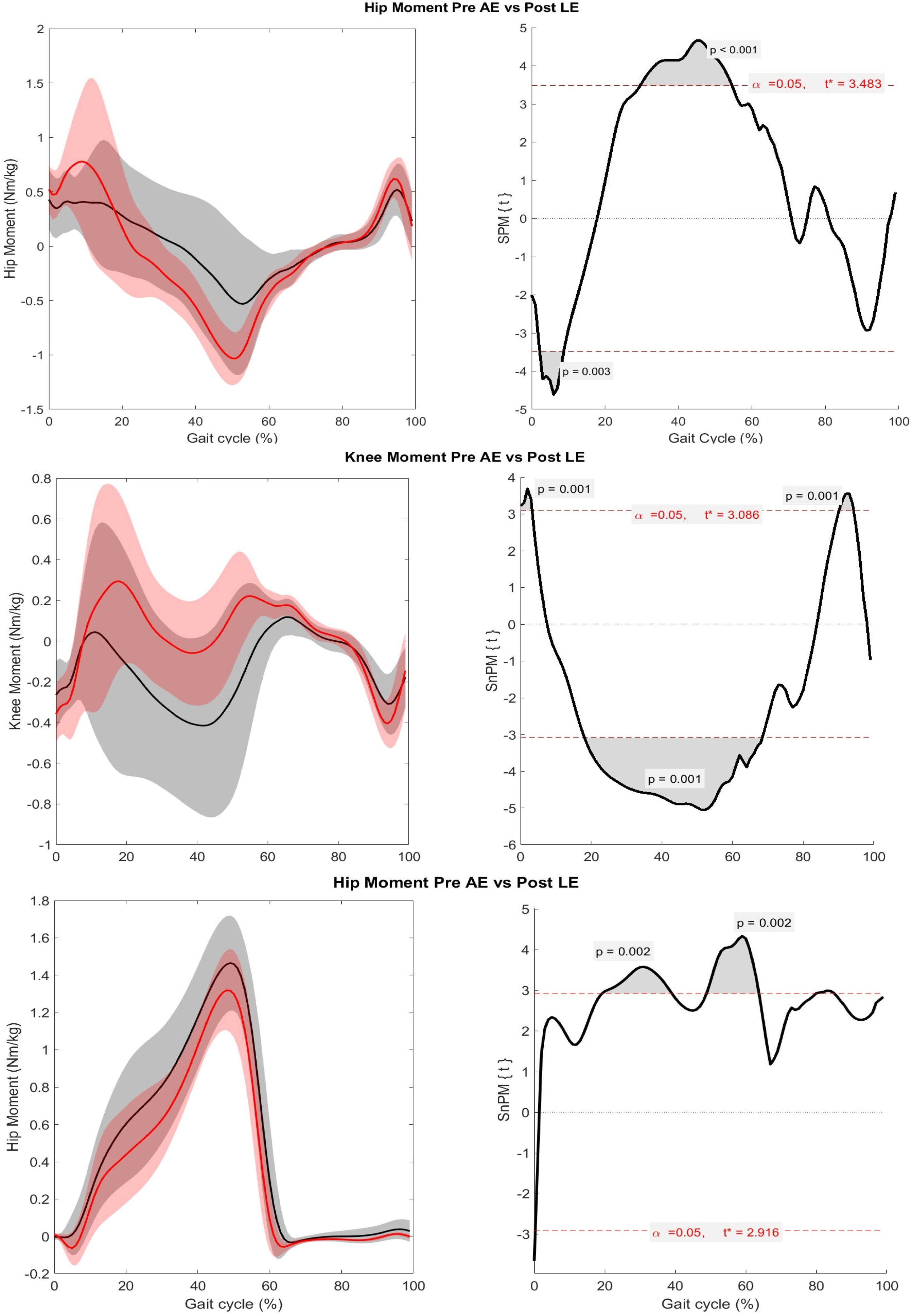
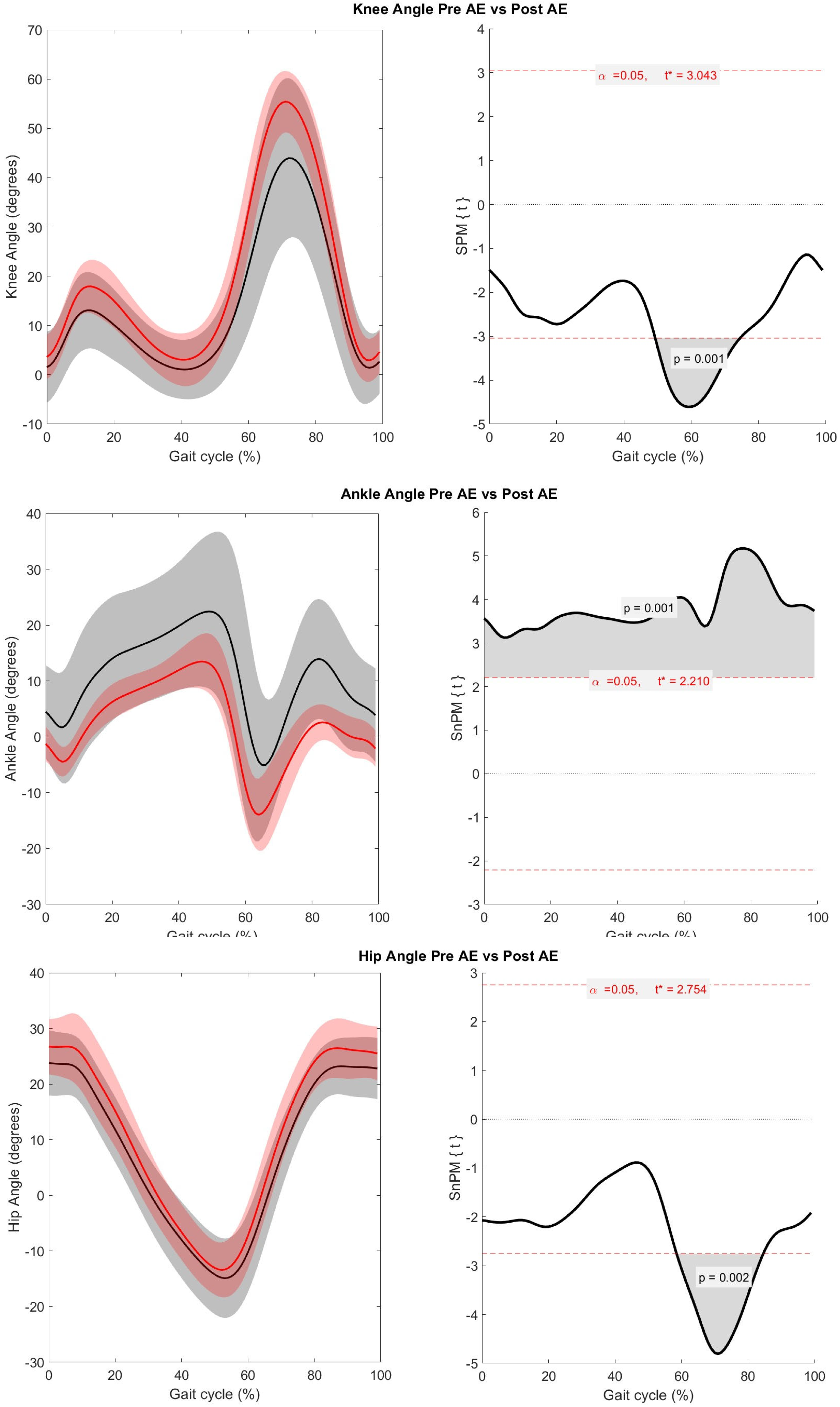
The SPM analysis revealed a difference only between pre-AE and post-LE. Figure 4 and Figure 5 illustrate the trajectories of joint kinetics and kinematics before AE and after LE, along with the t-values calculated via SPM.

4. Discussion

In this study, we investigated biomechanical changes in the gait of individuals with Parkinson’s disease (PD) over nine months while they participated in a multimodal exercise program in both aquatic and land environments. Following training in both environments, we observed changes in spatiotemporal, kinematic, and kinetic gait parameters.
A spatiotemporal reorganization of gait was evident, characterized by a decrease in double support time and an increase in cadence (see Table 1). There was a significant increase in gait speed following land-based exercise (post-LE) with an effect size classified as medium. Also, the observed change is larger than the moderate clinically meaningful difference in gait speed among individuals with PD in the on-medication state, which is 0.14 m/s [41]. The observed reduction in double support time might indicate enhanced postural control, as the center of mass remains within the base of support primarily during the double support phase of the gait cycle [42]. Notably, the decrease in stride length did not compensate for the increase in cadence. This observation is significant, given that a reduction in step length associated with increased cadence is characteristic of natural PD progression [43].
Gait speed consistently increased throughout the study, even during the retention period, which is unexpected in a neurodegenerative disease like PD. This may be attributed to the development of new motor strategies prompted by aquatic exercises. In fact, Volpe et al. [6] found differences in muscle activation among PD participants even several days after discontinuing underwater training activities. Moreover, recent research suggests that shared muscle synergies across different motor tasks maximize the generalization of motor learning effects [24]. Thus, our results hint at the transfer of skills from water to land environments. Future studies could examine this issue by analyzing kinematic, kinetic, or muscle activity patterns and identifying synergies.
Joint moments change significantly from pre-AE to post-LE (Figure 4). The knee flexor moment increases, associated with the eccentric contraction of the knee extensors to control weight acceptance [44]. The ankle moment shifts towards a dorsiflexor curve, indicating improved plantar flexion control after heel contact and better weight absorption in post-LE. The increased hip and knee extensor moments during the loading response relate to anterior trunk acceleration control due to the rapid body weight transfer onto the foot [45].
Due to the action of bi-articular muscles, joint moments influence adjacent segments. Therefore, changes in the hip moment are expected to occur concurrently with changes in the knee and ankle moments, as observed here. Considering the bi-articular nature of the rectus femoris, which spans both the hip and knee, coordinated improvements in hip and knee extensor moments—previously described by Winter [46] as part of a synergistic pattern—are expected. Such changes can also be influenced by improved motor unit recruitment, reduced co-contraction, and more efficient timing of muscle activation, all of which contribute to increased net joint torque production. Therefore, the observed improvements in gait cadence and velocity are likely the result of integrated neuromuscular and biomechanical enhancements, rather than isolated strength gains. However, as we did not use any other form of assessment, such as electromyography, we cannot categorically state that this was the mechanism underlying the observation.
During the mid and terminal stance phases, the hip flexor moment gradually increased, peaking at approximately 50% of the gait cycle (see Figure 4). In pre-AE, the knee exhibited a flexor moment throughout the stance phase. However, in post-LE, this pattern shifted towards a trajectory like that of healthy individuals, characterized by a double extension curve, with the peak of the extensor moment occurring at mid-stance [44]. This increase in hip flexion and knee extension moments may suggest improved alignment of the ground reaction vector, facilitating lower limb extension. In individuals with PD, this alignment may be associated with reduced energy consumption, as improved positioning and higher moments can help spare muscles from generating unnecessary contractions. To validate these hypotheses, future studies should analyze the ground reaction forces.
The ankle exhibited a reduced plantar flexor moment throughout the stance phase, including its peak value during the terminal stance/pre-swing post-LE (see Figure 4). This appears unexpected at first glance because the increase in velocity requires a larger impulse at foot-off. Our explanation is that subjects increased the knee extension moment to enhance the impulse without increasing the ankle plantarflexion moment. It is well-documented that the ankle is the most affected joint by kinematic and kinetic deficits in PD [10] and that individuals with PD generate less ankle power during the terminal stance phase than their healthy counterparts [9]. Moreover, Skinner et al. [47] reported that people with PD have a diminished capacity to produce ankle joint moments, forcing the adoption of alternative control strategies. We believe this is what occurred with the individuals in this study.
This compensatory mechanism aligns with previous findings in the literature. For instance, a recent treadmill-based study in individuals with PD demonstrated that, although dopaminergic medication increased gait speed and propulsion, improvements in joint torque occurred primarily at the hip, while ankle plantarflexion moments remained unchanged [48]. Similarly, Albani et al. [9] reported reduced ankle power in individuals with PD walking at their preferred speed, suggesting that distal musculature contributes less to propulsion under dopaminergic deficits. These observations support the hypothesis that, when ankle push-off capacity is compromised, individuals with PD increasingly rely on proximal joints—particularly the hip and knee—to maintain gait velocity. A similar redistribution of joint kinetics has been reported in healthy older adults [8,49]. Those authors emphasize that increased torque generation at proximal joints may relieve the ankle of its mechanical demands. Taken together, this body of evidence reinforces the interpretation that the reduction in ankle plantarflexor moment observed in this study—despite increased walking speed—reflects a proximal compensation strategy rather than a contradiction of expected gait mechanics.
In the terminal swing phase, a significantly higher knee flexor moment is observed post-LE (see Figure 4). This increase leads to typical values, as reported by Sloot et al. [44], and may be associated with the eccentric contraction of knee flexors, which decelerates the knee and prepares for ground contact [45].
Regarding the kinematics (Figure 5), significant increases in hip and knee peak flexion are observed post-LE during the initial swing phase. The coordinated movement of these joints facilitates the swing phase, enhancing foot clearance. Significantly, this increase in peak flexion during the initial swing phase does not interfere with the extension of either joint. This observation is significant given the tendency in PD to exhibit increased lower limb and trunk flexor patterns [50]. Furthermore, the ankle angle trajectory has shifted towards lower values throughout the entire stride, coming closer to normal gait. It is well-known that PD gait typically shows increased ankle dorsiflexion compared to normal [9,51]. After nine months, dorsiflexion at foot strike was absent. Across the entire stride, dorsiflexion was reduced, while plantarflexion was increased.
It is important to emphasize that this is an interventional cohort study, without a control group, which is a significant limitation. As such, it is not possible to attribute the observed changes solely to the exercise program. Results could be attributable to confounding factors, such as the natural progression of the disease or placebo effects. However, it is worth noticing that our results contrast with the expected decline in spatiotemporal variables as Parkinson’s disease progresses. It is well known that, over the years, Parkinson’s disease leads to a reduction in step length, with an increase in double support phase and cadence, resulting in shorter and more frequent step exchanges [1,43].
A key limitation of this study is its small sample size, especially due to the loss of five participants. As a result, the statistical power is limited. Additionally, the dropout of participants might have introduced bias, since those who remained could be more motivated or more concerned about their health.
Additionally, they were all recruited from the same Parkinson’s Association, which might have introduced some bias, as those who participate in such institutions could be more motivated and in better overall health than those who do not. We recognize that these factors reduce the external validity of the study. On the other hand, participants’ involvement in the same PD association facilitated consistent communication and monitoring, which likely supported adherence to the study guidelines.
Despite the study design and sample limitations, we consider our results valuable, given the burden of PD on healthcare systems. Families and patients also suffer from the hopelessness of a degenerative disease. The exercise protocols are sound and based on recognized international guidelines. Therefore, we recommend that future studies with more robust designs, such as multicentric randomized clinical trials or action observation therapy with one or both exercise protocols, be conducted. The challenges of standardizing the therapy across centers could be addressed using tele-rehabilitation strategies [52]. Moreover, the biomechanical evaluation could be performed in clinical settings using markerless motion capture systems [53].
Regarding biomechanical data, we have not analyzed ground reaction forces (GRFs) in this study. Although we recognize its importance, we believe it would not add much information, as our focus was on joint moments. Since the moments were calculated using Nexus software, we do not have the exact equations from it. Thus, establishing a mechanistic relationship between GRFs and the moments would not be possible anyway. Future studies could address this limitation through biomechanical and musculoskeletal modeling [53,54]. Incorporating musculoskeletal modeling would help in understanding the underlying mechanisms of the observed biomechanical changes and provide insights for developing motion strategies such as those in [54].
In summary, despite this study’s limitations, our findings collectively suggest beneficial changes in gait biomechanics, as hypothesized. Thus, encouraging future studies with more robust designs, adding other measures such as EMG, using markerless motion tracking, and incorporating musculoskeletal modeling.

5. Conclusions

Changes in spatiotemporal parameters, kinetics, and gait kinematics occurred following the nine months of intervention. Walking speed increased, while the duration of the double support phase decreased. Additionally, the knee extensor moment consistently increased throughout the interval from midstance to midswing (20% to 70% of the stride period), approaching standard gait patterns. Significant increases were also observed in both hip and knee peak flexion angles. Furthermore, the abnormal ankle dorsiflexion observed at foot strike disappeared.

Author Contributions

Conceptualization, D.D.I. and V.L.I.; data curation, D.D.I.; formal analysis, E.F.M.; funding acquisition, D.D.I. and V.L.I.; investigation, D.D.I.; methodology, D.D.I., E.F.M. and V.L.I.; project administration, V.L.I.; resources, D.D.I. and V.L.I.; software, E.F.M.; supervision, E.F.M. and V.L.I.; validation, D.D.I., E.F.M. and V.L.I.; visualization, E.F.M.; writing—original draft, D.D.I.; writing—review and editing, D.D.I. and E.F.M. All authors have read and agreed to the published version of the manuscript.

Funding

This study was financed in part by the Coordenação de Aperfeiçoamento de Pessoal de Nível Superior—Brasil (CAPES)—Finance Code 001.

Institutional Review Board Statement

This study was conducted in accordance with the Declaration of Helsinki and approved by the Federal University of Paraná Human Research Ethics Committee (protocol 3.780.224) for studies involving humans.

Informed Consent Statement

Informed consent was obtained from all subjects involved in this study.

Data Availability Statement

According to the Research Ethics Committee of the Federal University of Paraná (UFPR), Brazil, research data cannot be shared due to ethical issues regarding their secrecy and nondisclosure. Only the study results may be shared.

Acknowledgments

The authors would like to thank Luiz Augusto Kalva for his help with the scripts for data processing.

Conflicts of Interest

The authors declare no conflicts of interest.

Abbreviations

The following abbreviations are used in this manuscript:
PDParkinson’s Disease
SPMStatistical Parametric Mapping
AEAquatic Exercise
H&YModified Hoehn and Yahr
UPDRSUnified Parkinson’s Disease Rating Scale
LELand Exercise

References

  1. Kalia, L.V.; Lang, A.E. Parkinson’s disease. Lancet 2015, 386, 896–912. [Google Scholar] [CrossRef]
  2. Titova, N.; Padmakumar, C.; Lewis, S.J.G.; Chaudhuri, K.R. Parkinson’s: A syndrome rather than a disease? J. Neural Transm. 2017, 124, 907–914. [Google Scholar] [CrossRef]
  3. Mirelman, A.; Bonato, P.; Camicioli, R.; Ellis, T.D.; Giladi, N.; Hamilton, J.L.; Hass, C.J.; Hausdorff, J.M.; Pelosin, E.; Almeida, Q.J. Gait impairments in Parkinson’s disease. Lancet Neurol. 2019, 18, 697–708. [Google Scholar] [CrossRef] [PubMed]
  4. Zanardi, A.P.J.; Silva, E.S.; Costa, R.R.; Passos-Monteiro, E.; Santos, I.O.; Kruel, L.F.M.; Peyré-Tartaruga, L.A. Gait parameters of Parkinson’s disease compared with healthy controls: A systematic review and meta-analysis. Sci. Rep. 2021, 11, 752. [Google Scholar] [CrossRef]
  5. Spolaor, F.; Volpe, D.; Pavan, D.; Guiotto, A.; Fichera, F.; Torresin, P.; Fantinato, E.; Sawacha, Z. Surface EMG analysis in Parkinson disease patients before and after underwater gait training. Gait Posture 2017, 57, 185–191. [Google Scholar] [CrossRef]
  6. Volpe, D.; Spolaor, F.; Sawacha, Z.; Guiotto, A.; Pavan, D.; Bakdounes, L.; Urbani, V.; Frazzitta, G.; Iansek, R. Muscular activation changes in lower limbs after underwater gait training in Parkinson’s disease: A surface EMG pilot study. Gait Posture 2020, 80, 185–191. [Google Scholar] [CrossRef]
  7. Morris, M.E.; McGinley, J.; Huxham, F.; Collier, J.; Iansek, R. Constraints on the kinetic, kinematic and spatiotemporal parameters of gait in Parkinson’s disease. Hum. Mov. Sci. 1999, 18, 461–483. [Google Scholar] [CrossRef]
  8. Kuhman, D.; Hammond, K.G.; Hurt, C.P. Altered joint kinetic strategies of healthy older adults and individuals with Parkinson’s disease to walk at faster speeds. J. Biomech. 2018, 79, 112–118. [Google Scholar] [CrossRef] [PubMed]
  9. Albani, G.; Cimolin, V.; Fasano, A.; Trotti, C.; Galli, M.; Mauro, A. “Masters and servants” in parkinsonian gait: A three-dimensional analysis of biomechanical changes sensitive to disease progression. Funct. Neurol. 2014, 29, 99–105. [Google Scholar]
  10. Sofuwa, O.; Nieuwboer, A.; Desloovere, K.; Willems, A.-M.; Chavret, F.; Jonkers, I. Quantitative gait analysis in Parkinson’s disease: Comparison with a healthy control group. Arch. Phys. Med. Rehabil. 2005, 86, 1007–1013. [Google Scholar] [CrossRef]
  11. Ernst, M.; Folkerts, A.K.; Gollan, R.; Lieker, E.; Caro-Valenzuela, J.; Adams, A.; Cryns, N.; Monsef, I.; Dresen, A.; Roheger, M.; et al. Physical exercise for people with Parkinson’s disease: A systematic review and network meta-analysis. Cochrane Database Syst. Rev. 2023, 1, CD013856. [Google Scholar] [CrossRef] [PubMed]
  12. Ni, M.; Hazzard, J.B.; Signorile, J.F.; Luca, C. Exercise Guidelines for Gait Function in Parkinson’s Disease: A Systematic Review and Meta-analysis. Neurorehabil. Neural Repair 2018, 32, 872–886. [Google Scholar] [CrossRef]
  13. Hvingelby, V.S.; Glud, A.N.; Sørensen, J.C.H.; Tai, Y.; Andersen, A.S.M.; Johnsen, E.; Moro, E.; Pavese, N. Interventions to improve gait in Parkinson’s disease: A systematic review of randomized controlled trials and network meta-analysis. J. Neurol. 2022, 269, 4068–4079. [Google Scholar] [CrossRef]
  14. Keus, S.H.; Munneke, M.; Graziano, M.; Paltamaa, J.; Pelosin, E.; Domingos, J.; Bloem, B. European Physiotherapy Guideline for Parkinson’s Disease; KNGF/ParkinsonNet: Nijmegen, The Netherlands, 2014. [Google Scholar]
  15. Osborne, J.A.; Botkin, R.; Colon-Semenza, C.; DeAngelis, T.R.; Gallardo, O.G.; Kosakowski, H.; Martello, J.; Pradhan, S.; Rafferty, M.; Readinger, J.L.; et al. Physical therapist management of Parkinson disease: A clinical practice guideline from the American Physical Therapy Association. Phys. Ther. 2022, 102, pzab302. [Google Scholar] [CrossRef]
  16. Rafferty, M.R.; Prodoehl, J.; Robichaud, J.A.; David, F.J.; Poon, C.; Goelz, L.C.; Vaillancourt, D.E.; Kohrt, W.M.; Comella, C.L.; Corcos, D.M. Effects of two years of exercise on gait impairment in people with Parkinson’s Disease: The PRET-PD randomized trial. J. Neurol. Phys. Ther. 2017, 41, 21–30. [Google Scholar] [CrossRef]
  17. Shen, X.; Mak, M.K. Balance and gait training with augmented feedback improves balance confidence in people with Parkinson’s disease: A randomized controlled trial. Neurorehabil. Neural Repair 2014, 28, 524–535. [Google Scholar] [CrossRef]
  18. Pinto, C.; Salazar, A.P.; Marchese, R.R.; Spector, N.D.; Mukherjee, D. The effects of hydrotherapy on balance, functional mobility, motor status, and quality of life in patients with Parkinson disease: A systematic review and meta-analysis. PMR 2019, 11, 278–291. [Google Scholar] [CrossRef]
  19. Gomes Neto, M.; Pontes, S.S.; Almeida, L.D.; da Silva, C.M.; Sena, C.; Saquetto, M.B. Effects of water-based exercise on functioning and quality of life in people with Parkinson’s disease: A systematic review and meta-analysis. Clin. Rehabil. 2020, 34, 1425–1435. [Google Scholar] [CrossRef] [PubMed]
  20. Volpe, D.; Giantin, M.G.; Maestri, R.; Frazzitta, G. Comparing the effects of hydrotherapy and land-based therapy on balance in patients with Parkinson’s disease: A randomized controlled pilot study. Clin. Rehabil. 2014, 8, 1210–1217. [Google Scholar] [CrossRef]
  21. Volpe, D.; Pavan, D.; Morris, M.; Guiotto, A.; Iansek, R.; Fortuna, S.; Frazzitta, G.; Sawacha, Z. Underwater gait analysis in Parkinson’s disease. Gait Posture 2017, 52, 87–94. [Google Scholar] [CrossRef] [PubMed]
  22. Orselli, M.I.; Duarte, M. Joint forces and torques when walking in shallow water. J. Biomech. 2011, 44, 1170–1175. [Google Scholar] [CrossRef] [PubMed]
  23. Miyoshi, T.; Shirota, T.; Yamamoto, S.I.; Nakazawa, K.; Akai, M. Functional roles of lower-limb joint moments while walking in water. Clin. Biomech. 2005, 20, 194–201. [Google Scholar] [CrossRef] [PubMed]
  24. Yokoyama, H.; Kato, T.; Kaneko, N.; Kobayashi, H.; Hoshino, M.; Kokubun, T.; Nakazawa, K. Basic locomotor muscle synergies used in land walking are finely tuned during underwater walking. Sci. Rep. 2021, 11, 18480. [Google Scholar] [CrossRef] [PubMed]
  25. Bakhtiyarian, R.; Majlesi, M.; Azadian, E.; Ali, M.J. Examining virtual reality’s influence on kinetic variables for obstacle crossing in Parkinson’s disease. Gait Posture 2025, 121, 85–92. [Google Scholar] [CrossRef]
  26. Russo, M.; Amboni, M.; Pisani, N.; Volzone, A.; Calderone, D.; Barone, P.; Amato, F.; Ricciardi, C.; Romano, M. Biomechanics Parameters of Gait Analysis to Characterize Parkinson’s Disease: A Scoping Review. Sensors 2025, 25, 338. [Google Scholar] [CrossRef]
  27. Pataky, T.C. Generalized n-dimensional biomechanical field analysis using statistical parametric mapping. J. Biomech. 2010, 43, 1976–1982. [Google Scholar] [CrossRef]
  28. Goetz, C.G.; Fahn, S.; Martinez-Martin, P.; Poewe, W.; Sampaio, C.; Stebbins, G.T.; Stern, M.B.; Tilley, B.C.; Dodel, R.; Dubois, B.; et al. Movement Disorder Society-sponsored revision of the Unified Parkinson’s Disease Rating Scale (MDS-UPDRS): Process, format, and clinimetric testing plan. Mov. Disord. 2007, 22, 41–47. [Google Scholar] [CrossRef]
  29. Schenkman, M.L.; Clark, K.; Xie, T.; Kuchibhatla, M.; Shinberg, M.; Ray, L. Spinal movement and performance of a standing reach task in participants with and without Parkinson disease. Phys. Ther. 2001, 81, 1400–1411. [Google Scholar] [CrossRef]
  30. Davis, R.B., III; Ounpuu, S.; Tyburski, D.; Gage, J.R. A gait analysis data collection and reduction technique. Hum. Mov. Sci. 1991, 10, 575–587. [Google Scholar] [CrossRef]
  31. Carroll, L.M.; Morris, M.E.; O’Connor, W.T.; Volpe, D.; Salsberg, J.; Clifford, A.M. Evidence-Based Aquatic Therapy Guidelines for Parkinson’s Disease: An International Consensus Study. J. Parkinsons Dis. 2022, 12, 621–637. [Google Scholar] [CrossRef]
  32. Siega, J.; Iucksch, D.D.; Israel, V.L. Multicomponent Aquatic Training (MAT) Program for People with Parkinson’s Disease: A Protocol for a Controlled Study. Int. J. Environ. Res. Public Health 2022, 19, 1727. [Google Scholar] [CrossRef]
  33. Iucksch, D.D. Análise dos Efeitos de um Programa de Exercícios Físicos Multicomponentes em Ambiente Aquático E Terrestre na Funcionalidade, na Função Muscular, no Controle Postural e na Marcha de Pessoas Com Doença de Parkinson. Ph.D. Thesis, Federal University of Paraná, Curitiba, Brazil, 2022. [Google Scholar]
  34. Kanitz, A.C.; Delevatti, R.S.; Reichert, T.; Liedtke, G.V.; Ferrari, R.; Almada, B.P.; Pinto, S.S.; Alberton, C.L.; Kruel, L.F.M. Effects of two deep water training programs on cardiorespiratory and muscular strength responses in older adults. Exp. Gerontol. 2015, 64, 55–61. [Google Scholar] [CrossRef]
  35. Thompson, P.D.; Arena, R.; Riebe, D.; Pescatello, L.S.; American College of Sports Medicine. ACSM’s new preparticipation health screening recommendations from ACSM’s guidelines for exercise testing and prescription, ninth edition. Curr. Sports Med. Rep. 2013, 12, 215–217. [Google Scholar] [CrossRef] [PubMed]
  36. Lima, L.O.; Scianni, A.; Rodrigues-De-Paula, F. Progressive resistance exercise improves strength and physical performance in people with mild to moderate Parkinson’s disease: A systematic review. J. Physiother. 2013, 59, 7–13. [Google Scholar] [CrossRef] [PubMed]
  37. Cunha, M.C.B.; Alonso, A.C.; Silva, T.M.; de Raphael, A.C.B.; Mota, C.F. Ai Chi: Efeitos do relaxamento aquático no desempenho funcional e qualidade de vida em idosos. Fisioter. Em Mov. 2010, 23, 409–417. [Google Scholar] [CrossRef]
  38. Pereira, M.P.; Silva, N.A.; Matida, A.B.; Costa, J.N.A.; Gonçalves, C.D.; Safons, M.P.; Vianna, L.G. Protocolo de intervenção de Tai Chi Chuan para idosos. Lect. Educ. Física Y Deportes 2009, 14, 139. [Google Scholar]
  39. Cohen, J. Statistical Power Analysis for the Behavioral Sciences; Lawrence Erlbaum Associates: Hillsdale, NJ, USA, 1988. [Google Scholar]
  40. Donnelly, C.J.; Alexander, C.; Pataky, T.C.; Stannage, K.; Reid, S.; Robinson, M. Vector-field statistics for the analysis of time varying clinical gait data. Clin. Biomech. 2017, 41, 87–91. [Google Scholar] [CrossRef]
  41. Hass, C.J.; Bishop, M.; Moscovich, M.; Stegemöller, E.L.; Skinner, J.; Malaty, I.A.; Shukla, A.W.; McFarland, N.; Okun, M.S. Defining the clinically meaningful difference in gait speed in persons with Parkinson disease. J. Neurol. Phys. Ther. 2014, 38, 233–238. [Google Scholar] [CrossRef]
  42. Winter, D.A. Human balance and posture control during standing and walking. Gait Posture 1995, 3, 193–214. [Google Scholar] [CrossRef]
  43. Fasano, A.; Bloem, B.R. Gait disorders. Continuum 2013, 19, 1344–1382. [Google Scholar] [CrossRef]
  44. Sloot, L.H.; van der Krogt, M.M. Interpreting Joint Moments and Powers in Gait. In Handbook of Human Motion; Müller, B., Wolf, S.I., Brueggemann, G.P., Deng, Z., McIntosh, A.S., Miller, F., Selbie, W.S., Eds.; Springer: Cham, Switzerland, 2018; pp. 625–643. [Google Scholar] [CrossRef]
  45. Winter, D.A. Biomechanics and Motor Control of Human Movement, 4th ed.; John Wiley & Sons: Hoboken, NJ, USA, 2009. [Google Scholar] [CrossRef]
  46. Winter, D.A. Biomechanics of normal and pathological gait: Implications for understanding human locomotor control. J. Mot. Behav. 1989, 21, 337–355. [Google Scholar] [CrossRef]
  47. Skinner, J.W.; Lee, H.K.; Roemmich, R.T.; Amano, S.; Hass, C.J. Execution of activities of daily living in persons with Parkinson disease. Med. Sci. Sports Exerc. 2015, 47, 1906–1912. [Google Scholar] [CrossRef]
  48. Baudendistel, S.T.; Schmitt, A.C.; Roemmich, R.T.; Harrison, I.L.; Hass, C.J. Levodopa facilitates improvements in gait kinetics at the hip, not the ankle, in individuals with Parkinson’s disease. J. Biomech. 2021, 121, 110366. [Google Scholar] [CrossRef]
  49. Devita, P.; Hortobagyi, T. Age causes a redistribution of joint torques and powers during gait. J. Appl. Physiol. 2000, 88, 1804–1811. [Google Scholar] [CrossRef] [PubMed]
  50. Morris, M.E.; Huxham, F.; McGinley, J.; Dodd, K.; Iansek, R. The biomechanics and motor control of gait in Parkinson disease. Clin. Biomech. 2001, 16, 459–470. [Google Scholar] [CrossRef] [PubMed]
  51. Pistacchi, M.; Gioulis, M.; Sanson, F.; De Giovannini, E.; Filippi, G.; Rossetto, F.; Marsala, S.Z. Gait analysis and clinical correlations in early Parkinson’s disease. Funct. Neurol. 2017, 32, 28–34. [Google Scholar] [CrossRef] [PubMed]
  52. Truijen, S.; Abdullahi, A.; Bijsterbosch, D.; van Zoest, E.; Conijn, M.; Wang, Y.; Struyf, N.; Saeys, W. Effect of home-based virtual reality training and telerehabilitation on balance in individuals with Parkinson disease, multiple sclerosis, and stroke: A systematic review and meta-analysis. Neurol. Sci. 2022, 43, 2995–3006. [Google Scholar] [CrossRef] [PubMed] [PubMed Central]
  53. Uhlrich, S.D.; Falisse, A.; Kidziński, Ł.; Muccini, J.; Ko, M.; Chaudhari, A.S.; Hicks, J.L.; Delp, S.L.; Marsden, A.L. OpenCap: Human movement dynamics from smartphone videos. PLoS Comput. Biol. 2023, 19, e1011462. [Google Scholar] [CrossRef]
  54. Xu, D.; Zhou, H.; Quan, W.; Ma, X.; Chon, T.-E.; Fernandez, J.; Gusztav, F.; Kovács, A.; Baker, J.S.; Gu, Y. New Insights Optimize Landing Strategies to Reduce Lower Limb Injury Risk. Cyborg Bionic Syst. 2024, 5, 0126. [Google Scholar] [CrossRef]
Figure 1. Timeline of exercise programs and assessments. Abbreviations: Pre AE, Post AE, Pre LE, and Post LE stand for assessment sessions, pre-aquatic exercise, post-aquatic exercise, pre-land exercise, and post-land exercise, respectively; 3D GA, three-dimensional gait analysis; UPDRS, Unified Parkinson’s Disease Rating Scale; and MMSE, Mini-Mental State Examination.
Biomechanics 05 00053 g001
Figure 2. Results of the SPM two-way repeated measures ANOVA for hip, knee, and ankle joint moments. Main A: main factor A corresponds to the most and least compromised lower limbs; Main B: main factor B corresponds to assessments (Pre AE, Post AE, Pre LE, and Post LE). Grey areas above or below the red dotted lines indicate a significant difference (p < 0.05). Abbreviations: Pre AE, pre-aquatic exercise assessment; Post AE, post-aquatic exercise assessment; Pre LE, pre-land exercise assessment; Post LE, post-land exercise assessment.
Biomechanics 05 00053 g002
Figure 3. Result of the SPM two-way repeated measures ANOVA analysis for hip, knee, and ankle joint angles. Main A: main factor A corresponds to the most and least compromised lower limbs; Main B: main factor B corresponds to assessments (Pre AE, Post AE, Pre LE, and Post LE). Grey areas above or below the red dotted lines indicate a significant difference (p < 0.05). Abbreviations: Pre AE, pre-aquatic exercise assessment; Post AE, post-aquatic exercise assessment; Pre LE, pre-land exercise assessment; Post LE, post-land exercise assessment.
Biomechanics 05 00053 g003
Figure 4. Hip, knee, and ankle joint moments in Pre AE and Post LE assessments. In the three left panels, the black lines correspond to the average of Pre AE trajectories, while the red lines represent Post LE; shaded areas indicate the standard deviation. Positive moment values indicate extension and plantarflexion moments, while negative values denote flexion and dorsiflexion moments. In the three right panels, the black lines correspond to the SPM or SnPM calculated t-value, and t* is the critical threshold. Grey areas above or below the red dotted lines indicate a significant difference (p < 0.008). Abbreviations: Pre AE, pre-aquatic exercise assessment; Post LE, post-land exercise assessment.
Biomechanics 05 00053 g004
Figure 5. Hip, knee, and ankle joint angles in Pre AE and Post LE assessments. In the three left panels, the black lines correspond to the average of Pre AE trajectories, while the red lines correspond to Post LE; shaded areas represent the standard deviation; positive angle values indicate flexion and dorsiflexion, whereas negative values represent extension and plantarflexion angles. In the three right panels, black lines represent the SPM or SnPM calculated t-value, and t* is the critical threshold. Grey areas above or below the red dotted lines indicate a significant difference (p < 0.008). Abbreviations: Pre AE, pre-aquatic exercise assessment; Post LE, post-land exercise assessment.
Biomechanics 05 00053 g005
Table 1. Comparison of the spatiotemporal variables across assessments. Values are expressed as mean ± standard deviation; the p-values from the post hoc test are indicated along with Cohen’s d effect size when there was statistical significance.
ConditionAquatic Exercise ProgramLand Exercise ProgramWhole Program (AE, Follow Up, LE)
Pre AEPost AEp-Value
Cohen’s d
Pre LEPost LEp-Value
Cohen’s d
Pre AEPost LEp-Value
Cohen’s d
Gait Speed (m/s)1.0 ± 0.21.1 ± 0.1 p = 0.301.1 ± 0.21.2 ± 0.2p = 0.111.0 ± 0.21.2 ± 0.2p = 0.02 *
d = 0.71
Cadence
(steps/
min)
108.7 ± 10.3108.1 ± 6.3p = 0.78110.4 ± 8.5114.9±10.0p < 0.01 *
d = 0.48
108.7 ± 10.3114.9 ± 10.0p < 0.01 *
d = 0.61
Stride Length (cm)1.1 ± 0.21.2 ± 0.1p = 0.971.2 ± 0.11.2 ± 0.2p = 0.871.13±0.161.21±0.16p = 0.07
Swing Time (%)38.0 ± 2.738.0 ± 2.4p = 0.9737.7 ± 2.538.7 ± 2.9p = 0.03 *
d = 0.37
38.0 ± 2.738.7 ± 2.9p = 0.21
Double Support
(%)
25.0 ± 3.523.9 ± 3.1p = 0.2024.0 ± 3.523.0 ± 3.6p = 0.03 *
d = 0.29
25.0 ± 3.523.0 ± 3.6p < 0.01 *
d = 0.56
Abbreviations: Pre AE, pre-aquatic exercise assessment; Post AE, post-aquatic exercise assessment; Pre LE, pre-land exercise assessment; Post LE, post-land exercise assessment. (*) indicates statistical significance.
Disclaimer/Publisher’s Note: The statements, opinions and data contained in all publications are solely those of the individual author(s) and contributor(s) and not of MDPI and/or the editor(s). MDPI and/or the editor(s) disclaim responsibility for any injury to people or property resulting from any ideas, methods, instructions or products referred to in the content.