Article

Bioactive Properties and Fatty Acid Profile of Seed Oil from Amomyrtus luma

by
Claudia Giovagnoli-Vicuña
1,*,
Rafael Viteri
2,
Javiera Aparicio
1,
Issis Quispe-Fuentes
3 and
Ady Giordano
1,*
1
Facultad de Química y de Farmacia, Pontificia Universidad Católica de Chile, Santiago 7820436, Chile
2
Escuela de Ciencias Ambientales, Universidad Espíritu Santo, Guayaquil 092301, Ecuador
3
Departamento de Ingeniería en Alimentos, Universidad de La Serena, Av. Raúl Bitrán 1305, La Serena 1700000, Chile
*
Authors to whom correspondence should be addressed.
Compounds 2025, 5(3), 31; https://doi.org/10.3390/compounds5030031
Submission received: 4 July 2025 / Revised: 3 August 2025 / Accepted: 6 August 2025 / Published: 11 August 2025
(This article belongs to the Special Issue Compounds–Derived from Nature)

Abstract

Amomyrtus luma (A. luma), a native Chilean tree species, produces fruits containing 1–3 non-edible seeds, which are typically discarded as waste during processing. This study evaluated the fatty acid composition and bioactive properties of A. luma seed oil obtained through maceration, ultrasound extraction, and Soxhlet extraction, using hexane as the extraction solvent. Fatty acid methyl esters (FAMEs) were quantified using gas chromatography–mass spectrometry (GC–MS), revealing that linoleic acid was the most abundant (79.79–80.09%), followed by oleic acid (8.89–9.18%) and palmitic acid (7.29–7.40%), with no significant differences (p < 0.05) among extraction methods. However, extraction conditions significantly influenced the concentration of bioactive compounds, including total phenolics, flavonoids, tannins, lycopene, carotenoids, and antioxidant capacity, as determined through DPPH and FRAP assays. A strong correlation was observed between polyphenol content and antioxidant activity, particularly in maceration and ultrasound extraction, whereas Soxhlet extraction favored tocopherols and carotenoids due to the thermal degradation of polyphenols. Soxhlet extraction yielded the highest oil recovery, while ultrasound extraction preserved the highest levels of bioactive compounds and antioxidant capacity. No antimicrobial activity was detected against Staphylococcus aureus and Escherichia coli. These findings underscore the key role of extraction methods in determining the nutritional and functional quality of A. luma seed oil. Given its high unsaturated fatty acid content and bioactive potential, A. luma seed oil represents a promising ingredient for cosmetic and pharmaceutical applications, while contributing to waste valorization and sustainable resource utilization.

1. Introduction

In recent years, waste valorization has emerged as a strategic approach to transform waste into valuable resources, promoting sustainability and fostering a circular economy [1]. In a world where waste reduction and efficient resource utilization become increasingly critical, leveraging agricultural by-products minimizes environmental impact while creating economic opportunities. Fruit seeds, traditionally considered as waste, are rich in beneficial compounds, such as nutrient-dense oils, with applications in food, cosmetics, and pharmaceuticals [2].
Amomyrtus, a genus comprising two species (Amomyrtus luma Molina and Amomyrtus meli (Phil.) D.Legrand & Kausel), grows in southern Chile and Argentina and belongs to the Myrtaceae family. The edible berry of A. luma, known as cauchao or cauchahue, is violet-black and used in jams and chicha (a traditional fermented beverage) [3]. Its non-edible seeds, often discarded during processing, are a source of bioactive compounds. Their valorization aligns with circular economy, benefiting both industry and society.
Infusions made from the leaves of the Myrtaceae family are known to possess anti-inflammatory, analgesic, and antioxidant properties [4,5]. From the ethanolic extract of A. luma leaves, compounds such as 1-phenylpentan-3-one, 1-phenylhexan-3-one, β-caryophyllene, and linalool oxide have been isolated [6]. Additionally, tocopherols and plastoquinone-9 have been identified in the leaves [7], and their extracts have demonstrated the ability to inhibit platelet aggregation [8]. In contrast, A. meli leaves have been traditionally used by the Mapuche people to lower blood pressure, reduce cholesterol levels, and treat liver diseases [9]. Other medicinal applications include pain and inflammation relief (stems and leaves) and astringent uses of the roots [10].
While most of the research on these species has focused on the medicinal properties of their leaves, the potential applications of their fruit seeds remain largely unexplored, particularly in terms of their bioactive and functional properties. Given their oil-rich composition, these seeds represent a valuable resource for sustainable product development. Several plant-derived oils, such as grape seed (Vitis vinifera), pomegranate seed (Punica granatum), and raspberry seed (Rubus idaeus) oils, have gained attention due to their high content of polyunsaturated fatty acids (PUFAs), antioxidants, and bioactive compounds with potential applications [11,12,13,14,15,16,17].
The method of extraction plays a fundamental role in determining the yield, composition, and bioactive properties of plant-derived oils, influencing their suitability for various industrial applications. Maceration is a conventional solvent-based technique that relies on prolonged contact at room temperature, minimizing thermal degradation but requiring extended extraction times [18]. Ultrasound extraction enhances extraction efficiency through cavitation, which increases solvent penetration, accelerates mass transfer, and preserves thermosensitive bioactive compounds [12]. Soxhlet extraction, a widely used exhaustive method, continuously recycles solvent to achieve maximum lipid recovery, though prolonged heat exposure may lead to the degradation of certain bioactive compounds [19]. Therefore, this study investigates the fatty acid composition, antioxidant capacity, and bioactive compound content of A. luma seed oil obtained using these different extraction techniques. The findings could provide insights into its potential applications in the pharmaceutical and cosmetic industries, while simultaneously contributing to sustainable waste valorization and resource optimization.

2. Materials and Methods

2.1. Preparation of Seed and Extraction of Oil

Cauchao (A. luma) seeds were obtained from non-commercial fruits (mechanically damaged, smaller than 4 mm, dehydrated, and free from microbial spoilage) collected in the Los Lagos Region of Chile (42°37′26″ S, 73°46′21″ W) in February 2023. The fruit was manually separated from the seeds, and any remaining pulp was removed. The seeds were thoroughly washed to eliminate any residual fruit material and then air-dried in the dark using an oven (Model UF 110, Schwabach, FRG, Memmert, Germany). After drying, the seeds were stored at 20 °C for a maximum of four weeks to preserve their integrity until further analysis.

2.2. Oil Extraction Techniques

2.2.1. Maceration

The ground seed powder was mixed with hexane at a ratio of 1:30 (w/v) and left to macerate at room temperature for 24 h with continuous stirring. After the extraction, the mixture was filtered through Whatman No. 1 filter paper to remove solid residues. The solvent was removed through evaporation under reduced pressure (Rotavapor R-300, Büchi, Flawil, Switzerland) at 40 °C, and the extracted oil was collected and stored at 4 °C until further analysis.

2.2.2. Ultrasound Extraction

A mixture of ground seeds and hexane (1:30 w/v) was sonicated at 40 kHz for 30 min. After extraction, the mixture was filtered through Whatman No. 1 filter paper to remove solid residues. The solvent was then evaporated to recover the extracted oil. The extracted oil was collected and stored at 4 °C until further use.

2.2.3. Soxhlet Extraction

Ground seed material was placed in a Soxhlet apparatus, with hexane used as the solvent at a 1:30 (w/v) ratio. The extraction process lasted 6 h, after which the solvent was evaporated to obtain the oil.

2.2.4. Oil Yield Determination

The oil yield for each extraction method and solvent system was determined by calculating the ratio of the extracted oil mass (m2) to the initial mass of raw material (m1) [14]. The percentage yield was obtained using the following equation:
O i l   y i e l d % = m 2 m 1 × 100

2.3. Bioactive Compound Extraction

According to Ginocchio et al. [20], bioactive compounds were extracted by mixing seed oil with methanol and hexane in a 1:5:1 (v/v/v) ratio. The mixture was then subjected to ultrasonic treatment (Ultrasonic Cleaner VWR, Shanghai, China) at 20 Hz for 20 min in a room temperature water bath to enhance solvent penetration and facilitate the release of bioactive compounds. Following sonication, the solution was centrifuged at 4000 rpm for 20 min (MSE Super minor, Fisons, Ipswich, UK) at room temperature. The resulting mixture was stored at 4 °C for 1 h, after which the supernatant was filtered through a 0.22 µm syringe filter membrane to obtain the extract. This extract was subsequently used to evaluate the antioxidant capacity through DPPH and FRAP assays.

2.3.1. Total Phenolic Content

Total phenolic content (TPC) was determined using the Folin–Ciocalteu method [21] with slight modifications. A 20 µL aliquot of the extract was mixed with 100 µL of Folin–Ciocalteu reagent, diluted at a 1:10 (v/v) ratio, followed by the addition of 80 µL of a 7.5% sodium carbonate solution. The mixture was allowed to react in the dark for 40 min before measuring the absorbance at 765 nm using a FlexA-200 microplate reader (ALLSHENG Instrument Co., Ltd., Hangzhou, China). TPC was quantified using a calibration curve (y = 0.0048x − 0.0026; R2 = 0.9963) prepared with gallic acid (GA) as the standard and expressed as milligrams of gallic acid equivalents (GAE) per 100 grams of oil.

2.3.2. Total Flavonoid

Total flavonoid content (TFC) was determined using the aluminum chloride method [22]. In this procedure, 100 µL of each sample was mixed with 100 µL of a 2% aluminum chloride (AlCl3) solution in ethanol, followed by a 30 min incubation at room temperature. After incubation, the absorbance was measured at 420 nm using a microplate reader. TFC was calculated using a calibration curve (y = 0.0094x − 0.0294; R2 = 0.9969) with quercetin as the standard and expressed as milligrams of quercetin equivalents (QE) per 100 grams of oil.

2.3.3. Total Tannin Content

The determination of total tannin content (TTC) in each sample was conducted using insoluble polyvinylpolypyrrolidine (PVPP), following the method described by Makkar et al. [23]. To bind the tannins, 0.1 g of PVPP was mixed with 400 µL of the diluted sample (1:2 with methanol, v/v). After centrifugation at 10,000 rpm for 5 min, non-tannin phenolics were measured using a spectrophotometer, following the same procedure as for total phenolic content. Tannin content was then calculated as the difference between total phenolic content and the non-tannin phenolic content of the oil. The results are expressed as milligrams of gallic acid equivalent (GAE) per 100 grams of oil.

2.4. Carotenoid Content

Carotenoid levels were assessed using spectrophotometric techniques, which enable rapid evaluation of lycopene (LC) and β-carotene (β-CC) concentrations in various vegetable products [24]. The spectrophotometric analysis was performed on a 1:7 (w/v) dilution, measuring absorbance at 503 nm for lycopene and 478 nm for β-carotene. A blank solution was prepared using hexane. Lycopene and β-carotene content were calculated using Equations (1) and (2):
L C ( m g / 100   g   o i l ) = A 503 · M w · V m · ξ   100
where A503 = absorbance at 503 nm; Mw = molecular weight of lycopene (537 g/mol); V = hexane phase volume (mL); m = sample weight (g); ξ = lycopene extinction coefficient in hexane (172,000 m/m) [25].
β C C ( m g / 100   g   o i l ) = [ ( A 478 ( A 503 · 0.9285 ) ] · M w · V m · ξ   100
where A503 = absorbance at 503 nm; A478 = absorbance at 478 nm; Mw = molecular weight of β-carotene (533.85 g/mol); V = hexane phase volume (mL); m = sample weight (g); ξ = β-carotene extinction coefficient in hexane (139,000 m/m) [25].

2.5. Determination of α-Tocopherol

The content of α-tocopherol (α-TC) was determined using the method described by Quispe-Fuentes et al. [26]. A 25 mg portion of oil was weighed and mixed with 1 mL of a methanol/BHT solution, followed by agitation for 3 min. The supernatant was then filtered through 0.45 µm syringe filters and injected into the HPLC system. Identification and quantification of α-tocopherol were conducted using HPLC with fluorescence detection. A Kromasil 100-5 C18 column (250 mm × 4.6 mm) was utilized, employing a mobile phase composed of methanol and acetonitrile in a 1:1 (v/v) ratio at a flow rate of 1.2 mL/min. Excitation and emission wavelengths were set at 295 nm and 325 nm, respectively. All measurements were performed in triplicate, and α-tocopherol content was expressed as micrograms of α-tocopherol per gram of oil extract.

2.6. Antioxidant Capacity Assays

The antioxidant potential of the oil extracts was evaluated using two complementary spectrophotometric methods: the 2,2′-diphenyl-1-picrylhydrazyl (DPPH) radical scavenging assay [27] and the ferric-reducing antioxidant power (FRAP) assay [28]. For the DPPH assay, a 0.1 mM DPPH solution was prepared in methanol. In each well of a 96-well microplate, 50 µL of the oil extract was mixed with 150 µL of the DPPH solution. The reaction mixture was incubated in the dark at room temperature for 30 min, and absorbance was measured at 517 nm using a microplate reader. Antioxidant activity was quantified using a Trolox calibration curve in the range of 10–60 µg/mL (y = −2.6678x + 0.8063; R2 = 0.9987). For the FRAP assay, the reagent was freshly prepared by mixing 0.1 M acetate buffer (pH 3.6), 10 mM TPTZ (in 40 mM HCl), and 20 mM FeCl3 in a 10:1:1 (v/v/v) ratio. A volume of 50 µL of the extract was added to 150 µL of FRAP reagent in a 96-well plate. After a 30-min incubation at room temperature, the absorbance was measured at 593 nm. Quantification was performed using a Trolox standard curve ranging from 5 to 40 µg/mL (y = 6.7292x + 0.0294; R2 = 0.9972). All analyses were performed in triplicate, and results were expressed as micromoles of Trolox equivalents (µmol TE) per 100 grams of oil.

2.7. Preparation of FAMEs and Determination of Fatty Acid Profile via GC-FID

Fatty acid methyl esters (FAMEs) of A. luma seed oil were prepared via alkaline transesterification using methanolic potassium hydroxide [29,30]. Briefly, 50 µL of oil were placed in an Eppendorf tube and mixed with 200 µL of freshly prepared 2N KOH in methanol (1.122 g KOH in 10 mL methanol). The mixture was vortexed for 1 min to promote transesterification. Then, 500 µL of n-hexane was added, vortexed for 3 min, and allowed to stand at room temperature for 1 h to ensure phase separation. The upper organic phase, containing the FAMEs, was recovered and transferred into chromatographic vials for analysis.
FAMEs were analyzed using a Shimadzu GC-2010 gas chromatograph (Kyoto, Japan) equipped with an autosampler and a flame ionization detector (FID) set at 250 °C. Separation was performed on a BPX-90 capillary column (100 m × 0.25 mm ID, 0.25 µm film thickness). The oven temperature program was as follows: an initial hold at 100 °C for 13 min; increase to 180 °C at 10 °C/min (hold for 6 min); then to 200 °C at 1 °C/min (hold for 20 min); and a final ramp to 230 °C at 4 °C/min (hold for 7 min). A 1 µL aliquot of the sample was injected in split mode (1:100), using helium as carrier gas (linear velocity: 20 cm/s). Identification of fatty acids was based on retention times compared to a 37-component FAME Mix standard (Sigma-Aldrich, St. Louis, MO, USA). Quantification was performed by normalizing individual peak areas to the total chromatographic area.

2.8. Functional Quality and Oxidative Stability Indices

The functional quality of the oil was evaluated based on various lipid health indices, including the atherogenicity index (AI), thrombogenicity index (TI), hypocholesterolemic/hypercholesterolemic (H/H) ratio, and the oxidative stability index (COX value). The calculations were performed according to established equations [31,32].
The atherogenicity index (AI), which evaluates the potential of fatty acids to promote atherosclerosis, was calculated as follows:
A I = [ 4 ( C 14 : 0 ) + ( C 16 : 0 ) ] M U F A + ω 3   + ω 6
The thrombogenicity index (TI), which indicates the potential of fatty acids to contribute to blood clot formation, was determined using the following equation:
T I = C 14 : 0 + C 16 : 0 + C 18 : 0 0.5 ( M U F A ) + 3 ω 3   + 0.5 ω 6 + ω 3 ω 6
The hypocholesterolemic/hypercholesterolemic (H/H) ratio, which represents the balance between cholesterol-lowering and cholesterol-raising fatty acids, was calculated as follows:
H H = C 18 : 1 + C 18 : 2 + C 18 : 3 C 14 : 0 + C 16 : 0 .
The oxidative stability index (COX value), which estimates the susceptibility of oil to oxidative degradation, was determined using the following equation:
C O X   v a l u e = [ C 18 : 1   ( % ) + 10.3 C 18 : 2   ( % ) + 21.6 C 18 : 3   ( % ) ] 100
The results were expressed as dimensionless indices.

2.9. Measurement of Antimicrobial Activity

Antimicrobial activity was determined using the agar diffusion method and the minimum bactericidal concentration (MBC) [33]. Bacterial suspensions of Staphylococcus aureus (ATCC 25923) and Escherichia coli (ATCC 25922), as recommended in the CLSI M02-A11 standard [33], were adjusted to a 0.5 McFarland standard (≈1.5 × 10⁸ CFU/mL) and inoculated onto Mueller–Hinton agar plates (90 mm) for the diffusion assay. Sterile 6 mm filter paper disks were loaded with 20 µL of the oil samples or fractions (25% and 50% in DMSO) and placed on the agar surface. Plates were incubated at 37 °C for 24 h, and inhibition zones were measured. Streptomycin (1 mg/mL) and DMSO were used as positive and negative controls, respectively. For the MBC assay, serial two-fold dilutions of the oil samples were prepared in Mueller–Hinton broth using 96-well microplates. Each well received 50 µL of bacterial suspension (0.5 McFarland), and the plates were incubated at 37 °C for 24 h. Then, 4 µL from each well were subcultured onto Mueller–Hinton agar plates and incubated for an additional 24 h. The MBC was defined as the lowest concentration at which no bacterial growth was observed on the agar surface.

2.10. Statistical Analysis

Statgraphics Plus® 5.1 software was employed to conduct data analysis. To evaluate the impact of analysis on dependent variables, analysis of variance (ANOVA) was implemented. Tukey’s test was applied to determine significant differences among multiple means, with the level of significance set at p < 0.05. Pearson correlation coefficient was calculated to assess the relationship between bioactive compounds and antioxidant capacities across the three extraction methods.

3. Results and Discussion

3.1. Seed Oil Extraction

Oil yield is a critical parameter for evaluating the quality of seed oil and its potential commercial viability. Figure 1 presents the extraction yields of A. luma seed oil obtained using maceration, ultrasound, and Soxhlet extraction with n-hexane at a 1:30 m/v ratio. Among the methods, Soxhlet extraction achieved the highest yield (56.35 ± 1.25%), significantly exceeding the yields from ultrasound (17.87 ± 1.39%) and maceration (17.68 ± 0.65%). No significant differences were observed between maceration and ultrasound methods (p < 0.05).
The superior yield of Soxhlet extraction can be attributed to continuous solvent recirculation and high temperatures, which enhance the solubilization of lipophilic compounds. However, this method may also increase the risk of degradation for heat-sensitive bioactive compounds. In comparison, ultrasound and maceration operate under milder conditions, potentially preserving more thermolabile compounds but yielding lower oil quantities [34].
Previous studies reported oil yields from Morus alba seeds extracted with hexane using ultrasound, ranging from 22.0% to 28.5% [35]. The lower yields observed in the current study for A. luma seed oil may reflect differences in seed composition, extraction conditions, or solvent efficiency. These findings highlight the trade-off between maximizing extraction yield and preserving bioactive compounds, suggesting that the choice of extraction method should align with the intended application of the oil.

3.2. Bioactive Compounds and Antioxidant Capacity

Table 1 summarizes the content of phenols, flavonoids, tannins, lycopene, β-carotene, and α-tocopherol in A. luma seed oil. Phenolic compounds are essential indicators of oil quality due to their ability to neutralize free radicals and inhibit lipid peroxidation [36]. These compounds encompass a variety of chemical structures, including phenolic acids, flavonoids, tannins, coumarins, chalcones, and iridoids, which confer antioxidant properties and health benefits [37]. In this study, the total phenolic content (TPC) of A. luma seed oil extracted via ultrasound (113.12 ± 2.49 mg GAE/100 g seed oil) and maceration (112.13 ± 1.95 mg GAE/100 g seed oil) was significantly higher than that obtained using Soxhlet extraction (66.93 ± 8.19 mg GAE/100 g seed oil). In the absence of previous studies on the oil or any other part of the plant species under study, results were compared with oils from other seeds. In this regard, the TPC values reported here better those of other seed oils, including grape varieties such as “Blatina” (69.33 mg GAE/100 g oil) and “Merlot” (50.21 mg GAE/100 g oil) extracted using Soxhlet [38], as well as berry seed oils like black currant, raspberry, and strawberry, which range from 8.9 to 19.3 mg GAE/100 g oil when extracted via cold pressing [39].
Flavonoids, another key group of antioxidants, are known for their capacity to neutralize free radicals, making them valuable components of a healthy diet. The total flavonoid content (TFC) in A. luma seed oil was highest in the maceration extraction (21.06 ± 0.29 mg QE/100 g seed oil), followed by ultrasound (19.68 ± 0.22 mg QE/100 g seed oil) and Soxhlet (11.33 ± 0.06 mg QE/100 g seed oil) (Table 1). These values exceed those reported for sesame seed oil extracted using microwave (0.18 mg QE/g oil) [40] and are comparable to the TFC in blue honeysuckle seed oil (30.00 mg CE/100 g seed oil) extracted using enzyme-assisted aqueous methods [41]. The relatively low solubility of polyphenols in oil, due to their hydrophilic nature, may explain the variations in TPC and TFC among the extraction methods.
Tannins, typically insoluble in oils, play a protective role in plants against pathogens and environmental stressors [42]. In this study, ultrasound (84.97 ± 2.00 mg GAE/100 g seed oil) and maceration (82.38 ± 2.76 mg GAE/100 g seed oil) yielded higher tannin contents than Soxhlet extraction (53.36 ± 8.79 mg GAE/100 g seed oil). This highlights the suitability of non-thermal extraction methods in preserving bioactive compounds in A. luma seed oil, suggesting its potential as a natural nutrient source.
The content of lycopene and β-carotene, known for their antioxidant and health-promoting properties, were highest in oils extracted via maceration, with values of 1.42 ± 0.03 mg/100 g seed oil and 1.29 ± 0.10 mg/100 g seed oil, respectively. These compounds are associated with the chronic disease prevention, including cardiovascular conditions and cancer [43,44].
The α-tocopherol content varied significantly among the extraction methods, with Soxhlet yielding the highest levels (12.46 mg/100 g seed oil), followed by maceration (10.68 mg/100 g seed oil) and ultrasound (9.62 mg/100 g seed oil). The use of heat and mechanical processes in Soxhlet extraction may facilitate the release of α-tocopherol without causing its degradation, as reported in previous studies [26]. These findings suggest that Soxhlet extraction could be more efficient at isolating certain bioactive compounds, while maceration and ultrasound methods may better preserve polyphenols and antioxidants.
The antioxidant capacity of oils is closely linked to their concentration of phenolic compounds. This capacity can be assessed using various laboratory methods, including the DPPH (2,2-diphenyl-1-picrylhydrazyl) free radical assay and the ferric-reducing antioxidant power (FRAP) assay. These methods evaluate the oil’s ability to neutralize free radicals and prevent oxidative damage to cells and tissues [45].
The DPPH assay measures the ability of the sample to scavenge free radicals through a hydrogen transfer mechanism. In this study, the antioxidant capacity assessed using the DPPH decolorization method was highest in maceration-extracted oil (1060.15 ± 12.75 µmol TE/100 g oil), followed by ultrasound (868.00 ± 25.13 µmol TE/100 g oil) and Soxhlet (34.50 ± 1.10 µmol TE/100 g oil) (Figure 2). These results are lower than those reported for blue honeysuckle seed oil (1967 ± 32 µmol TE/100 g oil) [41].
The FRAP assay evaluates the ability of antioxidants to reduce ferric ions (Fe3+) to ferrous ions (Fe2+), forming a colored complex with 2,4,6-Tripyridyl-s-Triazine (TPTZ). As shown in Figure 2, the FRAP values for A. luma seed oil were significantly higher in maceration (2106.07 ± 49.47 µmol TE/100 g oil) and ultrasound extraction (2077.62 ± 5.54 µmol TE/100 g oil) compared to Soxhlet extraction (34.60 ± 2.76 µmol TE/100 g oil). Although the temperature reached during Soxhlet extraction with hexane (~64 °C) is below the typical values for thermal degradation of phenolic compounds, a significant reduction in the levels of polyphenols, flavonoids, and tannins was observed compared to extracts obtained through maceration or ultrasound (Table 1). This decrease may be attributed to a combination of factors, including prolonged heating time and the nature of the continuous reflux process. These findings demonstrated that the extraction method significantly influences the content of bioactive compounds obtained, beyond the expected thermal effects. In contrast, maceration and ultrasound methods, which operate under milder conditions, better preserve antioxidant compounds such as polyphenols and flavonoids. Consequently, for applications prioritizing antioxidant capacity, maceration or ultrasound extraction are more suitable methods [46].

3.3. Effect of Extraction Methods on the Correlation Between Bioactive Compounds and Antioxidant Capacity

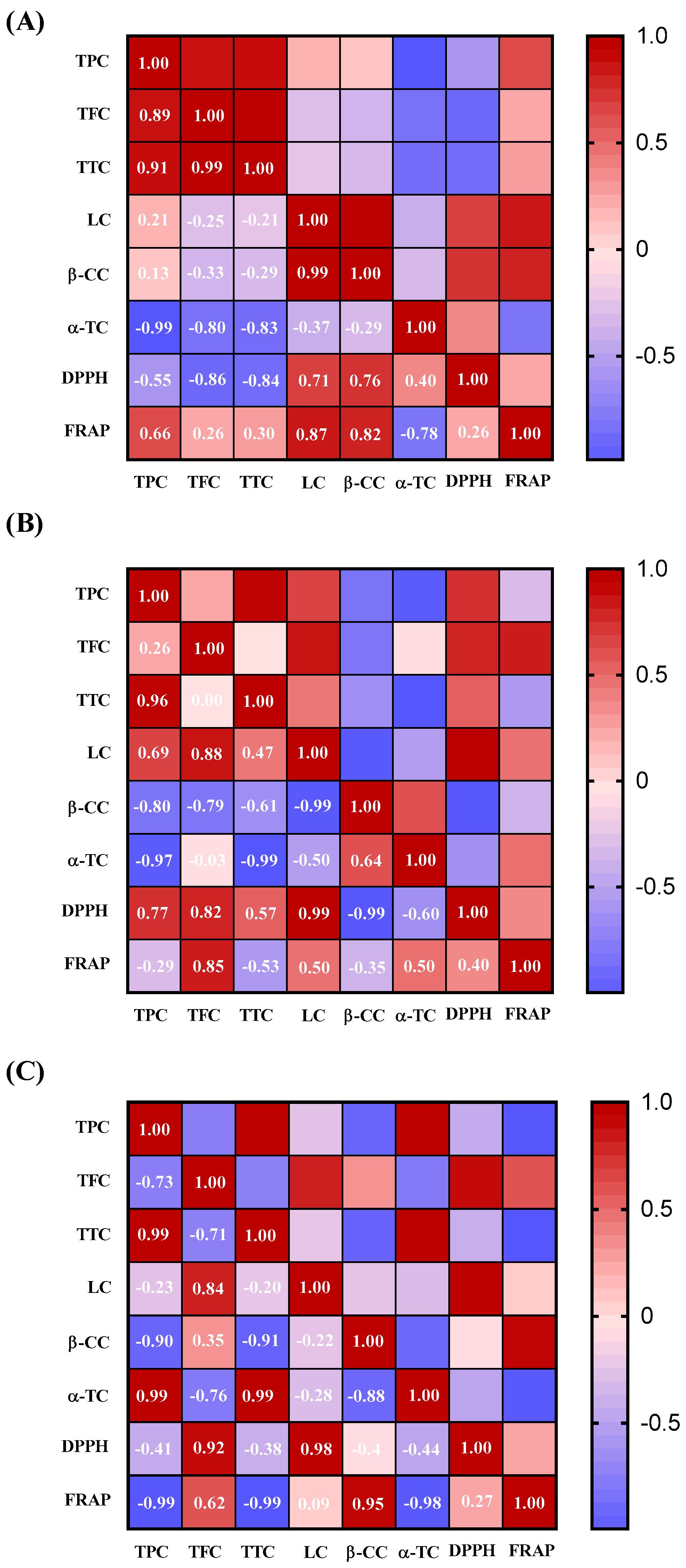
Correlation analysis (Figure 3: A–Maceration, B–Ultrasound, and C–Soxhlet) reveals the relationships between bioactive compounds and the antioxidant activity of A. luma seed oil, highlighting the impact of solvent type, temperature, and extraction conditions on the retention and functionality of polyphenols, flavonoids, carotenoids, and tocopherols. Maceration and ultrasound extraction showed strong positive correlations between TPC, TFC, and TTC with antioxidant activity (DPPH and FRAP assays, r > 0.90), indicating their primary role in antioxidant potential. Carotenoids such as LC and β-CC contributed to a lesser extent (r ≈ 0.70). In contrast, α-TC exhibited weak or negative correlations with polyphenols and antioxidant capacity, suggesting that conditions favoring tocopherol recovery may reduce polyphenol retention (r = −0.97).
Soxhlet extraction, characterized by prolonged solvent reflux and high temperatures, led to reduced polyphenol retention, as indicated by the negative correlation between TPC and antioxidant capacity (TPC vs. FRAP, r = −0.99), demonstrating significant thermal degradation [47]. However, TFC remained positively correlated with DPPH (r = 0.94), indicating greater heat resistance in certain flavonoid subclasses [48]. Additionally, carotenoids (LC and β-CC) and α-TC exhibited stronger correlations with antioxidant potential, suggesting a shift toward thermally stable compounds.
Maceration effectively preserved polyphenols, making it preferable for applications where high antioxidant activity is desired. Soxhlet extraction, despite its efficiency in lipid recovery, resulted in significant polyphenol degradation, shifting the antioxidant contribution to carotenoids and tocopherols.

3.4. Fatty Acid Profile

The fatty acid profile of A. luma seed oil obtained using the three extraction methods is summarized in Table 2. The fatty acids identified in A. luma seed oil were categorized as saturated and unsaturated. Saturated fatty acids detected included palmitic acid (C16:0), stearic acid (C18:0), and arachidic acid (C20:0), while unsaturated fatty acids comprised oleic acid (C18:1n−9), linoleic acid (C18:2n−6), and gondoic acid (C20:1n−9). Linoleic acid was the most abundant, with percentages ranging from 79.79% to 80.09%, followed by oleic acid (8.89–9.18%) and palmitic acid (7.29–7.40%) across all extraction methods.
Linoleic acid, an essential omega-6 polyunsaturated fatty acid, plays a vital role in human health, acting as a blood vessel cleanser, reducing serum cholesterol levels, and inhibiting arterial thrombosis [49]. It also exhibits significant anti-cancer activity, particularly in breast cancer treatment [50]. Oleic acid, the second most abundant fatty acid, is associated with cardiovascular benefits, including reduction in low-density lipoprotein (LDL) levels and increase in high-density lipoprotein (HDL) levels, thereby potentially reducing the risk of cardiovascular disease [51]. Additionally, oleic acid possesses anti-inflammatory and antioxidant properties [52].
Interestingly, A. luma seed oil contained 89% unsaturated fatty acids, exceeding the levels reported for strawberry seed oil (77–81%) and blackberry seed oil (67%) [39]. The intake of unsaturated fatty acids has been shown to regulate blood lipids and prevent cardiovascular diseases, whereas dietary saturated fatty acids are associated with increased LDL cholesterol levels and a higher risk of cardiovascular diseases [53]. Notably, A. luma seed oil obtained through all three extraction methods exhibited low levels of saturated fatty acids (10.50–10.68%).
The functional quality indices of A. luma seed oil (Table 3) indicate a favorable nutritional profile, particularly for cardiovascular health. The PUFA/SFA ratio (7.47–7.62) is consistent with high-quality edible oils, supporting its cardioprotective properties [54]. The low atherogenicity index (AI: 0.081–0.083) and thrombogenicity index (TI: 0.23) indicate a reduced risk of cholesterol accumulation and thrombosis, comparable to values reported for white and red grape seed oils [32]. Additionally, the high hypocholesterolemic/hypercholesterolemic (H/H) index (10.79–10.97) highlights its role in lipid metabolism regulation, as higher values are associated with improved nutritional quality and cardiovascular benefits [55]. While its oxidative stability (COX: 8.22–8.25) is characteristic of PUFA-rich oils, proper storage is necessary to prevent lipid oxidation.
These findings indicate that A. luma seed oil may contribute to cardiovascular health and overall metabolic balance. Furthermore, its high content of unsaturated fatty acids suggests potential applications in pharmaceutical formulations, presenting opportunities for therapeutic and nutraceutical development.

3.5. Antimicrobial Activity

The antimicrobial activity of cauchao seed oil, extracted via maceration, ultrasound, and Soxhlet extraction, was tested against Staphylococcus aureus (ATCC 25923) and Escherichia coli (ATCC 25922), but no antimicrobial effects were observed. Therefore, the bioactive compounds and fatty acids reported in this study may contribute to other biological properties unrelated to antimicrobial activity. These findings enhance the understanding of cauchao seed oil’s bioactive potential. Future research on compound isolation, alternative extraction methods, and synergistic effects could provide further insights into its functional properties.

4. Conclusions

This study evaluated the bioactive properties and fatty acid profile of A. luma seed oil obtained through maceration, ultrasound, and Soxhlet extraction. Among the extraction methods, Soxhlet yielded the highest oil recovery; however, maceration and ultrasound preserved greater concentrations of phenolic compounds and exhibited higher antioxidant capacity, likely due to the shorter extraction time and lower thermal exposure. Antioxidant capacity, measured via DPPH and FRAP assays, showed a strong correlation with polyphenol content, particularly in oils obtained using maceration and ultrasound. In contrast, Soxhlet extraction favored the recovery of tocopherols and carotenoids, suggesting partial polyphenol degradation under prolonged exposure. A strong inverse correlation between α-tocopherol and polyphenols (r = −0.97) was observed in oils obtained through ultrasound, underscoring the importance of optimizing sonication parameters to balance antioxidant composition. Linoleic acid (C18:2n-6) and oleic acid (C18:1n-9) were identified as the predominant fatty acids, highlighting the nutritional value of the oil. These findings demonstrate that A. luma seed oil is a promising source of bioactive compounds for potential applications in the food, nutraceutical, and cosmetic industries. Furthermore, the valorization of these seeds contributes to waste reduction and supports a circular economy approach by offering sustainable and profitable alternatives to conventional oil sources.

Author Contributions

C.G.-V., writing—review and editing, writing—original draft, visualization, resources, project administration, methodology, funding acquisition, formal analysis, and conceptualization; R.V., writing—review and editing, writing—original draft, data curation, and validation; J.A., data curation and formal analysis; I.Q.-F., writing—review and editing, resources, and formal analysis; A.G., writing—review and editing, resources, and supervision. All authors have read and agreed to the published version of the manuscript.

Funding

This research was funded by Fondecyt Postdoctorado Grant No. 3220661.

Institutional Review Board Statement

Not applicable.

Informed Consent Statement

Not applicable.

Data Availability Statement

The data that support this study are contained within the article.

Acknowledgments

The authors would like to thank ANID-Fondecyt Postdoctorado Project No. 3220661, Fondecyt Regular Project No. 1231835, Pontificia Universidad Católica de Chile, and Karol Rojas of the Universidad de La Serena for their technical support.

Conflicts of Interest

The authors declare that they have no known competing financial interests or personal relationships that could have appeared to influence the work reported in this paper.

References

  1. Chenavaz, R.Y.; Dimitrov, S. From Waste to Wealth: Policies to Promote the Circular Economy. J. Clean. Prod. 2024, 443, 141086. [Google Scholar] [CrossRef]
  2. Mármol, I.; Quero, J.; Ibarz, R.; Ferreira-Santos, P.; Teixeira, J.A.; Rocha, C.M.R.; Pérez-Fernández, M.; García-Juiz, S.; Osada, J.; Martín-Belloso, O.; et al. Valorization of Agro-Food by-Products and Their Potential Therapeutic Applications. Food Bioprod. Process. 2021, 128, 247–258. [Google Scholar] [CrossRef]
  3. García, N.; Ormazabal, C. Arboles Nativos de Chile; Enersis S.A.: Santiago, Chile, 2008. [Google Scholar]
  4. Maiolini, T.C.S.; Nicácio, K.J.; Rosa, W.; Miranda, D.O.; Santos, M.F.C.; Bueno, P.C.P.; Lago, J.H.G.; Sartorelli, P.; Dias, D.F.; Chagas de Paula, D.A.; et al. Potential Anti-Inflammatory Biomarkers from Myrtaceae Essential Oils Revealed by Untargeted Metabolomics. Nat. Prod. Res. 2023, 39, 985–992. [Google Scholar] [CrossRef]
  5. Velásquez, P.; Orellana, J.; Muñoz-Carvajal, E.; Faúndez, M.; Gómez, M.; Montenegro, G.; Giordano, A. Biological Activity of Native Myrtaceae Fruits from Chile as a Potential Functional Food. Nat. Prod. Res. 2021, 36, 3138–3142. [Google Scholar] [CrossRef]
  6. Weyerstahl, P.; Marschall, H.; Landrum, L.R. Constituents of the Leaf Extract of Amomyrtus Meli (R. A. Philippi) Legrand et Kausel, Amomyrtus Luma (Molina) Legrand et Kausel and of Amomyrtella Guili (Speg.) Kausel. Flavour Fragr. J. 1992, 7, 247–251. [Google Scholar] [CrossRef]
  7. Strzałka, K.; Szymańska, R.; Ewa, Ś.; Skorupińska-Tudek, K.; Suwalsky, M. Tocochromanols, Plastoquinone and Polyprenols in Selected Plant Species from Chilean Patagonia. Acta Biol. Cracoviensia Ser. Bot. 2009, 51, 39–44. [Google Scholar]
  8. Falkenberg, S.S.; Tarnow, I.; Guzman, A.; Mølgaard, P.; Simonsen, H.T. Mapuche Herbal Medicine Inhibits Blood Platelet Aggregation. Evid.-Based Complement. Altern. Med. 2012, 2012, 647620. [Google Scholar] [CrossRef]
  9. Mølgaard, P.; Holler, J.G.; Asar, B.; Liberna, I.; Rosenbæk, L.B.; Jebjerg, C.P.; Jørgensen, L.; Lauritzen, J.; Guzman, A.; Adsersen, A.; et al. Antimicrobial Evaluation of Huilliche Plant Medicine Used to Treat Wounds. J. Ethnopharmacol. 2011, 138, 219–227. [Google Scholar] [CrossRef]
  10. Schmeda-Hirschmann, G.; Jiménez-Aspee, F.; Theoduloz, C.; Ladio, A. Patagonian Berries as Native Food and Medicine. J. Ethnopharmacol. 2019, 241, 111979. [Google Scholar] [CrossRef]
  11. Rajagukguk, Y.V.; Islam, M.; Siger, A.; Fornal, E.; Tomaszewska-Gras, J. Oxidative Stability Assessment of Industrial and Laboratory-Pressed Fresh Raspberry Seed Oil (Rubus Idaeus L.) by Differential Scanning Calorimetry. Food Chem. Adv. 2023, 2, 100186. [Google Scholar] [CrossRef]
  12. de Menezes, M.L.; Johann, G.; Diório, A.; Schuelter Boeing, J.; Visentainer, J.V.; Raimundini Aranha, A.C.; Curvelo Pereira, N. Comparison of the Chemical Composition of Grape Seed Oil Extracted by Different Methods and Conditions. J. Chem. Technol. Biotechnol. 2023, 98, 1103–1113. [Google Scholar] [CrossRef]
  13. Yang, C.; Shang, K.; Lin, C.; Wang, C.; Shi, X.; Wang, H.; Li, H. Processing Technologies, Phytochemical Constituents, and Biological Activities of Grape Seed Oil (GSO): A Review. Trends Food Sci. Technol. 2021, 116, 1074–1083. [Google Scholar] [CrossRef]
  14. Liu, N.; Ren, G.; Faiza, M.; Li, D.; Cui, J.; Zhang, K.; Yao, X.; Zhao, M. Comparison of Conventional and Green Extraction Methods on Oil Yield, Physicochemical Properties, and Lipid Compositions of Pomegranate Seed Oil. J. Food Compos. Anal. 2022, 114, 104747. [Google Scholar] [CrossRef]
  15. Kapcsándi, V.; Hanczné Lakatos, E.; Sik, B.; Linka, L.Á.; Székelyhidi, R. Characterization of Fatty Acid, Antioxidant, and Polyphenol Content of Grape Seed Oil from Different Vitis Vinifera L. Varieties. OCL—Oilseeds Fats Crops Lipids 2021, 28, 30. [Google Scholar] [CrossRef]
  16. Ispiryan, A.; Viškelis, J.; Viškelis, P. Red Raspberry (Rubus Idaeus l.) Seed Oil: A Review. Plants 2021, 10, 944. [Google Scholar] [CrossRef]
  17. Bahmani, M.; Shokri, S.; Akhtar, Z.N.; Abbaszadeh, S.; Manouchehri, A. The Effect of Pomegranate Seed Oil on Human Health, Especially Epidemiology of Polycystic Ovary Syndrome; a Systematic Review. JBRA Assist. Reprod. 2022, 26, 631–636. [Google Scholar] [CrossRef] [PubMed]
  18. Bitwell, C.; Indra, S.S.; Luke, C.; Kakoma, M.K. A Review of Modern and Conventional Extraction Techniques and Their Applications for Extracting Phytochemicals from Plants. Sci. Afr. 2023, 19, e01585. [Google Scholar] [CrossRef]
  19. Rajesh, Y.; Khan, N.M.; Raziq Shaikh, A.; Mane, V.S.; Daware, G.; Dabhade, G. Investigation of Geranium Oil Extraction Performance by Using Soxhlet Extraction. In Proceedings of the Materials Today: Proceedings; Elsevier Ltd.: Amsterdam, The Netherlands, 2023; Volume 72, pp. 2610–2617. [Google Scholar]
  20. Ginocchio, R.; Muñoz-Carvajal, E.; Velásquez, P.; Giordano, A.; Montenegro, G.; Colque-Perez, G.; Sáez-Navarrete, C. Mayten Tree Seed Oil: Nutritional Value Evaluation According to Antioxidant Capacity and Bioactive Properties. Foods 2021, 10, 729. [Google Scholar] [CrossRef]
  21. Singleton, V.L.; Orthofer, R.; Lamuela-Raventós, R.M. Analysis of Total Phenols and Other Oxidation Substrates and Antioxidants by Means of Folin-Ciocalteu Reagent. In Methods in Enzymology; Academic Press: Cambridge, MA, USA, 1999; pp. 152–178. [Google Scholar]
  22. Woisky, R.G.; Salatino, A. Analysis of Propolis: Some Parameters and Procedures for Chemical Quality Control. J. Apic. Res. 1998, 37, 99–105. [Google Scholar] [CrossRef]
  23. Makkar, H.P.S.; Blümmel, M.; Borowy, N.K.; Becker, K. Gravimetric Determination of Tannins and Their Correlations with Chemical and Protein Precipitation Methods. J. Sci. Food Agric. 1993, 61, 161–165. [Google Scholar] [CrossRef]
  24. Biehler, E.; Mayer, F.; Hoffmann, L.; Krause, E.; Bohn, T. Comparison of 3 Spectrophotometric Methods for Carotenoid Determination in Frequently Consumed Fruits and Vegetables. J. Food Sci. 2010, 75, C55–C61. [Google Scholar] [CrossRef]
  25. Sadler, G.; Davis, J.; Dezman, D. Rapid Extraction of Lycopene and Β-Carotene from Reconstituted Tomato Paste and Pink Grapefruit Homogenates. J. Food Sci. 1990, 55, 1460–1461. [Google Scholar] [CrossRef]
  26. Quispe-Fuentes, I.; Vega-Gálvez, A.; Aranda, M.; Poblete, J.; Pasten, A.; Bilbao-Sainz, C.; Wood, D.; McHugh, T.; Delporte, C. Effects of Drying Processes on Composition, Microstructure and Health Aspects from Maqui Berries. J. Food Sci. Technol. 2020, 57, 2241–2250. [Google Scholar] [CrossRef]
  27. Brand-Williams, W.; Cuvelier, M.E.; Berset, C. Use of a Free Radical Method to Evaluate Antioxidant Activity. LWT—Food Sci. Technol. 1995, 28, 25–30. [Google Scholar] [CrossRef]
  28. Benzie, I.F.F.; Strain, J.J. The Ferric Reducing Ability of Plasma (FRAP) as a Measure of “Antioxidant Power”: The FRAP Assay. Anal. Biochem. 1996, 239, 70–76. [Google Scholar] [CrossRef]
  29. Bligh, E.G.; Dyer, W.J. The National Research Council Od Canada a Rapid Method of Total Lipid Extraction and Purification. Can. J. Biochem. Physiol. 1959, 37, 911–917. [Google Scholar] [CrossRef] [PubMed]
  30. Quispe-Fuentes, I.; Valenzuela, P.; Roco, T.; Pérez-Won, M.; Espinoza, J.; Bernal, G.; Bernal, C.; Martínez, R. Isolation and Characterization of Polyunsaturated Fatty Acid-Rich Oil from Jumbo Squid (Dosidicus gigas) Viscera: Antioxidant Potential and Anticancer Activity on Colorectal Cancer Cells. Waste Biomass Valorization 2025, 16, 3507–3517. [Google Scholar] [CrossRef]
  31. Dimić, I.; Teslić, N.; Putnik, P.; Kovačević, D.B.; Zeković, Z.; Šojić, B.; Mrkonjić, Ž.; Čolović, D.; Montesano, D.; Pavlić, B. Innovative and Conventional Valorizations of Grape Seeds from Winery By-Products as Sustainable Source of Lipophilic Antioxidants. Antioxidants 2020, 9, 568. [Google Scholar] [CrossRef]
  32. Carmona-Jiménez, Y.; Igartuburu, J.M.; Guillén-Sánchez, D.A.; García-Moreno, M.V. Fatty Acid and Tocopherol Composition of Pomace and Seed Oil from Five Grape Varieties Southern Spain. Molecules 2022, 27, 6980. [Google Scholar] [CrossRef]
  33. CLSI Performance Standards for Antimicrobial Disk Susceptibility Tests; Approved Standard—Eleventh Edition, CLSI Document M02-A11; CLSI: Wayne, PA, USA, 2012.
  34. Patrice Didion, Y.; Gijsbert Tjalsma, T.; Su, Z.; Malankowska, M.; Pinelo, M. What Is next? The Greener Future of Solid Liquid Extraction of Biobased Compounds: Novel Techniques and Solvents Overpower Traditional Ones. Sep. Purif. Technol. 2023, 320, 124147. [Google Scholar] [CrossRef]
  35. Yao, X.-H.; Shen, Y.-S.; Hu, R.-Z.; Xu, M.; Huang, J.-X.; He, C.-X.; Cao, F.-L.; Fu, Y.-J.; Zhang, D.-Y.; Zhao, W.-G.; et al. The Antioxidant Activity and Composition of the Seed Oil of Mulberry Cultivars. Food Biosci. 2020, 37, 100709. [Google Scholar] [CrossRef]
  36. Akbari, S.; Abdurahman, N.H.; Yunus, R.M.; Alara, O.R.; Abayomi, O.O. Extraction, Characterization and Antioxidant Activity of Fenugreek (Trigonella-Foenum Graecum) Seed Oil. Mater. Sci. Energy Technol. 2019, 2, 349–355. [Google Scholar] [CrossRef]
  37. Larrazábal-Fuentes, M.J.; Fernández-Galleguillos, C.; Palma-Ramírez, J.; Romero-Parra, J.; Sepúlveda, K.; Galetovic, A.; González, J.; Paredes, A.; Bórquez, J.; Simirgiotis, M.J.; et al. Chemical Profiling, Antioxidant, Anticholinesterase, and Antiprotozoal Potentials of Artemisia copa Phil. (Asteraceae). Front. Pharmacol. 2020, 11, 594174. [Google Scholar] [CrossRef] [PubMed]
  38. Banjanin, T.; Özcan, M.M.; Al Juhaimi, F.; Ranković-Vasić, Z.; Uslu, N.; Mohamed, I.A.; Ghafoor, K.; Babiker, E.E.; Osman, M.A.; Gassem, M.A.; et al. Effect of Varieties on Bioactive Compounds, Fatty Acids, and Mineral Contents in Different Grape Seed and Oils from Bosnia and Herzegovina. J. Food Process. Preserv. 2019, 43, e13981. [Google Scholar] [CrossRef]
  39. De Filette, M.; Schatteman, K.; Geuens, J. Characterization of Six Cold-Pressed Berry Seed Oils and Their Seed Meals. Appl. Sci. 2024, 14, 439. [Google Scholar] [CrossRef]
  40. El-Beltagi, H.S.; Maraei, R.W.; El-Ansary, A.E.; Rezk, A.A.; Mansour, A.T.; Aly, A.A. Characterizing the Bioactive Ingredients in Sesame Oil Affected by Multiple Roasting Methods. Foods 2022, 11, 2261. [Google Scholar] [CrossRef]
  41. Sun, J.; Li, D.; Huyan, W.; Hong, X.; He, S.; Huo, J.; Jiang, L.; Zhang, Y. Blue Honeysuckle Seeds and Seed Oil: Composition, Physicochemical Properties, Fatty Acid Profile, Volatile Components, and Antioxidant Capacity. Food Chem. X 2024, 21, 101176. [Google Scholar] [CrossRef]
  42. Dehghanian, Z.; Habibi, K.; Dehghanian, M.; Aliyar, S.; Asgari Lajayer, B.; Astatkie, T.; Minkina, T.; Keswani, C. Reinforcing the Bulwark: Unravelling the Efficient Applications of Plant Phenolics and Tannins against Environmental Stresses. Heliyon 2022, 8, e09094. [Google Scholar] [CrossRef]
  43. Neha, K.; Haider, M.R.; Pathak, A.; Yar, M.S. Medicinal Prospects of Antioxidants: A Review. Eur. J. Med. Chem. 2019, 178, 687–704. [Google Scholar] [CrossRef]
  44. Holzapfel, N.; Holzapfel, B.; Champ, S.; Feldthusen, J.; Clements, J.; Hutmacher, D. The Potential Role of Lycopene for the Prevention and Therapy of Prostate Cancer: From Molecular Mechanisms to Clinical Evidence. Int. J. Mol. Sci. 2013, 14, 14620–14646. [Google Scholar] [CrossRef]
  45. Moon, J.-K.; Shibamoto, T. Antioxidant Assays for Plant and Food Components. J. Agric. Food Chem. 2009, 57, 1655–1666. [Google Scholar] [CrossRef]
  46. Daud, N.M.; Putra, N.R.; Jamaludin, R.; Md Norodin, N.S.; Sarkawi, N.S.; Hamzah, M.H.S.; Mohd Nasir, H.; Abang Zaidel, D.N.; Che Yunus, M.A.; Md Salleh, L. Valorisation of Plant Seed as Natural Bioactive Compounds by Various Extraction Methods: A Review. Trends Food Sci. Technol. 2022, 119, 201–214. [Google Scholar] [CrossRef]
  47. Shi, L.; Zhao, W.; Yang, Z.; Subbiah, V.; Suleria, H.A.R. Extraction and Characterization of Phenolic Compounds and Their Potential Antioxidant Activities. Environ. Sci. Pollut. Res. 2022, 29, 81112–81129. [Google Scholar] [CrossRef]
  48. Langston, F.M.A.; Nash, G.R.; Bows, J.R. The Retention and Bioavailability of Phytochemicals in the Manufacturing of Baked Snacks. Crit. Rev. Food Sci. Nutr. 2023, 63, 2141–2177. [Google Scholar] [CrossRef]
  49. Chang, J.; Kang, X.; Yuan, J. Enhancing Emulsification and Antioxidant Ability of Egg Albumin by Moderately Acid Hydrolysis: Modulating an Emulsion-Based System for Mulberry Seed Oil. Food Res. Int. 2018, 109, 334–342. [Google Scholar] [CrossRef]
  50. Tao, X.-M.; Wang, J.; Wang, J.; Feng, Q.; Gao, S.; Zhang, L.-R.; Zhang, Q. Enhanced Anticancer Activity of Gemcitabine Coupling with Conjugated Linoleic Acid against Human Breast Cancer in Vitro and in Vivo. Eur. J. Pharm. Biopharm. 2012, 82, 401–409. [Google Scholar] [CrossRef] [PubMed]
  51. Pastor, R.; Bouzas, C.; Tur, J.A. Beneficial Effects of Dietary Supplementation with Olive Oil, Oleic Acid, or Hydroxytyrosol in Metabolic Syndrome: Systematic Review and Meta-Analysis. Free Radic. Biol. Med. 2021, 172, 372–385. [Google Scholar] [CrossRef] [PubMed]
  52. Lu, Y.; Zhao, J.; Xin, Q.; Yuan, R.; Miao, Y.; Yang, M.; Mo, H.; Chen, K.; Cong, W. Protective Effects of Oleic Acid and Polyphenols in Extra Virgin Olive Oil on Cardiovascular Diseases. Food Sci. Hum. Wellness 2024, 13, 529–540. [Google Scholar] [CrossRef]
  53. Siri-Tarino, P.W.; Sun, Q.; Hu, F.B.; Krauss, R.M. Saturated Fatty Acids and Risk of Coronary Heart Disease: Modulation by Replacement Nutrients. Curr. Atheroscler. Rep. 2010, 12, 384–390. [Google Scholar] [CrossRef]
  54. Popović, M.; Fuentes, J.B.; Papović, N.; Okuka, N.; Suručić, R.; Torović, L. Comparative Advantages of Fatty Acid Composition and Nutritional Indices of Specific Edible Plant Oils. Proceedings 2023, 91, 357. [Google Scholar]
  55. Fonseca, V.F.; Duarte, I.A.; Matos, A.R.; Reis-Santos, P.; Duarte, B. Fatty Acid Profiles as Natural Tracers of Provenance and Lipid Quality Indicators in Illegally Sourced Fish and Bivalves. Food Control 2022, 134, 108735. [Google Scholar] [CrossRef]
Figure 1. Yields (w/w %) of A. luma seed oil content through different extraction methods. Bars with different letters (a, b) indicate significant differences between means according to Tukey’s test (p < 0.05, n = 3).
Compounds 05 00031 g001
Figure 2. Antioxidant capacity (DPPH and FRAP) of Amomyrtus luma seed oil obtained using different extraction methods. Bars with different letters (a, b, c) within the same assay indicate significant differences between means according to Tukey’s test (p < 0.05, n = 3).
Compounds 05 00031 g002
Figure 3. Heatmaps of Pearson correlation coefficients showing the relationship between bioactive compounds and antioxidant capacity for the three extraction methods: (A) maceration, (B) ultrasound extraction, and (C) Soxhlet. Correlations were considered significant at p < 0.05. TPC, total polyphenol content; TFC, total flavonoid content; TTC, total tannin content; LC, lycopene content; β-CC, β-carotene content; α-T, α-tocopherol content; DPPH: 2,2-difenil-1-picrilhidrazilo assay; FRAP, ferric reducing antioxidant power assay.
Compounds 05 00031 g003
Table 1. Bioactive compound analysis of A. luma.
Bioactive CompoundsExtraction Method
MacerationUltrasoundSoxhlet
Polyphenols (mg GAE/100 g oil)112.13 ± 1.95 a113.12 ± 2.49 a66.93 ± 8.19 b
Flavonoids (mg QE/100 g oil)21.06 ± 0.29 a19.68 ± 0.22 a11.33 ± 0.06 b
Tannins (mg GAE/100 g oil)82.38 ± 2.76 a84.97 ± 2.00 a53.36 ± 8.79 b
Lycopene (mg/100 g oil)1.42 ± 0.03 a0.92 ± 0.05 b0.69 ± 0.04 c
β-Carotene (mg/100 oil g)1.29 ± 0.10 a1.04 ± 0.07 b0.19 ± 0.05 c
α-Tocopherol (mg/100 oil g)10.68± 0.10 a9.62 ± 0.11 b12.46 ± 0.26 c
Different letters (a, b, c) in the same row indicate significant differences between means according to Tukey’s test (p < 0.05, n = 3).
Table 2. Fatty acid (FA) profile of A. luma. seed oil obtained using different extraction methods.
Fatty AcidExtraction Method
Maceration (%)Ultrasound (%)Soxhlet (%)
16:07.40 ± 0.13 a7.29 ± 0.05 a7.36 ± 0.07 a
18:02.89 ± 0.06 a2.83 ± 0.02 a2.84 ± 0.03 a
18:1n-99.18 ± 0.24 a9.15 ± 0.54 a8.89 ± 0.13 a
18:2n-679.79 ± 0.44 a80.01 ± 0.55 a80.09 ± 0.15 a
20:00.39 ± 0.01 a0.38 ± 0.01 a0.38 ± 0.01 a
20:1n-90.35 ± 0.01 a0.34 ± 0.00 a0.36 ± 0.00 a
Saturated10.68 ± 0.20 a10.50 ± 0.04 a10.57 ± 0.06 a
Unsaturated89.32 ± 0.20 a89.50 ± 0.04 a89.43 ± 0.06 a
Identical letters (a) in the same row indicate no significant differences between means according to Tukey’s test (p < 0.05, n = 3).
Table 3. Functional quality and oxidative stability indices of A. luma seed oil obtained using different extraction methods.
IndexExtraction Method
MacerationUltrasoundSoxhlet
USFA/SFA8.68±0.18 a8.85±0.04 a8.77±0.06 a
PUFA/SFA7.76±0.18 a7.90±0.09 a7.86±0.05 a
AI0.08±0.00 a0.08±0.00 a0.08±0.00 a
TI0.23±0.00 a0.23±0.00 a0.23±0.00 a
H/H10.79±0.24 a10.97±0.14 a10.88±0.12 a
COX8.22±0.05 a8.24±0.02 a8.25±0.02 a
SFA, saturated fatty acids; USFA, unsaturated fatty acids; PUFA, polyunsaturated fatty acids; TI, thrombogenic index; AI, atherogenic index; H/H, hypocholesterolemic fatty acids/hypercholesterolemic fatty acids; COX value, oxidative stability index. Identical letters (a) in the same row indicate no significant differences between means according to Tukey’s test (p < 0.05, n = 3).
Disclaimer/Publisher’s Note: The statements, opinions and data contained in all publications are solely those of the individual author(s) and contributor(s) and not of MDPI and/or the editor(s). MDPI and/or the editor(s) disclaim responsibility for any injury to people or property resulting from any ideas, methods, instructions or products referred to in the content.