
Binding Affinity Modeling to Predict Human CD4 T Cell Epitopes in Leishmania Proteins

 ,
,  , , and
Abstract
1. Introduction
2. Materials and Methods
3. Results
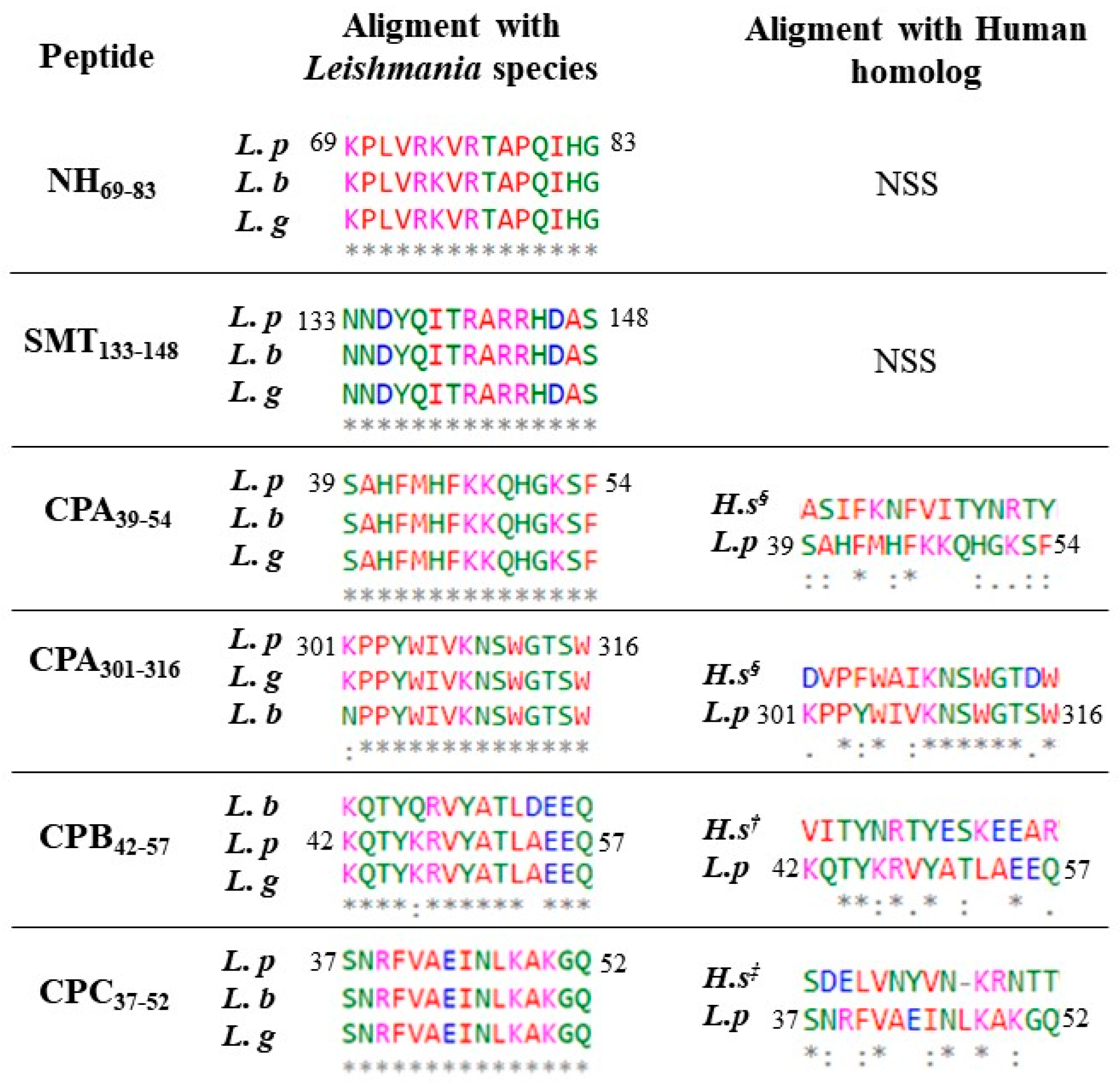
3.1. Binding Prediction
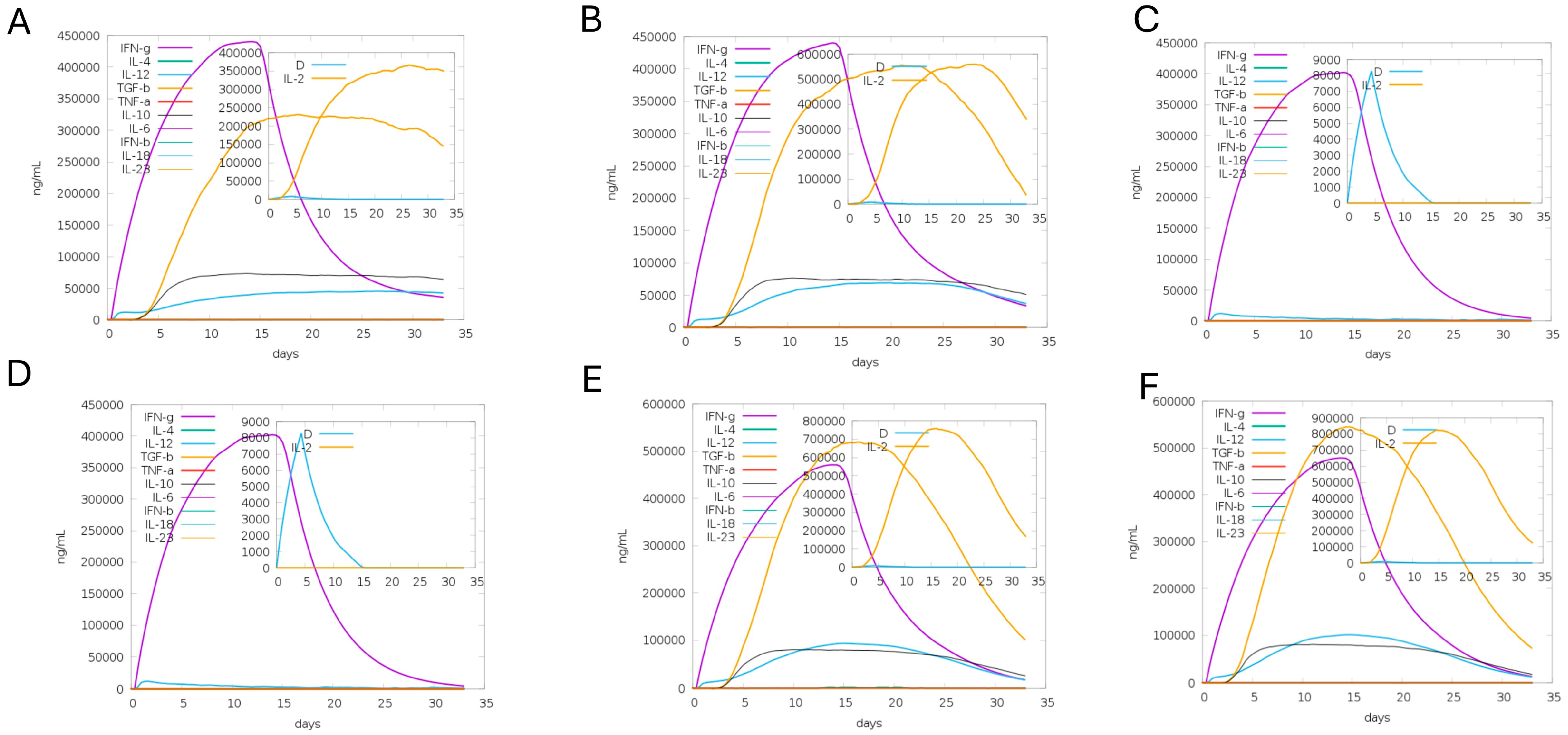
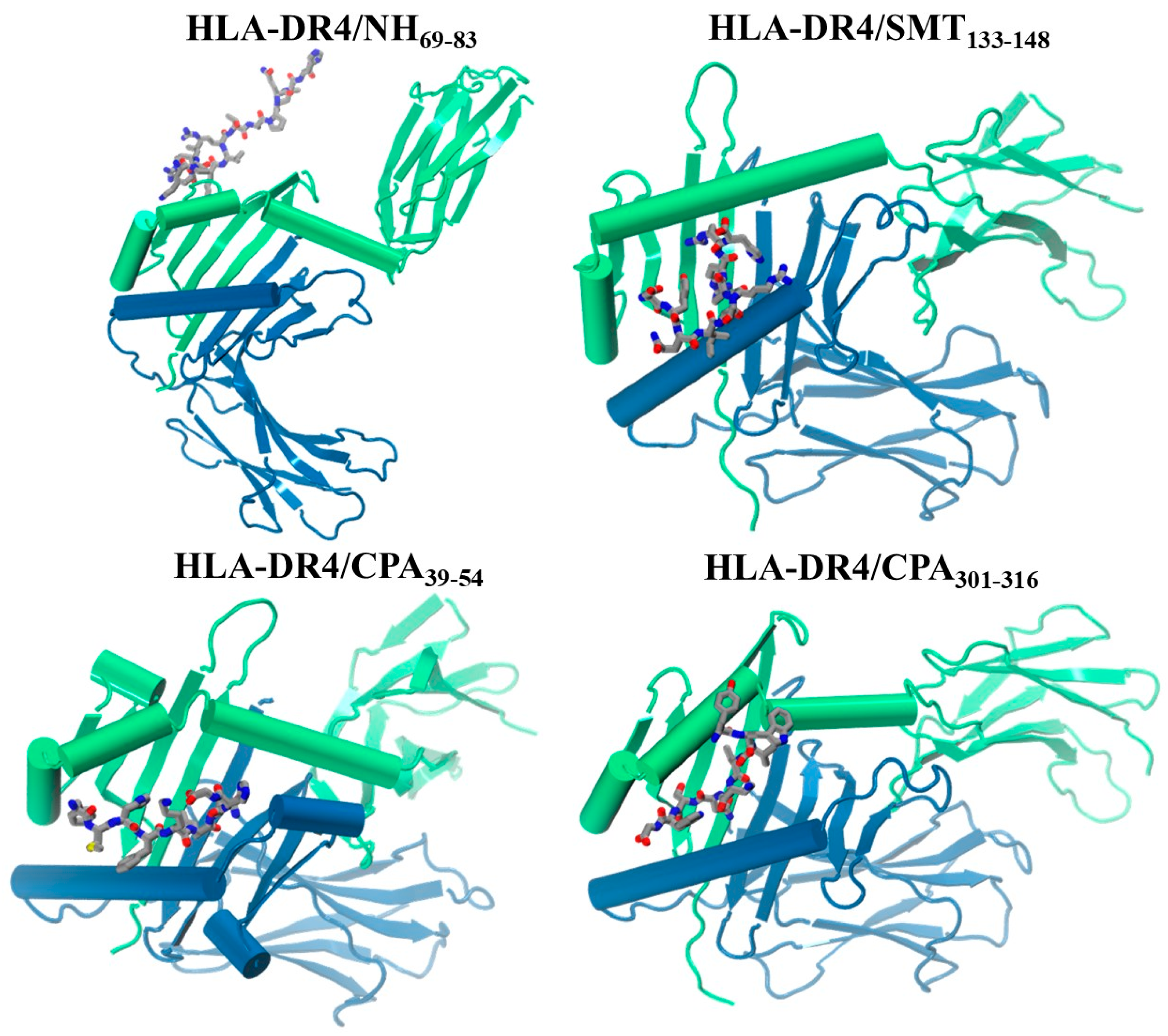
3.2. Molecular Docking and Immune Response
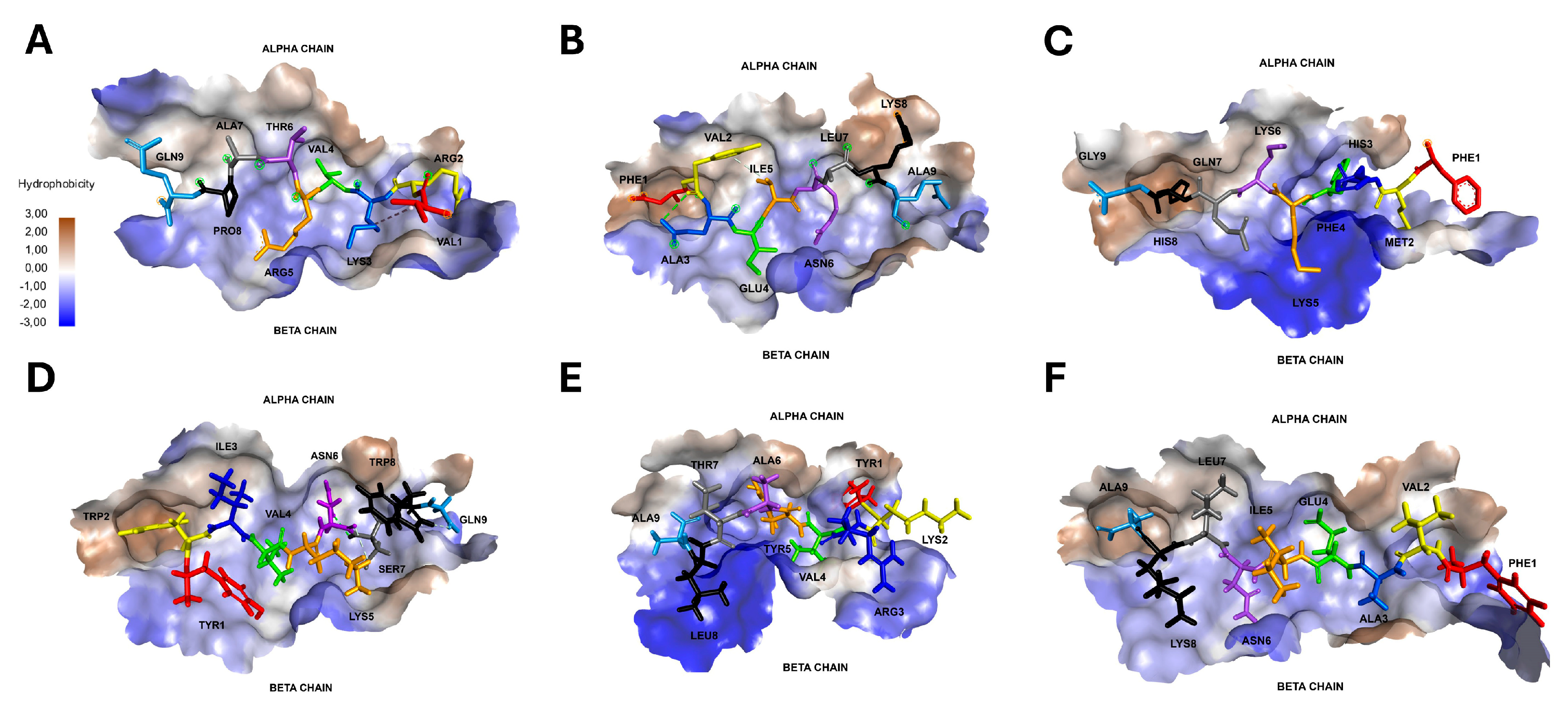
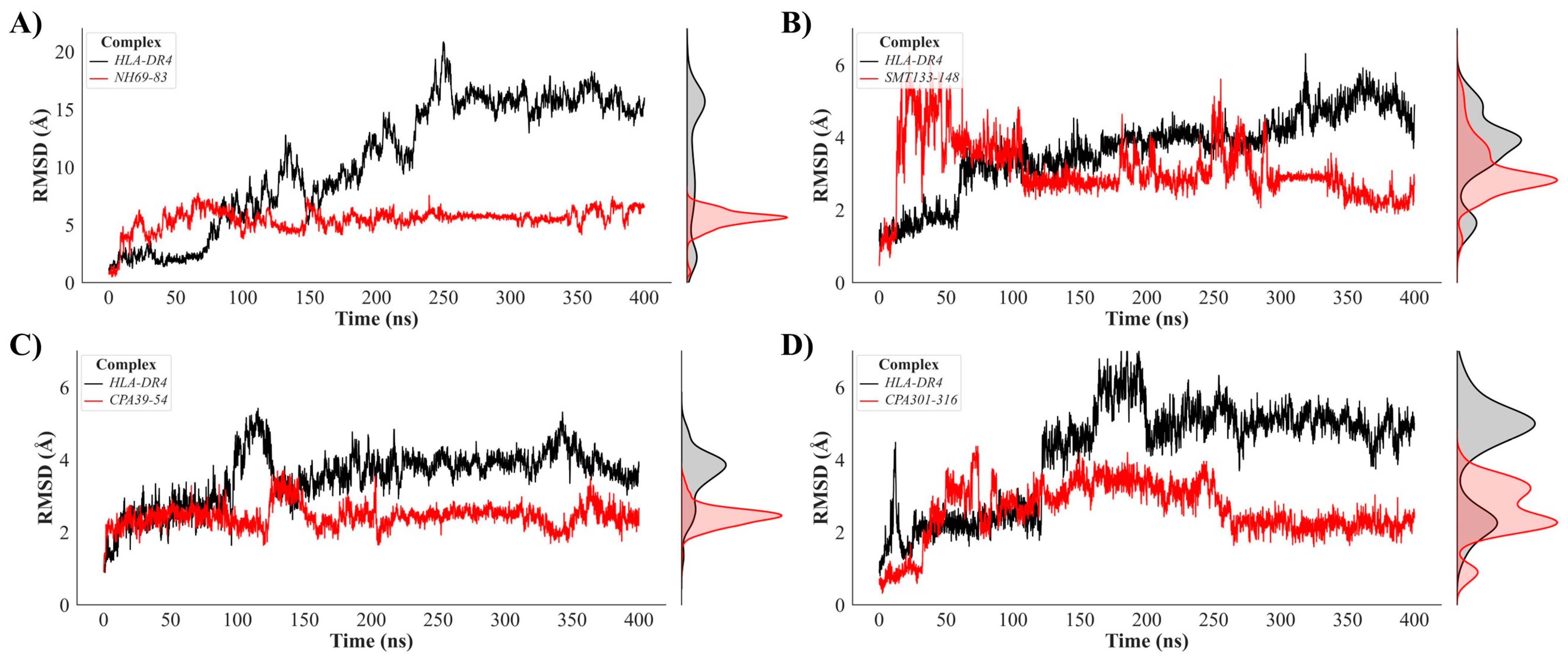
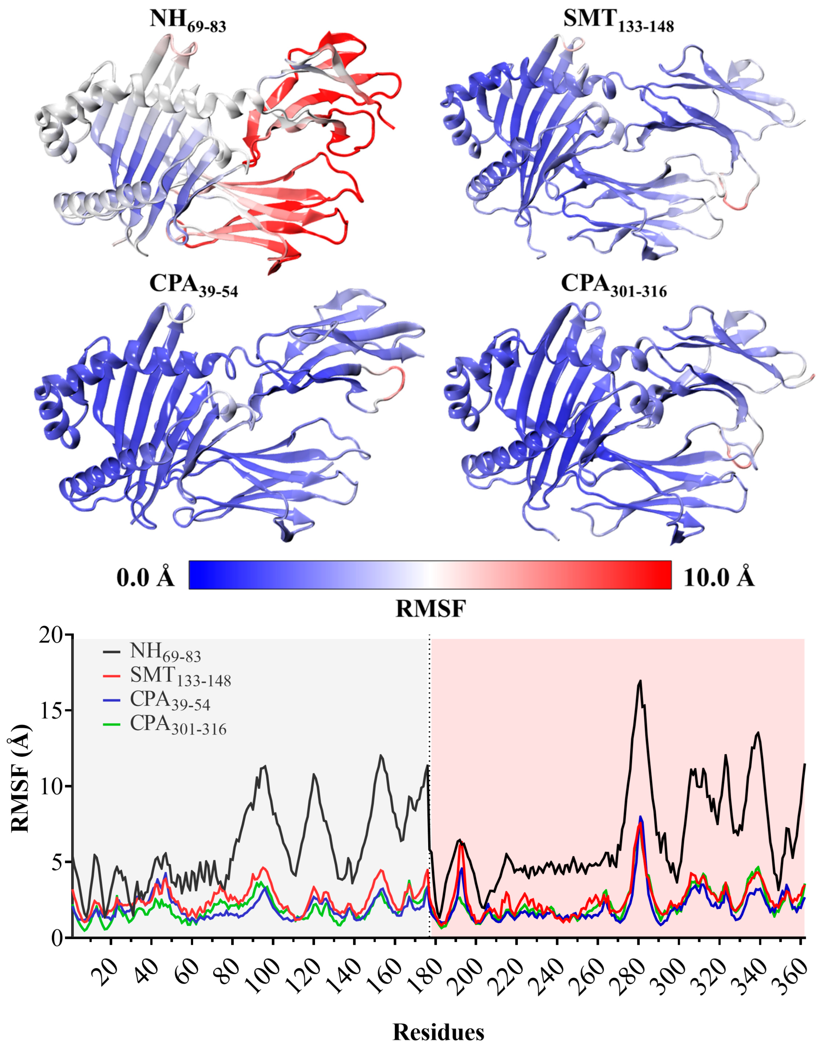
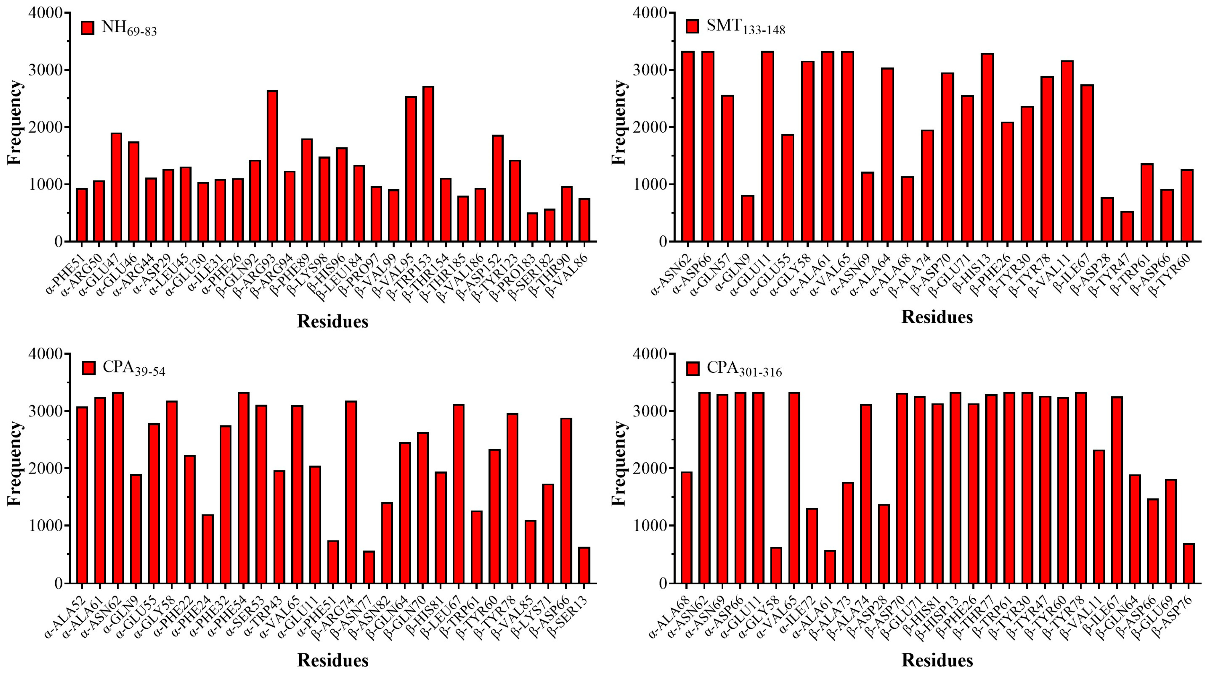
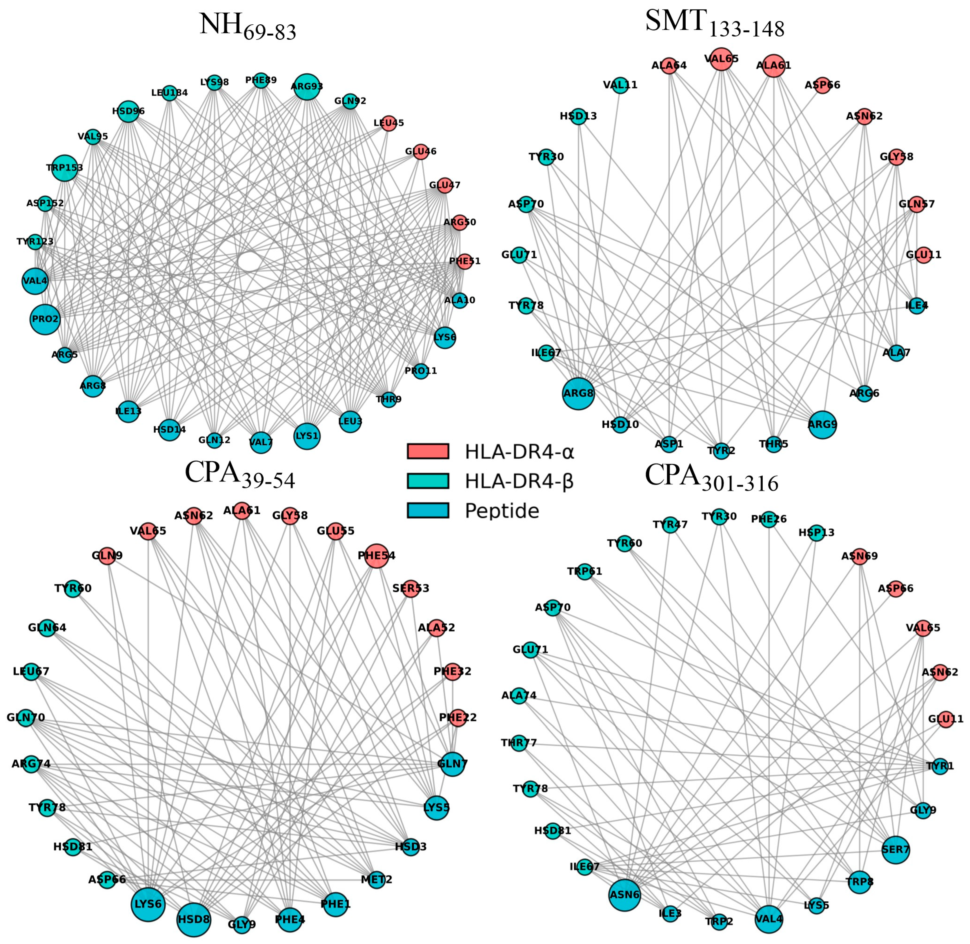
3.3. Molecular Insights into HLA-DR4–Peptide Binding Interfaces
3.4. Estimation of Binding Affinity in HLA-DR4–Peptide Complexes via Free Energy Calculations
4. Discussion
5. Conclusions
Supplementary Materials
Author Contributions
Funding
Institutional Review Board Statement
Informed Consent Statement
Data Availability Statement
Conflicts of Interest
Abbreviations
| HLA | Human Leucocyte Antigen |
| MHC | Major Histocompatibility Complex |
| NH | Nucleoside Hydrolase |
| SMT | Sterol 24-c-Methyltransferase |
| CPA | Cysteine Peptidase A |
| CPB | Cysteine Peptidase B |
| CPC | Cysteine Peptidase C |
| CL | Cutaneous Leishmaniasis |
| VL | Visceral Leishmaniasis |
| Th | T Helper |
| IFNγ | Interferon γ |
| IL | Interleucina |
| TGFβ | Transforming Growth Factor β |
| NO | Nitric Oxide |
| APS | Antigen Presenting Cell |
| TCR | T Cell Receptor |
| MD | Molecular Dynamics |
| PME | Particle Mesh Ewald |
| RMSD | Root Mean Square Deviation |
| RMSF | Root Mean Square Fluctuation |
References
- World Health Organization Leishmaniasis. Available online: https://www.who.int/health-topics/leishmaniasis#tab=tab_1 (accessed on 29 March 2022).
- Instituto Nacional de Salud Carlos III. Protocolo de Vigilancia de Leishmaniasis. Available online: https://cne.isciii.es/documents/d/cne/protocolo-20de-20vigilancia-20de-20leishmaniasis-pdf (accessed on 13 December 2023).
- Azim, M.; Khan, S.A.; Ullah, S.; Ullah, S.; Anjum, S.I. Therapeutic advances in the topical treatment of cutaneous leishmaniasis: A review. PLoS Negl. Trop. Dis. 2021, 15, e0009099. [Google Scholar] [CrossRef] [Scilit]
- Ponte-Sucre, A.; Gamarro, F.; Dujardin, J.C.; Barrett, M.P.; López-Vélez, R.; García-Hernández, R.; Pountain, A.W.; Mwenechanya, R.; Papadopoulou, B. Drug resistance and treatment failure in leishmaniasis: A 21st century challenge. PLoS Negl. Trop. Dis. 2017, 11, e0006052. [Google Scholar] [CrossRef] [Scilit]
- Li, S.; Qiu, T.; Zhao, N.; Liu, M.; He, X.; Wang, X.; Jiang, S.; Luo, M.; Wang, S.; Zhang, L.; et al. Global burden of leishmaniasis, 1990–2021: Systematic analysis of the global burden of disease study. Int. J. Equity Health 2025, 24, 329. [Google Scholar] [CrossRef] [Scilit]
- Belkaid, Y.; Kamhawi, S.; Modi, G.; Valenzuela, J.; Noben-Trauth, N.; Rowton, E.; Ribeiro, J.; Sacks, D.L. Development of a Natural Model of Cutaneous Leishmaniasis: Powerful Effects of Vector Saliva and Saliva Preexposure on the Long-Term Outcome of Leishmania major Infection in the Mouse Ear Dermis. J. Exp. Med. 1998, 188, 1941. [Google Scholar] [CrossRef] [Scilit]
- Bacon, K.M.; Hotez, P.J.; Kruchten, S.D.; Kamhawi, S.; Bottazzi, M.E.; Valenzuela, J.G.; Lee, B.Y. The potential economic value of a cutaneous leishmaniasis vaccine in seven endemic countries in the Americas. Vaccine 2012, 31, 480–486. [Google Scholar] [CrossRef] [Scilit]
- Dinc, R. Leishmania Vaccines: The Current Situation with Its Promising Aspect for the Future. Korean J. Parasitol. 2022, 60, 379–391. [Google Scholar] [CrossRef] [Scilit] [PubMed]
- Sacks, D.; Noben-Trauth, N. The immunology of susceptibility and resistance to Leishmania major in mice. Nat. Rev. Immunol. 2002, 2, 845–858. [Google Scholar] [CrossRef] [Scilit]
- Gollob, K.J.; Viana, A.G.; Dutra, W.O. Immunoregulation in human American leishmaniasis: Balancing pathology and protection. Parasite Immunol. 2014, 36, 367–376. [Google Scholar] [CrossRef] [Scilit] [PubMed]
- Tripathi, P.; Singh, V.; Naik, S. Immune response to leishmania: Paradox rather than paradigm. FEMS Immunol. Med. Microbiol. 2007, 51, 229–242. [Google Scholar] [CrossRef] [Scilit] [PubMed]
- Desai, D.V.; Kulkarni-Kale, U. T-cell epitope prediction methods: An overview. Methods Mol. Biol. 2014, 1184, 333–364. [Google Scholar] [CrossRef] [Scilit]
- Burza, S.; Croft, S.L.; Boelaert, M. Leishmaniasis. Lancet 2018, 392, 951–970. [Google Scholar] [CrossRef] [Scilit]
- Maxfield, L.; Crane, J.S. Leishmaniasis. In StatPearls; StatPearls: Treasure Island, FL, USA, 2023. [Google Scholar]
- Azam, S.S.; Abro, A.; Raza, S.; Saroosh, A. Structure and dynamics studies of sterol 24-C-methyltransferase with mechanism based inactivators for the disruption of ergosterol biosynthesis. Mol. Biol. Rep. 2014, 41, 4279–4293. [Google Scholar] [CrossRef] [Scilit]
- Besteiro, S.; Williams, R.A.M.; Coombs, G.H.; Mottram, J.C. Protein turnover and differentiation in Leishmania. Int. J. Parasitol. 2007, 37, 1063–1075. [Google Scholar] [CrossRef] [Scilit]
- Saha, S.; Srivastava, R.; Sarma, P.; Bhatt, T.K.; Prakash, A.; Kumar, D. Identification of potential inhibitors of Leishmania donovani Sterol 24-C-methyltransferase: In silico and in vitro studies. Mol. Simul. 2023, 49, 1311–1323. [Google Scholar] [CrossRef] [Scilit]
- Casgrain, P.A.; Martel, C.; McMaster, W.R.; Mottram, J.C.; Olivier, M.; Descoteaux, A. Cysteine Peptidase B Regulates Leishmania mexicana Virulence through the Modulation of GP63 Expression. PLoS Pathog. 2016, 12, e1005658. [Google Scholar] [CrossRef] [Scilit]
- Mottram, J.C.; Coombs, G.H.; Alexander, J. Cysteine peptidases as virulence factors of Leishmania. Curr. Opin. Microbiol. 2004, 7, 375–381. [Google Scholar] [CrossRef] [Scilit]
- Siqueira-Neto, J.L.; Debnath, A.; McCall, L.I.; Bernatchez, J.A.; Ndao, M.; Reed, S.L.; Rosenthal, P.J. Cysteine proteases in protozoan parasites. PLoS Negl. Trop. Dis. 2018, 12, e0006512. [Google Scholar] [CrossRef] [Scilit] [PubMed]
- MLB; Nico, D.; de Oliveira, F.A.; Barreto, A.S.; Palatnik-de-Sousa, I.; Carrillo, E.; Moreno, J.; de Luca, P.M.; Morrot, A.; Rosa, D.S.; et al. Leishmania donovani nucleoside Hydrolase (NH36) domains induce T-cell cytokine responses in human visceral leishmaniasis. Front. Immunol. 2017, 8, 227. [Google Scholar] [CrossRef] [Scilit] [PubMed]
- Nico, D.; Claser, C.; Borja-Cabrera, G.P.; Travassos, L.R.; Palatnik, M.; Soares Ida, S.; Rodrigues, M.M.; de Palatnik-Sousa, C.B. Adaptive Immunity against Leishmania Nucleoside Hydrolase Maps Its C-Terminal Domain as the Target of the CD4+ T Cell–Driven Protective Response. PLoS Negl. Trop. Dis. 2010, 4, e866. [Google Scholar] [CrossRef] [Scilit] [PubMed]
- Goto, Y.; Bhatia, A.; Raman, V.S.; Vidal, S.E.Z.; Bertholet, S.; Coler, R.N.; Howard, R.F.; Reed, S.G. Leishmania infantum sterol 24-c-methyltransferase formulated with MPL-SE induces cross-protection against L. major infection. Vaccine 2009, 27, 2884–2890. [Google Scholar] [CrossRef] [Scilit]
- Nico, D.; Gomes, D.C.; Palatnik-de-Sousa, I.; Morrot, A.; Palatnik, M.; Palatnik-de-Sousa, C.B. Leishmania donovani nucleoside hydrolase terminal domains in cross-protective immunotherapy against Leishmania amazonensis murine infection. Front. Immunol. 2014, 5, 88478. [Google Scholar] [CrossRef] [Scilit]
- Khoshgoo, N.; Zahedifard, F.; Azizi, H.; Taslimi, Y.; Alonso, M.J.; Rafati, S. Cysteine proteinase type III is protective against Leishmania infantum infection in BALB/c mice and highly antigenic in visceral leishmaniasis individuals. Vaccine 2008, 26, 5822–5829. [Google Scholar] [CrossRef] [Scilit]
- Doroud, D.; Zahedifard, F.; Vatanara, A.; Najafabadi, A.R.; Taslimi, Y.; Vahabpour, R.; Torkashvand, F.; Vaziri, B.; Rafati, S. Delivery of a cocktail DNA vaccine encoding cysteine proteinases type I, II and III with solid lipid nanoparticles potentiate protective immunity against Leishmania major infection. J. Control. Release 2011, 153, 154–162. [Google Scholar] [CrossRef] [Scilit]
- Pascalis, H.; Lavergne, A.; Bourreau, E.; Prévot-Linguet, G.; Kariminia, A.; Pradinaud, R.; Rafati, S.; Launois, P. Th1 cell development induced by cysteine proteinases A and B in localized cutaneous leishmaniasis due to Leishmania guyanensis. Infect. Immun. 2003, 71, 2924–2926. [Google Scholar] [CrossRef] [Scilit]
- Noormehr, H.; Zavaran Hosseini, A.; Soudi, S.; Beyzay, F. Enhancement of Th1 immune response against Leishmania cysteine peptidase A, B by PLGA nanoparticle. Int. Immunopharmacol. 2018, 59, 97–105. [Google Scholar] [CrossRef] [Scilit]
- Sidney, J.; Peters, B.; Sette, A. Epitope prediction and identification- adaptive T cell responses in humans. Semin. Immunol. 2020, 50, 101418. [Google Scholar] [CrossRef] [Scilit] [PubMed]
- Patronov, A.; Doytchinova, I. T-cell epitope vaccine design by immunoinformatics. Open Biol. 2013, 3, 120139. [Google Scholar] [CrossRef] [Scilit] [PubMed]
- Trachtenberg, E.A.; Keyeux, G.; Bernal, J.E.; Rhodas, M.C.; Erlich, H.A. Results of Expedition Humana. Tissue Antigens 1996, 48, 174–181. [Google Scholar] [CrossRef] [Scilit]
- Correa, P.A.; Whitworth, W.C.; Kuffner, T.; McNicholl, J.; Anaya, J.M. HLA-DR and DQB1 gene polymorphism in the North-western Colombian population. Tissue Antigens 2002, 59, 436–439. [Google Scholar] [CrossRef] [Scilit] [PubMed]
- Ossa Reyes, H.; Manrique, A.; Quintanilla, S.; Peña, A. Polimorfismos del sistema HLA (loci A*, B* y DRB1*) en población colombiana. Nova 2007, 5, 25–30. [Google Scholar] [CrossRef] [Scilit]
- Rocío Arias-Murillo, Y.; Ángel Castro-Jiménez, M.; Ríos-Espinosa, F.; López-Rivera, J.J.; Johanna Echeverry-Coral, S.; Esp, B.; Martínez-Nieto, O. Analysis of HLA-A, HLA-B, HLA-DRB1 allelic, genotypic, and haplotypic frequencies in colombian population. Colomb. Med. 2010, 41, 336–343. [Google Scholar] [CrossRef] [Scilit]
- Ávila-Portillo, L.M.; Carmona, A.; Franco, L.; Briceño, I.; Casas, M.C.; Gómez, A. Bajo polimorfismo en el sistema de antígenos de leucocitos humanos en población mestiza colombiana. Univ. Medica 2010, 51, 359–370. [Google Scholar] [CrossRef] [Scilit]
- Reynisson, B.; Barra, C.; Kaabinejadian, S.; Hildebrand, W.H.; Peters, B.; Peters, B.; Nielsen, M.; Nielsen, M. Improved Prediction of MHC II Antigen Presentation through Integration and Motif Deconvolution of Mass Spectrometry MHC Eluted Ligand Data. J. Proteome Res. 2020, 19, 2304–2315. [Google Scholar] [CrossRef] [Scilit] [PubMed]
- Racle, J.; Michaux, J.; Rockinger, G.A.; Arnaud, M.; Bobisse, S.; Chong, C.; Guillaume, P.; Coukos, G.; Harari, A.; Jandus, C.; et al. Robust prediction of HLA class II epitopes by deep motif deconvolution of immunopeptidomes. Nat. Biotechnol. 2019, 37, 1283–1286. [Google Scholar] [CrossRef] [Scilit] [PubMed]
- Racle, J.; Guillaume, P.; Schmidt, J.; Michaux, J.; Larabi, A.; Lau, K.; Perez, M.A.S.; Croce, G.; Genolet, R.; Coukos, G.; et al. Machine learning predictions of MHC-II specificities reveal alternative binding mode of class II epitopes. Immunity 2023, 56, 1359–1375.e13. [Google Scholar] [CrossRef] [Scilit]
- Chen, B.; Khodadoust, M.S.; Olsson, N.; Wagar, L.E.; Fast, E.; Liu, C.L.; Muftuoglu, Y.; Sworder, B.J.; Diehn, M.; Levy, R.; et al. Predicting HLA class II antigen presentation through integrated deep learning. Nat. Biotechnol. 2019, 37, 1332–1343. [Google Scholar] [CrossRef] [Scilit]
- Edgar, R.C. MUSCLE: Multiple sequence alignment with high accuracy and high throughput. Nucleic Acids Res. 2004, 32, 1792–1797. [Google Scholar] [CrossRef] [Scilit]
- Edgar, R.C. MUSCLE: A multiple sequence alignment method with reduced time and space complexity. BMC Bioinform. 2004, 5, 113. [Google Scholar] [CrossRef] [Scilit]
- Bui, H.H.; Sidney, J.; Dinh, K.; Southwood, S.; Newman, M.J.; Sette, A. Predicting population coverage of T-cell epitope-based diagnostics and vaccines. BMC Bioinform. 2006, 7, 153. [Google Scholar] [CrossRef] [Scilit] [PubMed]
- Xu, X.; Zou, X. MDockPeP: A Web Server for Blind Prediction of Protein-Peptide Complex Structures. Methods Mol. Biol. 2020, 2165, 259–272. [Google Scholar] [CrossRef] [Scilit]
- Zhou, P.; Jin, B.; Li, H.; Huang, S.Y. HPEPDOCK: A web server for blind peptide-protein docking based on a hierarchical algorithm. Nucleic Acids Res. 2018, 46, W443–W450. [Google Scholar] [CrossRef] [Scilit]
- Xu, X.; Yan, C.; Zou, X. MDockPeP: An ab-initio protein–peptide docking server. J. Comput. Chem. 2018, 39, 2409–2413. [Google Scholar] [CrossRef] [Scilit] [PubMed]
- Pyrkov, T.V.; Ozerov, I.V.; Balitskaya, E.D.; Efremov, R.G. Molecular docking: The role of noncovalent interactionsin the formation of protein-nucleotide and protein-peptide complexes. Russ. J. Bioorg. Chem. 2010, 36, 446–455. [Google Scholar] [CrossRef] [Scilit] [PubMed]
- Ling, R.; Dai, Y.; Huang, B.; Huang, W.; Yu, J.; Lu, X.; Jiang, Y. In silico design of antiviral peptides targeting the spike protein of SARS-CoV-2. Peptides 2020, 130, 170328. [Google Scholar] [CrossRef] [Scilit]
- Pantsar, T.; Poso, A. Binding Affinity via Docking: Fact and Fiction. Molecules 2018, 23, 1899. [Google Scholar] [CrossRef] [Scilit] [PubMed]
- Rapin, N.; Lund, O.; Bernaschi, M.; Castiglione, F. Computational Immunology Meets Bioinformatics: The Use of Prediction Tools for Molecular Binding in the Simulation of the Immune System. PLoS ONE 2010, 5, 9862. [Google Scholar] [CrossRef] [Scilit]
- Stolfi, P.; Castiglione, F.; Mastrostefano, E.; Di Biase, I.; Di Biase, S.; Palmieri, G.; Prisco, A. In-silico evaluation of adenoviral COVID-19 vaccination protocols: Assessment of immunological memory up to 6 months after the third dose. Front. Immunol. 2022, 13, 998262. [Google Scholar] [CrossRef] [Scilit]
- Ragone, C.; Manolio, C.; Cavalluzzo, B.; Mauriello, A.; Tornesello, M.L.; Buonaguro, F.M.; Castiglione, F.; Vitagliano, L.; Iaccarino, E.; Ruvo, M.; et al. Identification and validation of viral antigens sharing sequence and structural homology with tumor-associated antigens (TAAs). J. Immunother. Cancer 2021, 9, e002694. [Google Scholar] [CrossRef] [Scilit]
- Jo, S.; Kim, T.; Iyer, V.G.; Im, W. CHARMM-GUI: A web-based graphical user interface for CHARMM. J. Comput. Chem. 2008, 29, 1859–1865. [Google Scholar] [CrossRef] [Scilit]
- Olsson, M.H.M.; Søndergaard, C.R.; Rostkowski, M.; Jensen, J.H. PROPKA3: Consistent Treatment of Internal and Surface Residues in Empirical pKa Predictions. J. Chem. Theory Comput. 2011, 7, 525–537. [Google Scholar] [CrossRef] [Scilit]
- Neria, E.; Fisher, S.; Karplus, M. Simulation of activation free energies in molecular systems. J. Chem. Phys. 1996, 105, 1902–1921. [Google Scholar] [CrossRef] [Scilit]
- MacKerell, A.D.; Bashford, D.; Bellott, M.; Dunbrack, R.L.; Evanseck, J.D.; Field, M.J.; Fischer, S.; Gao, J.; Guo, H.; Ha, S.; et al. All-Atom Empirical Potential for Molecular Modeling and Dynamics Studies of Proteins. J. Phys. Chem. B 1998, 102, 3586–3616. [Google Scholar] [CrossRef] [Scilit]
- Huang, J.; MacKerell, A.D., Jr. CHARMM36 all-atom additive protein force field: Validation based on comparison to NMR data. J. Comput. Chem. 2013, 34, 2135–2145. [Google Scholar] [CrossRef] [Scilit]
- Kalé, L.; Skeel, R.; Bhandarkar, M.; Brunner, R.; Gursoy, A.; Krawetz, N.; Phillips, J.; Shinozaki, A.; Varadarajan, K.; Schulten, K. NAMD2: Greater scalability for parallel molecular dynamics. J. Comput. Phys. 1999, 151, 283–312. [Google Scholar] [CrossRef] [Scilit]
- Phillips, J.C.; Hardy, D.J.; Maia, J.D.C.; Stone, J.E.; Ribeiro, J.V.; Bernardi, R.C.; Buch, R.; Fiorin, G.; Hénin, J.; Jiang, W.; et al. Scalable molecular dynamics on CPU and GPU architectures with NAMD. J. Chem. Phys. 2020, 153, 044130. [Google Scholar] [CrossRef] [Scilit]
- Humphrey, W.; Dalke, A.; Schulten, K. VMD: Visual molecular dynamics. J. Mol. Graph. 1996, 14, 33–38. [Google Scholar] [CrossRef] [Scilit]
- Hayes, J.M.; Archontis, G. MM-GB(PB)SA Calculations of Protein-Ligand Binding Free Energies. In Molecular Dynamics; Wang, L., Ed.; IntechOpen: Rijeka, Croatia, 2012. [Google Scholar]
- Song, L.; Lee, T.-S.; Zhu, C.; York, D.M.; Merz, K.M., Jr. Validation of AMBER/GAFF for Relative Free Energy Calculations. ChemRxiv 2019. [Google Scholar] [CrossRef] [Scilit]
- Götz, A.W.; Williamson, M.J.; Xu, D.; Poole, D.; Le Grand, S.; Walker, R.C. Routine Microsecond Molecular Dynamics Simulations with AMBER on GPUs. 1. Generalized Born. J. Chem. Theory Comput. 2012, 8, 1542–1555. [Google Scholar] [CrossRef] [Scilit]
- Abroshan, H.; Akbarzadeh, H.; Parsafar, G.A. Molecular dynamics simulation and MM-PBSA calculations of sickle cell hemoglobin in dimer form with Val, Trp, or Phe at the lateral contact. J. Phys. Org. Chem. 2010, 23, 866–877. [Google Scholar] [CrossRef] [Scilit]
- Soleymani, S.; Tavassoli, A.; Housaindokht, M.R. An overview of progress from empirical to rational design in modern vaccine development, with an emphasis on computational tools and immunoinformatics approaches. Comput. Biol. Med. 2022, 140, 105057. [Google Scholar] [CrossRef] [Scilit]
- Seyed, N.; Zahedifard, F.; Safaiyan, S.; Gholami, E.; Doustdari, F.; Azadmanesh, K.; Mirzaei, M.; Eslami, N.; Sadegh, A.K.; Eslami Far, A.; et al. In Silico Analysis of Six Known Leishmania major Antigens and In Vitro Evaluation of Specific Epitopes Eliciting HLA-A2 Restricted CD8 T Cell Response. PLoS Negl. Trop. Dis. 2011, 5, e1295. [Google Scholar] [CrossRef] [Scilit]
- Motamedpour, L.; Dalimi, A.; Pirestani, M.; Ghaffarifar, F. In silico analysis and expression of a new chimeric antigen as a vaccine candidate against cutaneous leishmaniasis. Iran. J. Basic. Med. Sci. 2020, 23, 1409–1418. [Google Scholar] [CrossRef] [Scilit]
- Ahmadpour, N.B.; Dalimi, A.; Pirestani, M.; Sadraei, J. A novel chimeric antigen as a vaccine candidate against leishmania major: In silico analysis. Iran. J. Parasitol. 2021, 16, 186–198. [Google Scholar] [CrossRef] [Scilit]
- Rabienia, M.; Roudbari, Z.; Ghanbariasad, A.; Abdollahi, A.; Mohammadi, E.; Mortazavidehkordi, N.; Farjadfar, A. Exploring membrane proteins of Leishmania major to design a new multi-epitope vaccine using immunoinformatics approach. Eur. J. Pharm. Sci. 2020, 152, 105423. [Google Scholar] [CrossRef] [Scilit] [PubMed]
- Saha, S.; Vashishtha, S.; Kundu, B.; Ghosh, M. In-silico design of an immunoinformatics based multi-epitope vaccine against Leishmania donovani. BMC Bioinform. 2022, 23, 319. [Google Scholar] [CrossRef] [Scilit]
- Bhattacharjee, M.; Banerjee, M.; Mukherjee, A. In silico designing of a novel polyvalent multi-subunit peptide vaccine leveraging cross-immunity against human visceral and cutaneous leishmaniasis: An immunoinformatics-based approach. J. Mol. Model 2023, 29, 99. [Google Scholar] [CrossRef] [Scilit] [PubMed]
- Günther, S.; Schlundt, A.; Sticht, J.; Roske, Y.; Heinemann, U.; Wiesmüller, K.H.; Jung, G.; Falk, K.; Rötzschke, O.; Freund, C. Bidirectional binding of invariant chain peptides to an MHC class II molecule. Proc. Natl. Acad. Sci. USA 2010, 107, 22219–22224. [Google Scholar] [CrossRef] [Scilit]
- Günther, S.; Schlundt, A.; Sticht, J.; Roske, Y.; Heinemann, U.; Wiesmüller, K.H.; Jung, G.; Falk, K.; Rötzschke, O.; Freund, C. In silico methods for design of biological therapeutics. Methods 2017, 131, 33–65. [Google Scholar] [CrossRef] [Scilit] [PubMed]
- Murthy, V.L.; Stern, L.J. The class II MHC protein HLA-DR1 in complex with an endogenous peptide: Implications for the structural basis of the specificity of peptide binding. Structure 1997, 5, 1385–1396. [Google Scholar] [CrossRef] [Scilit]



| # | Code | Peptide Sequence | NetMHCIIpan | MixMHC2pred | MARIA | Population Coverage * | |||
|---|---|---|---|---|---|---|---|---|---|
| HLA-DRβ Allele as Strong Binder (%Rank ≤ 1) | Average of %Rank (Min–Max) § | HLA-DRβ Allele as Strong Binder (%Rank ≤ 1) | Average of %Rank (Min–Max) § | HLA-DRβ Allele with Probability of Being Presented (≥95%) | Average of % Probability (Min–Max) § | ||||
| 1 | NH69-83 | KPLVRKVRTAPQIHG | 04:02, 04:04, 04:03. | 7.83 (0.13–24.94) | 04:07, 04:04, 04:03, 04:11, 04:01, 11:04, 04:02. | 9.80 (0.009–47.3) | 12:01, 13:01, 11:04, 14:02, 11:01, 01:01, 01:02, 14:01, 07:01, 04:02, 13:03, 10:01, 04:11, 13:02, 16:02, 04:04, 09:01, 15:01, 04:05, 04:03, 04:01, 03:01, 04:07, 08:02, 03:02. | 97.8 (97.3–98.6) | World: 95.80% |
| South America: 96.34 | |||||||||
| Colombia: 97.31% | |||||||||
| 2 | SMT133-148 | NNDYQITRARRHDAS | 07:01, 08:02. | 6.40 (0.4–16.57) | 7:01 | 16.46 (0.38–42.7) | 12:01, 13:01, 11:04, 14:02, 01:01, 11:01, 03:01, 01:02, 14:01, 04:02, 13:02, 07:01, 13:03, 04:11, 04:04, 10:01, 04:05, 04:03, 04:01, 16:02, 08:02, 15:01, 09:01, 04:07, 03:02. | 98.5 (98.2–99.1) | World: 95.80% |
| South America: 96.34% | |||||||||
| Colombia: 97.31% | |||||||||
| 3 | CPA39-54 | SAHFMHFKKQHGKSF | 08:02, 11:01, 11:04. | 13.53 (0.33–28.63) | 11:01, 13:01, 13:02. | 22.70 (0.27–75.9) | 11:04, 12:01, 13:01, 14:02, 11:01. | 93.2 (90.6–95.8) | World: 36.25% |
| South America: 47.72% | |||||||||
| Colombia: 48.51% | |||||||||
| 4 | CPA301-316 | KPPYWIVKNSWGTSW | 04:01, 04:03, 04:04, 04:05, 04:07, 08:02, 16:02. | 5.46 (0.13–32.57) | 04:01, 04:05, 16:02. | 28.92 (0.22–87.4) | 12:01, 01:02, 01:01, 07:01, 13:01, 04:11, 14:02, 11:04, 04:04, 10:01, 11:01, 04:02, 09:01, 04:05, 04:03, 16:02, 04:01, 14:01, 15:01, 13:03, 13:02, 04:07, 08:02, 03:02, 03:01. | 98.8 (98.1–99.2) | World: 95.80% |
| South America: 96.34% | |||||||||
| Colombia: 97.31% | |||||||||
| 5 | CPB42-57 | KQTYKRVYATLAEEQ | 01:01, 01:02, 04:01, 07:01, 08:02, 09:01, 10:01; 11:01, 16:02. | 6.73 (0.03–44.83) | 01:01, 04:07, 16:02. | 19.47 (0.06–59.2) | 01:01, 01:02, 12:01, 09:01, 07:01, 04:11, 10:01, 04:04, 04:01, 11:04, 04:07, 04:05, 13:01, 04:03, 14:02, 04:02, 11:01, 16:02, 14:01, 13:03, 08:02, 13:02, 15:01, 03:02. | 96.5 (94.5–97.4) | World: 91.09% |
| South America: 94.43% | |||||||||
| Colombia: 95.83% | |||||||||
| 6 | CPC37-52 | SNRFVAEINLKAKGQ | 01:01, 01:02, 03:02, 04:01, 04:02, 04:03, 04:04, 04:05, 04:07, 04:11, 08:02, 10:01, 11:01, 13:02, 14:02, 16:02. | 1.43 (0–6.8) | 01;01, 04;01, 04:04, 04:05, 04:07, 11:01, 16:02. | 6.06 (0.004–24.6) | 01:01, 12:01, 01:02, 11:04, 13:01, 11:01, 14:02, 10:01, 09:01, 04:04, 04:01, 07:01, 04:02, 16:02, 04:11, 14:01, 04:07, 13:03, 04:05, 13:02, 04:03, 08:02, 15:01, 03:02, 03:01. | 97.5 (96.3–98.0) | World: 95.80% |
| South America: 96.34% | |||||||||
| Colombia: 97.31% | |||||||||
| Peptide Code | Energy (kJ/mol) ‡ | Peptide Amino Acid | Type of Interaction | DRβ1*04, α or β Chain | Distance (Å) ‡ |
|---|---|---|---|---|---|
| NH69-83 | −165.3 | ARG2 (R) | HB | PHE24α | 4.5 |
| HB | SER53α | 3.1 | |||
| Attractive | ASP28β | 4.8 | |||
| Hydrophobic | TRP61β | 4.0 | |||
| VAL4 (V) | Attractive | ASP70β | 4.5 | ||
| ARG5 (R) | HB | ASN62α | 3.0 | ||
| HB | GLU11α | 3.3 | |||
| ALA7 (A) | HB | GLN9α | 2.7 | ||
| Hydrophobic | VAL65α | 4.7 | |||
| PRO8 (P) | HB | ASN82β | 2.4 | ||
| SMT133-148 | −208.3 | TYR2 (Y) | Attractive | ASP66β | 3.9 |
| HB | GLN64β | 2.9 | |||
| Attractive | ILE67β | 5.2 | |||
| GLN3 (Q) | HB | ASN69α | 2.0 | ||
| ILE4 (I) | Hydrophobic | TRP61β | 4.1 | ||
| HB | HIS13β | 3.7 | |||
| ARG6 (R) | HB | ASN62α | 2.2 | ||
| HB | GLN9α | 2.8 | |||
| HB | ASP70β | 3.1 | |||
| Attractive | GLU71β | 5.3 | |||
| Hydrophobic | TYR78β | 3.6 | |||
| ARG8 (R) | HB | ASN62α | 3.1 | ||
| HB | GLY58α | 3.0 | |||
| ARG9 (R) | Hydrophobic | HIS81β | 4.7 | ||
| HB | THR77β | 3.2 | |||
| CPA39-54 | −203.8 | PHE1 (F) | Hydrophobic | PHE24α | 4.8 |
| Hydrophobic | VAL85β | 5.2 | |||
| Hydrophobic | VAL86β | 4.9 | |||
| MET2 (M) | HB | GLN70β | 2.3 | ||
| Hydrophobic | HIS81β | 4.4 | |||
| Hydrophobic | LEU67β | 4.0 | |||
| PHE4 (F) | Hydrophobic | ASN62α | 2.6 | ||
| Hydrophobic | ALA74β | 4.7 | |||
| Hydrophobic | TYR78β | 3.7 | |||
| LYS5 (K) | HB | ARG74β | 2.8 | ||
| HB | ASP70β | 3.1 | |||
| Hydrophobic | TYR78β | 5.3 | |||
| LYS6 (K) | HB | ASN62α | 3.2 | ||
| GLN7 (Q) | HB | ASN77β | 2.7 | ||
| HIS8 (H) | HB | ASN82β | 3.4 | ||
| CPA310-316 | −210.3 | TYR1 (Y) | HB | HIS81β | 3.6 |
| Hydrophobic | PHE24α | 4.5 | |||
| Hydrophobic | PHE32α | 5.2 | |||
| TRP2 (W) | HB | ASN82β | 3.2 | ||
| VAL4 (V) | HB | ASN62α | 3.7 | ||
| HB | GLN9α | 2.6 | |||
| Hydrophobic | HIS13β | 4.5 | |||
| Hydrophobic | TYR78β | 3.9 | |||
| ASN6 (N) | HB | GLU11α | 2.5 | ||
| HB | ASP70β | 2.8 | |||
| HB | GLU71β | 2.9 | |||
| SER7 (S) | HB | ASN69α | 2.3 | ||
| HB | TYR30β | 4.3 | |||
| TRP8 (W) | HB | ASN69α | 3.4 | ||
| CPB42-57 | −196.9 | TYR1 (Y) | Hydrophobic | ARG74β | 4.4 |
| LYS2 (K) | Hydrophobic | ARG74β | 4.6 | ||
| ARG3 (R) | Hydrophobic | PHE54α | 4.0 | ||
| TYR5 (Y) | HB | TYR30β | 3.5 | ||
| LEU8 (L) | HB | ASN69α | 2.5 | ||
| Hydrophobic | LEU67β | 4.9 | |||
| Hydrophobic | TRP61β | 3.7 | |||
| CPC37-52 | −165.3 | PHE1 (F) | Attractive | ASP57β | 3.3 |
| Hydrophobic | TYR60β | 4.2 | |||
| VAL2 (V) | HB | ASN69α | 2.1 | ||
| Hydrophobic | VAL65α | 4.5 | |||
| ALA3 (A) | Hydrophobic | TRP61β | 3.3 | ||
| GLU4 (E) | Attractive | ASN62α | 3.0 | ||
| HB | ARG74β | 3.2 | |||
| HB | LYS71β | 3.2 | |||
| ASN6 (N) | HB | ARG74β | 2.4 |
| Peptide | Calculated Free Energy Decomposition (kcal·mol−1) | ||||
|---|---|---|---|---|---|
| ΔGbinding | ΔEvdW | ΔEelect | ΔEgas | ΔEsasa | |
| NH69-83 | 41.1 ± 0.3 | −53.9 ± 0.4 | 21.7 ± 0.3 | −32.2 ± 0.3 | −8.9 ± 0.04 |
| SMT133-148 | −260.3 ± 0.4 | −48.3 ± 0.2 | −243.4 ± 0.5 | −254.1 ± 0.4 | −6.1 ± 0.02 |
| CPA39-54 | −30.5 ± 0.3 | −61.7 ± 0.2 | 38.9 ± 0.3 | −22.8 ± 0.3 | −7.7 ± 0.02 |
| CPA301-316 | −57.2 ± 0.2 | −60.4 ± 0.2 | 12.4 ± 0.3 | −48.0 ± 0.2 | −9.2 ± 0.01 |
Disclaimer/Publisher’s Note: The statements, opinions and data contained in all publications are solely those of the individual author(s) and contributor(s) and not of MDPI and/or the editor(s). MDPI and/or the editor(s) disclaim responsibility for any injury to people or property resulting from any ideas, methods, instructions or products referred to in the content. |
© 2026 by the authors. Licensee MDPI, Basel, Switzerland. This article is an open access article distributed under the terms and conditions of the Creative Commons Attribution (CC BY) license.
Share and Cite
Flórez, M.M.; Larios-Illidge, D.; Martínez, W.D.; Rojas, K.; Uribe, Y.; Delgado, D.R.; Aguilar, E.; Yáñez, O.; Torres, F.E. Binding Affinity Modeling to Predict Human CD4 T Cell Epitopes in Leishmania Proteins. Parasitologia 2026, 6, 28. https://doi.org/10.3390/parasitologia6030028
Flórez MM, Larios-Illidge D, Martínez WD, Rojas K, Uribe Y, Delgado DR, Aguilar E, Yáñez O, Torres FE. Binding Affinity Modeling to Predict Human CD4 T Cell Epitopes in Leishmania Proteins. Parasitologia. 2026; 6(3):28. https://doi.org/10.3390/parasitologia6030028
Chicago/Turabian StyleFlórez, Magda Melissa, Dariannis Larios-Illidge, Wilson David Martínez, Karel Rojas, Yajaira Uribe, Daniel Ricardo Delgado, Eliasid Aguilar, Osvaldo Yáñez, and Francy Elaine Torres. 2026. "Binding Affinity Modeling to Predict Human CD4 T Cell Epitopes in Leishmania Proteins" Parasitologia 6, no. 3: 28. https://doi.org/10.3390/parasitologia6030028
APA StyleFlórez, M. M., Larios-Illidge, D., Martínez, W. D., Rojas, K., Uribe, Y., Delgado, D. R., Aguilar, E., Yáñez, O., & Torres, F. E. (2026). Binding Affinity Modeling to Predict Human CD4 T Cell Epitopes in Leishmania Proteins. Parasitologia, 6(3), 28. https://doi.org/10.3390/parasitologia6030028