Article

Binding Affinity Modeling to Predict Human CD4 T Cell Epitopes in Leishmania Proteins

by
Magda Melissa Flórez
1,*,
Dariannis Larios-Illidge
1,
Wilson David Martínez
1,
Karel Rojas
1,
Yajaira Uribe
1,
Daniel Ricardo Delgado
2,
Eliasid Aguilar
3,
Osvaldo Yáñez
4 and
Francy Elaine Torres
1
1
Facultad de Medicina, Centro de Investigación en Salud para el Trópico, Universidad Cooperativa de Colombia, Santa Marta 470003, Colombia
2
Grupo de Investigación de Ingenierías, Programa de Ingeniería Civil, Facultad de Ingeniería, Universidad Cooperativa de Colombia, Neiva 410006, Colombia
3
Facultad Ciencias Básicas, Universidad del Magdalena, Santa Marta 470004, Colombia
4
Centro de Modelación Ambiental y Dinámica de Sistemas (CEMADIS), Facultad de Ingeniería y Negocios, Universidad de Las Américas, Santiago 7500975, Chile
*
Author to whom correspondence should be addressed.
Parasitologia 2026, 6(3), 28; https://doi.org/10.3390/parasitologia6030028

Submission received: 24 March 2026 / Revised: 20 May 2026 / Accepted: 21 May 2026 / Published: 31 May 2026

Abstract

Leishmaniasis causes skin ulcers to complex visceral involvement, and available treatment options for humans are highly toxic and have prolonged application schemes. So far, there are no licensed vaccines for humans, so it is necessary to develop a strategy that can prevent the development of the disease. A cellular immune response of a CD4+ Th1 profile is essential to eliminate intracellular Leishmania amastigotes; therefore, the identification of sequences that bind to HLA class II pockets could induce a protective immune response. This study aimed to identify CD4+ T epitopes from immunogenic Leishmania proteins. First, three prediction tools were used to compare 15-mer sequences throughout the complete protein sequence against 25 HLA-DR alleles using NH, SMT, CPA, CPB, and CPC proteins. Six peptides were identified as strong HLA-DR binders using the three bioinformatic prediction tools. After alignment, molecular docking analysis, and molecular dynamics, the stability and affinity of the peptide–DR4 complex were confirmed for three sequences. This bioinformatics strategy allowed a sequential screening from 1857 to three promising candidates, namely, SMT133-148, CPA39-54, and CPA301-316, which increases the probability of being natural Leishmania spp. CD4+ T cell epitopes in humans.

1. Introduction

Leishmaniasis is a neglected protozoan disease transmitted by sandflies to humans. Worldwide, there are between 0.9 and 1.6 million cases every year in 98 tropical and subtropical countries [1]. Cutaneous leishmaniasis (CL) is the most prevalent clinical manifestation of the disease and is caused by several species belonging to the Viannia and Leishmania subgenera of the parasite. In the Americas, approximately 64,000 cases of leishmaniasis are reported annually, mainly in Brazil. In Colombia, an average of 7481 cases were reported each year in the last decade, and the most prevalent species are Leishmania panamensis (L. panamensis), L. braziliensis, and L. guyanesis [1,2].
Currently, treatment options for leishmaniasis rely on chemotherapy, including pentavalent antimonials, amphotericin B, and miltefosine [3]. However, these pharmacological interventions face major hurdles: high toxicity, severe side effects, prolonged application schemes, and increasing reports of parasite resistance [4]. Furthermore, the high costs of healthcare services and their impact on Disability-Adjusted Life Years (DALYs) emphasize the urgent need for alternative strategies [5]. In this context, the development of an effective vaccine represents a more cost-effective and sustainable solution compared with current therapies, as it could prevent the disease’s appearance and bypass the toxicity of current drugs [6,7]. However, there are currently no vaccines licensed for use in humans, as many attempts have failed to scale from animal models to human clinical trials because of the complex immune response needed to eliminate the parasite inside the macrophage [8].
The Leishmania parasite survives and multiplies as intracellular amastigotes inside the phagolysosomes of macrophages in mammals; thus, a T helper (Th) cellular immune response is crucial for parasite control. In murine models, CD4+ Th1 T cells are associated with resistance to leishmaniasis, whereas the Th2 response is associated with susceptibility in mice models [6,9]. However, in humans, there are mixed profile responses in which an appropriate balance between pro-inflammatory and anti-inflammatory cytokines defines the asymptomatic course of the infection [10]. However, it has been reported that a predominance of Interferon γ (IFNγ) produced by Th1 lymphocytes is essential for the synthesis of nitric oxide (NO) by macrophages and, therefore, efficient phagocytosis of Leishmania amastigotes [10,11]. Th1 epitopes are short linear sequences that antigen-presenting cells (APCs) bind to HLA class II pockets, which are presented and recognized by TCRs of CD4+ T lymphocytes. After their identification, those sequences can be synthesized and evaluated as leishmaniasis vaccine candidates [12].
Pathogenesis of leishmaniasis is fundamentally dictated by the biological plasticity of the parasite and its ability to subvert host immunity. The infectious cycle is initiated when metacyclic promastigotes are inoculated into the host dermis during the blood meal of an infected female sandfly. These forms are subsequently internalized by APCs, primarily macrophages, establishing an intracellular niche within the phagolysosome through their differentiation into amastigotes [13,14]. This adaptive transition is orchestrated by critical enzymatic proteins: nucleoside hydrolase (NH) ensures the supply of essential purines for RNA metabolism, while Sterol-24C-methyltransferase (SMT) preserves membrane structural integrity and modulates host immune polarization [15,16,17]. The disruption of antigen presentation is a decisive factor in infection chronicity. Parasitic Cysteine Proteases (CPs) act as virulence factors that impair protein processing and the subsequent loading of epitopes onto MHC class II molecules, thereby limiting T cell receptor (TCR) recognition [18,19,20]. Such immunomodulation triggers an IL-4-rich environment that skews the host response toward an ineffective Th2 profile, bypassing the protective Th1 immunity—characterized by IFN-Ƴ secretion and nitric oxide (NO) production—required for effective leishmanicidal activity and parasite clearance.
Second-generation vaccine strategies rely on the identification of immunogenic proteins, particularly recombinant proteins or DNA vaccines, to demonstrate their immunogenicity in various research models. In addition to their role in the pathogenicity of the parasite, NH and SMT proteins induce immune responses in murine and human models. They promoted a Th1 immune response and reduced lesions caused by Cutaneous leishmaniasis (CL) or visceral leishmaniasis (VL) species. Both proteins were included in the LEISH+F3 vaccine candidate, which has been evaluated in human clinical trials [21,22,23,24]. Leishmania Cysteine Proteinases A, B and C (CPA, CPB and CPC) have also triggered a Th1 response and provided protection in mice infected with CL and VL species when used as recombinant proteins or DNA constructs, making these proteins good options to identify T epitopes along their sequence that can activate efficient T cells in humans [25,26,27,28].
In recent decades, the rise of algorithm-based computational tools has allowed the creation of computational systems that “learn” based on the biological results of protein–protein interaction and can predict outcomes. In the field of vaccines, tools can be used to predict the binding of protein fragments to the pockets of class I and II antigen-presenting molecules based on the nature of the sequences and HLA variants. These bioinformatics advances have allowed the analysis of many potential epitopes, thereby optimizing time and resources in research and accelerating important discoveries for the development of vaccines against pathogens that depend on an effective cellular immune response [29,30].
Despite several attempts, no licensed human vaccine exists, largely due to the complexity of the immune response required to eliminate the parasite within the macrophage. Therefore, this study aimed to identify possible CD4+ human T cell epitopes from NH, SMT, CPA, CPB and CPC leishmania proteins through bioinformatic analysis and their binding ability to HLA class II pockets.

2. Materials and Methods

Protein selection: Leishmania proteins were selected because of their reported antigenicity and/or immunogenicity as recombinant proteins in animal models or humans. Additionally, these proteins are highly conserved among Leishmania species, especially those causing New World CL. Five L. panamensis proteins were selected for this study: (i) Nucleoside Hydrolase—NH (XP_010697986.1); (ii) Sterol 24-c-Methyltransferase—SMT (NCBI accession number XP_010703182.1); (iii) Cysteine Peptidase A—CPA (XP_010698124.1); (iv) Cysteine Peptidase B—CPB (ABX74953.1) and (v) Cysteine Peptidase C—CPC (XP_010700690.1).
Epitope identification: The linear sequence of each protein was analyzed to identify T epitope regions (strong binder) for the following HLA-DR alleles frequent in the Colombian population: HLA-DRβ1*01:01, HLA-DRβ1*01:02, HLA-DRβ1*03:01, HLA-DRβ1*03:02, HLA-DRβ1*04:01, HLA-DRβ1*04:02, HLA-DRβ1*04:03, HLA-DRβ1*04:04, HLA-DRβ1*04:05, HLA-DRβ1*04:07, HLA-DRβ1*04:11, HLA-DRβ1*07:01, HLA-DRβ1*08:02, HLA-DRβ1*09:01, HLA-DRβ1*10:01, HLA-DRβ1*11:01, HLA-DRβ1*11:04, HLA-DRβ1*12:01, HLA-DRβ1*13:01, HLA-DRβ1*13:02, HLA-DRβ1*13:03, HLA-DRβ1*14:01, HLA-DRβ1*14:02, HLA-DRβ1*15:01 and HLA-DRβ1*16:02 [31,32,33,34,35]. All sequences compared with each allele were evaluated by three bioinformatic servers: NetMHCIIpan 4.0 (https://services.healthtech.dtu.dk/services/NetMHCIIpan-4.0/ accessed on 10 March 2023) [36], MixMHC2pred (http://mixmhc2pred.gfellerlab.org/ accessed on 28 April 2023) [37,38] and MARIA-MHC Analyzer with Recurrent Integrated Architecture—(https://maria.stanford.edu/about.php accessed on 7 May 2023) [39]. A strong binder cutoff was set in %Rank ≤ 1 for the first two tools and ≥95% for the third. Finally, sequences identified as strong binders for several alleles by all the tools simultaneously were selected for further analyses. Multiple alignments were made by MUSCLE—Multiple Sequence Comparison by Log-Expectation—(https://www.ebi.ac.uk/Tools/msa/muscle/ accessed on 1 August 2023) [40,41].
Population Coverage: Population coverage was assessed with the IEDB Population Coverage tool (http://tools.iedb.org/population/ accessed on 10 September 2025). Selected areas were set in the World, South America and Colombia. Calculation options were set as class II, and separate and MHC-restricted alleles were set according to previous binding prediction results of the three bioinformatic tools [42].
MHC class II protein selection and receptor preparation for molecular docking studies: MHC class II was selected according to its distribution in the Latin American population and the availability of crystallographic structures in databases. The HLA-DRB1-0401 (PDB:4MDJ) protein was used for docking. The receptors (HLA molecules) were prepared by removing water molecules and specific ligands using AutoDock Tools 1.5.6, while polar hydrogens and Kollman charges were added to the proteins.
Molecular peptide–protein docking by MDoCkPEP and HPEPDOCK 2.0 tools: Molecular docking between the peptide sequences and MHC class II protein (4MDJ) was performed using the MDoCkPEP server (https://zougrouptoolkit.missouri.edu/mdockpep/index.html) (accessed on 20 July 2023) [43] and HPEPDOCK 2.0 (http://huanglab.phys.hust.edu.cn/hpepdock/) (accessed on 25 July 2023) [44]. The best models generated by the tools were selected based on the interaction energy (kJ/mol). Chimera version 1.17.3 and DS-studio 2021 were used to visualize the models generated by the molecular docking tools. In these, favorable, unfavorable, HBond, hydrophobic interaction, and salt bridge bonds were analyzed [45,46,47,48]. The best models were selected based on the linearity and location of the peptide inside the HLA-DR pocket. Molecular docking for each peptide was performed three times, and the results show the average energy and the consensus between three different runs.
Immune response simulation: The C-immsim server was used (https://kraken.iac.rm.cnr.it/C-IMMSIM/index.php?page=1 accessed on 18 May 2024) to predict cytokine production by each individual peptide sequence. DR MHC class II was set for DRB1_0401 alleles. All other parameters were set as default [49,50,51].
Molecular dynamics (MD) simulations: The complexes with the highest docking affinities were selected—specifically, HLA-DR4 bound with peptides NH69-83, SMT133-148, CPA39-54, and CPA301-316. Each protein–peptide complex was meticulously prepared using the Charmm-GUI server (https://www.charmm-gui.org/ accessed on 15 September 2025) [52], which provides an automated system setup for both proteins and peptides. Reference pKa values for titratable residues were predicted using the empirical PROPKA 3 algorithm, a structure-based tool that considers desolvation, hydrogen bonding, and local environment effects [53]. The prepared models were solvated with TIP3P water [54] molecules, a commonly used three-point model compatible with CHARMM force fields. Additionally, Na+ and Cl ions were introduced to achieve neutrality and maintain physiological ionic strength, ensuring realistic simulation conditions and pH stability throughout the MD runs.
MD simulations were performed using the modeled CHARMM36 force fields [55,56] within NAMDv3.0 software [57,58]. First, each system started with 20,000 conjugate gradient energy minimization steps and then underwent 20 ns of simulation, during which the temperature was ramped up from 0 to 310 K using a controlled thermal equilibration protocol. The purpose of this step is to harmonize protein–peptide interactions, regulate atomic velocities, and prevent sudden structural clashes while bringing the system to equilibrium at the target temperature. Each MD simulation was conducted for approximately 400 ns per system, providing extensive sampling of conformational dynamics. The simulations were conducted under NPT ensemble conditions to ensure constant particle number, pressure, and temperature, with pressure maintained at 1 atm via the Nosée–Hoover–Langevin piston algorithm for effective barostatting. Motion equations were integrated at each 2 fs timestep, and the temperature was maintained at 310 K using the Nosée–Hoover thermostat method, which applied a relaxation time of 1 picosecond to stabilize temperature fluctuations. The SHAKE algorithm was used throughout the trajectory to constrain the geometry of all bonds involving hydrogen atoms, allowing a larger timestep and more stable integration. Non-bonded van der Waals interactions were calculated with a cutoff of 12 Å, ensuring computational efficiency while retaining accuracy. Long-range electrostatic forces were rigorously computed using the particle-mesh Ewald (PME) algorithm, and simulation data were collected every 60 ps. Trajectory analysis and molecular visualizations were performed using the VMD software package for windows v 1.9.4 [59], providing detailed structural insights into system behavior throughout the simulations.
Free Energy Calculation: The binding free energy of HLA-DR4 bound with NH69-83, SMT133-148, CPA39-54, and CPA301-316 peptides was estimated using the MM/GBSA method. The last 100 ns were extracted for analysis from a total of 400 ns of MD, and the explicit water molecules and ions were removed. The MM/GBSA analysis was performed on three subsets of each system: the protein alone, the ligand alone, and the complex (protein–ligand). For each of these subsets, the total free energy ( G t o t ) was calculated as follows:
G t o t = E M M + G s o l v T S c o n f
where E M M denotes the bonded and Lennard–Jones energy terms; G s o l v denotes the polar contribution of solvation energy and non-polar contribution to the solvation energy; T denotes the temperature; and S c o n f corresponds to the conformational entropy [60]. Both E M M and G s o l v were calculated using NAMD software with the generalized Born implicit solvent model [61,62]. G t o t was calculated as a linear function of the solvent-accessible surface area, with a probe radius of 1.4 Å [63]. The binding free energies of the HLA-DR4 and peptide complexes ( G b i n d ) were calculated as the difference, where G t o t values are the averages over the simulation.
G b i n d = G t o t c o m p l e x G t o t p r o t e i n G t o t ( l i g a n d )
No GenAI was used in this manuscript.

3. Results

3.1. Binding Prediction

For the identification of possible CD4+ T epitopes from immunogenic Leishmania proteins, NH, SMT, CPA, CPB, and CPC were selected using L. panamensis as a template, and then, linear sequences were divided into 15-mer peptides along the protein: for example, the peptide from the 1−15 position, the peptide from the 2−16 position and so on. A total of 1856 peptides were evaluated. Each peptide was compared against 25 HLA-DR alleles by both NetMHCIIpan and MixMHC2pred, and the sequences identified as strong binders for several alleles by both tools (30 sequences) were then run using the MARIA tool. Overall, 93,500 predictions were made, where most sequences did not bind to any allele by any tool, and some showed good results only by one of the three tools. Table 1 shows the best CD4+ T epitopes predicted simultaneously by the three prediction tools, where 13 sequences were found to be strong binders to several alleles and to have >90% probability of being presented.
The %Rank score is a normalization between the predicted binding score of the peptide to the HLA-DR allele in the study and the predicted scores of a set of random peptides. This means that the lower the value, the stronger the binding. Meanwhile, when using MARIA, the results are expressed as the probability that the peptide is presented by such an HLA-DR allele, which means that the higher the value, the higher possibility that the peptide is a true epitope. The results show the difference between the tools in terms of the alleles identified as strong binders. However, to select the best sequences, we detected those that all the tools identified as promiscuous natural Leishmania CD4+ T cell epitopes in humans. This strategy allowed us to go from 1856 to six promising sequences (Table 1). In addition, population coverage in the World, South America and Colombia was assessed for each peptide with prediction tools results, and 5/6 sequences would cover more than 90% for the given population, except for CPA39-54, whose coverage was less than 50% in each population. This result is expected because of the lower number of alleles predicted as strong binders compared with the other sequences.
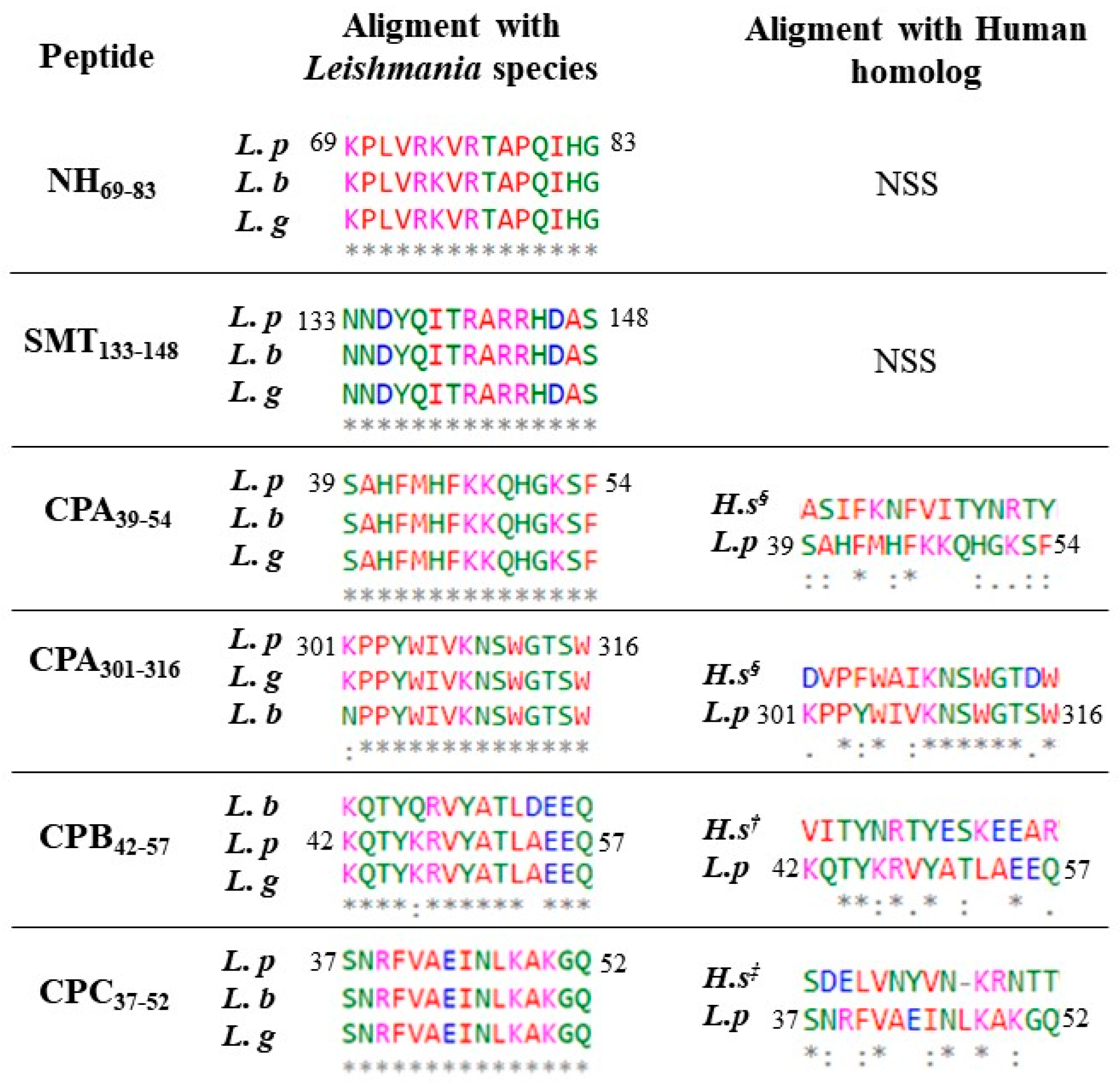
The next step was to define the percentage of sequence identities with different New World LC species. We compared each peptide from the L. panamensis sequence with L. braziliensis and L. guyanensis and with human homologs. Figure 1 shows that most peptides are 100% identical between the Leishmania species assessed, except for CPA301-316 and CPB42-57, which had 93.3% and 86.6% identity, respectively. Additionally, when sequences were compared to human homologs, only CPA301-316 showed significant identity (60%) with H. sapiens.

3.2. Molecular Docking and Immune Response

Molecular docking was performed to validate the strong interactions predicted by bioinformatic servers by comparing these six sequences against the pocket of HLA-DR4. Molecular docking was performed using the MDockPep server to simulate and analyze the interaction between the HLA-DR4 receptor and peptide sequences comprising a 9-mer core of each peptide (Figure 1, bold letters), such as a ligand.
The total binding energy (kJ·mol−1) is the result of the consensus of different intermolecular interactions between the ligand and receptor, where lower binding energy indicates higher binding affinity. The MHC class II pocket is formed by the alpha (α) and beta (β) chains of DR4. Global analysis (Table 2) showed that the epitope CPA301-316 had the strongest binding affinity (−210.3 kJ·mol−1), followed by SMT133-148 (−208.3 kJ·mol−1) and CPA39-54 (−203.8 kJ·mol−1). Finally, among the selected peptides, those with lower binding affinities were NH69-83 (−165.3 kJ·mol−1) and CPC37-52 (−165.3 kJ·mol−1).
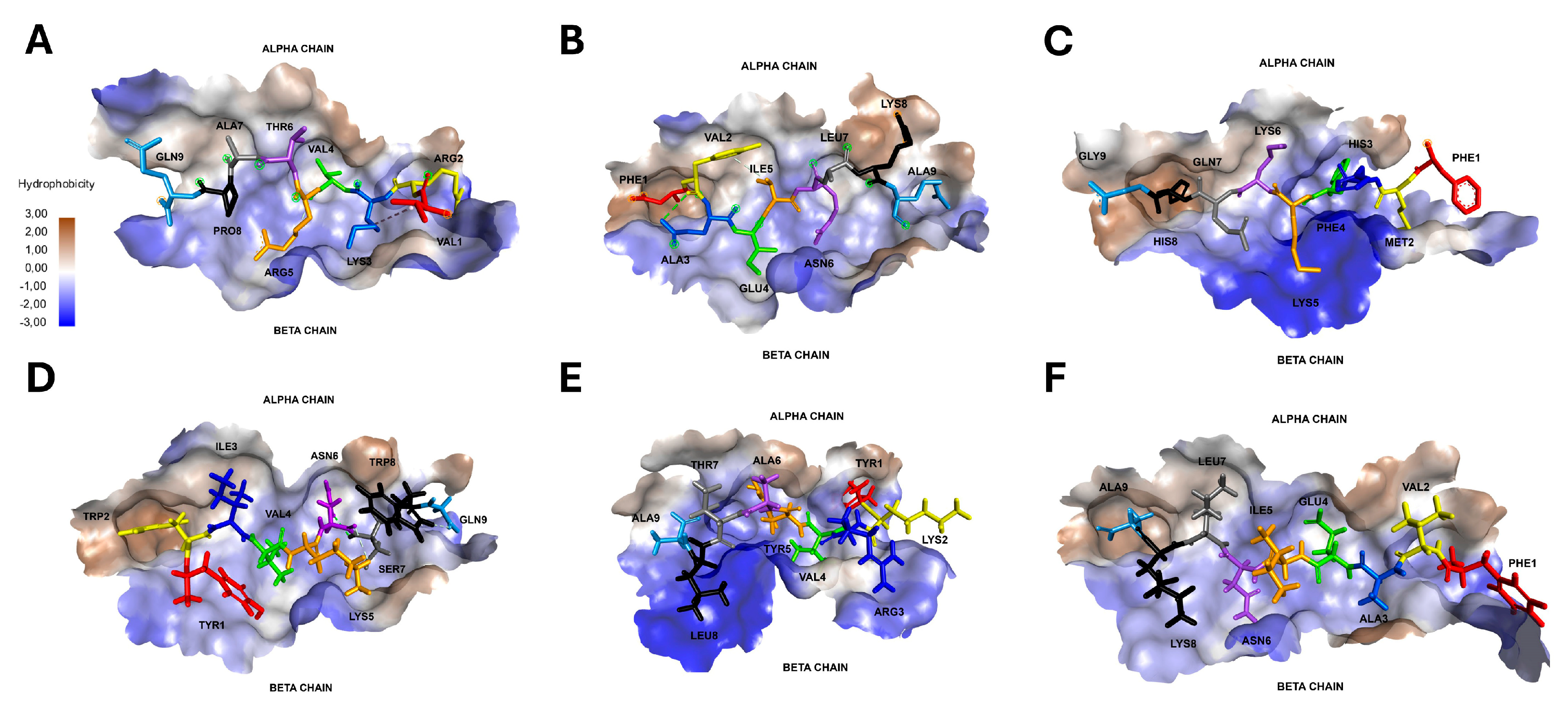
A deep analysis of molecular docking showed interactions between peptides and HLADR*04 α and β chain pocket amino acids (Table 2 and Figure 2). The overall forces that interact inside the epitope–receptor system can be classified into four main bonds from strongest to weakest: (i) hydrogen bonds—HBs, (ii) hydrophobic interactions, (iii) attractive forces, and (vi) unfavorable interactions. The β chain showed a higher number of interactions (44) compared with α chain amino acids (26); however, both chains had a similar number of HBs and hydrophobic bonds among all peptides studied, showing a similar contribution of both the α and β chains to peptide binding and stability through strong forces. Among the α chain, several positions had repeated interactions with different peptides, such as ASN62α, which interacted with seven residues, mainly forming HBs with hydrophobic and polar charged amino acids, as well as ASN69α, which formed HBs with five mainly polar residues. Similarly, among the β chain, ARG74β formed five bonds, and TRP61β, TYR78β, TRP61β and ASP70β were the positions with four bonds each.
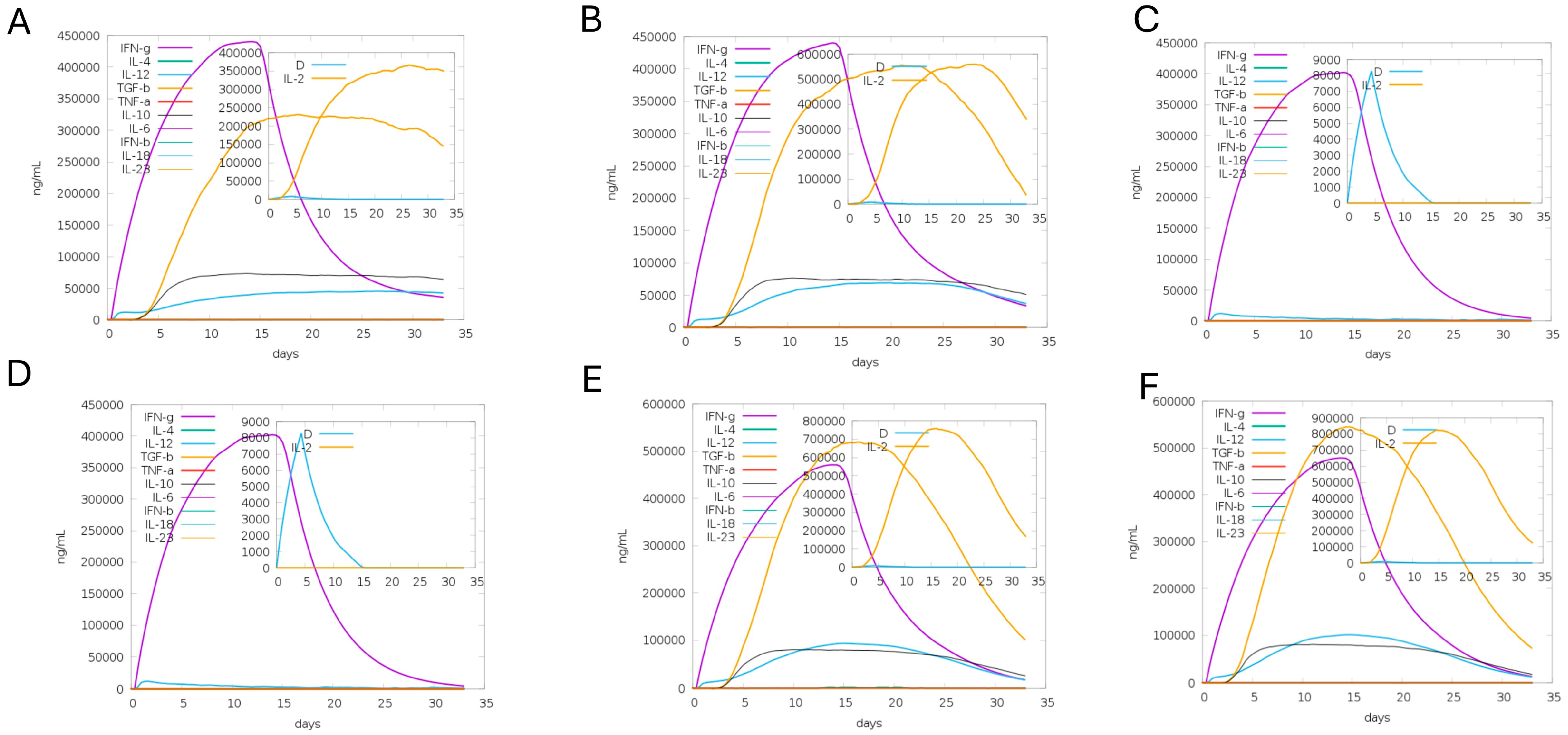
On the peptide side, SMT133-148 and CPA301-316 bonded to the DRβ1*04 pocket in a canonical way from N terminus to C terminus (Figure 2B,D). Surprisingly, NH69-83, CPA39-54, CPB42-57 and CPC37-52 bonded in a reverse way, from C to N terminus (Figure 2A,C,E,F). When a peptide binds to HLA class II molecules, it may indicate that the sequence acts as a CD4+ T epitope; however, each epitope may trigger different immune response profiles. Finally, the immune response triggered by each peptide was predicted by the C-immSim server, injecting a single dose of antigen (each 15-mer peptide) plus adjuvant. Figure 3 shows the dynamic cytokine production for over 30 days after immunization in humans carrying HLADRβ*04:01. The results showed that 15 days after immunization, NH69-83 and SMT133-148 showed a peak of a predominant pro-inflammatory Th1-related response, with higher levels of IFNγ and IL-2 (Figure 3A,B). In contrast, CPA peptides induced only IFNγ (Figure 3C,D), and CPB42-57 and CPC37-52 induced IL-2 and TGFβ at higher levels related to anti-inflammatory response (Figure 3E,F).

3.3. Molecular Insights into HLA-DR4–Peptide Binding Interfaces

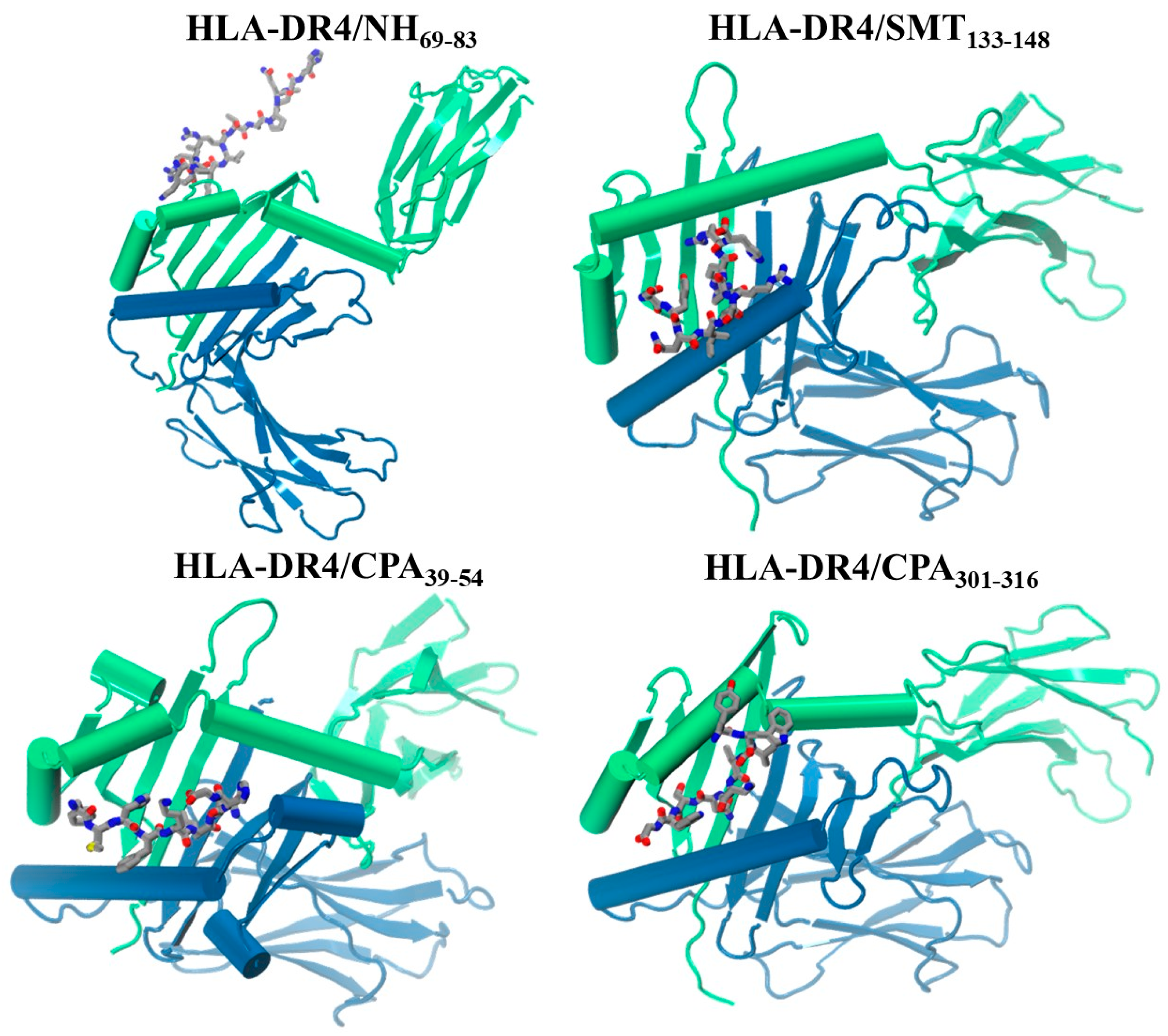
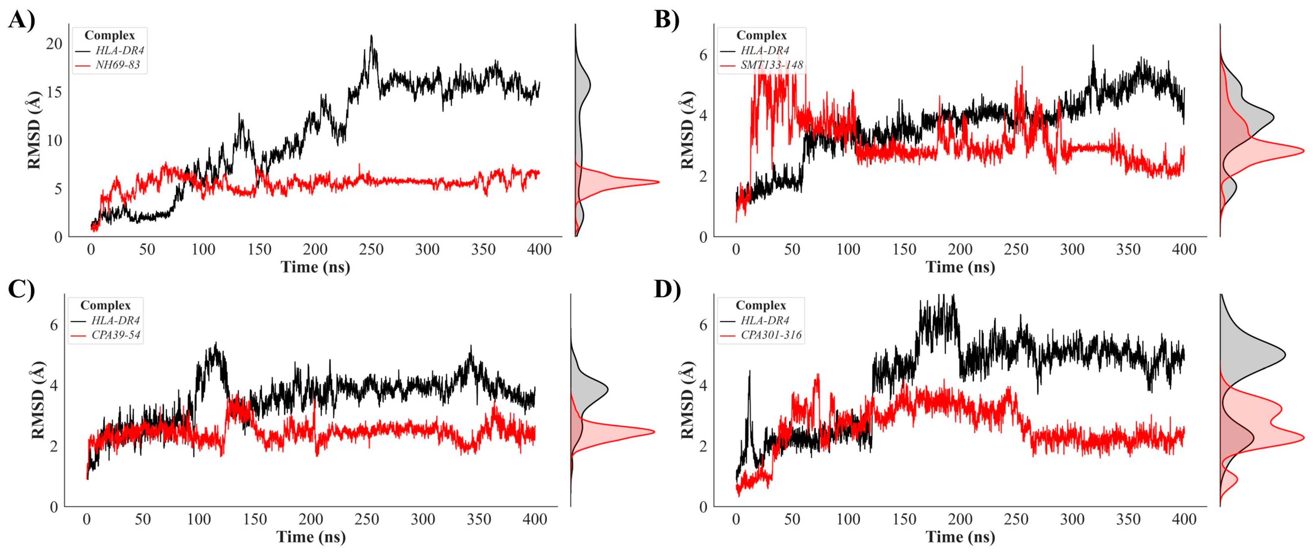
To deepen our understanding of the findings derived from the molecular docking studies conducted between the HLA-DR4 receptor and the NH69-83, SMT133-148, CPA39-54, and CPA301-316 peptides, molecular dynamics simulations were implemented to meticulously examine the dynamic behavior of each protein–peptide system over time (Videos S1–S4). Trajectory analyses revealed distinctive binding patterns, as schematically illustrated in Figure 4, which presents the final frames of each simulation. The complexes formed with the SMT133-148, CPA39-54, and CPA301-316 peptides demonstrated remarkable conformational stability, remaining consistently associated with the predicted binding site throughout the course of the molecular simulations. In contrast, the HLA-DR4/NH69-83 complex exhibited significant dissociation behavior, with the peptide shifting from the initial binding site to an alternative region located at the end of the receptor’s alpha chain, suggesting reduced affinity compared to the initial docking predictions. Temporal structural stability was quantified using root mean square deviation (RMSD) analysis, the results of which are presented in detail in Figure 5.
The molecular dynamics simulations demonstrated the differential stability patterns among the HLA-DR4 complexes with various Leishmania-derived peptides (Figure 5). RMSD analysis revealed that HLA-DR4 exhibits markedly higher structural fluctuations when complexed with NH69-83 (mean RMSD: 10.58 ± 5.42 Å) compared to the other peptide complexes, indicating reduced binding stability and increased conformational flexibility. This is because the peptide uncouples from the binding site and disrupts the protein stability. In contrast, the CPA39-54 complex demonstrates the highest structural stability with the lowest mean RMSD (2.44 ± 0.31 Å for peptide, 3.53 ± 0.76 Å for HLA-DR4), suggesting optimal peptide–MHC interactions and minimal conformational drift throughout the simulation. The SMT133-148 and CPA301-316 complexes exhibit intermediate stability profiles, with mean RMSD values of 3.10 ± 0.81 Å and 2.60 ± 0.75 Å for the peptides, respectively. The consistently lower standard deviations observed for the peptides compared to the HLA-DR4 receptor indicate that the bound peptides maintain more rigid conformations within the binding groove, while the MHC molecule undergoes expected thermal fluctuations characteristic of protein dynamics simulations. These findings suggest that CPA39-54 and CPA301-316 represent the most thermodynamically favorable peptide–HLA-DR4 interactions for potential immunotherapeutic applications.
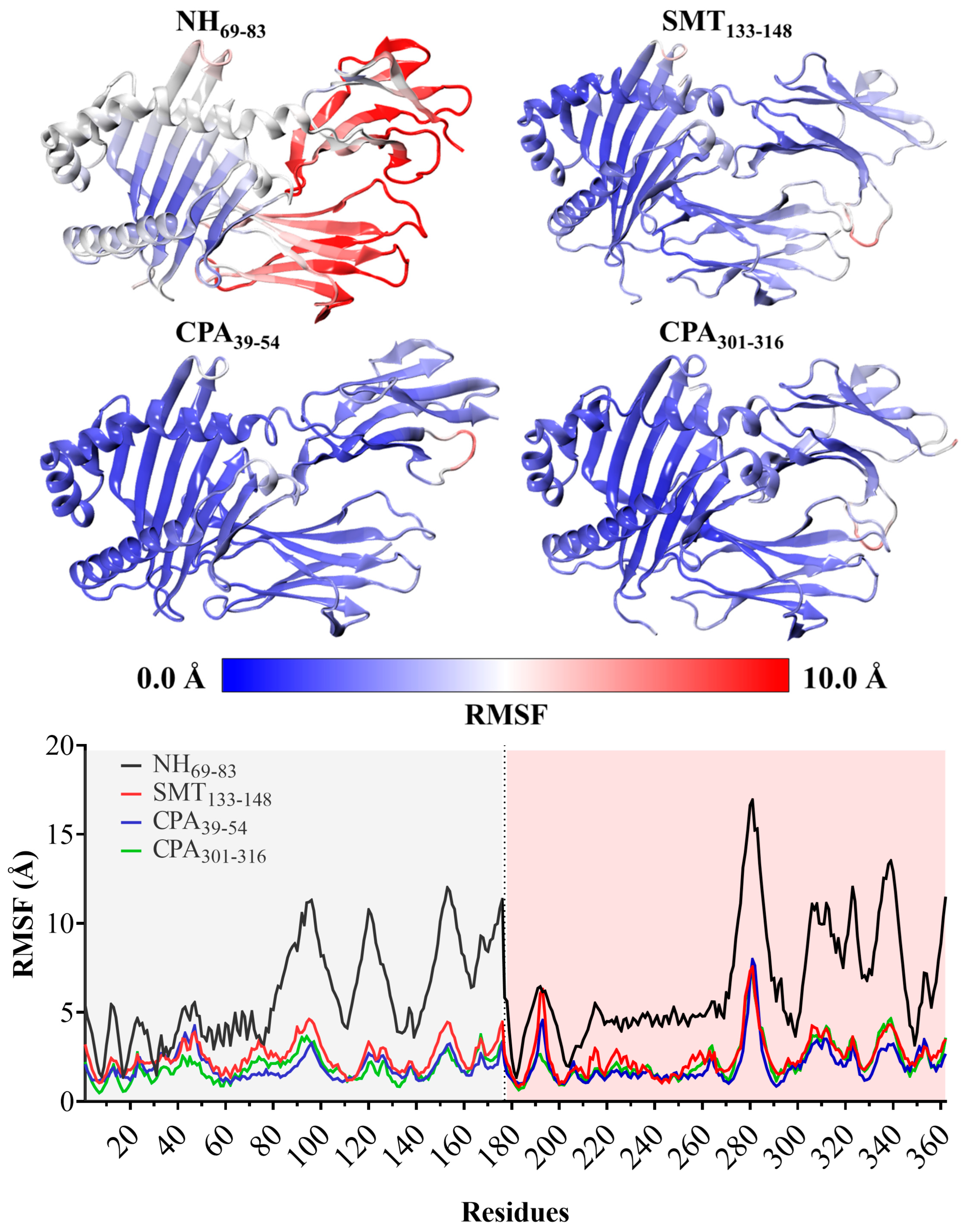
The comprehensive Root Mean Square Fluctuation (RMSF) analysis of the HLA-DR4 complexes with peptides NH69-83, SMT133-148, CPA39-54, and CPA301-316 revealed significant differences in conformational stability across the systems (Figure 6). The HLA-DR4/NH69-83 complex exhibited the most pronounced fluctuations, directly attributable to the dissociative behavior of the NH69-83 peptide, which progressively detached from the canonical binding site during the simulation trajectory and migrated toward the terminal region of the HLA-DR4 α-chain. This peptidic relocation induced considerable structural perturbations that compromised the receptor’s architectural integrity, manifesting as elevated fluctuations, particularly in the terminal regions of both the α and β chains. The thermodynamic color-coding visualization clearly illustrates these dynamics, where red hues indicate high RMSF fluctuation zones while blue regions indicate stable areas. The complexes with SMT133-148, CPA39-54, and CPA301-316 predominantly displayed blue coloration, indicating reduced fluctuations and notable structural stability, especially within the peptide binding site, where the blue coloration remained constant. The graphical RMSF profiles complemented this visualization, demonstrating remarkably similar fluctuation patterns among the three stable complexes in both the α and β chains, with RMSF values typically ranging below 2.0 Å for most residues. These findings collectively demonstrate that SMT133-148, CPA39-54, and CPA301-316 maintain optimal binding conformations and structural integrity throughout the MD simulations, while NH69-83 represents a less stable interaction characterized by significant conformational drift and receptor destabilization.
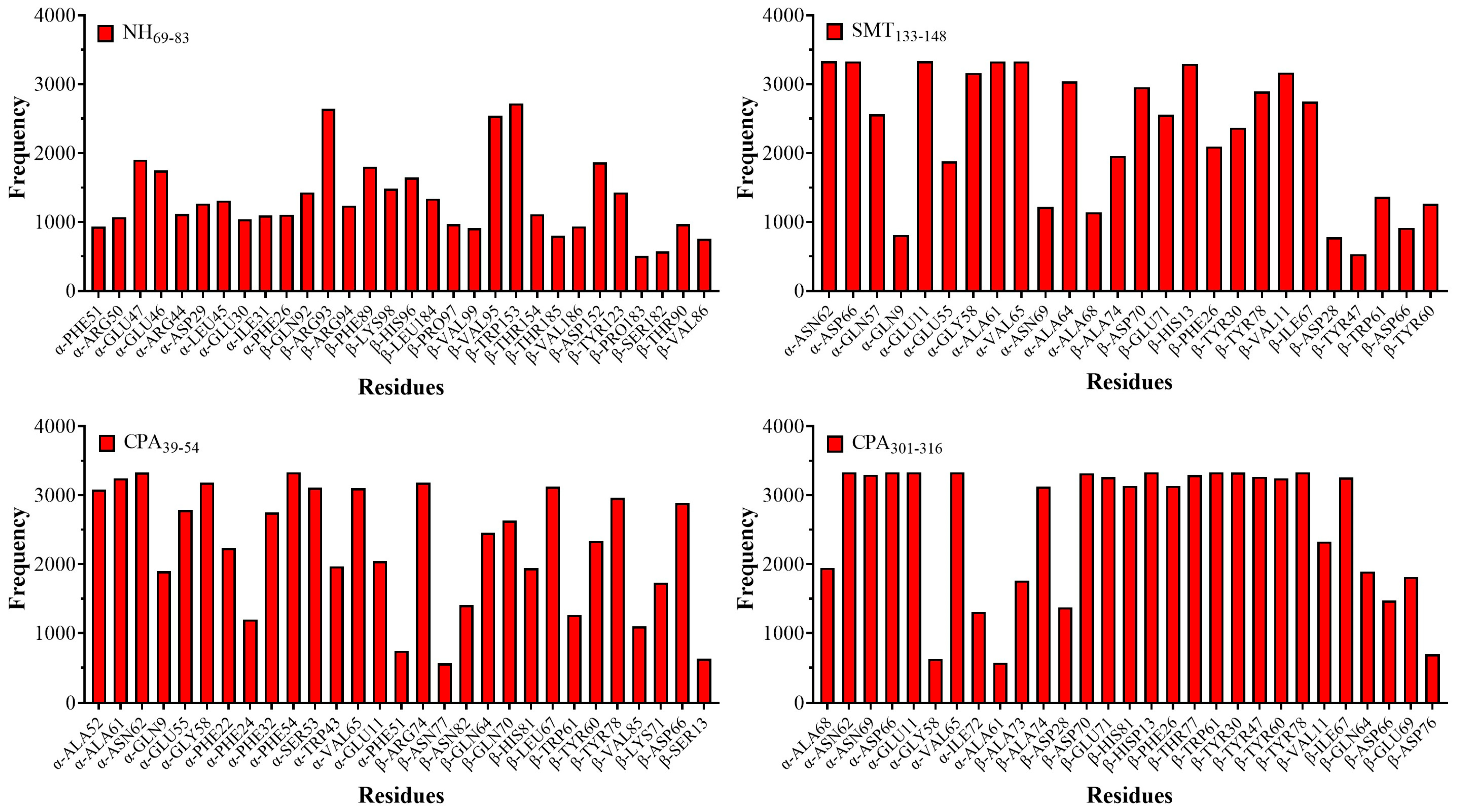
Detailed analysis of the molecular interactions between the HLA-DR4 receptor and the peptides NH69-83, SMT133-148, CPA39-54, and CPA301-316 allowed the identification of the critical amino acid residues responsible for the recognition and stabilization of each protein–peptide complex (Figure 7 and Figure 8). For the NH69-83 peptide, residue-specific interactions proved to be of limited biological relevance due to the observed dissociation phenomenon, whereby the peptide progressively decouples from the canonical binding site of HLA-DR4, thus compromising the formation of stable and functionally significant intermolecular contacts. In stark contrast, the complexes formed with the peptides SMT133-148, CPA39-54, and CPA301-316 demonstrated highly relevant and stable interaction patterns, maintaining their location within the receptor-specific binding site throughout the duration of the molecular simulations, which facilitated the accurate characterization of the residue–residue interactions.
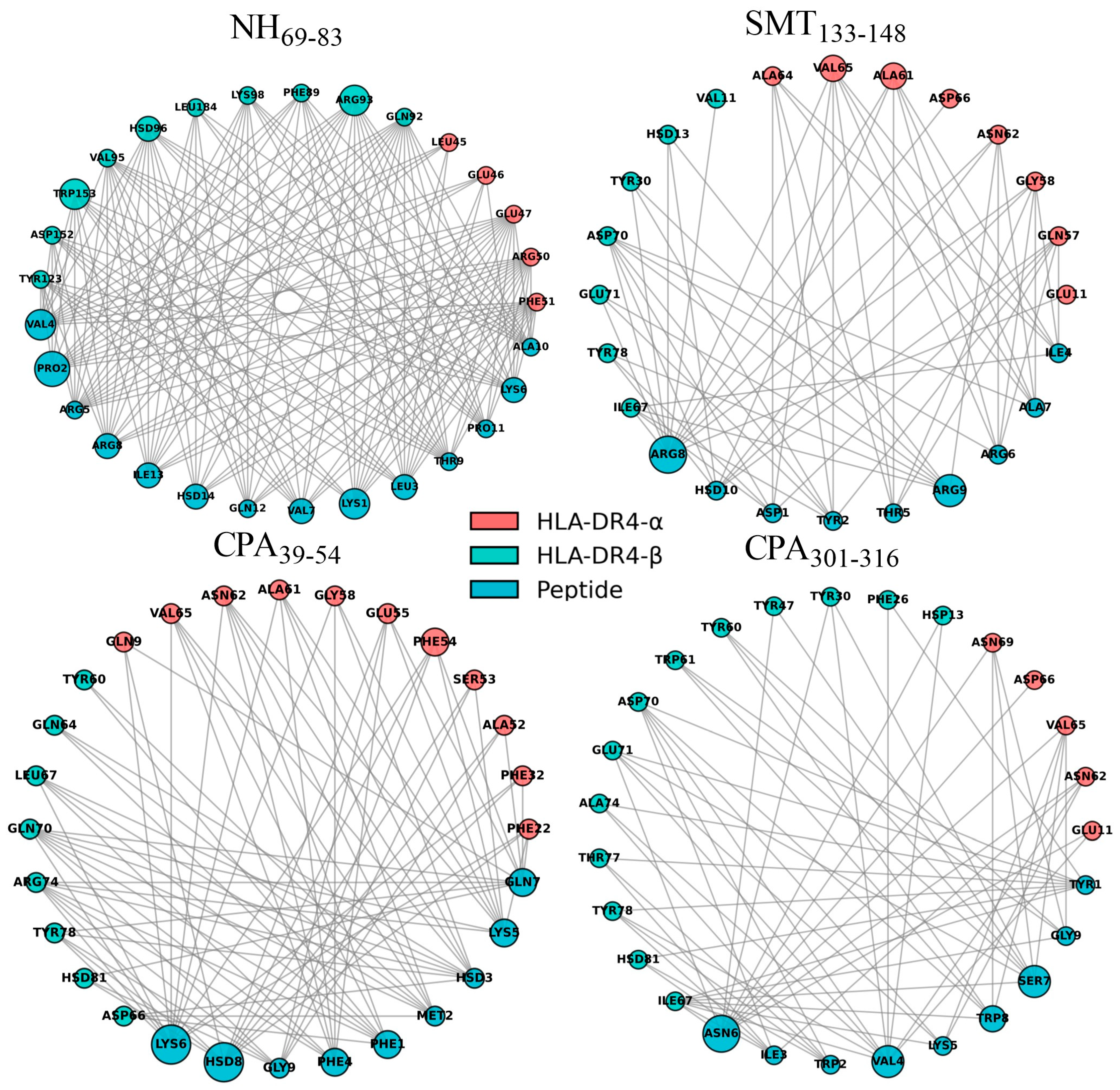
Comprehensive analysis of critical amino acid residues responsible for HLA-DR4 recognition and stabilization of SMT133-148, CPA39-54, and CPA301-316 peptide complexes revealed distinct interaction patterns and binding preferences characteristic of stable peptide–MHC class II associations (Figure 7). For SMT133-148, the α-chain residues ASN62, GLU11, and ASP66 exhibited the highest interaction frequencies (3333 occurrences each), while β-chain residues HIS13 (3294) and VAL11 (3169) significantly contributed to complex stability through sustained electrostatic and hydrogen bonding interactions. The CPA39-54 complex showed pronounced interactions with α-PHE54 (3332) and α-ASN62 (3328), complemented by β-ARG74 (3183) and β-LEU67 (3123), indicating a distinct binding mode favoring hydrophobic and aromatic interactions that facilitate peptide anchoring within the MHC binding groove. CPA301-316 displayed the most uniform interaction profile, with multiple residues achieving maximum occurrence frequencies (3333), including α-ASN62, α-ASP66, α-GLU11, α-VAL65, β-HIS13, β-TRP61, and β-TYR78, suggesting exceptionally stable peptide–receptor interactions throughout the 400 ns simulation trajectory. The dynamic residue interaction networks (Figure 8) revealed that peptide position-specific contacts drive binding specificity, with SMT133-148 residue ARG8 forming the most extensive network involving both α and β chains through multiple hydrogen bonds and salt bridges, CPA39-54 showing preferential interactions through histidine and lysine residues that stabilize the peptide backbone, and CPA301-316 demonstrating balanced interactions across multiple peptide positions that maximize binding surface area. Comparison with molecular docking results showed excellent concordance between predicted and observed binding patterns, where docking initially predicted SMT133-148 as the strongest binder (−233.9 kJ/mol), followed by CPA39-54 (−229.14 kJ/mol) and CPA301-316 (−228.65 kJ/mol). The molecular dynamics simulations validated these predictions through sustained interaction frequencies and revealed that key residues identified in static docking studies (α-ASN62, β-HIS13, α-VAL65) maintained consistent high-frequency contacts throughout the entire simulation, confirming the reliability of docking predictions for identifying key binding determinants and validating the therapeutic potential of these peptide candidates for leishmaniasis vaccine development.
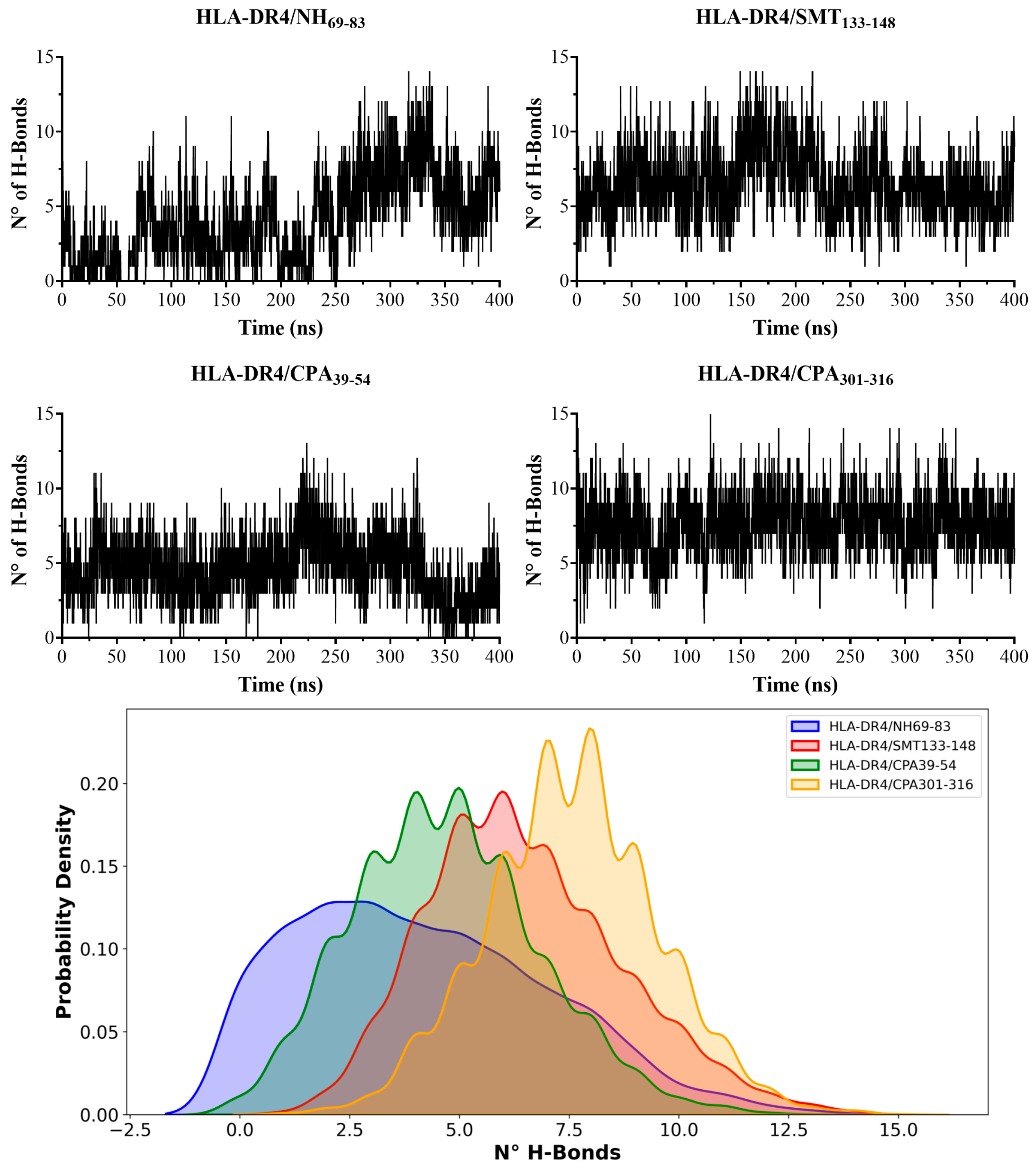
The hydrogen bond analysis of HLA-DR4 complexes with peptides NH69-83, SMT133-148, CPA39-54, and CPA301-316 revealed significant differences in conformational stability and binding characteristics between the systems, with hydrogen bond formation serving as a critical determinant of peptide–MHC class II complex stability (Figure 9). The HLA-DR4/NH69-83 complex exhibited severely compromised hydrogen bonding capacity due to the peptide’s progressive dissociation from the canonical binding site, resulting in minimal stable intermolecular contacts and confirming the destabilizing nature of this interaction observed in previous RMSD and RMSF analyses. In stark contrast, the three stable complexes demonstrated robust hydrogen bonding networks with distinct characteristics: SMT133-148 showed predominant acceptor behavior (6.16 average acceptors per frame, 0.31 donors) totaling 6.48 hydrogen bonds per frame, indicating strong electrostatic stabilization through negatively charged residues. CPA39-54 exhibited a more balanced donor–acceptor profile (1.34 donors, 3.33 acceptors) with 4.68 total hydrogen bonds per frame, suggesting a different binding mode involving both hydrogen bond donation and acceptance. CPA301-316 demonstrated the highest hydrogen bonding capacity with 2.74 donors and 4.81 acceptors, resulting in 7.56 hydrogen bonds per frame, indicating the most extensive and stable intermolecular interactions among all the studied complexes. The kernel density estimation curves provided complementary visualization of these binding patterns, where CPA39-54 displayed concentrated values between 2 and 6 bonds with multiple minor peaks, SMT133-148 exhibited similar but slightly higher values (4–8 range), and CPA301-316 demonstrated the strongest hydrogen bonding profile, with a main maximum near seven bonds, and the greatest dispersion toward high values. These findings correlate directly with the conformational stability patterns observed in molecular dynamics simulations, where hydrogen bond formation density directly corresponds to structural integrity and peptide retention within the MHC binding groove, establishing hydrogen bonding as a fundamental predictor of peptide–MHC class II binding affinity.

3.4. Estimation of Binding Affinity in HLA-DR4–Peptide Complexes via Free Energy Calculations

The MM-GBSA binding free energy calculations (Table 3) revealed pronounced thermodynamic differences among the HLA-DR4–peptide complexes, with SMT133-148 demonstrating exceptionally strong binding affinity (−260.3 ± 0.4 kcal·mol−1) compared to CPA301-316 (−57.2 ± 0.2 kcal·mol−1), CPA39-54 (−30.5 ± 0.3 kcal·mol−1), and the dissociated NH69-83 complex (-41.1 ± 0.3 kcal·mol−1). Energy decomposition analysis revealed that SMT133-148 binding was primarily driven by highly favorable electrostatic interactions (−243.4 ± 0.5 kcal·mol−1), indicating extensive hydrogen bonding and ionic interactions that stabilize the peptide–MHC interface. In contrast, CPA39-54 exhibited unfavorable electrostatic contributions (38.9 ± 0.3 kcal·mol−1) counterbalanced by strong van der Waals interactions (−61.7 ± 0.2 kcal·mol−1), suggesting a binding mode dominated by hydrophobic contacts. CPA301-316 showed moderate electrostatic favorability (12.4 ± 0.3 kcal·mol−1) with substantial van der Waals contributions (−60.4 ± 0.2 kcal·mol−1), representing a balanced interaction profile. Despite dissociation from the canonical binding, the NH69-83 complex retained some binding affinity through residual van der Waals interactions (−53.9 ± 0.4 kcal·mol−1), partially offset by unfavorable electrostatic repulsion (21.7 ± 0.3 kcal·mol−1), confirming its compromised binding stability. All complexes exhibited comparable solvent-accessible surface area contributions (ΔEsasa: −6.1 to −9.2 kcal·mol−1), indicating similar desolvation penalties upon binding. These thermodynamic profiles correlate directly with the structural stability patterns observed in MD simulations, establishing SMT133-148 as the most promising therapeutic candidate based on its exceptional binding affinity and electrostatic stabilization mechanisms.

4. Discussion

To date, protozoa vaccines have been elusive to humanity due to immune evasion by obligate intracellular parasites. Leishmania spp. also survive and multiply inside macrophages, the immune cells that are designed to eliminate them. Several attempts have been made for decades, using first-, second-, and third-generation vaccines; however, none have been licensed for use in humans [8].
With the rise of informatics and machine learning, it was possible to “feed” machines biological experiment results and then predict different outcomes through algorithms. Bioinformatic servers have been used to predict T epitopes by assessing the interaction between linear short sequences and the structure of the pocket of MHC molecules, considering that APCs may present those strong binders [64].
Researchers working on the leishmaniasis vaccine have designed several chimeric antigens that include T and/or B epitopes based on bioinformatic tools for cutaneous leishmaniasis. Seyed et al., 2011, designed CD8+ T cell-restricted epitopes from CPB, CPC, Sti1, TSA, Leif, and LPG-3 proteins from L. major, and pools from some predicted sequences showed IFN-γ induction in human Peripheral Blood Mononuclear Cells—PBMCs—in vitro [65]. Other studies have constructed polytopes made of T epitopes or a combination of T- and B-predicted epitopes from different L. major intracellular and membrane proteins (TSA, LPG3, GP63, STI1, LACK, SMT, LEIF, KMP11, and HASPB). These showed high predicted antigenicity in addition to non-allergenic properties, according to in silico results [66,67,68]. Another group of studies was designed to analyze visceral leishmaniasis species, such as L. donovani T and B multiepitopes, through bioinformatics [69], and researchers simultaneously included LC and LV species [70].
In our study, the choice of target proteins (NH, SMT, and CPs) was strategically significant. As established in the Introduction, NH and SMT are vital for the metabolic survival and structural integrity of the parasite during the transition to the amastigote stage [15,16,17,18,19]. By targeting these essential enzymes, a vaccine could theoretically interrupt the parasite’s adaptation to the host’s intracellular environment. Furthermore, since CPs are known virulence factors, identifying epitopes within these proteins that may successfully induce protective immunity is a therapeutic immunomodulatory strategy [18,19,20]. Taking this into account, strategies that allowed the selection of vaccine candidate epitopes were established.
T epitope prediction tools allow us to perform a high number of predictions; however, each server is based on different algorithms, so it is not clear which one predicts closer to reality. To overcome this issue, three different servers (based on different algorithms) were used in this study, and the sequences selected were those that the three tools predict simultaneously. Additionally, MD analyses were also considered to identify the best epitopes for improving epitope screening because this approach shows in-depth intermolecular interactions that mediate the binding and specificity between the peptide sequences and the MHC class II immune receptor. Population coverage analysis showed that using promiscuous epitopes, a large population can be protected not only in Colombia or South America, but also worldwide.
Molecular docking showed the possibility for sequences to bind in canonical and reverse ways, a behavior that has been reported for CLIP peptides, where CLIP102-120 binds in a canonical way while CLIP106-120 binds in a reverse way to the HLA-DRβ1 pocket, opening the possibilities for some antigens to also bind in reverse ways [71]. Recently, using machine learning and MHC-II peptidomics data, it has been reported that not only the HLA-DR pocket allows reverse binding but also HLA-DP and HLA-DQ [38], which can be used to improve prediction algorithms.
Immunoinformatics represents a great alternative in vaccinology research because it allows the screening of a large number of proteins and sequences through rational design. This leads to the control of different infectious and/or immune diseases, saving time, effort, and even research budgets in the first development steps. However, without subsequent in vitro and in vivo proofs of their utilities, the real scope of bioinformatic strategies cannot be known.
Given that MD simulations track the stability of the peptide–HLA-DR pocket complex and serve as a biologically relevant filtering mechanism [72], our findings demonstrated that the majority of peptides—specifically CPA39-54, SMT133-148, and CPA301-316—maintained successful binding. CPA39-54 was particularly notable for its structural rigidity (mean RMSD:2.44 ± 0.31 A), while CPA301-316 exhibited a robust hydrogen bond profile (7.56 per frame). In sharp contrast, the NH69-83 peptide presented high RMSD values (10.58 ± 5.42 A) and clear patterns of dissociation and repositioning within the pocket, which correlated with the complete absence of stable hydrogen bonds in the complex. These results strongly suggest that NH69-83 may be a false positive from the initial candidate screening, as it was unable to sustain stable interactions over the simulation time. The subsequent affinity validation, quantified via binding free energy (MM-GBSA), revealed that the SMT133-148 peptide possesses extremely high affinity, primarily driven by highly favorable electrostatic contributions (−243.4 ± 0.5 kcal·mol−1). This superior affinity is attributed to the presence of numerous hydrogen bonds and salt bridges within the complex, which strongly correlates with the interaction energies (MdoCKpep Score) derived from molecular docking [43].
Although the CPA39-54 peptide showed excellent structural stability (low RMSD), it exhibited low MM-GBSA affinity. This disparity is likely due to unfavorable electrostatic contributions (+38.9 ± 0.3 kcal·mol−1) that are only partially offset by favorable van der Waals (hydrophobic) terms. This suggests a binding mode dominated by hydrophobic forces but with a substantial electrostatic penalty. These findings underscore a critical distinction between compound stability (RMSD/RMSF) and affinity (MM-GBSA): while MD demonstrated CPA39-54’s structural rigidity, MM-GBSA revealed that SMT133-148 features the strongest total free energy of interaction, propelled by polar/charged interactions. In the biological context, successful antigen presentation relies on processes involving the fit of the peptide into the canonical anchor pockets (P1, P4, P6, and P9). It observed the involvement of high-frequency residues (α-ASN62, α-ASP66, β-HIS13, and β-TRP61) essential for the presentation of the peptide by the HLA-DR4 complex [73].

5. Conclusions

From the initial pool of 1856 peptides, a manageable set of HLA-DR4 candidates exhibiting relevant biological potential was identified by combining initial peptide selection via consensus-based multiple prediction algorithms with structural filtering through molecular docking. The rigorous application of MD simulations served as an essential, biologically relevant filter, effectively discarding weak and low-quality interactions. The SMT133-148 peptide was positioned as the most robust candidate, exhibiting the highest affinity, driven by a strong contribution from electrostatic interactions with canonical anchor residues. Ultimately, the methodology successfully narrowed the search space to three primary candidates (SMT133-148, CPA39-54, and CPA301-316). Based on the profile of maximal thermodynamic affinity, SMT133-148 must be prioritized for synthesis and subsequent in vitro experimental validation, thereby optimizing resources and accelerating the preclinical development phase of a potential chimeric vaccine. Finally, these sequences will be synthesized and proven for their immunogenicity in vitro in human PBMCs to validate their utility as prophylactic or therapeutic alternative options to fight leishmaniasis.

Supplementary Materials

The following supporting information can be downloaded at https://www.mdpi.com/article/10.3390/parasitologia6030028/s1: Video S1: Molecular dynamics CPA39-54; Video S2: Molecular dynamics CPA301-316; Video S3: Molecular dynamics NH69-83; Video S4: Molecular dynamics SMT133-148.

Author Contributions

Conceptualization, M.M.F., F.E.T. and O.Y.; methodology, D.L.-I., W.D.M., K.R., Y.U., O.Y. and F.E.T.; validation, E.A.; formal analysis, M.M.F., E.A. and O.Y.; investigation, D.R.D., E.A. and F.E.T.; data curation, K.R. and Y.U.; writing—original draft preparation, M.M.F. and F.E.T.; writing—review and editing, D.R.D., O.Y., M.M.F. and F.E.T.; supervision, D.R.D. and F.E.T.; project administration, M.M.F.; funding acquisition, M.M.F. and O.Y. All authors have read and agreed to the published version of the manuscript.

Funding

M.M.F. received funds from the Cooperative University of Colombia, Grant INV3340. O.Y. gratefully acknowledges the UDLA-PIR202409 and FONDECYT project 1251871. Powered@NLHPC: This research was partially supported by the supercomputing infrastructure of the NLHPC (CCSS210001).

Institutional Review Board Statement

Not applicable.

Informed Consent Statement

Not applicable.

Data Availability Statement

Datasets obtained in this project can be found at https://doi.org/10.57924/I5HS1I and https://doi.org/10.57924/BF9PR9.

Conflicts of Interest

The authors declare no conflicts of interest.

Abbreviations

The following abbreviations are used in this manuscript:
HLAHuman Leucocyte Antigen
MHCMajor Histocompatibility Complex
NHNucleoside Hydrolase
SMTSterol 24-c-Methyltransferase
CPACysteine Peptidase A
CPBCysteine Peptidase B
CPCCysteine Peptidase C
CLCutaneous Leishmaniasis
VLVisceral Leishmaniasis
ThT Helper
IFNγInterferon γ
ILInterleucina
TGFβTransforming Growth Factor β
NONitric Oxide
APSAntigen Presenting Cell
TCRT Cell Receptor
MDMolecular Dynamics
PMEParticle Mesh Ewald
RMSDRoot Mean Square Deviation
RMSFRoot Mean Square Fluctuation

References

  1. World Health Organization Leishmaniasis. Available online: https://www.who.int/health-topics/leishmaniasis#tab=tab_1 (accessed on 29 March 2022).
  2. Instituto Nacional de Salud Carlos III. Protocolo de Vigilancia de Leishmaniasis. Available online: https://cne.isciii.es/documents/d/cne/protocolo-20de-20vigilancia-20de-20leishmaniasis-pdf (accessed on 13 December 2023).
  3. Azim, M.; Khan, S.A.; Ullah, S.; Ullah, S.; Anjum, S.I. Therapeutic advances in the topical treatment of cutaneous leishmaniasis: A review. PLoS Negl. Trop. Dis. 2021, 15, e0009099. [Google Scholar] [CrossRef]
  4. Ponte-Sucre, A.; Gamarro, F.; Dujardin, J.C.; Barrett, M.P.; López-Vélez, R.; García-Hernández, R.; Pountain, A.W.; Mwenechanya, R.; Papadopoulou, B. Drug resistance and treatment failure in leishmaniasis: A 21st century challenge. PLoS Negl. Trop. Dis. 2017, 11, e0006052. [Google Scholar] [CrossRef]
  5. Li, S.; Qiu, T.; Zhao, N.; Liu, M.; He, X.; Wang, X.; Jiang, S.; Luo, M.; Wang, S.; Zhang, L.; et al. Global burden of leishmaniasis, 1990–2021: Systematic analysis of the global burden of disease study. Int. J. Equity Health 2025, 24, 329. [Google Scholar] [CrossRef]
  6. Belkaid, Y.; Kamhawi, S.; Modi, G.; Valenzuela, J.; Noben-Trauth, N.; Rowton, E.; Ribeiro, J.; Sacks, D.L. Development of a Natural Model of Cutaneous Leishmaniasis: Powerful Effects of Vector Saliva and Saliva Preexposure on the Long-Term Outcome of Leishmania major Infection in the Mouse Ear Dermis. J. Exp. Med. 1998, 188, 1941. [Google Scholar] [CrossRef]
  7. Bacon, K.M.; Hotez, P.J.; Kruchten, S.D.; Kamhawi, S.; Bottazzi, M.E.; Valenzuela, J.G.; Lee, B.Y. The potential economic value of a cutaneous leishmaniasis vaccine in seven endemic countries in the Americas. Vaccine 2012, 31, 480–486. [Google Scholar] [CrossRef]
  8. Dinc, R. Leishmania Vaccines: The Current Situation with Its Promising Aspect for the Future. Korean J. Parasitol. 2022, 60, 379–391. [Google Scholar] [CrossRef] [PubMed]
  9. Sacks, D.; Noben-Trauth, N. The immunology of susceptibility and resistance to Leishmania major in mice. Nat. Rev. Immunol. 2002, 2, 845–858. [Google Scholar] [CrossRef]
  10. Gollob, K.J.; Viana, A.G.; Dutra, W.O. Immunoregulation in human American leishmaniasis: Balancing pathology and protection. Parasite Immunol. 2014, 36, 367–376. [Google Scholar] [CrossRef] [PubMed]
  11. Tripathi, P.; Singh, V.; Naik, S. Immune response to leishmania: Paradox rather than paradigm. FEMS Immunol. Med. Microbiol. 2007, 51, 229–242. [Google Scholar] [CrossRef] [PubMed]
  12. Desai, D.V.; Kulkarni-Kale, U. T-cell epitope prediction methods: An overview. Methods Mol. Biol. 2014, 1184, 333–364. [Google Scholar] [CrossRef]
  13. Burza, S.; Croft, S.L.; Boelaert, M. Leishmaniasis. Lancet 2018, 392, 951–970. [Google Scholar] [CrossRef]
  14. Maxfield, L.; Crane, J.S. Leishmaniasis. In StatPearls; StatPearls: Treasure Island, FL, USA, 2023. [Google Scholar]
  15. Azam, S.S.; Abro, A.; Raza, S.; Saroosh, A. Structure and dynamics studies of sterol 24-C-methyltransferase with mechanism based inactivators for the disruption of ergosterol biosynthesis. Mol. Biol. Rep. 2014, 41, 4279–4293. [Google Scholar] [CrossRef]
  16. Besteiro, S.; Williams, R.A.M.; Coombs, G.H.; Mottram, J.C. Protein turnover and differentiation in Leishmania. Int. J. Parasitol. 2007, 37, 1063–1075. [Google Scholar] [CrossRef]
  17. Saha, S.; Srivastava, R.; Sarma, P.; Bhatt, T.K.; Prakash, A.; Kumar, D. Identification of potential inhibitors of Leishmania donovani Sterol 24-C-methyltransferase: In silico and in vitro studies. Mol. Simul. 2023, 49, 1311–1323. [Google Scholar] [CrossRef]
  18. Casgrain, P.A.; Martel, C.; McMaster, W.R.; Mottram, J.C.; Olivier, M.; Descoteaux, A. Cysteine Peptidase B Regulates Leishmania mexicana Virulence through the Modulation of GP63 Expression. PLoS Pathog. 2016, 12, e1005658. [Google Scholar] [CrossRef]
  19. Mottram, J.C.; Coombs, G.H.; Alexander, J. Cysteine peptidases as virulence factors of Leishmania. Curr. Opin. Microbiol. 2004, 7, 375–381. [Google Scholar] [CrossRef]
  20. Siqueira-Neto, J.L.; Debnath, A.; McCall, L.I.; Bernatchez, J.A.; Ndao, M.; Reed, S.L.; Rosenthal, P.J. Cysteine proteases in protozoan parasites. PLoS Negl. Trop. Dis. 2018, 12, e0006512. [Google Scholar] [CrossRef] [PubMed]
  21. MLB; Nico, D.; de Oliveira, F.A.; Barreto, A.S.; Palatnik-de-Sousa, I.; Carrillo, E.; Moreno, J.; de Luca, P.M.; Morrot, A.; Rosa, D.S.; et al. Leishmania donovani nucleoside Hydrolase (NH36) domains induce T-cell cytokine responses in human visceral leishmaniasis. Front. Immunol. 2017, 8, 227. [Google Scholar] [CrossRef] [PubMed]
  22. Nico, D.; Claser, C.; Borja-Cabrera, G.P.; Travassos, L.R.; Palatnik, M.; Soares Ida, S.; Rodrigues, M.M.; de Palatnik-Sousa, C.B. Adaptive Immunity against Leishmania Nucleoside Hydrolase Maps Its C-Terminal Domain as the Target of the CD4+ T Cell–Driven Protective Response. PLoS Negl. Trop. Dis. 2010, 4, e866. [Google Scholar] [CrossRef] [PubMed][Green Version]
  23. Goto, Y.; Bhatia, A.; Raman, V.S.; Vidal, S.E.Z.; Bertholet, S.; Coler, R.N.; Howard, R.F.; Reed, S.G. Leishmania infantum sterol 24-c-methyltransferase formulated with MPL-SE induces cross-protection against L. major infection. Vaccine 2009, 27, 2884–2890. [Google Scholar] [CrossRef]
  24. Nico, D.; Gomes, D.C.; Palatnik-de-Sousa, I.; Morrot, A.; Palatnik, M.; Palatnik-de-Sousa, C.B. Leishmania donovani nucleoside hydrolase terminal domains in cross-protective immunotherapy against Leishmania amazonensis murine infection. Front. Immunol. 2014, 5, 88478. [Google Scholar] [CrossRef]
  25. Khoshgoo, N.; Zahedifard, F.; Azizi, H.; Taslimi, Y.; Alonso, M.J.; Rafati, S. Cysteine proteinase type III is protective against Leishmania infantum infection in BALB/c mice and highly antigenic in visceral leishmaniasis individuals. Vaccine 2008, 26, 5822–5829. [Google Scholar] [CrossRef]
  26. Doroud, D.; Zahedifard, F.; Vatanara, A.; Najafabadi, A.R.; Taslimi, Y.; Vahabpour, R.; Torkashvand, F.; Vaziri, B.; Rafati, S. Delivery of a cocktail DNA vaccine encoding cysteine proteinases type I, II and III with solid lipid nanoparticles potentiate protective immunity against Leishmania major infection. J. Control. Release 2011, 153, 154–162. [Google Scholar] [CrossRef]
  27. Pascalis, H.; Lavergne, A.; Bourreau, E.; Prévot-Linguet, G.; Kariminia, A.; Pradinaud, R.; Rafati, S.; Launois, P. Th1 cell development induced by cysteine proteinases A and B in localized cutaneous leishmaniasis due to Leishmania guyanensis. Infect. Immun. 2003, 71, 2924–2926. [Google Scholar] [CrossRef]
  28. Noormehr, H.; Zavaran Hosseini, A.; Soudi, S.; Beyzay, F. Enhancement of Th1 immune response against Leishmania cysteine peptidase A, B by PLGA nanoparticle. Int. Immunopharmacol. 2018, 59, 97–105. [Google Scholar] [CrossRef]
  29. Sidney, J.; Peters, B.; Sette, A. Epitope prediction and identification- adaptive T cell responses in humans. Semin. Immunol. 2020, 50, 101418. [Google Scholar] [CrossRef] [PubMed]
  30. Patronov, A.; Doytchinova, I. T-cell epitope vaccine design by immunoinformatics. Open Biol. 2013, 3, 120139. [Google Scholar] [CrossRef] [PubMed]
  31. Trachtenberg, E.A.; Keyeux, G.; Bernal, J.E.; Rhodas, M.C.; Erlich, H.A. Results of Expedition Humana. Tissue Antigens 1996, 48, 174–181. [Google Scholar] [CrossRef]
  32. Correa, P.A.; Whitworth, W.C.; Kuffner, T.; McNicholl, J.; Anaya, J.M. HLA-DR and DQB1 gene polymorphism in the North-western Colombian population. Tissue Antigens 2002, 59, 436–439. [Google Scholar] [CrossRef] [PubMed]
  33. Ossa Reyes, H.; Manrique, A.; Quintanilla, S.; Peña, A. Polimorfismos del sistema HLA (loci A*, B* y DRB1*) en población colombiana. Nova 2007, 5, 25–30. [Google Scholar] [CrossRef]
  34. Rocío Arias-Murillo, Y.; Ángel Castro-Jiménez, M.; Ríos-Espinosa, F.; López-Rivera, J.J.; Johanna Echeverry-Coral, S.; Esp, B.; Martínez-Nieto, O. Analysis of HLA-A, HLA-B, HLA-DRB1 allelic, genotypic, and haplotypic frequencies in colombian population. Colomb. Med. 2010, 41, 336–343. [Google Scholar] [CrossRef]
  35. Ávila-Portillo, L.M.; Carmona, A.; Franco, L.; Briceño, I.; Casas, M.C.; Gómez, A. Bajo polimorfismo en el sistema de antígenos de leucocitos humanos en población mestiza colombiana. Univ. Medica 2010, 51, 359–370. [Google Scholar] [CrossRef]
  36. Reynisson, B.; Barra, C.; Kaabinejadian, S.; Hildebrand, W.H.; Peters, B.; Peters, B.; Nielsen, M.; Nielsen, M. Improved Prediction of MHC II Antigen Presentation through Integration and Motif Deconvolution of Mass Spectrometry MHC Eluted Ligand Data. J. Proteome Res. 2020, 19, 2304–2315. [Google Scholar] [CrossRef] [PubMed]
  37. Racle, J.; Michaux, J.; Rockinger, G.A.; Arnaud, M.; Bobisse, S.; Chong, C.; Guillaume, P.; Coukos, G.; Harari, A.; Jandus, C.; et al. Robust prediction of HLA class II epitopes by deep motif deconvolution of immunopeptidomes. Nat. Biotechnol. 2019, 37, 1283–1286. [Google Scholar] [CrossRef] [PubMed]
  38. Racle, J.; Guillaume, P.; Schmidt, J.; Michaux, J.; Larabi, A.; Lau, K.; Perez, M.A.S.; Croce, G.; Genolet, R.; Coukos, G.; et al. Machine learning predictions of MHC-II specificities reveal alternative binding mode of class II epitopes. Immunity 2023, 56, 1359–1375.e13. [Google Scholar] [CrossRef]
  39. Chen, B.; Khodadoust, M.S.; Olsson, N.; Wagar, L.E.; Fast, E.; Liu, C.L.; Muftuoglu, Y.; Sworder, B.J.; Diehn, M.; Levy, R.; et al. Predicting HLA class II antigen presentation through integrated deep learning. Nat. Biotechnol. 2019, 37, 1332–1343. [Google Scholar] [CrossRef]
  40. Edgar, R.C. MUSCLE: Multiple sequence alignment with high accuracy and high throughput. Nucleic Acids Res. 2004, 32, 1792–1797. [Google Scholar] [CrossRef]
  41. Edgar, R.C. MUSCLE: A multiple sequence alignment method with reduced time and space complexity. BMC Bioinform. 2004, 5, 113. [Google Scholar] [CrossRef]
  42. Bui, H.H.; Sidney, J.; Dinh, K.; Southwood, S.; Newman, M.J.; Sette, A. Predicting population coverage of T-cell epitope-based diagnostics and vaccines. BMC Bioinform. 2006, 7, 153. [Google Scholar] [CrossRef] [PubMed]
  43. Xu, X.; Zou, X. MDockPeP: A Web Server for Blind Prediction of Protein-Peptide Complex Structures. Methods Mol. Biol. 2020, 2165, 259–272. [Google Scholar] [CrossRef]
  44. Zhou, P.; Jin, B.; Li, H.; Huang, S.Y. HPEPDOCK: A web server for blind peptide-protein docking based on a hierarchical algorithm. Nucleic Acids Res. 2018, 46, W443–W450. [Google Scholar] [CrossRef]
  45. Xu, X.; Yan, C.; Zou, X. MDockPeP: An ab-initio protein–peptide docking server. J. Comput. Chem. 2018, 39, 2409–2413. [Google Scholar] [CrossRef] [PubMed]
  46. Pyrkov, T.V.; Ozerov, I.V.; Balitskaya, E.D.; Efremov, R.G. Molecular docking: The role of noncovalent interactionsin the formation of protein-nucleotide and protein-peptide complexes. Russ. J. Bioorg. Chem. 2010, 36, 446–455. [Google Scholar] [CrossRef] [PubMed]
  47. Ling, R.; Dai, Y.; Huang, B.; Huang, W.; Yu, J.; Lu, X.; Jiang, Y. In silico design of antiviral peptides targeting the spike protein of SARS-CoV-2. Peptides 2020, 130, 170328. [Google Scholar] [CrossRef]
  48. Pantsar, T.; Poso, A. Binding Affinity via Docking: Fact and Fiction. Molecules 2018, 23, 1899. [Google Scholar] [CrossRef] [PubMed]
  49. Rapin, N.; Lund, O.; Bernaschi, M.; Castiglione, F. Computational Immunology Meets Bioinformatics: The Use of Prediction Tools for Molecular Binding in the Simulation of the Immune System. PLoS ONE 2010, 5, 9862. [Google Scholar] [CrossRef]
  50. Stolfi, P.; Castiglione, F.; Mastrostefano, E.; Di Biase, I.; Di Biase, S.; Palmieri, G.; Prisco, A. In-silico evaluation of adenoviral COVID-19 vaccination protocols: Assessment of immunological memory up to 6 months after the third dose. Front. Immunol. 2022, 13, 998262. [Google Scholar] [CrossRef]
  51. Ragone, C.; Manolio, C.; Cavalluzzo, B.; Mauriello, A.; Tornesello, M.L.; Buonaguro, F.M.; Castiglione, F.; Vitagliano, L.; Iaccarino, E.; Ruvo, M.; et al. Identification and validation of viral antigens sharing sequence and structural homology with tumor-associated antigens (TAAs). J. Immunother. Cancer 2021, 9, e002694. [Google Scholar] [CrossRef]
  52. Jo, S.; Kim, T.; Iyer, V.G.; Im, W. CHARMM-GUI: A web-based graphical user interface for CHARMM. J. Comput. Chem. 2008, 29, 1859–1865. [Google Scholar] [CrossRef]
  53. Olsson, M.H.M.; Søndergaard, C.R.; Rostkowski, M.; Jensen, J.H. PROPKA3: Consistent Treatment of Internal and Surface Residues in Empirical pKa Predictions. J. Chem. Theory Comput. 2011, 7, 525–537. [Google Scholar] [CrossRef]
  54. Neria, E.; Fisher, S.; Karplus, M. Simulation of activation free energies in molecular systems. J. Chem. Phys. 1996, 105, 1902–1921. [Google Scholar] [CrossRef]
  55. MacKerell, A.D.; Bashford, D.; Bellott, M.; Dunbrack, R.L.; Evanseck, J.D.; Field, M.J.; Fischer, S.; Gao, J.; Guo, H.; Ha, S.; et al. All-Atom Empirical Potential for Molecular Modeling and Dynamics Studies of Proteins. J. Phys. Chem. B 1998, 102, 3586–3616. [Google Scholar] [CrossRef]
  56. Huang, J.; MacKerell, A.D., Jr. CHARMM36 all-atom additive protein force field: Validation based on comparison to NMR data. J. Comput. Chem. 2013, 34, 2135–2145. [Google Scholar] [CrossRef]
  57. Kalé, L.; Skeel, R.; Bhandarkar, M.; Brunner, R.; Gursoy, A.; Krawetz, N.; Phillips, J.; Shinozaki, A.; Varadarajan, K.; Schulten, K. NAMD2: Greater scalability for parallel molecular dynamics. J. Comput. Phys. 1999, 151, 283–312. [Google Scholar] [CrossRef]
  58. Phillips, J.C.; Hardy, D.J.; Maia, J.D.C.; Stone, J.E.; Ribeiro, J.V.; Bernardi, R.C.; Buch, R.; Fiorin, G.; Hénin, J.; Jiang, W.; et al. Scalable molecular dynamics on CPU and GPU architectures with NAMD. J. Chem. Phys. 2020, 153, 044130. [Google Scholar] [CrossRef]
  59. Humphrey, W.; Dalke, A.; Schulten, K. VMD: Visual molecular dynamics. J. Mol. Graph. 1996, 14, 33–38. [Google Scholar] [CrossRef]
  60. Hayes, J.M.; Archontis, G. MM-GB(PB)SA Calculations of Protein-Ligand Binding Free Energies. In Molecular Dynamics; Wang, L., Ed.; IntechOpen: Rijeka, Croatia, 2012. [Google Scholar]
  61. Song, L.; Lee, T.-S.; Zhu, C.; York, D.M.; Merz, K.M., Jr. Validation of AMBER/GAFF for Relative Free Energy Calculations. ChemRxiv 2019. [Google Scholar] [CrossRef]
  62. Götz, A.W.; Williamson, M.J.; Xu, D.; Poole, D.; Le Grand, S.; Walker, R.C. Routine Microsecond Molecular Dynamics Simulations with AMBER on GPUs. 1. Generalized Born. J. Chem. Theory Comput. 2012, 8, 1542–1555. [Google Scholar] [CrossRef]
  63. Abroshan, H.; Akbarzadeh, H.; Parsafar, G.A. Molecular dynamics simulation and MM-PBSA calculations of sickle cell hemoglobin in dimer form with Val, Trp, or Phe at the lateral contact. J. Phys. Org. Chem. 2010, 23, 866–877. [Google Scholar] [CrossRef]
  64. Soleymani, S.; Tavassoli, A.; Housaindokht, M.R. An overview of progress from empirical to rational design in modern vaccine development, with an emphasis on computational tools and immunoinformatics approaches. Comput. Biol. Med. 2022, 140, 105057. [Google Scholar] [CrossRef]
  65. Seyed, N.; Zahedifard, F.; Safaiyan, S.; Gholami, E.; Doustdari, F.; Azadmanesh, K.; Mirzaei, M.; Eslami, N.; Sadegh, A.K.; Eslami Far, A.; et al. In Silico Analysis of Six Known Leishmania major Antigens and In Vitro Evaluation of Specific Epitopes Eliciting HLA-A2 Restricted CD8 T Cell Response. PLoS Negl. Trop. Dis. 2011, 5, e1295. [Google Scholar] [CrossRef]
  66. Motamedpour, L.; Dalimi, A.; Pirestani, M.; Ghaffarifar, F. In silico analysis and expression of a new chimeric antigen as a vaccine candidate against cutaneous leishmaniasis. Iran. J. Basic. Med. Sci. 2020, 23, 1409–1418. [Google Scholar] [CrossRef]
  67. Ahmadpour, N.B.; Dalimi, A.; Pirestani, M.; Sadraei, J. A novel chimeric antigen as a vaccine candidate against leishmania major: In silico analysis. Iran. J. Parasitol. 2021, 16, 186–198. [Google Scholar] [CrossRef]
  68. Rabienia, M.; Roudbari, Z.; Ghanbariasad, A.; Abdollahi, A.; Mohammadi, E.; Mortazavidehkordi, N.; Farjadfar, A. Exploring membrane proteins of Leishmania major to design a new multi-epitope vaccine using immunoinformatics approach. Eur. J. Pharm. Sci. 2020, 152, 105423. [Google Scholar] [CrossRef] [PubMed]
  69. Saha, S.; Vashishtha, S.; Kundu, B.; Ghosh, M. In-silico design of an immunoinformatics based multi-epitope vaccine against Leishmania donovani. BMC Bioinform. 2022, 23, 319. [Google Scholar] [CrossRef]
  70. Bhattacharjee, M.; Banerjee, M.; Mukherjee, A. In silico designing of a novel polyvalent multi-subunit peptide vaccine leveraging cross-immunity against human visceral and cutaneous leishmaniasis: An immunoinformatics-based approach. J. Mol. Model 2023, 29, 99. [Google Scholar] [CrossRef] [PubMed]
  71. Günther, S.; Schlundt, A.; Sticht, J.; Roske, Y.; Heinemann, U.; Wiesmüller, K.H.; Jung, G.; Falk, K.; Rötzschke, O.; Freund, C. Bidirectional binding of invariant chain peptides to an MHC class II molecule. Proc. Natl. Acad. Sci. USA 2010, 107, 22219–22224. [Google Scholar] [CrossRef]
  72. Günther, S.; Schlundt, A.; Sticht, J.; Roske, Y.; Heinemann, U.; Wiesmüller, K.H.; Jung, G.; Falk, K.; Rötzschke, O.; Freund, C. In silico methods for design of biological therapeutics. Methods 2017, 131, 33–65. [Google Scholar] [CrossRef] [PubMed]
  73. Murthy, V.L.; Stern, L.J. The class II MHC protein HLA-DR1 in complex with an endogenous peptide: Implications for the structural basis of the specificity of peptide binding. Structure 1997, 5, 1385–1396. [Google Scholar] [CrossRef]
Figure 1. Multiple alignments between selected peptides and Leishmania species or human homologs. L.p: L. panamensis; L.b: L. braziliensis; L.g: L. guyanensis; H.s: Homo sapiens. NSS: No significant similarity found; § Cathepsin AAC78838,1; Cathepsin AAC78839,1; CathepsinB NP_001371643; an * (asterisk) indicates positions with a single, fully conserved residue. A: (colon) indicates conservation between groups of strongly similar properties. A. (period) indicates conservation between groups of weakly similar properties. Red: small (small + hydrophobic (incl. aromatic-Y)); blue: acidic; magenta: basic–H; green: hydroxyl + sulfhydryl + amine + G.
Parasitologia 06 00028 g001
Figure 2. Molecular interactions between epitopes and the DRβ1*04:01 pocket (A) NH69-83. (B) SMT133-148. (C) CPA39-54. (D) CPA301-316. (E) CPB42-57. (F) CPC37-52.
Parasitologia 06 00028 g002
Figure 3. Prediction of cytokine production, expressed as ng/mL, by each peptide in HLADRβ 04:01 individuals. Injection of a single dose plus adjuvant for 30 days post-immunization. (A) NH69-83. (B) SMT133-148. (C) CPA39-54. (D) CPA301-316. (E) CPB42-57. (F) CPC37-52.
Parasitologia 06 00028 g003
Figure 4. Schematic depictions of peptide binding to HLA-DR4, indicating that SMT133-148, CPA39-54, and CPA301-316 localize to the active site, whereas NH69-83 is absent from it. The α chain is shown in blue and the β chain in green.
Parasitologia 06 00028 g004
Figure 5. Variation in Root Mean Square Deviation (RMSD) values as a function of simulation time for complexes of HLA-DR4 with (A) NH69-83, (B) SMT133-148, (C) CPA39-54, and (D) CPA301-316. Black lines represent the protein, and red lines represent the peptide.
Parasitologia 06 00028 g005
Figure 6. Structural visualization of Root Mean Square Fluctuation (RMSF) values for HLA-DR4 complexes with NH69-83, SMT133-148, CPA39-54, and CPA301-316. Lower fluctuations are shown in blue, while higher fluctuations appear in red. An RMSF plot over the 400 ns MD simulations for these complexes is also presented, where the gray background represents the α chain and light red represents the β chain of the HLA-DR4 protein.
Parasitologia 06 00028 g006
Figure 7. Analysis of residue occurrence at distances up to 3.5 Å from peptides NH69-83, SMT133-148, CPA39-54, and CPA301-316, computed using MD.
Parasitologia 06 00028 g007
Figure 8. Dynamic residue interaction network diagrams for HLA-DR4 complexes with NH69-83, SMT133-148, CPA39-54, and CPA301-316. The node size reflects the number of interactions during the MD simulation. Red nodes correspond to the HLA-DR4 α chain, green nodes to the β chain, and blue nodes to the peptides.
Parasitologia 06 00028 g008
Figure 9. Intermolecular hydrogen bonding patterns between HLA-DR4 and peptides NH69-83, SMT133-148, CPA39-54, and CPA301-316 analyzed through MD simulations, complemented with a kernel density estimate (KDE) graph showing their distributions.
Parasitologia 06 00028 g009
Table 1. Predicted T CD4+ epitopes by three bioinformatic tools.
#CodePeptide SequenceNetMHCIIpanMixMHC2predMARIAPopulation Coverage *
HLA-DRβ Allele as Strong Binder (%Rank ≤ 1)Average of %Rank (Min–Max) §HLA-DRβ Allele as Strong Binder (%Rank ≤ 1)Average of %Rank (Min–Max) §HLA-DRβ Allele with Probability of Being Presented (≥95%)Average of % Probability (Min–Max) §
1NH69-83KPLVRKVRTAPQIHG04:02, 04:04, 04:03.7.83 (0.13–24.94)04:07, 04:04, 04:03, 04:11, 04:01, 11:04, 04:02.9.80 (0.009–47.3)12:01, 13:01, 11:04, 14:02, 11:01, 01:01, 01:02, 14:01, 07:01, 04:02, 13:03, 10:01, 04:11, 13:02, 16:02, 04:04, 09:01, 15:01, 04:05, 04:03, 04:01, 03:01, 04:07, 08:02, 03:02.97.8 (97.3–98.6)World: 95.80%
South America: 96.34
Colombia: 97.31%
2SMT133-148NNDYQITRARRHDAS07:01, 08:02.6.40 (0.4–16.57)7:0116.46 (0.38–42.7)12:01, 13:01, 11:04, 14:02, 01:01, 11:01, 03:01, 01:02, 14:01, 04:02, 13:02, 07:01, 13:03, 04:11, 04:04, 10:01, 04:05, 04:03, 04:01, 16:02, 08:02, 15:01, 09:01, 04:07, 03:02.98.5 (98.2–99.1)World: 95.80%
South America: 96.34%
Colombia: 97.31%
3CPA39-54SAHFMHFKKQHGKSF08:02, 11:01, 11:04.13.53 (0.33–28.63)11:01, 13:01, 13:02.22.70 (0.27–75.9)11:04, 12:01, 13:01, 14:02, 11:01.93.2 (90.6–95.8)World: 36.25%
South America: 47.72%
Colombia: 48.51%
4CPA301-316KPPYWIVKNSWGTSW04:01, 04:03, 04:04, 04:05, 04:07, 08:02, 16:02.5.46 (0.13–32.57)04:01, 04:05, 16:02.28.92 (0.22–87.4)12:01, 01:02, 01:01, 07:01, 13:01, 04:11, 14:02, 11:04, 04:04, 10:01, 11:01, 04:02, 09:01, 04:05, 04:03, 16:02, 04:01, 14:01, 15:01, 13:03, 13:02, 04:07, 08:02, 03:02, 03:01.98.8 (98.1–99.2)World: 95.80%
South America: 96.34%
Colombia: 97.31%
5CPB42-57KQTYKRVYATLAEEQ01:01, 01:02, 04:01, 07:01, 08:02, 09:01, 10:01; 11:01, 16:02.6.73 (0.03–44.83)01:01, 04:07, 16:02.19.47 (0.06–59.2)01:01, 01:02, 12:01, 09:01, 07:01, 04:11, 10:01, 04:04, 04:01, 11:04, 04:07, 04:05, 13:01, 04:03, 14:02, 04:02, 11:01, 16:02, 14:01, 13:03, 08:02, 13:02, 15:01, 03:02.96.5 (94.5–97.4)World: 91.09%
South America: 94.43%
Colombia: 95.83%
6CPC37-52SNRFVAEINLKAKGQ01:01, 01:02, 03:02, 04:01, 04:02, 04:03, 04:04, 04:05, 04:07, 04:11, 08:02, 10:01, 11:01, 13:02, 14:02, 16:02.1.43 (0–6.8)01;01, 04;01, 04:04, 04:05, 04:07, 11:01, 16:02.6.06 (0.004–24.6)01:01, 12:01, 01:02, 11:04, 13:01, 11:01, 14:02, 10:01, 09:01, 04:04, 04:01, 07:01, 04:02, 16:02, 04:11, 14:01, 04:07, 13:03, 04:05, 13:02, 04:03, 08:02, 15:01, 03:02, 03:01.97.5 (96.3–98.0)World: 95.80%
South America: 96.34%
Colombia: 97.31%
*: Population coverage of individual peptide and HLA-DR alleles found by all three bioinformatic tools. §: Average and range of all alleles evaluated.
Table 2. Intermolecular interactions and binding energies of molecular docking between MHC class II molecules (DR4) and Leishmania spp. peptide sequences.
Peptide CodeEnergy (kJ/mol) Peptide Amino AcidType of InteractionDRβ1*04, α or β ChainDistance (Å)
NH69-83−165.3ARG2 (R)HBPHE24α4.5
HBSER53α3.1
AttractiveASP28β4.8
HydrophobicTRP61β4.0
VAL4 (V)AttractiveASP70β4.5
ARG5 (R)HBASN62α3.0
HBGLU11α3.3
ALA7 (A)HBGLN9α2.7
HydrophobicVAL65α4.7
PRO8 (P)HBASN82β2.4
SMT133-148−208.3TYR2 (Y)AttractiveASP66β3.9
HBGLN64β2.9
AttractiveILE67β5.2
GLN3 (Q)HBASN69α2.0
ILE4 (I)HydrophobicTRP61β4.1
HBHIS13β3.7
ARG6 (R)HBASN62α2.2
HBGLN9α2.8
HBASP70β3.1
AttractiveGLU71β5.3
HydrophobicTYR78β3.6
ARG8 (R)HBASN62α3.1
HBGLY58α3.0
ARG9 (R)HydrophobicHIS81β4.7
HBTHR77β3.2
CPA39-54−203.8PHE1 (F)HydrophobicPHE24α4.8
HydrophobicVAL85β5.2
HydrophobicVAL86β4.9
MET2 (M)HBGLN70β2.3
HydrophobicHIS81β4.4
HydrophobicLEU67β4.0
PHE4 (F)HydrophobicASN62α2.6
HydrophobicALA74β4.7
HydrophobicTYR78β3.7
LYS5 (K)HBARG74β2.8
HBASP70β3.1
HydrophobicTYR78β5.3
LYS6 (K)HBASN62α3.2
GLN7 (Q)HBASN77β2.7
HIS8 (H)HBASN82β3.4
CPA310-316−210.3TYR1 (Y)HBHIS81β3.6
HydrophobicPHE24α4.5
HydrophobicPHE32α5.2
TRP2 (W)HBASN82β3.2
VAL4 (V)HBASN62α3.7
HBGLN9α2.6
HydrophobicHIS13β4.5
HydrophobicTYR78β3.9
ASN6 (N)HBGLU11α2.5
HBASP70β2.8
HBGLU71β2.9
SER7 (S)HBASN69α2.3
HBTYR30β4.3
TRP8 (W)HBASN69α3.4
CPB42-57−196.9TYR1 (Y)HydrophobicARG74β4.4
LYS2 (K)HydrophobicARG74β4.6
ARG3 (R)HydrophobicPHE54α4.0
TYR5 (Y)HBTYR30β3.5
LEU8 (L)HBASN69α2.5
HydrophobicLEU67β4.9
HydrophobicTRP61β3.7
CPC37-52−165.3PHE1 (F)AttractiveASP57β3.3
HydrophobicTYR60β4.2
VAL2 (V)HBASN69α2.1
HydrophobicVAL65α4.5
ALA3 (A)HydrophobicTRP61β3.3
GLU4 (E)AttractiveASN62α3.0
HBARG74β3.2
HBLYS71β3.2
ASN6 (N)HBARG74β2.4
DRβ1*04 amino acids that interact with the peptide. HB: hydrogen bond. Interaction distance in Angstrom (Å). Average of 3 independent runs.
Table 3. MM-GBSA-based binding free energies and decomposition of individual energy components for HLA-DR4–peptide complexes from MD.
PeptideCalculated Free Energy Decomposition (kcal·mol−1)
ΔGbindingΔEvdWΔEelectΔEgasΔEsasa
NH69-8341.1 ± 0.3−53.9 ± 0.421.7 ± 0.3−32.2 ± 0.3−8.9 ± 0.04
SMT133-148−260.3 ± 0.4−48.3 ± 0.2−243.4 ± 0.5−254.1 ± 0.4−6.1 ± 0.02
CPA39-54−30.5 ± 0.3−61.7 ± 0.238.9 ± 0.3−22.8 ± 0.3−7.7 ± 0.02
CPA301-316−57.2 ± 0.2−60.4 ± 0.212.4 ± 0.3−48.0 ± 0.2−9.2 ± 0.01
Disclaimer/Publisher’s Note: The statements, opinions and data contained in all publications are solely those of the individual author(s) and contributor(s) and not of MDPI and/or the editor(s). MDPI and/or the editor(s) disclaim responsibility for any injury to people or property resulting from any ideas, methods, instructions or products referred to in the content.