
Article

Immediate Loading After Implant Placement with Relocation of the Inferior Alveolar Nerve in Atrophic Mandibles: A Four-Year Retrospective Evaluation


1
Post-Graduate School of Oral Surgery, Dental School, Vita-Salute and IRCCS San Raffaele University, 20132 Milan, Italy
2
Department of Stomatology, Tuscan Stomatologic Institute, Foundation for Dental Clinic, Research and Continuing Education, 55041 Camaiore, Italy
*
Author to whom correspondence should be addressed.
Submission received: 2 March 2026 / Revised: 2 April 2026 / Accepted: 13 April 2026 / Published: 15 April 2026

Highlights

What are the main findings?
  • Implant placement after inferior alveolar nerve relocation achieved a high survival rate (98%) over a four-year follow-up with no definitive prosthetic failure.
  • All provisional prostheses were successfully delivered with no prosthetic failures, although neurosensory disturbances were common initially but showed progressive recovery over time.
What are the implications of the main findings?
  • Inferior alveolar nerve relocation enables placement of longer implants in severely atrophic mandibles (<6 mm available bone), allowing immediate functional rehabilitation without the need for complex bone augmentation procedures.
  • Despite being an effective alternative to vertical bone grafting, the technique is surgically demanding and associated with a higher risk of neurosensory complications, requiring careful patient selection and clinician expertise.

Abstract

Objectives: To retrospectively evaluate the survival rate and complications of immediately loaded implant-supported restorations in atrophic mandibles of patients subjected to inferior alveolar nerve relocation for the placement of dental implants. Methods: Consecutively treated patients having a follow-up of four years after loading of implants immediately placed after inferior alveolar nerve repositioning were included. Outcome measures were implant and prothesis survival rates and any type of complications related to the surgery, in particular related to post-operative neurosensory disturbance. Results: Data from 14 consecutive patients rehabilitated with 51 implants were analyzed. All provisional prosthetic restorations could be immediately placed after implant placement, none failed, and no patient dropped out over the four years after definitive loading. Only one implant failed. One day postoperatively, all patients experienced dysesthesia or paresthesia. At two weeks, nine patients had a total neurological recovery and one had partial recovery, while the remaining four patients did not recover. One mandible fractured three weeks after implant placement. At one-year post-loading, one patient was still affected by neurosensory dysfunction and three partially recovered (one of these had it preoperatively). After three years, two patients still presented partial recovery. After four years, no further changes were recorded, and no patients reported total neurosensory dysfunction. The patient with the fractured mandible recovered completely with no neurosensory disturbances. Conclusions: Implant placement in atrophic mandibles following inferior alveolar nerve transposition or lateralization may represent a viable alternative to vertical ridge augmentation, with the added advantage of allowing immediate fixed provisional prothesis. Although severe complications and neurosensory disturbances are not uncommon, all patients experience gradual recovery within one to three years.

1. Introduction

Edentulism represents a major life event and is often considered the final indicator of cumulative oral disease burden. It affects approximately 300 million individuals worldwide [1]. The condition results in alveolar bone atrophy, soft tissue deficiencies, reduced masticatory efficiency, loss of vertical dimension, and a range of functional and esthetic impairments—collectively leading to a significant decline in quality of life. The resulting resorption patterns have been classified by Cawood and Howell [2].
Following tooth extraction, the alveolar bone undergoes a remodeling process that persists for several months, mainly due to the loss of functional stimulation and the interruption of the blood supply previously provided by the periodontal ligament, particularly after significant surgical trauma [3]. Bone resorption occurs in both vertical and horizontal dimensions, with a predominant reduction at the buccal aspect [4]. Treatment options for the implant prosthetic rehabilitation of atrophic mandible include bone augmentation procedures and the use of short implants, defined in the literature as 4 to 6 mm in length [4,5,6]. Various augmentation techniques are available, such as inlay or onlay bone grafting, distraction osteogenesis, and vertical guided bone regeneration. However, these procedures are often associated with high morbidity, prolonged treatment duration (up to one year or more), multiple surgical interventions and clinical visits, significant financial costs, and limited predictability [7,8,9,10]. Short implants (4–6 mm) have been successfully employed in the management of posterior edentulism [4,5,6], but they require a minimum of 4 mm of residual bone above the mandibular canal and are typically not suitable for immediate loading [6,7,8,9,10].
For many years, onlay bone grafting techniques have been considered the gold standard and have demonstrated favorable implant survival rates. However, complications such as graft resorption and exposure have been frequently reported in the literature [11,12,13]. In addition, simultaneous implant placement and immediate loading are often not feasible with this approach [12].
In thee atrophic posterior mandible, immediate loading without grafting might be possible using short implants or, if it is not possible due to the lack of bone, an alternative option might be the placement of longer implants after the relocation of the inferior alveolar nerve (IAN) by lateralization/transposition procedures [14,15].
IAN lateralization is performed by creating a bone window in the cortical buccal bone of the mandible, posterior to the mental foramen, without involving it, to laterally move the nerve. In the transposition variant, the osteotomy includes the mental foramen to allow greater mobilization of the IAN cutting the terminal incisal portion of the nerve, so that the mental nerve becomes the terminal portion of the IAN itself. A recent review including 33 studies reported that, among 269 patients who underwent nerve transposition, 93% experienced immediate neurosensory disturbances and 15% developed persistent symptoms. In comparison, among 350 patients treated with lateralization, immediate neurosensory disturbance was also observed in 93% of cases, while persistent disturbances occurred in 6% [16]. Among medium- and long-term complications reported, there were neurosensory disturbance, implants failures, fracture of the mandible, pain and necrosis of the adjacent mesial tooth.
The aim of this retrospective evaluation with four years of follow-up is to evaluate implant and prothesis success and any type of complications, with particular attention to post-operative neurosensory disturbance of patients treated with dental implants and immediate protheses in atrophic mandibles subjected to inferior alveolar nerve relocation.

2. Materials and Methods

2.1. Study Design

This study included a retrospective evaluation of clinical cases with unblinded assessment of consecutive partially or totally edentulous patients treated with inferior alveolar nerve lateralization or transposition and immediate provisional implant-supported prothesis rehabilitated from 2017 to 2021 at the Department of Dentistry, IRCCS San Raffaele, Milan, Italy.
The study was approved by the local Ethics Committee with number No. 190/INT/2021. All patients signed an informed consent form for the treatment, which clearly indicated all potential surgical risks, especially paresthesia, and the possible alternative rehabilitation procedures. The study was reported according to the Reporting of Observational Studies in Epidemiology (STROBE: https://www.strobe-statement.org/ accessed on 21 February 2023).

2.2. Participants

Inclusion criteria were:
  • Partially or totally edentulous patients having less than 6 mm of bone height above the mandibular canal who required a fixed implant-supported prothesis.
Exclusion criteria were:
  • History of severe systemic diseases contraindicating oral surgical intervention.
  • Any report of bisphosphonate therapy.
  • History of recent bone resection or radiation therapy as part of an oncological treatment.

2.3. Surgical Procedures

All subjects were treated by the same surgeon (R.V.) with administration of 2 g of amoxicillin plus clavulanic acid or 0.6 g of clindamycin, for patients allergic to penicillin 1 h before nerve transposition. Local anesthesia and intravenous sedation were administered, and patients rinsed for 1 min with 0.2% chlorhexidine mouthwash prior to surgery.
If extractions were needed, teeth were extracted with an atraumatic technique to preserve the alveolar bone [17]. A para-crestal incision was made from the canines, on the buccal side in the alveolar mucosa, and a full-thickness buccal flap was raised to expose the buccal bone plate up to the retromolar trigone. Great care was given to avoid tension at the emergence of the mental nerve. Divergent mesial and distal vertical incisions were performed, taking into account the vascularization of mandible. A minimally invasive vestibular corticotomy was performed using a round bur mounted on a straight handpiece under abundant irrigation, from 3 mm distal to the mental foramen up to 6 to 7 mm distal to the future most distal implant planned position, at the same height as the mental foramen. This osteotomy was enlarged to expose the inferior mandibular canal with a piezo-surgery device (Piezo-surgery Touch, Mectron, Carasco, Italy). The vestibular access to the IAN was coronal to the nerve from mesial to distal, guided by the position of the exposed mental nerve or by a surgical guide. At this point, different surgical approaches for relocating the IAN (lateralization or transposition) were used. Ideally, it is recommended to perform only lateralization procedures; however, when at the planning stage the IAN was located too deeply bucco-lingually (more than 3 mm) within the bone or when the lack of elasticity of the nerve, noticed during surgery, precluded lateralization, a transposition procedure was used instead.

2.4. Lateralization of IAN

A bone slot was created with piezo-surgery on the coronal part of the residual bone, in a different position than the planned implant site, to facilitate retrieval and mobilization of the vascular nerve bundle. Through this slot, the nerve was then pulled outside its bony canal with a mini blunt spatula and kept dislocated by applying a collagen sponge, which limited bleeding and kept space for the osteoinductive properties of the coagulum. The lateralization procedure, being less invasive than the transposition, was our first choice.

2.5. Transposition of IAN

If the IAN was too deeply located or in the presence of intra-surgical difficulties due to the lack of elasticity of the IAN, a transposition was implemented using minimally invasive instruments. The medullary bone 3 mm mesial to the mental foramen was removed to intercept the incisive nerve. It was isolated and cut to allow better mobility and distal transposition of the mental nerve. Also in this case, resorbable collagen sponges were applied.
Three types of titanium implants from the same manufacturer (Winsix Implants, BIOSAFIN, Trezzano Rosa, Italy) were placed: TTx (Torque Type, external hexagon); TTi (Torque Type, internal hexagon) with a cylindrical shape and a conical apex; and K (Kappa, internal hexagon with a different thread design) with a cylindrical shape. These implants have a micro rough surface (MRS) made by means of subtractive processes entailing sandblasting and acid-etching. All implants had a 3.8 mm diameter and different lengths (Table 1).
The implant site preparation was performed following the drill sequence as described by the manufacturer, under abundant saline irrigation, to engage the lower cortex of the mandible and achieve higher primary stability. Implants were placed by a handpiece at low speed and without irrigation, with an insertion torque greater than 50 Ncm. The exposed implant vestibular surfaces, accessing the mandibular canal, were covered with collagen sponges, on which the IAN vascular–nervous bundle was positioned. Healing screws at least 4 mm long were placed to have enough height compared to the thickness of the surrounding soft tissues. The remaining bone defect was filled with autologous bone taken with a bone scraper near the surgical site. Flaps were then sutured by primary intention with Vicryl 4.0, leaving the healing screws exposed.

2.6. Prosthetic Procedures

Immediate provisional prothesis were manufactured from a diagnostic impression obtained before surgery. Healing screws were removed, and abutments were positioned. Intermediate EA (Extreme Abutment) of the same implant brand was used, since it allows modification to the vertical axis of emergence of the implant platform according to the occlusal plan. The access holes on the provisional prothesis were enlarged to make them fit onto the abutments. After checking the height and occlusal contacts of the provisional prothesis, a self-curing resin was used to fill in the gap between the prostheses and the abutments. In partially edentulous patients, whenever possible, the contacts in static and lateral occlusion were completely removed (non-occlusal immediate loading), whereas contacts were made homogeneous in full prostheses. Abutments were unscrewed with the provisional protheses, which were finished and polished and then screwed back with 25 Ncm torque. Prostheses were polished again after a second occlusal check. Definitive protheses were delivered six months after implantation.
Antibiotic prophylaxis was carried on for six days, taking 1 g of amoxicillin plus clavulanic acid or 0.3 g of clindamycin every 12 h, and patients were suggested to take non-steroidal anti-inflammatory drugs (NSAIDs) to control pain once a day for at least three days.
Post-operative checks were performed immediately after surgery, and then at one day; one, two and four weeks; three and six months; and one, two, three and four years.
The following clinical outcome variables were recorded:
  • Prothesis failures, defined as the need for prothesis replacement for any reason;
  • Implant failures; defined as the need for implant removal (mobile implants were considered failures);
  • Any complication;
  • Neurosensory dysfunction test: used to assess cutaneous sensitivity related to the function of the inferior alveolar and mental nerves. A standardized and reproducible method, known as the two-point discrimination (2PD) test [18,19], was administered preoperatively and at two weeks, six months, and yearly postoperatively. Briefly, two points of a compass, referred to as point A and point B, were placed 15 mm apart. Point A was positioned at the center of the lower lip, while point B was placed 15 mm laterally on the same side as the surgical site. With eyes closed, patients were asked whether they could distinguish the two points as separate stimuli. A “YES” response indicated a positive sensitivity evaluation; a “NO” response indicated a negative one. If sensitivity had improved but remained negative, the neurosensory dysfunction was considered partially recovered. All assessments were performed by the same surgeon who carried out the nerve transpositions (R.V.).
Since each patient’s pre- and post-operative CBCT scans were available in the records, it was possible, for the purposes of this study, to measure the bone volume above the mandibular canal at each implant site, in order to better clarify the rationale for selecting nerve transposition instead of short implants.
CBCT images were superimposed at each planned implant position to assess the available bone both vertically and horizontally prior to implant placement. This allowed calculation of the bone height and width above the mandibular canal, as well as the total mandibular height between the crestal and basal bone.
All volumetric, area, and linear measurements were performed by a single examiner (S.C.) without prior calibration.

2.7. Statistical Analysis

Descriptive statistics were calculated using the Matrix Laboratory software (Microsoft Excel 2024, Microsoft Corp., Redmond, WA, USA). Variables are described as mean and standard deviation or median (interquartile range).

3. Results

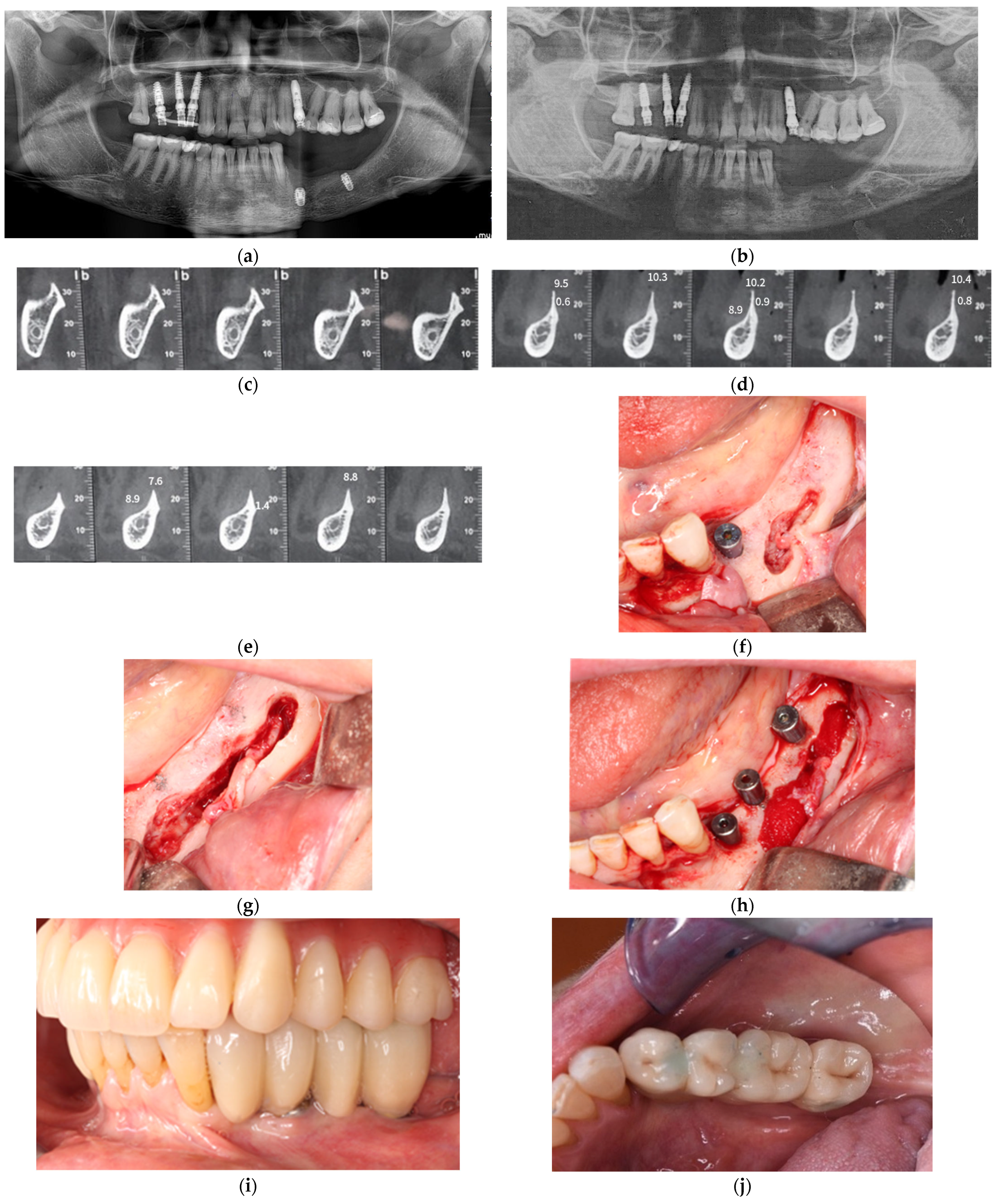
Data of fourteen patients six males and eight females) consecutively rehabilitated with fifty-one implants were placed (Table 1; Figure 1a–l). No patients dropped out, and all could be assessed at all planned time intervals. Patients were aged 64.8 ± 5.8 years at the time of intervention. The mean available bone height above the mandibular canal was 3.3 ± 1.1 mm (range 1 to 5 mm), highlighting the impossibility of placing short implants, and the mean total mandibular height was 11.7 ± 1.5 mm (range from 9 to 15 mm), suggesting a potential risk of post-operative mandibular fracture. Details of patients and procedures characteristics are presented in Table 1. Eight patients were treated with a full-arch prothesis and a double transposition, while the other six were treated with partial fixed prostheses and mono-lateral relocations of the nerve (five lateralizations and one transposition).
No intra-operative complications occurred. All patients presented episodes of minor inflammation, edema and paresthesia during the first days after implant placement. Five post-operative complications occurred in four patients: one mandibular fracture three weeks after implant placement and four cases of post-operative dysesthesia (four cases of neurosensory dysfunction, with one showing partial recovery after two weeks). The mandibular fracture resulted in the loss of the implant involved in the fracture line without affecting the provisional prothesis, which remained supported by four other implants. The fracture healed spontaneously after four months with a soft-food diet. No other implant or prothesis failed over the four years of follow-up. At one-year post-loading four patients were still affected by partial paresthesia, three of them with partial recovery; however, one of these patients was already affected by paresthesia preoperatively due to iatrogenic damage caused by a short implant placed in the area of the mental foramen.
The neurosensory dysfunction test was evaluated before surgery, and all patients did not report any neurosensory dysfunction, with the exception of one patient who had paresthesia caused by an implant touching the emergence of the mental nerve placed in another dental clinic. Table 2 reports the outcome of the individual sensory tests after surgery.
After two weeks, nine patients reported complete recovery from neurosensory disturbance, two of them treated with lateralization procedure and seven with double transposition.
At one month, no changes were recorded compared with those at two weeks. At three months, the patient initially presenting neurosensory dysfunction before the surgery had partial recovery, and another patient who received lateralization had complete recovery. At six months, no other changes were recorded.
After one year, one patient treated with a double transposition procedure still presented neurosensory dysfunction, while three patients reported partial recovery; however, one of them had complete neurosensory dysfunction before the surgery (Figure 1a–l), while the patient who suffered a mandibular fracture had complete recovery.
After two years, the patient treated with a double transposition procedure with neurosensory dysfunction had partial recovery, and one out of three patients who reported partial recovery had total recovery. No patient reported total neurosensory dysfunction after two years of follow-up.
After three years, the patient treated with double transposition had complete recovery. Only two patients still presented partial recovery, but one of them had complete neurosensory dysfunction before the surgery.
After four years, no other changes were recorded.

4. Discussion

Transposition and lateralization of the IAN, in the present study, have shown a high success rate at four years after implant placement, with only one implant lost due to a mandibular fracture. These results are comparable to the results of other studies [14,20], but implants in our study were loaded immediately. As reported in the Results, the potential risk of mandibular fracture was quite high in this sample of patients due to the mean available bone above the mandibular canal of 3.3 ± 1.1 mm (range 1 to 5 mm), highlighting the impossibility to place short implants. The mean total mandibular height of the patients with mandibular fracture was 11.0 mm (total sample ranged from 9 to 15 mm), but with no change in the final results in term of immediate loading and definitive full-arch prothesis.
However, the main problem with this procedure is the possibility of the onset of neurosensory disorders, which may be permanent. This altered sensitivity manifests itself in the form of hypoesthesia and, from the results of the present study, appears to be temporary in most cases, although partial recovery after two years was observed in three out of fourteen patients, reducing to two cases after three and four years of follow-up. Other authors reported similar results in terms of paresthesia and neurosensory disturbance [19,20,21].
A recent systematic review on these techniques highlighted that dental implants can be placed in atrophic posterior mandibles following nerve relocation, with high levels of patient satisfaction. However, the overall level of evidence remains low due to the high risk of bias among the included studies [16].
Moreover, according to another systematic review, lateralization appears to be associated with fewer persistent neurosensory disturbances compared to transposition [22]. Notably, none of the studies included in these reviews reported immediate loading protocols similar to those adopted in the present study. Only one case report described immediate loading of implants placed using a modified inferior alveolar nerve lateralization technique [23], in addition to a case series derived from the same cohort as the present study [14]. Autogenous bone grafts are commonly used to augment mandibular bone defects in height using intraoral donor sites, such as the symphysis and ramus of the mandible [24]. Autologous bone is considered the gold standard for augmentation procedures because of its osteogenic, osteoinductive, and osteoconductive properties [10,11,24].
Grafts can be used according to the inlay technique (also known as the interpositional or sandwich technique) or as onlay grafts directly on the area to be reconstructed to allow the placement of dental implants above the mandibular canal. The sandwich osteotomy technique was first described by Schettler in 1976, and it consisted of an interpositional graft of iliac bone [25]. One of the main difficulties was the management of soft tissues to preserve the blood supply of the displaced bone fragment. Furthermore, the original technique included harvesting bone from the iliac crest, which requires general anesthesia and hospitalization. In 2007, Marchetti and co-workers modified the technique by describing a dislocation of the mandibular crestal bone with concomitant preservation of the lingual periosteum to facilitate the healing process [25].
Considering the morbidity associated with intra- and extraoral bone grafting procedures, various bone substitutes, including allografts and xenografts, are widely used due to their lower morbidity compared with autologous bone harvesting. In addition, novel materials such as dentin-derived tooth grafts have recently been proposed [26,27].
Several studies have compared different vertical bone augmentation techniques for the posterior mandible, including the interpositional approach. Autologous bone blocks have demonstrated clinical and radiographic outcomes comparable to those achieved with bovine anorganic blocks, with the latter being less invasive and potentially preferable to iliac crest harvesting [28,29].
The primary limitation of the interpositional technique is the requirement of at least 4 mm of residual bone above the mandibular canal to ensure technical feasibility. Onlay grafting techniques serve as an alternative to inlay augmentation; however, the vertical bone gain achievable with onlay grafts is generally slightly less than that obtained through interpositional grafting [6,30,31].
Moreover, the concept of “perigraftitis”, a type of biomedical device-associated infection (BAI), is increasingly recognized and investigated in the literature [32,33]. Bone grafting has been identified as a significant risk factor for implant failure in the context of early infection. Implants placed in conjunction with bone grafts show lower survival rates following infection [32,33].
Moreover, bone substitutes blocks should not be used as onlay grafts to augment mandibles due to the exceedingly high failure rates; therefore, short implants, 4 to 6 mm long, could be a preferable choice, especially in mandibles, since the treatment is faster cheaper, and associated with a lower number of complications, with similar clinical results as longer implants placed in augmented bone [7,8,34,35,36,37].
With the approach adopted in the present study, longer implants could be placed by fully exploiting the available native bone height, thereby enabling immediate loading in all treated patients. This represents a clinically relevant advantage, particularly in cases of severely atrophic posterior mandibles where conventional treatment strategies may be limited.
Alternative approaches, such as the use of short or narrow implants, as well as customized subperiosteal implants, have been widely reported in the literature and may provide predictable outcomes with reduced morbidity in selected cases [38,39,40]. These options are often less invasive and can shorten treatment time, making them highly attractive in contemporary implant dentistry. However, their applicability is not universal and may be constrained by specific anatomical conditions, such as extremely reduced bone volume, unfavorable bone quality, or the need to achieve adequate primary stability for immediate loading.
In this context, inferior alveolar nerve relocation allows the placement of longer implants engaging the full vertical dimension of the native bone, which may be critical to obtain sufficient primary stability and to support immediate provisionalization, even in borderline clinical scenarios. This aspect is particularly relevant for patients requiring immediate functional and aesthetic rehabilitation, where delayed loading protocols or removable solutions may be poorly accepted.
Nevertheless, this technique should not be considered a first-line approach, but rather a valuable alternative in carefully selected cases where other less invasive options are not feasible or predictable. The decision-making process must always be individualized, taking into account patient-related factors, anatomical limitations, surgical risks, and the clinician’s experience. In particular, the higher incidence of neurosensory disturbances associated with nerve relocation procedures must be weighed against the potential benefits of immediate loading and the use of longer implants.
In the present study, the complications that occurred were paresthesia and mandibular fracture. While the fracture healed without negative sequelae, one year after loading, three patients were still affected by paresthesia, though two patients improved over time. The long-term neurosensory outcomes in patients undergoing inferior alveolar nerve relocation appear encouraging, even in the case of a patient with a previous neurosensory disorder, as shown for case 03 (Figure 1). After one year, while one patient treated with a double transposition procedure still experienced neurosensory dysfunction, three others showed partial improvement. It is noteworthy that one of these patients had complete neurosensory loss prior to surgery due to a previous implant procedure, and another, who sustained a mandibular fracture, achieved full recovery within the first year.
At the two-year follow-up, further improvements were observed. The patient initially treated with double transposition demonstrated partial recovery, and one of the three patients who previously had partial improvement experienced full recovery. Importantly, no patients exhibited total neurosensory loss at this stage.
At the three-year mark, the patient treated with the transposition procedure achieved complete recovery. Only two patients continued to report partial neurosensory symptoms, one of whom had pre-existing total dysfunction prior to the intervention.
At four years, the clinical status remained stable, with no further changes reported, and the native bone showed a stable long-term follow-up. One patient treated with double transposition continued to present with partial neurosensory symptoms, as did the patient with prior complete dysfunction. Notably, no cases of total neurosensory loss were observed at this follow-up, indicating a favorable long-term prognosis for most patients, as also observed in a recent systematic review [41].
The surgeon tried to perform lateralization whenever possible because, with transposition, cutting the incisal nerve may lead to more frequent sensory deficits [42]. Interestingly, in the presence of a higher degree of atrophy, the nerve is more superficial, making the procedure simpler and less traumatic, while in cases where the nerve is less elastic and deeper within the bone, transposition could be the most sensible approach [43].
The main limitations of the present study are its retrospective nature and the small sample size. However, the present technique might be predictable in terms of implant and prothesis survival but is a complex procedure. Computer-guided planning on CBCT, with a bony-supported guided surgical mask could help in guiding the osteotomy over the mandibular canal, decreasing the risk of intra- and post-surgical complications.

5. Conclusions

Within the limitations of this study, the use of all available native bone through inferior alveolar nerve lateralization or transposition allows for immediate loading with high success rates. This rehabilitation protocol may therefore represent a valid therapeutic option for patients with severe mandibular atrophy, particularly when immediate prosthetic rehabilitation is required.
However, this approach is associated with a relatively high risk of neurosensory disturbances, which tend to improve over time, with most cases showing recovery within two years. Compared with augmentation procedures, this technique may enable a faster rehabilitation and can be considered when insufficient bone is available for the placement of short or narrow implants.
Nevertheless, randomized controlled trials with larger sample sizes are needed to further evaluate the effectiveness and safety of this treatment approach in comparison with alternative protocols.

Author Contributions

Conceptualization, R.V. and S.C.; methodology, M.E.; software, G.L.P.; validation, R.V., S.C. and M.E.; formal analysis, G.L.P.; investigation, S.C.; resources, R.V.; data curation, S.C.; writing—original draft preparation, S.C.; writing—review and editing, M.E.; visualization, G.L.P.; supervision, M.E.; project administration, R.V.; funding acquisition, R.V. All authors have read and agreed to the published version of the manuscript.

Funding

This research received no external funding. This study was completely self-supported, and no financial support has been sought or obtained, not even in form of free materials.

Institutional Review Board Statement

The study was conducted in accordance with the Declaration of Helsinki and approved by the local Ethics Committee with number No. 190/INT/2021 (15 December 2021) at the Department of Dentistry, IRCCS San Raffaele, Milan, Italy.

Informed Consent Statement

Informed consent was obtained from all subjects involved in the study.

Data Availability Statement

The raw data supporting the conclusions of this article will be made available by the authors on request.

Acknowledgments

The authors have reviewed and edited the output and take full responsibility for the content of this publication.

Conflicts of Interest

The authors declare no conflicts of interest.

Abbreviations

The following abbreviations are used in this manuscript:
IANInferior Alveolar Nerve
EAExtreme Abutment
NSAIDsNon-Steroidal Anti-Inflammatory Drugs
CBCTCone-Beam Computer Tomography

References

  1. Rodrigues, A.; Abi-Nader, S.; Durand, R.; Rompré, P.; Janati, A.I.; Atsu, S.; Morris, M.; Emami, E. Effectiveness of zygomatic-implant fixed rehabilitation for the atrophic edentulous maxilla: Protocol for a systematic review and network meta-analysis. Syst. Rev. 2024, 13, 146. [Google Scholar] [CrossRef] [PubMed]
  2. Cawood, J.I.; Howell, R.A. A classification of the edentulous jaws. Int. J. Oral Maxillofac. Surg. 1988, 17, 232–236. [Google Scholar] [CrossRef]
  3. Ahamed, S.K.; Menchini-Fabris, G.B.; Alqarni, A.; Alarabi, S.M.; Alharbi, A.A.; Alshamrani, A.; Covani, U.; Cosola, S. Insights into the Current Management Techniques for Peri-Implant Gaps: A Systematic Review. J. Clin. Med. 2025, 14, 3351. [Google Scholar] [CrossRef]
  4. Tan, W.L.; Wong, T.L.; Wong, M.C.; Lang, N.P. A systematic review of post-extractional alveolar hard and soft tissue dimensional changes in humans. Clin. Oral Implant. Res. 2012, 23, 1–21. [Google Scholar] [CrossRef] [PubMed]
  5. Carosi, P.; Lorenzi, C.; Laureti, M.; Ferrigno, N.; Arcuri, C. Short Dental Implants (≤6 mm) to Rehabilitate Severe Mandibular Atrophy: A Systematic Review. Int. J. Oral Maxillofac. Implant. 2021, 36, 30–37. [Google Scholar] [CrossRef]
  6. Chaware, S.H.; Thakare, V.; Chaudhary, R.; Jankar, A.; Thakkar, S.; Borse, S. The rehabilitation of posterior atrophic maxilla by using the graftless option of short implant versus conventional long implant with sinus graft: A systematic review and meta-analysis of randomized controlled clinical trial. J. Indian Prosthodont. Soc. 2021, 21, 28–44. [Google Scholar] [CrossRef] [PubMed]
  7. Papaspyridakos, P.; De Souza, A.; Vazouras, K.; Gholami, H.; Pagni, S.; Weber, H.P. Survival rates of short dental implants (≤6 mm) compared with implants longer than 6 mm in posterior jaw areas: A meta-analysis. Clin. Oral Implant. Res. 2018, 29, 8–20. [Google Scholar] [CrossRef]
  8. Esposito, M.; Buti, J.; Barausse, C.; Gasparro, R.; Sammartino, G.; Felice, P. Short implants versus longer implants in vertically augmented atrophic mandibles: A systematic review of randomised controlled trials with a 5-year post-loading follow-up. Int. J. Oral Implantol. 2019, 12, 267–280. [Google Scholar]
  9. Pachiou, A.; Naka, O.; Sykara, M.; Sykaras, N.; Tsirogiannis, P.; Thoma, D.S.; Naenni, N. Survival rate and prosthetic complications of short dental implants supporting implant overdentures: A systematic review and meta-analysis. J. Dent. 2026, 166, 106351. [Google Scholar] [CrossRef]
  10. Arbildo-Vega, H.I.; Cruzado-Oliva, F.H.; Infantes-Ruíz, E.D.; Luján-Valencia, S.A.; Meza-Málaga, J.M.; Marroquín-Soto, C.; Coronel-Zubiate, F.T. Clinical performance of short implants vs. standard implants in edentulous patients. An umbrella review. Front. Oral Health 2025, 6, 1670095. [Google Scholar] [CrossRef] [PubMed]
  11. Esposito, M.; Grusovin, M.G.; Coulthard, P.; Worthington, H.V. The efficacy of various bone augmentation procedures for dental implants: A Cochrane systematic review of randomized controlled clinical trials. Int. J. Oral Maxillofac. Implant. 2006, 21, 696–710. [Google Scholar]
  12. Schwartz-Arad, D.; Levin, L. Intraoral autogenous block onlay bone grafting for extensive reconstruction of atrophic maxillary alveolar ridges. J. Periodontol. 2005, 76, 636–641. [Google Scholar] [CrossRef] [PubMed]
  13. Calciolari, E.; Ravanetti, F.; Strange, A.; Mardas, N.; Bozec, L.; Cacchioli, A.; Kostomitsopoulos, N.; Donos, N. Degradation pattern of a porcine collagen membrane in an in vivo model of guided bone regeneration. J. Periodontal Res. 2018, 53, 430–439. [Google Scholar] [CrossRef] [PubMed]
  14. Vinci, R.; Cosola, S.; Borsi, A.; Speroni, S.; Esposito, M. Immediate loading after implant placement with relocation of the inferior alveolar nerve in atrophic mandibles: A 1-year retrospective case series. Clin. Trials Dent. 2024, 06, 37–48. [Google Scholar]
  15. Pimentel, A.C.; Sanches, M.A.; Ramalho, G.C.; Roman-Torres, C.V.; Manzi, M.R.; Sendyk, W.R. Lateralization Technique and Inferior Alveolar Nerve Transposition. Case Rep. Dent. 2016, 2016, 4802637. [Google Scholar] [CrossRef]
  16. Allavéna, J.; Nicot, R.; Majoufre, C.; Schlund, M. Inferior alveolar nerve repositioning surgical techniques and outcomes—A systematic review. J. Stomatol. Oral Maxillofac. Surg. 2024, 125, 101631. [Google Scholar] [CrossRef]
  17. Crespi, R.; Toti, P.; Covani, U.; Trasarti, S.; Crespi, G.; Menchini-Fabris, G.B. Conventional avulsion versus less traumatic extraction of ankylosed roots with a magnetoelectric device in anterior maxillary areas. J. Cranio-Maxillofac. Surg. 2023, 51, 760–765. [Google Scholar] [CrossRef]
  18. Kan, J.Y.; Lozada, J.L.; Goodacre, C.J.; Davis, W.H.; Hanisch, O. Endosseous implant placement in conjunction with inferior alveolar nerve transposition: An evaluation of neurosensory disturbance. Int. J. Oral Maxillofac. Implant. 1997, 12, 463–471. [Google Scholar] [PubMed]
  19. Abayev, B.; Juodzbalys, G. Inferior Alveolar Nerve Lateralization and Transposition for Dental Implant Placement. Part II: A Systematic Review of Neurosensory Complications. J. Oral Maxillofac. Res. 2015, 6, e3. [Google Scholar] [CrossRef] [PubMed][Green Version]
  20. Jensen, J.; Reiche-Fischel, O.; Sindet-Pedersen, S. Nerve transposition and implant placement in the atrophic posterior mandibular alveolar ridge. J. Oral Maxillofac. Surg. 1994, 52, 662–668. [Google Scholar] [CrossRef]
  21. Rathod, M.; Kshirsagar, R.A.; Joshi, S.; Pawar, S.; Tapadiya, V.; Gupta, S.; Mahajan, V. Evaluation of Neurosensory Function Following Inferior Alveolar Nerve Lateralization for Implant Placement. J. Maxillofac. Oral Surg. 2019, 18, 273–279. [Google Scholar] [CrossRef] [PubMed]
  22. Palacio García-Ochoa, A.; Pérez-González, F.; Negrillo Moreno, A.; Sánchez-Labrador, L.; Cortés-Bretón Brinkmann, J.; Martínez-González, J.M.; López-Quiles Martínez, J. Complications associated with inferior alveolar nerve reposition technique for simultaneous implant-based rehabilitation of atrophic mandibles. A systematic literature review. J. Stomatol. Oral Maxillofac. Surg. 2020, 121, 390–396. [Google Scholar] [CrossRef]
  23. Eldibany, R.; Rodriguez, J.G. Immediate loading of one-piece implants in conjunction with a modified technique of inferior alveolar nerve lateralization: 10 years follow-up. Craniomaxillofacial Trauma Reconstr. 2014, 7, 55–62. [Google Scholar] [CrossRef]
  24. Laviv, A.; Jensn, O.T.; Tarazi, E.; Casap, N. Alveolar sandwich osteotomy in resorbed alveolar ridge for dental implants: A4-year prospective study. J. Oral Maxillofac. Surg. 2014, 72, 292. [Google Scholar] [CrossRef]
  25. Schettler, D. Sandwich technic with cartilage transplant for raising the alveolar process in the lower jaw. Fortschr. Kiefer Gesichtschir 1976, 20, 61. [Google Scholar] [PubMed]
  26. Minetti, E.; Gianfreda, F.; Bollero, P.; Annicchiarico, C.; Daniele, M.; Padula, R.; Mastrangelo, F. Comparative Histological Analysis of Dentine-Derived Tooth Grafts in Maxillary vs Mandibular Socket Preservation: A Retrospective Study of 178 Cases. Dent. J. 2024, 12, 320. [Google Scholar] [CrossRef] [PubMed]
  27. Dioguardi, M.; Spirito, F.; Quarta, C.; Sovereto, D.; Basile, E.; Ballini, A.; Caloro, G.A.; Troiano, G.; Lo Muzio, L.; Mastrangelo, F. Guided Dental Implant Surgery: Systematic Review. J. Clin. Med. 2023, 12, 1490. [Google Scholar] [CrossRef]
  28. Starch-Jensen, T.; Nielsen, H.B. Sandwich osteotomy of the atrophic posterior mandible with interpositional autogenous bone block graft compared with bone substitute material: A systematic review and meta-analysis. Br. J. Oral Maxillofac. Surg. 2020, 58, e237–e247. [Google Scholar] [CrossRef]
  29. Felice, P.; Marchetti, C.; Iezzi, G.; Piattelli, A.; Worthington, H.; Pellegrino, G.; Esposito, M. Vertical ridge augmentation of the atrophic posterior mandible with interpositional bloc grafts: Bone from the iliac crest vs. bovine anorganic bone. Clinical and histological results up to one year after loading from a randomized-controlled clinical trial. Clin. Oral Implants Res. 2009, 20, 1386–1393. [Google Scholar] [CrossRef]
  30. Mijiritsky, E.; Ferroni, L.; Gardin, C.; Bressan, E.; Zanette, G.; Piattelli, A.; Zavan, B. Porcine Bone Scaffolds Adsorb Growth Factors Secreted by MSCs and Improve Bone Tissue Repair. Materials 2017, 10, 1054. [Google Scholar] [CrossRef]
  31. Barone, A.; Toti, P.; Menchini-Fabris, G.B.; Felice, P.; Marchionni, S.; Covani, U. Early volumetric changes after vertical augmentation of the atrophic posterior mandible with interpositional block graft versus onlay bone graft: A retrospective radiological study. J. Cranio-Maxillofac. Surg. 2017, 45, 1438–1447. [Google Scholar] [CrossRef] [PubMed]
  32. Tabrizi, R.; Zarchini, R.; Ozkan, B.T.; Majdi, S. Dental Implant Survival after Postoperative Infection. J. Maxillofac. Oral Surg. 2022, 21, 796–801. [Google Scholar] [CrossRef]
  33. Do, J.H. Peri-Implantitis and Concomitant Perigraftitis of an Implant Placed in a Site That Had Alveolar Ridge Preservation Three Decades Earlier: A Case Report With Human Histology. Clin. Adv. Periodontics 2022, 12, 44–50. [Google Scholar] [CrossRef]
  34. Rameh, S.; Menhall, A.; Younes, R. Key factors influencing short implant success. Oral Maxillofac. Surg. 2020, 24, 263–275. [Google Scholar] [CrossRef] [PubMed]
  35. Sáenz-Ravello, G.; Ossandón-Zúñiga, B.; Muñoz-Meza, V.; Mora-Ferraro, D.; Baeza, M.; Fan, S.; Sagheb, K.; Schiegnitz, E.; Díaz, L. Short implants compared to regular dental implants after bone augmentation in the atrophic posterior mandible: Umbrella review and meta-analysis of success outcomes. Int. J. Implant Dent. 2023, 9, 18. [Google Scholar] [CrossRef]
  36. Felice, P.; Barausse, C.; Pistilli, R.; Ippolito, D.R.; Esposito, M. Short implants versus longer implants in vertically augmented posterior mandibles: Result at 8 years after loading from a randomised controlled trial. Eur. J. Oral Implantol. 2018, 11, 385–395. [Google Scholar] [PubMed]
  37. Amine, M.; Guelzim, Y.; Benfaida, S.; Bennani, A.; Andoh, A. Short implants (5–8 mm) vs. long implants in augmented bone and their impact on peri-implant bone in maxilla and/or mandible: Systematic review. J. Stomatol. Oral Maxillofac. Surg. 2019, 120, 133–142. [Google Scholar] [CrossRef]
  38. Toti, P.; Marchionni, S.; Menchini-Fabris, G.B.; Marconcini, S.; Covani, U.; Barone, A. Surgical techniques used in the rehabilitation of partially edentulous patients with atrophic posterior mandibles: A systematic review and meta-analysis of randomized controlled clinical trials. J. Cranio-Maxillofac. Surg. 2017, 45, 1236–1245. [Google Scholar] [CrossRef]
  39. Vatteroni, E.; Covani, U.; Menchini Fabris, G.B. The New Generation of Subperiosteal Implants for Patient-Specific Treatment of Atrophic Dental Arches: Literature Review and Two Case Reports. Int. J. Periodontics Restor. Dent. 2023, 43, 735–741. [Google Scholar] [CrossRef] [PubMed]
  40. Cosola, S.; Vatteroni, E.; Asadi, M.; Covani, U.; Cardarelli, A.; Menchini-Fabris, G.B. Clinical Outcomes, Survival, and Complications of Customized Computer-Aided Design and Manufacturing 3-Dimensional-Printed Titanium Subperiosteal Implants. J. Oral Maxillofac. Surg. 2026, 24, S0278-2391. [Google Scholar] [CrossRef] [PubMed]
  41. Vinci, R.; Cosola, S.; Varkey, M.K.; Gunasekaran, S.; George, J.; Covani, U. Neurosensory Disturbances Following Inferior Alveolar Nerve Relocation and Implant Placement: A Systematic Review and Meta-Analysis. J. Clin. Med. 2025, 14, 5741. [Google Scholar] [CrossRef] [PubMed]
  42. Ferrigno, N.; Laureti, M.; Fanali, S. Inferior alveolar nerve transposition in conjunction with implant placement. Int. J. Oral Maxillofac. Implant. 2005, 20, 610–620. [Google Scholar]
  43. Kikuta, S.; Iwanaga, J.; Kusukawa, J.; Tubbs, R.S. Anatomical study: The potential movability of the inferior alveolar nerve. Oral Surg. Oral Med. Oral Pathol. Oral Radiol. 2019, 128, 353–356. [Google Scholar] [CrossRef] [PubMed]
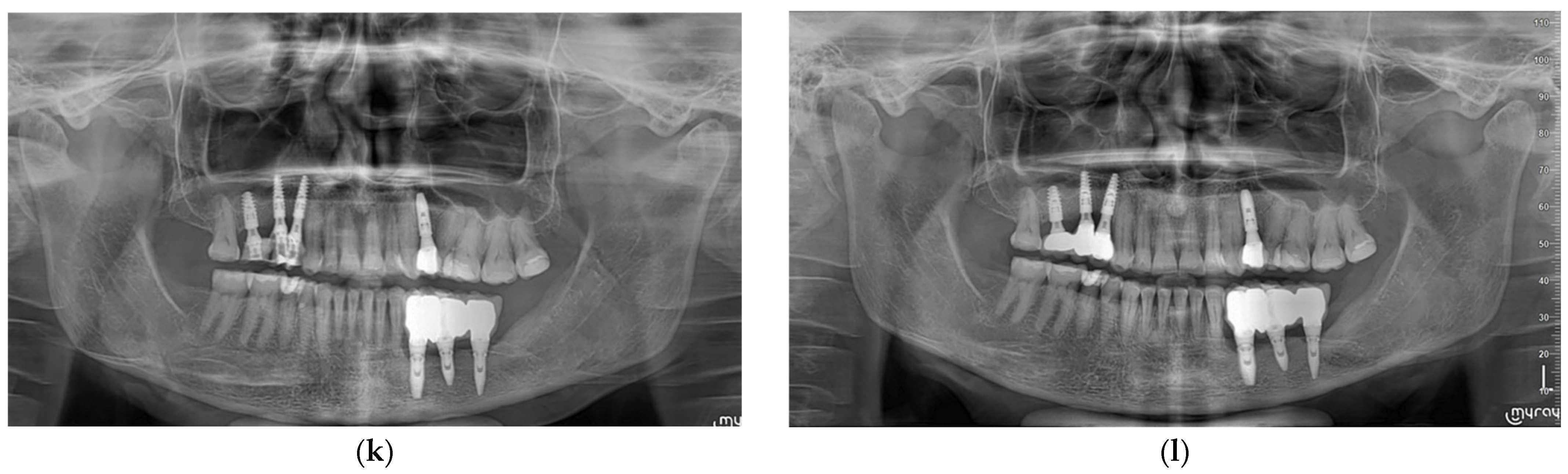
Figure 1. Pre-operative OPT at the time of first visit. The patient presented total paresthesia of the left side of the lip, possibly due to an implant placed in site 35 in proximity to the mental nerve in another dental office. The previous prosthetic rehabilitation fractured in correspondence with the implant in site 37 (a). Both implants were extracted to try to reduce the neurosensory disorder (b). Screenshots of CBCT in correspondence with implants planned in position 34 (c), position 35 (d) and position 37 (e) were used to evaluate the remaining native bone by the transposition technique due to the buccal and superficial positioning of the mental nerve. After full-thickness flap elevation, a vertical incision was performed in correspondence with 32, and the mental nerve was isolated (f). Implants in positions 35, 36 and 37 were placed after the IAN lateralization technique (g) to have sufficient bone in length and bicortical implant engagement A collagen sponge was placed, separating the implants from the nerve (h). An immediate non-occluding provisional screw-retained prothesis was delivered (i). Six months after surgery a definitive prothesis was delivered (j), and an OPT was performed (k). After four years, another OPT was performed (l).
Oral 06 00045 g001aOral 06 00045 g001b
Table 1. Characteristics of the included patients. L = lateralization, T = transposition, and 2T = double transposition. All implants had a 3.8 mm diameter. TTx = Torque Type, external hexagon; TTi = Torque Type, internal hexagon; K = internal hexagon with a different threads design. * Patient 03 had a neurosensory dysfunction before lateralization and Patient 11 had a post-operative mandibular fracture.
InterventionSexAgePartial/Total EdentulismImplant Position and LengthBone Height Above Mandibular Canal (<6 mm)Total Mandibular HeightType of Implants
01 (T)M66Partial37–11 mm4 mm12 mmK
36–11 mm4 mm12 mmK
34–13 mm 3 mm13 mmTTI
02 (L)F65Partial36–9 mm2 mm10 mmTTI
35–9 mm3 mm10 mmTTI
03 * (L)F58Partial37–9 mm4 mm11 mmTTx
35–11 mm4 mm12 mmTTx
34–11 mm3 mm12 mmTTx
04 (L)M72Partial35–11 mm2 mm13 mmTTI
36–11 mm1 mm12 mmTTI
05 (L)M63Partial37–9 mm3 mm9 mmTTI
35–11 mm4 mm12 mmTTI
34–11 mm3 mm12 mmTTI
06 (T)F57Partial44–13 mm5 mm14 mmTTx
46–11 mm2 mm13 mmTTx
07 (2T)F62Total35–13 mm3 mm14 mmTTx
32–13 mm5 mm14 mmTTx
42–11 mm4 mm13 mm TTx
45–13 mm3 mm14 mmTTx
08 (2T)M70Total34–9 mm3 mm10 mmTTx
32–9 mm3 mm10 mmTTx
42–9 mm3 mm10 mmTTx
44–9 mm5 mm10 mm TTx
09 (2T)F55Total36–11 mm5 mm12 mmTTI
34–9 mm4 mm10 mmTTI
32–11 mm5 mm11 mmTTI
42–11 mm5 mm12 mmTTI
44–9 mm3 mm9 mmTTI
46–11 mm2 mm12 mmTTI
10 (2T)F69Total35–9 mm3 mm10 mmTTI
32–11 mm5 mm12 mmTTI
42–11 mm5 mm12 mmTTI
44–9 mm3 mm10 mmTTI
46–11 mm1 mm11 mmTTI
11 * (2T)F65Total36–11 mm3 mm11 mmTTI
35–9 mm5 mm10 mmTTI
32–9 mm5 mm10 mmTTI
42–11 mm3 mm12 mmTTI
45–11 mm2 mm12 mmTTI
12 (2T)M75Total35–11 mm2 mm12 mmTTI
32–11 mm3 mm12 mmTTI
42–11 mm3 mm12 mmTTI
45–11 mm1 mm12 mmTTI
13 (2T)M62Total35–13 mm3 mm15 mmTTI
32–13 mm4 mm15 mmTTI
42–11 mm3 mm12 mmTTI
45–11 mm2 mm14 mmTTI
14 (2T)F68Total35–9 mm2 mm11 mmTTI
32–9 mm3 mm12 mmTTI
42–9 mm4 mm12 mmTTI
45–9 mm3 mm12 mmTTI
Table 2. Results of the neurosensory dysfunction test at each time point. L = lateralization; T = transposition; 2T = double transposition; NSD = presence of neurosensory dysfunction; PR = partial recovery; R = complete recovery. * Patient 03 had a neurosensory dysfunction before lateralization and Patient 11 had a post-operative mandibular fracture.
Patient NumberOne DayTwo WeeksOne MonthThree MonthsSix MonthsOne YearTwo Years Three YearsFour Years
01 (T)NSDNSDNSDRRRRRR
02 (L)NSDPRPRPRPRPRRRR
03 (L) *NSDNSDNSDPRPRPRPRPRPR
04 (L)NSDRRRRRRRR
05 (L)NSDRRRRRRRR
06 (T)NSDNSDNSDNSDNSDPRPRRR
07 (2T)NSDRRRRRRRR
08 (2T)NSDRRRRRRRR
09 (2T)NSDRRRRRRRR
10 (2T)NSDNSDNSDNSDNSDNSDPRPRPR
11 (2T) *NSDRRRRRRRR
12 (2T)NSDRRRRRRRR
13 (2T)NSDRRRRRRRR
14 (2T)NSDRRRRRRRR
Disclaimer/Publisher’s Note: The statements, opinions and data contained in all publications are solely those of the individual author(s) and contributor(s) and not of MDPI and/or the editor(s). MDPI and/or the editor(s) disclaim responsibility for any injury to people or property resulting from any ideas, methods, instructions or products referred to in the content.