Article

Assessing the Genotoxic Impact of Ni-Cr Alloys in Dental Prosthodontics: A Preliminary Comparative Analysis with and Without Beryllium

by
Florentina Caministeanu
1,†,
Viorel Stefan Perieanu
1,†,
Andrei Sabin Popa
2,
Loredana Sabina Cornelia Manolescu
3,*,
Andreea Angela Stetiu
4,†,
Radu Catalin Costea
1,
Mihai Burlibasa
1,*,
Andrei Vorovenci
5,
Raluca Mariana Costea
6,
Cristina Maria Serbanescu
1,
Andi Ciprian Dragus
1,
Maria Antonia Stetiu
4,
Madalina Adriana Malita
1 and
Liliana Burlibasa
7
1
Department of Dental Technology, Faculty of Midwifery and Nursing, “Carol Davila” University of Medicine and Pharmacy, 050474 Bucharest, Romania
2
Faculty of Biology, University of Bucharest, 91-95 Splaiul Independenței, 060101 Bucharest, Romania
3
Faculty of Midwives and Nursing, Department of Microbiology, Parasitology and Virology, “Carol Davila” University of Medicine and Pharmacy, 020021 Bucharest, Romania
4
Faculty of Medicine, Lucian Blaga University of Sibiu, B-dul Victoriei Nr. 10, 550024 Sibiu, Romania
5
Prosthodontics Residency, “Carol Davila” University of Medicine and Pharmacy, 4 Poligrafiei Avenue, Sector 1, 013705 Bucharest, Romania
6
S.C. Dentexpert Magic, 050514 Bucharest, Romania
7
Genetics Department, Faculty of Biology, University of Bucharest, 91-95 Splaiul Independenței, 060101 Bucharest, Romania
*
Authors to whom correspondence should be addressed.
These authors contributed equally to this work.
Submission received: 2 March 2025 / Revised: 13 April 2025 / Accepted: 28 April 2025 / Published: 7 May 2025
(This article belongs to the Special Issue Advanced Dental Materials for Oral Rehabilitation)

Abstract

:
Objective: This study aims to evaluate cell proliferation capacity and micronuclei incidence in the presence of nickel–chromium (Ni-Cr)-based dental alloys, with and without the addition of beryllium (Be). The use of these alloys in dental prosthetics is widespread; however, the potential risks associated with their genotoxicity and cytotoxicity require further investigation. The study seeks to provide insight into the safety of these materials and their long-term impact on the health of both patients and dental professionals. Methods: The study was conducted through a comparative analysis of genotoxicity and cytotoxicity using human lymphocyte cultures exposed to two types of Ni-Cr-based dental alloys, one containing beryllium and the other without beryllium. The evaluations were performed according to the OECD Test No. 487 guideline, employing the micronucleus assay and cell proliferation assay. Lymphocytes were exposed to three different alloy concentrations (5 mg/mL, 10 mg/mL, and 20 mg/mL), and the effects on genetic material were analyzed microscopically. Descriptive statistics (mean, standard deviation, and variance) were calculated, and one-way ANOVA was used to assess statistical significance between groups, with a significance threshold of p < 0.05. Results: A significant increase in cytotoxicity and micronuclei incidence was observed in the samples containing beryllium compared to those without beryllium. Statistical analysis revealed significant differences (p < 0.001) between the test and control groups and between different concentrations. Additionally, a direct proportional relationship was noted between alloy concentration and the intensity of genotoxic effects. Microscopic analysis confirmed genetic material damage, indicating a potentially increased risk associated with the use of this type of dental material. Conclusions: The data obtained suggest that Ni-Cr-based dental alloys containing beryllium may present a significant risk of genotoxicity and cytotoxicity. Therefore, the selection of materials used in dental prosthetics should be based on solid scientific evidence, and the use of these alloys should be approached with caution. The study highlights the need for further research to better understand the long-term impact of these materials on human health.

1. Introduction

Modern dental prosthodontics is based on the use of a wide range of dental alloys to create durable and esthetic metal–ceramic restorations for a single tooth or multiple teeth. These alloys have different compositions and properties. The correct choice of alloy is crucial, as it must meet several criteria, including biocompatibility, corrosion resistance, adequate mechanical properties, and the ability to form a solid bond with dental ceramics. They must also have a considerably higher melting point than the ceramic that is applied over it [1]. There are other essential requirements for an alloy intended for metal–ceramic technology, such as a higher thermal expansion coefficient than the ceramic material, ensuring a tight bond of the ceramic to the metal base and dimensional stability when fired at the temperatures used for the crystallization of ceramic materials [2]. Hence, Ni-Cr alloys have become popular due to their affordability and their performance [2,3,4]. They are used both in the manufacturing of fixed teeth or implant-supported prosthetic restorations, regardless of whether the metal structure is obtained by the classic lost-wax technique or by computerized milling. The introduction of beryllium in Ni-Cr alloys was made for the benefits it has in the metallurgical process. Beryllium enhances the melting process by reducing the alloy’s melting temperature [4,5,6]. This is achieved by the formation of a secondary Ni-Be eutectic phase [5]. At the same time, beryllium increases the fluidity of the alloy, improving the casting process. On the other hand, beryllium produces a slight decrease in corrosion resistance, which helps the formation of the oxide layer on the surface of the alloy [4,5,7]. This can be an advantage when we talk about the etching of alloys or the layer of oxides required for applying ceramic masses. At the same time, the decrease in corrosion resistance allows the component elements to leave the alloy and reach the level of soft tissues, especially Ni and beryllium [4]. The study by Cheng et al. (1990) showed that the Ni-beryllium eutectic phase is the least corrosion-resistant component, the amount of Ni-beryllium being directly proportional to the amount of beryllium added to the alloy and the appearance of corrosion being detected from amounts of beryllium exceeding 0.5 wt.% [4]. Low corrosion resistance is an extremely important factor, especially since Ni, Cr, and beryllium are responsible for numerous adverse reactions in the body [4,8]. Various institutions have detailed the absorption pathways and associated hazards of beryllium. In principle, exposure and absorption of beryllium can be achieved through the respiratory, gastrointestinal, and dermal routes. The inhalation route, the most worrying and the most common, leads to the storage of beryllium in the lungs and subsequently its distribution to the other parts of the body [9,10]. Oral absorption, although lower, results in the accumulation of beryllium in bone or liver. Dermal exposure does not result in the accumulation of beryllium in the organs, but only the appearance of a sensitivity of the body to it [10,11,12]. For these reasons, there have been numerous studies on different compounds of beryllium in terms of mutagenicity and genotoxicity [10]. Exposure to beryllium vapor or powders containing beryllium can be dangerous and is an occupational hazard, especially for dental technicians who handle alloys containing beryllium, leading to serious health problems if adequate protective measures are not followed. Apostoli et al. (1989) conducted a study in which they found that the average urinary beryllium level in dental technicians exposed to beryllium was almost double the average value recorded in the general population in an area with a high density of metallurgical manufacturing industries [13]. For this reason, more and more manufacturers are choosing to remove beryllium from the composition of dental alloys based on Ni-Cr. However, alloys containing beryllium are still found on the Romanian (Central and Eastern European) dental markets. The biomaterials used and developed in recent years mainly aim to replace, for a prolonged period, morphological elements that are missing or damaged and must be replaced. It is preferable that the material from which the implant or other prosthetic product is made avoids triggering allergic reactions, thus shortening its lifespan. Another important characteristic for biomaterials is their resistance not only to the biochemical environment inside the oral cavity, but also to external mechanical factors. The most widely used metal alloys in the oral cavity for the manufacture of fixed prosthetic restorations, either by the classic lost-wax technique or computerized milling, are usually stainless alloys based on Ni-Cr, Co-Cr, but also commercially pure titanium and its alloys. Because the component elements that make up the prosthetic restorations can exert effects that increase genotoxicity in the surrounding tissues, there are a series of tests that evaluate the degree of risk in their use. Examples of tests used are the “micronucleus assay”, the “comet assay”, the “Ames test”, and the “chromosomal structural aberrations test”. Most of the research carried out up to this point has focused on evaluating the degree of release of components from Ni-Cr alloys because of the corrosion process. There have also been studies that used the micronucleus test in lymphocytes to monitor human exposure to chromium, nickel, vanadium, and silica [12,14]. The first studies on the mutagenicity and genotoxicity of beryllium were carried out by Nishioka in 1975, investigating gene mutation generated by beryllium chloride using the B. subtilis rec assay [15]. Later, the tests also targeted beryllium compounds, based on gene mutation, forward mutation, genomic instability, genetic alteration, etc. [10]. Regarding the use of the micronucleus test for the analysis of mutagenicity and genotoxicity of beryllium, it was used by Ashby et al. (1990) for beryllium sulfate analysis without noticing an obvious effect, but also by Fritzenschaf et al. (1993), who noted a genotoxic effect of soluble beryllium compounds [16,17]. In general, the studies that analyzed dental alloys, especially Ni-Cr alloys, and used the micronucleus assay did not take beryllium into account. The tested alloys were beryllium-free and Cu-free [18]. Genotoxicity is the ability of an agent to cause DNA damage, meaning that, to be considered genotoxic, a chemical agent must interact with genetic material [19]. In the scientific literature, several chemical agents are classified as genotoxic. It is believed that the human genome is constantly affected by various chemicals. However, eukaryotic cells are biological units specialized in neutralizing genotoxic insults by promoting DNA repair. A xenobiotic metabolism system and a DNA repair machinery are essential for maintaining the integrity of the human genome [1,2,18,20]. In the context of concerns about the safety of Ni-Cr dental alloys, our study aims to evaluate their genotoxic potential, i.e., the ability to cause genetic damage that could lead to mutations or cancer. Testing includes the following:
-
Cell proliferation assay: This measures the growth rate of cells to determine if the alloys affect normal cell division, an indicator of possible toxicity or carcinogenic effects.
-
In vitro micronucleus assay: This test assesses the appearance of micronuclei in mammalian cells, which are indicators of chromosomal damage or chromosome loss. The test is performed on cultures of human lymphocytes extracted from peripheral blood, providing a sensitive and relevant method for the assessment of genotoxicity.
By addressing these aspects, this study not only aims to clarify the safety of using Ni-Cr dental alloys but also to contribute to the improvement of practices in the field of dental prosthetics, ensuring that the materials used are both effective and safe for patients and professionals in the field. This research is important for the future development of dental prosthodontics, providing clear directions in the selection of materials and the implementation of the necessary protective measures. The objectives of this study were to identify the cell proliferation capacity and also to observe under a microscope the incidence of micronuclei in the presence of chemical substances, namely the Ni-Cr-based alloy, as well as the alloy which, in addition to the two previously mentioned elements, Ni and Cr, also contains beryllium.
In modern dental prosthodontics, metallic alloys are essential for creating durable and esthetic metal–ceramic restorations. Among these, nickel–chromium (Ni-Cr) alloys are widely used due to their low cost, favorable mechanical properties, and good compatibility with ceramic materials [1,2]. Some of these alloys include beryllium in their composition, an element that contributes to lowering the melting temperature, increasing fluidity, and promoting the formation of an oxide layer necessary for ceramic adhesion [3,4,5,6]. However, beryllium may slightly reduce the corrosion resistance of the alloy, facilitating the release of metal ions into the oral environment [4,7,8].
This release may lead to local or systemic biological reactions, considering that beryllium is recognized for its toxic, allergenic, and genotoxic potential [9,10]. Exposure routes include inhalation, ingestion, and dermal contact, with the most significant effects observed among dental technicians [9,10,11]. In addition, nickel and chromium—common components of these alloys—are also associated with allergic reactions and cytotoxic effects, especially in their soluble or nanoparticulate forms [12,14].
It is well established that any chemical substance can become toxic depending on its dose or concentration—an essential aspect in cytotoxicity and cell viability studies. The first studies on the genotoxicity of beryllium compounds date back to 1975 [15], and subsequent investigations have utilized tests such as the micronucleus assay to further explore these effects [16,17]. The assay has also been employed in biomonitoring studies evaluating exposure to dental alloys containing nickel, chromium, and other metals [12,14,18].
Therefore, an important hypothesis of the present study is that the tested materials—depending on their composition and concentration—may inherently compromise cell viability. Considering this factor is crucial for correctly distinguishing between cytotoxic and genotoxic effects, reinforcing the scientific rationale of the investigation [19,20].
Although numerous studies have examined the behavior of Ni-Cr alloys, most have excluded variants containing beryllium. Consequently, the scientific literature provides limited data regarding their genotoxic potential in the context of dental use. Thus, applying validated methods such as the micronucleus assay and cell proliferation assay is essential for evaluating the safety of these materials.
The objective of this study is to compare the genotoxic and cytotoxic effects of Ni-Cr dental alloys, with and without beryllium, on human lymphocyte cultures. Through this approach, the research contributes to a deeper understanding of the risks associated with these biomaterials and supports informed material selection in dental practice.

2. Materials and Methods

2.1. Ethical Compliance

This study was conducted in accordance with the Declaration of Helsinki and was approved by the Ethics Committee of Carol Davila University of Medicine and Pharmacy, Bucharest (Approval Code: PO-35-F-43).

2.2. Study Design

This study was designed as an in vitro comparative analysis of two types of nickel–chromium (Ni-Cr)-based dental alloys: Type 1 (without beryllium) and Type 2 (with 2.1% beryllium). As a biological model, peripheral blood lymphocyte cultures obtained from a healthy donor were used, in accordance with OECD Test Guideline No. 487. The lymphocytes were exposed to each type of alloy at three different concentrations (5 mg/mL, 10 mg/mL, and 20 mg/mL). A negative control group (unexposed) was also included. Each experimental condition was tested in duplicate to ensure the reproducibility of results. The dependent variables analyzed were the frequency of micronuclei (‰), as a marker of genotoxicity, and the relative increase in cell count (RICC), as an indicator of cytotoxicity. The independent variables were the type of alloy and the concentration of the tested substance.

2.3. Sample Preparation

2.3.1. Types of Dental Alloys Used

Two types of nickel–chromium (Ni-Cr)-based dental alloys were used in this study, distinguished by the presence or absence of beryllium: Type 1—Ni-Cr Test Alloy without beryllium (approximate composition: Ni 63%, Cr 24%, Mo 10%, Si 1%, Fe 1%), and Type 2—Ni-Cr Test Alloy with beryllium (approximate composition: Ni 78%, Cr 12%, beryllium 2.1%, Mo 5%). Both types of metal alloys were used in the form of ingots, one ingot per alloy type.

2.3.2. Milling and Powder Preparation

To facilitate in vitro testing, the ingots were processed into fine metal powders using a dental technique micromotor and special carbide burs. The resulting powder was collected and stored in sterile conditions before further processing.

2.3.3. Sample Sterilization

Before being introduced into the cell culture, the metal powders were sterilized by exposure to UV light for 15 min to eliminate any potential microbial contamination.

2.4. Experimental Group Classification

2.4.1. Biological Model—Human Lymphocyte Cultures

Human peripheral venous blood was collected from a donor who had no recent exposure to genotoxic agents, following OECD guidelines for in vitro micronucleus assays [21].
The cell lines used were peripheral human lymphocyte cultures obtained from a single healthy donor, in accordance with OECD Test Guideline No. 487. This biological model was chosen for its relevance in assessing human genotoxicity, offering the advantage of using primary cells that more accurately reflect biological responses compared to transformed cell lines.

2.4.2. Experimental Groups and Alloy Concentrations

The study included a control group and experimental groups classified based on exposure to metal powders at different concentrations: Control Group—Lymphocytes cultured without metal exposure, and Experimental Groups—Lymphocytes exposed to Ni-Cr alloy powders at three different concentrations: 5 mg/mL, 10 mg/mL, and 20 mg/mL. These concentration levels were selected based on previous studies evaluating the dose-dependent effects of metal alloy exposure on cell viability and genotoxicity [22,23]. To ensure reproducibility, two independent replicates were performed for each condition.
Each experimental condition was tested in two independent replicates to ensure data reproducibility. The variables analyzed were the frequency of micronuclei and the relative increase in cell count (RICC).

2.5. Sample Processing and Testing Methods

2.5.1. Lymphocyte Culture and Incubation

A total of 0.5 mL of heparinized venous blood was transferred into tubes containing PB-MAX culture medium. The cultures were incubated at 37 °C in a 5% CO2 atmosphere for 72 h.

2.5.2. Exposure to Metal Powders

The sterilized metal powders were introduced into the cultures 24 h after incubation started. The test concentrations were 5 mg/mL, 10 mg/mL, and 20 mg/mL, consistent with previous studies evaluating the cytotoxic and genotoxic effects of Ni-Cr alloys [18].

2.5.3. Culture Termination and Cell Fixation

A total of 48 h after the introduction of the test materials, the cultures were terminated using a hypotonic potassium citrate solution (75 mM). Fixation was performed in a methanol: acetic acid solution (3:1).

2.5.4. Micronucleus Assay

The micronucleus assay, an in vitro test for assessing genotoxicity, was performed following OECD Test No. 487 guidelines. Micronuclei are small nuclear fragments formed due to chromosome breakage or mis-segregation during cell division, indicating genotoxic stress. A variant of this test, the Cytokinesis-Block Micronucleus Assay (CBMA), was used, which involves inhibition of cytokinesis using colchicine B. This method prevents additional micronucleus formation by blocking actin filament formation, ensuring accurate genotoxicity assessment.

2.5.5. Staining and Microscopic Analysis

Microscopic preparations were made by Giemsa staining. Analysis was conducted using an Olympus BX40 microscope. Ten microscope slides were examined for each sample.
For each sample, 1000 cells per slide were analyzed microscopically to ensure high statistical accuracy.

2.6. Statistical Analysis

The collected data were statistically analyzed using IBM SPSS Statistics 25.0 (Armonk, NY, USA). Descriptive statistics were used to evaluate mean values, standard deviation, and variance for each condition. Analysis of Variance (ANOVA) was applied to determine statistical significance between groups. Comparative analysis of micronucleus frequency and cytotoxicity levels was performed to establish correlations between beryllium exposure and genotoxic effects.
Dependent variables: micronucleus frequency (‰) and the RICC index for cytotoxicity assessment. Independent variables: type of alloy and concentration of the tested substance. Prior to applying ANOVA, the assumption of normality was verified and met using the Kolmogorov–Smirnov test, with the obtained values falling within acceptable limits. Homogeneity of variances was also tested using Levene’s Test. Where necessary, multiple comparisons were followed by post hoc tests to identify significant differences between specific groups.

3. Results

3.1. Cell Proliferation Assay

An effective approach for estimating genotoxicity involves the analysis of cell proliferation. Genotoxicity can only be accurately assessed if cytotoxicity remains below a specific threshold, as exceeding this threshold may lead to false results. Due to the absence of cytochalasin B in the cultures, measurement of the relative increase in cell count (RICC) index was required. The RICC index evaluates whether lymphocytes in culture undergo cell division. When the RICC index surpasses the threshold value of 60%, the observed micronuclei result from cytotoxic effects rather than reflecting the degree of genotoxicity. Genotoxicity cannot be reliably estimated beyond this threshold, making the calculation of the RICC index essential to avoid false-negative results.
Cytotoxicity according to the RICC index for the three concentrations was calculated, and as seen in Table 1, a correct assessment can be made without false positives.

3.2. Micronucleus Assay

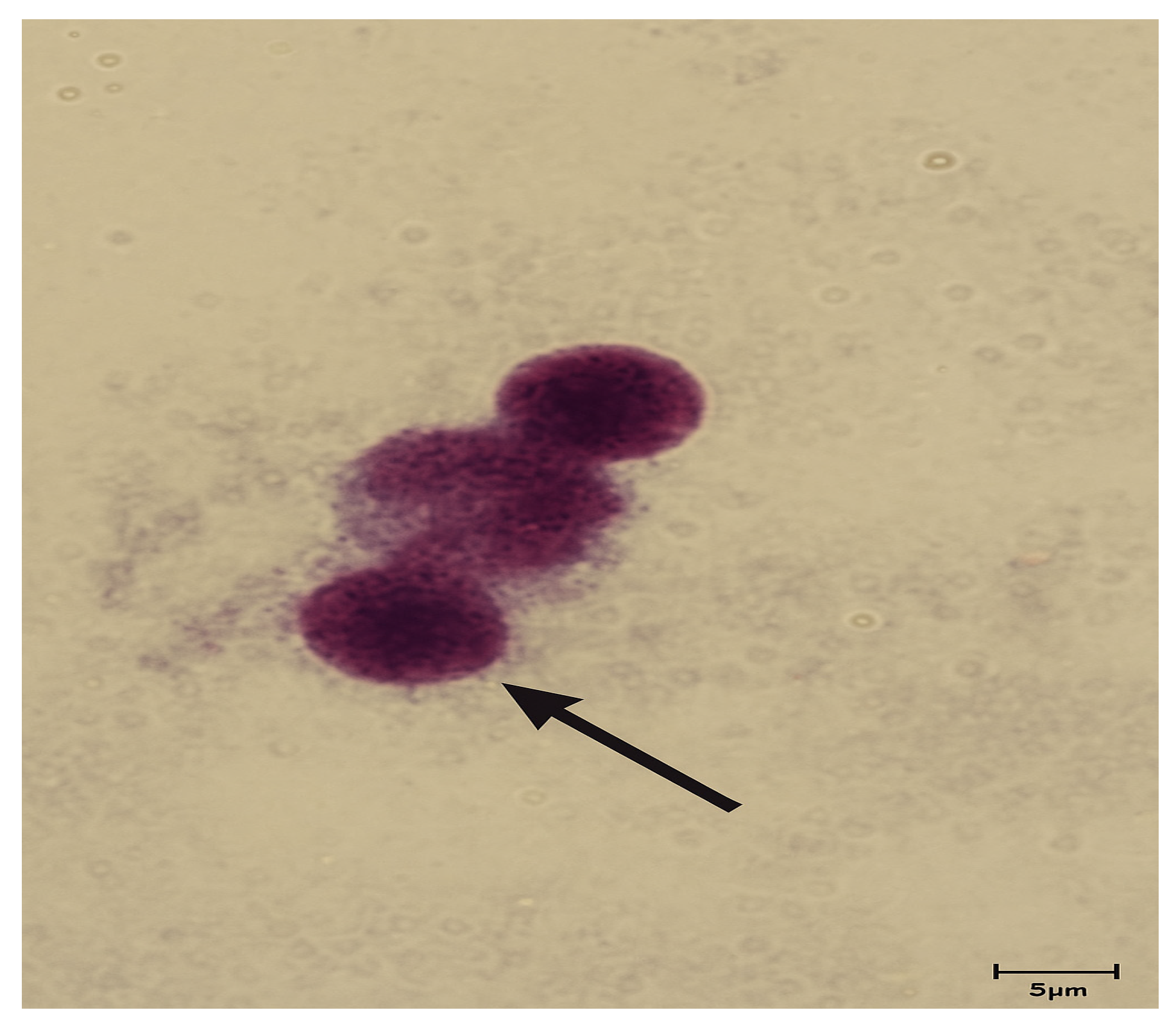
The control sample consists of a lymphocyte culture that was not exposed to the tested metal alloys, (Figure 1). Under the conditions provided by the culture medium, the appearance of micronuclei is observed due to a delay in the attachment of chromosomes to the mitotic spindle, Table 2.
Figure 2 presents the nuclei and micronuclei detected in lymphocytes exposed to the type 1 Ni-Cr test alloy, which does not contain beryllium, at a concentration of 5 mg/mL. In cultures exposed to Ni-Cr-beryllium alloy powder at a concentration of 5 mg/mL (Figure 3), the appearance of micronuclei is observed because of genetic material damage, leading to abnormal cell division.
In lymphocyte cultures exposed to type 1 Ni-Cr test alloy powder at a concentration of 10 mg/mL, a higher number of micronuclei was observed, highlighting an increase in genotoxicity compared to the sample with 5 mg/mL, (Figure 4).
A concentration of 10 mg/mL of type 2 Ni-Cr test alloy with beryllium resulted in significantly higher cytotoxicity and genotoxicity in both replicates compared to samples containing 5 mg/mL of the substance with or without beryllium, as well as 10 mg/mL without beryllium. Damage to the genetic material is evident (Figure 5).
For the concentration of 20 mg/mL of the type 1 test alloy Ni-Cr without beryllium, the appearance of micronuclei was also observed, due to the genotoxicity of the substances used, (Figure 6).
At a concentration of 20 mg/mL, type 2 Ni-Cr test alloy containing beryllium (Figure 7) induced significant damage to the genetic material, with recorded cytotoxicity and genotoxicity exceeding those observed in lymphocyte nuclei treated with 20 mg/mL of type 1 Ni-Cr test alloy without beryllium. Statistical analysis of micronuclei incidence, based on microscopic observations, was conducted using IBM® SPSS® Statistics 25.0 (Armonk, NY, USA) (Table 3, Table 4, Table 5, Table 6, Table 7 and Table 8).
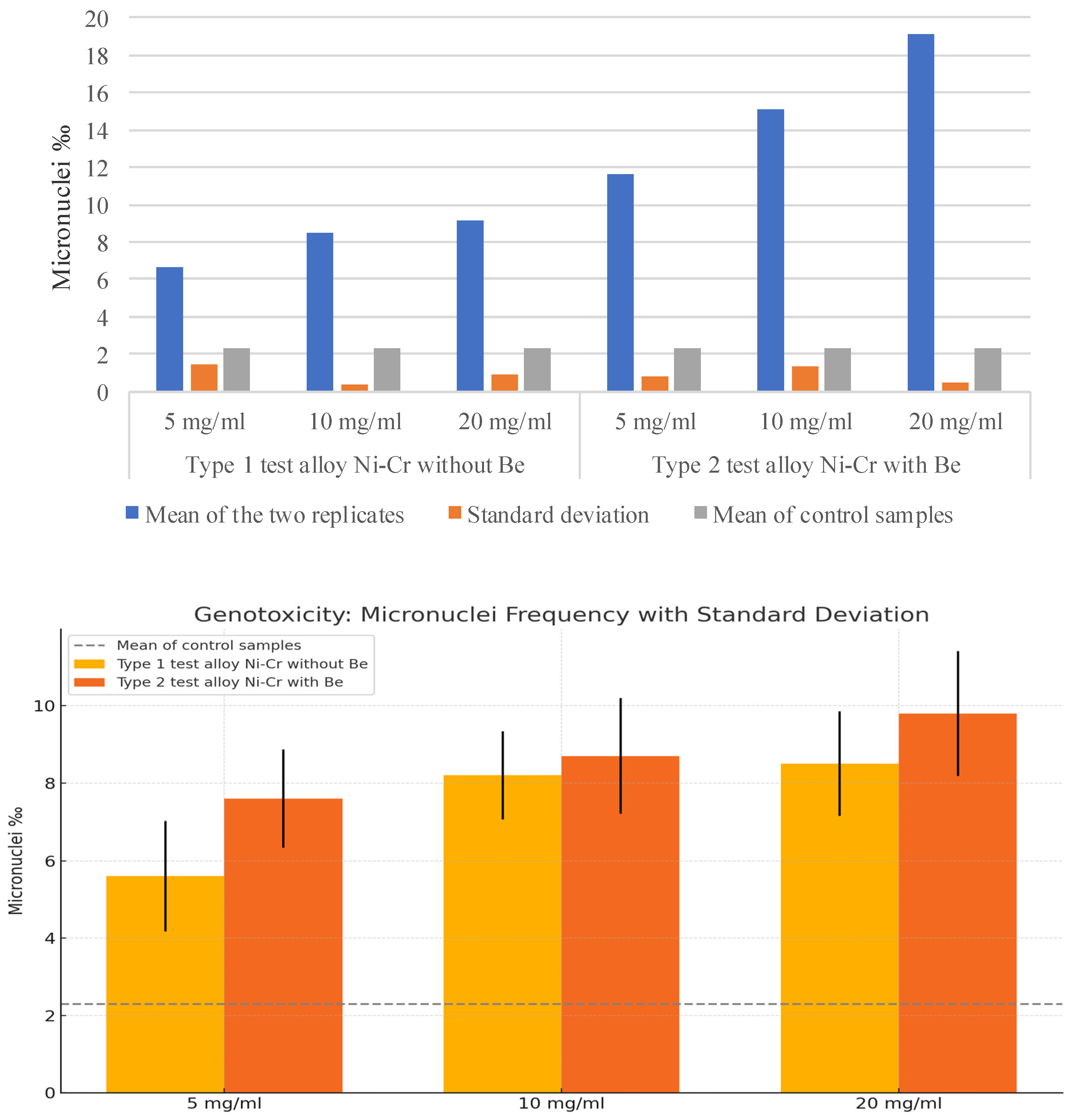
The previous graph (Figure 8) demonstrates that genotoxicity increases with the amount of substance added to the lymphocyte culture. Significant differences are also observed in the incidence of micronuclei between samples without beryllium and those containing beryllium, with beryllium exhibiting high toxicity even when combined with other metals in an alloy. Data obtained from the analysis of the two replicates were used for statistical analysis.

4. Discussion

Two test alloys were used in this study, designated Type 1 Ni-Cr test alloy without beryllium (approximate composition: Ni 63%, Cr 24%, Mo 10%, Si 1%, Fe 1%) and Type 2 Ni-Cr test alloy with beryllium (approximate composition: Ni 78%, Cr 12%, beryllium 2.1%, Mo 5%). In the specialized literature, both genotoxicity and cytotoxicity of metallic alloys with dental applications, having similar compositions, have been investigated [18,20,24,25,26,27].
Drăguș L. et al. evaluated the genotoxicity and cytotoxicity of a Ni-Cr-based metal alloy for prosthetic applications, without the addition of beryllium, with a composition like that of the Type 1 Ni-Cr test alloy. The analyzed metal alloy exhibited high adhesion. The activity of alkaline phosphatase was assessed after 3 and 6 days, showing that the studied alloy ranked lowest in comparison to other alloys when analyzing the activity of this marker [28].
Another relevant study, conducted by Zhihong et al. (2011) [29], examined the cytotoxicity and genotoxicity of a Ni-Cr-based metal alloy with a beryllium addition of approximately 1.9%, a composition closely resembling the Type 2 Ni-Cr test alloy used in this study. The findings indicated that the cytotoxicity of the alloy was higher than that of the two other alloys tested, primarily due to the presence of nickel. The addition of beryllium was identified as a contributing factor to increased cytotoxic effects. In contrast, chromium and molybdenum formed a passive layer, limiting ion release due to their lower toxicity [29].
The compositional differences between the two tested alloys—beyond the presence of beryllium—may influence cellular responses through variations in metal ion release, corrosion behavior, and oxide layer formation, all of which can directly affect cell viability and genomic stability.
Nickel (Ni), chromium (Cr), titanium (Ti), and their respective alloys are widely used in various branches of dentistry, including orthodontics, endodontics, prosthetics, and oral implantology, due to their mechanical strength and corrosion resistance. However, their application in biomedical contexts is strictly regulated by their biocompatibility, which determines their safety and acceptability in contact with human tissues. This requirement is governed by European Council Directive 93/42/EEC of 14 June 1993, and its subsequent amendments, which define essential health and safety requirements for medical devices, including dental materials. According to this directive, all metallic components must not release substances that can endanger the health of patients or healthcare professionals, directly addressing the cytotoxic and genotoxic potential of such materials [30].
The primary objective of this study was to investigate the cytotoxicity and genotoxicity of Ni-Cr and Ni-Cr-beryllium alloys. The scientific literature has extensively discussed the role of beryllium-containing dental alloys, particularly in relation to their cytotoxic and genotoxic potential. Beryllium is often added to Ni-Cr alloys to improve fluidity during casting and to promote oxide layer formation for better ceramic adhesion. However, studies have shown that beryllium may increase the release of toxic ions and provoke inflammatory or mutagenic responses in surrounding tissues. Furthermore, occupational exposure to beryllium-containing dust or vapors, especially in dental laboratories, has been linked to systemic toxicity and hypersensitivity reactions in dental technicians. These findings underscore the need to critically evaluate beryllium-containing alloys and explore safer alternatives in clinical and laboratory practice [31].
The scientific literature identifies beryllium as a carcinogenic substance responsible for acute chemical pneumonitis and chronic beryllium disease. Studies conducted in the United States have shown an increased cancer risk among workers exposed to beryllium in industrial processing facilities. Furthermore, experiments on rodents have demonstrated that high doses of beryllium (410–980 mg/m3 in inhaled air) can lead to tumor formation in approximately 64% of cases. These findings are consistent with the results of the present study, where beryllium-containing alloys induced a higher frequency of micronuclei, indicating a significant genotoxic effect.
Nickel (Ni), widely used in industry due to its favorable physicochemical properties, is also one of the most potent allergens and a frequent cause of contact dermatitis, especially among dental technicians. Moreover, nickel is recognized as a carcinogen and exists in various chemical forms, including oxides, sulfates, and nanoparticles. Among these, insoluble nickel compounds exhibit a significantly higher carcinogenic risk than their water-soluble counterparts. In vitro studies on mammalian cells have shown that nickel nanoparticles can activate molecular pathways involved in carcinogenesis. The results of this study confirm these observations, as the increased frequency of micronuclei in groups exposed to Ni-based alloys suggests a negative impact on genomic integrity [32,33].
Toxicity evaluations on lymphocytes have demonstrated that nickel chloride induces the formation of reactive oxygen species (ROS), particularly hydroperoxides. These free radicals are involved in pathogenic processes, including cancer, aging, and arteriosclerosis [34]. The toxicity and carcinogenicity of nickel are strongly influenced by both its physical and chemical properties and the route of entry into the body. Nickel exposure occurs through ingestion, dermal absorption (via cosmetic products), and inhalation. Inhaled nickel-based compounds accumulate in the alveoli, tracheobronchial region, and nasopharynx [35].
Chromium (Cr) absorption occurs through three primary pathways: respiratory, dermal, and oral. The oxidation state of chromium compounds significantly affects absorption efficiency. Hexavalent chromium (Cr VI) is absorbed more efficiently than trivalent chromium (Cr III), and this characteristic is associated with carcinogenicity. Under normal biological conditions, the reduction of Cr VI to Cr III does not occur spontaneously, while the reverse process takes place in cellular metabolic pathways, generating reactive oxygen species (ROS) that damage essential biomolecules [36,37].
Industrial exposure to high levels of chromium is linked to severe toxicity and an elevated risk of respiratory cancers, primarily due to Cr VI, which is more soluble and mobile than Cr III. Because of direct airway exposure, cancers most frequently develop in the lungs, sinuses, and nasal cavity [36]. Multiple in vivo studies have demonstrated the genotoxic effects of chromium, including structural DNA damage, strand breaks, and cross-links between DNA strands [36,38].
The findings of this study indicate that samples containing beryllium exhibited a higher genotoxic potential than those without beryllium, suggesting an increased risk associated with the use of Be-containing alloys in dental materials. These results confirm the necessity of caution when selecting alloy compositions for dental applications.
Differences in cytotoxicity were also observed between the tested alloys, with higher cytotoxic levels recorded in Be-containing samples. These findings highlight the importance of toxicity assessment in dental prosthetics. The incidence of micronuclei, which serve as indicators of chromosomal fragmentation or loss, was significantly higher in samples containing beryllium, suggesting increased genetic instability and an elevated risk of genotoxic effects associated with this element.

5. Conclusions

The results of this study highlight the importance of carefully selecting materials used in the fabrication of dental restorations to minimize health risks for both patients and healthcare professionals. There is a clear need for further research to better understand the long-term effects of exposure to various metal alloy compositions used in dentistry, with a particular focus on the assessment of genotoxicity and cytotoxicity.
Awareness of the potential risks associated with these materials is essential, and appropriate safety measures must be implemented. These include selecting materials based on current scientific evidence and in accordance with existing safety regulations.
Although preliminary, the data obtained in this study provide valuable insight into the safety of dental alloys and reinforce the need for continued research aimed at optimizing both dental materials and clinical practice in prosthodontics.

6. Study Limitations and Future Directions

An important limitation of this study lies in the fact that the tested alloys differed not only in the presence or absence of beryllium, but also in the concentration of other chemical elements (such as nickel, chromium, and molybdenum), which may influence cellular responses and limit the ability to isolate the specific effect of beryllium. Additionally, the use of lymphocytes from a single human donor, although compliant with OECD guidelines, restricts the generalizability of the findings.
For future studies, we recommend testing alloys with identical base compositions, differing only in their beryllium content, to allow a more precise evaluation of this element’s specific effect. We also propose expanding the biological model by including multiple human donors, as well as applying complementary genotoxicity assays (e.g., comet assay, γ-H2AX foci detection) to validate and further explore the results obtained.

Author Contributions

Conceptualization, F.C.; methodology, V.S.P. and M.A.M.; software, A.S.P.; validation, L.S.C.M.; formal analysis, A.A.S.; investigation, R.C.C.; resources, M.B.; data curation, A.V.; writing—original draft preparation, L.B.; writing—review and editing, M.B.; visualization, R.M.C.; supervision, C.M.S.; project administration, A.C.D.; funding acquisition, M.A.S. All authors have read and agreed to the published version of the manuscript.

Funding

This research received no external funding.

Institutional Review Board Statement

The study was conducted in accordance with the Declaration of Helsinki and approved by the Institutional Review Board of Carol Davila University of Medicine and Pharmacy, Ethics Committee Approval Code: PO-35-F03, date of approval: 9 February 2024.

Informed Consent Statement

Written informed consent was obtained from all subjects involved in the study.

Data Availability Statement

All data are within the manuscript.

Acknowledgments

Publication of this paper was supported by the Carol Davila University of Medicine and Pharmacy, through the institutional program Publish not Perish.

Conflicts of Interest

The authors declare no conflicts of interest.

References

  1. Wataha, J.C. Alloys for prosthodontic restorations. J. Prosthet. Dent. 2002, 87, 351–363. [Google Scholar] [CrossRef] [PubMed]
  2. Desai, S.; Bidanda, B.; Bártolo, P. Metallic and Ceramic Biomaterials: Current and Future Developments. In Bio-Materials and Prototyping Applications in Medicine; Bártolo, P., Bidanda, B., Eds.; Springer: Boston, MA, USA, 2008; pp. 1–14. [Google Scholar]
  3. Bezzon, O.L.; Ribeiro, R.F.; Rollo, J.M.; Crosara, S. Castability and resistance of ceramometal bonding in Ni-Cr and Ni-Cr-Be alloys. J. Prosthet. Dent. 2001, 85, 299–304. [Google Scholar] [CrossRef]
  4. Cheng, T.-P.; Tsai, W.-T.; Lin, J.-H.C.; Lee, J.-T. The effect of beryllium on the corrosion resistance of nickel-chromium dental alloys. J. Mater. Sci. Mater. Med. 1990, 1, 211–218. [Google Scholar] [CrossRef]
  5. Cohen, S.; Vaidyanathan, T.; Schulman, A. The effect of limited beryllium additions on a Ni-Cr alloy. J. Prosthet. Dent. 1988, 60, 688–692. [Google Scholar] [CrossRef] [PubMed]
  6. Vineeth, G.; Alla, R.K.; Srinivasa Raju, D.; Suresh Sajjan, M.C.; Ramaraju, A.V.; Yeleti, H. Casting alloys: The saga of their existence and the recipe of their blend. Int. J. Dent. Mater. 2019, 1, 25–35. [Google Scholar] [CrossRef]
  7. Geis-Gerstorfer, J.; Pässler, K. Studies on the influence of BE content on the corrosion behavior and mechanical properties of Ni-25Cr-10Mo alloys. Dent. Mater. 1993, 9, 177–181. [Google Scholar] [CrossRef]
  8. Bezzon, O.L.; Mattos, M.d.G.d.; Ribeiro, R.F.; Rollo, J.M.A. Effect of beryllium on the castability and resistance of ceramometal bonds in nickel-chromium alloys. J. Prosthet. Dent. 1998, 80, 570–574. [Google Scholar] [CrossRef]
  9. Registry Aftsad. Toxicological Profile for Beryllium. 2003. Available online: https://www.atsdr.cdc.gov/toxprofiles/tp4.pdf (accessed on 1 March 2025).
  10. Agency, U.S.E.P. Toxicological Review of Beryllium and Compounds. 2008. Available online: https://iris.epa.gov/static/pdfs/0012tr.pdf (accessed on 1 March 2025).
  11. UKEA. A Review of the Toxicity of Beryllium in Air 2008. Available online: https://assets.publishing.service.gov.uk/media/5a7c6f8ded915d696ccfcc2f/scho0508bobo-e-e.pdf (accessed on 26 December 2024).
  12. Annangi, B.; Bonassi, S.; Marcos, R.; Hernández, A. Biomonitoring of humans exposed to arsenic, chromium, nickel, vanadium, and complex mixtures of metals by using the micronucleus test in lymphocytes. Mutat. Res. Mol. Mech. Mutagen. 2016, 770, 140–161. [Google Scholar] [CrossRef]
  13. Apostoli, P.; Porru, S.; Alessio, L. Behaviour of urinary beryllium in general population and in subjects with low-level occupational exposure. Med. Lav. 1989, 80, 390–396. [Google Scholar]
  14. Halder, A.; De, M. Increase in DNA damage in lymphocytes and micronucleus frequency in buccal cells in silica-exposed workers. Indian J. Occup. Environ. Med. 2012, 16, 34–37. [Google Scholar] [CrossRef]
  15. Nishioka, H. Mutagenic activities of metal compounds in bacteria. Mutat. Res. 1975, 31, 185–189. [Google Scholar] [CrossRef] [PubMed]
  16. Ashby, J.; Ishidate, M.; Stoner, G.; Morgan, M.; Ratpan, F.; Callander, R. Studies on the genotoxicity of beryllium sulphate in vitro and in vivo. Mutat. Res. Toxicol. 1990, 240, 217–225. [Google Scholar] [CrossRef] [PubMed]
  17. Fritzenschaf, H.; Kohlpoth, M.; Rusche, B.; Schiffmann, D. Testing of known carcinogens and noncarcinogens in the Syrian hamster embryo (SHE) micronucleus test in vitro; correlations with in vivo micronucleus formation and cell transformation. Mutat. Res. Toxicol. 1993, 319, 47–53. [Google Scholar] [CrossRef]
  18. Tripuraneni, S.C.; Namburi, S.K. Evaluation of genotoxicity of recycled Ni-Cr dental casting alloys: An in vitro study. J. Appl. Biomater. Biomech. 2008, 6, 47–54. [Google Scholar] [PubMed]
  19. Ribeiro, D.A.; Yujra, V.Q.; De Moura, C.F.G.; Handan, B.A.; Viana, M.D.B.; Yamauchi, L.Y.; Castelo, P.M.; Aguiar, O. Genotoxicity Induced by Dental Materials: A Comprehensive Review. Anticancer. Res. 2017, 37, 4017–4024. [Google Scholar] [CrossRef]
  20. Altas, E.; Gurhan, I.D.; Urkmez, A.S. Influence of processing flaws on cytotoxicity and genotoxicity of Co-Cr and Ni-Cr based dental crowns and bridges. In Proceedings of the 10th International Workshop on Biomedical Engineering, Kos, Greece, 5–7 October 2011; pp. 1–4. [Google Scholar] [CrossRef]
  21. OCED Test No. 487: In Vitro Mammalian Cell Micronucleus Test Paris: 21. Publishing. 2014. Available online: https://www.oecd.org/en/publications/test-no-487-in-vitro-mammalian-cell-micronucleus-test_9789264264861-en.html (accessed on 1 March 2025).
  22. Ye, C.J.; Sharpe, Z.; Alemara, S.; Mackenzie, S.; Liu, G.; Abdallah, B.; Horne, S.; Regan, S.; Heng, H.H. Micronuclei and Genome Chaos: Changing the System Inheritance. Genes 2019, 10, 366. [Google Scholar] [CrossRef]
  23. Sommer, S.; Buraczewska, I.; Kruszewski, M. Micronucleus Assay: The State of Art, and Future Directions. Int. J. Mol. Sci. 2020, 21, 1534. [Google Scholar] [CrossRef]
  24. Al-Hiyasat, A.S.; Bashabsheh, O.M.; Darmani, H. Elements released from dental casting alloys and their cytotoxic effects. Int. J. Prosthodont. 2002, 15, 473–478. [Google Scholar]
  25. Alp, G.; Çakmak, G.; Sert, M.; Burgaz, Y. Corrosion potential in artificial saliva and possible genotoxic and cytotoxic damage in buccal epithelial cells of patients who underwent Ni-Cr based porcelain-fused-to-metal fixed dental prostheses. Mutat. Res. Genet. Toxicol. Environ. Mutagen. 2018, 827, 19–26. [Google Scholar] [CrossRef]
  26. Ellakany, P.; AlGhamdi, M.A.; Alshehri, T.; Abdelrahman, Z. Cytotoxicity of Commercially Pure Titanium (cpTi), Silver-Palladium (Ag-Pd), and Nickel-Chromium (Ni-Cr) Alloys Commonly Used in the Fabrication of Dental Prosthetic Restorations. Cureus 2022, 14, e31679. [Google Scholar] [CrossRef]
  27. Morán-Martínez, J.; Luna, K.M.-D.; Betancourt-Martínez, N.; Carranza-Rosales, P.; Contreras-Martínez, J.; López-Meza, M.; Rodríguez-Villarreal, O. Genotoxicity in oral epithelial cells in children caused by nickel in metal crowns. Genet. Mol. Res. 2013, 12, 3178–3185. [Google Scholar] [CrossRef] [PubMed]
  28. Dragus, L.; Ghergic, D.L.; Comaneanu, R.M.; Bechir, A.; Coman, C.; Botoaca, O. In vitro comparative tests about the biocompatibility of some dental alloys. Rev. Chim. 2019, 70, 610–613. [Google Scholar] [CrossRef]
  29. Zhihong, C.; Yezhen, L.; Zhiyuan, G.; Zhongqiao, Y.; Xiaoxue, L.; Yifeng, R.; Weikun, F.; Jiliang, H. Comparison of the cytogenotoxicity induced by five different dental alloys using four in vitro assays. Dent. Mater. J. 2011, 30, 861–868. [Google Scholar] [CrossRef]
  30. Union, E. Consolidated Text: Council Directive 93/42/EEC of 14 June 1993 Concerning Medical Devices. EUR-Lex—Access to European Union Law 2007. Available online: https://eur-lex.europa.eu/eli/dir/1993/42/oj/eng (accessed on 1 March 2025).
  31. Strupp, C. Beryllium metal I. experimental results on acute oral toxicity, local skin and eye effects, and genotoxicity. Ann. Occup. Hyg. 2011, 55, 30–42. [Google Scholar] [CrossRef] [PubMed]
  32. Gordon, T. Beryllium: Genotoxicity and carcinogenicity. Mutat. Res. Mol. Mech. Mutagen. 2003, 533, 99–105. [Google Scholar] [CrossRef]
  33. Jiménez-Lamana, J.; Godin, S.; Aragonès, G.; Bladé, C.; Szpunar, J.; Łobinski, R. Nickel Nanoparticles Induce the Synthesis of a Tumor-Related Polypeptide in Human Epidermal Keratinocytes. Nanomaterials 2020, 10, 992. [Google Scholar] [CrossRef] [PubMed]
  34. Chen, C.-Y.; Wang, Y.-F.; Huang, W.-R.; Huang, Y.-T. Nickel induces oxidative stress and genotoxicity in human lymphocytes. Toxicol. Appl. Pharmacol. 2003, 189, 153–159. [Google Scholar] [CrossRef]
  35. Genchi, G.; Carocci, A.; Lauria, G.; Sinicropi, M.S.; Catalano, A. Nickel: Human Health and Environmental Toxicology. Int. J. Environ. Res. Public Health 2020, 17, 679. [Google Scholar] [CrossRef]
  36. Sawicka, E.; Jurkowska, K.; Piwowar, A. Chromium (III) and chromium (VI) as important players in the induction of genotoxicity—Current view. Ann. Agric. Environ. Med. 2021, 28, 1–10. [Google Scholar] [CrossRef]
  37. Otelea, M.; Handra, C.; Rascu, A. Registered cases of occupational n-hexane intoxication in Bucharest. Romanian J. Leg. Med. 2015, 23, 279–284. [Google Scholar] [CrossRef]
  38. Pircalabioru, G.G.; Ilie, I.; Oprea, L.; Picu, A.; Petcu, L.M.; Burlibasa, L.; Chifiriuc, M.-C.; Musat, M. Microbiome, Mycobiome and Related Metabolites Alterations in Patients with Metabolic Syndrome—A Pilot Study. Metabolites 2022, 12, 218. [Google Scholar] [CrossRef] [PubMed]
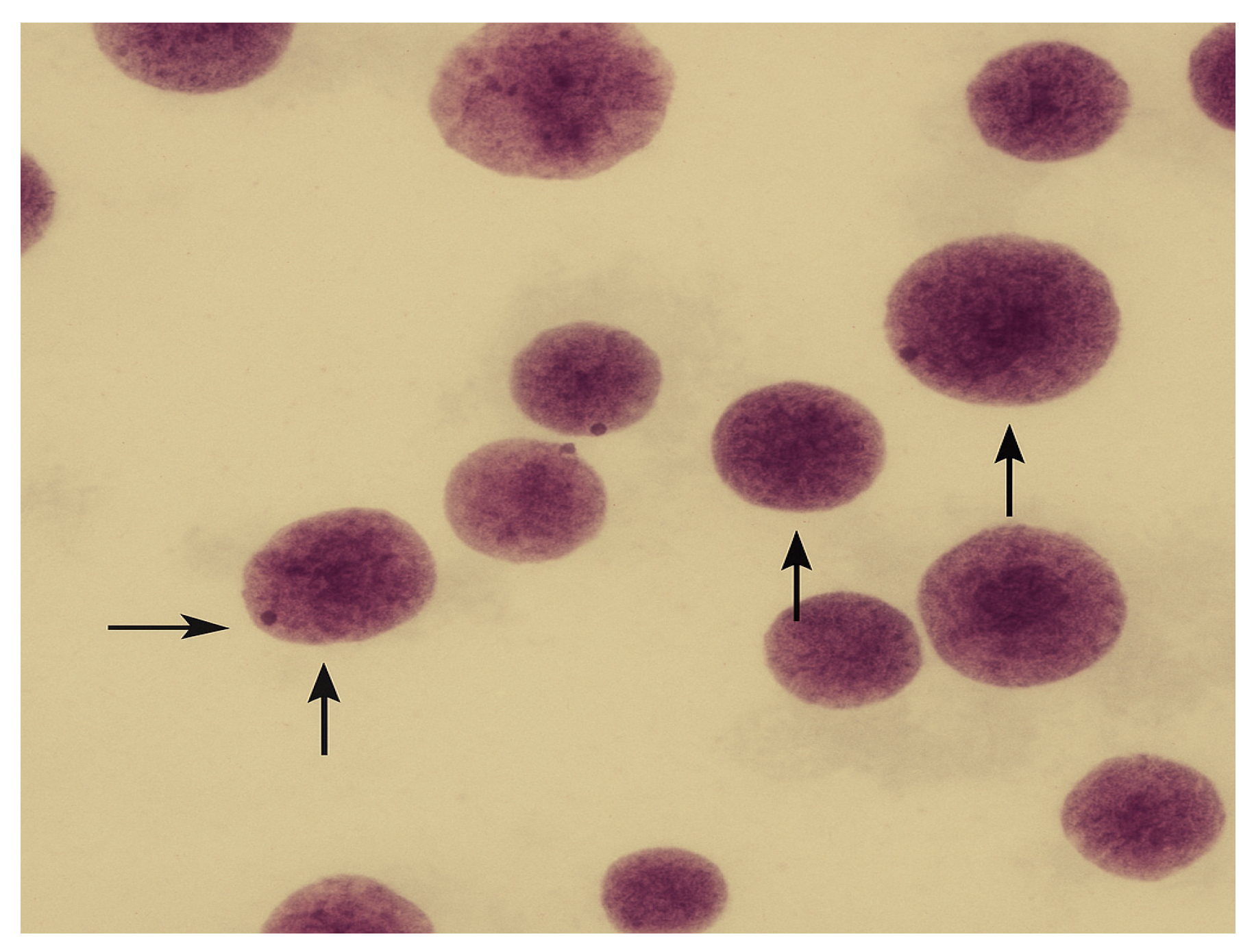
Figure 1. The control sample used to compare the natural state and the rate of appearance of micronuclei normally in relation to the other samples exposed to the substances of interest.
Oral 05 00032 g001
Figure 2. Type 1 test alloy Ni-Cr without beryllium, 5 mg/mL.
Oral 05 00032 g002
Figure 3. Atypical nuclei from lymphocyte culture treated with 5 mg/mL type 2 test alloy Ni-Cr with beryllium in the composition.
Oral 05 00032 g003
Figure 4. Lymphocyte culture put in contact with 10 mg/mL type 1 test alloy Ni-Cr without beryllium.
Oral 05 00032 g004
Figure 5. Image from lymphocyte culture in which type 2 test alloy Ni-Cr with beryllium was added in a concentration of 10 mg/mL.
Oral 05 00032 g005
Figure 6. The presence of micronuclei in the lymphocyte culture in which type 1 test alloy Ni-Cr without beryllium in a concentration of 20 mg/mL was added.
Oral 05 00032 g006
Figure 7. Atypical nuclei and micronuclei appearing in lymphocyte culture in which type 2 test alloy Ni-Cr with beryllium in a concentration of 20 mg/mL was added.
Oral 05 00032 g007
Figure 8. Comparison of the incidence of micronuclei in the different samples analyzed against the control sample.
Oral 05 00032 g008
Table 1. Cytotoxicity of substances depends on the concentration in the culture medium and type.
Substance TypeCytotoxicity Depends on the Concentration of the
Substance in the Culture Medium
5 mg/mL10 mg/mL20 mg/mL
Type 1 test alloy Ni-Cr without beryllium40%49.2%58.2%
Type 2 test alloy Ni-Cr with beryllium56.2%57.4%60%
Table 2. Analysis of the frequency of micronuclei according to the concentration in the culture medium and the type of substance.
Substance TypeThe Concentration of Substances in the Culture Medium
5 mg/mL10 mg/mL20 mg/mL
Type 1 Ni-Cr test alloy without beryllium
Replica 1
5.6‰8.2‰8.5‰
Replica 27.6‰8.7‰9.8‰
Negative control
Replica 1
2.6‰
Replica 22‰
Table 3. Type 1 test alloy Ni-Cr without beryllium—Replica 1.
Descriptive Statistics
NRangeMinimumMaximumMeanStd. ErrorStd. DeviationVariancep-Value
FB.R1.5 mg/mL104485.600.4521.4302.044<0.05
FB.R1.10 mg/mL1037108.200.3591.1351.289<0.01
FB.R1.20 mg/mL1046108.500.4281.3541.833<0.01
Valid N (listwise)10
Table 4. Type 1 test alloy Ni-Cr without beryllium—Replica 2.
Descriptive Statistics
NRangeMinimumMaximumMeanStd. ErrorStd. DeviationVariancep-Value
FB.R2.5 mg/mL1046107.600.4001.2651.600<0.05
FB.R2.10 mg/mL1047118.700.4731.4942.233<0.01
FB.R2.20 mg/mL10689149.800.5121.6192.622<0.001
Valid N (listwise)10
Table 5. Ni-Cr type 2 test alloy solution with beryllium—Replica 1.
Descriptive Statistics
NRangeMinimumMaximumMeanStd. ErrorStd. DeviationVariancep-Value
CB.R1.5 mg/mL10591412.200.6111.9323.733<0.05
CB.R1.10 mg/mL105141916.000.6832.1604.667<0.01
CB.R1.20 mg/mL107142118.800.8272.6166.844<0.001
Valid N (listwise)10
Table 6. Ni-Cr type 2 test alloy solution with beryllium—Replica 2.
Descriptive Statistics
NRangeMinimumMaximumMeanStd. ErrorStd. DeviationVariancep-Value
CB.R2.5 mg/mL10491311.000.4221.3331.778<0.05
CB.R2.10 mg/mL107111814.100.6902.1834.767<0.01
CB.R2.20 mg/mL109132219.400.7632.4135.822<0.001
Valid N (listwise)10
Table 7. Witness Replica.
Descriptive Statistics
NRangeMinimumMaximumMeanStd. ErrorStd. DeviationVariancep-Value
M.R1103142.600.3401.0751.156<0.05
M.R2104042.000.3651.1551.333<0.05
Valid N (listwise)10
Table 8. Witness Replica 2.
Experimental ConditionMeanStd. ErrorStd. DeviationVariancep-Value
FB.R1.5 mg/mL5.60.4521.432.044<0.05
FB.R1.10 mg/mL8.20.3591.1351.289<0.01
FB.R1.20 mg/mL8.50.4281.3541.833<0.01
FB.R2.5 mg/mL7.60.41.2651.6<0.05
FB.R2.10 mg/mL8.70.4731.4942.233<0.01
FB.R2.20 mg/mL9.80.5121.6192.622<0.001
CB.R1.5 mg/mL12.20.6111.9323.733<0.05
CB.R1.10 mg/mL16.00.6832.164.667<0.01
CB.R1.20 mg/mL18.80.8272.6166.844<0.001
CB.R2.5 mg/mL11.00.4221.3331.778<0.05
CB.R2.10 mg/mL14.10.692.1834.767<0.01
CB.R2.20 mg/mL19.40.7632.4135.822<0.001
M.R12.60.341.0751.156<0.05
M.R22.00.3651.1551.333<0.05
Disclaimer/Publisher’s Note: The statements, opinions and data contained in all publications are solely those of the individual author(s) and contributor(s) and not of MDPI and/or the editor(s). MDPI and/or the editor(s) disclaim responsibility for any injury to people or property resulting from any ideas, methods, instructions or products referred to in the content.