Article

Stress Distribution in a Mandibular Kennedy Class I with Bilateral Implant-Assisted Removable Partial Denture: A Finite Element Analysis

by
Dagny Ochoa-Escate
1,2,
Freddy Valdez-Jurado
3,
Romel Watanabe
3,
Martha Pineda-Mejía
3,
Edwin Antonio Córdova Huayanay
3,
Maria Soledad Ventocilla Huasupoma
3,
Marcos Herrera Cisneros
3,
Giovanna Lujan Larreátegui
3,
Angela Quispe-Salcedo
4,
Doris Salcedo-Moncada
3,* and
Jesús Julio Ochoa Tataje
3,*
1
Faculty of Dentistry, National University of San Marcos, Av. Amezaga 375, Lima 15081, Peru
2
Division of Bio-Prosthodontics, Department of Oral Health Science, Graduate School of Medical and Dental Sciences, Niigata University, 2-5274, Gakkocho-dori, Chuo-ku, Niigata 951-8514, Japan
3
Department of Rehabilitation Stomatology, Faculty of Dentistry, National University of San Marcos, Av. Amezaga 375, Lima 15081, Peru
4
Division of Anatomy and Cell Biology of the Hard Tissue, Department of Tissue Regeneration, Graduate School of Medical and Dental Sciences, Niigata University, 2-5274 Gakkocho-dori, Chuo-ku, Niigata 951-8514, Japan
*
Authors to whom correspondence should be addressed.
Submission received: 20 February 2025 / Revised: 24 March 2025 / Accepted: 3 April 2025 / Published: 5 May 2025

Abstract

:
Objectives: This study evaluated the dental and alveolar bone stress distribution of a mandibular Kennedy Class I restored with a bilateral implant-assisted removable partial denture (IARPD) compared with a conventional removable partial denture (CRPD) through the application of finite element analysis (FEA). Methods: Kennedy Class I plaster models were made, including teeth from the lower left first premolar and lower right canine. The models were scanned, resin-based replicated and digitized. Using Solidworks software, internal hexagonal implants (10 mm × 4 mm) were virtually placed at the level of the first molars. Each model was grouped into a unit, and a load of 200 N was applied, simulating masticatory forces. Von Mises stress distributions were calculated via FEA for the vertical, diagonal and combined forces. Results: In the IARPD, the stress generated in the alveolar bone by the vertical (4.2 Mpa), diagonal (12.2 MPa) and combined forces (12.3 MPa) was lower than that of the CRPD (7 MPa, 26.3 MPa and 32 MPa, respectively). Similarly, at the lower central incisor, the IARPD generated less stress than the CRPD due to the action of the vertical, diagonal and combined forces. Conclusions: Our preliminary data suggest that bilateral implant placement may result in less stress on bone and teeth during rehabilitation with a Kennedy Class I IARPD, with different orientations of the forces applied.

1. Introduction

In emerging countries, characterized by the high prevalence of edentulism and limited access to oral health services, conventional partial dentures are still a viable and economic treatment option for oral rehabilitation [1,2]. As life expectancy has increased, so has the number of users of partial dentures. In this context, there is a need to improve rehabilitation techniques that ensure aesthetics, chewing function and protection of remaining bone and teeth [1,2,3,4].
Among the different types of edentulism, the rehabilitation of a Kennedy Class I with a conventional removable partial denture (CRPD) represents a significant challenge [4,5,6]. Due to the lack of support in the posterior teeth, this treatment is reported to have low retention during chewing, which can lead to instability of the CRPD [7,8,9]. Similarly, biomechanical knowledge is essential for correct treatment. Unfavorable movements, such as rotation during function, often occur in a mandibular Kennedy Class I due to the resiliency between the two main supports: the teeth and the buccal mucosa [4,5,6,7]. The teeth are anchored to the alveolar bone by the periodontal ligament, which is approximately 0.2 mm thick, while the buccal mucosa, which is in contact with the acrylic base of the prosthesis, is approximately 2 mm thick [5]. The disparity in the thickness of these tissues creates unequal resilience, which can lead to unwanted and damaging lever movements on the teeth and the mucosa [5,10].
An implant-assisted removable partial denture (IARPD) has been an alternative to a CRPD since the 1990s, when it was reported that posterior implant placement in conjunction with an RPD resulted in more clinically stable rehabilitation and better RPD support [8,9,10,11,12,13,14]. As a result, masticatory function, biomechanics and patient satisfaction are improved. Likewise, this option could eliminate the intrusion movements of the RPD, as the placement of two distal implants in a posterior bilateral edentulism transforms into a pseudo-Kennedy Class III model [1,9,13,14,15,16,17].
Previous studies have shown that the best site for implant placement is the first molar area due to lower levels of displacement and stress within the metal structure, the peri-implant bone area and the implant [2,11,15,16,18,19]. In addition, patients with an IARPD have a good implant survival rate of 99.44% in the mandible [7,11]. Moreover, this alternative would reduce treatment costs and be more accessible to patients with limited economic resources [7,11,20,21].
Several studies have investigated clinical outcomes, feasibility and patient satisfaction when using an IARPD [11,12,13,14,15,16,17]. However, there is still a paucity of literature analyzing the stresses generated by the forces exerted by this type of prosthesis on the remaining dental tissues. This is because it is ethically impossible to carry out clinical trials on the mechanical behavior of IARPDs, so it is necessary to use appropriate methods that can help to improve our understanding of how this type of RPD might affect dental and remaining tissue.
A well-known numerical technique for stress and strain analysis of geometric structures is finite element analysis (FEA) [20,21,22]. This technique does not adversely affect the physical properties of the materials analyzed and is an easily repeatable technique [21,22]. In recent years, FEA has become a key research tool for predicting the biomechanical behaviors of the human tissues, materials and techniques used in dentistry [16,18,22].
In this context, our study aimed to evaluate the tooth and alveolar bone stress distribution of a mandibular Kennedy Class I restored with a conventional removable partial denture (CRPD) compared to a bilateral implant-assisted removable partial denture (IARPD) using finite element analysis (FEA). It is hypothesized that the IARPD will distribute masticatory forces more efficiently over the remaining teeth and alveolar bone compared to the CRPD, reducing stress on the alveolar bone and improving the stability of the remaining teeth in a mandibular Kennedy class I.

2. Materials and Methods

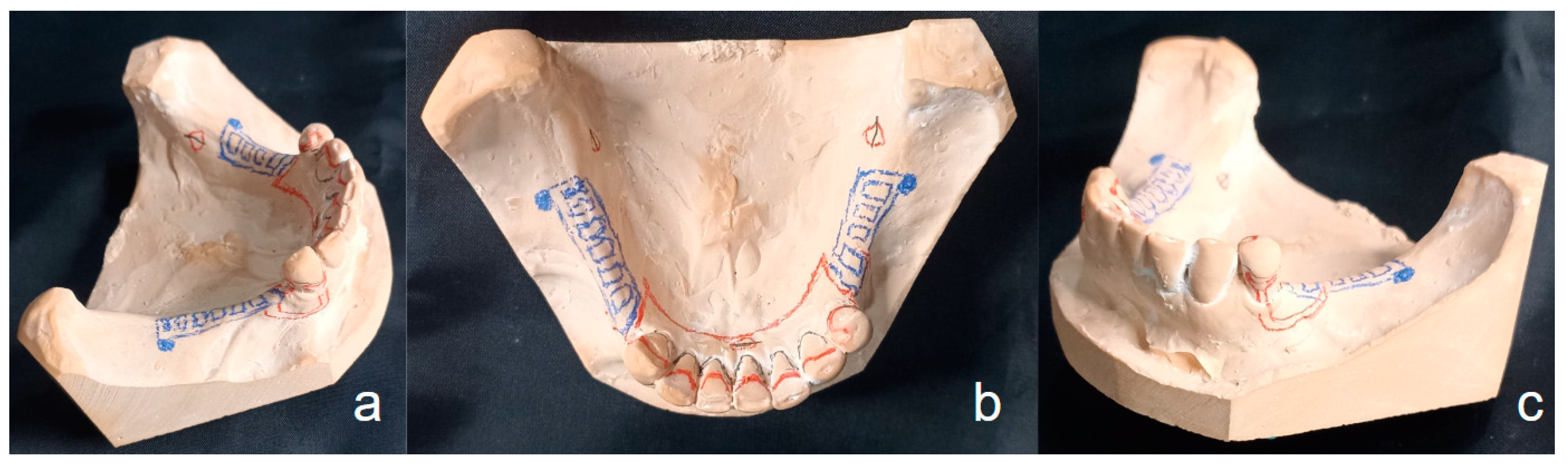
2.1. Plaster Model Preparation

A Kennedy Class I mandibular model with preserved teeth from the lower left first premolar to the lower right canine—the abutments—was used. An alginate impression (orthoprint, Zhermack) was taken to replicate the model, and then type IV stone (Elite Rock, Zhermack) was applied. After a paralleling process, the design of the removable partial denture was realized. The occlusal rests were placed on the mesial surface of the lower left first premolar and at the level of the cingulum of both present canines. Additionally, T-type retainers and a lingual plate as a major connector were selected. (Figure 1a–c). This design was maintained in both the CRPD and IARPD.

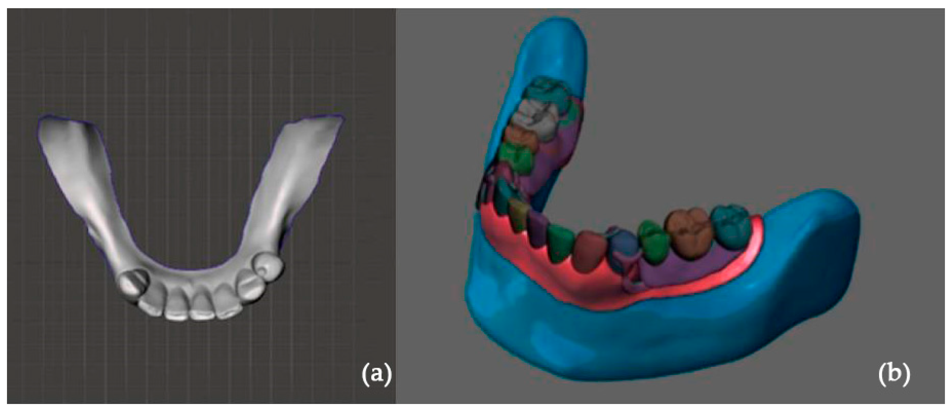
2.2. Digital Preparation

The plaster model was scanned with an optical scanner (Steinbichler Comet l3 d 5 m; Steinbichler Optotechnik GmbH, Neubeuern, Germany), replicated in resin and then digitized twice using SolidWorks version 14 software (Dassault Systems SolidWorks Corp., Waltham, MA, USA) (Figure 2a). The mucosa was modeled with depreciable values to simulate an extreme scenario. A single geometry was generated where the prosthesis apparently makes contact with the mandibular bone. The digital design for the teeth and bone anatomy was created considering normal dental occlusion, as well as the anatomy of the roots and mandibular bone, assuming a single structure between the removable partial denture, teeth and bone. A conventional removable partial denture was also digitally designed (Figure 2b). For the IARPD model, internal hexagonal implants (10 mm × 4 mm) were digitally placed at the level of the first molars.

2.3. Finite Element Analysis
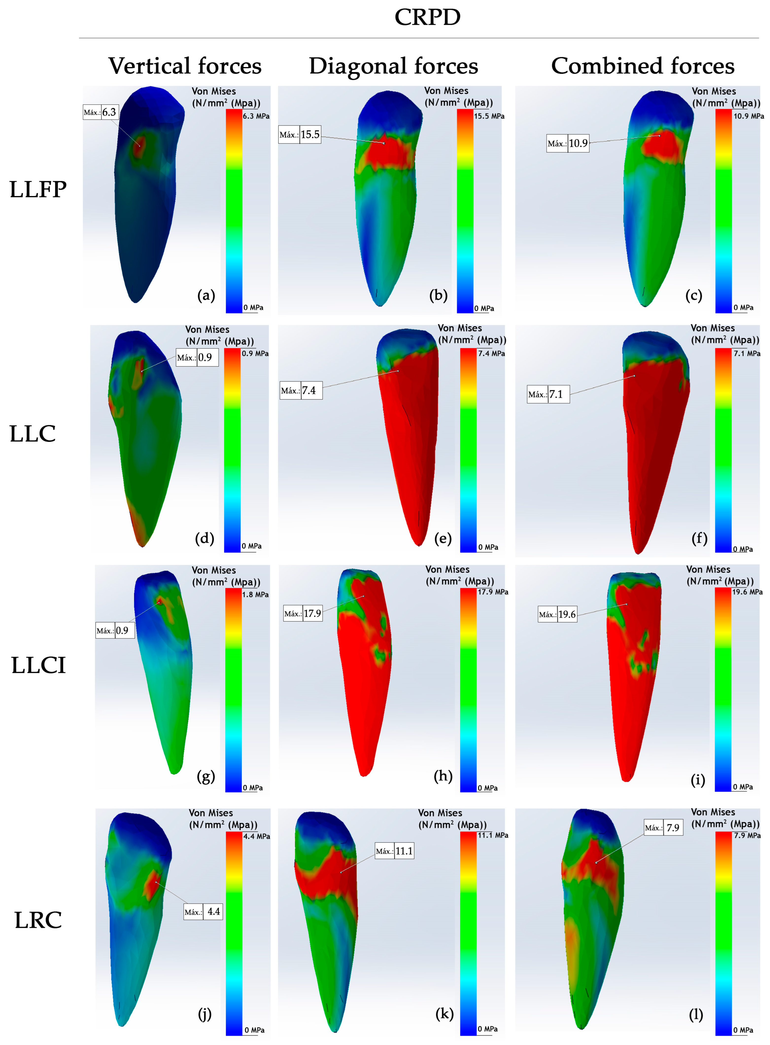
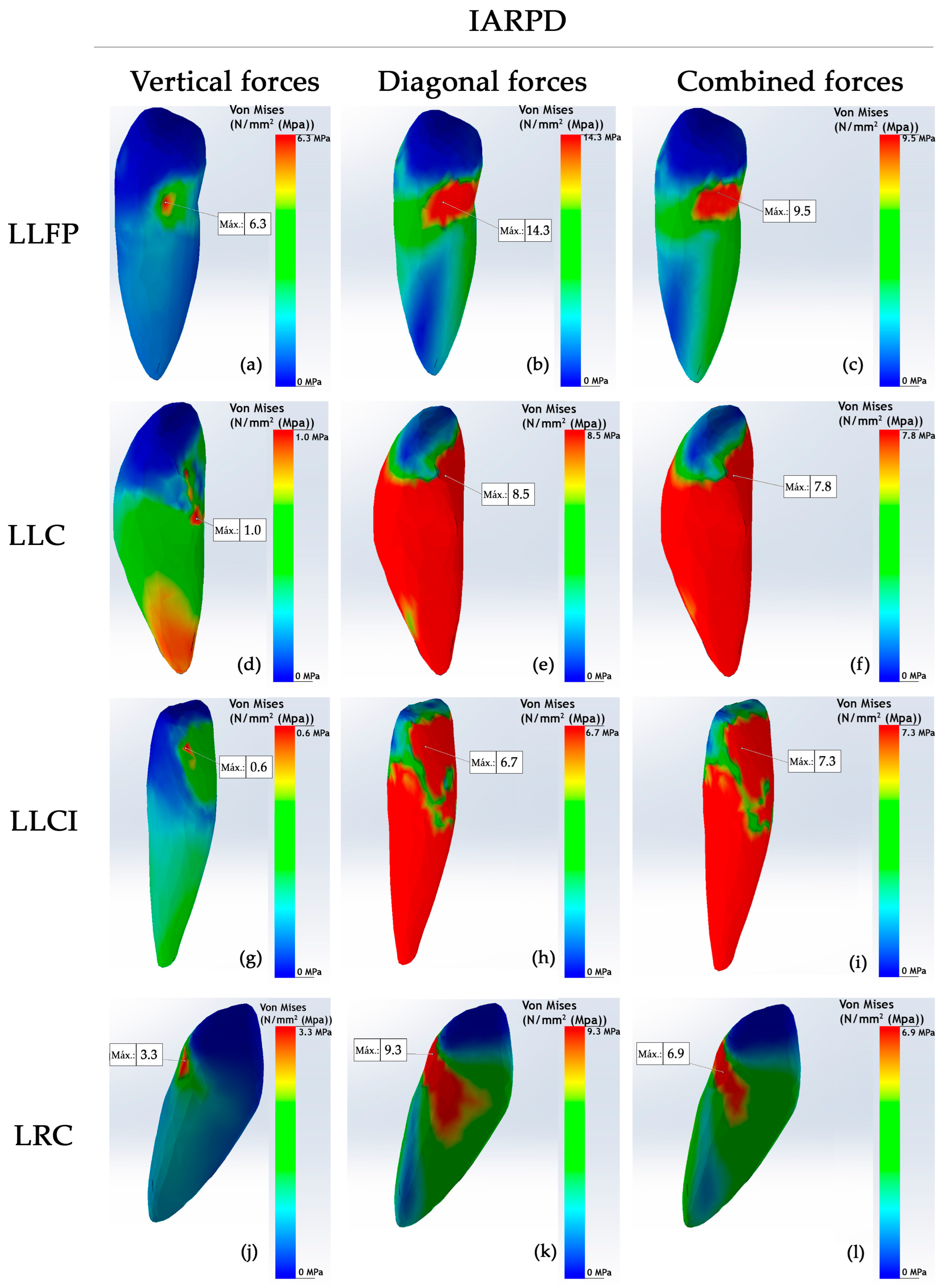
The physical properties of all the materials used were represented by two key constants: Young’s modulus of elasticity in megapascals (MPa) and Poisson’s ratio. The properties were taken from a similar study conducted in previous research, and the values are shown in Table 1 [11]. The FEA also considered biological and non-biological structures, such as isotropic bodies [10]. Although the mandibular bone is a non-homogeneous, anisotropic and non-ductile structure, for this study, the mandibular cortical bone was considered, as it is more homogeneous and isotropic. The relation between the different structures in the study of digital models was considered as a unit. A 3D simulation was performed to analyze the complex structure. Full adhesion contact between the prosthesis and the hard and soft tissues was assumed in the general contact simulation. Tetrahedral mesh was generated numerically due to being a complex structure, as the RPD, tooth and bone were considered a unit. A mesh convergence analysis was used to reduce the size of the tetrahedral elements in order to improve the accuracy of the analysis, achieving an ideal balance of all the elements generated by the software.
A force of 200 N was selected as an average value to simulate the chewing process [9,17]. Three types of forces were considered to simulate the complex movements of mastication: vertical, diagonal and combined. The vertical forces applied at 90° on the occlusal surfaces and the diagonal forces, with an angle of 30°, represent the crushing and compressive forces, simulating the masticatory movement related to the interactions between the mandible and the temporomandibular joint. The combined forces are the result of the interaction between the two previous ones. Each type of force was applied on the removable partial denture in both the CRPD and IARPD and was equally distributed.
To evaluate the biomechanical behavior, displacement values in mm, von Mises stress maps in MPa and strain percentages of the different structures were considered. Von Mises stress distributions were calculated using FEA for the vertical, diagonal and combined forces exerted. A qualitative analysis was performed with the results obtained according to the Von Mises stress analysis, represented in numerical values and color maps. An increase in the stress distribution was registered in both models using the following ascending color code sequence: blue, green, yellow, orange and red. Moreover, descriptive analysis of the data acquired from the finite element simulation was conducted.

3. Results

In the IARPD, it is observed that vertical forces produce a lower stress on the mandibular bone than on the CRPD, with a maximum value of 4.2 MPa (Table 2). This tension distribution is limited to the buccal shelf area, following the shape of the metal structure (Figure 3d). On the other hand, the diagonal forces on the IARPD generated a lower stress of 12.2 MPa (Figure 3e). Likewise, a low tension of 12.3 MPa is observed as a result of the combined forces on the IARPD (Figure 3f). These two tensions were mainly distributed in the buccal shelf and the interdental bone regions (Figure 3e,f).
Related to the teeth, the abutment teeth (lower left first premolar and lower right canine), lower left central incisor and lower left canine exhibited the highest stress values in both the CRPD and IARPD models (Table 3). Regarding the lower left first premolar, lower stress was identified in the IARPD model. The highest recorded stress was 15.5 MPa in the CRPD, which decreased to 14.3 MPa in the IARPD, resulting from diagonal forces (Figure 4b and Figure 5b). Under combined forces, the stress value of 10.9 MPa observed in the CRPD decreased to 9.5 MPa in the IARPD (Figure 4c and Figure 5c). In both models, the stress areas were localized in the distal region of the cemento-enamel junction (Figure 4a–c and Figure 5a–c).
Regarding the lower right canine, when a CRPD was utilized, vertical forces induced a stress value of 4.4 MPa, localized in a smaller region at the distal cemento-enamel junction (Figure 4j). This value decreased to 3.3 MPa with the use of an IARPD (Figure 5j). Diagonal forces generated the highest stress, reaching 11.1 MPa in the CRPD, primarily concentrated around the cemento-enamel junction of the tooth (Figure 4k). In contrast, the IARPD reduced the stress to 9.3 MPa, resulting in a smaller stress distribution area (Figure 5k). Furthermore, under combined loading conditions, the CRPD induced a stress of 7.9 MPa, while the IARPD produced only 6.9 MPa (Figure 4l and Figure 5l). Moreover, the slight orange stress area observed on the lingual side of the root in the CRPD was absent in the IARPD (Figure 4l and Figure 5l).
According to the lower left central incisor, a stress value of 1.8 MPa was observed on the lingual aspect of the crown under vertical loading conditions (Figure 4g). The IARPD model demonstrated a significant reduction in stress, with a value of 0.6 MPa localized in a small area of the crown (Figure 5g). Under diagonal forces, a substantial stress of 17.9 MPa was distributed from the lingual side of the crown toward the root when using the CRPD (Figure 4h). In contrast, the IARPD model exhibited a reduced stress value of 6.7 MPa (Figure 5h). When combined loading forces were applied, the highest stress recorded across all teeth was observed, with a value of 19.6 MPa in the CRPD, which decreased significantly to 7.3 MPa in the IARPD (Figure 4i and Figure 5i). This reduction reflects a notable decrease in stress within the anterior sector, likely attributed to the presence of the implant.
In the lower left canine, a slight increase in stress was observed in the IARPD model. Specifically, a stress value of 0.9 MPa was detected in small regions on both the crown and the root apex when using the CRPD (Figure 4d). Conversely, a value of 1.0 MPa was observed in the IARPD (Figure 5d). Under diagonal forces, the CRPD generated a stress of 7.4 MPa, while combined forces produced a stress of 7.1 MPa, with both stresses extending from the mid-crown region to encompass the entire root surface (Figure 4e,f). In the IARPD model, diagonal and combined forces induced stresses of 8.5 MPa and 7.8 MPa, respectively, with a similar stress distribution area (Figure 5e,f).

4. Discussion

The present study was conducted to analyze the stress distribution on the remaining teeth and bone under simulated masticatory forces in IARPD and CRPD models using finite element analysis. The selection of the Kennedy Class I edentulous model was based on the biomechanical complexity of prosthetic rehabilitation in cases of bilateral lower partial edentulism.
A static, uniformly distributed force of 200 N was selected, as it is considered an average value to simulate the forces involved in mastication [9,16], although the force applied during real chewing varies due to factors such as the stage of food processing, from crushing to bolus formation, and food consistency. However, previous studies have used static values for finite element analysis, employing a meshing technique with tetrahedral elements [2,4,11,16,17]. For consistency with prior research, this study applied a uniformly distributed force to the removable partial denture in both models, the CRPD and IARPD.
The results confirmed the main hypothesis, as the forces applied to the IARPD model generated less tension in the bone and better stress distribution among the remaining teeth compared to the mandibular Kennedy Class I CRPD.
Our data indicate that under different applied forces, lower stresses were generated in the mandibular bone of the IARPD model compared to the CRPD model. These findings are consistent with several reports that emphasize the importance of placing the implant at the level of the first molars to reduce bone stress values to optimal levels [11,13,16,18,23]. The placement of distal implants in a Kennedy Class I situation places the mandibular arch in a clinically more favorable state, similar to Class III [1,10]. This results in a more efficient distribution of stress in the IARPD model, improving support, stability and retention and minimizing vertical and anteroposterior displacements [13,17,18,24].
For vertical forces, the stress generated was greater in the abutment teeth of both the CRPD and IARPD models. The main function of the occlusal rest of the premolar is to absorb vertical occlusal forces and transmit them to the distal–proximal plate [2,3,5]. The anterior teeth showed lower similar stress values, except for the lower right canine and the lower left central incisor, which had higher stress values. This can be explained by the fact that the canine acts as an abutment tooth. In addition, as the central incisor is located in the most anterior region of the opposite quadrant of the longest edentulous space, it is subject to indirect forces. These stresses are reduced in the IARPD model because the lingual plate acts as an indirect retainer, resisting vertical posterior movements. Moreover, the placement of implants affects the distribution of stresses on the supporting elements, including the remaining teeth, the implants and the mucosa under the denture base [25,26,27,28,29].
Diagonal and combined forces generated higher stress on both bone and teeth, influenced by the presence of retainers that respond according to the direction of the applied force. In contrast, vertical forces caused lower stress in the teeth due to the positioning of the rests, which direct forces along the axial axis in a controlled manner. The highest overall stress was observed in the central incisor of the CRPD model. This can be explained by the biomechanical aspects of rehabilitating a Kennedy Class I with a CRPD, where different movements occur due to differences in resilience between the abutment teeth and the mucosa. However, the lower left canine showed slightly higher stress in the IARPD model, probably due to the design of a lingual rest, which is supported by the tooth’s larger root and greater stability [5].
In the color map, vertical forces were observed to generate cooler areas, such as blue, indicating low tension. These forces are better distributed due to the positioning of the rests and the major connector, with the lingual plate acting as an indirect retainer. In addition, in the alveolar bone, the residual ridge area also showed blue colors, indicating lower tension, while the extremes showed warmer colors, indicating higher tension. Diagonal and combined forces created higher stress in both the teeth and alveolar bone, especially in the distal part of the edentulous space (buccal shelf), where the longer lever arm increased pressure on the residual ridge. This is because the fulcrum effect amplifies the force at the farthest point from the rests. However, in a CRPD, the mucosa bears loads transferred to the residual bone, while in an IARPD with distal implants, forces are better distributed.
A previous study reported that with an implant at the level of the first molar, the bone stress reached 13.02 MPa, while with a conventional RPD, it was 14.75 MPa [11]. It has been reported that in an IARPD model with the implant positioned at the level of the first molar, the maximum bone stress was 13.02 MPa, while the conventional RPD recorded 14.75 MPa [11]. In our study, the maximum stress value obtained with the IARPD due to combined forces was 12.3 MPa. This difference could be explained by the fact that the abutment teeth in that study were canines, whereas our study has one premolar. This suggests that the presence of more posterior abutment teeth positively influences the distribution of stresses [1,2,3,4,5,6,7,11].
Evidence suggests that placing the implant in the position of the first and second molars reduces stress on the remaining teeth and the alveolar bone in a Kennedy Class I IARPD [29,30,31,32,33]. This position is associated with the buccal shelf, an area considered ideal for absorbing forces and generating maximum bone tension [16,17,18,25]. This is supported by a study that reported that an implant placed in the region corresponding to the second premolar could cause an unphysiological extrusive force on the abutment teeth due to the load on the artificial second molar [25].
Similarly, the placement of distal implants in IARPD models has been reported to be beneficial with significantly higher occlusal force values being found when compared to the CRPD [28,29,30,31,32,33,34]. The use of the IARPD model would increase the support provided by the implants and prevent the partial dentures from settling. As a result, they could better withstand total occlusal loads, improving stress distribution to all supporting tissues [35,36,37,38,39].
Based on our findings, new questions arise regarding the values, distribution and intensity of stresses at the implant level. Future research could investigate the stresses generated using FEA, considering different removable partial denture designs, as well as different implant placements and types. Additionally, stresses could vary if real values of the key mechanical properties of both the prostheses and the hard and soft tissues were incorporated, along with simulations of dynamic loading to improve the biological functionality of modeling.
Some limitations of the study include the selection of the lower left first premolar and the lower right canine as posterior abutment teeth. Also, for the FEA, the prostheses and hard and soft tissues were treated as isotropic, linear and elastic materials, and our study only considered static force application, using material mechanical properties values from previous studies. It is important to acknowledge that FEA is useful for understanding biomechanical behavior and functions well in controlled settings. Also, the periodontal ligament (PDL) was not modeled due to its complexity and the time it would take to create the model. However, it is important to note that modeling the PDL with real biomechanical parameters could change both the distribution and intensity of the stresses.

Clinical Implications

Several studies have examined the clinical outcomes and viability of IARPDs. Most of these focused on patient satisfaction, implant survival rates or complications related to prostheses and materials, noting an increased survival rate in IARPD patients [3,8,11]. However, the reasons for this improvement were not clearly understood. Patient satisfaction and quality of life have shown notable improvements while using IARPDs. However, a systematic review and meta-analysis demonstrated that certain mechanical and biological complications have been linked to this treatment, necessitating regular patient monitoring to prevent these issues from arising [7].
Our study, a predictive simulation model, demonstrates that forces transmitted to the bone in a Kennedy Class I with removable partial dentures are more evenly distributed to the teeth and implants in an IARPD, reducing bone stress. This reduction could explain the long-term success of IARPDs previously mentioned. However, we also observed an increased load on the lower left central incisor due to the presence of a fulcrum, which may have clinical implications, emphasizing the need for individualized patient considerations and precautions.
The results of our study also emphasize the critical role of implant positioning in force distribution. Implants placed more distally to the abutment teeth improves stability and force distribution, while a nearby implant generates leverage, especially when forces are applied diagonally, which could harm the supporting tissues and could increase the risk of bone resorption. The results obtained for the analyzed geometry in this Kennedy Class I denture with an RPD can be applied, with some limitations, to similar geometries with unilateral or bilateral free ends, both conventional and implant assisted. However, it is essential to consider the mucosa and periodontal tissue to improve the accuracy of the model.

5. Conclusions

Taking into account the limitations, we can conclude that the IARPD model distributes the stress to the remaining teeth and bone better than the CPRD model. However, the high tension values observed at the level of the central incisors suggest the need to explore alternatives for implant placement based on the abutment teeth, aiming to optimize the system’s biomechanics and minimize loads on critical areas. Considering the importance of using FEA to analyze biomechanical properties in digital simulation models, our intention is not to use our findings for clinical decision making but rather as guidance for taking clinical considerations into account, based on the predictive power of this type of analysis. Future research can follow up with users of implant-assisted prostheses designed based on the findings of our study in order to evaluate clinical aspects and user perception.

Author Contributions

Conceptualization, J.J.O.T., R.W., E.A.C.H., M.H.C. and G.L.L.; methodology, F.V.-J., D.S.-M., M.P.-M., M.S.V.H., J.J.O.T., R.W., A.Q.-S. and D.O.-E.; software, J.J.O.T., M.H.C. and F.V.-J.; validation, R.W., D.S.-M. and A.Q.-S.; formal analysis, D.O.-E., F.V.-J., R.W., M.P.-M., E.A.C.H. and M.S.V.H.; investigation, D.O.-E., D.S.-M. and J.J.O.T.; resources, J.J.O.T., M.S.V.H. and G.L.L.; data curation, D.O.-E., F.V.-J., R.W. and A.Q.-S.; writing—original draft preparation, D.O.-E., F.V.-J., R.W., M.P.-M., D.S.-M., A.Q.-S. and J.J.O.T.; writing—review and editing, D.O.-E., F.V.-J., R.W. and M.H.C.; visualization, D.O.-E., E.A.C.H., M.S.V.H. and G.L.L.; supervision, D.S.-M., A.Q.-S. and J.J.O.T.; project administration, D.S.-M., R.W. and J.J.O.T. All authors have read and agreed to the published version of the manuscript.

Funding

This research received no external funding.

Institutional Review Board Statement

Not applicable.

Informed Consent Statement

Not applicable.

Data Availability Statement

The article contains the original contributions made in the study. Any additional questions can be directed to the corresponding author.

Conflicts of Interest

The authors declare no conflicts of interest.

Abbreviations

The following abbreviations are used in this manuscript:
CRPDConventional removable partial denture
IARPDImplant-assisted removable partial denture
LLFPLower left first premolar
LLCLower left canine
LLLILower left lateral incisor
LLCILower left central incisor
LRCILower right central incisor
LRLILower right lateral incisor
LRCLower right canine

References

  1. Araujo, R.; Zancopé, K.; Moreira, R.; Barreto, T.; Neves, F. Mandibular implant-assisted removable partial denture—Kennedy Class I to Class III modification—Case series with masticatory performance and satisfaction evaluation. J. Clin. Exp. Dent. 2023, 15, e71–e78. [Google Scholar] [CrossRef] [PubMed]
  2. Kumar, N.; Kuma Koli, D.; Jain, V.; Nanda, A. Stress distribution and patient satisfaction in flexible and cast metal removable partial dentures: Finite element analysis and randomized pilot study. J. Oral Biol. Craniofac. Res. 2021, 11, 478–485. [Google Scholar] [CrossRef]
  3. Putra, A.Y.; Goto, T.; Iwawaki, Y.; Ishida, Y.; Watanabe, M.; Ichikawa, T. Treatment outcomes of implant-assisted removable partial denture with distal extension based on the Kennedy classification and attachment type: A systematic review. Int. J. Implant. Dent. 2021, 7, 111. [Google Scholar] [CrossRef]
  4. Shahmiri, R.; Aarts, J.; Bennani, V.; Atieh, M.; Swain, M. Finite Element Analysis of an Implant-Assisted Removable Partial Denture. J. Prosthodont. 2013, 22, 550–555. [Google Scholar] [CrossRef] [PubMed]
  5. Rendon-Yúdice, R. Prótesis Parcial Removible. Conceptos actuales. Atlas de diseño. 1° Ed. México. In Chapter 3: Difference Between Tooth-Supported and Dentomucosal (or Distal Extension) Removable Partial Dentures; Editorial Médica Panamericana: Buenos Aires, Argentina, 2006; pp. 9–11. [Google Scholar]
  6. Park, J.; Lee, J.; Shin, S.; Kim, H. Effect of conversion to implant-assisted removable partial denture in patients with mandibular Kennedy classification Ⅰ: A systematic review and meta-analysis. Clin. Oral Implant. Res. 2020, 31, 360–373. [Google Scholar] [CrossRef]
  7. Bandiaky, O.; Lokossou, D.; Soueidan, A.; Le Bars, P.; Gueye, M.; Mbodj, E.; Le Guéhennec, L. Implant-supported removable partial dentures compared to conventional dentures: A systematic review and meta-analysis of quality of life, patient satisfaction, and biomechanical complications. Clin. Exp. Dent. Res. 2022, 8, 294–312. [Google Scholar] [CrossRef] [PubMed]
  8. Silveira, R.; Laprida, A.; Macedo, A.; Chiarello, M.; Ribeiro, R. Retention and stress distribution in distal extension removable partial dentures with and without implant association. J. Prosthodont. Res. 2013, 57, 24–29. [Google Scholar] [CrossRef]
  9. Girotra, M.; Yadav, B.; Malhotra, P.; Ritwal, P.; Singh, D.; Madan, R. Comparative Evaluation of the Stresses on the Terminal Abutment and Edentulous Ridge in Unilateral Distal Extension Condition when Restored with Different ProstheticOptions: An FEA Analysis. J. Prosthodont. Restor. Dent. 2023, 13, 58–64. [Google Scholar] [CrossRef]
  10. Zancopé, K.; Abrão, G.; Karam, F.; Neves, F. Placement of a distal implant to convert a mandibular removable Kennedy class I to an implant-supported partial removable Class III dental prosthesis: A systematic review. J. Prosthet. Dent. 2015, 113, 528–533.e3. [Google Scholar] [CrossRef]
  11. Ortiz-Puigpelat, O.; Lázaro-Abdulkarim, A.; de Medrano-Reñé, J.-M.; Gargallo-Albiol, J.; Cabratosa-Termes, J.; Hernández-Alfaro, F. Influence of Implant Position in Implant-Assisted Removable Partial Denture: A Three-Dimensional Finite Element Analysis. J. Prosthodont. 2019, 28, e675–e681. [Google Scholar] [CrossRef]
  12. Rocca, S.; Tanaka, N.; Watanabe, R.; Cueva, L. Removable partial dentures associated with dental implants: Literature review. KIRU 2023, 20, 190–196. [Google Scholar] [CrossRef]
  13. Tun Naing, S.; Kanazawa, M.; Hada, T.; Iwaki, M.; Komagamine, Y.; Miyayasu, A.; Uehara, Y.; Minakuchi, S. In vitro study of the effect of implant position and attachment type on stress distribution of implant-assisted removable partial dentures. J. Dent. Sci. 2022, 17, 1697–1703. [Google Scholar] [CrossRef] [PubMed]
  14. Payne, A.; Tawse-Smith, A.; Wismeijer, D.; De Silva, R.; Sunyoung, M. Multicentre prospective evaluation of implant-assisted mandibular removable partial dentures: Surgical and prosthodontic outcomes. Clin. Oral Implant. 2017, 28, 116–125. [Google Scholar] [CrossRef]
  15. Mousa, M.; Abdullah, J.; Jamayet, N.; El-Anwar, M.; Ganji, K.; Alam, M.; Husein, A. Biomechanics in Removable Partial Dentures: A Literature Review of FEA-Based Studies. BioMed Res. Int. 2021, 2021, 5699962. [Google Scholar] [CrossRef] [PubMed]
  16. Rungsiyakull, C.; Rungsiyakull, P.; Suttiat, K.; Duangrattanaprathip, N. Stress Distribution Pattern in Mini Dental Implant-Assisted RPD with Different Clasp Designs: 3D Finite Element Analysis. Int. J. Dent. 2022, 2022, 2416888. [Google Scholar] [CrossRef]
  17. Messias, A.; Neto, M.; Amaro, A.; Lopes, V.; Nicolau, P. Mechanical Evaluation of Implant-Assisted Removable Partial Dentures in Kennedy Class I Patients: Finite Element Design Considerations. Appl. Sci. 2021, 11, 659. [Google Scholar] [CrossRef]
  18. Lemos, C.; Nunes, R.; Santiago-Júnior, J.; Gomes, J.; Oliveira, J.; Rosa, C.; Verri, F.; Pellizzer, E. Are implant-supported removable partial dentures a suitable treatment for partially edentulous patients? A systematic review and meta-analysis. J. Prosthet. Dent. 2023, 129, 538–546. [Google Scholar] [CrossRef]
  19. Solano, K.; Orejuela-Ramírez, F.; Castillo, D. Frecuencia de tratamientos con prótesis convencional y sobre implantes en pacientes atendidos en el centro dental de una universidad privada en Lima, Perú, por un período de cuatro años. Rev. Estomatol. Herediana 2024, 34, 221–231. [Google Scholar] [CrossRef]
  20. Cespedes, S.; Peche, N. Oral Rehabilitation with Removable Partial Dentures in Partially Edentulous Patients (2020–2023): A Bibliometric Analysis. Bachelor’s Thesis, Señor de Sipan University, Pimentel, Peru, 2024. [Google Scholar]
  21. Romanyk, D.; Vafaeian, B.; Addison, O.; Adeeb, S. The use of finite element analysis in dentistry and orthodontics: Critical points for model development and interpreting results. Semin. Orthod. 2020, 26, 162–173. [Google Scholar] [CrossRef]
  22. Shivakumar, S.; Kudagi, V.S.; Talwade, P. Applications of finite element analysis in dentistry: A review. J. Int. Oral Health 2021, 13, 415–422. [Google Scholar]
  23. Madruga, C.F.L.; Ramos, G.F.; Borges, A.L.S.; Saavedra, G.d.S.F.A.; Souza, R.O.; Marinho, R.M.d.M.; Penteado, M.M. Stress Distribution in Modified Veneer Crowns: 3D Finite Element Analysis. Oral 2021, 3, 272–280. [Google Scholar] [CrossRef]
  24. Murashima, N.; Takayama, Y.; Nogawa, T.; Yokoyama, A.; Sakaguchi, K. Mechanical Effect of an Implant Under Denture Base in Implant-Supported Distal Free-End Removable Partial Dentures. Dent. J. 2024, 12, 358. [Google Scholar] [CrossRef] [PubMed]
  25. Hedeșiu, M.; Pavel, D.G.; Almășan, O.; Pavel, S.G.; Hedeșiu, H.; Rafiroiu, D. Three-Dimensional Finite Element Analysis on Mandibular Biomechanics Simulation under Normal and Traumatic Conditions. Oral 2022, 3, 221–237. [Google Scholar] [CrossRef]
  26. Tribst, J.P.; de Araújo, R.M.; Ramanzine, N.P.; Santos, N.R.; Dal Piva, A.O.; Borges, A.L.; da Silva, J.M. Mechanical behavior of implant assisted removable partial denture for Kennedy class II. J. Clin. Exp. Dent. 2020, 12, e38–e45. [Google Scholar] [CrossRef] [PubMed] [PubMed Central]
  27. Matsudate, Y.; Yoda, N.; Nanba, M.; Ogawa, T.; Sasaki, K. Load distribution on abutment tooth, implant and residual ridge with distal-extension implant-supported removable partial denture. J. Prosthodont. Res. 2016, 60, 282–288. [Google Scholar] [CrossRef]
  28. Abdulkaremm, J.; Salloomi, K.; Faraj, S.; Al-Zahawi, A.; Abdullah, O.; Tulunoglu, I. Finite element analysis of class II mandibular unilateral distal extension partial dentures. J. Mech. Eng. Sci. 2022, 236, 9407–9418. [Google Scholar] [CrossRef]
  29. Yi, Y.; Heo, S.J.; Koak, J.Y.; Kim, S.K. Clinical Outcomes of Implant-Assisted Removable Partial Dentures According to Implant Strategic Position. J. Prosthodont. 2023, 32, 401–410. [Google Scholar] [CrossRef]
  30. You-Kyoung, O.; Eun-Bin, B.; Jung-Bo, H. Retrospective clinical evaluation of implant-assisted removable partial dentures combined with implant surveyed prostheses. J. Prosthet. Dent. 2021, 126, 76–82. [Google Scholar] [CrossRef]
  31. Toro, M.; Chaple-Gil, A.; Romo, F.; Díaz, L. Performance and Maximum Functional Masticatory Force in Patients with Dentomucosal-supported and Implant-supported Removable Partial Dentures. Rev. Cuba. Estomatol. 2024, 61, e4917. [Google Scholar]
  32. Ichikawa, H.; Yoda, N.; Ogawa, T.; Iwamoto, M.; Kawata, T.; Egusa, H.; Sasaki, K. Impact of implant location on load distribution of implant-assisted removable partial dentures: A review of in vitro model and finite-element analysis studies. Int. J. Implant Dent. 2023, 9, 31. [Google Scholar] [CrossRef]
  33. Jia-Mahasap, W.; Rungsiyakull, C.; Bumrungsiri, W.; Sirisereephap, N.; Rungsiyakull, N. Effect of Number and Location on Stress Distribution of Mini Dental Implant-Assisted Mandibular Kennedy Class I Removable Partial Denture: Three-Dimensional Finite Element Analysis. Int. J. Dent. 2022, 2022, 4825177. [Google Scholar] [CrossRef] [PubMed]
  34. Kuroshima, S.; Ohta, Y.; Uto, Y.; Al-Omari, F.; Sasaki, M.; Sawase, T. Implant-assisted removable partial dentures: Part I. A scoping review of clinical applications. J. Prosthodont. Res. 2024, 68, 20–39. [Google Scholar] [CrossRef] [PubMed]
  35. Kuroshima, S.; Sasaki, M.; Al-Omari, F.A.; Uto, Y.; Ohta, Y.; Uchida, Y.; Sawase, T. Implant-assisted removable partial dentures: Part II. a systematic review of the effects of implant position on the biomechanical behavior. J. Prosthodont. Res. 2024, 68, 40–49. [Google Scholar] [CrossRef] [PubMed]
  36. Duong, H.; Roccuzzo, A.; Stähli, A.; Salvi, G.; Lang, N.; Sculean, A. Oral health-related quality of life of patients rehabilitated with fixed and removable implant-supported dental prostheses. Perio 2000. 2022, 88, 201–237. [Google Scholar] [CrossRef]
  37. Lahoud, T.; Yu, A.Y.; King, S. Masticatory dysfunction in older adults: A scoping review. J. Oral Rehabil. 2023, 50, 724–737. [Google Scholar] [CrossRef]
  38. Zelig, R.; Goldstein, S.; Touger-Decker, R.; Firestone, E.; Golden, A.; Johnson, Z.; Kaseta, A.; Sackey, J.; Tomesko, J.; Parrott, J.S. Tooth Loss and Nutritional Status in Older Adults: A Systematic Review and Meta-analysis. JDR Clin. Trans. Res. 2022, 7, 4–15. [Google Scholar] [CrossRef]
  39. Murakami, K.; Ayukawa, Y.; Ogino, Y.; Nakagawa, A.; Horikawa, T.; Yamaguchi, E.; Takaki, K.; Koyano, K. Clinical effectiveness of implant support for distal extension removable partial dentures: Functional evaluation using occlusal force measurement and masticatory efficiency. Int. J. Implant Dent. 2021, 7, 101. [Google Scholar] [CrossRef]
Figure 1. Hard plaster mandibular Kennedy Class I model with the design of a removable partial denture. Right lateral view of the design of the removable partial denture (a). Occlusal view of the design of the removable partial denture (b). Left lateral view of the design of the removable partial denture (c). The red design corresponds to the metal components of the RPD, and the blue design corresponds to the metal grid where the teeth will be placed.
Oral 05 00031 g001
Figure 2. The 3D digital replication of the mandibular Kennedy Class I denture (a). The 3D digital design of the complete removable partial denture (b).
Oral 05 00031 g002
Figure 3. Color map of stress values (MPa) registered on mandibular bone in a CRPD (ac) and IARPD (df). Vertical (a,d), diagonal (b,e) and combined (c,f) forces. Areas of warm color indicate high-stress zones, while cooler colors indicate low-stress zones. Black arrows in the IARPD models indicate the locations of the implants placed.
Oral 05 00031 g003
Figure 4. Color map of stress values (MPa) registered on teeth in a CRPD. Vertical forces (a,d,g,j), diagonal forces (b,e,h,k) and combined forces (c,f,i,l) according to each tooth. Areas of warm color indicate high-stress zones, while cooler colors indicate low-stress zones.
Oral 05 00031 g004
Figure 5. Color map of stress values (MPa) registered on teeth in an IARPD. Vertical forces (a,d,g,j), diagonal forces (b,e,h,k) and combined forces (c,f,i,l) according to each tooth. Areas of warm color indicate high-stress zones, while cooler colors indicate low-stress zones.
Oral 05 00031 g005
Table 1. Mechanical properties of materials.
CharacteristicMandibular BoneTeethChrome–Cobalt Metal Structure
Young’s modulus (GPa)4.518.6206.9
Poisson’s ratio0.30.300.33
Material properties obtained from Ortiz-Puigpelat, O., et al. [11].
Table 2. Maximum stress (MPa) recorded on mandibular bone, due to vertical, diagonal and combined forces, in a conventional RPD and IARPD.
Direction of ForcesTypeMaximum Stress Level (MPa)
VerticalCRPD7
IARPD4.2
DiagonalCRPD32.4
IARPD12.2
CombinedCRPD26.3
IARPD12.3
CRPD: conventional removable partial denture. IARPD: implant-assisted removable partial denture.
Table 3. Maximum stress values (MPa) for each tooth, due to vertical, diagonal and combined forces, in a conventional RPD and IARPD.
Direction of ForcesRPD TypeTeeth
LLFPLLCLLLILLCILRCILRLILRC
VerticalCRPD6.30.90.61.80.40.94.4
IARPD6.31.00.60.60.30.93.3
DiagonalCRPD15.57.43.417.94.54.411.1
IARPD14.38.53.76.74.24.59.3
CombinedCRPD10.97.13.319.64.53.87.9
IARPD9.57.83.67.34.13.86.9
CRPD: conventional removable partial denture; IARPD: implant-assisted removable partial denture. LLFP: lower left first premolar; LLC: lower left canine; LLLI: lower left lateral incisor; LLCI: lower left central incisor; LRCI: lower right central incisor; LRLI: lower right lateral incisor; LRC: lower right canine.
Disclaimer/Publisher’s Note: The statements, opinions and data contained in all publications are solely those of the individual author(s) and contributor(s) and not of MDPI and/or the editor(s). MDPI and/or the editor(s) disclaim responsibility for any injury to people or property resulting from any ideas, methods, instructions or products referred to in the content.

