2. Materials and Methods
This study was conducted as a retrospective observational case series at the Department of Rheumatology, Allergy and Immunology, Tan Tock Seng Hospital, Singapore, a tertiary referral centre for drug hypersensitivity evaluation. Patients were identified through clinical records and departmental databases if they developed systemic symptoms temporally associated with exposure to ICM and were subsequently evaluated by an allergy specialist. Inclusion required the presence of systemic inflammatory features, such as fever, rigors, or haemodynamic instability, together with exclusion of an alternative infectious cause based on clinical assessment and microbiological investigations.
Electronic medical records were reviewed to obtain demographic characteristics, comorbidities, indications for imaging, type of contrast administered, timing and clinical manifestations of reactions, laboratory findings, allergy investigations, treatments administered, and clinical outcomes. Laboratory evaluation during acute episodes included complete blood count, C-reactive protein (CRP), renal and liver function tests, and serum tryptase levels where available. Cytokine measurements were performed using leftover stored serum samples obtained during routine clinical evaluation where available and stored at −70 °C. Serum cytokines were quantified using commercially available enzyme-linked immunosorbent assays (ELISA) according to the manufacturers’ instructions (BD Pharmingen, San Diego, CA, USA; or Thermo Fisher Scientific, Waltham, MA, USA). The lower limits of detection were as follows: interleukin (IL)-6 < 0.1 pg/mL, IL-1β < 5 pg/mL, IL-8 < 15 pg/mL, IL-10 < 2 pg/mL, IFN-γ < 2 pg/mL, and tumour necrosis factor (TNF)-α < 0.5 pg/mL. Total serum tryptase levels were measured using the ImmunoCAP Tryptase fluoroimmunoassay (Phadia, Thermo Fisher Scientific, Uppsala, Sweden) in accordance with the manufacturer’s instructions.
Skin prick and intradermal testing were performed using a commercially available solution of the tested ICM, in accordance with international guidelines in 2 of the 3 patients in this series [
6]. Skin prick testing was performed on the volar aspects of the forearms using undiluted commercially available ICM solutions (Iohexol 350 mg/mL, Iodixanol 320 mg/mL, Iopromide 370 mg/mL), with histamine and normal saline as positive and negative controls, respectively. Intradermal testing was performed using sequential dilutions (1:100, 1:10, and 1:1) of each ICM, with wheal size recorded at 0 and 15 min. A wheal of 3 mm or more in diameter is considered to represent a positive skin prick test response. A wheal increase of ≥3 mm over the initial injection bleb at 15 min with surrounding flare is considered to represent a positive intradermal response [
7]. Delayed readings were obtained at 48 h. Patients were required to have discontinued antihistamines for at least one week prior to testing. Drug provocation testing using graded dosing protocols was performed following negative skin testing when clinically indicated and deemed safe at the discretion of the treating allergist [
8]. Graded doses of 10 mL, 15 mL, 25 mL, and 50 mL of the skin test–negative ICM were administered intravenously at 30-min intervals to a cumulative dose of 100 mL, using solution strengths of 350 mg I/mL (iohexol), 320 mg I/mL (iodixanol), and 370 mg I/mL (iopromide). This protocol was chosen to approximate the actual volume of ICM used in clinical practice. Vital signs were measured and patients were assessed before and after each dose escalation, with monitoring for at least 2 h after the final dose in a setting with resuscitation facilities available. The study was approved by the institutional review board (National Healthcare Group Domain Specific Review Board; reference number 2022/00494) on 17 May 2023, and written informed consent was obtained from all participants.
3. Case Presentation and Results
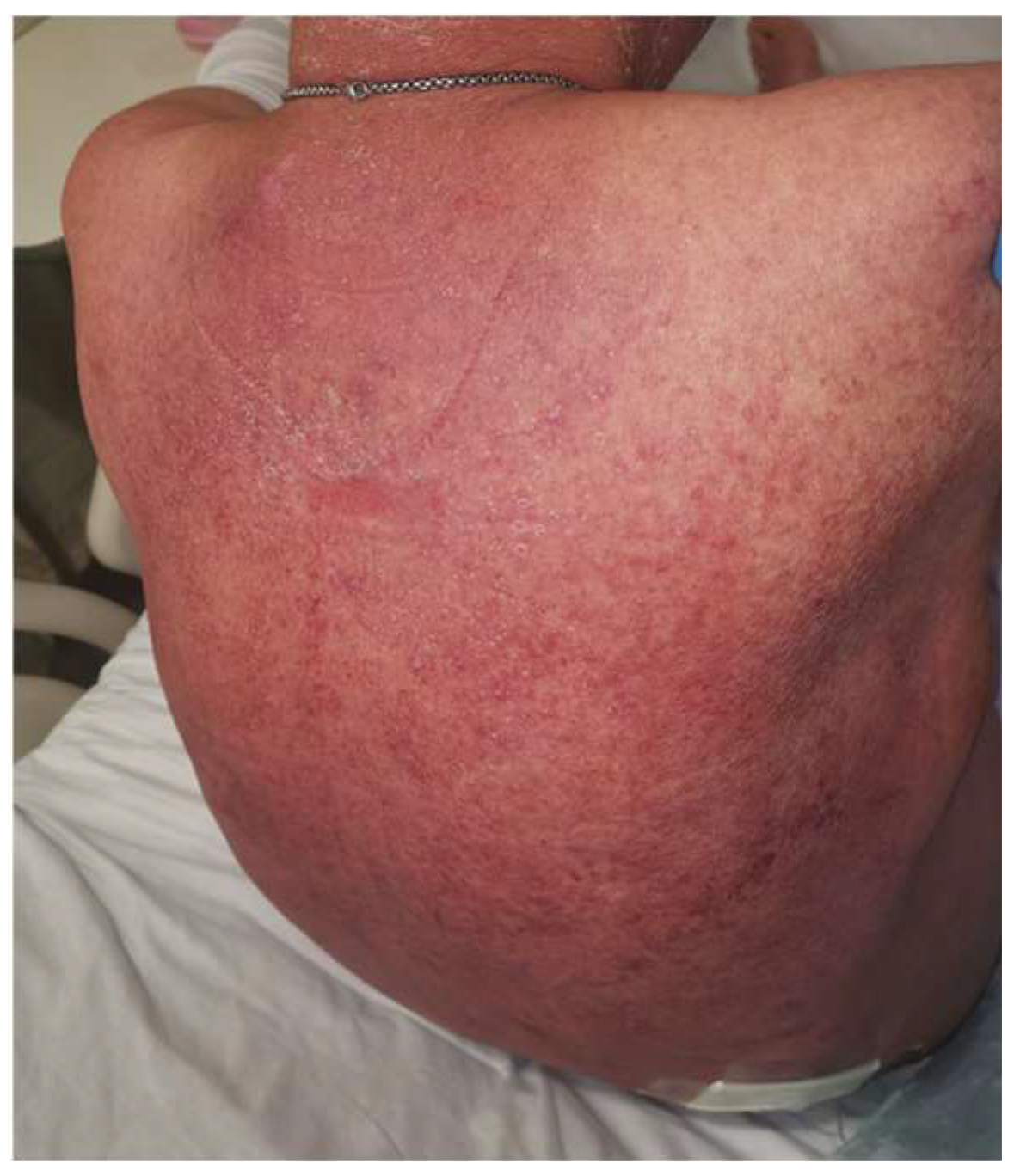
Patient 1, a 58-year-old Chinese male with well-controlled human immunodeficiency virus (HIV) infection on antiretroviral therapy (viral load < 20 copies/mL; CD4 count 534 cells/μL), hypertension, diabetes, and atrial fibrillation reported a prior delayed cutaneous reaction to an unidentified ICM in 2010. In 2022, he underwent computed tomography (CT) of the abdomen for acute abdominal pain, which revealed mild pancreatic tail swelling suggestive of pancreatitis. Despite once dose of intravenous (IV) hydrocortisone 100 mg premedication, he experienced a delayed reaction after iohexol, beginning with flushing and erythema at 20 h, followed by high-grade fever (Tmax 39–40 °C) and rigors at 46 h, and progressive hypotension requiring vasopressor support at 70 h. His cutaneous eruption worsened progressively, evolving into generalized diffuse, confluent erythematous eruption (
Figure 1). Skin biopsy showed psoriasiform dermatitis, accompanied by superficial perivascular chronic inflammation and dermal red cell extravasation. No granulomas or malignancy were seen, and direct immunofluorescence for IgG, IgA, IgM, C3, C1q and fibrinogen was negative. No infectious etiology was identified despite blood cultures (two sets), urine culture, and stool cultures. IL-6 levels were markedly elevated during the sepsis workup and normalized after 10 weeks. He continued to require vasopressor support despite empiric broad-spectrum antibiotics, with rapid clinical improvement observed only after initiation of high-dose intravenous hydrocortisone (50 mg every 6 h for one week), followed by oral prednisolone taper over the subsequent two weeks.
In 2024, he experienced a similar reaction 12 h after re-exposure to iohexol for evaluation of anorexia, again despite premedication with once dose hydrocortisone. Serum tryptase was normal, and all cultures remained negative. Both episodes required high-dose corticosteroids for resolution. Key laboratory findings, including cytokine and tryptase levels, are summarized in
Table 1.
Patient 2, a 37-year-old Malay female with end-stage renal failure on hemodialysis and non-ischemic cardiomyopathy had a remote history of rash to iohexol. During an arteriovenous fistuloplasty, she received iodixanol following premedication with a single dose of IV hydrocortisone 100 mg. Eight hours later, she developed high-grade fever (Tmax 38–39 °C), rigors, flushing, and hypotension requiring vasopressors. Repeated septic workups, including blood cultures (two sets) and urine culture, were negative. She did not improve with empiric antibiotics but responded promptly to intravenous hydrocortisone (100 mg every 8 h for one week). Review of her medical history revealed four prior episodes in 2020 characterized by fever and exanthem (with or without hypotension) 6–8 h after dialysis catheter exchange involving iodinated contrast, all with negative cultures. Skin prick and intradermal testing with iodixanol (320 mg/mL) and iohexol (350 mg/mL) up to 1:1 dilution were negative, including delayed readings at 48 h. A drug provocation test (DPT) with incremental iodixanol doses up to a cumulative 100 mL produced no immediate reaction; however, two hours post-test, she developed generalized pruritus and exanthema, which resolved with IV hydrocortisone (100 mg every 8 h for two days) followed by oral prednisolone (30 mg daily for five days) and antihistamines, without progression to a severe reaction.
Patient 3, a 70-year-old Chinese male had previously developed pruritus and rash within 15 min of iohexol exposure. Skin prick testing with 1:1 dilution of iohexol (350 mg/mL), iodixanol (320 mg/mL), and iopromide (370 mg/mL) were negative. Intradermal testing with a 1:1 dilution of iopromide produced an increment of 3 mm × 3 mm wheal without surrounding erythema, while results were negative for iohexol and iodixanol. No follow-up drug provocation test was performed, as approximately five minutes after completion of the intradermal test at 1:1 concentration, he developed facial pruritus without visible rash. Over the next hour, the pruritus intensified and became generalized. At 75 min, he developed generalized erythema and flushing, followed by severe rigors and hypotension five hours later, requiring inotropic support in the medical high dependency unit. He recovered after a week of intravenous hydrocortisone administration (100 mg every 8 h), followed by prednisolone taper over 2 weeks. Investigations showed normal serum tryptase (4.7 μg/L at 3 h; 3.3 μg/L at 72 h) and sterile blood and urine cultures. Cytokine assays demonstrated elevated IL-6, IL-8, IL-1β, and IFN-γ levels during the acute phase, which normalized over 72 h.
4. Discussion
Immediate reactions to ICM remain controversial. Some are IgE-mediated, while others result from direct mast cell or basophil activation through MRGPRX2 or complement pathways [
2]. CRS is characterized by aberrant release of proinflammatory cytokines, primarily IL-6 and IFN-γ, as well as immunoregulatory cytokines such as IL-10 which is elevated as part of the counter-regulatory response to inflammation, leading to fever, hypotension, and systemic inflammation [
4]. Although well described in settings such as chimeric antigen receptor (CAR) T-cell therapy and severe infections, CRS-like reactions have also been reported in non-infectious and non-oncologic contexts, including drug exposures. Notably, a case series describing amoxicillin–clavulanate–induced CRS in two pediatric patients highlights that medications can trigger cytokine-mediated inflammatory responses independent of malignancy-directed therapies [
9]. These observations broaden the recognized triggers of CRS beyond infections and immunotherapy.
CRS-like reactions following ICM appear to be rare and likely underrecognized, as their presentation with fever, hypotension, and elevated inflammatory markers may closely mimic sepsis and therefore be misattributed to infection. Reports of delayed febrile or flu-like syndromes after ICM exposure exist [
10,
11,
12], but these reactions are generally mild, and cytokine profiling has seldom been performed. Our report extends current classifications by demonstrating a cytokine signature consistent with CRS-like reactions in the context of ICM exposure.
Across all three patients, several consistent clinical and laboratory features were observed, including fever and rigors, exanthema, normal serum tryptase levels, negative microbiological investigations, leukocytosis, markedly elevated CRP, and rapid clinical improvement following corticosteroid therapy. The absence of elevated tryptase argues against classical anaphylaxis, which is predominantly mast cell-mediated. Furthermore, the presence of high fever, rigors, and hypotension is atypical for T cell–mediated delayed hypersensitivity reactions, which more commonly manifest with delayed cutaneous eruptions and organ-specific involvement rather than systemic inflammatory instability. The constellation of high fever, rigors, hypotension, and markedly elevated inflammatory markers supports a cytokine-driven systemic inflammatory response distinct from established IgE- or T cell–mediated mechanisms.
It is important to consider alternative immunological mechanisms that could account for the observed clinical features. IgE-mediated anaphylaxis typically presents with rapid onset of urticaria, angioedema, bronchospasm, and hypotension, with elevated serum tryptase reflecting mast cell degranulation. In our patients, there was no demonstrable rise in serum tryptase, arguing against significant mast cell activation. Furthermore, the clinical course characterized by delayed onset of high-grade fever, rigors, and progressive hypotension developing over hours is atypical for IgE-mediated reactions. Leukotriene-mediated reactions, while capable of producing cutaneous and respiratory symptoms, do not typically cause high-grade fever, rigors, or the significant persistent hypotension observed in our patients. We acknowledge that basophil activation testing and histamine release assays to exclude IgE-mediated reactions were not performed in our patients and represent a limitation of this retrospective series.
An alternative mechanism worth considering is endotoxin-mediated activation. However, several observations argue against exogenous endotoxin contamination of the contrast media. The reactions in our patients occurred over different time periods, months to years apart, and involved different batches of ICM from different manufacturers (iohexol and iodixanol). If the reactions were attributable to endotoxin-contaminated contrast media, a cluster of similar reactions among other patients receiving the same batch would be expected, which was not observed.
The question of why the vast majority of patients tolerate ICM without developing CRS-like reactions remains unanswered. It is plausible that individual host factors, including genetic polymorphisms in innate immune pathways, baseline inflammatory status, or comorbidities, may predispose certain individuals to an exaggerated cytokine-mediated response. Notably, Patient 1 had well-controlled HIV infection, a condition associated with chronic immune activation even under antiretroviral therapy, and Patient 2 had end-stage renal failure, which is associated with uremia-related immune dysregulation. These underlying conditions may lower the threshold for cytokine-mediated inflammatory responses upon ICM exposure. Further research, including genetic and immunophenotyping studies, is needed to identify risk factors and susceptibility markers for this reaction phenotype.
Both CRS and delayed-type radiocontrast hypersensitivity reactions exhibit a broad spectrum of cutaneous manifestations, ranging from mild erythematous or maculopapular eruptions to severe presentations such as erythroderma and Stevens–Johnson syndrome/toxic epidermal necrolysis–like reactions [
13,
14,
15]. Similar to patient 1, erythrodermic psoriasis triggered by radiologic contrast media has been previously reported [
16], supporting the ability of contrast exposure to precipitate severe systemic inflammatory skin reactions.
Skin testing is recommended in evaluating ICM reactions for both immediate (IgE-mediated) and non-immediate (T cell–mediated) mechanisms [
17,
18,
19]. In our case series of these three patients, the underlying mechanism was initially unclear and thought to be either an immediate or non-immediate phenotype; therefore, skin testing was performed as part of the diagnostic evaluation. However, in Patient 3, a CRS-like episode was precipitated by intradermal testing alone, highlighting the need for caution. This observation challenges assumptions regarding the safety of skin testing and demonstrates its potential to provoke systemic inflammatory responses. Whether the intradermal test represented a false-positive result remains uncertain, as repeat testing was not performed. Repeat intradermal testing was not pursued due to the severity of the systemic reaction precipitated by the initial test, which required inotropic support and prolonged hospitalization. In the interest of patient safety, the clinical team and patient jointly decided against repeating the skin testing. It is also worth noting that a positive intradermal test is conventionally defined as the development of a wheal ≥3 mm in diameter accompanied by surrounding erythema (flare). In this patient, although a wheal response was observed, the absence of a surrounding flare indicates that the test did not meet the standard criteria for a definitive positive result.
The timing of drug-associated CRS has been reported to range from minutes to several days after exposure, with variable severity [
20]. Fever is a hallmark feature, and the presentation often mimics infection, with symptoms such as rash, nausea, vomiting, azotemia, and transaminitis. The considerable clinical overlap between infection and CRS highlights the importance of maintaining a high index of suspicion, early exclusion of sepsis, and prompt corticosteroid therapy to control inflammation.
All three patients responded rapidly to high-dose corticosteroids and supportive care, consistent with current management guidelines for the treatment of CRS [
21,
22]. Although premedication with corticosteroids and antihistamines can reduce the risk of hypersensitivity reactions to iodinated contrast media (ICM), its protective effect is not complete [
23]. The development of CRS-like reactions in our patients despite premedication suggests that prophylactic strategies with corticosteroids may be inadequate to suppress cytokine-driven inflammatory responses. The failure of standard prophylactic corticosteroid doses to prevent CRS-like reactions, contrasted with the efficacy of sustained high-dose corticosteroid therapy, suggests that the intensity and duration of immunosuppression required to control cytokine-driven inflammation exceeds that provided by single-dose premedication protocols designed to attenuate immediate type hypersensitivity reactions.
Whether patients who experience CRS-like reactions to iodinated contrast media (ICM) can safely undergo future iodinated contrast exposure remains uncertain. At present, there is no established prophylactic strategy for preventing recurrence of cytokine-mediated reactions. In other CRS settings such as COVID-19 and CAR T-cell therapy, IL-6 receptor blockade with tocilizumab has demonstrated efficacy [
24,
25,
26]; however, its role in contrast-associated CRS remains entirely unexplored.
Several limitations of this case series should be acknowledged. Cytokine profiling was not uniformly available across all three patients. For Patient 1, only IL-6 was measured during the index episode using stored serum, and no cytokine data were available for Patient 2 due to the retrospective nature of this study and the absence of stored samples. The detailed cytokine panel for Patient 3 was obtained in the setting of an intradermal test–provoked reaction, which may not be directly representative of reactions occurring after full intravenous contrast administration. In addition, basophil activation testing, histamine release assays, and immunoglobulin levels (including total IgE) were not systematically obtained and represent important investigations for future prospective studies. Despite these limitations, the convergence of clinical features across all three patients, including fever, rigors, hypotension, normal tryptase, negative cultures, and corticosteroid responsiveness, combined with the available cytokine data showing IL-6 elevation in Patients 1 and 3, provides a basis for the hypothesis that these reactions are cytokine-mediated rather than IgE- or T cell–driven. This case series is hypothesis-generating rather than mechanistically definitive.
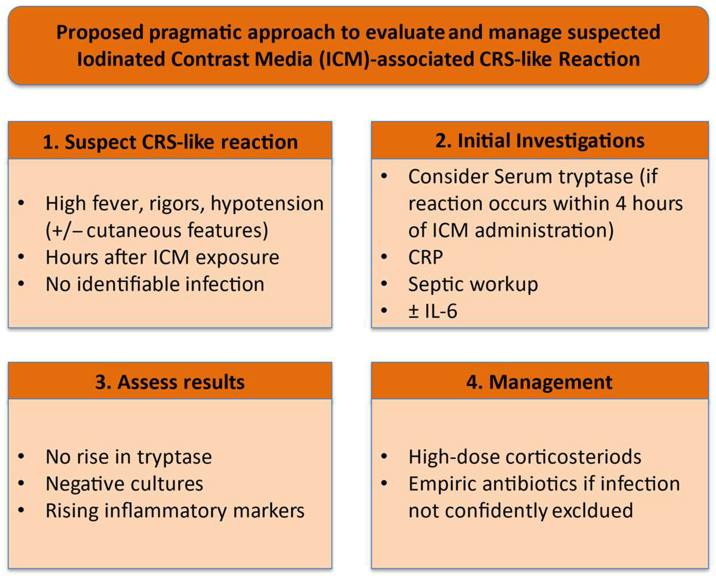
Based on the clinical patterns observed in this case series, we propose a pragmatic approach (
Figure 2) to the evaluation of suspected ICM-associated CRS-like reaction. First, CRS-like reactions should be suspected when systemic inflammatory features such as high-grade fever, rigors, and hypotension develop hours after ICM exposure in the absence of an identifiable infectious source. Second, initial investigations should include serum tryptase (particularly if the reaction occurs within the first 4 h of ICM administration to exclude IgE mediated reaction), inflammatory markers such as C-reactive protein (CRP), and a comprehensive septic workup, including blood and urine cultures as well as other site-specific cultures as clinically indicated; measurement of interleukin-6 (IL-6) may be considered where feasible. Third, in the absence of a rise in tryptase and with negative microbiological studies despite escalating inflammatory markers, a CRS-like reaction should be considered. Fourth, prompt initiation of high-dose intravenous corticosteroids is recommended, with empiric antimicrobial coverage where infection cannot be confidently excluded. Although this proposed framework requires prospective validation, it may provide a useful clinical guide for recognising and managing this uncommon presentation.





 ,
,