Review

Molecular Programming of Stem-Cell Differentiation: From Soluble Factors to Agonist Antibodies

Department of Biological Sciences and Biotechnology, Hannam University, Daejeon 34054, Republic of Korea
*
Author to whom correspondence should be addressed.
Immuno 2026, 6(1), 11; https://doi.org/10.3390/immuno6010011
Submission received: 24 December 2025 / Revised: 27 January 2026 / Accepted: 30 January 2026 / Published: 31 January 2026

Abstract

Stem-cell differentiation technologies have traditionally relied on recombinant growth factors, cytokines, and morphogens to initiate and guide lineage specification toward clinically relevant cell types. These approaches have enabled substantial progress in regenerative medicine, as exemplified by recent advances in cell-replacement therapies for Parkinson’s disease, type 1 diabetes, and retinal degeneration. However, protein-based ligands and soluble factors are often limited by short half-lives, pleiotropic signaling, condition-dependent effects, and challenges in achieving precise spatial and temporal control in scalable systems. In this review, we survey differentiation strategies driven by administered substances, organizing the field into five material-centric modules: recombinant growth factors, cytokines, morphogens, exogenous ligands, and agonist antibodies. For each module, we summarize mechanistic principles, representative studies, controllable variables, and translational considerations. While growth factors, cytokines, morphogens, and exogenous ligands remain central tools for directing lineage commitment and maturation, recent studies indicate that agonist antibodies offer an additional and distinct means of controlling differentiation outcomes. These antibodies can function as receptor agonists while also imparting tissue-selective effects, enabling lineage specification with coordinated spatial targeting. By focusing on differentiation methods driven by administered molecules and excluding direct physical stimulation or complex 3D constructs, this review provides a framework that is particularly relevant to immunology and translational practice. We highlight agonist antibody-based induction as an emerging strategy that complements established ligand-based approaches and may broaden the design space for clinically applicable stem-cell differentiation.

1. Introduction

Technologies for directing stem and progenitor cell differentiation continue to advance along four interlocking pillars: (i) recombinant growth factors and morphogens; (ii) small-molecule modulators; (iii) biomaterials and mechanotransduction; and (iv) genetic and synthetic-biology tools [1,2,3,4,5]. Among these, exogenously applied biochemical signals remain the most widely adopted and clinically tractable means of controlling lineage specification in scalable systems. Recent work has placed particular emphasis on how biomaterial-induced mechanical regulations interface with metabolic reprogramming, and on engineering spatiotemporal signal patterning in 3D environments [6]. Together, these advances reflect a transition from trial-and-error culture condition optimization toward more rational control of signaling magnitude, duration, and sequence during differentiation.
On governance and ethics, the International Society for Stem Cell Research (ISSCR) Guidelines (2021) underwent targeted updates in 2025 to address stem cell-based embryo models and related oversight. These revisions underscore growing expectations for standardization, reproducibility, and data transparency as differentiation platforms advance toward clinical translation, particularly in complex developmental and immunology-facing applications [7,8,9,10].
Looking ahead, we foresee four converging directions: (i) integrated 3D scaffolds/organoids/co-cultures with precise delivery (gradients, staged release); (ii) process optimization that couples mechanotransduction with metabolic control to improve efficiency and maturation; (iii) combination regimens that include immune modulation; and (iv) programmable signaling designs built from antibodies, multi-ligand cocktails, and gene circuits [11,12,13,14,15,16]. Antibody-based induction is being increasingly studied as a conceptually distinct strategy. By mimicking morphogens and controlling receptor clustering geometry, antibodies offer a route to encode spatial and temporal signal logic with administered molecules alone potentially without additional physical platforms [17,18,19,20,21,22,23].
In this review, administered biochemical signals are treated as the primary drivers of stem-cell differentiation, while mechanical cues and three-dimensional context are referenced only as contextual modifiers that influence how these signals are interpreted, rather than as independent induction modalities.
To maintain a focused scope relevant to immunology and translational practice, this review deliberately excludes direct physical or biophysical stimulation, environmental conditioning, and composite 3D or organoid frameworks as primary drivers of differentiation. Instead, we concentrate on differentiation methods driven by administered substances and organize the field into five material-centric modules: (1) recombinant growth factors; (2) cytokines; (3) morphogens; (4) exogenous ligands (including nuclear-receptor, peptide, and nucleic-acid ligands); and (5) agonist antibody-driven induction. Each section is discussed using a common framework that highlights functional roles, representative studies, key control variables, and translational considerations.
Across all administered-signal modalities, differentiation outcomes are governed by a common set of parameters, including ligand dose, timing and duration of exposure, sequence of application, and mode of delivery, whether soluble, matrix-associated, or locally confined. While these variables are shared, each induction strategy relies on them in distinct ways, shaping both its strengths and limitations in practice.
To improve clarity and reduce repetition across sections, the shared control variables that influence differentiation—such as dose and exposure time, pulse versus tonic signaling, and the balance between specificity and pleiotropy—are summarized in a unified table (Table 1). The sections below, therefore, emphasize the modality-specific variables that distinguish each induction strategy and define their practical considerations.

2. Recombinant Growth Factors-Driven Induction

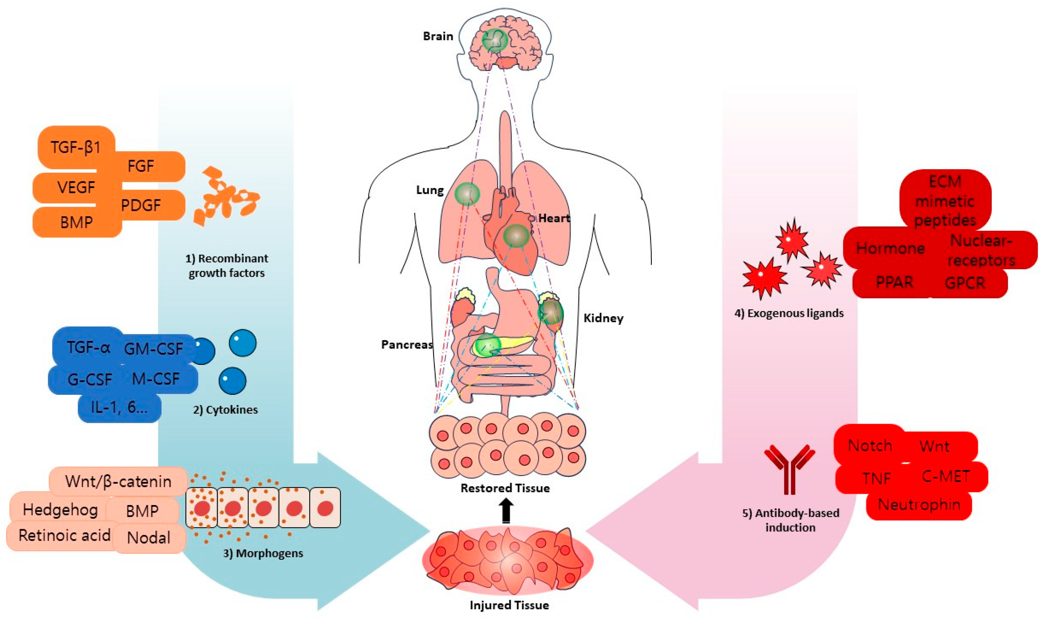
Recombinant growth factors provide the primary, receptor-proximal signals that initiate lineage programs in vitro and in vivo, moving pluripotent and adult stem cells from lineage priming toward specification and, ultimately, terminal differentiation. In most differentiation protocols, these factors establish the initial cell-fate path that subsequent signals further specify rather than replace. The subsequent sections on cytokines, morphogens, exogenous ligands, and agonist antibodies are positioned as complementary controls that tune duration, amplitude, and spatial presentation, align microenvironmental conditions, and reduce pleiotropy so that early fate decisions progress efficiently into stable lineage commitment and functional maturation. Taken as a whole, these modalities form a practical playbook in which growth factor signals establish trajectory, and the additional modules refine commitment, maturation, and immune compatibility across stem-cell differentiation protocols (Figure 1).
Recombinant growth factors are protein ligands that bind receptor tyrosine kinases or serine/threonine kinase receptors and activate downstream signaling to run lineage programs that drive differentiation. In the musculoskeletal differentiation, Transforming growth factor β1 (TGF-β1) functions early during chondrogenic condensation, promoting cell–cell aggregation and extracellular matrix remodeling through molecules such as fibronectin and N-CAM [24,25]. Bone morphogenetic proteins 2, 4, 7, and 9 guide osteochondral lineage commitment and maturation by engaging BMPR-I/II, which phosphorylate SMAD1/5/8, form SMAD4 complexes, and activate RUNX2, SP7, and DLX5. Non-canonical MAPK and PI3K signaling further modulates signal amplitude and duration, shaping maturation outcomes. [26,27,28]. Because the effects of TGF-β1 vary with timing and culture format in two- versus three-dimensional (2D/3D) systems, careful control of onset, duration, and termination is essential. Fibroblast growth factors (FGF) 2, 9, and 18, epidermal growth factor, platelet-derived growth factor, and vascular endothelial growth factor coordinate proliferation, angiogenesis, and tissue patterning to advance terminal differentiation. Controlled release of FGF-2 or FGF-18 increases bone formation and osteoblast expansion in vivo; delivering vascular endothelial growth factor (VEGF) followed by platelet-derived growth factor (PDGF) improves the maturation of neovessels. Given the short half-life of platelet-derived growth factor, delivery strategies that extend local retention are often required. Stage-sequenced combinations, such as insulin-like growth factor 1 (IGF-1) followed by bone morphogenetic protein 6 (BMP-6), can outperform single agents for osteoinduction [29,30,31,32].
In practice, growth factor–driven differentiation is highly sensitive to the identity and dose of the ligand, the timing and sequence of exposure during priming and maturation, and the mode of presentation, whether through soluble delivery or matrix-associated retention. Coordinated application of angiogenic and osteogenic factors further enables coupling of vascular development with tissue-specific maturation, providing a flexible design space for integrating patterning with functional differentiation.
A key distinction is the distinction between pulse and tonic exposure. Short, stage-specific pulses can initiate lineage programs, whereas prolonged tonic exposure often increases pleiotropy and promotes unwanted phenotypes. Because many growth factors have short half-lives, strategies that extend local retention, such as heparin- or perlecan-functionalized matrices, provide an additional strategy with which to improve reproducibility and efficacy.
Going forward, separating pulse and tonic exposure in space and time will be important to meter signaling strength and limit pleiotropy. Heparin- or perlecan-functionalized scaffolds and sulfated chitosan carriers provide effective means to extend growth factor half-life and localize activity. However, significant challenges persist, including the lack of predictive rules for optimal growth factor sequencing across cell sources, narrow therapeutic windows that separate productive differentiation from hypertrophic or fibrotic outcomes, and the difficulty in maintaining reproducible signaling gradients in scalable culture systems. Addressing these limitations will be essential for translating growth factor-driven induction strategies into robust and predictable differentiation platforms.

3. Cytokines-Driven Induction

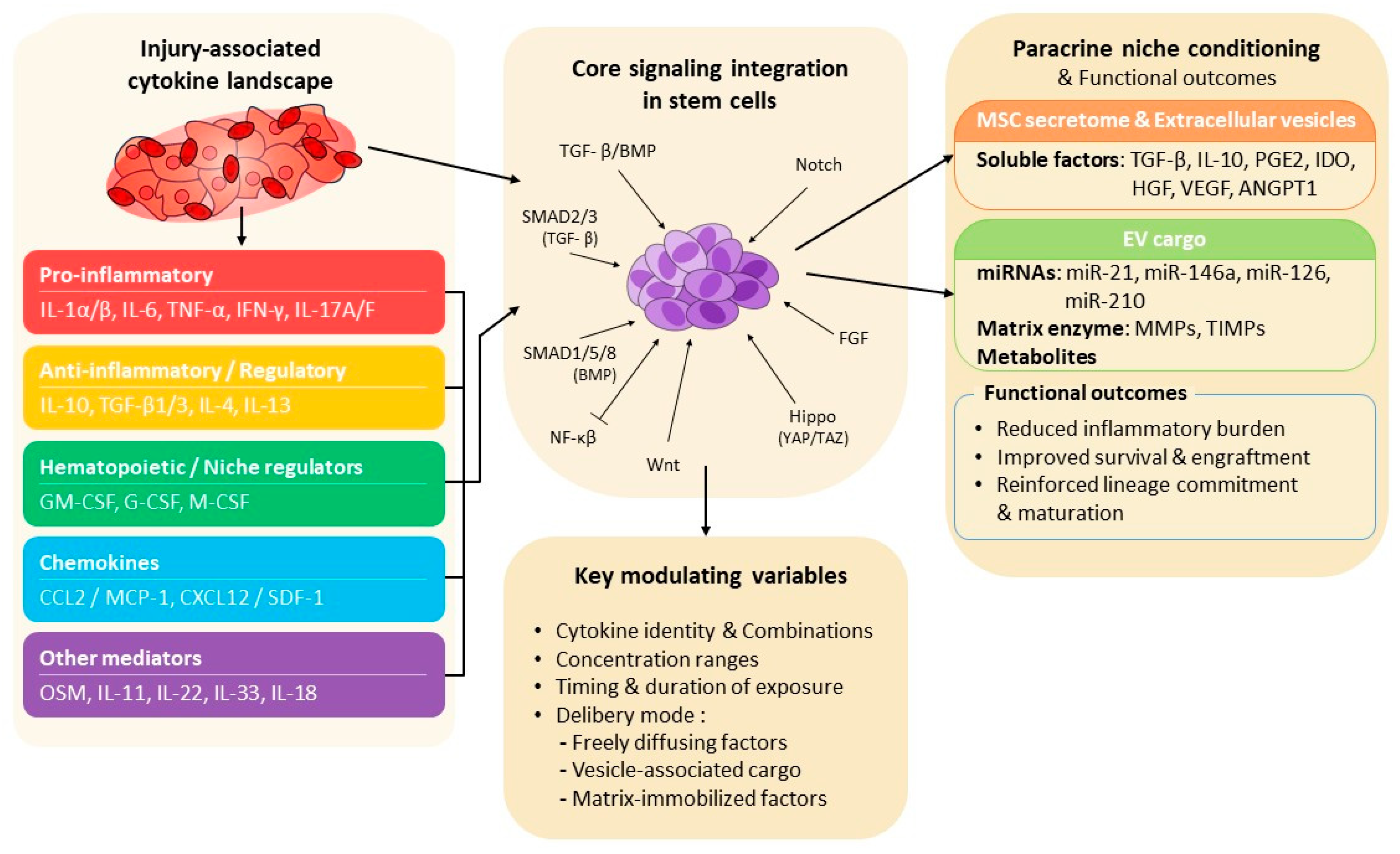
Cytokines guide stem-cell fate by turning lineage programs on or off and by reshaping the surrounding microenvironment. Key families include the TGF-β superfamily, such as bone morphogenetic proteins and Activin or Nodal, together with classic interleukins. In tissue engineering, cytokines function as core biochemical induction factors that set survival, proliferation, and differentiation. In contrast to recombinant growth factors, that primarily establish the lineage developmental path, cytokines act across multiple stages to bias commitment, stabilize newly formed phenotypes, and shape the local paracrine microenvironment through sustained signaling (Figure 2).
Early after tissue injury, interleukin 1 alpha, interleukin 1 beta, interleukin 6, tumor necrosis factor alpha(TNF-α), interferon gamma, interleukin 17A and 17F, interleukin 10, TGF-β 1 and 3, interleukin 4, interleukin 13, granulocyte-macrophage colony-stimulating factor(GM-CSF), granulocyte colony-stimulating factor(G-CSF), macrophage colony-stimulating factor(M-CSF), chemokines such as CCL2 or MCP-1 and CXCL12 or SDF-1, and mediators, such as oncostatin M, interleukin 11, interleukin 22, interleukin 33, and interleukin 18, collectively build pro- or anti-differentiation microenvironments that shape resident and transplanted cells for the long term [33,34,35,36,37]. These injury-associated cytokine conditions serve as well-established reference benchmarks for understanding how the level of inflammation influences stem-cell fate decisions in vivo and in engineered systems.
Mechanistically, the SMAD2 and SMAD3 branch and the SMAD1, SMAD5, and SMAD8 branch of TGF-β and bone morphogenetic protein signaling coordinate with Notch, Wnt, Hippo, and fibroblast growth factor pathways to align lineage commitment and maturation in pluripotent and adult stem cells [38,39]. In mesenchymal stem or stromal cell applications, secreted cytokines and extracellular vesicle cargo further reprogram the niche. Common mediators include TGF-β, interleukin 10, prostaglandin E2, indoleamine 2,3-dioxygenase, hepatocyte growth factor, vascular endothelial growth factor, and angiopoietin 1. Vesicles often carry microRNAs such as miR-21, miR-146a, miR-126, and miR-210, matrix enzymes including matrix metalloproteinases and tissue inhibitors of metalloproteinases, and metabolites [40,41,42]. Together, these biochemical signals promote immunosuppression through M2-like macrophage polarization, expansion of regulatory T cells, and dampening of NF-κB; stimulate angiogenesis through vascular endothelial growth factor and hepatocyte growth factor axes; tune extracellular matrix remodeling through the matrix metalloproteinase to tissue inhibitor balance; and support chemotaxis and homing through the SDF-1 to CXCR4 axis [43,44]. The overall effect is a reduction in inflammatory burden, improved survival and engraftment, and reinforcement of lineage commitment and maturation in vivo. Co-culture systems and conditioned media from differentiated tissues provide cytokine-rich paracrine environments that can steer embryonic stem cells and mesenchymal stromal cells toward chondrogenic or osteogenic outcomes, showing that sustained soluble exposure to cytokines and morphogens can partly substitute for direct cell–cell contact. In practice, researchers identify active factors using neutralizing antibodies, receptor antagonists, size fractionation, proteomics, and extracellular-vesicle analysis, implement gradients and staged delivery with controlled-release biomaterials, and set batch-to-batch quality metrics, such as absolute cytokine levels and functional bioassays, to support reproducibility and clinical applicability.
Key variables that can be controlled include the identity and combination of cytokines, their concentration ranges, the timing and duration of exposure, and whether signals are delivered as freely diffusing factors, vesicle-associated cargo, or matrix-immobilized factors.
In cytokine-driven induction, differentiation outcomes are particularly sensitive to inflammatory state and paracrine environment. The balance between pro- and anti-inflammatory cytokines, the duration of exposure to injury-mimetic signals, and the mode of delivery—freely soluble factors versus extracellular vesicle-associated cargo—strongly bias niche conditioning, immune modulation, and subsequent lineage stabilization. Accordingly, cytokines function less as primary fate determinants and more as milieu-setting regulators that influence survival, engraftment, and maturation once lineage paths have been initiated.
Several additional targets and regulatory pathways are frequently engaged. Activin and Nodal, together with bone morphogenetic protein and growth and differentiation factor branches, tune the balance between pluripotency and germ-layer entry [38,45]. Notch integrates with TGF-β, bone morphogenetic protein, and Wnt to stabilize tissues and adjust fate, although its net effect is strongly cell-state dependent [46,47]. The Hippo axis with YAP and TAZ senses matrix stiffness and cell density, and interacts with bone morphogenetic protein, Notch, and TGF-β pathways to bias differentiation. Because excessive Hippo activation can promote oncogenic or fibrotic responses, careful modulation is required [48,49].
Pro-inflammatory cytokines, such as interleukin 1 beta, TNF-α, and interferon gamma, are often used to license or prime mesenchymal stromal cells, which can enhance the activity of their extracellular vesicles and create repair settings in which differentiation proceeds more effectively [50,51]. From a delivery standpoint, controlled-release materials that immobilize or trigger factor release improve the timing and localization of cytokines and growth factors, supporting staged morphogenesis and more homogeneous outcomes. The transient pharmacological modulation of downstream pathway nodes offers an additional level of control, allowing for lineage biasing without sustained upstream pathway activation.
Despite these advances, challenges remain, including the pleiotropic and condition-dependent nature of cytokine signaling, difficulties in defining minimal effective combinations, and variability in cytokine production and stability across cell sources and manufacturing batches. Addressing these issues will be essential to translating cytokine-driven induction strategies into predictable and scalable differentiation platforms.

4. Morphogens-Driven Induction

Morphogens are secreted factors that diffuse to create concentration gradients and assign distinct cell fates according to dose and exposure time. In stem-cell systems, canonical families include Hedgehog, Wnt/β-catenin, bone morphogenetic proteins within the TGF-β superfamily, and FGFs; retinoic acid operates as a morphogen-like small molecule. These signals activate lineage-specific gene networks and coordinate the timing of commitment with tissue morphogenesis [52,53,54,55,56]. Unlike growth factors and cytokines, morphogens specify positional information, enabling cells to respond to relative signal strength rather than absolute ligand abundance.
Functionally, morphogens differ from growth factors and cytokines in that they encode positional and temporal information, allowing cells to interpret relative signal strength and duration to resolve pattern and identity. Because physiologically relevant differentiation reflects the combined action of soluble morphogens, cytokines, and biophysical inputs such as substrate stiffness and topography, rigorous spatial and temporal control of presentation is essential for reproducibility. BMP and Wnt or Hedgehog signaling are central to musculoskeletal patterning, while FGF signaling supports pluripotency yet also sculpts tissue pattern in a dose- and stage-dependent manner.
These principles are well illustrated by studies of osteogenic and patterning morphogens in the musculoskeletal system. BMP-2, BMP-4, BMP-6, BMP-7, and BMP-9 act as archetypal osteogenic morphogens: by engaging type I and type II BMP receptors they phosphorylate SMAD1, SMAD5, and SMAD8, assemble SMAD4 complexes, and activate osteochondral transcriptional programs driven by RUNX2, SP7, and DLX5. Depending on timing and combinatorial signaling environment, the same axis can promote osteogenic differentiation or chondrocyte maturation and hypertrophy, with non-canonical MAPK and PI3K inputs shaping amplitude and duration. Sonic Hedgehog signaling proceeds through Patched and Smoothened to GLI transcription factors and regulates the balance between self-renewal and differentiation in a gradient- and time-dependent manner. [28,57,58,59,60]. Co-culture systems and conditioned media supply morphogenetic factors from neighboring differentiated cells and can steer embryonic stem cells and mesenchymal stromal cells toward lineage commitment and the endochondral program, demonstrating that sustained soluble exposure can partially substitute for direct cell–cell contact. These systems provide reference models for how graded morphogen exposure determines fate decisions in both developmental and bioengineered environments.
Additional targets often pair with these morphogens. Wnt/β-catenin, through Frizzled (FZD) and LRP5/6 with downstream Dishevelled (DVL), drives tissue-specific transcription and cross-talks with BMP and Hippo. The FGF–FGFR axis signals through PI3K–AKT and RAS–MAPK to support pluripotency and pattern brain and skeleton [59,61,62,63,64]. On the delivery side, biomaterial-based controlled release and immobilization enable true gradients and stage-specific presentation, bringing in vitro delivery closer to developmental patterning in vivo.
Morphogen-driven induction depends primarily on spatial and temporal gradient properties. Gradient steepness threshold concentration ranges and the duration of exposure above or below specified signaling thresholds together instruct positional identity and pattern formation. Coupling soluble morphogens with matrix-bound signals and local mechanical environment further stabilizes lineage specification. Rather than acting permissively, morphogens function instructively by enabling cells to integrate relative signal strength and timing. This distinguishes morphogen-based induction from growth factor or cytokine-driven approaches, which rely more heavily on absolute ligand levels.
Future work should center on spatiotemporal gradient engineering. Programmable scaffolds with tunable kinetics and diffusion can deliver sequenced cues that initiate condensation with early TGF-β and BMP, then transition to BMP-dominant signals for maturation, thereby emulating embryonic patterning. Rational multi-morphogen cocktails combining BMP, Wnt, Hedgehog, and FGF pathways, together with modulators of Notch and Hippo signaling, may enhance productive crosstalk while limiting pleiotropic effects. Paracrine engineering approaches using co-culture, conditioned media, or extracellular-vesicle-rich formulations are also likely to amplify endogenous differentiation responses. Aligning morphogen gradients with extracellular-matrix ligands and mechanical inputs should further stabilize lineage specification. Finally, transient and stage-specific application of small-molecule agonists or antagonists targeting bone morphogenetic protein, Wnt, Hedgehog, and Hippo nodes provides a complementary control mechanism, enabling lineage biasing without sustained pathway rewiring. A canonical example is chemically controlled cardiomyocyte induction from human pluripotent stem cells, which relies on a brief pulse of Wnt activation followed by timely Wnt inhibition to consolidate cardiac identity [65,66].
Remaining challenges include defining quantitative gradient thresholds across cell sources, maintaining stable gradients in three-dimensional and scalable systems, and preventing unwanted lineage drift or hypertrophy caused by prolonged or excessive morphogen exposure.

5. Exogenous Ligands-Driven Induction

Exogenous ligands beyond classic growth factors include hormonal and nuclear-receptor ligands, designed peptides, and nucleic-acid ligands. These agents can program fate directly or stabilize acquired function [67,68,69,70]. In differentiation workflows, exogenous ligands are frequently used to complement protein-based factors by providing intracellular access and temporally confined signaling control. Functionally, exogenous ligands differ from recombinant proteins in that they can be delivered intracellularly or locally synthesized, enabling transient yet potent control over signaling and gene expression without continuous extracellular supplementation.
In tissue engineering, polynucleotides such as mRNA, miRNA, and siRNA are widely used to modulate intracellular gene expression. ECM-mimetic peptides serve as instructive ligands, often used alongside proteins, to enhance differentiation efficiency and maturation. Beyond nucleic-acid and peptide formats, hormonal and nuclear-receptor ligands constitute a well-established class of exogenous ligands that act through intracellular receptors to bias differentiation and promote functional maturation. Delivery of BMP-2 mRNA or anti-miR-221 elevates osteogenic and chondrogenic programs without the need for exogenous protein, while synthetic mRNA supports strong yet transient in situ production of instructive proteins. Ligands engaging PPAR signaling or thyroid hormone signaling (T3) are frequently applied at later stages of differentiation to reinforce metabolic identity, structural maturation, and tissue-specific function [71,72,73,74]. Immobilizing ECM-derived motifs within matrices provides integrin ligands that support survival and, when paired with morphogen signals, promote lineage specification. Endocrine and hormonal ligands are frequently combined with growth factors to advance maturation and function. Panels of miRNA or siRNA, including miR-29a and miR-7, regulate ECM synthesis, neurite outgrowth, and osteogenesis in a tissue-specific manner. GPCR ligands further expand the exogenous ligand toolkit by enabling rapid, tunable modulation of intracellular second-messenger pathways that influence survival, migration, and lineage bias [75,76]. Synthetic mRNA can also be used to express surface molecules that aid homing while reinforcing differentiation programs [77,78,79,80,81,82]. Together, these approaches represent how non-protein ligands can substitute for, amplify, or temporally confine traditional growth factor signaling.
For exogenous ligand-driven induction, molecular format is a dominant design parameter. Whether signals are delivered as peptides, synthetic mRNA, miRNA, or other nucleic-acid ligands determines expression amplitude, persistence, and innate immune engagement. For hormonal, nuclear-receptor, and GPCR ligands, dose, exposure timing, and receptor coupling define whether signaling acts instructively to initiate lineage programs or permissively to stabilize and mature differentiated states. These properties, together with dose and spatial presentation through matrix immobilization or localized delivery, influence whether exogenous ligands act instructively to initiate fate programs or permissively to stabilize and mature previously specified lineages. Intracellular activity profiles therefore distinguish exogenous ligands from extracellular protein signals, enabling transient pathway activation that can bias lineage outcomes without sustained receptor activation.
Future directions emphasize gene-coded ligand strategies, such as mRNA or miRNA, to replace or complement proteins with precisely controlled, on-site and time-limited expression. Peptidomimetic libraries that recapitulate receptor tyrosine kinase, Notch, and integrin engagement may further improve stability, manufacturability, and cost efficiency. Established hormonal, nuclear-receptor, and GPCR ligands provide a conceptual and practical foundation for integrating chemically defined exogenous ligands into stage-wise differentiation playbooks. Stage-wise differentiation playbooks that pair hormonal, peptide, and nucleic-acid ligands with pathway modulators offer a route to progressive maturation while limiting pleiotropy and improving reproducibility. Outstanding challenges include variability in transfection efficiency, avoidance of innate immune activation by nucleic acids, definition of safe and effective dosing windows across cell types, and integration of these ligands into scalable and clinically compliant manufacturing workflows.

6. Agonist Antibodies-Driven Induction

Agonist antibodies selected from combinatorial libraries can function as surrogate ligands for cell-surface receptors and membrane proteins, initiating differentiation programs while being simultaneously associated with tissue-level homing. In contrast to other applied ligands, agonist antibodies remain fully drug-like while actively shaping the ligand-receptor interface geometry and avidity. This property allows for fate decision and spatial targeting to be coordinated within a single molecular input.
Importantly, this mode of action is most relevant in differentiation settings where spatial restriction, lineage stabilization, or tissue-selective signaling are required, rather than in early germ-layer induction or highly standardized, large-scale differentiation workflows. A clear comparison with established differentiation modalities is essential for placing agonist antibodies in proper context. Recombinant growth factors and morphogens remain the most effective tools for early germ-layer induction and broad lineage priming, particularly in highly scalable and cost-sensitive differentiation workflows. Small-molecule pathway modulators likewise offer clear advantages in manufacturability, batch consistency, and rapid temporal control, making them well suited for early-stage and industrial-scale protocols. Agonist antibodies are therefore not expected to replace these approaches in the near term for early developmental patterning or fully standardized production. Rather, their strengths emerge at later stages of differentiation, where lineage stabilization, spatially restricted signaling, and tissue-selective activity become limiting factors for conventional ligands. In this setting, agonist antibodies are best viewed as complementary tools that occupy a distinct niche within stem-cell programming strategies, rather than as direct substitutes for existing modalities.
At this stage, it should be noted that many agonist antibodies have been identified through phenotypic screening, with target receptors and pathways clarified only after functional effects on differentiation or migration were observed. In some cases, these molecular targets remain only partially defined. This reflects both the opportunity and the current limitation of antibody-based approaches, which can uncover unexpected signaling dependencies but often require follow-up mechanistic analysis.
Compared with soluble growth factors and cytokines, agonist antibodies tend to generate more spatially confined signaling. This arises, in part, from their high specificity for defined cell-surface targets and their ability to engage receptors in particular clustering arrangements at the plasma membrane. Because effective signaling often depends on receptor density and local organization, antibody-mediated activation is favored in tissues where the target receptor is sufficiently expressed and appropriately organized. In contrast to freely diffusible ligands that broadly stimulate receptors across many cell types, agonist antibodies act through target-restricted binding and more limited spread, which reduces pleiotropic effects and off-target activation. Moreover, when antibodies are identified through in vivo migration- or function-based selection strategies, tissue tropism can emerge in selected antibody target contexts, allowing for lineage instruction and organ-specific homing to be linked within a unified antibody-mediated signal.
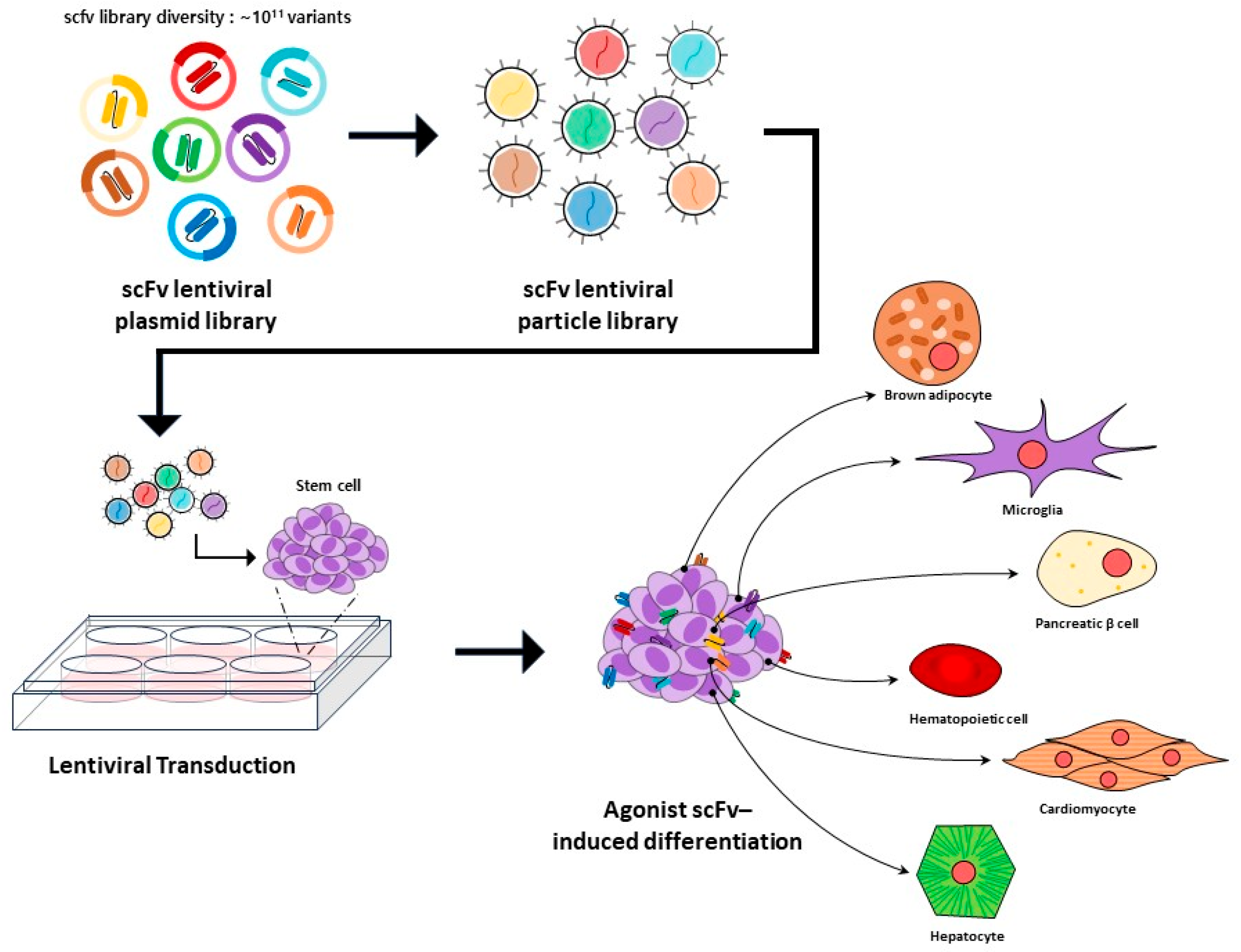
Previous work has shown that a single antibody input can convert human and mouse stem or progenitor cells into three distinct lineages with organ tropism: microglia-like cells that traffic to the brain; brown adipocyte-like cells that traffic to the heart; and beta-like cells that traffic to pancreatic islets (Figure 3) [83,84,85,86,87,88,89,90,91,92]. These observations suggest that antibody binding can couple lineage specification with tissue targeting rather than by acting solely as a differentiation signal. Conceptually, this mode of induction extends beyond individual case studies and is consistent with the agonist antibody engagement of diverse cell-surface signaling systems, including pathways traditionally activated by juxtacrine ligands, morphogens, and receptor tyrosine kinases.
Beyond the individual case studies, the screening strategy shown in Figure 3 points to a broader strength of agonist antibody-based approaches. High-throughput antibody screening makes it possible to examine cell-surface signaling in stem-cell differentiation in a direct and functional way, without first needing to predict which receptors or pathways are involved. When antibodies are selected based on observable outcomes—such as lineage conversion, cell migration, or tissue engraftment—this process can reveal surface targets and signaling behaviors that are often missed by ligand-focused experiments or transcriptome-driven analyses. Importantly, receptor engagement is evaluated side by side with its phenotypic consequences, allowing for surface signaling events to be linked more clearly to fate decisions and cell behavior. In this sense, antibody screening provides a practical and scalable means to broaden target discovery while deepening our understanding of how receptor-level signaling shapes stem-cell programming.
Mechanistic analyses have begun to reveal unconventional targets on stem-cell surfaces. Iodotyrosine deiodinase (IYD), behaves as a non-canonical receptor in stem cells. Agonism at IYD reroutes thyroid-hormone metabolism and the thermogenic axis, providing a coherent explanation for brown adipocyte-like differentiation and associated metabolic reprogramming. In representative reports, agonist antibody engagement of IYD in human progenitor cells has been associated with the acquisition of a brown adipocyte-like program and preferential homing to cardiovascular tissue in vivo. Notably, the tissue tropism observed in these systems should not be interpreted as a universal or purely direct targeting mechanism. In the examples reported so far, antibody engagement may influence localization through both direct receptor-mediated effects and indirect changes that accompany differentiation such as shifts in chemokine receptor expression, adhesion properties, or metabolic state. Further studies will be needed to clarify the relative contribution of these mechanisms. IYD agonism upregulates thermogenic gene expression and drives the downstream differentiation process [83,88]. This example illustrates how antibody engagement of non-classical surface targets can link metabolic rewiring to lineage commitment. Beyond this specific metabolic axis, related studies have demonstrated that agonist antibodies can similarly substitute for canonical ligands in pathways such as Notch and Wnt/β-catenin, in which receptor clustering geometry and signaling strength critically determine cell fate outcomes [93,94,95].
A second example involves the agonist antibody and the generation of microglia-like cells. In vivo selection based on migration identified an agonist antibody that converts bone marrow-derived progenitors into microglia-like cells that migrate to the brain. In an Alzheimer’s disease model, this conversion and trafficking coincided with a reduction in amyloid beta burden, consistent with functional integration of the induced cells within the central nervous system milieu [84]. A third example is the P1 antibody, which instructs beta-like differentiation while imparting islet tropism. This agonist antibody drives human CD34+ hematopoietic stem and progenitor cells and murine bone marrow toward pancreatic islets and activates a beta-like program in the absence of exogenous cytokines [90]. Together, these cases represent canonical exemplars of agonist antibody-driven induction, in which fate specification and homing are observed to co-occur in these examples. Beyond these specific systems, agonist antibodies targeting neurotrophin receptors, c-MET, and members of the TNF receptor family further support the generality of this approach, illustrating how antibody-mediated receptor engagement can regulate survival, maturation, and lineage bias across distinct progenitor populations [96,97,98,99].
As agonist biotherapeutic engineering matures, several design priorities are becoming clearer. A key need is to pair agonist discovery with systematic target identification and pathway mapping, allowing for receptor engagement to be connected to measurable transcriptional programs and phenotypes rather than relying on phenotypeic screening alone [20,22]. At the design level, new antibody formats include biepitopic constructs with intrinsic agonism and engineered “i-shaped” architectures that emphasize potency and signaling bias. These properties can be engineered by controlling receptor-clustering geometry, epitope spacing, and conformational stabilization [21]. In parallel, discovery pipelines should more routinely combine experimental screening with computational and rational engineering to improve developability and to anticipate safety liabilities associated with excessive or prolonged pathway activation. Progress in synthetic pathway agonists, including emerging Wnt agonist platforms, further illustrate how programmable ligand design principles can be extended beyond antibodies. Applying these principles together with time-coded dosing and localized presentation is expected to improve reproducibility across differentiation systems.
In agonist antibody-driven induction, epitope selection and receptor clustering geometry serve as practical determinants of signaling outcomes. Antibody affinity, dose, and exposure schedule further tune signaling strength, while the mode of delivery determines whether activity is localized or systemic. Together, these parameters distinguish whether an agonist antibody primarily functions as a fate inducer, a homing signal, or an integrated factor that performs both roles simultaneously. Control over receptor clustering geometry enables the ability of agonist antibodies to impose signaling bias. The spatial arrangement of binding sites influences whether antibody binding promotes lineage specification alone or simultaneously confers tissue tropism, setting this modality apart from conventional ligands.
Robust practical designs work best when the antibody is responsible for biochemical specificity and receptor clustering, while scaffolds or nanocarriers handle when and where the signal is presented. For reproducibility and safety, each batch should be qualified not only for binding and functional potency, but also for how it releases from the chosen delivery matrix, using a small set of mechanism-linked assays. Such qualification strategies are particularly important given the strength of agonist signaling and its potential cell-state dependence.
For many functional antibodies, the relevant molecular targets remain incompletely identified, and signaling outputs can shift with cellular microenvironment in human tissues. In addition, strong agonism requires careful modulation to preserve long-term safety. Addressing these challenges will be critical to establishing agonist antibodies as reliable and scalable building blocks for next-generation differentiation platforms.
Agonist antibody-driven induction introduces safety and immunological considerations that are distinct from those associated with conventional growth factors, cytokines, or small-molecule ligands. Because agonist antibodies can produce strong and geometry-dependent receptor activation, excessive or prolonged signaling may increase the risk of aberrant pathway engagement, altered cell states, or the unintended persistence of induced phenotypes. From an immunology perspective, additional risks include immunogenicity and anti-drug antibody formation, particularly for strongly agonistic formats or non-canonical targets. Accordingly, Fc engineering, epitope selection, and time-limited or localized dosing are increasingly used to mitigate immune activation, together with functional assays, such as PBMC-based cytokine release testing and Fcγ receptor reporter assays, to de-risk off-target immune responses. In addition, Fc-mediated interactions, including the engagement of Fcγ receptors and downstream immune effector functions, represent a potential source of off-target effects, particularly in inflammatory or disease-associated tissue environments.
Another consideration is tissue-targeting specificity. While antibody-mediated homing represents a key advantage of this modality, incomplete target restriction or cell state-dependent receptor expression could, in principle, lead to off-target localization or ectopic differentiation. Such risks underscore the importance of mapping receptor expression landscapes and signaling outputs across relevant cell states and tissues.
Several risk-management strategies are increasingly incorporated into agonist antibody design and application. These include, as follows: Fc engineering to minimize immune Fc effector activity; time-limited or pulse dosing to avoid sustained agonism; and localized or matrix-associated delivery to restrict spatial exposure. The qualification of agonist antibodies using mechanism-linked functional assays, in addition to standard binding and potency metrics, further supports reproducibility and safety. Together, these considerations highlight that careful optimization of antibody format, dosing, and delivery is essential for translating agonist antibody-driven induction into reliable and clinically compatible differentiation platforms. Viewed alongside recombinant growth factors and small-molecule modulators, agonist antibodies are therefore best positioned as late-stage or context-specific tools that complement, rather than supplant, established differentiation strategies.

7. Ongoing Clinical Trials in Stem-Cell Differentiation

There are currently nine ongoing clinical trials evaluating stem-cell differentiation strategies, with cell-replacement therapy for Parkinson’s disease emerging as the most advanced example (Table 2). In an early-phase study with 36-month follow-up, the pluripotent stem cell-derived dopaminergic product bemdaneprocel (BRT-DA01; BlueRock Therapeutics, Cambridge, MA, USA) demonstrated sustained safety and encouraging trends in motor outcomes, and a randomized, controlled pivotal trial is anticipated to initiate in 2025 [100,101]. These findings represent some of the clearest demonstrations to date that the transplantation of stem cell-derived neurons can yield measurable clinical benefit.
In type 1 diabetes, Vertex’s islet program (zimislecel; formerly VX-880) reported interim results in 2025 indicating that differentiated and implanted cells could restore endogenous insulin production. Notably, among fully dosed recipients followed for one year, some maintained target glycemic control without exogenous insulin. Although longer observation and broader cohorts are needed, these findings support the premise that differentiation–maturation–function workflows can operate effectively in clinical settings [102,103].
In retinal degeneration, particularly age-related macular degeneration, human pluripotent stem cell-derived retinal pigment epithelium (RPE) transplantation has similarly progressed toward clinical implementation. Recent reviews and clinical reports consistently show reassuring long-term safety for RPE grafts. Across studies, grafts generally survive with appropriate apical–basal orientation, show no evidence of tumorigenicity or uncontrolled proliferation, present manageable immune risk, and demonstrate stabilization of visual function metrics such as best-corrected visual acuity and visual fields [104,105].
Surgical techniques have also improved. Instead of injecting cell suspensions, many groups now implant single-layer sheets grown on ultrathin scaffolds such as parylene, collagen, and poly(lactic-co-glycolic acid). This makes the grafts easier to handle and helps them to lie flat and oriented after implantation [106,107]. At the same time, less-invasive procedures, such as small-incision vitrectomy, creation of a subretinal pocket, and use of dedicated patch inserters or catheters, have lowered complication rates. Noninvasive imaging is now standardized. Optical coherence tomography (OCT), fundus autofluorescence, and OCT angiography enable clinicians to track over time where the graft sits, how thick it is, how pigmented it looks, and how the local microvasculature changes, while keeping immunosuppression as light as possible [108,109].

8. Conclusions

Over the past decades, stem-cell differentiation has been driven primarily by recombinant growth factors, cytokines, and morphogens that recapitulate developmental signaling to guide lineage specification and maturation. These modalities, complemented by exogenous ligands such as small molecules, peptides, and nucleic acids, have enabled reproducible differentiation workflows and have supported the first wave of successful clinical translation, including cell-replacement therapies for neurodegenerative, metabolic, and retinal diseases. Collectively, they establish a practical framework in which growth factors determine lineage progression pathway, cytokines and morphogens shape microenvironmental signals and patterning, and exogenous ligands stabilize function and maturation.
Despite this progress, persistent limitations remain. Conventional ligands often exhibit pleiotropy, narrow therapeutic windows, and limited control over receptor engagement geometry. In addition, integrating fate instruction with spatial targeting typically requires multi-component or sequential strategies, complicating process design and scale-up. These constraints become increasingly apparent as differentiation platforms move toward clinically compliant manufacturing and systemic delivery.
In this framework, agonist antibodies are increasingly recognized as a conceptually distinct addition to the differentiation strategy. By functioning as surrogate ligands capable of controlling receptor clustering and signaling bias, agonist antibodies offer a mechanism with which to encode spatial, temporal, and functional logic within a fully drug-like format. The examples discussed in this review demonstrate that, in selected settings, agonist antibodies can induce lineage programs while simultaneously promoting organ tropism, thereby linking differentiation and homing within a single therapeutic molecule.
At the same time, current evidence for agonist antibody-driven induction remains limited to a small number of well-characterized systems; in many cases, the underlying molecular targets and downstream pathways are not yet fully elucidated. Signaling outputs may vary with cell state, receptor expression level, and delivery mode, underscoring the need for systematic target identification and pathway mapping. Accordingly, agonist antibodies should at present be viewed as a still-developing modality rather than a broadly established replacement for existing differentiation strategies.
Advances in antibody engineering, including biepitopic formats and geometry-controlled designs, provide a rational path to optimize potency, signaling bias, and safety. When combined with standardized dosing schedules and controlled presentation, these design features offer a route to reduce pleiotropy and improve reproducibility in specific differentiation settings. However, realizing this potential will require the close integration of agonist discovery with mechanistic validation, comparative benchmarking against established ligand-based protocols, and careful evaluation of long-term safety.
Looking forward, agonist antibody-driven induction is best viewed as a complementary and potentially enabling strategy within the broader landscape of stem-cell differentiation technologies. As target identification, pathway mapping, and delivery design mature, antibody-based approaches may, in selected applications, reduce the complexity of multi-factor differentiation protocols and improve the coupling of fate instruction with tissue targeting. Such progress is likely to proceed incrementally, guided by comparative performance and translational feasibility rather than the wholesale replacement of established modalities.
In summary, the integration of agonist antibodies into stem-cell differentiation workflows expands the conceptual scope for exogenous-signal control. By aligning biochemical specificity with receptor geometry and delivery logic, this approach highlights new opportunities for programmable differentiation, while also delineating clear priorities for future validation and development. Continued progress along these lines has the potential to enhance precision, scalability, and translational impact in stem cell-based regenerative therapies.

Author Contributions

Conceptualization, K.H.H.; investigation, H.I. and K.H.H.; resources, K.H.H.; data curation, H.I. and K.H.H.; writing—original draft preparation, K.H.H.; writing—review and editing, K.H.H.; visualization, H.I. and K.H.H.; supervision, K.H.H.; project administration, K.H.H.; funding acquisition, K.H.H. All authors have read and agreed to the published version of the manuscript.

Funding

This research was supported by the Regional Innovation System & Education (RISE) program through the Daejeon RISE Center, funded by the Ministry of Education (MOE) and the Daejeon Metropolitan City, Republic of Korea (2025-RISE-06-013).

Institutional Review Board Statement

Not applicable.

Informed Consent Statement

Not applicable.

Data Availability Statement

Not applicable.

Conflicts of Interest

The authors declare no conflicts of interest.

References

  1. Verbakel, J.; de Boer, J. Engineering stem cell metabolism using biomaterial-induced mechanotransduction. Cell Biomater. 2025, 1, 100192. [Google Scholar] [CrossRef] [Scilit]
  2. Qazi, T.H.; Blatchley, M.R.; Davidson, M.D.; Yavitt, F.M.; Cooke, M.E.; Anseth, K.S.; Burdick, J.A. Programming hydrogels to probe spatiotemporal cell biology. Cell Stem Cell 2022, 29, 678–691. [Google Scholar] [CrossRef] [Scilit]
  3. O’Grady, B.J.; Lippmann, E.S. Recent Advancements in Engineering Strategies for Manipulating Neural Stem Cell Behavior. Curr. Tissue Microenviron. Rep. 2020, 1, 41–47. [Google Scholar] [CrossRef] [Scilit]
  4. Vogel, A.M.; Persson, K.M.; Seamons, T.R.; Deans, T.L. Synthetic biology for improving cell fate decisions and tissue engineering outcomes. Emerg. Top. Life Sci. 2019, 3, 631–643. [Google Scholar] [CrossRef] [Scilit]
  5. Liu, K.; Yu, C.; Xie, M.; Li, K.; Ding, S. Chemical Modulation of Cell Fate in Stem Cell Therapeutics and Regenerative Medicine. Cell Chem. Biol. 2016, 23, 893–916. [Google Scholar] [CrossRef] [Scilit]
  6. Ma, Y.; Lin, M.; Huang, G.; Li, Y.; Wang, S.; Bai, G.; Lu, T.J.; Xu, F. 3D Spatiotemporal Mechanical Microenvironment: A Hydrogel-Based Platform for Guiding Stem Cell Fate. Adv. Mater. 2018, 30, e1705911. [Google Scholar] [CrossRef] [Scilit]
  7. Cao, J.; Hao, J.; Wang, L.; Tan, Y.; Tian, Y.; Li, S.; Ma, A.; Fu, B.; Dai, J.; Zhai, P.; et al. Developing standards to support the clinical translation of stem cells. Stem Cells Transl. Med. 2021, 10, S85–S95. [Google Scholar] [CrossRef] [Scilit] [PubMed]
  8. Kataoka, M.; Niikawa, T.; Nagaishi, N.; Lee, T.L.; Erler, A.; Savulescu, J.; Sawai, T. Beyond consciousness: Ethical, legal, and social issues in human brain organoid research and application. Eur. J. Cell Biol. 2025, 104, 151470. [Google Scholar] [CrossRef] [Scilit] [PubMed]
  9. Cave, E. Advocating distinct regulatory paths for embryos and embryo-like structures. J. Law Biosci. 2025, 12, lsaf008. [Google Scholar] [CrossRef] [Scilit] [PubMed]
  10. Clark, A.T.; Cook-Andersen, H.; Franklin, S.; Isasi, R.; Mathews, D.J.H.; Pasque, V.; Rugg-Gunn, P.J.; Tam, P.P.L.; Wang, H.; Zylicz, J.J.; et al. Stem cell-based embryo models: The 2021 ISSCR stem cell guidelines revisited. Stem Cell Rep. 2025, 20, 102514. [Google Scholar] [CrossRef] [Scilit]
  11. Schnider, S.T.; Vigano, M.A.; Affolter, M.; Aguilar, G. Functionalized Protein Binders in Developmental Biology. Annu. Rev. Cell Dev. Biol. 2024, 40, 119–142. [Google Scholar] [CrossRef] [Scilit]
  12. Kalogriopoulos, N.A.; Tei, R.; Yan, Y.; Klein, P.M.; Ravalin, M.; Cai, B.; Soltesz, I.; Li, Y.; Ting, A.Y. Synthetic GPCRs for programmable sensing and control of cell behaviour. Nature 2025, 637, 230–239. [Google Scholar] [CrossRef] [Scilit]
  13. Klumpe, H.E.; Langley, M.A.; Linton, J.M.; Su, C.J.; Antebi, Y.E.; Elowitz, M.B. The context-dependent, combinatorial logic of BMP signaling. Cell Syst. 2022, 13, 388–407 e310. [Google Scholar] [CrossRef] [Scilit]
  14. Knoedler, L.; Dean, J.; Diatta, F.; Thompson, N.; Knoedler, S.; Rhys, R.; Sherwani, K.; Ettl, T.; Mayer, S.; Falkner, F.; et al. Immune modulation in transplant medicine: A comprehensive review of cell therapy applications and future directions. Front. Immunol. 2024, 15, 1372862. [Google Scholar] [CrossRef] [Scilit]
  15. Cho, Y.; You, J.; Lee, J.H. Natural Polymer-Based Hydrogel Platforms for Organoid and Microphysiological Systems: Mechanistic Insights and Translational Perspectives. Polymers 2025, 17, 2109. [Google Scholar] [CrossRef] [Scilit]
  16. Dupont, S. Mechano-metabolism on the rise. Curr. Opin. Cell Biol. 2025, 95, 102529. [Google Scholar] [CrossRef] [Scilit]
  17. Voss, J.H.; Koszegi, Z.; Yan, Y.; Shorter, E.; Gratz, L.; Lanner, J.T.; Calebiro, D.; Schulte, G. WNT-induced association of Frizzled and LRP6 is not sufficient for the initiation of WNT/beta-catenin signaling. Nat. Commun. 2025, 16, 4848. [Google Scholar] [CrossRef] [Scilit]
  18. Tsutsumi, N.; Hwang, S.; Waghray, D.; Hansen, S.; Jude, K.M.; Wang, N.; Miao, Y.; Glassman, C.R.; Caveney, N.A.; Janda, C.Y.; et al. Structure of the Wnt-Frizzled-LRP6 initiation complex reveals the basis for coreceptor discrimination. Proc. Natl. Acad. Sci. USA 2023, 120, e2218238120. [Google Scholar] [CrossRef] [Scilit] [PubMed]
  19. Vanamee, E.S.; Faustman, D.L. The benefits of clustering in TNF receptor superfamily signaling. Front. Immunol. 2023, 14, 1225704. [Google Scholar] [CrossRef] [Scilit] [PubMed]
  20. Jhajj, H.S.; Schardt, J.S.; Khalasawi, N.; Yao, E.L.; Lwo, T.S.; Kwon, N.Y.; O’Meara, R.L.; Desai, A.A.; Tessier, P.M. Facile generation of biepitopic antibodies with intrinsic agonism for activating tumor necrosis factor receptors. Cell Chem. Biol. 2024, 31, 944–954.E5. [Google Scholar] [CrossRef] [Scilit] [PubMed]
  21. Romei, M.G.; Leonard, B.; Katz, Z.B.; Le, D.; Yang, Y.; Day, E.S.; Koo, C.W.; Sharma, P.; Bevers Iii, J.; Kim, I.; et al. i-shaped antibody engineering enables conformational tuning of biotherapeutic receptor agonists. Nat. Commun. 2024, 15, 642. [Google Scholar] [CrossRef] [Scilit] [PubMed]
  22. Schardt, J.S.; Jhajj, H.S.; O’Meara, R.L.; Lwo, T.S.; Smith, M.D.; Tessier, P.M. Agonist antibody discovery: Experimental, computational, and rational engineering approaches. Drug Discov. Today 2022, 27, 31–48. [Google Scholar] [CrossRef] [Scilit] [PubMed]
  23. Post, Y.; Lu, C.; Fletcher, R.B.; Yeh, W.C.; Nguyen, H.; Lee, S.J.; Li, Y. Design principles and therapeutic applications of novel synthetic WNT signaling agonists. iScience 2024, 27, 109938. [Google Scholar] [CrossRef] [Scilit]
  24. Singh, P.; Schwarzbauer, J.E. Fibronectin and stem cell differentiation—Lessons from chondrogenesis. J. Cell Sci. 2012, 125, 3703–3712. [Google Scholar] [CrossRef] [Scilit]
  25. Wang, W.; Rigueur, D.; Lyons, K.M. TGFbeta signaling in cartilage development and maintenance. Birth Defects Res. Part C Embryo Today 2014, 102, 37–51. [Google Scholar] [CrossRef] [Scilit]
  26. Lang, J.; Morya, V.K.; Kwak, M.K.; Park, S.H.; Noh, K.C. Molecular crosstalk in SP7-mediated osteogenesis: Regulatory mechanisms and therapeutic potential. Osteoporos. Sarcopenia 2025, 11, 31–37. [Google Scholar] [CrossRef] [Scilit] [PubMed]
  27. Lowery, J.W.; Rosen, V. The BMP Pathway and Its Inhibitors in the Skeleton. Physiol. Rev. 2018, 98, 2431–2452. [Google Scholar] [CrossRef] [Scilit]
  28. Wu, M.; Wu, S.; Chen, W.; Li, Y.P. The roles and regulatory mechanisms of TGF-beta and BMP signaling in bone and cartilage development, homeostasis and disease. Cell Res. 2024, 34, 101–123. [Google Scholar] [CrossRef] [Scilit]
  29. Yin, H.; Staples, S.C.R.; Pickering, J.G. The fundamentals of fibroblast growth factor 9. Differentiation 2024, 139, 100731. [Google Scholar] [CrossRef] [Scilit]
  30. Yang, X.; Ma, P.X. Role of FGF2 in Promoting Osteogenic Differentiation for Craniofacial Bone Regeneration. Regen. Eng. Transl. Med. 2025, 11, 874–892. [Google Scholar] [CrossRef] [Scilit]
  31. Oliveira, E.R.; Nie, L.; Podstawczyk, D.; Allahbakhsh, A.; Ratnayake, J.; Brasil, D.L.; Shavandi, A. Advances in Growth Factor Delivery for Bone Tissue Engineering. Int. J. Mol. Sci. 2021, 22, 903. [Google Scholar] [CrossRef] [Scilit]
  32. Chen, R.R.; Silva, E.A.; Yuen, W.W.; Mooney, D.J. Spatio-temporal VEGF and PDGF delivery patterns blood vessel formation and maturation. Pharm. Res. 2007, 24, 258–264. [Google Scholar] [CrossRef] [Scilit] [PubMed]
  33. Eming, S.A.; Wynn, T.A.; Martin, P. Inflammation and metabolism in tissue repair and regeneration. Science 2017, 356, 1026–1030. [Google Scholar] [CrossRef] [Scilit] [PubMed]
  34. Pasztoi, M.; Ohnmacht, C. Tissue Niches Formed by Intestinal Mesenchymal Stromal Cells in Mucosal Homeostasis and Immunity. Int. J. Mol. Sci. 2022, 23, 5181. [Google Scholar] [CrossRef] [Scilit]
  35. Allen, J.E. IL-4 and IL-13: Regulators and Effectors of Wound Repair. Annu. Rev. Immunol. 2023, 41, 229–254. [Google Scholar] [CrossRef] [Scilit] [PubMed]
  36. Mu, X.; Gu, R.; Tang, M.; Wu, X.; He, W.; Nie, X. IL-17 in wound repair: Bridging acute and chronic responses. Cell Commun. Signal. 2024, 22, 288. [Google Scholar] [CrossRef] [Scilit]
  37. Mata, R.; Yao, Y.; Cao, W.; Ding, J.; Zhou, T.; Zhai, Z.; Gao, C. The Dynamic Inflammatory Tissue Microenvironment: Signality and Disease Therapy by Biomaterials. Research 2021, 2021, 4189516. [Google Scholar] [CrossRef] [Scilit]
  38. Gordeeva, O. TGFbeta Family Signaling Pathways in Pluripotent and Teratocarcinoma Stem Cells’ Fate Decisions: Balancing Between Self-Renewal, Differentiation, and Cancer. Cells 2019, 8, 1500. [Google Scholar] [CrossRef] [Scilit]
  39. Luo, K. Signaling Cross Talk between TGF-beta/Smad and Other Signaling Pathways. Cold Spring Harb. Perspect. Biol. 2017, 9, a022137. [Google Scholar] [CrossRef] [Scilit]
  40. Ding, J.Y.; Chen, M.J.; Wu, L.F.; Shu, G.F.; Fang, S.J.; Li, Z.Y.; Chu, X.R.; Li, X.K.; Wang, Z.G.; Ji, J.S. Mesenchymal stem cell-derived extracellular vesicles in skin wound healing: Roles, opportunities and challenges. Mil. Med. Res. 2023, 10, 36. [Google Scholar] [CrossRef] [Scilit]
  41. Murphy, M.B.; Moncivais, K.; Caplan, A.I. Mesenchymal stem cells: Environmentally responsive therapeutics for regenerative medicine. Exp. Mol. Med. 2013, 45, e54. [Google Scholar] [CrossRef] [Scilit] [PubMed]
  42. Trigo, C.M.; Rodrigues, J.S.; Camoes, S.P.; Sola, S.; Miranda, J.P. Mesenchymal stem cell secretome for regenerative medicine: Where do we stand? J. Adv. Res. 2025, 70, 103–124. [Google Scholar] [CrossRef] [Scilit]
  43. Zheng, G.; Ge, M.; Qiu, G.; Shu, Q.; Xu, J. Mesenchymal Stromal Cells Affect Disease Outcomes via Macrophage Polarization. Stem Cells Int. 2015, 2015, 989473. [Google Scholar] [CrossRef] [Scilit]
  44. Jiang, W.; Xu, J. Immune modulation by mesenchymal stem cells. Cell Prolif. 2020, 53, e12712. [Google Scholar] [CrossRef] [Scilit]
  45. Pauklin, S.; Vallier, L. Activin/Nodal signalling in stem cells. Development 2015, 142, 607–619. [Google Scholar] [CrossRef] [Scilit]
  46. Sachan, N.; Sharma, V.; Mutsuddi, M.; Mukherjee, A. Notch signalling: Multifaceted role in development and disease. FEBS J. 2024, 291, 3030–3059. [Google Scholar] [CrossRef] [Scilit] [PubMed]
  47. Siebel, C.; Lendahl, U. Notch Signaling in Development, Tissue Homeostasis, and Disease. Physiol. Rev. 2017, 97, 1235–1294. [Google Scholar] [CrossRef] [Scilit]
  48. Dey, A.; Varelas, X.; Guan, K.L. Targeting the Hippo pathway in cancer, fibrosis, wound healing and regenerative medicine. Nat. Rev. Drug Discov. 2020, 19, 480–494. [Google Scholar] [CrossRef] [Scilit]
  49. Cai, X.; Wang, K.C.; Meng, Z. Mechanoregulation of YAP and TAZ in Cellular Homeostasis and Disease Progression. Front. Cell Dev. Biol. 2021, 9, 673599. [Google Scholar] [CrossRef] [Scilit]
  50. Ragni, E.; Perucca Orfei, C.; De Luca, P.; Mondadori, C.; Vigano, M.; Colombini, A.; de Girolamo, L. Inflammatory priming enhances mesenchymal stromal cell secretome potential as a clinical product for regenerative medicine approaches through secreted factors and EV-miRNAs: The example of joint disease. Stem Cell Res. Ther. 2020, 11, 165. [Google Scholar] [CrossRef] [Scilit] [PubMed]
  51. Cheng, A.; Choi, D.; Lora, M.; Shum-Tim, D.; Rak, J.; Colmegna, I. Human multipotent mesenchymal stromal cells cytokine priming promotes RAB27B-regulated secretion of small extracellular vesicles with immunomodulatory cargo. Stem Cell Res. Ther. 2020, 11, 539. [Google Scholar] [CrossRef] [Scilit]
  52. Dubey, A.; Rose, R.E.; Jones, D.R.; Saint-Jeannet, J.P. Generating retinoic acid gradients by local degradation during craniofacial development: One cell’s cue is another cell’s poison. Genesis 2018, 56, e23091. [Google Scholar] [CrossRef] [Scilit]
  53. Heemskerk, I.; Burt, K.; Miller, M.; Chhabra, S.; Guerra, M.C.; Liu, L.; Warmflash, A. Rapid changes in morphogen concentration control self-organized patterning in human embryonic stem cells. eLife 2019, 8, e40526. [Google Scholar] [CrossRef] [Scilit]
  54. Akieda, Y.; Ogamino, S.; Furuie, H.; Ishitani, S.; Akiyoshi, R.; Nogami, J.; Masuda, T.; Shimizu, N.; Ohkawa, Y.; Ishitani, T. Cell competition corrects noisy Wnt morphogen gradients to achieve robust patterning in the zebrafish embryo. Nat. Commun. 2019, 10, 4710. [Google Scholar] [CrossRef] [Scilit]
  55. Sagner, A.; Briscoe, J. Morphogen interpretation: Concentration, time, competence, and signaling dynamics. Wiley Interdiscip. Rev. Dev. Biol. 2017, 6, e271. [Google Scholar] [CrossRef] [Scilit] [PubMed]
  56. Gurdon, J.B.; Bourillot, P.Y. Morphogen gradient interpretation. Nature 2001, 413, 797–803. [Google Scholar] [CrossRef] [Scilit] [PubMed]
  57. Jing, J.; Wu, Z.; Wang, J.; Luo, G.; Lin, H.; Fan, Y.; Zhou, C. Hedgehog signaling in tissue homeostasis, cancers, and targeted therapies. Signal Transduct. Target. Ther. 2023, 8, 315. [Google Scholar] [CrossRef] [Scilit]
  58. Ogasawara, T.; Kawaguchi, H.; Jinno, S.; Hoshi, K.; Itaka, K.; Takato, T.; Nakamura, K.; Okayama, H. Bone morphogenetic protein 2-induced osteoblast differentiation requires Smad-mediated down-regulation of Cdk6. Mol. Cell. Biol. 2004, 24, 6560–6568. [Google Scholar] [CrossRef] [Scilit]
  59. Mossahebi-Mohammadi, M.; Quan, M.; Zhang, J.S.; Li, X. FGF Signaling Pathway: A Key Regulator of Stem Cell Pluripotency. Front. Cell Dev. Biol. 2020, 8, 79. [Google Scholar] [CrossRef] [Scilit]
  60. Kozhemyakina, E.; Lassar, A.B.; Zelzer, E. A pathway to bone: Signaling molecules and transcription factors involved in chondrocyte development and maturation. Development 2015, 142, 817–831. [Google Scholar] [CrossRef] [Scilit] [PubMed]
  61. Azami, T.; Matsumoto, K.; Jeon, H.; Waku, T.; Muratani, M.; Niwa, H.; Takahashi, S.; Ema, M. Klf5 suppresses ERK signaling in mouse pluripotent stem cells. PLoS ONE 2018, 13, e0207321. [Google Scholar] [CrossRef] [Scilit]
  62. Kim, M.; Jho, E.H. Cross-talk between Wnt/beta-catenin and Hippo signaling pathways: A brief review. BMB Rep. 2014, 47, 540–545. [Google Scholar] [CrossRef] [Scilit]
  63. Arya, P.N.; Saranya, I.; Selvamurugan, N. Crosstalk between Wnt and bone morphogenetic protein signaling during osteogenic differentiation. World J. Stem Cells 2024, 16, 102–113. [Google Scholar] [CrossRef] [Scilit] [PubMed]
  64. Wang, L.; Zhu, R.; Wen, Z.; Fan, H.S.; Norwood-Jackson, T.; Jathan, D.; Lee, H.J. Structural and Functional Insights into Dishevelled-Mediated Wnt Signaling. Cells 2024, 13, 1870. [Google Scholar] [CrossRef] [Scilit]
  65. Riege, D.; Herschel, S.; Fenkl, T.; Schade, D. Small-Molecule Probes as Pharmacological Tools for the Bone Morphogenetic Protein Signaling Pathway. ACS Pharmacol. Transl. Sci. 2023, 6, 1574–1599. [Google Scholar] [CrossRef] [Scilit]
  66. Najafi, S.; Barasa, L.; Huang, S.Y.; Yoganathan, S.; Perron, J.C. Discovery of a novel class of benzimidazoles as highly effective agonists of bone morphogenetic protein (BMP) receptor signaling. Sci. Rep. 2022, 12, 12146. [Google Scholar] [CrossRef] [Scilit] [PubMed]
  67. Bilem, I.; Chevallier, P.; Plawinski, L.; Sone, E.D.; Durrieu, M.C.; Laroche, G. RGD and BMP-2 mimetic peptide crosstalk enhances osteogenic commitment of human bone marrow stem cells. Acta Biomater. 2016, 36, 132–142. [Google Scholar] [CrossRef] [Scilit] [PubMed]
  68. Ligorio, C.; Mata, A. Synthetic extracellular matrices with function-encoding peptides. Nat. Rev. Bioeng. 2023, 1, 518–536. [Google Scholar] [CrossRef] [Scilit]
  69. Castanon-Cortes, L.G.; Bravo-Vazquez, L.A.; Santoyo-Valencia, G.; Medina-Feria, S.; Sahare, P.; Duttaroy, A.K.; Paul, S. Current advances in the development of microRNA-integrated tissue engineering strategies: A cornerstone of regenerative medicine. Front. Bioeng. Biotechnol. 2024, 12, 1484151. [Google Scholar] [CrossRef] [Scilit]
  70. Qin, S.; Tang, X.; Chen, Y.; Chen, K.; Fan, N.; Xiao, W.; Zheng, Q.; Li, G.; Teng, Y.; Wu, M.; et al. mRNA-based therapeutics: Powerful and versatile tools to combat diseases. Signal Transduct. Target. Ther. 2022, 7, 166. [Google Scholar] [CrossRef] [Scilit]
  71. Lee, Y.K.; Ng, K.M.; Chan, Y.C.; Lai, W.H.; Au, K.W.; Ho, C.Y.; Wong, L.Y.; Lau, C.P.; Tse, H.F.; Siu, C.W. Triiodothyronine promotes cardiac differentiation and maturation of embryonic stem cells via the classical genomic pathway. Mol. Endocrinol. 2010, 24, 1728–1736. [Google Scholar] [CrossRef] [Scilit] [PubMed]
  72. Yang, X.; Rodriguez, M.; Pabon, L.; Fischer, K.A.; Reinecke, H.; Regnier, M.; Sniadecki, N.J.; Ruohola-Baker, H.; Murry, C.E. Tri-iodo-l-thyronine promotes the maturation of human cardiomyocytes-derived from induced pluripotent stem cells. J. Mol. Cell. Cardiol. 2014, 72, 296–304. [Google Scholar] [CrossRef] [Scilit]
  73. Kim, D.H.; Kim, D.H.; Heck, B.E.; Shaffer, M.; Yoo, K.H.; Hur, J. PPAR-delta agonist affects adipo-chondrogenic differentiation of human mesenchymal stem cells through the expression of PPAR-gamma. Regen. Ther. 2020, 15, 103–111. [Google Scholar] [CrossRef] [Scilit]
  74. Yuan, S.M.; Guo, Y.; Zhou, X.J.; Shen, W.M.; Chen, H.N. Rosiglitazone accentuates the adipogenesis of hemangioma-derived mesenchymal stem cells induced by adipogenic media. Int. J. Clin. Exp. Med. 2014, 7, 1741–1746. [Google Scholar]
  75. Hoggatt, J.; Singh, P.; Sampath, J.; Pelus, L.M. Prostaglandin E2 enhances hematopoietic stem cell homing, survival, and proliferation. Blood 2009, 113, 5444–5455. [Google Scholar] [CrossRef] [Scilit]
  76. Lin, C.I.; Chen, C.N.; Lin, P.W.; Lee, H. Sphingosine 1-phosphate regulates inflammation-related genes in human endothelial cells through S1P1 and S1P3. Biochem. Biophys. Res. Commun. 2007, 355, 895–901. [Google Scholar] [CrossRef] [Scilit]
  77. Levy, O.; Zhao, W.; Mortensen, L.J.; Leblanc, S.; Tsang, K.; Fu, M.; Phillips, J.A.; Sagar, V.; Anandakumaran, P.; Ngai, J.; et al. mRNA-engineered mesenchymal stem cells for targeted delivery of interleukin-10 to sites of inflammation. Blood 2013, 122, e23–e32. [Google Scholar] [CrossRef] [Scilit]
  78. Kapinas, K.; Kessler, C.; Ricks, T.; Gronowicz, G.; Delany, A.M. miR-29 modulates Wnt signaling in human osteoblasts through a positive feedback loop. J. Biol. Chem. 2010, 285, 25221–25231. [Google Scholar] [CrossRef] [Scilit]
  79. Stevens, D.A.; Hasserjian, R.P.; Robson, H.; Siebler, T.; Shalet, S.M.; Williams, G.R. Thyroid hormones regulate hypertrophic chondrocyte differentiation and expression of parathyroid hormone-related peptide and its receptor during endochondral bone formation. J. Bone Miner. Res. 2000, 15, 2431–2442. [Google Scholar] [CrossRef] [Scilit] [PubMed]
  80. Gresham, R.C.H.; Bahney, C.S.; Leach, J.K. Growth factor delivery using extracellular matrix-mimicking substrates for musculoskeletal tissue engineering and repair. Bioact. Mater. 2021, 6, 1945–1956. [Google Scholar] [CrossRef] [Scilit] [PubMed]
  81. Lolli, A.; Sivasubramaniyan, K.; Vainieri, M.L.; Oieni, J.; Kops, N.; Yayon, A.; van Osch, G. Hydrogel-based delivery of antimiR-221 enhances cartilage regeneration by endogenous cells. J. Control Release 2019, 309, 220–230. [Google Scholar] [CrossRef] [Scilit] [PubMed]
  82. Balmayor, E.R. Synthetic mRNA—Emerging new class of drug for tissue regeneration. Curr. Opin. Biotechnol. 2022, 74, 8–14. [Google Scholar] [CrossRef] [Scilit] [PubMed]
  83. Han, K.H.; Arlian, B.M.; Lin, C.W.; Jin, H.Y.; Kang, G.H.; Lee, S.; Lee, P.C.; Lerner, R.A. Agonist Antibody Converts Stem Cells into Migrating Brown Adipocyte-Like Cells in Heart. Cells 2020, 9, 256. [Google Scholar] [CrossRef] [Scilit]
  84. Han, K.H.; Arlian, B.M.; Macauley, M.S.; Paulson, J.C.; Lerner, R.A. Migration-based selections of antibodies that convert bone marrow into trafficking microglia-like cells that reduce brain amyloid beta. Proc. Natl. Acad. Sci. USA 2018, 115, E372–E381. [Google Scholar] [CrossRef] [Scilit]
  85. Han, K.H.; Gonzalez-Quintial, R.; Peng, Y.; Baccala, R.; Theofilopoulos, A.N.; Lerner, R.A. An agonist antibody that blocks autoimmunity by inducing anti-inflammatory macrophages. FASEB J. 2016, 30, 738–747. [Google Scholar] [CrossRef] [Scilit]
  86. Han, K.H.; Li, Y.C.; Parveen, R.; Venkataraman, S.; Lin, C.W. Technologies for Monoclonal Antibody Discovery and Development. Int. J. Mol. Sci. 2025, 26, 10470. [Google Scholar] [CrossRef] [Scilit]
  87. In, H.; Choi, Y.H.; Kang, S.; Han, K.H. In vivo antibody library screening identifies PKM2-targeting M1 antibody with antitumor activity in melanoma. Am. J. Cancer Res. 2025, 15, 3460–3470. [Google Scholar] [CrossRef] [Scilit]
  88. Kang, S.; Park, H.W.; Han, K.H. Antibodies Regulate Dual-Function Enzyme IYD to Induce Functional Synergy between Metabolism and Thermogenesis. Int. J. Mol. Sci. 2022, 23, 7834. [Google Scholar] [CrossRef] [Scilit]
  89. Kim, J.H.; Lee, E.J.; Han, K.H. An Antagonist Antibody That Inhibits Cancer Cell Growth In Vitro through RACK1. Pharmaceuticals 2024, 17, 1303. [Google Scholar] [CrossRef] [Scilit]
  90. Lee, E.J.; Baek, S.H.; Song, C.H.; Choi, Y.H.; Han, K.H. Agonist (P1) Antibody Converts Stem Cells into Migrating Beta-Like Cells in Pancreatic Islets. J. Microbiol. Biotechnol. 2022, 32, 1615–1621. [Google Scholar] [CrossRef] [Scilit] [PubMed]
  91. Lerner, R.A.; Grover, R.K.; Zhang, H.; Xie, J.; Han, K.H.; Peng, Y.; Yea, K. Antibodies from combinatorial libraries use functional receptor pleiotropism to regulate cell fates. Q. Rev. Biophys. 2015, 48, 389–394. [Google Scholar] [CrossRef] [Scilit] [PubMed]
  92. Song, C.H.; Lin, C.W.; Han, K.H. Cell cycle-based antibody selection for suppressing cancer cell growth. FASEB J. 2025, 39, e70402. [Google Scholar] [CrossRef] [Scilit] [PubMed]
  93. Janda, C.Y.; Dang, L.T.; You, C.; Chang, J.; de Lau, W.; Zhong, Z.A.; Yan, K.S.; Marecic, O.; Siepe, D.; Li, X.; et al. Surrogate Wnt agonists that phenocopy canonical Wnt and beta-catenin signalling. Nature 2017, 545, 234–237. [Google Scholar] [CrossRef] [Scilit]
  94. Machuca-Parra, A.I.; Bigger-Allen, A.A.; Sanchez, A.V.; Boutabla, A.; Cardona-Velez, J.; Amarnani, D.; Saint-Geniez, M.; Siebel, C.W.; Kim, L.A.; D’Amore, P.A.; et al. Therapeutic antibody targeting of Notch3 signaling prevents mural cell loss in CADASIL. J. Exp. Med. 2017, 214, 2271–2282. [Google Scholar] [CrossRef] [Scilit]
  95. Li, K.; Li, Y.; Wu, W.; Gordon, W.R.; Chang, D.W.; Lu, M.; Scoggin, S.; Fu, T.; Vien, L.; Histen, G.; et al. Modulation of Notch signaling by antibodies specific for the extracellular negative regulatory region of NOTCH3. J. Biol. Chem. 2008, 283, 8046–8054. [Google Scholar] [CrossRef] [Scilit]
  96. Yu, X.; James, S.; Felce, J.H.; Kellermayer, B.; Johnston, D.A.; Chan, H.T.C.; Penfold, C.A.; Kim, J.; Inzhelevskaya, T.; Mockridge, C.I.; et al. TNF receptor agonists induce distinct receptor clusters to mediate differential agonistic activity. Commun. Biol. 2021, 4, 772. [Google Scholar] [CrossRef] [Scilit]
  97. Desole, C.; Gallo, S.; Vitacolonna, A.; Vigna, E.; Basilico, C.; Montarolo, F.; Zuppini, F.; Casanova, E.; Miggiano, R.; Ferraris, D.M.; et al. Engineering, Characterization, and Biological Evaluation of an Antibody Targeting the HGF Receptor. Front. Immunol. 2021, 12, 775151. [Google Scholar] [CrossRef] [Scilit] [PubMed]
  98. Kim, G.S.; Cho, S.; Nelson, J.W.; Zipfel, G.J.; Han, B.H. TrkB agonist antibody pretreatment enhances neuronal survival and long-term sensory motor function following hypoxic ischemic injury in neonatal rats. PLoS ONE 2014, 9, e88962. [Google Scholar] [CrossRef] [Scilit]
  99. Merkouris, S.; Barde, Y.A.; Binley, K.E.; Allen, N.D.; Stepanov, A.V.; Wu, N.C.; Grande, G.; Lin, C.W.; Li, M.; Nan, X.; et al. Fully human agonist antibodies to TrkB using autocrine cell-based selection from a combinatorial antibody library. Proc. Natl. Acad. Sci. USA 2018, 115, E7023–E7032. [Google Scholar] [CrossRef] [Scilit]
  100. Paul, G.; Morizane, A.; Kirkeby, A.; Takahashi, J.; Henchcliffe, C. Chapter Ten—The future: Stem cells? Current clinical trials using stem cells for dopaminergic cell replacement. In International Review of Movement Disorders; Odin, P., Chaudhuri, K.R., Falup-Pecurariu, C., Eds.; Academic Press: Cambridge, MA, USA, 2024; Volume 7, pp. 191–220. [Google Scholar]
  101. Morizane, A. Cell therapy for Parkinson’s disease with induced pluripotent stem cells. Inflamm. Regen. 2023, 43, 16. [Google Scholar] [CrossRef] [Scilit]
  102. Piemonti, L. The Last Mile in Beta-Cell Replacement Therapy for Type 1 Diabetes: Time to Grow Up. Transpl. Int. 2025, 38, 14565. [Google Scholar] [CrossRef] [Scilit]
  103. Hogrebe, N.J.; Ishahak, M.; Millman, J.R. Developments in stem cell-derived islet replacement therapy for treating type 1 diabetes. Cell Stem Cell 2023, 30, 530–548. [Google Scholar] [CrossRef] [Scilit]
  104. Chen, Q.; Zg, T.; Chen, Z.; Zeng, J.; O’Connor, A.; Zhu, M.; Gillies, M.C.; Lu, F.; Zhu, L. Retinal Pigment Epithelium Transplantation in Retinal Disease: Clinical Trial Development, Challenges, and Future Directions. Biomolecules 2025, 15, 1167. [Google Scholar] [CrossRef] [Scilit]
  105. Liu, H.; Huang, S.S.; Lingam, G.; Kai, D.; Su, X.; Liu, Z. Advances in retinal pigment epithelial cell transplantation for retinal degenerative diseases. Stem Cell Res. Ther. 2024, 15, 390. [Google Scholar] [CrossRef] [Scilit]
  106. Gullapalli, V.K.; Zarbin, M.A. New Prospects for Retinal Pigment Epithelium Transplantation. Asia Pac. J. Ophthalmol. 2022, 11, 302–313. [Google Scholar] [CrossRef] [Scilit] [PubMed]
  107. Chee, P.L.; Hao, M.; Liu, G.; Yew, P.Y.M.; Kim, E.; Liu, H.; Sathasivam, T.; Xu, G.; Liu, Z.; Kai, D. Functional scaffolds design strategies for retinal repair and regeneration. Mater. Today Bio 2025, 35, 102283. [Google Scholar] [CrossRef] [Scilit] [PubMed]
  108. Kashani, A.H.; Lebkowski, J.S.; Rahhal, F.M.; Avery, R.L.; Salehi-Had, H.; Chen, S.; Chan, C.; Palejwala, N.; Ingram, A.; Dang, W.; et al. One-Year Follow-Up in a Phase 1/2a Clinical Trial of an Allogeneic RPE Cell Bioengineered Implant for Advanced Dry Age-Related Macular Degeneration. Transl. Vis. Sci. Technol. 2021, 10, 13. [Google Scholar] [CrossRef] [Scilit] [PubMed]
  109. Kashani, A.H.; Lebkowski, J.S.; Rahhal, F.M.; Avery, R.L.; Salehi-Had, H.; Dang, W.; Lin, C.M.; Mitra, D.; Zhu, D.; Thomas, B.B.; et al. A bioengineered retinal pigment epithelial monolayer for advanced, dry age-related macular degeneration. Sci. Transl. Med. 2018, 10, eaao4097. [Google Scholar] [CrossRef] [Scilit]
Figure 1. Molecular strategies for programming stem-cell differentiation through administered signals. Schematic overview of differentiation strategies driven by five major sections: (1) recombinant growth factors; (2) cytokines; (3) morphogens; (4) exogenous ligands; and (5) agonist antibody-based induction. Conventional approaches rely on recombinant growth factors and cytokines to promote lineage commitment and expansion, while morphogens provide concentration-dependent patterning cues during fate specification. Exogenous ligands, including hormonal and nuclear-receptor ligands, GPCR ligands, and ECM-mimetic peptides, enable chemically defined and temporally confined modulation of intracellular signaling to stabilize differentiation and promote maturation. In contrast, agonist antibodies function as drug-like surrogate ligands that directly engage cell-surface receptors, encoding receptor clustering geometry, signaling strength, and tissue tropism within a single molecule. Antibody-mediated induction uniquely couples fate specification with organ-specific homing, enabling differentiated cells to migrate to injured tissues and contribute to functional restoration. Together, these sections illustrate a continuum from traditional soluble factors to programmable antibody-based signaling platforms for stem-cell differentiation and regenerative repair.
Immuno 06 00011 g001
Figure 2. Cytokine-driven modulation of stem-cell differentiation and niche conditioning. Early after tissue injury, diverse cytokines including interleukins, transforming growth factor-β family members, colony-stimulating factors, chemokines, and inflammatory mediators establish pro- or anti-differentiation conditions that influence stem-cell behavior. These cytokines engage canonical signaling pathways, such as SMAD2/3 and SMAD1/5/8, together with NF-κB, Notch, Wnt, Hippo, FGF, and related pathways, which integrate signals in a context-dependent manner to bias lineage commitment and maturation rather than directly determining fate. In mesenchymal stem or stromal cell-based systems, secreted cytokines and extracellular vesicles further condition the local niche by promoting immunomodulation, angiogenesis, extracellular matrix remodeling, and chemotaxis. Key variables including cytokine composition, concentration, exposure duration, and delivery mode shape these outcomes. Collectively, cytokine-driven induction functions primarily by stabilizing emerging phenotypes, improving survival and engraftment, and reinforcing lineage maturation within inflammatory and regenerative environments.
Immuno 06 00011 g002
Figure 3. Combinatorial scFv library-based discovery of agonist antibodies for stem-cell differentiation. Schematic illustration of an in vivo-inspired discovery pipeline for agonist antibody-driven induction. Highly diverse scFv plasmid libraries are first constructed and packaged into lentiviral particle libraries, enabling stable expression of individual scFv molecules in stem or progenitor cells following lentiviral transduction. Each transduced cell expresses a single scFv variant, effectively coupling antibody genotype with cellular phenotype. Functional screening and selection identify scFv clones that act as agonist antibodies, engaging cell-surface targets to induce lineage-specific differentiation programs. Remarkably, individual agonist scFv molecules can direct differentiation toward distinct cell fates, including brown adipocyte-like cells, microglia-like cells, pancreatic β-like cells, hematopoietic cells, cardiomyocytes, and hepatocyte-like cells. This strategy demonstrates how combinatorial antibody libraries can be used to program cell fate decisions without exogenous growth factors or cytokines, establishing agonist antibodies as programmable, drug-like inputs for stem-cell differentiation.
Immuno 06 00011 g003
Table 1. Shared Control Variables Across Administered Differentiation Modalities.
Differentiation ModalityPrimary Role in DifferentiationKey Controllable VariablesTypical Mode of ExposureSpecificity vs. PleiotropyMost Effective Stage
Recombinant growth factorsLineage induction,
early priming
Dose, timing,
sequence of application
Soluble or staged
(often sustained)
Moderate specificity,
relatively pleiotropic
Early to mid
CytokinesContext modulation,
lineage stabilization,
immune conditioning
Cytokine identity
& combinations,
concentration range,
duration,
inflammatory balance
Soluble, paracrine, or
vesicle-associated
Highly context-dependent,
pleiotropic
Mid to late
MorphogensSpatial patterning,
fate specification
Gradient shape,
dose,
exposure time
Gradient-based,
spatially distributed
Context-dependentEarly
Exogenous ligands (hormonal, peptide, nucleic-acid)Maturation,
functional stabilization
Expression level,
duration,
localization
Transient, localized, or
matrix-associated
Variable, target-dependentMid to late
Agonist antibodiesLineage stabilization,
spatially restricted signaling
Epitope selection,
receptor clustering geometry,
affinity,
dose
Localized or
target-restricted
High specificity,
reduced pleiotropy
Late-stage or context-specific
Table 2. Summarizing The Clinical Pipeline.
Disease AreaDifferentiation/Cell Type & ProductMethodDevelopment StageCompanyCountryNCT Number
Parkinson’s diseasehESC-derived dopaminergic progenitor cell graft/
bemdaneprocel (BRT-DA01)
Growth factors,
Stage-wise protocols
Phase 3BlueRock Therapeutics/
Bayer
USA/GermanyNCT06944522
Type 1 diabetesStem cell-derived islet cells/zimislecel (VX-880)Growth factors,
Cytokines,
Small molecules,
Stage-wise protocols
Phase 3Vertex PharmaceuticalsUSANCT04786262
OphthalmologyPSC-derived RPE transplantation/
sheet, patch, strip and other delivery formats
Growth factors,
Biomaterials,
Stage-wise protocols
Multiple early-to
mid-stage trials
London Project to Cure
Blindness
UK/Japan/
USA
Clinical program
Intrabony
periodontal
defect
Periodontium/recombinant human PDGF, enamel matrix derivativesGrowth factors,
Cytokines,
Biomaterials
Phase 1, 2Nova Southeastern
University
USANCT05442034
Spinal cord injuryAxon/concentrated growth factorsGrowth factors,
Cytokines
Early Phase 1Sun Yat-Sen UniversityChinaNCT07253233
Biliary atresiaNeutrophils and hematopoietic stem cell precursors/
granulocyte colony stimulating factor
CytokinesPhase 2-USA/Pakistan/
Vietnam
NCT04373941
Horizontal alveolar
bone Loss
Alveolar ridge/curcuminSmall moleculesPhase 4Ain Shams UniversityEgyptNCT04971382
Vertebral compression
fracture,
Osteoporotic Fractures
Osteoblasts/teriparatideHormonesPhase 2CHA UniversityRepublic of KoreaNCT05018637
Traumatic spinal cord
injury
Spinal cord neural lineage cells/
functional self-assembling peptide nanofiber hydrogels
Growth factors,
Biomaterials,
Stage-wise protocols
Not ApplicableKunming Tongren HospitalChinaNCT05967325
Disclaimer/Publisher’s Note: The statements, opinions and data contained in all publications are solely those of the individual author(s) and contributor(s) and not of MDPI and/or the editor(s). MDPI and/or the editor(s) disclaim responsibility for any injury to people or property resulting from any ideas, methods, instructions or products referred to in the content.