Review

Lessons Learned from Our First Concurrent Liver Transplant with Off-Pump Coronary Artery Bypass Surgery: Five Critical Key Factors


1
Department of Anesthesiology, University of Oklahoma College of Medicine, Oklahoma, OK 73104, USA
2
Department of Cardiac Surgery, University of Oklahoma College of Medicine, Oklahoma, OK 73104, USA
*
Author to whom correspondence should be addressed.
Livers 2026, 6(2), 31; https://doi.org/10.3390/livers6020031
Submission received: 6 February 2026 / Revised: 15 March 2026 / Accepted: 2 April 2026 / Published: 16 April 2026
(This article belongs to the Special Issue Transforming Liver Transplantation: Breakthroughs and Boundaries)

Abstract

Liver transplantation (LT) is the definitive treatment for patients with end-stage liver disease. Since its inception in the 1960s, transplant medicine has undergone substantial advances in surgical technique, immunosuppression, organ preservation, and organ allocation policies. According to the 2023 WHO census, approximately 47,180 LT procedures occur worldwide each year, with living donors contributing to up to 23% of cases. Additional milestones include the expansion of transplant eligibility to patients with hilar cholangiocarcinoma and advanced colorectal liver metastasis, the incorporation of viscoelastic testing into perioperative blood management algorithms, and the increasing use of mechanical circulatory support for pre-transplant optimization. In parallel, medical training has evolved to meet the complexities associated with these high-risk procedures. Structured fellowship programs now provide focused expertise, and guide investigations to resolve complex clinical dilemmas. Experience accumulated over decades has improved clinicians’ ability to manage the expanding spectrum of comorbidities seen in contemporary transplant candidates. Key perioperative challenges include accurate assessment of fluid status, optimization of intravascular volume, management of vasoplegia, intraoperative renal replacement therapy, treatment of right-ventricular failure, and the mitigation of severe lactic acidosis. As transplant recipients increasingly present at older ages and with multiple comorbidities, perioperative management has become more demanding. One emerging strategy for select high-risk patients involves performing concurrent surgical procedures within a single operative session. This narrative review focuses on the intraoperative management of five variables that proved challenging during the first case of concurrent liver transplantation and off-pump coronary artery bypass surgery in our institution.

Graphical Abstract

1. Introduction

Liver transplant (LT) is the definitive treatment for patients with end-stage liver disease (ESLD). Since its introduction in the 1960s, the field of transplant medicine has evolved remarkably, driven by advancements in surgical techniques, immunosuppressive therapies, organ preservation strategies, and donor organ allocation systems. According to 2024 World Health Organization census, approximately 47,180 liver transplants occurred worldwide each year, with living donors accounting for about 23% of these procedures [1].
Major milestones in the evolution of LT include the extension of transplant eligibility to patients with conditions previously deemed unsuitable—such as hilar cholangiocarcinoma and advanced colorectal liver metastasis [2]—the adoption of viscoelastic testing into perioperative blood management protocols [3], and the utilization of mechanical circulatory support for pre-transplant optimization [4]. As transplant candidates increasingly present at older ages with multiple comorbidities, their perioperative management has grown progressively more complex [5].
Patients with ESLD awaiting LT are frequently admitted to the intensive care unit (ICU) for perioperative optimization. Despite achieving apparent clinical stability, these critically ill patients often experience recurrent decompensation due to the inherently unstable and evolving hemodynamic milieu of ESLD. This clinical reality presents significant intraoperative management challenges, particularly in settings where existing guidelines offer limited or insufficient direction.
One emerging approach that conveys these challenges and adds complexity is the performance of concurrent surgical procedures within a single operative session in carefully selected high-risk patients [6]. Essential knowledge based on evolving research in LT has resulted in the clinical confidence necessary to undertake such complex concurrent interventions. In such cases, multidisciplinary collaboration in enabling the integration of liver transplantation with off-pump coronary artery bypass surgery (OPCAB) is a cornerstone.
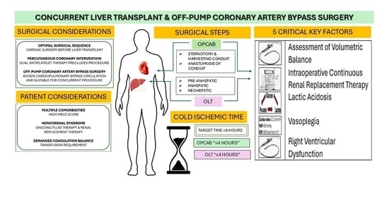
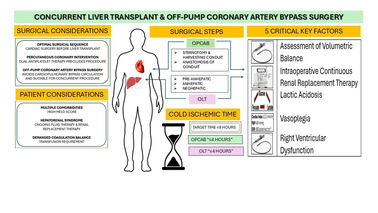
Among the multiple factors requiring optimization in patients undergoing concurrent LT and OPCAB, the authors have found the most critical ones to include the following: (1) volume status, (2) continuous renal replacement therapy, (3) lactic acidosis, (4) vasoplegia, and (5) acute right ventricular dysfunction. These variables are dynamically interrelated, forming a continuous pathophysiological cascade. Consequently, a problem-solving approach must address all components, even when the initial clinical presentation involves only one single factor. Failure to optimize any one element is likely to precipitate rapid deterioration of the remaining physiological parameters.

2. Methods

In this narrative review, an in-depth literature search was undertaken to synthesize the importance of five focal points, which were instrumental in the conduct of concurrent liver transplant and off-pump coronary artery bypass surgery.

2.1. Optimization of Patient Volumetric Balance

Patients with cirrhosis frequently appear fluid-overloaded due to peripheral edema and ascites yet may be intravascularly volume-depleted. This paradox places them at high risk for acute kidney injury (AKI) and often warrants parenteral fluid administration [7].
During fluid resuscitation, patients may easily become overloaded. Even when total body water appears optimized, fluctuations between the intravascular and extravascular compartments persist due to altered vascular permeability, a phenomenon exacerbated by hypoproteinemia and the profound vasoplegia associated with ESLD.

2.1.1. Tests Based on Waveform Analysis

Emerging machine learning-based models using pulse oximetry waveform analysis have demonstrated promise in estimating intravascular volume overload and, in certain settings, may outperform traditional biomarkers such as B-type natriuretic peptide (BNP) [8]. Davenport et al. demonstrated that while total body water may be comparable between stable and decompensated cirrhotic patients, extracellular volume is significantly expanded in the decompensated state, likely reflecting increased vascular permeability [9].
Importantly, static hemodynamic indices such as central venous pressure and pulmonary capillary wedge pressure are unreliable indicators of true circulatory volume status in this population [10,11].

2.1.2. Blood Chemistry

Hypovolemia, resulting from significant fluid or blood loss, leads to a critical reduction in intravascular volume, hypotension, and impaired tissue perfusion. This cascade promotes cellular hypoxia, anerobic metabolism, and ultimately organ failure [12]. Laboratory assessment—including serum lactate, arterial blood gas analysis, and urinary sodium concentration—can assist in determining both etiology and severity.
Conversely, hypervolemia may also impair tissue perfusion by increasing interstitial pressure, thus promoting capillary leakage, microcirculatory dysfunction, reduced organ perfusion, and end-organ injury, particularly AKI [13].
Elevated BNP and N-terminal pro-BNP (NT-proBNP) levels are strong indicators of increased ventricular filling pressures, diastolic dysfunction, and severe volume overload in cirrhotics. BNP levels correlate positively with the severity of liver disease and are typically higher in decompensated cirrhosis, particularly in the presence of significant ascites. Elevated BNP is an independent negative prognostic marker, associated with increased mortality and complications. In liver transplant candidates, BNP monitoring aids in assessing cardiac reserve and identifying patients at risk for post-operative heart failure. However, BNP interpretation in cirrhosis may be confused by cirrhotic cardiomyopathy, renal dysfunction, and the hyperdynamic circulatory state characteristic of ESLD [14,15].
Laboratory indices are available to differentiate the etiology of oliguria. Fractional excretion of sodium (FENa) has traditionally been employed to distinguish prerenal AKI (<1%) from intrinsic renal injury (>2–3%). However, Alsaad et al. demonstrated that FENa < 1% is common in cirrhotic patients with renal dysfunction and does not reliably differentiate hepatorenal syndrome (HRS) from other causes of AKI [16]. Additionally, diuretic therapy significantly limits FENa utility by artificially increasing urinary sodium excretion.
Patidar et al. proposed the fractional excretion of urea (FEUrea) as an alternative marker for distinguishing prerenal AKI and HRS from intrinsic renal injury in cirrhosis [17]. Subsequent work by Sim et al. demonstrated that FEUrea correlates with inflammatory markers and liver dysfunction and independently predicts survival beyond Model for End-stage Liver Disease (MELD) score and AKI stage, with stratification identifying patients at higher risk for poor 90-day outcomes [18].

2.1.3. Radiological Examination

Chest X-ray (CXR) is frequently utilized to assess volume status in ESLD by identifying pulmonary edema, vascular congestion, and pleural effusions, particularly hepatic hydrothorax. While useful for detecting thoracic fluid overload, CXR has limited sensitivity for assessing intravascular volume and cannot reliably distinguish among different etiologies of pulmonary congestion [19].
Lung ultrasound has emerged as a superior alternative, offering greater diagnostic accuracy for pulmonary congestion and interstitial edema compared with CXR [19].

2.1.4. Ultrasound Examination

Accurate assessment of volume status in cirrhotic patients requires integration of clinical evaluation with advanced diagnostic modalities such as point-of-care ultrasound (POCUS). POCUS enables real-time assessment of venous congestion through evaluation of inferior vena cava diameter and collapsibility, as well as hepatic, portal, and renal venous Doppler flow patterns [20].

2.1.5. Echocardiography

Transthoracic echocardiography provides valuable information regarding volume status through assessment of cardiac chamber size and end-diastolic volumes [21], although these static indices are not consistently incorporated into routine clinical decision-making [22]. By contrast, transesophageal echocardiography (TEE) has become integral to modern interventional cardiology practice [23]. During liver transplantation, TEE is generally limited to upper and mid-esophageal views, as esophageal varices and portal hypertension are considered relative contraindications [24].
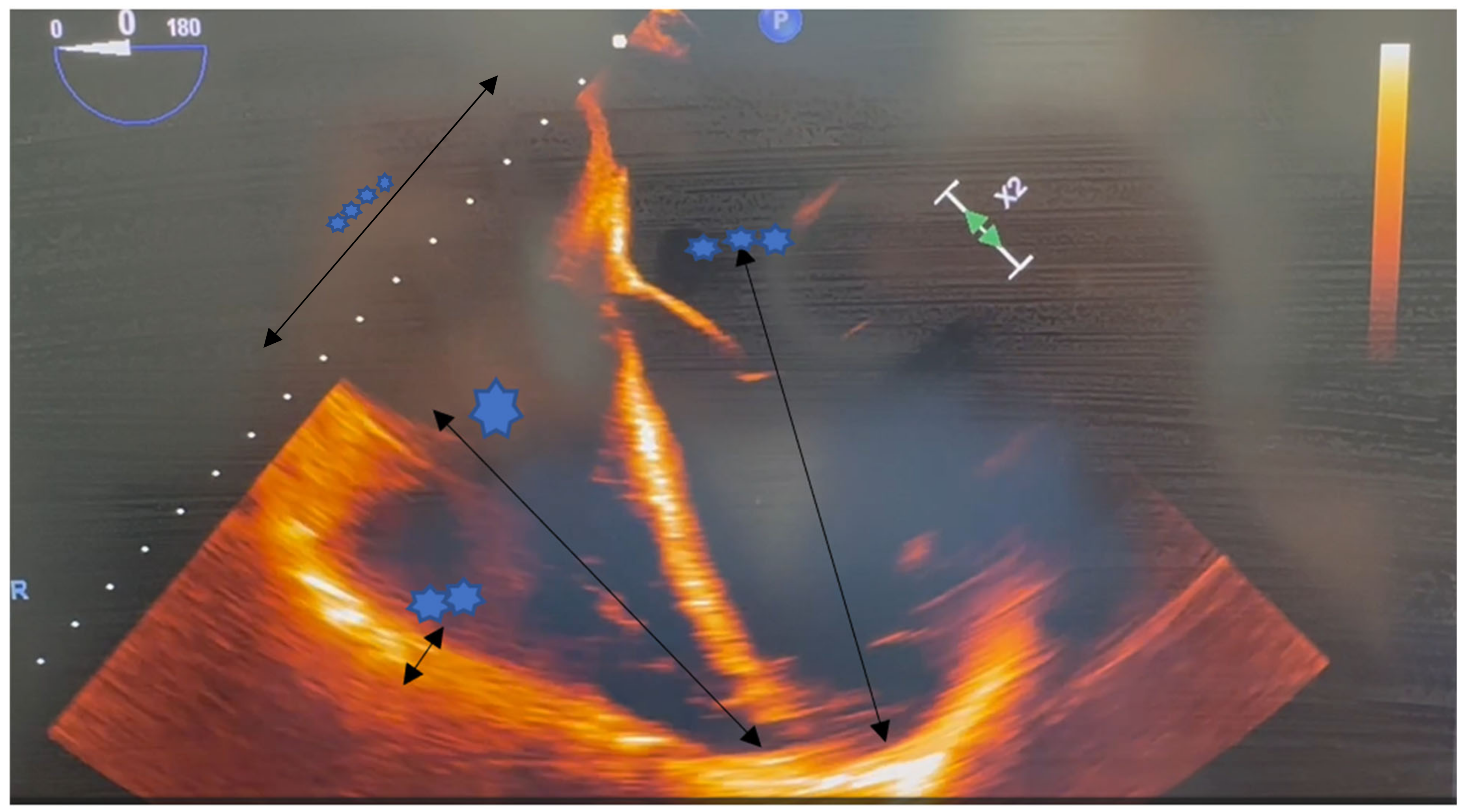
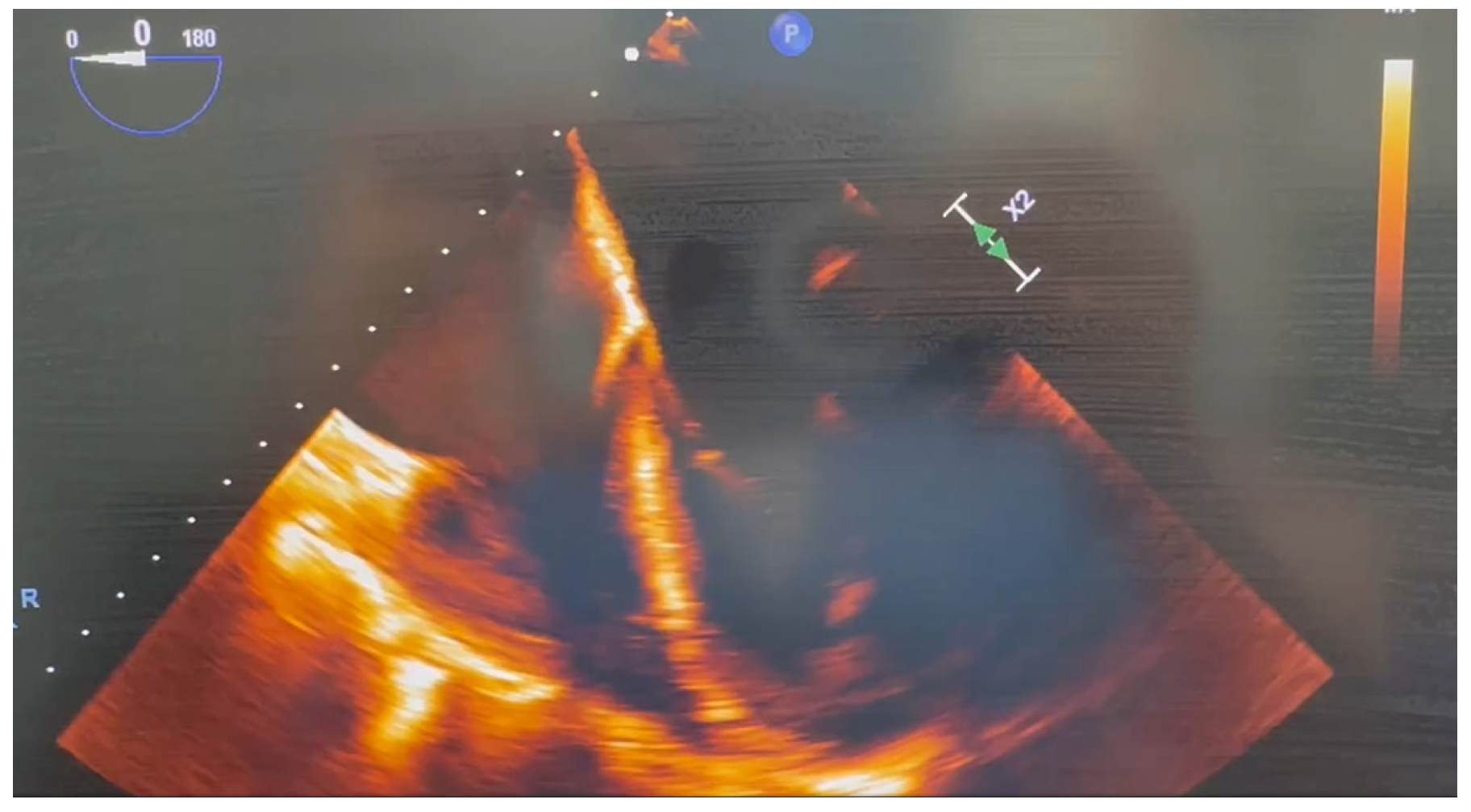
Numerous studies have demonstrated associations between liver disease severity and cardiac dysfunction [25], including left atrial enlargement, increased left ventricular end-diastolic volume, elevated cardiac output, and augmented aortic flow [26]. Diastolic dysfunction—often defined by a transmitral Doppler E/A ratio < 1.0—is frequently observed in cirrhotic patients [27]. Accordingly, the American Association for the Study of Liver Diseases (AASLD) recommends comprehensive echocardiographic evaluation in all LT candidates, including assessment of chamber dimensions, myocardial hypertrophy, systolic and diastolic function, valvular abnormalities, and left ventricular outflow tract obstruction (Figure 1) [28].
Right ventricular end-systolic pressure may be elevated (>30 mmHg) in patients with acute cirrhotic decompensation despite normal pulmonary valve flow velocities [9]. Increased left ventricular end-diastolic volumes (>128 mL) may also be present with preserved aortic valve flow. The most consistent echocardiographic abnormalities involve right-sided cardiac changes, including atrial enlargement, mildly elevated right ventricular end-systolic pressures, variable right atrial pressures, and prominent left ventricular diastolic dysfunction [9].
More recently, studies comparing TEE with right-ventricular pressure catheterization have demonstrated that dynamic right ventricular outflow tract obstruction of varying severity is common during liver transplantation and is readily detectable using intraoperative TEE [29].

2.1.6. Intravascular Volume Expansion

Intravascular volume expansion—often required to prevent or mitigate AKI—may exacerbate ascites, pleural effusions, or cardiac failure. Renal dysfunction is highly prevalent in this population, with reported AKI rates reaching up to 60% among hospitalized patients with cirrhosis [30].
Ascites
Ascites is the pathological accumulation of fluid within the peritoneal cavity and is most associated with liver disease (particularly cirrhosis), although it may also result from cardiac or renal disease, or chylous disorders. In cirrhotic patients, the development of ascites often signifies the transition from compensated to decompensated disease.
The initial event in the pathogenesis of cirrhotic ascites is portal hypertension. Once portal hypertension exceeds a critical threshold, circulating nitric oxide levels increase, resulting in progressive splanchnic arterial vasodilatation. This vasodilatation leads to reduced effective arterial blood volume, increased permeability, and elevated hydrostatic pressure within splanchnic capillaries. These changes activate sodium and water retention through stimulation of the renin–angiotensin–aldosterone system (RAAS), arginine vasopressin secretion, and the sympathetic nervous system (SNS) [31]. Increased hepatic and splanchnic lymph production that exceeds lymphatic reabsorption further contributes to ascitic fluid accumulation [32]. Similarly, in both high- and low-output heart failure, reduced effective arterial blood volume triggers neurohormonal activation, renal vasoconstriction, and sodium and water retention [33].
Management of ascites requires identification and treatment of the underlying mechanism of fluid retention. The principal therapeutic goals are a reduction in ascitic volume and peripheral edema while preserving adequate intravascular volume and end-organ perfusion [34,35,36]. Therapeutic paracentesis provides rapid symptomatic relief in patients with tense or diuretic-refractory ascites. Albumin administration following large-volume paracentesis reduces the risk of hypotension and post-paracentesis circulatory dysfunction (PPCD), defined as an increase in plasma renin activity exceeding 50% above baseline. PPCD is associated with hyponatremia, renal impairment, and hepatorenal syndrome [37].
The American Association for the Study of Liver Diseases recommends albumin replacement after removal of ≥5 L of ascitic fluid, typically at a dose of 6 to 8 g per liter removed [38]. Evidence supporting albumin administration after removal of 3–5 L remains limited [39]. More recent data suggest albumin dosing on total volume removed: 25 g for 5–6 L, 50 g for 7–10 L, and 75 g for volumes exceeding 10 L [39].
Large-volume paracentesis can lead to rapid reaccumulation of ascites and systemic hypotension. Although the precise mechanism remains incompletely understood, proposed contributors include intravascular fluid shifts and mechanical decompression of the splanchnic circulation, leading to impaired systemic vascular responsiveness to increased shear stress and perfusion [40,41].
Pleural Effusion (Hepatic Hydrothorax)
Hepatic hydrothorax is a recognized complication of decompensated cirrhosis and portal hypertension. It is a pleural effusion—typically exceeding 500 mL—in patients with cirrhosis in the absence of primary cardiac or pulmonary disease [42,43]. Reported incidence varies widely, ranging from 16% to 70% depending on diagnostic criteria and study population [42,44].
Hepatic hydrothorax commonly occurs in conjunction with ascites. However, unlike ascites—where large fluid volumes may be tolerated due to the expansile capacity of the peritoneal cavity—relatively small pleural fluid volumes (1–2 L) can cause significant cardiopulmonary compromise, including dyspnea and hypoxemia [45].
The most widely accepted mechanism involves the movement of ascitic fluid into the pleural cavity through diaphragmatic defects, driven by positive intra-abdominal pressure and negative intrathoracic pressure gradients [46,47,48,49]. Additional risk factors include higher MELD scores, hypoalbuminemia, hyperbilirubinemia, diabetes mellitus, and non-use of non-selective beta-blockers [50,51]. In some cases, pleural effusion may develop in the absence of clinically detectable ascites when ascitic formation and pleural reabsorption occur at the same rates [52].
In patients with symptomatic hepatic hydrothorax, thoracocentesis relives dyspnea or coughing. However, recurrence is common, and definitive resolution typically requires liver transplantation.
Pericardial Effusion
Pericardial effusion is common in ESLD, with reported prevalence ranging from 4% to 10% in general cirrhotic populations to as high as 63% among patients with severe ascites [53,54]. These effusions can be small, asymptomatic, and associated with volume overload, sodium and water retention, and hypoalbuminemia.
Echocardiography is the primary modality for assessing effusion size and evaluating for hemodynamic compromise [55]. Signs of tamponade physiology include chamber collapse and characteristic respiratory variation in mitral and tricuspid inflow velocities [56].
A preemptive pericardial drainage relieves hemodynamic compromise in patients with large effusions [57]. However, recurrence is common. Timely intervention is particularly important in the perioperative LT setting, as untreated pericardial effusion may precipitate catastrophic circulatory collapse during major vascular manipulation or graft reperfusion.

2.2. Intraoperative Continuous Renal Replacement Therapy

Renal dysfunction is common in patients with cirrhosis and is associated with substantial morbidity and mortality. The major etiologies of AKI in this population include prerenal (hypovolemic) AKI, hepatorenal syndrome (HRS), and acute tubular necrosis (ATN). Although HRS accounts for a minority of AKI cases (approximately 12%), both ATN and HRS are associated with up to 50% 90-day mortality. AKI severity and lack of response to therapy are strong predictors of short-term mortality [30]. Patients with persistent renal dysfunction frequently require renal replacement therapy (RRT) as a bridge to renal recovery or liver transplantation.

2.2.1. Practice Guidelines

At present, no standardized algorithms or evidence-based guidelines exist to direct the use of continuous renal replacement therapy (CRRT) during liver transplantation. This lack of consensus is consistent with conclusions from the Acute Disease Quality Initiative Conference, which emphasized the need to individualize RRT modality and prescription based on patient-specific clinical factors [58].
Often patients undergoing LT initiate dialysis preoperatively and continue RRT intraoperatively. Paine et al. reported CRRT utilization in approximately 25% of LT procedures at their institution, particularly among patients receiving preoperative dialysis or those with renal dysfunction and high perioperative risk [59]. Zimmermann et al. identified additional indications for intraoperative CRRT, including serum creatinine > 2.0 mg/dL, oliguria or anuria, high MELD scores, pre-transplant ICU admission, use of two or more vasopressors, and an anticipated transfusion requirement exceeding eight units of blood products [60].

2.2.2. Benefits of Intraoperative CRRT

Intraoperative CRRT may prevent life-threatening complications by enabling precise fluid management, facilitating clearance of water-soluble toxins such as ammonia, correcting electrolyte disturbances, and maintaining metabolic homeostasis during liver transplantation. However, evidence supporting routine intraoperative CRRT remains limited.
Townsend et al., in a review of 41 liver transplant procedures utilizing intraoperative CRRT, demonstrated that the technique was safe and enabled achievement of an even or negative fluid balance in 92% of cases [61]. Similarly, Koscielska et al. reported a post-reperfusion syndrome incidence of only 13.5% in patients receiving intraoperative CRRT. Fehervari et al. observed significantly lower intraoperative cytokine concentrations in CRRT-treated patients, suggesting improved hemodynamic stability and attenuation of the inflammatory response [62].
Conversely, Adelman et al. reviewed 172 patients requiring preoperative RRT who did not undergo intraoperative dialysis and found no significant differences in intraoperative acidosis or hyperkalemia [63]. Safwan et al. similarly concluded that CRRT did not confer a clear clinical benefit, despite significantly higher MELD scores in the CRRT cohort. Notably, however, 10 of 57 patients in the non-CRRT group developed severe hyperkalemia (>6 mmol/L) or significant acidosis (pH < 7.2), and one patient died intraoperatively [64].
Collectively, these findings highlight the heterogeneity of patient responses to similar treatment strategies. While optimization of baseline functional status remains essential, clinicians should consider initiating CRRT when clinically indicated on a case-by-case basis.

2.2.3. Renal Replacement Strategies

Intraoperative CRRT encompasses modalities such as continuous venovenous hemofiltration, continuous venovenous hemodialysis, and continuous venovenous hemodiafiltration [65]. Owing to its gradual solute and fluid clearance, CRRT is generally preferred in patients with cardiovascular instability [66].
A 2007 Cochrane review concluded that rates of hemodynamic instability, hypotension, and escalation of supportive therapies did not differ significantly between intermittent and continuous RRT modalities; however, patients receiving CRRT demonstrated higher mean arterial pressure [67].
The literature does not clearly define optimal rates for electrolyte correction using CRRT under standard intraoperative conditions. Although ultrafiltration rates of 1–2 L/h theoretically allow significant volume removal [68], excessive ultrafiltration increases the risk of hemoconcentration, filter clotting, reduced dialyzer efficiency, and treatment interruptions. To mitigate these risks, ultrafiltration is generally limited to a filtration fraction of 16–20%, or up to 30–33% when adequate regional anticoagulation is employed [69].

2.2.4. Role of CRRT in the Management of Intraoperative Lactic Acidosis

The use of bicarbonate-based replacement solutions during continuous hemofiltration may facilitate rapid correction of acidosis in patients with acute lactic acidosis (LA). LA commonly develops during the pre-anhepatic phase and worsens during the anhepatic phase of transplantation, making it an important indication for intraoperative dialysis [70].
Alkalinization through bicarbonate administration during dialysis can stimulate 6-phosphofructokinase activity, potentially augmenting lactate production. Nonetheless, dialysis can reduce lactic acid, improving acidemia, and both hemodialysis and hemofiltration are available as therapeutic options for LA [71,72]. Although lactate clearance achieved through CRRT accounts for only approximately 3% of total lactate removal [73], dialysis contributes to correction of acidemia primarily through buffer delivery and modulation of acid-base balance.
Despite limited randomized controlled data, the use of CRRT in patients with LA and concomitant renal dysfunction during liver transplantation remains an attractive therapeutic strategy. Definitive trials may be difficult to conduct given the widespread current use of this intervention.
Acidosis
Severe metabolic acidosis is frequently encountered during liver transplantation, particularly following reperfusion. This is especially relevant in recipients with preexisting acidosis, with intraoperative development of acidosis during the pre-anhepatic phase, or receiving marginal grafts. Expanded donor criteria—including advanced donor age and hepatic steatosis—have increased susceptibility to ischemia–reperfusion injury [74]. Cold ischemic times exceeding 6 h are also associated with increased preservation injury and heightened risk of severe metabolic disturbances [74].
Intraoperatively, optimization of fluid resuscitation and sodium bicarbonate administration help maintain pH > 7.25. In patients with chronic kidney disease or new-onset AKI, bicarbonate therapy alone is often insufficient to correct metabolic acidosis. Moreover, bicarbonate administration may exacerbate hypocalcemia [75], a common issue in liver transplantation due to citrate toxicity from massive transfusion. Intraoperative CRRT enables more precise modulation of the bicarbonate buffer system and supports maintenance of metabolic homeostasis.

2.2.5. Intraoperative CRRT and Electrolyte Management

Hyperkalemia
Hyperkalemia during liver transplantation may result from potassium release from damaged tissues, massive transfusion of stored blood products, bleeding, and metabolic acidosis. The reperfusion phase is particularly hazardous, as a substantial potassium load enters the systemic circulation from the graft while acidosis simultaneously drives intracellular-to-extracellular potassium shifts. These changes may precipitate hemodynamic instability, hypotension, bradyarrhythmia, and malignant cardiac arrhythmias.
CRRT facilitates rapid potassium clearance through manipulation of dialysate composition [76]. However, as potassium levels normalize, transition to more physiologic solutions is essential to avoid hypokalemia. Studies initiating dialysis early during transplantation demonstrated safe and effective maintenance of normokalemia [77], although many patients had normal potassium levels at baseline. In patients with acute-on-chronic kidney disease, inadequate potassium control may significantly exacerbate post-reperfusion syndrome.
Blood transfusion significantly influences serum potassium levels, with reported increases ranging from 0.05 to 0.4 mEq/L per unit transfused [78]. Prolonged storage leads to red blood cell membrane degradation and potassium leakage into the supernatant. Large-volume transfusion (>7 units of packed red blood cells) has been identified as a risk factor for post-transfusion hyperkalemia [79], although this effect is confounded by hypotension, acidosis, and tissue injury. Intraoperative cell salvage may mitigate the potassium load and offers a cost-effective alternative, with processing times of approximately 5–10 min per unit [79].
Hyponatremia
Hyponatremia is common in ESLD and is typically dilutional due to excess arginine vasopressin secretion. Additional risk factors include malnutrition, diuretic therapy, and alcohol use. Severe hyponatremia is associated with a poor prognosis [80,81]. Rapid correction during reperfusion may cause abrupt osmotic shifts, increasing the risk of osmotic demyelination syndrome [82]. CRRT allows controlled, gradual correction of serum sodium, minimizing fluctuations in intracranial pressure [83].

2.3. Lactic Acidosis

Lactic acidosis (LA) in patients with ESLD is among the most complex metabolic disturbances encountered in advanced cirrhosis and is associated with increased morbidity and mortality. Multiple-organ failure accompanying advanced liver disease disrupts important components of lactate metabolism, resulting in increased lactate production and impaired clearance, leading to severe LA-induced acidemia. These derangements become particularly pronounced in ESLD patients undergoing liver transplantation. Consequently, perioperative management of LA and associated acid-base disturbances represents a critical component of anesthetic, postoperative, and intensive care management in this population.

2.3.1. Lactic Acidosis: Definition and Types

Historically, lactic acidosis has been defined by the presence of metabolic acidosis (pH < 7.35) in conjunction with serum lactate levels > 5 mmol/L [84]. However, more recent literature suggests that overt hyperlactatemia and acidosis do not always coexist [85]. A universally accepted definition remains elusive due to variability in laboratory cut-off values and clinical context [85]. Nonetheless, hyperlactatemia—or a significant increase in serum lactate from baseline, even within the normal range—remains an essential diagnostic criterion. Acidemia may be absent due to respiratory compensation or concurrent metabolic alkalosis [85].
LA is traditionally classified into two categories based on underlying pathophysiology: Type A and Type B [85]. Type A LA results from hypoperfusion and hypoxia, as seen in shock, hypovolemia, or severe hypoxemia. In contrast, Type B LA arises from derangements in normal metabolic processes independent of hypoxia, including underlying disease states, drug or toxin exposure, and inborn errors of metabolism [85]. At the cellular level, mechanisms differ between types, ranging from anerobic glycolysis to aerobic glycolysis (Warburg effect) under normoxic conditions. Although the coexistence of Type A and Type B LA is physiologically plausible, it has been infrequently reported in the clinical literature [86].

2.3.2. Lactic Acidosis in End-Stage Liver Disease

Lactate production and metabolism are well described elsewhere [85]. The liver accounts for approximately 70% of whole-body lactate clearance; therefore, decompensated cirrhosis or acute hepatic failure is associated with elevated lactate levels and poor clinical outcomes [87]. Hyperlactatemia and LA thus serve as important prognostic indicators in ESLD [88]. Persistent LA further impairs lactate clearance, establishing a self-perpetuating vicious cycle. Acidemia reduces hepatic lactate uptake, and in the setting of severe hypoxia and acidemia, the liver may effectively become a net lactate-producing organ.
Bleeding is a common contributor to hyperlactatemia in cirrhotic patients. Increased lactate production results from both overt tissue hypoperfusion (e.g., hemorrhagic shock) and impaired cellular oxygen utilization.

2.3.3. Lactic Acidosis: Intraoperative Fluctuations

Hepatic lactate clearance is directly dependent on adequate liver perfusion (Table 1). Liver transplantation induces profound alterations in hepatic blood flow and systemic physiology. In a clinical study involving 488 patients undergoing liver resection, 72% developed elevated lactate levels, which correlated with peak postoperative bilirubin, coagulopathy, renal dysfunction, diabetes, extent of resection, blood loss, and transfusion requirements. The authors concluded that early postoperative lactate concentration is a useful predictor of outcomes following hepatic surgery [89].
Intraoperative liver tissue hypoxia promotes increased conversion of pyruvate to lactate. This process is exacerbated by surgical stressors, including blood loss, endogenous catecholamine release, and administration of vasoactive agents [90]. Serum lactate may also rise following transfusion of stored blood products, which contains an increasing concentration of lactate proportional to storage duration [91].
During the anhepatic phase of transplantation, lactate clearance becomes entirely dependent on extrahepatic pathways, primarily the kidneys. Renal dysfunction—such as hepatorenal syndrome or AKI—further impairs lactate clearance [92]. While acidemia markedly suppresses hepatic lactate uptake, it enhances renal lactate metabolism. Consequently, the renal contribution to lactate removal increases from approximately 16% at pH 7.45 to 44% at pH 6.75, partially compensating for hepatic loss and accounting for half of total lactate clearance under severe academic conditions.
Immediately following graft reperfusion, acidemia often worsens and typically reaches its nadir. Paradoxically, pre-reperfusion acidosis may confer cytoprotective effects. Experimental data suggest that naturally occurring acidosis protects against cell death in multiple organs, including hepatocytes. This phenomenon—known as the “pH paradox”—is independent of oxygen availability. Reperfusion at low pH (<6.5) inhibits mitochondrial permeability transition pore opening, allowing mitochondrial repolarization and preventing cell death [93].
The capacity of a newly reperfused graft to resume lactate metabolism depends in part on the graft-to-recipient weight ratio. This ratio is an important predictor of post-reperfusion lactate clearance in living-donor liver transplantation. Larger grafts demonstrate a greater lactate elimination capacity, whereas relatively small grafts may be overwhelmed by the lactate load, resulting in persistent hyperlactatemia after reperfusion [94].

2.3.4. Effects of Catecholamines and Vasoactive Agents on Lactate Production and Elimination

During liver transplantation, lactate production and clearance change to a substantial degree following changes in endogenous and exogenous catecholamine concentrations. The use of vasoactive agents—particularly exogenous catecholamines—modulates lactate dynamics through multiple, and sometimes opposing, mechanisms.
Norepinephrine can induce significant peripheral vasoconstriction, which impair microcirculatory flow and tissue oxygen delivery, thereby promoting anerobic metabolism and metabolic (lactic) acidosis [95]. In contrast, epinephrine increases serum lactate concentrations while often maintaining a stable lactate-to-pyruvate ratio. This effect reflects enhanced aerobic glycolysis rather than tissue hypoxia. Epinephrine stimulates glycogenolysis, increases pyruvate production, and augments adenosine triphosphate consumption via activation of the Na+/K+—ATPase, collectively accelerating glycolytic flux and lactate generation [96].
Thus, catecholamine-induced hyperlactatemia during liver transplantation does not uniformly reflect impaired tissue perfusion and should be interpreted within the broader hemodynamic and metabolic context.

2.3.5. Lactic Acidosis: Management Options

Hemodynamic optimization aimed at improving tissue perfusion and enhancing lactate clearance by metabolically active organs—principally the liver and kidneys—is the cornerstone of lactic acidosis management. However, in advance ESLD and during LT, excessive lactate production combined with impaired elimination may overwhelm the corrective capacity of hemodynamic optimization alone.
In such circumstances, adjunctive therapeutic strategies—including modulation of vasoactive support, correction of acid-base disturbances, and selective use of RRT—can mitigate severe acidemia and its systemic consequences (Table 2)

2.4. Vasoplegia

Vasoplegia is an increasingly recognized perioperative challenge, particularly in patients with ESLD undergoing LT. In adults, vasoplegia has multiple operational definitions but is most commonly characterized by a normal or elevated cardiac output state (cardiac index ≥ 2.2 L/min/m2) with persistent hypotension—typically an inability to maintain mean arterial pressure (MAP) ≥ 60 mmHg—due to markedly reduced systemic vascular resistance (<800 dynes*s*cm−5), despite high-dose vasopressor therapy (commonly ≥0.5 µg/kg/min norepinephrine equivalents) [97,98].
Although no universally accepted treatment algorithm exists, recent advances in pharmacologic therapy allow effective treatment in most cases. Selection of therapy must account for the distinct mechanisms of action and potential adverse effects of each agent.

2.4.1. Pathophysiology

The pathophysiology of vasoplegia is multifactorial. Proposed mechanisms include increased inducible nitric oxide synthase activity, activation of cyclic guanylate cyclase, cytokine-mediated vasodilation, hydrogen sulfide signaling, adrenal axis dysregulation, and relative vasopressin deficiency. Together, these processes result in inappropriate vascular smooth muscle relaxation and impaired organ perfusion [99].
Experimental and clinical data demonstrate reduced vascular responsiveness to vasoconstrictors as a key contributor to splanchnic and systemic vasodilation [100]. Defects in smooth muscle intracellular signaling pathways further exacerbate hyporesponsiveness to endogenous and exogenous vasoconstrictors [100].
Additional risk factors include prolonged allograft ischemia time, preexisting infection, or systemic inflammation, and exposure to medications such as angiotensin-converting enzyme inhibitors and angiotensin receptor blockers. Mechanical circulatory support techniques, including cardiopulmonary bypass, also predispose patients to profound vasodilation [99].

2.4.2. Vasoplegic Shock

Vasoplegic shock, often considered a subtype of distributive shock, represents a more severe circulatory disturbance characterized by vasoplegia accompanied by evidence of tissue hypoperfusion. This state is frequently associated with hyperlactatemia and metabolic acidosis [101].

2.4.3. Clinical Assessment

Clinical evaluation includes assessment of skin temperature and color, jugular venous distention, capillary refill, and peripheral edema. Point-of-care echocardiography plays a central role in refining the diagnosis and should include evaluation for pericardial effusion, assessment of left and right ventricular size and systolic function, respiratory variation in vena cava dimensions, and measurement of the aortic velocity-time integral as a surrogate of stroke volume [102,103].

2.4.4. Treatment Options

Initial management of vasoplegia typically involves catecholamines and vasopressin analogs. Norepinephrine is the first-line therapy. Current Surviving Sepsis Campaign guidelines suggest that early combination therapy—most commonly norepinephrine and vasopressin—may improve vascular responsiveness while limiting excessive adrenergic stimulation and receptor desensitization [104]. This approach may also mitigate deleterious effects of sympathetic overactivation on inflammation and myocardial function.
Based on emerging pathophysiological insights, novel agents such as selepressin and angiotensin II are under investigation. A recent double-blind, randomized, placebo-controlled trial is evaluating angiotensin II during LT, with norepinephrine requirements as the primary endpoint [105]. Results from this study will inform future standardized treatment strategies.
In refractory vasoplegia, adjunctive therapies such as methylene blue, hydroxocobalamin, and ascorbic acid have been employed to increase systemic vascular resistance [97,106,107,108]. While retrospective studies suggest hemodynamic benefit in LT patients, high-quality prospective data remains limited.
Published case reports describe successful treatment of LT-associated vasoplegia with hydroxocobalamin [109,110]. Two retrospective studies—one evaluating hydroxocobalamin as a primary therapy and another as rescue therapy following methylene blue failure—represent the strongest available evidence supporting its use [111,112]. Importantly, hydroxocobalamin may interfere with laboratory assays, optical sensors, and renal replacement therapy circuits; one case report described intraoperative dialysis interruption due to false activation of a blood-leak alarm [109,113].
The role of methylene blue (MB), a nitric oxide synthetase inhibitor, is helpful during LT. In a randomized controlled trial, administration of a 1.5 mg/kg bolus one minute before reperfusion significantly reduced vasopressor requirements, improved hemodynamic stability, and lowered serum lactate compared with placebo [114]. Additional case series support its perioperative use, with most reported doses not exceeding 2 mg/kg [115,116].
Ascorbic acid, an essential cofactor for endogenous catecholamine synthesis, is frequently depleted in patients undergoing extracorporeal circulation [117]. High-dose ascorbic acid has been associated with reduced vasopressor requirements in vasoplegic states following extracorporeal support [118]. Of note, both methylene blue and ascorbic acid are contraindicated in patients with glucose-6-phosphate dehydrogenase deficiency due to the risk of hemolytic anemia [119].
Use of angiotensin II during LT remains uncommon; however, one case report describes its successful application in combined heart–liver transplantation [120].

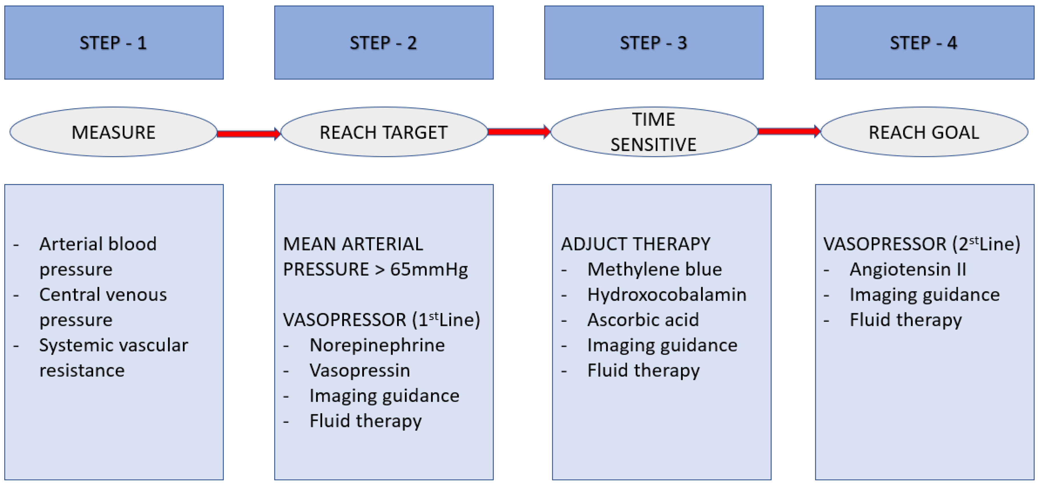
2.4.5. Vasoplegia Syndrome: Treatment Algorithm (Figure 2)

Despite the presence of a high cardiac output state, vasoplegia is frequently accompanied by myocardial dysfunction. Early fluid resuscitation is crucial to restore an effective circulating volume and tissue perfusion. Goal-directed therapy will guide fluid administration [121]. Use of central venous pressure alone is insufficient. Its ability to predict fluid responsiveness is minimal within the “normal” range (8–12 mmHg) [121,122]. Dynamic measures of cardiac output, such as arterial pulse pressure variation, demonstrate superior predictive accuracy [123].
Figure 2. Recommendations to assist management of vasoplegia.
Livers 06 00031 g002
MAP is the principal determinant of tissue perfusion pressure. Although autoregulation may preserve cerebral and renal blood flow across a range of pressures, below a critical MAP threshold, organ perfusion becomes pressure-dependent. Studies evaluating higher MAP targets (>65 mmHg) through increased norepinephrine dosing have not demonstrated improvements in urine output, lactate clearance, oxygen delivery, or consumption, but have shown increased arrhythmia risk [124]. Consequently, targeting an initial MAP of 65 mmHg is appropriate, as it balances adequate perfusion with lower vasopressor exposure, reduced arrhythmia risk, and similar mortality.
Elevated serum lactate may reflect tissue hypoxia, catecholamine-driven aerobic glycolysis, impaired hepatic clearance, or a combination of mechanisms. Regardless of etiology, hyperlactatemia is consistently associated with worse outcomes [104]. Two meta-analyses demonstrated reduced mortality when early lactate clearance strategies were employed, compared with usual care or central venous oxygen saturation-guided therapy [125,126].

2.5. Right Ventricular Dysfunction and Failure

The cardiovascular abnormalities, commonly associated with cirrhosis, may meet diagnostic criteria for cirrhotic cardiomyopathy (CCM) when sufficiently severe [127]. The bidirectional dependence between the heart and liver is critical for maintaining hemodynamic stability during the perioperative period of liver transplantation. In patients with ESLD, pre-transplant hemodynamic alterations may remain clinically occult due to limited functional capacity and reduced physiological reserve [128]. Moreover, concomitant pulmonary disorders—including pulmonary hypertension (PH), hepatopulmonary syndrome and portopulmonary hypertension (PoPH)—are frequently present [128]. Collectively, these factors underscore the growing need for meticulous preoperative cardiovascular evaluation and risk stratification in LT candidates.

2.5.1. Strategy for Assessment and Management of Pulmonary Hypertension

Standard transthoracic echocardiography helps to evaluate PH in all LT candidates [129]. PoPH refers to pulmonary arterial hypertension (PAH) occurring in the setting of portal hypertension of hepatic or extrahepatic origin. Up to 4–8% of LT candidates have PoPH [129,130]. The diagnosis of PoPH consists of four essential elements [129]: (i) mean pulmonary arterial pressure (mPAP) > 20 mmHg; (ii) pulmonary vascular resistance (PVR) > 2 Woods Units (WU); (iii) pulmonary capillary wedge pressure (PCWP) ≤ 15 mmHg; and (iv) clinical evidence of portal hypertension.
Although no single etiology has been consistently linked to PoPH development [131,132,133], severe PoPH is associated with worse outcomes following LT compared with recipients without PoPH [134]. The pathophysiology remains incompletely understood; however, the prevailing hypothesis involves dysregulation of vasoconstrictive and vasodilatory mediators. Humoral substances such as endothelin-1 may bypass hepatic metabolism via portosystemic shunts and enter the pulmonary circulation, promoting pulmonary vascular remodeling and PAH [135].
Screening transthoracic echocardiography focuses on estimation of right ventricular systolic pressure (RVSP) and assessment of tricuspid regurgitation (TR). Peak TR velocity is the principal echocardiographic variable used to determine the probability of PH [129]. A peak TR velocity > 2.8 m/s is suggestive of PH and should prompt confirmatory right heat catheterization (RHC).
If the mPAP is <20 mmHg with preserved RV function, LT may proceed. Repeat RHC when mPAP is ≥20 mmHg with an elevated PCWP (>15 mmHg) following optimization of volume status. If mPAP is ≥20 mmHg and PVR is ≥2 WU in the setting of a PCWP of ≤15 mmHg, clinically significant PAH is present. Mild PAH (mPAP 20 to 34 mmHg) is not an absolute contraindication for LT, particularly when RV function is normal. In contrast, moderate to severe PAH (mPAP ≥ 35 mmHg) warrants consideration of PAH-targeted therapy prior to transplantation [136,137]. A mPAP of >45 mmHg is a contraindication for LT [129]. In carefully selected patients, a combination of PAH-directed therapy and LT has been associated with excellent long-term survival, with reported 5-year survival rates of up to 81% [138].
During LT, a pulmonary artery catheter is helpful for hemodynamic monitoring [139]. This guides the administration of pulmonary vasodilator therapy, inotropes, and inhaled nitric oxide (NO) during the perioperative period, although reported outcomes are variable [140].

2.5.2. Functional Stratification

Under-recognition and suboptimal management of right ventricular dysfunction (RVD) and right ventricular failure (RVF) are associated with substantial perioperative morbidity and mortality [141]. Both pressure and volume overload adversely affect RV mechanics and performance [142]. RVD is an impaired RV filling or ejection in the absence of overt heart failure symptoms [143]. In contrast, RVF is inadequate forward flow through the pulmonary circulation, resulting in low cardiac output, hypotension, and systemic venous congestion, manifesting as jugular venous distension, peripheral edema, oliguria, and congestive hepatopathy [142,144,145].
Acute RVF is a rapidly progressive syndrome marked by systemic congestion due to impaired RV filling and/or reduced RV output [143]. Chronic RVF most commonly results from sustained elevations in RV afterload, typically secondary to pulmonary hypertension from left ventricular dysfunction, PAH, chronic lung disease, or persistent volume overload from right-sided valvular lesions such as TR. Identification and optimization of modifiable risk factors for RVD are essential. Appropriate preoperative volume optimization and surgical strategies aimed at minimizing blood loss and ensuring myocardial protection will mitigate the risk of RVF [146].
Explantation of the cirrhotic liver relieves portal hypertension and may increase RV preload. Additionally, graft reperfusion can trigger the release of vasoactive cytokines capable of acutely elevating pulmonary artery pressure (PAP) [147]. In reperfusion syndrome, abrupt increases in PAP may precipitate acute RVF, cardiogenic shock, and graft failure. Inhaled NO, intravenous prostacyclin, and milrinone help to attenuate these effects [148,149]. In extreme cases of refractory RV systolic failure with shock, extracorporeal membrane oxygenation has been employed as salvage therapy [150].

2.5.3. Right Ventricle Dysfunction During Liver Transplantation

During the anhepatic phase of LT, cardiac output (CO) may decrease by up to 50% in patients not supported with venovenous bypass, primarily due to reduced venous return. Simultaneously, systemic vascular resistance increases because of inferior vena cava clamping, while the heart rate typically rises as a compensatory response. In patients with cirrhotic cardiomyopathy, chronotropic incompetence may limit heart rate augmentation, impairing compensation for reduced preload. In this setting, optimization of intravascular volume and judicious use of vasopressors are essential to maintain mean arterial pressure (Figure 3). As the anhepatic phase progresses, coagulopathy and metabolic acidosis often worsen.
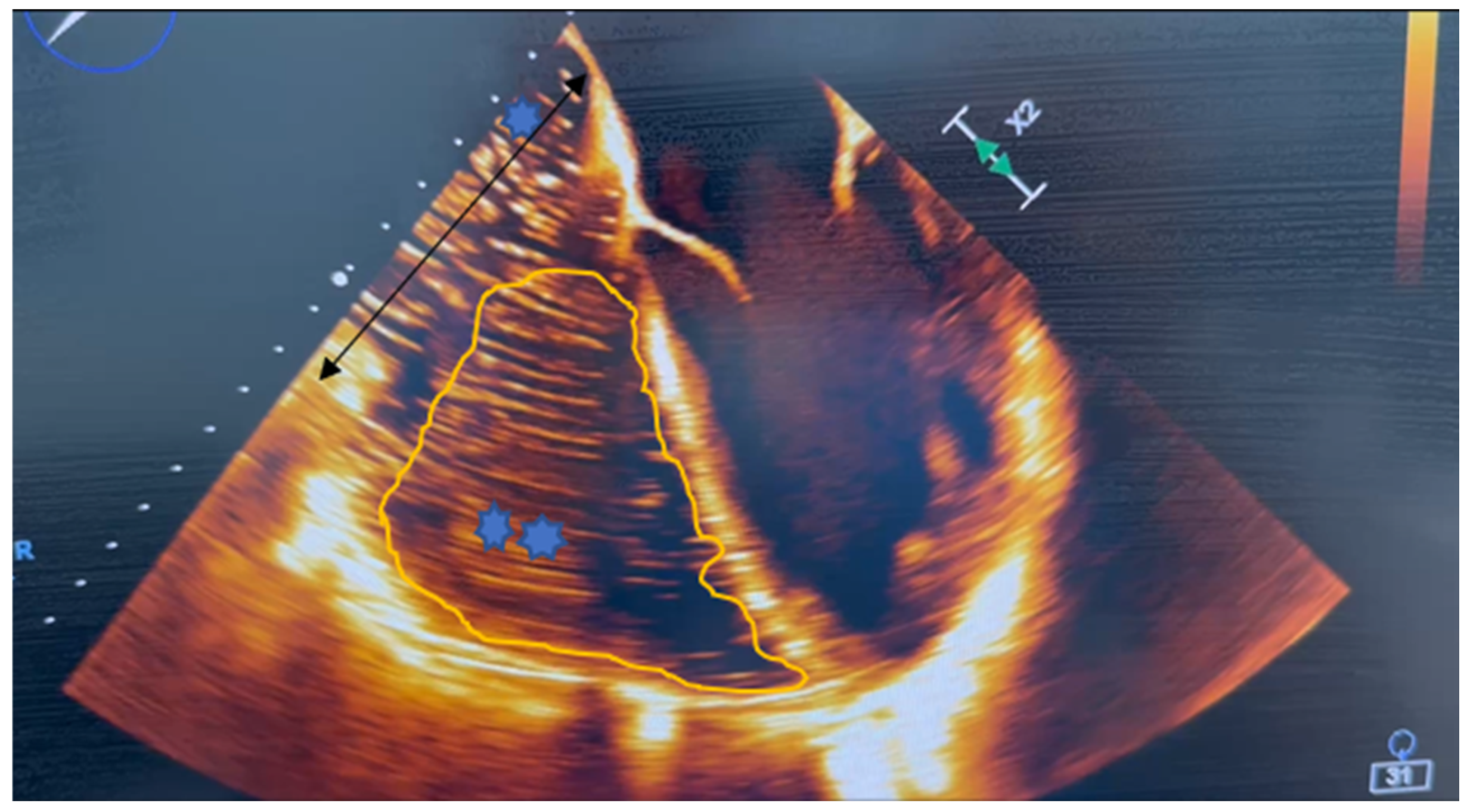
During the neohepatic (reperfusion) phase, restoration of blood flow through the allograft may precipitate marked hemodynamic instability. Rapid return of cold, acidotic, and potentially hyperkalemic blood to the right heart and pulmonary circulation can acutely elevate PAP, leading to RV dilation and dysfunction (Figure 4). During post-reperfusion syndrome, these features worsen.

2.5.4. Medical Management

(i) Preload Optimization
Patients with ESLD frequently exhibit reduced effective right-sided preload due to hypovolemia, diminished venous tone from medications, vasoplegia, or positive-pressure ventilation. Although the Frank–Starling mechanism is functional in the failing human RV myocardium [151], the RV operates on a flatter Frank–Starling curve than the left ventricle, resulting in limited augmentation of contractility across a wide range of filling pressures.
In conditions where RV afterload is normal, but contractility is impaired (e.g., acute RV infarction), a higher preload improves forward flow. However, most etiologies of RVF in LT have elevated RV afterload. In these settings, reduction in excessive preload using diuretics or hemofiltration is critical to limit RV dilation, reduce free-wall tension, minimize ischemia, and optimize contractility. A moderately elevated RV diastolic filling pressure (8–12 mmHg) is often necessary.
(ii) Afterload Reduction
Strategies to reduce RV afterload include general supportive measures and targeted pharmacologic therapy. General measures aim to correct factors that increase PVR, such as acidosis, hypoxia, and hypercapnia. Lung-protective ventilation strategies—employing the lowest effective plateau pressures, tidal volumes, and positive end-expiratory pressure while avoiding hypoxemia and hypercarbia—optimize both RV preload and afterload [152].
Pulmonary vasodilators represent the cornerstone of pharmacologic afterload reduction. While systemic vasodilators may cause hypotension and worsen ventilation–perfusion mismatch, inhaled nitric oxide (iNO), used off-label, provides selective pulmonary vasodilation with rapid onset and a short half-life. iNO demonstrated efficacy in improving pulmonary hemodynamics in RVF [153]. Inhaled prostacyclin analogues are likewise safe and effective in patients with PAH, refractory hypoxemia, or RV dysfunction and may represent a cost-effective alternative to iNO [154].
(iii) Improving Contractility
General management principles emphasize avoidance of RV free wall overstretch through optimization of preload and afterload. Maintenance of sinus rhythm is critical, as supraventricular tachyarrhythmias further impair RV filling and output [155].
Among inotropic agents, low-dose dobutamine (5–10 mcg/kg/min) improves RV-pulmonary artery coupling and increases cardiac output more effectively than norepinephrine owing to its stronger inotropic properties [156]. As an inodilator, dobutamine is best suited for normotensive patients.
Epinephrine improves cardiac output without significantly raising PVR [157], whereas norepinephrine provides α1-mediated vasoconstriction with modest β1 inotropic effects. Although norepinephrine may increase PVR, an improved RV–pulmonary artery coupling offsets this effect, making both agents appropriate for hypotensive patients.
Vasopressin, acting on V1 receptors, induces pulmonary vasodilation at low doses (0.01–0.03 U/min) via endothelial nitric oxide release; however, at higher doses, it may cause coronary vasoconstriction by enhancing catecholamine responsiveness [158,159].
In 2022, Gouvea et al. evaluated RV function during LT using transesophageal echocardiography with quantitative assessment across five surgical stages: baseline, hepatectomy, anhepatic, post-reperfusion, and closure [160]. Tricuspid annular plane systolic excursion demonstrated a non-significant trend toward reduction during the anhepatic phase compared with baseline (2.0 cm vs. 2.4 cm; p = 0.24).
In 2024, Arora et al. proposed the Perioperative Quality Initiative (POQI)—9 Risk Score to guide perioperative monitoring and management of RV function [161]. This tool stratifies patients based on anesthetic factors, surgical technique, and procedural risk, providing a pragmatic framework for anticipatory decision-making.
When RV decompensation is refractory to medical therapy, escalation to mechanical circulatory support (MCS) will be necessary. Timing is critical, as earlier MCS initiation is associated with improved survival in RVF. As early as 2012, Itagaki et al. described modifications to MCS strategies to support ventricular function in critically ill patients undergoing LT [162].

2.6. Concurrent Liver Transplant and Off-Pump Coronary Artery Bypass Graft Surgery

Patient requiring transplant span a broad clinical spectrum, from individuals with multiple organ failure who require multiorgan transplantation to patients who may need transplantation of a single organ accompanied by corrective surgery for another dysfunctional organ. Despite challenges such as donor shortages and complex logistical constraints, the demand for these lifesaving procedures continues to rise [163].

2.6.1. Clinical Challenges

The presence of significant cardiac disease frequently renders patient ineligible for isolated liver transplantation [164]. Evidence from single-center experiences with combined heart–liver transplantation suggests that this approach is safe when performed in carefully selected patients and is associated with excellent outcomes [165]. However, the complexity of performing combined cardiac surgery (CS) and LT prolongs cold ischemic time (CIT) inherent to liver allografts.
To minimize CIT, combined procedures utilize living donor grafts. A short CIT—ideally no longer than 8 h—is necessary for good graft function [166]. Maintaining a limited CIT is further complicated by procedural sequencing, as LT typically follows completion of the cardiothoracic operation. Consequently, even grafts procured from low-risk donors suffer prolonged ischemia, which can precipitate significant hemodynamic instability upon portal vein reperfusion.
In patients with concomitant cardiac disease, perioperative mortality and morbidity are high when CS or LT is performed separately [167]. Patients have an increased bleeding risk associated with dual antiplatelet therapy that precludes the use of percutaneous coronary intervention (PCI) prior to LT. Additionally, certain coronary anatomies are unsuitable for PCI, and diffuse coronary artery disease may be unreconstructable percutaneously. Patients with high MELD scores or short anticipated wait times for transplant often cannot safely delay LT to complete the required three-month course of dual antiplatelet therapy following PCI. For these patients, simultaneous LT and CS represents a viable therapeutic option [167].
Cardiopulmonary bypass (CPB) is frequently employed during combined LT and CS; however, its use amplifies the systemic inflammatory response and contributes to coagulopathy due to mandatory anticoagulation. To mitigate these complications, off-pump coronary artery bypass grafting (OPCAB) has emerged as an alternative strategy. Nonetheless, experience with OPCAB in the setting of combined LT and CS remains limited to small case series and isolated case reports [6,168,169,170] (Table 3).
A recent study by Wehrle et al. [171] evaluated predictors of success and long-term outcomes in patients undergoing combined LT and CS. The authors developed a four-variable LT + CS risk score, identifying preoperative renal dysfunction and coronary artery disease as significant negative predictors of mortality.
Table 3. Case series of combined liver transplant and cardiac surgery. LT, liver transplant; OPCAB, off-pump coronary artery bypass surgery; CT, cardiothoracic; CABG, coronary artery bypass surgery; LKT, liver and kidney transplant; AF, atrial fibrillation; MOF, multiorgan failure; CRRT, continuous renal replacement therapy; AKI, acute kidney injury; DVT, deep vein thrombosis; LE, lower extremity; PCI, percutaneous intervention; GVHD, graft-versus-host disease.
Published Case SeriesSample SizeSurgical SequenceComplicationsMortalityGraft Outcomes
Juneja R, et al. [169]4OPCAB followed by LTAF (25%), sepsis (25%)Death < 1 yr (n = 1)No allograft rejection during hospital stays
Jacob S, et al. [172]12Cardiac surgery followed by LTOne patient each had stroke, sternal osteomyelitis, peritonitis, meningitis and encephalitis, and renal hemodialysisDeath < 30 days (n = 1)No allograft rejection during hospital stays
Wehrle CJ, et al. [173]31Cardiac surgery followed by LTAF (75%), MI (n = 1), severe diastolic failure (n = 1), sternal wound infection (n = 1), sepsis/MOF (n = 1), seizures (n = 1), pulmonary artery thrombosis + stroke (n = 1)Death < 90 days (n = 3), death < 1 yr (n = 2)GVHD (1 pt)
Uludag YT, et al. [6]2Cardiac surgery followed by LTBleeding, re-exploration (n = 2)No death < 1 yrNo allograft rejection during hospital stays
Chai J, et al. [174]1Cardiac surgery followed by LTSternal wound infectionNo death < 1 yrNo allograft rejection during hospital stays
Bui CCM, et al. [175]25Cardiac surgery followed by LTHepatic artery thrombosis (n = 1)No death < 90 daysSecond LT < 30 days (n = 1)
Vohra V, et al. [170]8Cardiac surgery followed by LTAtrial arrhythmia (n = 4), sepsis/MOF (n = 2)Death in postoperative period (n = 2)None reported
Kazimi M, et al. [176]1Cardiac surgery followed by LTNo complicationsNo death at 1 yrNone reported
Wood A, et al. [167]15Cardiac surgery followed by LTAKI requiring CRRT (26.7%), hypoxic respiratory failure requiring reintubation (26.7%), operative re-exploration for hemorrhage (n = 2)Death < 1 yr (n = 4)None reported
Lin E, et al. [177]1Cardiac surgery followed by LTNone reportedNo death < 1 yrNone reported
Garvanovic SH, et al. [178]1Cardiac surgery followed by LTBiliary stricture requiring biliary stent placement, DVT of LENo death < 1 yrNone reported
Brozzi NA, et al. [168]1Cardiac surgery followed by LTPCI after 3 monthsNo death < 1 yrNone reported
Chaubey S, et al. [179]8Cardiac surgery followed by LTAortic rupture (fungal infection) (n = 1), anoxic brain injury (n = 1), MOF (n = 1)In hospital mortality at 3 months (25%), >1 yr (n = 1)Liver failure (n = 1)
Yazici SE, et al. [180]2Cardiac surgery followed by LTBiliary leak (n = 1)No mortality < 1 yrNone reported
Karatas C, et al. [181]1Cardiac surgery followed by LTNone reportedNo mortality < 1 yrNone reported
Rogers JL, et al. [182]1Cardiac surgery followed by LTBiliary leakNo mortality < 1 yrNone reported
Kwon YIC, et al. [183]1Cardiac surgery followed by LTAKI requiring hemodialysisNo mortality < 1 yrNone reported

2.6.2. Intraoperative Fluid Management During Concurrent LT and OPCAB

A major intraoperative challenge arises from the opposing fluid management strategies required for OPCAB and LT. OPCAB procedures typically rely on an adequate intravascular volume to maintain hemodynamic stability, whereas LT favors restrictive fluid strategies to minimize graft congestion and dysfunction. Reconciling these opposing requirements represents a significant challenge for transplant anesthesiologists.
Multiple studies support the use of goal-directed fluid therapy (GDFT) during both OPCAB and LT to guide intravenous fluid administration and inotropic support using cardiac output or dynamic preload parameters. GDFT involves targeted manipulation of cardiac preload, contractility, and afterload to optimize the balance between systemic oxygen delivery and demand [184]. The current literature suggests maintaining a stroke volume variation below 10% during OPCAB and between 13% and 15% during LT [185,186]. This approach minimizes excessive fluid administration—thereby reducing complications such as pulmonary edema and delayed recovery—while avoiding inadequate resuscitation that may precipitate prerenal AKI or acute tubular necrosis.
Given the profound disturbances in fluid homeostasis associated with advanced liver disease, a multimodal approach to intravascular volume assessment is essential.

2.6.3. Intraoperative Continuous Renal Replacement Therapy During Concurrent LT and OPCAB

Intraoperative CRRT is particularly important in patients with AKI who require preoperative dialysis and are undergoing combined LT and OPCAB. The intravascular volume supplementation necessary during OPCAB may become excessive during the LT phase. Anuric patients are often unable to tolerate this hypervolemic state, leading to cardiorespiratory compromise. Furthermore, non-anion gap metabolic acidosis typically worsens during the anhepatic and neohepatic phases of LT, resulting in diminished responsiveness to vasopressor therapy.
Maintenance of intraoperative serum potassium concentration during OPCAB is critical to prevent arrhythmias and reduce morbidity, with general targets aimed at avoiding hypokalemia (<3.5–4.0 mEq/L). Although off-pump surgery avoids the hemodilution and extreme potassium shifts associated with CPB, significant fluctuations may still occur. Maintaining potassium levels in the high-normal range (>4.0 mEq/L), is often necessary to ensure cardiac electrical stability [187].
Once LT begins, the anhepatic phase begins within a few hours, leaving a limited window to reduce serum potassium to a target range of 3.5–4.0 mEq/L prior to reperfusion. CRRT typically achieves potassium clearance of approximately 1 mmol/L per hour, with modest reductions in clearance as serum potassium normalizes [188]. Lower potassium before reperfusion mitigates hemodynamic instability associated with hyperkalemia during graft reperfusion. Without CRRT, achieving this target in a timely manner is often difficult [189].

2.6.4. Lactic Acidosis During Concurrent LT and OPCAB

Hyperlactatemia occurs in approximately 10–20% of patients following cardiac surgery [190]. Although off-pump techniques aim to avoid complications associated with CPB, elevated lactate levels remain a significant predictor of mortality, acute renal failure, and prolonged ICU stay. Severe lactic acidosis (typically lactate >10 mmol/L) following cardiac surgery, including OPCAB, represents a critical event strongly associated with postoperative cardiogenic shock and systemic inflammation [191].
While lactic acidosis is a serious indicator of tissue hypoxia and metabolic stress, it is not uniformly fatal. Although severe hyperlactatemia (>10 mmol/L) carries an estimated mortality risk of 40–50%, survival is possible with timely recognition and intervention, as more than 50% of affected patients may survive to hospital discharge [192].
The deleterious effects of lactic acidosis include reductions in cardiac contractility and cardiac output, as well as an increased propensity for cardiac arrhythmias, which may contribute to sudden death. A decrease in intracellular pH reduces myocardial contractility through competitive inhibition of calcium binding to troponin. Although sympathetic activation accompanies metabolic acidosis, responsiveness to both endogenous and exogenous catecholamines is less.
Metabolic acidosis also induces vascular smooth muscle relaxation via activation of ATP-sensitive potassium channels and upregulation of inducible nitric oxide synthetase in endothelial and vascular smooth muscle cells, resulting in excessive nitric oxide production and direct vasodilatory effects on the systemic vasculature.

2.6.5. Vasoplegia During Concurrent LT and OPCAB

During the OPCAB phase, maintaining MAP ≥ 65 mmHg may be particularly challenging in patients with ESLD. GDFT frequently necessitates the use of vasopressors in combination with inotropic support. Adjunct therapies such as methylene blue and high-dose hydroxocobalamin are frequently required. Acute fluctuations in cardiovascular hemodynamics can severely impair renal function, and patients with pre-existing CKD are especially susceptible to superimposed AKI. Progressive metabolic acidosis further diminishes vasopressor responsiveness.
The anhepatic and neohepatic phases of LT are similarly associated with marked reductions in MAP despite vasopressor support. Worsening lactic acidosis and anion-gap metabolic acidosis often necessitate renal replacement therapy to restore metabolic homeostasis.

2.6.6. Right Ventricle Dysfunction During Concurrent LT and OPCAB

Displacement of the beating heart during positioning for graft anastomosis—particularly to the obtuse marginal artery—may cause significant derangement of RV function and reductions in cardiac output. In contrast, changes in right ventricular ejection fraction (RVEF) and cardiac index during anastomosis of the left anterior descending or right coronary artery are generally less pronounced [193]. A thermo-dilution pulmonary artery catheter capable of continuous measurement of cardiac output and RVEF may be valuable for monitoring dynamic changes in RV function and volume status during OPCAB. Intraoperative TEE also plays a critical role in guiding hemodynamic management in patients with impaired cardiac function.
Regardless of CPB use, pulmonary hypertension remains an important prognostic factor in cardiac surgery. Elevated pulmonary vascular resistance is often responsive to iNO [153]. The vasodilatory effects of iNO on pulmonary circulation are short-lived due to the brief half-life of cyclic guanosine monophosphate, allowing rapid cessation of effect upon discontinuation.
Right ventricular dysfunction commonly occurs during off-pump cardiac surgery. The characteristic features include transient reductions in long-axis RV function, often peaking within the first few postoperative days. Although OPCAB avoids the systemic inflammatory response associated with CPB, mechanical manipulation and positioning of the heart—particularly for posterior wall grafting—can result in acute, reversible RV impairment that typically resolves within three months [194].
While conventional on-pump CABG is associated with more severe, widespread, and sustained post-bypass ventricular dysfunction due to ischemia–reperfusion injury, off-pump surgery still carries a risk of acute, transient, mechanically mediated RV dysfunction. Some, but not all, studies suggest superior long-term preservation of RV function with off-pump techniques compared with on-pump surgery [195].
To prevent severe dysfunction, close collaboration between surgeons and anesthesiologists is essential, with careful, gradual positioning of the heart to minimize hemodynamic compromise [194].

3. Conclusions and Future Directions

Patients undergoing concurrent surgical procedures represent a uniquely complex and high-risk population. They frequently present with profound hemodynamic instability, systemic inflammatory responses, thrombocytopenia, and derangements in coagulation, fibrinolysis, and metabolism. Persistent vasoplegia despite vasopressor support is common and often culminates in multiorgan dysfunction, including acute kidney injury, a condition further exacerbated by prolonged and technically demanding surgical interventions.
These procedures are associated with substantial intraoperative blood loss, necessitating large-volume resuscitation with fluid and blood products. Consequently, perioperative fluid management plays a pivotal role in patient outcomes and remains a cornerstone of enhanced recovery pathways.
Advances in intraoperative fluid management have shifted paradigms toward individualized, goal-directed fluid therapy [184]. Excessive fluid administration has been linked to complications such as pulmonary edema and delayed recovery, whereas inadequate resuscitation increases the risk of pre-renal acute tubular necrosis and other ischemic injuries [185]. Balancing these competing risks is particularly challenging in transplant recipients with tenuous hemodynamics and impaired metabolic reserve.
Continuous renal replacement therapy offers an important adjunct in this setting by facilitating fluid balance, electrolyte control, and acid-base homeostasis. Beyond volume management, CRRT may mitigate lactic acidosis by limiting fluid overload, reducing bicarbonate-associated tonicity shifts, and optimizing metabolic conditions for tissue oxygen delivery [186,187]. Intraoperative application, however, demands close interdisciplinary coordination, as ultrafiltration rates, transfusion-related electrolyte shifts, and ventilatory changes can rapidly alter systemic acid-base balance—particularly when pH-targeted rather than bicarbonate-targeted dialysis strategies are employed.
Advanced renal failure remains the clearest indication for intraoperative CRRT. In patients already receiving renal replacement therapy—especially those with anuria, oliguria, or severe catabolic states—continuation during surgery may be essential. Interruption of therapy risks post-dialysis rebound phenomena, including rapid increases in serum urea, potassium, and hydrogen ion concentration during liver transplantation.
Outcomes following combined surgery are multifactorial, including recipient age, viral status, graft quality, ischemia times, intraoperative complications, and surgical technique. Among biochemical markers, lactate had emerged as a simple yet powerful prognostic indicator in liver disease. Its incorporation into the Chronic Liver Failure Consortium Organ Failure score and the Asian Pacific association for the study of the liver score has significantly improved prognostic performance [188,189].
Despite its prognostic value, optimal management of perioperative lactic acidosis remains unresolved. No consensus exists regarding the timing, magnitude, or rate of lactate correction. Severe acidemia often obscures the relative contribution of lactate itself, complicating therapeutic decisions. Strategies focused solely on correcting acidemia without addressing lactate production risk inefficacy, as lactate accumulation further impairs hepatic and renal clearance. Given the poor prognosis associated with persistent lactic acidosis, early active and cause-oriented interventions appear warranted. Lactate-guided therapy aimed at normalization has demonstrated potential benefits, though robust evidence remains limited [190].
The selection and dosing of vasoactive agents during liver transplantation should consider not only hemodynamic efficacy but also their potential to exacerbate lactic acidosis. Maintenance of systemic vascular resistance is essential to preserve right ventricle function and systemic perfusion. Norepinephrine and vasopressin remain first-line agents, while inotropes are also necessary in the setting of reduced cardiac output. However, high-quality data guiding agent selection are lacking [191]. Inodilators such as milrinone and levosimendan can improve cardiac output but worsen vasodilation under general anesthesia [192,193]. Similarly, inhaled pulmonary vasodilators may benefit select patients with acute right ventricle failure, yet evidence-based recommendations for their routine intraoperative use are absent [194].
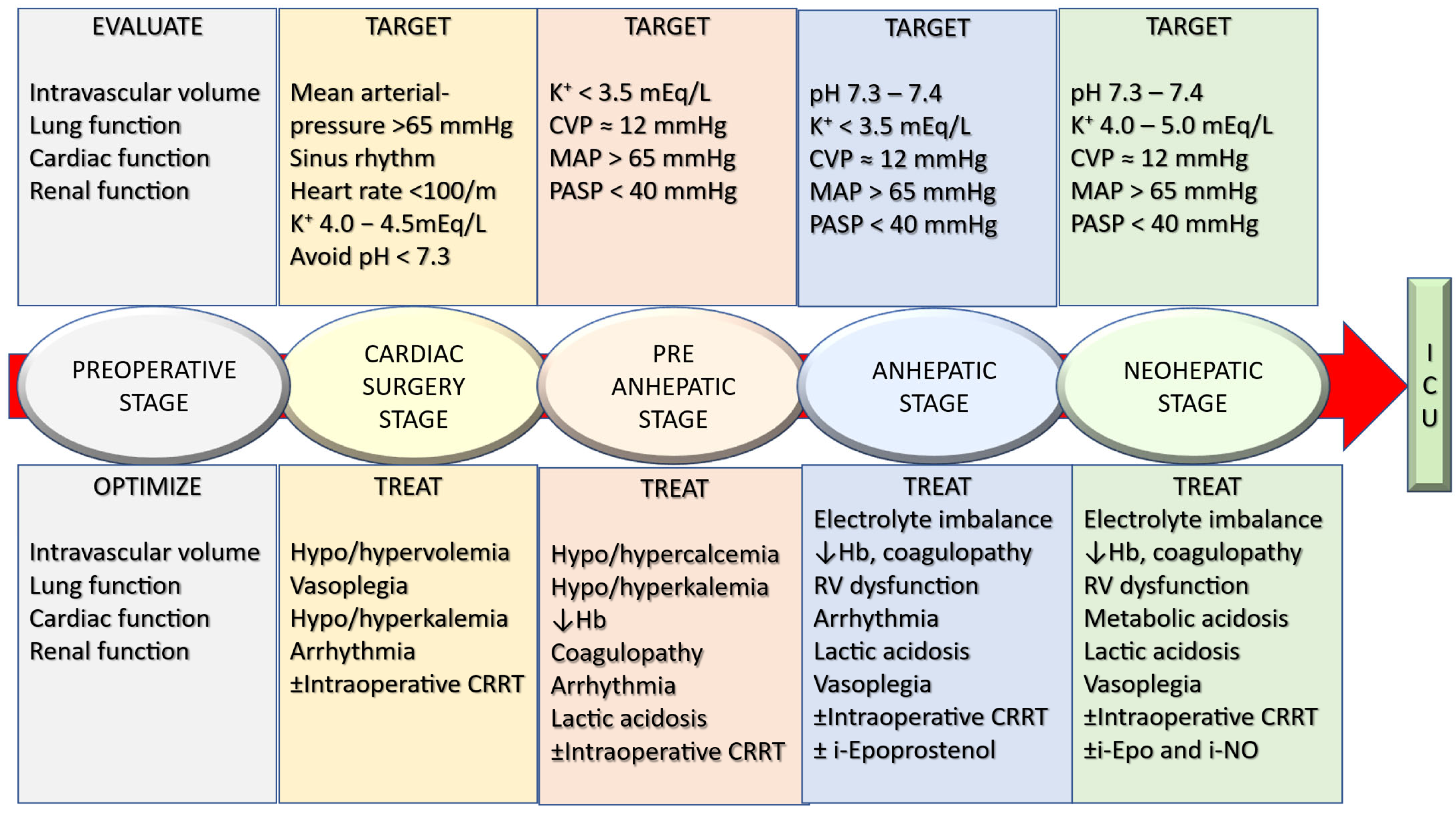
Overall, assessment and optimization of volume status, acidosis, vasoplegia, continuous renal replacement therapy and right ventricle dysfunction are crucial points for the successful management of concurrent LT and OPCAB (Figure 5).
Despite widespread adoption of Enhanced Recovery After Surgery (ERAS) protocols across surgical specialties, implementation in transplantation is sparse, resulting in a relative paucity of high-quality evidence. Moreover, changes in U.S. organ allocation policies—which prioritize preoperative illness severity—may inadvertently discourage preoperative optimization, a key principle of ERAS pathways [195]. Nevertheless, emerging data suggest that ERAS implementation in liver transplantation can significantly reduce hospital length of stay, with reductions approaching 47%. Future research should focus on defining standardized, evidence-based protocols for intraoperative CRRT use, lactate-guided therapy, and vasoactive agent selection in transplantation. Prospective studies integrating hemodynamic monitoring, metabolic targets, and ERAS principles can improve outcomes in this vulnerable population.

Author Contributions

Conceptualization, S.R.; data curation, S.R.; writing—original draft preparation, S.R., M.G., S.I., M.Q.B. and B.R.; writing—review and editing, S.R., M.G., S.I., M.Q.B. and B.R.; supervision, S.R. All authors have read and agreed to the published version of the manuscript.

Funding

This review received no external funding.

Institutional Review Board Statement

Not applicable.

Informed Consent Statement

Not applicable.

Data Availability Statement

No new data studied in this study. Data sharing is not applicable to this article.

Conflicts of Interest

The authors declare no conflicts of interest.

Abbreviations

The following abbreviations apply in this manuscript: LT: liver transplantation; ESLD, end-stage liver disease; ICU, intensive care unit; AKI, acute kidney injury; BNP, B-type natriuretic peptide; NT-proBNP, N-terminal pro-BNP; FENa, fractional excretion of sodium; HRS, hepatorenal syndrome; FEUrea, fractional excretion of urea; MELD, model for end-stage liver disease; CXR, chest X-ray; POCUS, point-of-care ultrasound; TEE, transesophageal echocardiography; RAAS, renin–angiotensin–aldosterone system; AASLD, American Association for the Study of Liver Diseases; SNS, sympathetic nervous system; RRT, renal replacement therapy; CRRT, continuous renal replacement therapy; LA, lactic acidosis; Na+/K+—ATPase, sodium/potassium—adenosine triphosphatase; MAP, mean arterial pressure; CCM, cirrhotic cardiomyopathy; PH, pulmonary hypertension, PoPH, portopulmonary hypertension; PAH, pulmonary arterial hypertension; mPAP, mean pulmonary arterial pressure; PVR, pulmonary vascular resistance; WU, Woods Units; PCWP, pulmonary capillary wedge pressure; RVSP, right ventricular systolic pressure; TR, tricuspid regurgitation; RHC, right heat catheterization; RV, right ventricle; RVD, right ventricular dysfunction; RVF, right ventricular failure; PAP, pulmonary artery pressure; CO, cardiac output; iNO, inhaled nitric oxide; MCS, mechanical circulatory support; CS, cardiac surgery; CIT, cold ischemic time; OPCAB, off-pump coronary artery bypass grafting; PCI, percutaneous coronary intervention; CPB, cardiopulmonary bypass, GDFT, goal-directed fluid therapy.

References

  1. Martin, F.; Carmona, M.; Mahillo, B.; Alvarez, M.; Luengo, A.; Chatzixiros, E.; López-Fraga, M.; Domínguez-Gil, B.; Tullius, S.G. Organ Donation and Transplantation Worldwide: The Global Observatory on Donation and Transplantation 2024 Report. Transplantation 2026, 110, e655–e669. [Google Scholar] [CrossRef] [Scilit]
  2. Lang, S.A.; Bednarsch, J.; Czigany, Z.; Joechle, K.; Kroh, A.; Amygdalos, I.; Strnad, P.; Bruns, T.; Heise, D.; Ulmer, F.; et al. Liver transplantation in malignant disease. World J. Clin. Oncol. 2021, 12, 623–645. [Google Scholar] [CrossRef] [Scilit] [PubMed]
  3. Pérez-Calatayud, A.A.; Hofmann, A.; Pérez-Ferrer, A.; Escorza-Molina, C.; Torres-Pérez, B.; Zaccarias-Ezzat, J.R.; Sanchez-Cedillo, A.; Paez-Zayas, V.M.; Carrillo-Esper, R.; Görlinger, K. Patient Blood Management in Liver Transplant-A Concise Review. Biomedicines 2023, 11, 1093. [Google Scholar] [CrossRef] [Scilit] [PubMed]
  4. Patel, S.; Gutmann, C.; Loveridge, R.; Pirani, T.; Willars, C.; Vercueil, A.; Angelova-Chee, M.; Aluvihare, V.; Heneghan, M.; Menon, K.; et al. Perioperative extracorporeal membrane oxygenation in liver transplantation—Bridge to transplantation, intraoperative salvage, and postoperative support: Outcomes and predictors for survival in a large-volume liver transplant center. Am. J. Transplant. 2025, 25, 396–405. [Google Scholar] [CrossRef] [Scilit] [PubMed]
  5. Benjamin, E.J.; Virani, S.S.; Callaway, C.W.; Chamberlain, A.M.; Chang, A.R.; Cheng, S.; Chiuve, S.E.; Cushman, M.; Delling, F.N.; Deo, R.; et al. Heart Disease and Stroke Statistics-2018 Update: A Report From the American Heart Association. Circulation 2018, 137, e67–e492. [Google Scholar] [CrossRef] [Scilit]
  6. Uludag Yanaral, T.; Ertugrul, G.; Ulukan, M.O.; Karaaslan, P.; Karaca, I.O.; Dayangac, M. Combined liver transplantation and off-pump coronary artery bypass grafting: A report of two cases. Acta Chir. Belg. 2022, 122, 269–274. [Google Scholar] [CrossRef] [Scilit]
  7. Wong, F.; Nadim, M.K.; Kellum, J.A.; Salerno, F.; Bellomo, R.; Gerbes, A.; Angeli, P.; Moreau, R.; Davenport, A.; Jalan, R.; et al. Working Party proposal for a revised classification system of renal dysfunction in patients with cirrhosis. Gut 2011, 60, 702–709. [Google Scholar] [CrossRef] [Scilit]
  8. Mazumder, N.R.; Kazen, A.; Carek, A.; Etemadi, M.; Levitsky, J. The answer at our fingertips: Volume status in cirrhosis determined by machine learning and pulse oximeter waveform. Physiol. Rep. 2022, 10, e15223. [Google Scholar] [CrossRef] [Scilit]
  9. Davenport, A.; Argawal, B.; Wright, G.; Mantzoukis, K.; Dimitrova, R.; Davar, J.; Vasianopoulou, P.; Burroughs, A.K. Can non-invasive measurements aid clinical assessment of volume in patients with cirrhosis? World J Hepatol 2013, 5, 433–438. [Google Scholar] [CrossRef] [Scilit]
  10. Davenport, A.; Ahmad, J.; Al-Khafaji, A.; Kellum, J.A.; Genyk, Y.S.; Nadim, M.K. Medical management of hepatorenal syndrome. Nephrol. Dial. Transplant. 2012, 27, 34–41. [Google Scholar] [CrossRef] [Scilit]
  11. Marik, P.E.; Baram, M.; Vahid, B. Does central venous pressure predict fluid responsiveness? A systematic review of the literature and the tale of seven mares. Chest 2008, 134, 172–178. [Google Scholar] [CrossRef] [Scilit]
  12. Taghavi, S.; Nassar, A.K.; Askari, R. Hypovolemia and Hypovolemic Shock; StatPearls Publishing LLC: Treasure Island, FL, USA, 2025. [Google Scholar]
  13. Wallace, H.A.; Regunath, H. Fluid Resuscitation; StatPearls Publishing LLC: Treasure Island, FL, USA, 2025. [Google Scholar]
  14. Metwaly, A.; Khalik, A.A.; Nasr, F.M.; Sabry, A.I.; Gouda, M.F.; Hassan, M. Brain Natriuretic Peptide in Liver Cirrhosis and Fatty Liver: Correlation with Cardiac Performance. Electron. Physician 2016, 8, 1984–1993. [Google Scholar] [CrossRef] [Scilit] [PubMed]
  15. Kansagra, C.; Kumar, A.; Abhisheka, T.; Joshi, N.; Sukanya, B.; Sandip, S.; Nikhil, S.; Kunal, V. NT Pro-BNP Levels in Patients With Cirrhosis: Correlation with Cardiovascular Dysfunction and Severity of Disease. Clin. Gastroenterol. Hepatol. 2015, 13, 214. [Google Scholar] [CrossRef] [Scilit]
  16. Alsaad, A.A.; Wadei, H.M. Fractional excretion of sodium in hepatorenal syndrome: Clinical and pathological correlation. World J. Hepatol. 2016, 8, 1497–1501. [Google Scholar] [CrossRef] [Scilit] [PubMed]
  17. Patidar, K.R.; Kang, L.; Bajaj, J.S.; Carl, D.; Sanyal, A.J. Fractional excretion of urea: A simple tool for the differential diagnosis of acute kidney injury in cirrhosis. Hepatology 2018, 68, 224–233. [Google Scholar] [CrossRef] [Scilit]
  18. Sim, J.; Toka, C.; Forrest, E. P188 The prognostic value of the fractional excretion of urea in patients with cirrhosis. Gut 2021, 70, A141–A141. [Google Scholar] [CrossRef] [Scilit]
  19. Kearney, D.; Reisinger, N.; Lohani, S. Integrative Volume Status Assessment. Pocus J. 2022, 7, 65–77. [Google Scholar] [CrossRef] [Scilit]
  20. Koratala, A.; Ronco, C.; Kazory, A. Hepatocardiorenal Syndrome: Integrating Pathophysiology with Clinical Decision-Making via Point-Of-Care Ultrasound. Cardiorenal Med. 2025, 15, 184–197. [Google Scholar] [CrossRef] [Scilit]
  21. Renner, J.; Gruenewald, M.; Brand, P.; Steinfath, M.; Scholz, J.; Lutter, G.; Bein, B. Global end-diastolic volume as a variable of fluid responsiveness during acute changing loading conditions. J. Cardiothorac. Vasc. Anesth. 2007, 21, 650–654. [Google Scholar] [CrossRef] [Scilit]
  22. Singh, S.; Kuschner, W.G.; Lighthall, G. Perioperative intravascular fluid assessment and monitoring: A narrative review of established and emerging techniques. Anesthesiol. Res. Pract. 2011, 2011, 231493. [Google Scholar] [CrossRef] [Scilit]
  23. Hahn, R.T.; Saric, M.; Faletra, F.F.; Garg, R.; Gillam, L.D.; Horton, K.; Khalique, O.K.; Little, S.H.; Mackensen, G.B.; Oh, J.; et al. Recommended Standards for the Performance of Transesophageal Echocardiographic Screening for Structural Heart Intervention: From the American Society of Echocardiography. J. Am. Soc. Echocardiogr. 2022, 35, 1–76. [Google Scholar] [CrossRef] [Scilit] [PubMed]
  24. Zhang, L.; Xie, Y.; Ren, Z.; Xie, M. Transesophageal echocardiography related complications. Front. Cardiovasc. Med. 2024, 11, 1410594. [Google Scholar] [CrossRef] [Scilit]
  25. Nadim, M.K.; Kellum, J.A.; Davenport, A.; Wong, F.; Davis, C.; Pannu, N.; Tolwani, A.; Bellomo, R.; Genyk, Y.S.; ADQI Workgroup. Hepatorenal syndrome: The 8th International Consensus Conference of the Acute Dialysis Quality Initiative (ADQI) Group. Crit. Care 2012, 16, R23. [Google Scholar] [CrossRef] [Scilit] [PubMed]
  26. Sun, F.R.; Meng, Y.-M.; Wang, B.-Y.; Liu, Y.-F.; Liu, C.-X.; Xie, D.-W.; Ding, Y.-Y.; Li, J.-P.; Ma, L. Correlations between MELD score and left ventricular function in patients with end-stage liver disease. Zhonghua Gan Zang Bing Za Zhi 2010, 18, 758–762. [Google Scholar] [PubMed]
  27. Rabie, R.N.; Cazzaniga, M.; Salerno, F.; Wong, F. The use of E/A ratio as a predictor of outcome in cirrhotic patients treated with transjugular intrahepatic portosystemic shunt. Am. J. Gastroenterol. 2009, 104, 2458–2466. [Google Scholar] [CrossRef] [Scilit]
  28. Murray, K.F.; Carithers, R.L., Jr. AASLD practice guidelines: Evaluation of the patient for liver transplantation. Hepatology 2005, 41, 1407–1432. [Google Scholar] [CrossRef] [Scilit]
  29. Miles, L.F.; Couture, E.J.; Potes, C.; Makar, T.; Fernando, M.C.; Hungenahally, A.; Mathieson, M.D.; Perlman, H.; Perini, M.V.; Thind, D.; et al. Preliminary experience with continuous right ventricular pressure and transesophageal echocardiography monitoring in orthotopic liver transplantation. PLoS ONE 2022, 17, e0263386. [Google Scholar] [CrossRef] [Scilit]
  30. Patidar, K.R.; Belcher, J.M.; Regner, K.R.; Hillien, S.A.S.; Simonetto, D.A.; Asrani, S.K.; Neyra, J.A.; Sharma, P.; Velez, J.C.Q.; Wadei, H.; et al. Incidence and outcomes of acute kidney injury including hepatorenal syndrome in hospitalized patients with cirrhosis in the US. J. Hepatol. 2023, 79, 1408–1417. [Google Scholar] [CrossRef] [Scilit]
  31. Bernardi, M.; Moreau, R.; Angeli, P.; Schnabl, B.; Arroyo, V. Mechanisms of decompensation and organ failure in cirrhosis: From peripheral arterial vasodilation to systemic inflammation hypothesis. J. Hepatol. 2015, 63, 1272–1284. [Google Scholar] [CrossRef] [Scilit]
  32. Jalan, R.; D’aMico, G.; Trebicka, J.; Moreau, R.; Angeli, P.; Arroyo, V. New clinical and pathophysiological perspectives defining the trajectory of cirrhosis. J. Hepatol. 2021, 75, S14–S26. [Google Scholar] [CrossRef] [Scilit]
  33. Fukui, H.; Kawaratani, H.; Kaji, K.; Takaya, H.; Yoshiji, H. Management of refractory cirrhotic ascites: Challenges and solutions. Hepat. Med. 2018, 10, 55–71. [Google Scholar] [CrossRef] [Scilit] [PubMed]
  34. Burgos, A.C.; Thornburg, B. Transjugular Intrahepatic Portosystemic Shunt Placement for Refractory Ascites: Review and Update of the Literature. Semin. Interv. Radiol. 2018, 35, 165–168. [Google Scholar] [CrossRef] [Scilit] [PubMed]
  35. Adebayo, D.; Neong, S.F.; Wong, F. Refractory Ascites in Liver Cirrhosis. Am. J. Gastroenterol. 2019, 114, 40–47. [Google Scholar] [CrossRef] [Scilit] [PubMed]
  36. Storni, F.; Stirnimann, G.; Banz, V.; De Gottardi, A. Treatment of Malignant Ascites Using an Automated Pump Device. Am. J. Gastroenterol. 2018, 113, 1060–1061. [Google Scholar] [CrossRef] [Scilit]
  37. Kulkarni, A.V.; Kumar, P.; Sharma, M.; Sowmya, T.R.; Talukdar, R.; Rao, P.N.; Reddy, D.N. Pathophysiology and Prevention of Paracentesis-induced Circulatory Dysfunction: A Concise Review. J. Clin. Transl. Hepatol. 2020, 8, 42–48. [Google Scholar] [CrossRef] [Scilit]
  38. Biggins, S.W.; Angeli, P.; Garcia-Tsao, G.; Ginès, P.; Ling, S.C.; Nadim, M.K.; Wong, F.; Kim, W.R. Diagnosis, Evaluation, and Management of Ascites, Spontaneous Bacterial Peritonitis and Hepatorenal Syndrome: 2021 Practice Guidance by the American Association for the Study of Liver Diseases. Hepatology 2021, 74, 1014–1048. [Google Scholar] [CrossRef] [Scilit]
  39. Tanaka, T.; Weg, M.V.; Jones, M.P.; Wehby, G. Assessment of the 2021 AASLD Practice Guidance for Albumin Infusion in Elective Therapeutic Paracentesis: A Regression Discontinuity Design. Am. J. Gastroenterol. 2024, 119, 2045–2051. [Google Scholar] [CrossRef] [Scilit]
  40. Lindsay, A.J.; Burton, J.; Ray, C.E., Jr. Paracentesis-induced circulatory dysfunction: A primer for the interventional radiologist. Semin. Interv. Radiol. 2014, 31, 276–278. [Google Scholar] [CrossRef] [Scilit]
  41. Cabrera, J.; Falcón, L.; Gorriz, E.; Pardo, M.D.; Granados, R.; Quinones, A.; Maynar, M. Abdominal decompression plays a major role in early postparacentesis haemodynamic changes in cirrhotic patients with tense ascites. Gut 2001, 48, 384–389. [Google Scholar] [CrossRef] [Scilit]
  42. Badillo, R.; Rockey, D.C. Hepatic hydrothorax: Clinical features, management, and outcomes in 77 patients and review of the literature. Medicine 2014, 93, 135–142. [Google Scholar] [CrossRef] [Scilit]
  43. Krok, K.L. Hepatic hydrothorax: Current concepts. Clin. Liver Dis. 2014, 4, 35–37. [Google Scholar] [CrossRef] [Scilit] [PubMed]
  44. Xiol, X.; Castellote, J.; Cortes-Beut, R.; Delgado, M.; Guardiola, J.; Sesé, E. Usefulness and complications of thoracentesis in cirrhotic patients. Am. J. Med. 2001, 111, 67–69. [Google Scholar] [CrossRef] [Scilit] [PubMed]
  45. Kiafar, C.; Gilani, N. Hepatic hydrothorax: Current concepts of pathophysiology and treatment options. Ann. Hepatol. 2008, 7, 313–320. [Google Scholar] [CrossRef] [Scilit] [PubMed]
  46. Chaaban, T.; Kanj, N.; Akl, I.B. Hepatic Hydrothorax: An Updated Review on a Challenging Disease. Lung 2019, 197, 399–405. [Google Scholar] [CrossRef] [Scilit]
  47. Garbuzenko, D.V.; Arefyev, N.O. Hepatic hydrothorax: An update and review of the literature. World J. Hepatol. 2017, 9, 1197–1204. [Google Scholar] [CrossRef] [Scilit]
  48. Sharma, P.; Nagarajan, V. Pleural effusion from leaky diaphragm--the hepatic hydrothorax. J. Gen. Intern. Med. 2014, 29, 1309. [Google Scholar] [CrossRef] [Scilit]
  49. Banini, B.A.; Alwatari, Y.; Stovall, M.; Ogden, N.; Gershman, E.; Shah, R.D.; Strife, B.J.; Shojaee, S.; Sterling, R.K. Multidisciplinary Management of Hepatic Hydrothorax in 2020: An Evidence-Based Review and Guidance. Hepatology 2020, 72, 1851–1863. [Google Scholar] [CrossRef] [Scilit]
  50. Hou, F.; Qi, X.; Ning, Z.; Zhao, J.; Zhang, X.; Deng, H.; Peng, Y.; Li, J.; Yao, H.; Li, H.; et al. Prevalence, risk factors, and in-hospital outcome of pleural effusion in liver cirrhosis: A retrospective observational study. Int. J. Clin. Exp. Med. 2016, 9, 3265–3279. [Google Scholar]
  51. Matei, D.; Pasca, S.; David, A.; Groza, I.; Negrutiu, D.-E.; Procopet, B.; Andreica, V.; Vesa, S.; Tantau, M. Hepatic hydrothorax in patients with cirrhosis and ascites–its prevalence and association with other cirrhosis complications. J. Hepatol. 2017, 1, S135. [Google Scholar] [CrossRef] [Scilit]
  52. Serrat, J.; Roza, J.J.; Planella, T. Hepatic hydrothorax in the absence of ascites: Respiratory failure in a cirrhotic patient. Anesth. Analg. 2004, 99, 1803–1804. [Google Scholar] [CrossRef] [Scilit]
  53. Muralimohan, R.; Delu, A.; Ma, T. Pericardial effusion of obscure origin. Dig. Dis. Sci. 2014, 59, 2909–2912. [Google Scholar] [CrossRef] [Scilit]
  54. Naschitz, J.E.; Slobodin, G.; Lewis, R.J.; Zuckerman, E.; Yeshurun, D. Heart diseases affecting the liver and liver diseases affecting the heart. Am. Heart J. 2000, 140, 111–120. [Google Scholar] [CrossRef] [Scilit]
  55. Garg, A.; Armstrong, W.F. Echocardiography in liver transplant candidates. JACC Cardiovasc. Imaging 2013, 6, 105–119. [Google Scholar] [CrossRef] [Scilit]
  56. Sagristà-Sauleda, J.; Mercè, A.S.; Soler-Soler, J. Diagnosis and management of pericardial effusion. World J. Cardiol. 2011, 3, 135–143. [Google Scholar] [CrossRef] [Scilit]
  57. Park, J.; Lee, M.; Kim, J.; Choi, H.; Kwon, A.; Chung, H.; Hong, S.; Park, C.; Choi, J.; Chae, M. Intraoperative Management to Prevent Cardiac Collapse in a Patient with a Recurrent, Large-volume Pericardial Effusion and Paroxysmal Atrial Fibrillation During Liver Transplantation: A Case Report. Transplant. Proc. 2019, 51, 568–574. [Google Scholar] [CrossRef] [Scilit] [PubMed]
  58. Kellum, J.A.; Ronco, C. The 17th Acute Disease Quality Initiative International Consensus Conference: Introducing Precision Renal Replacement Therapy. Blood Purif. 2016, 42, 221–223. [Google Scholar] [CrossRef] [Scilit] [PubMed]
  59. Paine, C.H.; Pichler, R.H. Intraoperative Renal Replacement Therapy for Liver Transplantation: Is There Really a Benefit? Liver Transplant. 2020, 26, 971–972. [Google Scholar] [CrossRef] [Scilit] [PubMed]
  60. Zimmerman, M.A.; Selim, M.; Kim, J.; Regner, K.; Saeian, K.; Zanowski, S.; Martin, A.; Connolly, L.A.; Lauer, K.K.; Woehlck, H.J.; et al. Outcome analysis of continuous intraoperative renal replacement therapy in the highest acuity liver transplant recipients: A single-center experience. Surgery 2017, 161, 1279–1286. [Google Scholar] [CrossRef] [Scilit]
  61. Townsend, D.R.; Bagshaw, S.M.; Jacka, M.J.; Bigam, D.; Cave, D.; Gibney, N.R. Intraoperative renal support during liver transplantation. Liver Transpl. 2009, 15, 73–78. [Google Scholar] [CrossRef] [Scilit]
  62. Fehervari, I.; Fazakas, J.; Gerlei, Z.; Nemes, B.; Kobori, L. Mitigation of cytokine storm by intraoperative use of renal replacement therapy during combined liver-kidney transplantation. Transplant. Proc. 2010, 42, 2353–2356. [Google Scholar] [CrossRef] [Scilit]
  63. Adelmann, D.; Olmos, A.; Liu, L.L.; Feiner, J.R.; Roll, G.R.; Burdine, L.; Tavakol, M.; Syed, S.; Orandi, B.J.; Niemann, C.U. Intraoperative Management of Liver Transplant Patients Without the Routine Use of Renal Replacement Therapy. Transplantation 2018, 102, e229–e235. [Google Scholar] [CrossRef] [Scilit] [PubMed]
  64. Safwan, M.; Gosnell, J.; Collins, K.; Rizzari, M.; Yoshida, A.; Abouljoud, M.; Nagai, S. Effects of Intraoperative Continuous Renal Replacement Therapy on Outcomes in Liver Transplantation. Transplant. Proc. 2020, 52, 265–270. [Google Scholar] [CrossRef] [Scilit] [PubMed]
  65. Kanda, H.; Hirasaki, Y.; Iida, T.; Kanao-Kanda, M.; Toyama, Y.; Chiba, T.; Kunisawa, T. Perioperative Management of Patients With End-Stage Renal Disease. J. Cardiothorac. Vasc. Anesth. 2017, 31, 2251–2267. [Google Scholar] [CrossRef] [Scilit] [PubMed]
  66. Matuszkiewicz-Rowińska, J.; Małyszko, J.; Wieliczko, M. Renal support during liver transplantation: When to consider it? Transplant. Proc. 2013, 45, 3157–3162. [Google Scholar] [CrossRef] [Scilit]
  67. Rabindranath, K.S.; Adams, J.; MacLeod, A.M.; Muirhead, N. Intermittent versus continuous renal replacement therapy for acute renal failure in adults. Cochrane Database Syst. Rev. 2007, 3, Cd003773. [Google Scholar] [CrossRef] [Scilit]
  68. Baxter. Prismaflex ST Set; Baxter: Deerfield, IL, USA, 2019. [Google Scholar]
  69. Honore, P.; Spapen, H. What a Clinician Should Know About a Renal Replacement Membrane? J. Transl. Intern. Med. 2018, 6, 62–65. [Google Scholar] [CrossRef] [Scilit]
  70. Vitin, A.; Muczynski, K.; Bakthavatsalam, R.; Martay, K.; Dembo, G.; Metzner, J. Treatment of severe lactic acidosis during the pre-anhepatic stage of liver transplant surgery with intraoperative hemodialysis. J. Clin. Anesth. 2010, 22, 466–472. [Google Scholar] [CrossRef] [Scilit]
  71. De Corte, W.; Vuylsteke, S.; De Waele, J.J.; Dhondt, A.W.; Decruyenaere, J.; Vanholder, R.; Hoste, E.A. Severe lactic acidosis in critically ill patients with acute kidney injury treated with renal replacement therapy. J. Crit. Care 2014, 29, 650–655. [Google Scholar] [CrossRef] [Scilit]
  72. Luft, F.C. Lactic acidosis update for critical care clinicians. J. Am. Soc. Nephrol. 2001, 12, S15–S19. [Google Scholar] [CrossRef] [Scilit]
  73. Bellomo, R. Bench-to-bedside review: Lactate and the kidney. Crit. Care 2002, 6, 322–326. [Google Scholar] [CrossRef] [Scilit]
  74. Busuttil, R.W.; Tanaka, K. The utility of marginal donors in liver transplantation. Liver Transpl. 2003, 9, 651–663. [Google Scholar] [CrossRef] [Scilit]
  75. Adeva-Andany, M.M.; Fernández-Fernández, C.; Mouriño-Bayolo, D.; Castro-Quintela, E.; Domínguez-Montero, A. Sodium bicarbonate therapy in patients with metabolic acidosis. Sci. World J. 2014, 2014, 627673. [Google Scholar] [CrossRef] [Scilit] [PubMed]
  76. Singh, T.; Alagasundaramoorthy, S.; Gregory, A.; Astor, B.C.; Maursetter, L. Low dialysis potassium bath is associated with lower mortality in end-stage renal disease patients admitted to hospital with severe hyperkalemia. Clin. Kidney J. 2021, 14, 2059–2063. [Google Scholar] [CrossRef] [Scilit] [PubMed]
  77. Kościelska, M.; Matuszkiewicz-Rowińska, J.; Zieniewicz, K.; Krawczyk, M.; Giercuszkiewicz, D.; Sierdziński, J.; Żebrowski, P.; Malyszko, J. Intraoperative dialysis during liver transplantation. Nephrol. Dial. Transplant. 2020, 35, gfaa142.P0627. [Google Scholar] [CrossRef] [Scilit]
  78. Altun, D.; Arnaz, A.; Doğan, A.; Yalçınbaş, Y.; Türköz, R.; Yüksek, A.; Altun, D.; Abdullah, T.; Özgen, S.U.; Toraman, F.; et al. Measuring potassium level in packed red blood cells before using: Word of caution for congenital cardiac surgery. J. Card. Surg. 2022, 37, 535–541. [Google Scholar] [CrossRef] [Scilit]
  79. Cardigan, R.; New, H.V.; Tinegate, H.; Thomas, S. Washed red cells: Theory and practice. Vox Sang. 2020, 115, 606–616. [Google Scholar] [CrossRef] [Scilit]
  80. Dawwas, M.F.; Lewsey, J.D.; Neuberger, J.M.; Gimson, A.E. The impact of serum sodium concentration on mortality after liver transplantation: A cohort multicenter study. Liver Transpl. 2007, 13, 1115–1124. [Google Scholar] [CrossRef] [Scilit]
  81. Karapanagiotou, A.; Kydona, C.; Papadopoulos, S.; Dimitriadis, C.; Giasnetsova, T.; Rempelakos, G.; Passakiotou, M.; Fouzas, I.; Papanikolaou, V.; Gritsi-Gerogianni, N. The effect of hyponatremia on the outcome of patients after orthotopic liver transplantation. Transplant. Proc. 2012, 44, 2724–2726. [Google Scholar] [CrossRef] [Scilit]
  82. Cascales Campos, P.; Ramirez, P.; Gonzalez, R.; Pons, J.; Miras, M.; Bueno, F.S.; Robles, R.; Parrilla, P. Central pontine and extrapontine myelinolysis: A rare and fatal complication after liver transplantation. Transplant. Proc. 2011, 43, 2237–2238. [Google Scholar] [CrossRef] [Scilit]
  83. Parmar, A.; Bigam, D.; Meeberg, G.; Cave, D.; Townsend, D.R.; Gibney, R.N.; Bagshaw, S.M. An evaluation of intraoperative renal support during liver transplantation: A matched cohort study. Blood Purif. 2011, 32, 238–248. [Google Scholar] [CrossRef] [Scilit]
  84. Fall, P.J.; Szerlip, H.M. Lactic Acidosis: From Sour Milk to Septic Shock. J. Intensiv. Care Med. 2005, 20, 255–271. [Google Scholar] [CrossRef] [Scilit] [PubMed]
  85. Kraut, J.A.; Madias, N.E. Lactic acidosis. N. Engl. J. Med. 2014, 371, 2309–2319. [Google Scholar] [CrossRef] [Scilit] [PubMed]
  86. He, Y.; Ong, J.; Ong, S. Refractory Lactic Acidosis and an Approach to its Management—A Case Report. J. Crit. Care Med. 2019, 5, 60–65. [Google Scholar] [CrossRef] [Scilit] [PubMed]
  87. Macquillan, G.C.; Seyam, M.S.; Nightingale, P.; Neuberger, J.M.; Murphy, N. Blood lactate but not serum phosphate levels can predict patient outcome in fulminant hepatic failure. Liver Transpl. 2005, 11, 1073–1079. [Google Scholar] [CrossRef] [Scilit]
  88. Jeppesen, J.B.; Mortensen, C.; Bendtsen, F.; Møller, S. Lactate metabolism in chronic liver disease. Scand. J. Clin. Lab. Investig. 2013, 73, 293–299. [Google Scholar] [CrossRef] [Scilit]
  89. Wiggans, M.G.; Starkie, T.; Shahtahmassebi, G.; Woolley, T.; Birt, D.; Erasmus, P.; Anderson, I.; Bowles, M.J.; Aroori, S.; A Stell, D. Serum arterial lactate concentration predicts mortality and organ dysfunction following liver resection. Perioper. Med. 2013, 2, 21. [Google Scholar] [CrossRef] [Scilit]
  90. Luchette, F.A.; Jenkins, W.A.; Friend, L.A.; Su, C.; Fischer, J.E.; James, J.H. Hypoxia is not the sole cause of lactate production during shock. J. Trauma 2002, 52, 415–419. [Google Scholar] [CrossRef] [Scilit]
  91. Uvizl, R.; Klementa, B.; Adamus, M.; Neiser, J. Biochemical changes in the patient’s plasma after red blood cell transfusion. Signa Vitae J. Intesive Care Emerg. Med. 2011, 6, 64–71. [Google Scholar]
  92. Wu, C.C.; Yeung, L.-K.; Tsai, W.-S.; Tseng, C.-F.; Chu, P.; Huang, T.-Y.; Lin, Y.-F.; Lu, K.-C. Incidence and factors predictive of acute renal failure in patients with advanced liver cirrhosis. Clin. Nephrol. 2006, 65, 28–33. [Google Scholar] [CrossRef] [Scilit]
  93. Fukazawa, K.; Vitin, A.A.; Pretto, E.A., Jr. Serum acidosis prior to reperfusion facilitates hemodynamic recovery following liver transplantation. J. Anesth. 2016, 30, 80–88. [Google Scholar] [CrossRef] [Scilit]
  94. Jun, J.; Kim, G.; Na, Y.; Gwak, M.; Ko, J.; Lee, S. Analysis of predictors for lactate elimination after reperfusion in recipients of living-donor liver transplantation. Transplant. Proc. 2014, 46, 709–711. [Google Scholar] [CrossRef] [Scilit]
  95. Levy, B.; Perez, P.; Perny, J.; Thivilier, C.; Gerard, A. Comparison of norepinephrine-dobutamine to epinephrine for hemodynamics, lactate metabolism, and organ function variables in cardiogenic shock. A prospective, randomized pilot study. Crit. Care Med. 2011, 39, 450–455. [Google Scholar] [CrossRef] [Scilit]
  96. Levy, B.; Mansart, A.; Bollaert, P.-E.; Franck, P.; Mallie, J.-P. Effects of epinephrine and norepinephrine on hemodynamics, oxidative metabolism, and organ energetics in endotoxemic rats. Intensive Care Med. 2003, 29, 292–300. [Google Scholar] [CrossRef] [Scilit] [PubMed]
  97. Jentzer, J.C.; Vallabhajosyula, S.; Khanna, A.K.; Chawla, L.S.; Busse, L.W.; Kashani, K.B. Management of Refractory Vasodilatory Shock. Chest 2018, 154, 416–426. [Google Scholar] [CrossRef] [Scilit] [PubMed]
  98. Levy, B.; Fritz, C.; Tahon, E.; Jacquot, A.; Auchet, T.; Kimmoun, A. Vasoplegia treatments: The past, the present, and the future. Crit. Care 2018, 22, 52. [Google Scholar] [CrossRef] [Scilit] [PubMed]
  99. Ortoleva, J.; Dalia, A.; Convissar, D.; Pisano, D.V.; Bittner, E.; Berra, L. Vasoplegia in Heart, Lung, or Liver Transplantation: A Narrative Review. J. Cardiothorac. Vasc. Anesth. 2025, 39, 988–1003. [Google Scholar] [CrossRef] [Scilit]
  100. Martell, M.; Coll, M.; Ezkurdia, N.; Raurell, I.; Genescà, J. Physiopathology of splanchnic vasodilation in portal hypertension. World J. Hepatol. 2010, 2, 208–220. [Google Scholar] [CrossRef] [Scilit]
  101. Vincent, J.L.; De Backer, D. Circulatory shock. N. Engl. J. Med. 2013, 369, 1726–1734. [Google Scholar] [CrossRef] [Scilit]
  102. Labovitz, A.J.; Noble, V.E.; Bierig, M.; Goldstein, S.A.; Jones, R.; Kort, S.; Porter, T.R.; Spencer, K.T.; Tayal, V.S.; Wei, K. Focused cardiac ultrasound in the emergent setting: A consensus statement of the American Society of Echocardiography and American College of Emergency Physicians. J. Am. Soc. Echocardiogr. 2010, 23, 1225–1230. [Google Scholar] [CrossRef] [Scilit]
  103. Vincent, J.L.; Rhodes, A.; Perel, A.; Martin, G.S.; Rocca, G.; Vallet, B.; Pinsky, M.R.; Hofer, C.K.; Teboul, J.-L.; de Boode, W.-P.; et al. Clinical review: Update on hemodynamic monitoring--a consensus of 16. Crit. Care 2011, 15, 229. [Google Scholar] [CrossRef] [Scilit]
  104. Rhodes, A.; Evans, L.E.; Alhazzani, W.; Levy, M.M.; Antonelli, M.; Ferrer, R.; Kumar, A.; Sevransky, J.E.; Sprung, C.L.; Nunnally, M.E.; et al. Surviving Sepsis Campaign: International Guidelines for Management of Sepsis and Septic Shock: 2016. Intensive Care Med. 2017, 43, 304–377. [Google Scholar] [CrossRef] [Scilit]
  105. Bokoch, M.P.; Tran, A.T.; Brinson, E.L.; Marcus, S.G.; Reddy, M.; Sun, E.; Roll, G.R.; Pardo, M.; Fields, S.; Adelmann, D.; et al. Angiotensin II in liver transplantation (AngLT-1): Protocol of a randomised, double-blind, placebo-controlled trial. BMJ Open 2023, 13, e078713. [Google Scholar] [CrossRef] [Scilit] [PubMed]
  106. Shaefi, S.; Mittel, A.; Klick, J.; Evans, A.; Ivascu, N.S.; Gutsche, J.; Augoustides, J.G. Vasoplegia After Cardiovascular Procedures-Pathophysiology and Targeted Therapy. J. Cardiothorac. Vasc. Anesth. 2018, 32, 1013–1022. [Google Scholar] [CrossRef] [Scilit] [PubMed]
  107. Datt, V.; Wadhhwa, R.; Sharma, V.; Virmani, S.; Minhas, H.S.; Malik, S. Vasoplegic Syndrome After Cardiovascular Surgery: A Review of Pathophysiology and Outcome Oriented Therapeutic Management. Authorea 2021, 36, 3749–3760. [Google Scholar] [CrossRef] [Scilit] [PubMed]
  108. Ortoleva, J.P.; Cobey, F.C. A Systematic Approach to the Treatment of Vasoplegia Based on Recent Advances in Pharmacotherapy. J. Cardiothorac. Vasc. Anesth. 2019, 33, 1310–1314. [Google Scholar] [CrossRef] [Scilit]
  109. Crouch, C.; Hendrickse, A.; Gilliland, S.; Mandell, M.S. Unexpected Complication of Hydroxocobalamin Administration for Refractory Vasoplegia in Orthotopic Liver Transplant: A Case Report. Semin. Cardiothorac. Vasc. Anesth. 2019, 23, 409–412. [Google Scholar] [CrossRef] [Scilit]
  110. An, S.S.; Henson, C.P.; Freundlich, R.E.; McEvoy, M.D. Case report of high-dose hydroxocobalamin in the treatment of vasoplegic syndrome during liver transplantation. Am. J. Transplant. 2018, 18, 1552–1555. [Google Scholar] [CrossRef] [Scilit]
  111. Boettcher, B.T.; Woehlck, H.J.; Makker, H.; Pagel, P.S.; Freed, J.K. Hydroxocobalamin for treatment of catecholamine-resistant vasoplegia during liver transplantation: A single-center series of 20 cases. Int. J. Surg. Case Rep. 2022, 98, 107488. [Google Scholar] [CrossRef] [Scilit]
  112. Pai, S.L.; Torp, K.D.; Insignares, V.C.; DeMaria, S.; Giordano, C.R.; Logvinov, I.I.; Li, Z.; Chadha, R.; Aniskevich, S. Use of hydroxocobalamin to treat intraoperative vasoplegic syndrome refractory to vasopressors and methylene blue during liver transplantation. Clin. Transplant. 2024, 38, e15271. [Google Scholar] [CrossRef] [Scilit]
  113. Ortoleva, J.; Shapeton, A.D. Seeing Red: Hydroxocobalamin and Result Interference. J. Cardiothorac. Vasc. Anesth. 2023, 37, 1339–1342. [Google Scholar] [CrossRef] [Scilit]
  114. Koelzow, H.; Gedney, J.A.; Baumann, J.; Snook, N.J.; Bellamy, M.C. The effect of methylene blue on the hemodynamic changes during ischemia reperfusion injury in orthotopic liver transplantation. Anesth. Analg. 2002, 94, 824–829. [Google Scholar] [CrossRef] [Scilit] [PubMed]
  115. Cheng, S.S.; Berman, G.W.; Merritt, G.R.; Hendrickse, A.; Fiegel, M.J.; Teitelbaum, I.; Campsen, J.; Wachs, M.; Zimmerman, M.; Mandell, M.S. The response to methylene blue in patients with severe hypotension during liver transplantation. J. Clin. Anesth. 2012, 24, 324–328. [Google Scholar] [CrossRef] [Scilit]
  116. Fischer, G.W.; Bengtsson, Y.; Scarola, S.; Cohen, E. Methylene blue for vasopressor-resistant vasoplegia syndrome during liver transplantation. J. Cardiothorac. Vasc. Anesth. 2010, 24, 463–466. [Google Scholar] [CrossRef] [Scilit] [PubMed]
  117. Anstey, M.H.; Aljeaidi, M.S.; Palmer, R.; Jacques, A.; Mevavala, B.; Litton, E.; Wibrow, B. Intravenous vitamin C for vasoplegia: A double-blinded randomised clinical trial (VALENCIA trial). J. Crit. Care 2023, 78, 154369. [Google Scholar] [CrossRef] [Scilit] [PubMed]
  118. Wieruszewski, P.M.; Nei, S.D.; Maltais, S.; Schaff, H.V.; Wittwer, E.D. Vitamin C for Vasoplegia After Cardiopulmonary Bypass: A Case Series. A&A Pract. 2018, 11, 96–99. [Google Scholar]
  119. Hwang, K.Y.; Phoon, P.H.Y.; Hwang, N.C. Adverse Clinical Effects Associated With Non-catecholamine Pharmacologic Agents for Treatment of Vasoplegic Syndrome in Adult Cardiac Surgery. J. Cardiothorac. Vasc. Anesth. 2024, 38, 802–819. [Google Scholar] [CrossRef] [Scilit]
  120. Running, K.; Weinberg, D.; Trudo, W.; Sullivan, C.L.; Patel, G.P. Intraoperative Use of Angiotensin II for Severe Vasodilatory Shock During Liver Transplantation: A Case Report. A&A Pract. 2021, 15, e01402. [Google Scholar]
  121. Cecconi, M.; De Backer, D.; Antonelli, M.; Beale, R.; Bakker, J.; Hofer, C.; Jaeschke, R.; Mebazaa, A.; Pinsky, M.R.; Teboul, J.L.; et al. Consensus on circulatory shock and hemodynamic monitoring. Task force of the European Society of Intensive Care Medicine. Intensive Care Med. 2014, 40, 1795–1815. [Google Scholar]
  122. Eskesen, T.G.; Wetterslev, M.; Perner, A. Systematic review including re-analyses of 1148 individual data sets of central venous pressure as a predictor of fluid responsiveness. Intensive Care Med. 2016, 42, 324–332. [Google Scholar] [CrossRef] [Scilit]
  123. Monnet, X.; Marik, P.; Teboul, J.-L. Passive leg raising for predicting fluid responsiveness: A systematic review and meta-analysis. Intensive Care Med. 2016, 42, 1935–1947. [Google Scholar] [CrossRef] [Scilit]
  124. Lamontagne, F.; Meade, M.O.; Hébert, P.C.; Asfar, P.; Lauzier, F.; Seely, A.J.; Day, A.G.; Mehta, S.; Muscedere, J.; Bagshaw, S.M.; et al. Higher versus lower blood pressure targets for vasopressor therapy in shock: A multicentre pilot randomized controlled trial. Intensive Care Med. 2016, 42, 542–550. [Google Scholar] [CrossRef] [Scilit]
  125. Gu, W.J.; Zhang, Z.; Bakker, J. Early lactate clearance-guided therapy in patients with sepsis: A meta-analysis with trial sequential analysis of randomized controlled trials. Intensive Care Med. 2015, 41, 1862–1863. [Google Scholar] [CrossRef] [Scilit] [PubMed]
  126. Simpson, S.Q.; Gaines, M.; Hussein, Y.; Badgett, R.G. Early goal-directed therapy for severe sepsis and septic shock: A living systematic review. J. Crit. Care 2016, 36, 43–48. [Google Scholar] [CrossRef] [Scilit] [PubMed]
  127. Izzy, M.; VanWagner, L.B.; Lin, G.; Altieri, M.; Findlay, J.Y.; Oh, J.K.; Watt, K.D.; Lee, S.S. on behalf of the Cirrhotic Cardiomyopathy Consortium. Redefining Cirrhotic Cardiomyopathy for the Modern Era. Hepatology 2020, 71, 334–345. [Google Scholar] [CrossRef] [Scilit] [PubMed]
  128. Goswami, R.M.; Khouzam, R.N. Acute right ventricular failure after orthotopic liver transplantation. Ann. Transl. Med. 2017, 6, 19. [Google Scholar] [CrossRef] [Scilit]
  129. Humbert, M.; Humbert, M.; Vachiéry, J.-L.; Gibbs, S.; Lang, I.M.; Torbicki, A.; Simonneau, G.; Peacock, A.; Noordegraaf, A.V.; Beghetti, M.; et al. 2022 ESC/ERS Guidelines for the diagnosis and treatment of pulmonary hypertension. Eur. Heart J. 2022, 43, 3618–3731. [Google Scholar] [CrossRef] [Scilit]
  130. Kochar, R.; Rubin, M.I.N.; Fallon, M.B. Pulmonary complications of cirrhosis. Curr. Gastroenterol. Rep. 2011, 13, 34–39. [Google Scholar] [CrossRef] [Scilit]
  131. Chen, H.S.; Xing, S.-R.; Xu, W.-G.; Yang, F.; Qi, X.-L.; Wang, L.-M.; Yang, C.-Q. Portopulmonary hypertension in cirrhotic patients: Prevalence, clinical features and risk factors. Exp. Ther. Med. 2013, 5, 819–824. [Google Scholar] [CrossRef] [Scilit]
  132. Le Pavec, J.; Souza, R.; Herve, P.; Lebrec, D.; Savale, L.; Tcherakian, C.; Jaïs, X.; Yaïci, A.; Humbert, M.; Simonneau, G.; et al. Portopulmonary hypertension: Survival and prognostic factors. Am. J. Respir. Crit. Care Med. 2008, 178, 637–643. [Google Scholar] [CrossRef] [Scilit]
  133. Simonneau, G.; Montani, D.; Celermajer, D.S.; Denton, C.P.; Gatzoulis, M.A.; Krowka, M.; Williams, P.G.; Souza, R. Haemodynamic definitions and updated clinical classification of pulmonary hypertension. Eur. Respir J. 2019, 53, 1801913. [Google Scholar] [CrossRef] [Scilit]
  134. DuBrock, H.M.; Runo, J.R.; Sadd, C.J.; Burger, C.D.; Cartin-Ceba, R.; Rosen, C.B.; Taner, T.; Nyberg, S.L.; Heimbach, J.K.; Findlay, J.Y.M.; et al. Outcomes of Liver Transplantation in Treated Portopulmonary Hypertension Patients with a Mean Pulmonary Arterial Pressure ≥ 35 mm Hg. Transplant. Direct 2020, 6, e630. [Google Scholar] [CrossRef] [Scilit] [PubMed]
  135. Huertas, A.; Guignabert, C.; Barberà, J.A.; Bärtsch, P.; Bhattacharya, J.; Bhattacharya, S.; Bonsignore, M.R.; Dewachter, L.; Dinh-Xuan, A.T.; Dorfmüller, P.; et al. Pulmonary vascular endothelium: The orchestra conductor in respiratory diseases: Highlights from basic research to therapy. Eur. Respir J. 2018, 51, 1700745. [Google Scholar] [CrossRef] [Scilit] [PubMed]
  136. VanWagner, L.B.; Harinstein, M.E.; Runo, J.R.; Darling, C.; Serper, M.; Hall, S.; Kobashigawa, J.A.; Hammel, L.L. Multidisciplinary approach to cardiac and pulmonary vascular disease risk assessment in liver transplantation: An evaluation of the evidence and consensus recommendations. Am. J. Transplant. 2018, 18, 30–42. [Google Scholar] [CrossRef] [Scilit] [PubMed]
  137. Raval, Z.; Harinstein, M.E.; Skaro, A.I.; Erdogan, A.; DeWolf, A.M.; Shah, S.J.; Fix, O.K.; Kay, N.; Abecassis, M.I.; Gheorghiade, M.; et al. Cardiovascular risk assessment of the liver transplant candidate. J. Am. Coll. Cardiol. 2011, 58, 223–231. [Google Scholar] [CrossRef] [Scilit]
  138. Savale, L.; Guimas, M.; Ebstein, N.; Fertin, M.; Jevnikar, M.; Renard, S.; Horeau-Langlard, D.; Tromeur, C.; Chabanne, C.; Prevot, G.; et al. Portopulmonary hypertension in the current era of pulmonary hypertension management. J. Hepatol. 2020, 73, 130–139. [Google Scholar] [CrossRef] [Scilit]
  139. Krowka, M.J.; Fallon, M.B.; Kawut, S.M.; Fuhrmann, V.; Heimbach, J.K.; Ramsay, M.A.; Olivier, S.; Sokol, R.J. International Liver Transplant Society Practice Guidelines: Diagnosis and Management of Hepatopulmonary Syndrome and Portopulmonary Hypertension. Transplantation 2016, 100, 1440–1452. [Google Scholar] [CrossRef] [Scilit]
  140. Fukazawa, K.; Pretto, E.A., Jr. Poor outcome following aborted orthotopic liver transplantation due to severe porto-pulmonary hypertension. J. Hepatobiliary Pancreat. Sci. 2010, 17, 505–508. [Google Scholar] [CrossRef] [Scilit]
  141. Ren, X.; Johns, R.A.; Gao, W.D. EXPRESS: Right Heart in Pulmonary Hypertension: From Adaptation to Failure. Pulm. Circ. 2019, 9, 2045894019845611. [Google Scholar] [CrossRef] [Scilit]
  142. Konstam, M.A.; Kiernan, M.S.; Bernstein, D.; Bozkurt, B.; Jacob, M.; Kapur, N.K.; Kociol, R.D.; Lewis, E.F.; Mehra, M.R.; Pagani, F.D.; et al. Evaluation and Management of Right-Sided Heart Failure: A Scientific Statement From the American Heart Association. Circulation 2018, 137, e578–e622. [Google Scholar] [CrossRef] [Scilit]
  143. Harjola, V.P.; Mebazaa, A.; Čelutkienė, J.; Bettex, D.; Bueno, H.; Chioncel, O.; Crespo-Leiro, M.G.; Falk, V.; Filippatos, G.; Gibbs, S.; et al. Contemporary management of acute right ventricular failure: A statement from the Heart Failure Association and the Working Group on Pulmonary Circulation and Right Ventricular Function of the European Society of Cardiology. Eur. J. Heart Fail. 2016, 18, 226–241. [Google Scholar] [CrossRef] [Scilit]
  144. Jabagi, H.; Nantsios, A.; Ruel, M.; Mielniczuk, L.M.; Denault, A.Y.; Sun, L.Y. A standardized definition for right ventricular failure in cardiac surgery patients. ESC Heart Fail. 2022, 9, 1542–1552. [Google Scholar] [CrossRef] [Scilit] [PubMed]
  145. Hrymak, C.; Strumpher, J.; Jacobsohn, E. Acute Right Ventricle Failure in the Intensive Care Unit: Assessment and Management. Can. J. Cardiol. 2017, 33, 61–71. [Google Scholar] [CrossRef] [Scilit] [PubMed]
  146. McGlothlin, D.P.; Granton, J.; Klepetko, W.; Beghetti, M.; Rosenzweig, E.B.; Corris, P.A.; Horn, E.; Kanwar, M.K.; McRae, K.; Roman, A.; et al. ISHLT consensus statement: Perioperative management of patients with pulmonary hypertension and right heart failure undergoing surgery. J. Heart Lung Transplant. 2022, 41, 1135–1194. [Google Scholar] [CrossRef] [Scilit] [PubMed]
  147. Krowka, M.J.; Plevak, D.J.; Findlay, J.Y.; Rosen, C.B.; Wiesner, R.H.; Krom, R.A. Pulmonary hemodynamics and perioperative cardiopulmonary-related mortality in patients with portopulmonary hypertension undergoing liver transplantation. Liver Transpl. 2000, 6, 443–450. [Google Scholar] [CrossRef] [Scilit]
  148. Fukazawa, K.; Poliac, L.C.; Pretto, E.A. Rapid assessment and safe management of severe pulmonary hypertension with milrinone during orthotopic liver transplantation. Clin. Transplant. 2010, 24, 515–519. [Google Scholar] [CrossRef] [Scilit]
  149. Minder, S.; Fischler, M.; Muellhaupt, B.; Zalunardo, M.P.; Jenni, R.; Clavien, P.A.; Speich, R. Intravenous iloprost bridging to orthotopic liver transplantation in portopulmonary hypertension. Eur. Respir. J. 2004, 24, 703–707. [Google Scholar] [CrossRef] [Scilit]
  150. Stratta, C.; Lavezzo, B.; Ballaris, M.; Panio, A.; Crucitti, M.; Andruetto, P.; Fanelli, V.; Marra, W.G.; Ranieri, M.; Salizzoni, M. Extracorporeal membrane oxygenation rescue therapy in a case of portopulmonary hypertension during liver transplantation: A case report. Transplant. Proc. 2013, 45, 2774–2775. [Google Scholar] [CrossRef] [Scilit]
  151. Milani-Nejad, N.; Canan, B.D.; Elnakish, M.T.; Davis, J.P.; Chung, J.-H.; Fedorov, V.V.; Binkley, P.F.; Higgins, R.S.D.; Kilic, A.; Mohler, P.J.; et al. The Frank-Starling mechanism involves deceleration of cross-bridge kinetics and is preserved in failing human right ventricular myocardium. Am. J. Physiol. Heart Circ. Physiol. 2015, 309, H2077–H2086. [Google Scholar] [CrossRef] [Scilit]
  152. Vieillard-Baron, A.; Jardin, F. Why protect the right ventricle in patients with acute respiratory distress syndrome? Curr. Opin. Crit. Care 2003, 9, 15–21. [Google Scholar] [CrossRef] [Scilit]
  153. Chester, A.H.; Yacoub, M.H.; Moncada, S. Nitric oxide and pulmonary arterial hypertension. Glob. Cardiol. Sci. Pract. 2017, 2017, 14. [Google Scholar] [CrossRef] [Scilit]
  154. De Wet, C.J.; Affleck, D.G.; Jacobsohn, E.; Avidan, M.S.; Tymkew, H.; Hill, L.L.; Zanaboni, P.B.; Moazami, N.; Smith, J.R. Inhaled prostacyclin is safe, effective, and affordable in patients with pulmonary hypertension, right heart dysfunction, and refractory hypoxemia after cardiothoracic surgery. J. Thorac. Cardiovasc. Surg. 2004, 127, 1058–1067. [Google Scholar] [CrossRef] [Scilit] [PubMed]
  155. Tongers, J.; Schwerdtfeger, B.; Klein, G.; Kempf, T.; Schaefer, A.; Knapp, J.-M.; Niehaus, M.; Korte, T.; Hoeper, M.M. Incidence and clinical relevance of supraventricular tachyarrhythmias in pulmonary hypertension. Am. Heart J. 2007, 153, 127–132. [Google Scholar] [CrossRef] [Scilit] [PubMed]
  156. Kerbaul, F.; Rondelet, B.; Motte, S.; Fesler, P.; Hubloue, I.; Ewalenko, P.; Naeije, R.; Brimioulle, S. Effects of norepinephrine and dobutamine on pressure load-induced right ventricular failure. Crit. Care Med. 2004, 32, 1035–1040. [Google Scholar] [CrossRef] [Scilit] [PubMed]
  157. Cheung, P.Y.; Barrington, K.J. The effects of dopamine and epinephrine on hemodynamics and oxygen metabolism in hypoxic anesthetized piglets. Crit. Care 2001, 5, 158–166. [Google Scholar] [CrossRef] [Scilit]
  158. Zamanian, R.T.; Haddad, F.; Doyle, R.L.; Weinacker, A.B. Management strategies for patients with pulmonary hypertension in the intensive care unit. Crit. Care Med. 2007, 35, 2037–2050. [Google Scholar] [CrossRef] [Scilit]
  159. Leather, H.A.; Segers, P.; Berends, N.; Vandermeersch, E.; Wouters, P.F. Effects of vasopressin on right ventricular function in an experimental model of acute pulmonary hypertension. Crit. Care Med. 2002, 30, 2548–2552. [Google Scholar] [CrossRef] [Scilit]
  160. Gouvêa, G.; Feiner, J.; Joshi, S.; Diaz, R.; Manso, J.E.F.; Assad, A.R.; Cavalcanti, I.L.; Salgado-Filho, M.F.; Pereira, A.D.; Verçosa, N. Evaluation of right ventricular function during liver transplantation with transesophageal echocardiography. PLoS ONE 2022, 17, e0275301. [Google Scholar] [CrossRef] [Scilit]
  161. Arora, R.C.; Brown, J.K.; Chatterjee, S.; Gan, T.J.; Singh, G.; Tong, M.Z.; on behalf of the POQI IX Consortium. Perioperative management of the vulnerable and failing right ventricle. Perioper. Med. 2024, 13, 40. [Google Scholar] [CrossRef] [Scilit]
  162. Itagaki, S.; Hosseinian, L.; Varghese, R. Right ventricular failure after cardiac surgery: Management strategies. Semin. Thorac. Cardiovasc. Surg. 2012, 24, 188–194. [Google Scholar] [CrossRef] [Scilit]
  163. Russo, F.P.; Murad, S.D.; Pillai, A.A.; Shingina, A.; Donahoe, L.; Mrzljak, A.; Avitzur, Y.; Burra, P.; Selzner, N. Combined liver with other solid organ transplants: Promises, pitfalls and ethical dilemmas. J. Hepatol. 2025, 83, 982–988. [Google Scholar] [CrossRef] [Scilit]
  164. Sharma, V.; Kleb, C.; Sheth, C.; Verma, B.R.; Jain, V.; Sharma, R.; Parikh, P.; Cywinski, J.; Menon, K.V.N.; Esfeh, J.M.; et al. Cardiac considerations in liver transplantation. Cleve Clin. J. Med. 2022, 89, 46–55. [Google Scholar] [CrossRef] [Scilit] [PubMed]
  165. Kiani, A.Z.; Lindemann, J.; Vachharajani, N.; Hill, A.L.; Chaparro, D.F.; Kulkarni, S.S.; Stoll, J.; Henkel, S.A.F.; Eghtesady, P.; Nath, D.; et al. Combined Heart and Liver Transplantation: A Single Center Experience and Review of National Data. Clin. Transplant. 2025, 39, e70277. [Google Scholar] [CrossRef] [Scilit] [PubMed]
  166. Sirivatanauksorn, Y.; Taweerutchana, V.; Limsrichamrern, S.; Kositamongkol, P.; Mahawithitwong, P.; Asavakarn, S.; Tovikkai, C.; Sanphasitvong, V. Analysis of donor risk factors associated with graft outcomes in orthotopic liver transplantation. Transplant. Proc. 2012, 44, 320–323. [Google Scholar] [CrossRef] [Scilit] [PubMed]
  167. Wood, A.; Eghtesad, B.; Menon, K.V.N.; Fares, M.; Tong, M.Z.-Y.; Sharma, V.; Lopez, R.; Esfeh, J.M. Safety and Outcomes of Combined Liver Transplantation and Cardiac Surgery in Cirrhosis. Ann. Thorac. Surg. 2021, 111, 62–68. [Google Scholar] [CrossRef] [Scilit]
  168. Brozzi, N.A.; Beduschi, T.; Salerno, T.; Cifuentes, R.; Ghodsizad, A.; Martin, E.; Vianna, R.; Loebe, M. Combined off-pump coronary bypass grafting without heparin and liver transplantation: A novel approach to a complex dilemma. J. Card. Surg. 2020, 35, 450–453. [Google Scholar] [CrossRef] [Scilit]
  169. Juneja, R.; Kumar, A.; Ranjan, R.; Hemantlal, P.M.; Mehta, Y.; Wasir, H.; Vohra, V.; Trehan, N. Combined off pump coronary artery bypass graft and liver transplant. Ann. Card. Anaesth. 2021, 24, 197–202. [Google Scholar] [CrossRef] [Scilit]
  170. Vohra, V.; Bhangui, P.; Bhangui, P.; Sharma, N.; Gupta, N.; Bansal, M.; Soin, A.S. Combined Living Donor Liver Transplantation With Coronary Artery Bypass Grafting: Possible but not Without a Struggle. J. Clin. Exp. Hepatol. 2025, 15, 102515. [Google Scholar] [CrossRef] [Scilit]
  171. Wehrle, C.J.; Schlegel, A.; Khalil, M.; Rotroff, D.; Del Prete, L.; Maspero, M.; Roma, R.; William, C.F.; Bijan, E.; Federico, A.; et al. Combined Liver Transplant and Cardiac Surgery: Long-term Outcomes and Predictors of Success. Ann. Surg. 2025, 281, 834–842. [Google Scholar] [CrossRef] [Scilit]
  172. Li, P.; Qu, L.-P.; Qi, D.; Shen, B.; Wang, Y.-M.; Xu, J.-R.; Jiang, W.-H.; Zhang, H.; Ding, X.-Q.; Teng, J. Significance of perioperative goal-directed hemodynamic approach in preventing postoperative complications in patients after cardiac surgery: A meta-analysis and systematic review. Ann. Med. 2017, 49, 343–351. [Google Scholar] [CrossRef] [Scilit]
  173. Benes, J.; Chytra, I.; Altmann, P.; Hluchy, M.; Kasal, E.; Svitak, R.; Pradl, R.; Stepan, M. Intraoperative fluid optimization using stroke volume variation in high risk surgical patients: Results of prospective randomized study. Crit. Care 2010, 14, R118. [Google Scholar] [CrossRef] [Scilit]
  174. Choi, S.-S.; Jun, I.-G.; Cho, S.-S.; Kim, S.-K.; Hwang, G.-S.; Kim, Y.-K. Effect of stroke volume variation-directed fluid management on blood loss during living-donor right hepatectomy: A randomised controlled study. Anaesthesia 2015, 70, 1250–1258. [Google Scholar] [CrossRef] [Scilit]
  175. Gupta, K.; Dayal, P.D.; Bhandari, S.; Kumar, A.; Todaro, C.; Gogia, A.R.; Malhotra, P. Is it time to rethink about protocols for managing intraoperative serum potassium and blood glucose levels during off-pump coronary artery bypass surgery? Egypt. J. Cardiothorac. Anesth. 2018, 12, 17–26. [Google Scholar] [CrossRef] [Scilit] [PubMed]
  176. Pun, P.H. Dialysate potassium concentration: Should mass balance trump electrophysiology? In Seminars in Dialysis; Wiley Online Library: Hoboken, NJ, USA, 2018. [Google Scholar]
  177. Sterns, R.H.; Grieff, M.; Bernstein, P.L. Treatment of hyperkalemia: Something old, something new. Kidney Int. 2016, 89, 546–554. [Google Scholar] [CrossRef] [Scilit] [PubMed]
  178. O’Connor, E.; Fraser, J.F. The interpretation of perioperative lactate abnormalities in patients undergoing cardiac surgery. Anaesth. Intensive Care 2012, 40, 598–603. [Google Scholar] [CrossRef] [Scilit] [PubMed]
  179. Ryan, T.; Balding, J.; McGovern, E.M.; Hinchion, J.; Livingstone, W.; Chughtai, Z.; Smith, O.P. Lactic acidosis after cardiac surgery is associated with polymorphisms in tumor necrosis factor and interleukin 10 genes. Ann. Thorac. Surg. 2002, 73, 1905–1909. [Google Scholar] [CrossRef] [Scilit]
  180. Renew, J.R.; Barbara, D.W.; Hyder, J.A.; Dearani, J.A.; Rivera, M.; Pulido, J.N. Frequency and outcomes of severe hyperlactatemia after elective cardiac surgery. J. Thorac. Cardiovasc. Surg. 2016, 151, 825–830. [Google Scholar] [CrossRef] [Scilit]
  181. Kwak, Y.; Oh, Y.; Jung, S.; Yoo, K.; Lee, J.; Hong, Y. Change in right ventricular function during off-pump coronary artery bypass graft surgery. Eur. J. Cardio-Thorac. Surg. 2004, 25, 572–577. [Google Scholar] [CrossRef] [Scilit]
  182. Khani, M.; Hosseintash, M.; Foroughi, M.; Naderian, M.; Khaheshi, I. Assessment of the effect of off-pump coronary artery bypass (OPCAB) surgery on right ventricle function using strain and strain rate imaging. Cardiovasc. Diagn. Ther. 2016, 6, 138–143. [Google Scholar] [CrossRef] [Scilit]
  183. Chassot, P.G.; van der Linden, P.; Zaugg, M.; Mueller, X.M.; Spahn, D.R. Off-pump coronary artery bypass surgery: Physiology and anaesthetic management. Br. J. Anaesth. 2004, 92, 400–413. [Google Scholar] [CrossRef] [Scilit]
  184. Edwards, M.R.; Forbes, G.; MacDonald, N.; Berdunov, V.; Mihaylova, B.; Dias, P.; Thomson, A.; Grocott, M.P.; Mythen, M.G.; A Gillies, M.; et al. Optimisation of Perioperative Cardiovascular Management to Improve Surgical Outcome II (OPTIMISE II) trial: Study protocol for a multicentre international trial of cardiac output-guided fluid therapy with low-dose inotrope infusion compared with usual care in patients undergoing major elective gastrointestinal surgery. BMJ Open 2019, 9, e023455. [Google Scholar]
  185. Makaryus, R.; Miller, T.; Gan, T. Current concepts of fluid management in enhanced recovery pathways. Br. J. Anaesth. 2018, 120, 376–383. [Google Scholar] [CrossRef] [Scilit] [PubMed]
  186. Cerdá, J.; Tolwani, A.J.; Warnock, D.G. Critical care nephrology: Management of acid-base disorders with CRRT. Kidney Int. 2012, 82, 9–18. [Google Scholar] [CrossRef] [Scilit] [PubMed]
  187. Cheungpasitporn, W.; Zand, L.; Dillon, J.J.; Qian, Q.; Leung, N. Lactate clearance and metabolic aspects of continuous high-volume hemofiltration. Clin. Kidney J. 2015, 8, 374–377. [Google Scholar] [CrossRef] [Scilit] [PubMed]
  188. Drolz, A.; Horvatits, T.; Rutter, K.; Landahl, F.; Roedl, K.; Meersseman, P.; Wilmer, A.; Kluwe, J.; Lohse, A.W.; Kluge, S.; et al. Lactate Improves Prediction of Short-Term Mortality in Critically Ill Patients With Cirrhosis: A Multinational Study. Hepatology 2019, 69, 258–269. [Google Scholar] [CrossRef] [Scilit]
  189. Choudhury, A.; Choudhury, A.; Jindal, A.; Maiwall, R.; Sharma, M.K.; Sharma, B.C.; Pamecha, V.; Mahtab, M.; Rahman, S.; Chawla, Y.K.; et al. Liver failure determines the outcome in patients of acute-on-chronic liver failure (ACLF): Comparison of APASL ACLF research consortium (AARC) and CLIF-SOFA models. Hepatol. Int. 2017, 11, 461–471. [Google Scholar] [CrossRef] [Scilit]
  190. Jansen, T.C.; Van Bommel, J.; Schoonderbeek, F.J.; Visser, S.J.S.; Van Der Klooster, J.M.; Lima, A.P.; Willemsen, S.P.; Bakker, J. Early lactate-guided therapy in intensive care unit patients: A multicenter, open-label, randomized controlled trial. Am. J. Respir. Crit. Care Med. 2010, 182, 752–761. [Google Scholar] [CrossRef] [Scilit]
  191. Price, L.C.; Wort, S.J.; Finney, S.J.; Marino, P.S.; Brett, S.J. Pulmonary vascular and right ventricular dysfunction in adult critical care: Current and emerging options for management: A systematic literature review. Crit. Care 2010, 14, R169. [Google Scholar] [CrossRef] [Scilit]
  192. Hollenberg, S.M. Vasoactive drugs in circulatory shock. Am. J. Respir. Crit. Care Med. 2011, 183, 847–855. [Google Scholar] [CrossRef] [Scilit]
  193. Parissis, J.T.; Paraskevaidis, I.; Bistola, V.; Farmakis, D.; Panou, F.; Kourea, K.; Nikolaou, M.; Filippatos, G.; Kremastinos, D. Effects of levosimendan on right ventricular function in patients with advanced heart failure. Am. J. Cardiol. 2006, 98, 1489–1492. [Google Scholar] [CrossRef] [Scilit]
  194. Liu, K.; Wang, H.; Yu, S.-J.; Tu, G.-W.; Luo, Z. Inhaled pulmonary vasodilators: A narrative review. Ann. Transl. Med. 2021, 9, 597. [Google Scholar] [CrossRef] [Scilit]
  195. Freeman, R.B., Jr.; Wiesner, R.H.; Harper, A.; McDiarmid, S.V.; Lake, J.; Edwards, E.; Merion, R.; Wolfe, R.; Turcotte, J.; Teperman, L. The new liver allocation system: Moving toward evidence-based transplantation policy. Liver Transpl. 2002, 8, 851–858. [Google Scholar] [CrossRef] [Scilit]
Figure 1. Mid-esophageal 4-chamber view showing dilated atria, ventricles, and ventricular hypertrophy. * Dilated right ventricle, ** hypertrophied ventricular wall, *** dilated left ventricle, **** dilated right atrium.
Livers 06 00031 g001
Figure 3. Transesophageal echocardiography—mid-esophageal four-chamber view showing hypovolemic right ventricle during anhepatic stage of liver transplantation (reduced right ventricle cross-sectional area).
Livers 06 00031 g003
Figure 4. Transesophageal echocardiography—mid-esophageal four-chamber view showing significant dilation of right atrium and right ventricle immediately after reperfusion. Multiple micro-air-bubbles are clearly visible in the right atrium and ventricle. * Air bubbles in the right atrium, ** air bubbles in the right ventricle. Increased right ventricle cross-sectional area is marked.
Livers 06 00031 g004
Figure 5. Schematic diagram showing perioperative targets and management strategy.
Livers 06 00031 g005
Table 1. Potential causes of lactic acidosis during orthotopic liver transplantation (OLT).
Phase of OLTCause
Pre-anhepaticLiver damage Livers 06 00031 i001 Livers 06 00031 i002 lactate clearance
Hemodynamic instability Livers 06 00031 i003 inadequate tissue perfusion Livers 06 00031 i004 Livers 06 00031 i005 lactate production
Livers 06 00031 i006 Hepatic blood flow Livers 06 00031 i007 Livers 06 00031 i008 lactate clearance
Kidney failure Livers 06 00031 i009 Livers 06 00031 i010 renal clearance
Use of vasopressors Livers 06 00031 i011 Livers 06 00031 i012 tissue perfusion Livers 06 00031 i013 Livers 06 00031 i014 lactate production
Vasoconstriction of portal and renal vessels Livers 06 00031 i015 Livers 06 00031 i016 liver and renal clearance
AnhepaticRapid lactate underutilization
ReperfusionSudden lactate release from the graft > utilization of lactate by the graft
Table 2. Available treatment options of lactic and metabolic acidosis.
MedicationRx of LARx of Metabolic AcidosisDisadvantages
Sodium bicarbonateNoYesSevere
DichloroacetateYesYesMinimal
Thromethamine (THAM)YesYesMinimal
CRRTYesYesMinimal
Disclaimer/Publisher’s Note: The statements, opinions and data contained in all publications are solely those of the individual author(s) and contributor(s) and not of MDPI and/or the editor(s). MDPI and/or the editor(s) disclaim responsibility for any injury to people or property resulting from any ideas, methods, instructions or products referred to in the content.