Review

Beyond the Liver: A Systematic Symptom-Based Approach to Extrahepatic Manifestations in Autoimmune Hepatitis


by
Dante Pio Pallotta
1,*,
Francesco Tovoli
1,2,
Elisa Barbaro
1,
Andrea De Sinno
1,
Matteo Cappelli Aimone Chiorat
1,
Ernestina Santangeli
1 and
Fabio Piscaglia
1,2
1
Department of Medical and Surgical Sciences, University of Bologna, 40138 Bologna, Italy
2
Division of Internal Medicine, Hepatobiliary and Immunoallergic Diseases, IRCCS Azienda Ospedaliero-Universitaria di Bologna, 40138 Bologna, Italy
*
Author to whom correspondence should be addressed.
Livers 2026, 6(2), 32; https://doi.org/10.3390/livers6020032
Submission received: 14 December 2025 / Revised: 1 February 2026 / Accepted: 10 April 2026 / Published: 17 April 2026

Abstract

Autoimmune hepatitis (AIH) has long been regarded as an organ-specific disorder. However, increasing evidence supports its systemic nature, with extrahepatic manifestations representing key aspects of clinical management. These manifestations can affect musculoskeletal, gastrointestinal, haematologic, and other systems. They also reflect the complex interplay between systemic inflammation, concomitant autoimmune diseases, and drug-related toxicity. A careful evaluation is therefore essential to distinguish between these scenarios, especially for symptoms like fatigue and cytopenias. This narrative review provides a comprehensive, symptom-based overview of extrahepatic clinical and laboratory findings in AIH. By integrating current evidence with practical diagnostic considerations, it aims to offer clinicians a patient-centred and clinically relevant framework for navigating the multifaceted systemic landscape of AIH.

1. Introduction

Autoimmune hepatitis is often considered a subtle and asymptomatic disease, with only a minority of patients developing liver decompensation in case of acute severe presentation or of delayed diagnosis with already developed liver cirrhosis [1]. However, the presence of extrahepatic symptoms is not uncommon in AIH patients [2]. Systemic and organ-specific extrahepatic manifestation can occur in both untreated and compensated AIH patients due to several factors. These factors include systemic inflammation secondary to hepatitis, autoimmune comorbidities, and toxicity of drugs used for the management of AIH [3,4]. While a recent meta-analysis from Slooter et al. did not demonstrate a significant impact of autoimmune comorbidities on survival and AIH progression, extrahepatic manifestation may still significantly impact patient health and quality of life [5]. This review aims to discuss the most reported extrahepatic clinical and laboratory findings in patient with AIH, analyze their possible causal mechanisms, and provide guidance for the differential diagnosis between those possible causes.

2. Methods

This narrative review summarizes current evidence regarding extrahepatic manifestations in patients with AIH. A literature search was conducted using the PubMed/MEDLINE database up to January 2026. The search strategies employed a combination of the following keywords: “autoimmune hepatitis”, “extrahepatic manifestations”, “comorbidities”, “arthritis”, “arthralgia”, “joint pain”, “myalgia”, “gastrointestinal manifestations”, “diarrhoea”, “malabsorption”, “fatigue”, “depression”, “anxiety”, “quality of life”, “health-related quality of life”, “shortness of breath”, “dyspnoea”, “cytopenia”, “anaemia”, “leukopenia”, “thrombocytopenia”, “aplastic anaemia”, “drug toxicity”, “diabetes mellitus”, “hyperglycaemia”, “hypoglycaemia”, “hypothyroidism”, “hyperthyroidism”, “thyroid disease”, “bone mass density”, “osteopenia”, “osteoporosis”, “sarcopenia”, “weight loss”, “neurologic comorbidities”, “neurologic disease”, “neuropathy”, and “peripheral neuropathy”. Articles in English were considered. We prioritized systematic reviews, meta-analyses, high-quality observational studies over small retrospective studies and case series; however, in most context, evidence was limited to small and retrospective studies. For each section, we provide a subsection summarizing the grade of evidence and whether current EASL guidelines (2025) provided specific recommendations.

3. Arthralgia, Arthritis and Myalgia

Arthralgia (non-specific joint pain) and myalgia (muscle pain) are relatively common in patients with AIH [2,6,7]. In this scenario, arthralgia usually reflects systemic inflammation, determined by the production of pro-inflammatory cytokines (IL 10, IL 17, etc…) by T-cells [8,9]. While these symptoms may occasionally correlate with hepatic disease activity, neither their presence nor their severity is considered as a reliable marker of activity, and treatment tapering should be based on the more accurate biochemical markers of activity (ALT, IgG) [8,10]. On the other hand, the persistence or appearance of arthralgia despite good biochemical control should raise suspicion of an associated autoimmune or inflammatory disorder [11].
The pattern of joint involvement often provides important diagnostic clues. Monoarthritis is typically the result of a localized pathological process (e.g., gout), while polyarthritis is more indicative of a systemic inflammatory disease [12,13]. Rheumatoid arthritis is among the most frequent comorbidities, with a prevalence between 1.8% and 5.7% [3,11,14]. Less commonly, arthritis may present as a feature of connective tissue disease, most often Sjogren’s syndrome [14]. Although historically linked to AIH, systemic lupus erythematosus appears be a rare co-occurrence [15,16,17]. The diagnosis of these systemic autoimmune diseases relies on a combination of clinical features (e.g., arthritis, dermatologic manifestation, serositis) and serological markers (e.g., anti-citrullinated peptide and antinuclear and anti-double-stranded DNA antibodies) [18]. Importantly, many AIH patients may exhibit these autoantibodies in the absence of systemic autoimmune disease [19]. Therefore, isolated serological positivity—particularly for ANA or anti-dsDNA—should be interpreted as part of the typical AIH immunological spectrum rather than as evidence of an additional systemic disorder.
Considerations and proposed approach: For patients presenting with articular symptoms, the first diagnostic step is to distinguish arthralgia from arthritis, based on the presence of clinical signs of inflammation (warmth, swelling, redness) [20]. Isolated arthralgia may occasionally accompany AIH activity, representing a consequence of ongoing systemic inflammation; however, its persistence despite AIH control should prompt further evaluations [2,6,21].
Arthralgia may be observed in several other situations, including osteoarthritis, viral infections, or connective tissue disease. Consequently, a detailed history and review of associated symptoms are essential [22].
In cases of overt arthritis, the number of joints involved could help distinguish between local processes and systemic diseases. Acute monoarthritis may result from traumas, infections (septic arthritis), crystal deposition, or, less commonly, from primary or secondary neoplasms involving the synovia. The differential diagnosis relies on history (traumas, fever, previous similar episodes) and physical examination (identification of specific joint involved, presence of extra-articular manifestations), and laboratory tests comprehensive of blood cell count, c-reactive protein, erythrocyte sedimentation rate and uric acid could help in this differential diagnosis [23]. Imaging studies, such as X-ray studies for chondrocalcinosis and ultrasound for synovial inflammation, are also valuable [24]. In patients with unexplained monoarthritis, arthrocentesis with synovial fluid analysis is crucial to rule out septic arthritis [25].
In patients with involvement of multiple joints, the number and type (small rather than large) of joints involved should be evaluated, and the presence of morning stiffness and relief from pain and stiffness after physical activity should lead to the suspicion of an inflammatory process [26]. Acute polyarthritis (less than 6 weeks) is often reactive to other pathological processes, like infections (rheumatic fever, viral infections), but it may also represent an early presentation of a chronic rheumatologic disease. Polyarthritis, or, more commonly, oligo-arthritis (more than one joint but less than ten joints involved) may also be observed in septic arthritis or in deposition of crystals [27]. In patients with chronic polyarthritis, a careful evaluation of clinical history (previous infections, previous episodes of arthritis, comorbidities like psoriasis), physical examination (pattern of arthritis, evaluation of extraarticular manifestations) and serological markers is essential [22]. The possible differential diagnosis of arthritis is summarized in Table 1.
The management of articular manifestations in AIH requires coordination between hepatologists and rheumatologists. Treatment should aim to control both hepatic and extrahepatic inflammation while maintaining caution in the use of hepatotoxic drugs such as methotrexate or leflunomide (especially in patients with severe liver fibrosis). Non-steroidal anti-inflammatory drugs can be used cautiously for symptomatic relief, and corticosteroid adjustments may be considered, especially if joint symptoms coincide with a hepatic flare.
Grade of evidence: Evidence regarding the prevalence of arthralgia, myalgia, arthritis (including rheumatoid arthritis), and connective tissue diseases is derived mostly from relatively small retrospective studies. Recommendations for differential diagnosis derive mostly from expert opinion and guidance not specifically designed for patients with AIH or liver disease, as evidence-based or expert-opinion-based diagnostic algorithms specific for AIH are lacking. EASL 2025 guidelines acknowledge rheumatoid arthritis, systemic lupus erythematosus and Sjogren’s syndrome as common extrahepatic manifestations of AIH, yet no specific recommendation is provided regarding screening for these diseases.

4. Gastrointestinal Manifestations

4.1. Chronic Diarrhoea

Chronic diarrhoea, defined as symptoms lasting at least four weeks, may occur in patients with autoimmune hepatitis (AIH). While functional disorders such as irritable bowel syndrome (IBS) are common, gastrointestinal symptoms in AIH warrant careful assessment, as specific autoimmune comorbidities remain more frequent than in the general population [28,29].
Celiac disease (CeD) is the most prevalent associated condition. CeD is an autoimmune enteropathy triggered by gluten, leading to villous atrophy and malabsorption. Beyond its classical presentation with diarrhoea, weight loss and anaemia, CeD may manifest with subtle or extraintestinal features such as osteoporosis or infertility [30,31,32]. The association between CeD and AIH reflects a shared HLA background (DR3, DQ2/DQ8) [32,33]. A meta-analysis by Haggård et al. found a pooled prevalence of biopsy-confirmed CD of 3.5% (95% CI: 1.6–5.3%) in a cohort of 567 individuals with AIH from eight studies [34]. This figure is higher than the prevalence of CeD in the general population (1%). Despite the recognized association, the clinical impact of routine screening and the optimal follow-up strategy for these comorbidities remain uncertain. Instead, a diagnostic work-up for CeD is warranted in patients with AIH and gastrointestinal symptoms [34]. This screening is particularly relevant, as undiagnosed CeD can be the cause of persistent or recurrent abnormal liver function tests despite a proper treatment for AIH [35,36]. Management follows standard gluten-free diet protocols, which can normalize intestinal symptoms and liver enzymes and help restore a proper intestinal absorption of steroid-sparing drugs for AIH.
Inflammatory bowel diseases (IBDs), particularly ulcerative colitis, may also coexist with AIH, although less frequently than with primary sclerosing cholangitis (PSC) [37,38]. The risk of IBDs (especially ulcerative colitis) is particularly high in patients with co-existent features of AIH and PSC, as observed in the PSC/AIH variant syndrome or in autoimmune sclerosing cholangitis, a mixed form of autoimmune liver disease typical of paediatric patients [39,40]. The occurrence of IBDs in AIH patients may suggest an AIH-PSC overlap syndrome rather than isolated AIH [41]. The link between IBD and liver autoimmunity resides probably in the abnormal immune response to antigens of microbiota and the cross-reaction between those antigens and host components, as documented by the common finding of atypical p-ANCAs (perinuclear anti-neutrophil cytoplasmic antibodies) in both UC and AIH [42,43]. Notably, those p-ANCAs differ from those found in small-vessel vasculitis like eosinophilic granulomatosis with polyangiitis, which are directed against the antigen myeloperoxidase (MPO) and whose positivity has been described in patients with the association of AIH and vasculitis but not UC [44]. Atypical p-ANCAs found in UC and AIH are, instead, usually directed against non-MPO antigens and are recognized with indirect immunofluorescence [45].
Considerations and proposed approach: While evaluating patients with chronic diarrhoea, the primary goal is to distinguish organic disease, such as IBDs or CeD, from functional disorders like IBS. Clinical features suggestive of IBD or CeD include weight loss, nocturnal symptoms, bloody stool, unexplained fever, increase in of inflammatory markers and/or anaemia [46]. While chronic diarrhoea, weight loss and anaemia are commonly reported in both CeD and IBDs, the presence of bloody stool and peri-anal fistula should strongly prompt towards the suspicion of IBDs [47].
Conversely, CeD can present in milder forms, mimicking IBS. Given the strong association between CeD and AIH, it appears reasonable to test all AIH patients with new-onset gastrointestinal symptoms for CeD [36]. Notably, a minority of CeD patients have associated IgA deficiency, a condition also described in AIH, especially in paediatric patients [48]. Patients with IgA deficiency are at increased risk of infection (including gastrointestinal and respiratory) but are also commonly affected by autoimmune diseases due to the immune-dysregulatory nature of the defect [49]. Due to its high frequency in patients with CeD and because standard screening relies on anti-tTG IgA-class antibodies, patients screened for CeD should also be tested for total IgA deficiency. If confirmed, IgG-class CeD-related antibodies should be used for the diagnosis [50].
A definitive diagnosis of organic enteropathies often requires endoscopic studies of the gastrointestinal tracts. All patients with positive CeD serology should undergo upper gastrointestinal endoscopy with histological sampling of duodenal mucosa. Additionally, selected patients with negative CeD but strong clinical suspicion (e.g., suspected seronegative CeD, suspected drug-induced enteropathy like that induced by Olmesartan) may also warrant an upper gastrointestinal endoscopy [51]. Colonoscopy should be reserved for patients with suspected IBDs; in these cases, even in the absence of macroscopic lesions, mucosal mapping with biopsies should be performed to identify microscopic colitis [52].
In conclusion, chronic diarrhoea in AIH should not be easily dismissed as functional: identifying associated autoimmune enteropathies may directly influence both hepatic and intestinal management, improving long-term outcomes. A summary of possible causes of diarrhoea in AIH patients is reported in Table 2.

4.2. Malabsorption

Malabsorption in patients with AIH is uncommon but clinically relevant, as it often signals an associated autoimmune gastrointestinal disorder. It should be suspected in patients with unexplained weight loss, anaemia, or deficiencies of iron, folate, or vitamin B12, regardless of the presence of diarrhoea [53]. CeD represents one of the most common causes of malabsorption in AIH; thus, screening for CeD should be extended to patients with unexplained signs of malabsorption even in the absence of diarrhoea [36].
Historically considered rare in AIH, AIG is now increasingly recognized as a possible cause of malabsorption, with recent studies reporting a high prevalence of anti-parietal-cell antibodies and mild histological gastritis in AIH cohorts [54]. This apparent discrepancy likely reflects the improved sensitivity of modern serological assays and earlier detection of subclinical disease [55]. AIG is associated with iron and B12 malabsorption, leading to cytopenia (up to pancytopenia) if supplementation is not started [56].
Considerations and proposed approach: The initial evaluation of patients with AIH and weight loss should include a thorough history, nutritional assessment, and laboratory tests for micronutrient deficiencies (iron, folate, vitamin B12). When deficiency is confirmed, targeted testing for autoimmune causes is warranted: anti-tissue transglutaminase (anti-TTG) for CeD and anti-parietal-cell or anti-intrinsic-factor antibodies for AIG [36,57]. Upper endoscopy with duodenal and gastric biopsies remains the gold standard for diagnosis. Other causes, such as chronic infection or malignancy, should also be considered and ruled out (especially in patients receiving high-dose steroids or immune-modulating agents) [58]. It should be noted that despite the increasing diagnostic accuracy, serological diagnosis of AIG presents, to this date, some pitfalls due to the limited sensitivity of anti-intrinsic-factor antibodies and the suboptimal accuracy and specificity of anti-parietal-cell antibodies; thus, in cases of strong suspicion, upper GI endoscopy with gastric sampling should be performed also in case of negative AIG serology [59].
Treatment focuses on correcting nutritional deficits and addressing the underlying autoimmune process. A gluten-free diet is indicated for CeD, while AIG management includes vitamin B12 replacement and surveillance for gastric neoplasia. An early identification and management of these conditions may favourably influence outcomes. A summary of possible causes of malabsorption in AIH patients is reported in Table 2.
Grade of evidence: Evidence regarding the prevalence of specific gastrointestinal comorbidities in patients with AIH (and vice versa) derives mostly from retrospective studies. Recommendations for differential diagnosis are derived from guidelines evaluating gastrointestinal syndromes not specifically in the context of AIH and from expert opinion reinforcing the potential relevance of screening AIH patients for specific diseases (e.g., CeD). EASL 2025 guidelines recommend screening for CeD in all AIH patients at diagnosis, while no specific indication is provided for AIG and IBDs (the latter recognized as common comorbidity of AIH).

5. Fatigue Syndrome

Fatigue is one of the most frequent and disabling extrahepatic manifestations of AIH, affecting up to 40–60% of patients at diagnosis and affecting both patients with acute and chronic presentation of AIH [60,61,62]. Despite being frequently overlooked, fatigue may significantly impact quality of life.
Its pathogenesis is multifactorial. Liver and systemic inflammation plays a key role, with pro-inflammatory mediators (IL-6, IL-17) negatively impacting serotonergic and dopaminergic transmission [63,64]. The same pro-inflammatory cytokines also influence the hypothalamus–hypophysis–adrenergic axis, leading to reduced cortisol response to physical and psychological stress [65,66]. This strict relation between inflammation and liver inflammation was also observed by Liu et al., who identified active liver inflammation as an independent risk factor for fatigue in patients with biopsy-proven chronic liver disease; notably, the authors also reported autoimmune liver disease as a risk factor for fatigue, further suggesting its connection with liver autoimmunity [67]. However, concurrent factors, including sleep disturbances, psychological comorbidities, and hypothyroidism, should not be overlooked when investigating fatigue [68,69]. In fact, fatigue in AIH does not reliably correlate with disease activity. Many patients experience persistent symptoms even after complete normalization of liver tests, and some turn to complementary or self-prescribed treatments (including caffeine, cannabidiol, acupuncture) to achieve partial relief [61,70].
Fatigue may also overlap with anxiety and depression, which may alter the perception of disease and reduce adherence to therapy [68,71]. While the association between autoimmunity and psychiatric disorder has been widely reported, with a key role of inflammatory mechanisms such as interleukin production and inflammasome activation, more recent evidence hints towards a genetic correlation between AIH and psychiatric comorbidities, depicting a complex scenario of interconnection between liver autoimmunity and psychiatric disease [72,73,74]. Often overlooked, psychiatric comorbidities negatively impact both quality of life and liver-related outcomes in AIH patients [75,76]. Current guidelines reinforce the importance of early identification of depression and anxiety disorders in AIH patients, with the use of validated tools such as PHQ-2, alone or in combination with PHQ-9, or GAD-7 [10,77,78].
Shortness of breath, frequently described alongside fatigue, requires separate evaluation [68,79]. Common causes include asthma—which is more prevalent in AIH patients—and, more rarely, interstitial lung disease or pulmonary manifestations of connective tissue disease [3,33,60,80,81].
Considerations and proposed approach: Evaluation should first focus on excluding reversible organic contributors. A comprehensive assessment includes history, physical examination, and baseline laboratory tests (cell blood count, kidney and liver function, CRP/ESR, thyroid function), along with targeted imaging such as chest X-ray or abdominal ultrasound when indicated [52]. Screening for sleep disorders, mood disturbances, and adverse effects of medications is recommended, as these frequently coexist in AIH patients and are often under-recognized.
When no organic cause is identified, fatigue should still be pro-actively addressed. Non-pharmacological interventions (including sleep hygiene, psychological support, management of mood disorders, and pacing strategies) represent the core of management. Collaboration with psychology, psychiatry, and primary care specialists can significantly improve outcomes. In case of shortness of breath, the work-up should follow standard algorithms (pulse oximetry, ECG, echocardiography, chest radiography, and 6 min walking test, spirometry or HRCT in selected cases).
In conclusion, fatigue in AIH remains an area of substantial unmet need, given its high prevalence and the absence of targeted pharmacological therapy. Dedicated studies focused on its mechanisms and management are required.
Grade of evidence: Evidence regarding fatigue and associated conditions derive mostly from retrospective studies. While several expert opinions, as well as EASL 2025 guidelines, recommend the use of standardized tools in the evaluation of quality of life in AIH patients, studies investigating and comparing the available tools in the specific setting of AIH are lacking.

6. Neurological Alterations

Neurological manifestations in patients with AIH are uncommon but clinically important, as they often reflect associated autoimmune diseases [82]. Despite the clinical relevance of neurologic comorbidities, data regarding the prevalence of concurrent neurologic autoimmune diseases in AIH remain scarce. Current evidence is primarily derived from small, descriptive studies, largely due to the rarity of these conditions. This limitation highlights the critical need for collaboration between hepatologists and neurologists to ensure timely and accurate recognition and treatment, thereby preventing disease progression and the onset of disability.
The most reported associations are multiple sclerosis and myasthenia gravis. The prevalence of multiple sclerosis was about 1% in various AIH cohorts [83,84,85,86]. The association with myasthenia gravis remains a rare but documented occurrence [87].
Historically, peripheral nervous system disorders were thought to be rare [88], but recent evidence has challenged this assumption. A prospective study demonstrated a higher prevalence of sensory neuronopathy in patients with AIH than in controls, with symptoms persisting despite adequate immunosuppression and biochemical remission [82]. These findings highlight that neuromuscular symptoms (such as weakness, sensory deficits, gait disturbance or fatigability) should not be dismissed as nonspecific manifestations of chronic illness. Instead, they may represent an independent autoimmune neurological process requiring targeted evaluation.
Considerations and proposed approach: the initial assessment should focus on distinguishing central from peripheral neurological symptoms. A detailed history (pattern and progression of symptoms, visual or bulbar manifestations, fluctuating weakness), neurological examination, and baseline laboratory testing are essential. Red-flag features include rapidly progressive weakness, asymmetrical deficits, sphincter involvement, or signs of neuromuscular junction dysfunction. Given the complexity of these conditions, early referral to neurology is recommended.
Management depends entirely on the underlying neurological diagnosis, A careful coordination between hepatologists and neurologists is essential to balance efficacy and hepatic safety. Treatment decisions should, in fact, consider the potential hepatotoxicity of some neurological medications.
Grade of evidence: Evidence regarding central nervous system autoimmunity derives mostly from small retrospective cohort or case series. The topic of peripheral neuropathy has recently been enriched by a prospective controlled study on 70 AIH patients (control group of 52 HBV patients. EASL 2025 guidelines do not provide specific recommendation for neurological manifestations in AIH patients.

7. Alterations in Blood Cell Counts

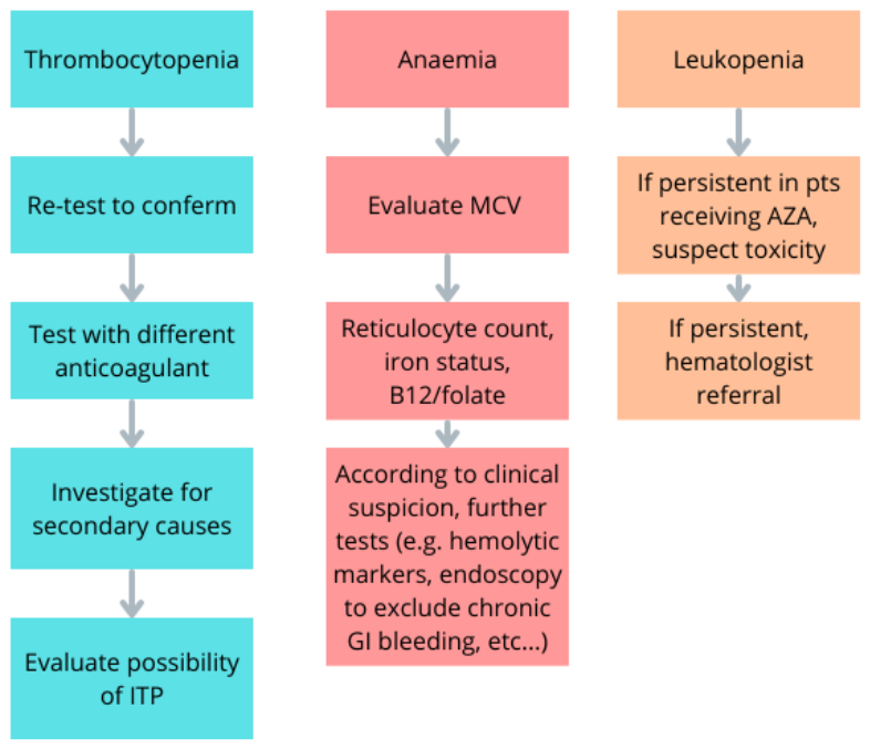
Cytopenia, affecting one or multiple blood lines, may occur in AIH patients due to several factors, including nutritional deficiency, autoimmune disruption of blood cells or precursors, cirrhosis and portal-hypertension-induced hypersplenism, reduced hepatic production of haematopoietic growth factors, and drug toxicity [89,90]. Less commonly, cytopenia may be a feature of associated non-haematologic autoimmune disease like systemic lupus erythematosus [91,92]. Due to the wide variety of conditions leading to cytopenia, the differential diagnosis should be based on clinical and laboratory findings and further examinations (e.g., bone marrow aspirate) in selected patients; Figure 1 summarizes the key points in the differential diagnosis of cytopenia.

7.1. Thrombocytopenia

Thrombocytopenia is a common finding in chronic liver disease, including AIH, and often reflects portal hypertension with splenic sequestration [93]. Reduced hepatic synthesis of thrombopoietin may further contribute to low platelet counts [94].
In the setting of AIH, immune thrombocytopenia (ITP) represents a well-documented cause of thrombocytopenia. Its diagnosis is clinical, based primarily on the exclusion of secondary causes [95,96,97,98].
Considerations and proposed approach: The first step is to confirm a true reduction in platelet count. Laboratory artefacts, especially EDTA-related pseudo-thrombocytopenia, should be excluded through smear review or by repeating the test in citrate or heparinized samples [99]. Reviewing prior blood counts helps determine whether thrombocytopenia is chronic or newly developed. Other relevant clinical information to collect includes evidence of portal hypertension (splenomegaly, varices), recent infections, medication exposure, bleeding symptoms, and coexisting cytopenia.
When thrombocytopenia is persistent and not clearly attributable to liver-related mechanisms, the diagnostic framework proposed in the updated international ITP consensus report (Table 3) can be useful [100]. Bone marrow examination should be reserved for selected cases, including as patients with unexplained additional cytopenias, atypical features, or inadequate response to initial management.
The management of thrombocytopenia depends on its cause. Portal-hypertension-related thrombocytopenia generally requires no specific treatment. ITP is treated according to standard guidelines: corticosteroids are often first-line and may overlap with AIH therapy, although the overall immunosuppressive regimen should be individualized. Coordination between the hepatology and haematology teams is important when establishing the timing of steroid tapering.

7.2. Anaemia

Anaemia in AIH may arise from several mechanisms, including nutritional deficiencies, chronic disease, autoimmune haemolysis, bone marrow suppression, and, more rarely, marrow failure syndromes. Correct classification of anaemia is essential, as management varies considerably according to the underlying cause.
The most common form of anaemia in liver disease is iron deficiency anaemia, which can result from malabsorption due to associated conditions like CeD and AIG, blood loss from occult or overt bleeding or nutritional deficiency [101,102,103,104].
Less frequently, anaemia can be caused by a deficiency in cobalamin (vitamin B12) and/or folic acid (vitamin B9), often linked to poor dietary intake or the association with AIG [105,106]. Severe deficiencies of those vitamins may even lead to pancytopenia [107].
Autoimmune haemolytic anaemia (AIHA) is an uncommon but well-recognized extrahepatic autoimmune manifestation associated with AIH. Typical features include symptomatic anaemia, jaundice with preserved liver function, elevated reticulocyte count, increased LDH, low haptoglobin, and a positive direct antiglobulin test [108,109]. The coexistence of AIHA and immune thrombocytopenia (Evans’ syndrome) has also been reported, though rarely [110,111].
Anaemia of chronic disease can occur, especially in patients with advanced liver disease, presenting with a characteristically low reticulocyte count, normal or reduced mean corpuscular volume and high ferritin values [112,113].
Hepatitis-associated aplastic anaemia (HAAA) is a rare but life-threatening complication of AIH. It is a syndrome characterized by pancytopenia, marked reticulocytopenia, and a hypocellular bone marrow on biopsy [114,115]. It has been reported after episodes of acute hepatitis, including seronegative or paediatric presentations of AIH [116]. Evidence about HAAA in AIH is scarce but indicates a limited response to corticosteroids and azathioprine, with a need for second-line treatment (including anti-thymocyte globulin plus cyclosporine, up to bone marrow transplantation) [116,117].
Considerations and proposed approach: Given the wide range of potential mechanisms, a systematic diagnostic work-up is usually needed to determine the underlying cause of anaemia [79]. The initial evaluation of anaemia in AIH should include complete blood count with reticulocyte count, blood smear, iron metabolism studies, vitamin B12 and folate levels, and haemolysis work-up (LDH, haptoglobin, bilirubin).
Table 4 summarizes the characteristic laboratory profiles of the main forms of anaemia encountered in patients with AIH. When iron, B12, or folate deficiency is identified, evaluation for CeD and AIG is recommended. Suspected haemolysis or unexplained cytopenias should prompt early haematology referral.
Severe or progressive pancytopenia requires rapid investigation to exclude HAAA or drug-induced marrow suppression.

7.3. Leukopenia

Leukopenia, despite being less common than thrombocytopenia and anaemia, should not be overlooked in AIH patients due to its possible association with clinically relevant phenomena such as infections and drug toxicity. Infections related to immune suppression and antimicrobial treatments needed for them can trigger a transitory drop in leukocyte counts [118]. Rarely, leukopenia may result from the immune-mediated destruction of leukocytes or their precursors, as seen in autoimmune neutropenia [118,119]. Compared with thrombocytopenia or anaemia, leukopenia is less common but often more clinically significant, as it may imply an increased risk of infections or an evolving bone marrow dysfunction.
Considerations and proposed approach: In patients with leukopenia, the first questions are (1) whether the alteration is acute (onset in days or week) or chronic (developed over months or more) and (2) whether or not it is accompanied by other manifestations (e.g., fever) [119]. Evaluation of previous blood cell counts may be helpful in enhancing progressive trends towards leukopenia. In all patients receiving azathioprine and/or mycophenolate, suspicion for drug-induced leukopenia should be raised [4]. Azathioprine-induced bone marrow suppression is strongly influenced by TPMT and NUDT15 polymorphisms [120]. Genotyping or phenotyping of these enzymes, now widely available, allows for the identification of patients at increased risk of severe cytopenia and may inform safer therapeutic strategies. Patients with unexplained and persisting neutropenia, especially if associated with other blood cell count alterations, should be referred to a haematology specialist for further investigations [121].

7.4. Drug-Induced Cytopenia

Cytopenias are a well-known side effect of several immunosuppressive drugs, including mofetil mycophenolate and azathioprine, the first-line treatments for AIH [10]. Both drugs can lead to bone marrow failure by interfering with purine metabolism [122]. Genetic variants affecting thiopurine methyltransferase activity have been identified as a risk factor for AZA-induced myelotoxicity [120,123,124]. Due to this risk, current guidelines recommend regular monitoring of CBC in both patients receiving AZA and MMF, especially during the first eight weeks of treatment [10].
Grade of evidence: Evidence regarding ITP and thrombocytopenia in the scenario of AIH are limited mostly to case reports and series. Similarly, evidence concerning anaemia, leukopenia (especially immune leukopenia) and HAAA derive mostly to series and descriptive retrospective studies. EASL 2025 guidelines address the topic of cytopenia mostly in the context of drug toxicity; both AIHA and ITP are cited as common comorbidities of AIH; however, no specific recommendations are provided.

8. Endocrine and Metabolic Comorbidities

Endocrine comorbidities are frequent and clinically relevant manifestations in AIH patients. They could develop as an expression of a concurrent autoimmune disease or of drug toxicity or because of disease activity. A periodic assessment of clinical manifestations and laboratory or instrumental tests suggestive of endocrine and metabolic comorbidities is thus necessary to recognize this condition early, which can develop subtly and become clinically manifested only in severe or complicated forms. Table 5 summarizes the main endocrine and metabolic manifestations that are discussed in the following subsections.

8.1. Glycaemic Abnormalities

Disorders of glucose metabolism are relatively common in patients with AIH, with an increased risk of both type 1 and type 2 diabetes mellitus (T1DM and T2DM). These associations reflect a combination of shared autoimmune predisposition, corticosteroid exposure, liver dysfunction, and the metabolic consequences of chronic inflammation. Thus, current guidelines recommending regular blood glucose monitoring in patients with AIH [10,125].
The association between AIH and T1DM is supported by shared genetic and immunological pathways [126,127]. Conversely, the risk of T2DM in AIH is increased by prolonged corticosteroid therapy, which can induce insulin resistance and hyperglycaemia. Concomitant liver cirrhosis and metabolic-associated steatotic liver disease can further contribute [128,129,130].
Although less common, hypoglycaemia may also occur in patients with AIH. In most cases, recurrent episodes are related to excessive doses of antidiabetic medications, particularly insulin or insulin secretagogues [131]. A relative excess of these medications may occur when steroids are tapered or discontinued. A minority of patients may present with spontaneous hypoglycaemia, which requires careful evaluation for rare causes, such as insulinoma or autoimmune insulin antibody syndrome (Hirata’s disease) [132,133].
Considerations and proposed approach: The finding of hyperglycaemia should prompt evaluations, including repeated fasting plasma glucose, oral glucose tolerance test (when appropriate), C-peptide and insulin levels, and HbA1c. If T1DM is suspected, serological tests for T1DM-associated autoantibodies are indicated [134]. In patients with recurring hypoglycaemia, history evaluation should exclude overdosing or inappropriate use (including self-prescription) of anti-diabetic drugs. Spontaneous or unexplained episodes require endocrinology referral and may need supervised fasting tests or imaging to exclude insulin-producing tumours or autoimmune hypoglycaemia [132].

8.2. Thyroid Disorders

Thyroid dysfunction is one of the most common extrahepatic autoimmune conditions associated with AIH, with a reported prevalence significantly higher than in the general population [135]. Both hypothyroidism, usually due to chronic autoimmune thyroiditis (Hashimoto’s disease), and hyperthyroidism, typically resulting from Graves’ disease, occur more frequently in AIH than in the general population [84,136]. AIH guidelines recommend screening thyroid function at diagnosis and at least annually to identify dysthyroidisms before its clinical presentation [10]. If not recognized and treated, thyroid disorders may lead to a worsening in quality of life, having been linked with fatigue, depression, anxiety and poorer quality of sleep [69,137,138].
Considerations and proposed approach: Initial testing should include TSH and free T4; additional markers (free T3, anti-TPO, anti-TSHR antibodies) are reserved in case of dysfunction [10]. As previously stated, thyroid function tests should be performed periodically in asymptomatic AIH patients. However, assessment becomes imperative in the presence of symptoms suggestive of hypothyroidism (e.g., fatigue, unexplained weight gain) or hyperthyroidism (e.g., sleep disturbance, tachycardia) [137,138].
In newly diagnosed thyroid abnormalities, clinicians should consider the potential impact on fatigue and metabolic control and the possibility of overlap with other autoimmune conditions. Management follows standard endocrine practice [138,139]. Attention is needed when prescribing thyreostatic drugs (methimazole or carbimazole), as they can rarely induce agranulocytosis, a complication that may be confused with AIH-related cytopenias [115].

8.3. Bone Metabolism

Low bone mass density occurs in AIH patients more commonly than in the general population, with retrospective series reporting a prevalence of 15% and 43% for osteoporosis and osteopenia respectively [134]. A large study from Lim et al. reported a significantly increased risk of pathological fractures in AIH patients when compared with healthy controls [135]. The risk of low bone mass density is particularly increased in patients with prolonged disease activity, prolonged steroid treatment and liver cirrhosis [140]. Autoimmune comorbidities like autoimmune thyroiditis and CeD may further increase the risk of mineral bone alterations and pathological fractures [141,142]. Often asymptomatic, if not recognized and treated, low bone mass density may lead to reduced autonomy, disability and increased risk of mortality; in this scenario, screening for low bone mass density is paramount [143,144].
Consideration and proposed approach: The actual EASL guidelines do not specifically address the topic of bone health in AIH, limiting their recommendation to an evaluation of bone mass density at the start of steroid treatment, reserving further evaluation to patients with non-specified features of risk for osteoporosis [10]. The topic is more strictly addressed by AASLD guidelines, where follow-up bone mass density evaluation is suggested every 2–3 years in patients with risk features for osteoporosis and pathologic fractures [145].
A reasonable approach should include a periodic (e.g., yearly) evaluation of risk factors for low bone mineral density (BMD), such as lifestyle factors (e.g., smoking), disease activity, and comorbidities. This strategy aims to maximize accuracy in identifying patients warranting longitudinal BMD assessment. Multidimensional fracture risk assessment tools should be employed to determine the need for anti-resorptive treatment (bisphosphonates, denosumab), while optimization of vitamin D levels should be ensured in all AIH patients [144].
In patients with prolonged steroid treatment (e.g., at least 7.5 mg of prednisone daily for at least 3 months), treatment with bisphosphonates, associated with vitamin D and calcium supplementation, is indicated to prevent steroid-induced osteoporosis and pathological fractures [146,147].
Lastly, in patients with low bone mass density in the absence of known risk factors, it could be reasonable to exclude comorbidities associated with elevated bone reabsorption or intestinal malabsorption, such as hyperthyroidism or CeD [30,141].

8.4. Sarcopenia

Sarcopenia is not uncommon in AIH patients of any age, with a significantly higher prevalence in elderly patients [148,149]. Risk factors for sarcopenia include disease activity, prolonged steroid treatment, age, malnutrition and vitamin D deficiency [149,150,151]. In patients with systemic autoimmune comorbidities like rheumatoid arthritis, ongoing systemic inflammation further increases the risk of sarcopenia [152]. The presence of sarcopenia is associated with increased mortality and morbidity as well as poorer quality of life [151].
Considerations and proposed approach: The use of a standardized questionnaire (SARC-F), functional evaluation (handgrip strength, chair-stand test, etc.) or composite tools like the Liver Frailty Index should be included in the standard periodic evaluation of AIH patients, especially where risk factors for sarcopenia are identified [153,154,155]. Instrumental evaluation of body composition could then help in quantifying the muscle mass [153].
When sarcopenia is diagnosed, multidisciplinary evaluation of potential modifiable determinants (e.g., smoking, undiagnosed systemic immune comorbidities) and nutritional support represent the keys in its management [152,156].
Grade of evidence: Evidence regarding the association between AIH and diabetes (both type 1 and type 2), thyroid disorders and low bone mass density is based on various reports and studies, and previous authors described the pathogenetic mechanisms of those associations. Evidence regarding the association with sarcopenia, on the other hand, is based mostly on studies on chronic liver disease, where patients with AIH were only part of the population. EASL 2025 guidelines clearly recommend screening for autoimmune thyroiditis and low bone mass density in all AIH patients; T1DM is cited as a common comorbidity in AIH, with no further specific recommendation, while T2DM is discussed mostly in the context of drug (steroid) toxicity; sarcopenia is not cited.

9. Impact of Extrahepatic Manifestations

The relationship between the course of AIH and extrahepatic manifestations appears unclear, with most studies focusing on autoimmune comorbidities and giving conflicting results [16,157,158]. Slooter et al. published a meta-analysis of 35 studies in 2025, where they did not demonstrate a significant impact of concurrent autoimmune disease on AIH prognosis [158]. Similarly, the impact of fatigue and anxious–depressive symptoms on survival appears to be limited [75,159]. This de-correlation between comorbidity and course of the hepatic disease can lead the physician to underestimate the relevance of extrahepatic manifestations, focusing the management of AIH only on “objective” data like aminotransferases or liver stiffness that are “silent” for the patient.
Extrahepatic comorbidities, however, significantly impact health-related quality of life (HRQoL). In this context, fatigue and anxiety interplay with disease-related parameters, such as duration of steroid treatment (independently from disease course) or perceived “limitation” due to disease, leading to a poor HRQoF [75,159]. The presence of comorbidities further decreases the HRQoF of AIH patients, highlighting how a careful recognition and management of those could represent a key point in improving the quality of life of people living with AIH [160].

10. Perspective

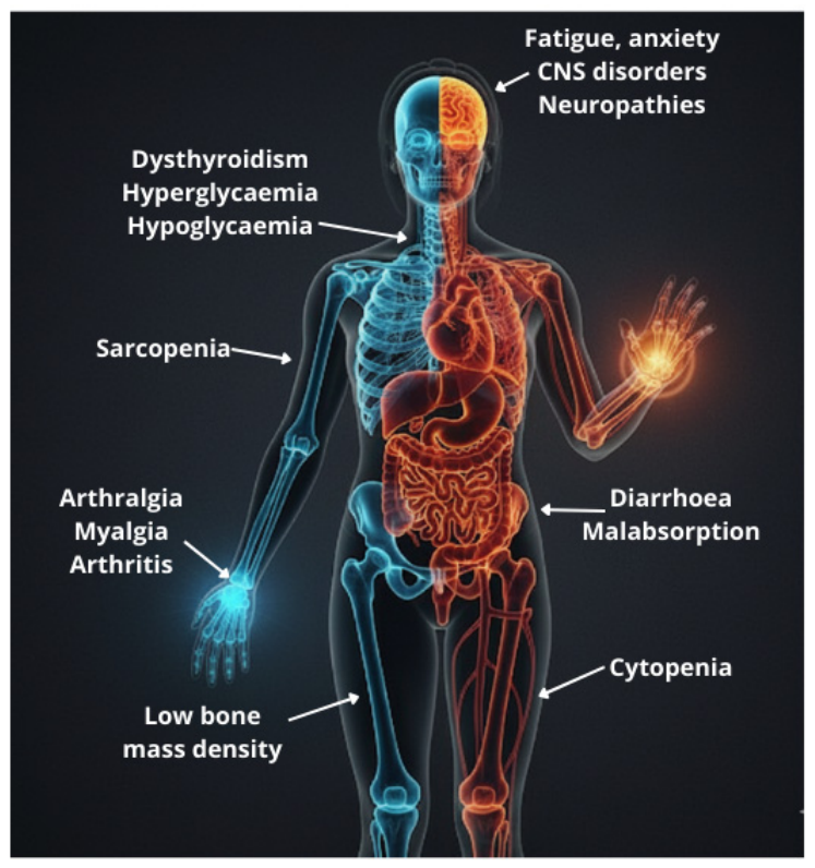
Far from being restricted to the hepatic compartment, AIH unfolds within a broad immunological, metabolic, and systemic context (Figure 2). Several layers contribute to this complexity, including susceptibility to other autoimmune conditions, complications related to a possible evolution toward liver cirrhosis, and drug-related adverse events. Moreover, clinicians increasingly follow AIH patients into ages at which cardiovascular, metabolic, and frailty-related comorbidities become prevalent.
Consequently, extrahepatic symptoms are not ancillary findings. In fact, while their impact on prognosis, especially when considering liver- and AIH-related outcomes (e.g., development of cirrhosis), appears to be minor, they are central determinants of quality of life [5]. Fatigue, arthralgias, anxiety, and gastrointestinal symptoms are among the most impactful and frequently reported complaints, yet they often remain under-investigated or attributed generically to chronic illness. One of the recurring themes across several manifestations is the poor correlation between hepatic activity and symptom burden. Many patients achieve biochemical remission yet continue to experience symptoms that impair daily functioning, social participation, sleep, and mental health.
This gap between laboratory remission and patient-reported outcomes highlights the need to reassess how success is defined in AIH management. While hepatic control remains essential, quality of life, functional status, and symptom relief should be integrated as therapeutic objectives. At present, few structured instruments exist to systematically capture patient-reported outcomes specific to AIH. As a result, extrahepatic symptoms often emerge only when they become severe enough to trigger additional consultations. This situation leads to a difficult patient journey, possibly leading to a fragmented management.
In perspective, priorities emerge for tackling these problems and improving the care of patients with AIH.
First, multidisciplinary models of care should be developed for AIH, similar to those already in place for other autoimmune diseases. Regular collaboration between hepatology, rheumatology, endocrinology, gastroenterology, and haematology would not only streamline diagnostic evaluation but also avoid redundant testing and accelerate targeted treatment.
Second, the systematic assessment of symptom burden should become routine. This may include the use of general quality-of-life tools but ideally should involve instruments specifically adapted to AIH. Developing and validating such patient-reported outcome measures would be welcome.
Third, clinicians should adopt a proactive approach to monitoring and prevention. Early testing for CeD or autoimmune thyroiditis, attention to bone health, and regular screening for diabetes and cytopenia can reduce delays in care.
Finally, research efforts must continue to elucidate the pathophysiology of extrahepatic manifestations and identify potential therapeutic strategies. Key priorities include validating standardized tools to screen for subtle or overlooked manifestations and evaluating how AIH medications influence the natural history of concurrent diseases. Furthermore, a more nuanced understanding of the impact of specific comorbidities—rather than the mere presence of an unspecified autoimmune condition—is required. Ultimately, this will aid in defining optimal management strategies that account for the potential hepatotoxicity of medications used to treat these comorbidities.

11. Limitations

This review acknowledges several limitations. Firstly, as a narrative review, it provides a comprehensive overview but does not adhere to systematic review protocols (e.g., PRISMA), preventing a statistical evaluation of bias. Secondly, the available evidence regarding extrahepatic manifestations in AIH is largely derived from retrospective, small-cohort studies. These study designs are inherently subject to selection bias and often lack adequate control groups. Finally, due to the scarcity of high-quality prospective data, specific recommendations often rely on expert opinion or are extrapolated from guidelines designed for other diseases, highlighting the urgent need for prospective, multicenter studies dedicated specifically to the AIH population.

12. Conclusions

AIH should be considered a systemic, rather than merely a liver, disease given the significant prevalence of extrahepatic manifestations. Those manifestations may reflect associated autoimmune conditions but also arise as adverse effects of treatment.
Often overlooked due to their limited impact on natural history of AIH, extrahepatic manifestations are key determinants of health-related quality of life. Symptoms like fatigue and depression may also lead to reduced adherence to AIH treatment due to the fear of medications (especially steroids) negatively affecting autonomy and quality of life.
The approach to the patients should not be limited to controlling hepatic inflammation and normalizing liver function tests but should extend to the evaluation of global health to recognize extrahepatic manifestations early. In this context, standardized tools to evaluate fatigue, depression, and the risk of sarcopenia should be implemented in the standard evaluation of people living with AIH.
The systemic nature of AIH requires a multidisciplinary strategy for the diagnosis and long-term care of people living with AIH, with the goal of improving patient outcomes, health and quality of life.

Author Contributions

Conceptualization, D.P.P. and E.B.; methodology, D.P.P. and F.T.; investigation, E.B., A.D.S., M.C.A.C. and E.S.; writing—original draft preparation, D.P.P., E.B. and A.D.S.; writing—review and editing, F.T. and F.P.; supervision, D.P.P. and F.T. All authors have read and agreed to the published version of the manuscript.

Funding

This research received no external funding.

Institutional Review Board Statement

Not applicable.

Informed Consent Statement

Not applicable.

Data Availability Statement

No new data were created or analyzed in this study.

Acknowledgments

During the preparation of the manuscript, the authors used Gemini 2.5 Flash Image for the purpose of producing the graphical abstract. The authors have reviewed and edited the output and take full responsibility for the content of this publication.

Conflicts of Interest

The authors declare no conflicts of interest.

Abbreviations

The following abbreviations are used in this manuscript:
AIGAutoimmune Gastritis
AIHAutoimmune Hepatitis
AIHAAutoimmune Haemolytic Anaemia
ANAAntinuclear Antibodies
AZAAzathioprine
CBCComplete Blood Count
CeDCeliac Disease
HAAAHepatitis-Associated Aplastic Anaemia
HbA1cGlycated Haemoglobin
HIVHuman Immunodeficiency Virus
HLAHuman Leukocyte Antigen
IBDsInflammatory Bowel Diseases
IBSIrritable Bowel Syndrome
ITPImmune Thrombocytopenia
LDHLactate Dehydrogenase
MCVMean Corpuscular Volume
MMFMycophenolate Mofetil
PSCPrimary Sclerosing Cholangitis
T1DMType 1 Diabetes Mellitus
T2DMType 2 Diabetes Mellitus
TSHThyroid-Stimulating Hormone
UCUlcerative Colitis

References

  1. van Gerven, N.M.; de Boer, Y.S.; Mulder, C.J.; van Nieuwkerk, C.M.; Bouma, G. Auto Immune Hepatitis. World J. Gastroenterol. 2016, 22, 4651–4661. [Google Scholar] [CrossRef] [PubMed]
  2. Golding, P.L.; Smith, M.; Williams, R. Multisystem Involvement in Chronic Liver Disease. Studies on the Incidence and Pathogenesis. Am. J. Med. 1973, 55, 772–782. [Google Scholar] [CrossRef]
  3. Teufel, A.; Weinmann, A.; Kahaly, G.J.; Centner, C.; Piendl, A.; Wörns, M.; Lohse, A.W.; Galle, P.R.; Kanzler, S. Concurrent Autoimmune Diseases in Patients with Autoimmune Hepatitis. J. Clin. Gastroenterol. 2010, 44, 208–213. [Google Scholar] [CrossRef]
  4. Behairy, B.E.-S.; El-Said, H.H.; Konswa, H.A.-A.; El-Deen, A.E.-S.N.; Adawy, N.M.; Sira, A.M. Predictors of Azathioprine Toxicity in Children with Autoimmune Hepatitis. Clin. Exp. Hepatol. 2021, 7, 47–54. [Google Scholar] [CrossRef]
  5. Slooter, C.D.; Troyer, I.D.M.; van den Brand, F.F.; de Vries, R.; Heymans, M.W.; Heneghan, M.A.; Drenth, J.P.H.; de Boer, Y.S. Early Prognostic Factors for Reduced Survival in Autoimmune Hepatitis: A Systematic Review with Meta-Analysis. Clin. Gastroenterol. Hepatol. 2026, 24, 328–341. [Google Scholar] [CrossRef]
  6. Al-Chalabi, T.; Boccato, S.; Portmann, B.C.; McFarlane, I.G.; Heneghan, M.A. Autoimmune Hepatitis (AIH) in the Elderly: A Systematic Retrospective Analysis of a Large Group of Consecutive Patients with Definite AIH Followed at a Tertiary Referral Centre. J. Hepatol. 2006, 45, 575–583. [Google Scholar] [CrossRef]
  7. Muratori, P.; Granito, A.; Pappas, G.; Muratori, L.; Lenzi, M.; Bianchi, F.B. Autoimmune Liver Disease 2007. Mol. Asp. Med. 2008, 29, 96–102. [Google Scholar] [CrossRef]
  8. Gatselis, N.K.; Zachou, K.; Koukoulis, G.K.; Dalekos, G.N. Autoimmune Hepatitis, One Disease with Many Faces: Etiopathogenetic, Clinico-Laboratory and Histological Characteristics. World J. Gastroenterol. 2015, 21, 60–83. [Google Scholar] [CrossRef]
  9. Fernandes, B.; Dias, E.; Mascarenhas-Saraiva, M.; Bernardes, M.; Costa, L.; Cardoso, H.; Macedo, G. Rheumatologic Manifestations of Hepatic Diseases. Ann. Gastroenterol. 2019, 32, 352–360. [Google Scholar] [CrossRef] [PubMed]
  10. Dalekos, G.; Gatselis, N.; Drenth, J.P.; Heneghan, M.; Jørgensen, M.; Lohse, A.W.; Londoño, M.; Muratori, L.; Papp, M.; Samyn, M.; et al. EASL Clinical Practice Guidelines on the Management of Autoimmune Hepatitis. J. Hepatol. 2025, 83, 453–501. [Google Scholar] [CrossRef] [PubMed]
  11. Czaja, A.J. Autoimmune Liver Disease and Rheumatic Manifestations. Curr. Opin. Rheumatol. 2007, 19, 74–80. [Google Scholar] [CrossRef] [PubMed]
  12. Till, S.H.; Snaith, M.L. Assessment, Investigation, and Management of Acute Monoarthritis. Emerg. Med. J. 1999, 16, 355–361. [Google Scholar] [CrossRef] [PubMed]
  13. Singh, S.; Mehra, S. Approach to Polyarthritis. Indian J. Pediatr. 2010, 77, 1005–1010. [Google Scholar] [CrossRef] [PubMed]
  14. Selmi, C.; Generali, E.; Gershwin, M.E. Rheumatic Manifestations in Autoimmune Liver Disease. Rheum. Dis. Clin. N. Am. 2018, 44, 65–87. [Google Scholar] [CrossRef]
  15. Adiga, A.; Nugent, K. Lupus Hepatitis and Autoimmune Hepatitis (Lupoid Hepatitis). Am. J. Med. Sci. 2017, 353, 329–335. [Google Scholar] [CrossRef]
  16. Birn-Rydder, R.; Jensen, M.D.; Jepsen, P.; Grønbaek, L. Extrahepatic Autoimmune Diseases in Autoimmune Hepatitis: Effect on Mortality. Liver Int. 2022, 42, 2466–2472. [Google Scholar] [CrossRef]
  17. Li, Z.; Xu, D.; Wang, Z.; Wang, Y.; Zhang, S.; Li, M.; Zeng, X. Gastrointestinal System Involvement in Systemic Lupus Erythematosus. Lupus 2017, 26, 1127–1138. [Google Scholar] [CrossRef]
  18. Tani, C.; Carli, L.; Vagnani, S.; Talarico, R.; Baldini, C.; Mosca, M.; Bombardieri, S. The Diagnosis and Classification of Mixed Connective Tissue Disease. J. Autoimmun. 2014, 48–49, 46–49. [Google Scholar] [CrossRef]
  19. Muratori, P.; Granito, A.; Pappas, G.; Pendino, G.M.; Quarneti, C.; Cicola, R.; Menichella, R.; Ferri, S.; Cassani, F.; Bianchi, F.B.; et al. The Serological Profile of the Autoimmune Hepatitis/Primary Biliary Cirrhosis Overlap Syndrome. Am. J. Gastroenterol. 2009, 104, 1420–1425. [Google Scholar] [CrossRef]
  20. American College of Rheumatology Ad Hoc Committee on Clinical Guidelines. Guidelines for the Initial Evaluation of the Adult Patient with Acute Musculoskeletal Symptoms. Arthritis Rheum. 1996, 39, 1–8. [Google Scholar] [CrossRef]
  21. van Gerven, N.M.F.; Verwer, B.J.; Witte, B.I.; van Erpecum, K.J.; van Buuren, H.R.; Maijers, I.; Visscher, A.P.; Verschuren, E.C.; van Hoek, B.; Coenraad, M.J.; et al. Epidemiology and Clinical Characteristics of Autoimmune Hepatitis in the Netherlands. Scand. J. Gastroenterol. 2014, 49, 1245–1254. [Google Scholar] [CrossRef] [PubMed]
  22. El-Gabalawy, H.S.; Duray, P.; Goldbach-Mansky, R. Evaluating Patients with Arthritis of Recent Onset: Studies in Pathogenesis and Prognosis. JAMA 2000, 284, 2368–2373. [Google Scholar] [CrossRef] [PubMed]
  23. Baker, D.G.; Schumacher, H.R. Acute Monoarthritis. N. Engl. J. Med. 1993, 329, 1013–1020. [Google Scholar] [CrossRef]
  24. Sakellariou, G.; Scirè, C.A.; Adinolfi, A.; Batticciotto, A.; Bortoluzzi, A.; Delle Sedie, A.; De Lucia, O.; Dejaco, C.; Epis, O.M.; Filippucci, E.; et al. Differential Diagnosis of Inflammatory Arthropathies by Musculoskeletal Ultrasonography: A Systematic Literature Review. Front. Med. 2020, 7, 141. [Google Scholar] [CrossRef] [PubMed]
  25. Carpenter, C.R.; Schuur, J.D.; Everett, W.W.; Pines, J.M. Evidence-Based Diagnostics: Adult Septic Arthritis. Acad. Emerg. Med. 2011, 18, 781–796. [Google Scholar] [CrossRef]
  26. Dao, K.; Cush, J.J. Acute Polyarthritis. Best Pract. Res. Clin. Rheumatol. 2006, 20, 653–672. [Google Scholar] [CrossRef]
  27. Foster, Z.J.; Day, A.L.; Miller, J. Polyarticular Joint Pain in Adults: Evaluation and Differential Diagnosis. Am. Fam. Physician 2023, 107, 42–51. [Google Scholar]
  28. Stotzer, P.-O.; Abrahamsson, H.; Bajor, A.; Kilander, A.; Sadik, R.; Sjövall, H.; Simrén, M. Are the Definitions for Chronic Diarrhoea Adequate? Evaluation of Two Different Definitions in Patients with Chronic Diarrhoea. United Eur. Gastroenterol. J. 2015, 3, 381–386. [Google Scholar] [CrossRef]
  29. Burgers, K.; Lindberg, B.; Bevis, Z.J. Chronic Diarrhea in Adults: Evaluation and Differential Diagnosis. Am. Fam. Physician 2020, 101, 472–480. [Google Scholar] [PubMed]
  30. Tovoli, F.; Pallotta, D.P.; Giamperoli, A.; Zavatta, G.; Skoracka, K.; Raiteri, A.; Faggiano, C.; Krela-Kaźmierczak, I.; Granito, A. Evolution of Bone Densitometry Parameters and Risk of Fracture in Coeliac Disease: A 10-Year Perspective. Intern. Emerg. Med. 2023, 18, 1405–1414. [Google Scholar] [CrossRef]
  31. Tovoli, F.; Faggiano, C.; Raiteri, A.; Giamperoli, A.; Catenaro, T.; Sansone, V.; Pallotta, D.P.; Granito, A. Risk of Drop-Out from Follow-Up Evaluations for Celiac Disease: Is It Similar for All Patients? Nutrients 2022, 14, 1223. [Google Scholar] [CrossRef] [PubMed]
  32. Ludvigsson, J.F.; Leffler, D.A.; Bai, J.; Biagi, F.; Fasano, A.; Green, P.H.; Hadjivassiliou, M.; Kaukinen, K.; Kelly, C.; Leonard, J.N.; et al. The Oslo Definitions for Coeliac Disease and Related Terms. Gut 2013, 62, 43–52. [Google Scholar] [CrossRef]
  33. Czaja, A.J.; Carpenter, H.A.; Santrach, P.J.; Moore, S.B. Genetic Predispositions for the Immunological Features of Chronic Active Hepatitis. Hepatology 1993, 18, 816–822. [Google Scholar] [CrossRef]
  34. Haggård, L.; Glimberg, I.; Lebwohl, B.; Sharma, R.; Verna, E.C.; Green, P.H.R.; Ludvigsson, J.F. High Prevalence of Celiac Disease in Autoimmune Hepatitis: Systematic Review and Meta-Analysis. Liver Int. 2021, 41, 2693–2702. [Google Scholar] [CrossRef]
  35. Tovoli, F.; De Giorgio, R.; Caio, G.; Grasso, V.; Frisoni, C.; Serra, M.; Caputo, C.; Stanghellini, V.; Bolondi, L.; Corinaldesi, R.; et al. Autoimmune Hepatitis and Celiac Disease: Case Report Showing an Entero-Hepatic Link. Case Rep. Gastroenterol. 2010, 4, 469–475. [Google Scholar] [CrossRef]
  36. Singh, P.; Singh, A.D.; Ahuja, V.; Makharia, G.K. Who to Screen and How to Screen for Celiac Disease. World J. Gastroenterol. 2022, 28, 4493–4507. [Google Scholar] [CrossRef]
  37. Ricciuto, A.; Kamath, B.M.; Hirschfield, G.M.; Trivedi, P.J. Primary Sclerosing Cholangitis and Overlap Features of Autoimmune Hepatitis: A Coming of Age or an Age-Ist Problem? J. Hepatol. 2023, 79, 567–575. [Google Scholar] [CrossRef] [PubMed]
  38. Ordonez, F.; Lacaille, F.; Canioni, D.; Talbotec, C.; Fournet, J.-C.; Cerf-Bensussan, N.; Goulet, O.; Schmitz, J.; Ruemmele, F.M. Pediatric Ulcerative Colitis Associated with Autoimmune Diseases: A Distinct Form of Inflammatory Bowel Disease? Inflamm. Bowel Dis. 2012, 18, 1809–1817. [Google Scholar] [CrossRef] [PubMed]
  39. Zhang, X.; Lin, X.; Li, X.; Guan, L.; Li, Y.; Wang, N. Ulcerative Colitis Complicated by Primary Sclerosing Cholangitis and Autoimmune Hepatitis Overlap Syndrome: A Case Report and Literature Review. Front. Immunol. 2023, 14, 1132072. [Google Scholar] [CrossRef]
  40. Deneau, M.; Jensen, M.K.; Holmen, J.; Williams, M.S.; Book, L.S.; Guthery, S.L. Primary Sclerosing Cholangitis, Autoimmune Hepatitis, and Overlap in Utah Children: Epidemiology and Natural History. Hepatology 2013, 58, 1392–1400. [Google Scholar] [CrossRef]
  41. Saich, R.; Chapman, R. Primary Sclerosing Cholangitis, Autoimmune Hepatitis and Overlap Syndromes in Inflammatory Bowel Disease. World J. Gastroenterol. 2008, 14, 331–337. [Google Scholar] [CrossRef] [PubMed]
  42. Lo, S.K.; Fleming, K.A.; Chapman, R.W. A 2-Year Follow-up Study of Anti-Neutrophil Antibody in Primary Sclerosing Cholangitis: Relationship to Clinical Activity, Liver Biochemistry and Ursodeoxycholic Acid Treatment. J. Hepatol. 1994, 21, 974–978. [Google Scholar] [CrossRef] [PubMed]
  43. Terjung, B.; Spengler, U. Atypical P-ANCA in PSC and AIH: A Hint toward a “Leaky Gut”? Clin. Rev. Allergy Immunol. 2009, 36, 40–51. [Google Scholar] [CrossRef]
  44. Tovoli, F.; Vannini, A.; Fusconi, M.; Frisoni, M.; Zauli, D. Autoimmune Liver Disorders and Small-Vessel Vasculitis: Four Case Reports and Review of the Literature. Ann. Hepatol. 2014, 13, 136–141. [Google Scholar] [CrossRef]
  45. Granito, A.; Muratori, P.; Tovoli, F.; Muratori, L. Anti-Neutrophil Cytoplasm Antibodies (ANCA) in Autoimmune Diseases: A Matter of Laboratory Technique and Clinical Setting. Autoimmun. Rev. 2021, 20, 102787. [Google Scholar] [CrossRef] [PubMed]
  46. Danese, S.; Fiorino, G.; Mary, J.-Y.; Lakatos, P.L.; D’Haens, G.; Moja, L.; D’Hoore, A.; Panes, J.; Reinisch, W.; Sandborn, W.J.; et al. Development of Red Flags Index for Early Referral of Adults with Symptoms and Signs Suggestive of Crohn’s Disease: An IOIBD Initiative. J. Crohn’s Colitis 2015, 9, 601–606. [Google Scholar] [CrossRef]
  47. Felice, C.; Leccese, P.; Scudeller, L.; Lubrano, E.; Cantini, F.; Castiglione, F.; Gionchetti, P.; Orlando, A.; Salvarani, C.; Scarpa, R.; et al. Red Flags for Appropriate Referral to the Gastroenterologist and the Rheumatologist of Patients with Inflammatory Bowel Disease and Spondyloarthritis. Clin. Exp. Immunol. 2019, 196, 123–138. [Google Scholar] [CrossRef] [PubMed]
  48. Jannone, G.; Triaille, C.; Touzot, F.; Alvarez, F. Autoimmune Hepatitis and Immune Dysregulation: A Case Series. Dig. Liver Dis. 2025, 57, 2152–2158. [Google Scholar] [CrossRef]
  49. Singh, K.; Chang, C.; Gershwin, M.E. IgA Deficiency and Autoimmunity. Autoimmun. Rev. 2014, 13, 163–177. [Google Scholar] [CrossRef] [PubMed]
  50. Raiteri, A.; Granito, A.; Pallotta, D.P.; Giamperoli, A.; Pratelli, A.; Monaco, G.; Faggiano, C.; Tovoli, F. Exploring Total Immunoglobulin A’s Impact on Non-Biopsy Diagnosis of Celiac Disease: Implications for Diagnostic Accuracy. Nutrients 2024, 16, 3195. [Google Scholar] [CrossRef] [PubMed]
  51. Aziz, I.; Peerally, M.F.; Barnes, J.-H.; Kandasamy, V.; Whiteley, J.C.; Partridge, D.; Vergani, P.; Cross, S.S.; Green, P.H.; Sanders, D.S. The Clinical and Phenotypical Assessment of Seronegative Villous Atrophy; a Prospective UK Centre Experience Evaluating 200 Adult Cases over a 15-Year Period (2000–2015). Gut 2017, 66, 1563–1572. [Google Scholar] [CrossRef]
  52. Miehlke, S.; Verhaegh, B.; Tontini, G.E.; Madisch, A.; Langner, C.; Münch, A. Microscopic Colitis: Pathophysiology and Clinical Management. Lancet Gastroenterol. Hepatol. 2019, 4, 305–314. [Google Scholar] [CrossRef]
  53. Montoro-Huguet, M.A.; Belloc, B.; Domínguez-Cajal, M. Small and Large Intestine (I): Malabsorption of Nutrients. Nutrients 2021, 13, 1254. [Google Scholar] [CrossRef]
  54. Massironi, S.; Dispinzieri, G.; Rossi, A.; Cristoferi, L.; Lenti, M.V.; Gerussi, A.; Elvevi, A.; Carbone, M.; Bonfichi, A.; Di Sabatino, A.; et al. Immunological and Clinical Overlap between Autoimmune Gastritis and Autoimmune Liver Diseases: A Prospective Cohort Study. Front. Immunol. 2025, 16, 1628478. [Google Scholar] [CrossRef]
  55. Osmola, M.; Hémont, C.; Romańczyk, M.; Druet, A.; Chapelle, N.; Matysiak-Budnik, T.; Lenti, M.V.; Martin, J.C. A Comparative Study of Different Assays for Autoantibodies Detection in Patients with Autoimmune Gastritis. J. Transl. Autoimmun. 2025, 10, 100294. [Google Scholar] [CrossRef]
  56. Andres, E.; Serraj, K. Optimal Management of Pernicious Anemia. J. Blood Med. 2012, 3, 97–103. [Google Scholar] [CrossRef]
  57. Harrington, D.J.; Stevenson, E.; Sobczyńska-Malefora, A. The Application and Interpretation of Laboratory Biomarkers for the Evaluation of Vitamin B12 Status. Ann. Clin. Biochem. 2025, 62, 22–33. [Google Scholar] [CrossRef]
  58. Rao, S.; Kikano, E.G.; Smith, D.A.; Guler, E.; Tirumani, S.H.; Ramaiya, N.H. Unintentional Weight Loss: What Radiologists Need to Know and What Clinicians Want to Know. Abdom. Radiol. 2021, 46, 2236–2250. [Google Scholar] [CrossRef] [PubMed]
  59. Shah, S.C.; Piazuelo, M.B.; Kuipers, E.J.; Li, D. AGA Clinical Practice Update on the Diagnosis and Management of Atrophic Gastritis: Expert Review. Gastroenterology 2021, 161, 1325–1332.e7. [Google Scholar] [CrossRef] [PubMed]
  60. Werner, M.; Prytz, H.; Ohlsson, B.; Almer, S.; Björnsson, E.; Bergquist, A.; Wallerstedt, S.; Sandberg-Gertzén, H.; Hultcrantz, R.; Sangfelt, P.; et al. Epidemiology and the Initial Presentation of Autoimmune Hepatitis in Sweden: A Nationwide Study. Scand. J. Gastroenterol. 2008, 43, 1232–1240. [Google Scholar] [CrossRef] [PubMed]
  61. Gelow, K.; Chalasani, S.; Green, K.; Lammert, C. Utilization and Impact of Complementary and Alternative Medicines in Symptomatic Autoimmune Hepatitis Patients. Dig. Dis. Sci. 2022, 67, 2891–2898. [Google Scholar] [CrossRef]
  62. Shen, Y.; Lu, C.; Men, R.; Liu, J.; Ye, T.; Yang, L. Clinical and Pathological Characteristics of Autoimmune Hepatitis with Acute Presentation. Can. J. Gastroenterol. Hepatol. 2018, 2018, 3513206. [Google Scholar] [CrossRef]
  63. Silverman, M.N.; Heim, C.M.; Nater, U.M.; Marques, A.H.; Sternberg, E.M. Neuroendocrine and Immune Contributors to Fatigue. PM R 2010, 2, 338–346. [Google Scholar] [CrossRef] [PubMed]
  64. Vuerich, M.; Wang, N.; Kalbasi, A.; Graham, J.J.; Longhi, M.S. Dysfunctional Immune Regulation in Autoimmune Hepatitis: From Pathogenesis to Novel Therapies. Front. Immunol. 2021, 12, 746436. [Google Scholar] [CrossRef] [PubMed]
  65. Morris, G.; Berk, M.; Galecki, P.; Walder, K.; Maes, M. The Neuro-Immune Pathophysiology of Central and Peripheral Fatigue in Systemic Immune-Inflammatory and Neuro-Immune Diseases. Mol. Neurobiol. 2016, 53, 1195–1219. [Google Scholar] [CrossRef]
  66. Zielinski, M.R.; Systrom, D.M.; Rose, N.R. Fatigue, Sleep, and Autoimmune and Related Disorders. Front. Immunol. 2019, 10, 1827. [Google Scholar] [CrossRef]
  67. Liu, J.; Gong, X.; Lv, H.; Liu, S.; Jiang, Y.; Zhu, G.; Ma, X.; Wang, J.; Ye, X.; Gao, Y.; et al. Is Fatigue Related to the Severity of Liver Inflammation in Patients with Chronic Liver Disease? A Cross-Sectional Study. BMJ Open 2023, 13, e069028. [Google Scholar] [CrossRef]
  68. Jones, E.; Watkins, M.; Anderson, E.; Gelow, K.; Green, K.; Draucker, C.; Lammert, C. Overlooked Symptoms in Autoimmune Hepatitis Negatively Impact Many Facets of Life. Dig. Dis. Sci. 2023, 68, 77–86. [Google Scholar] [CrossRef]
  69. Louwerens, M.; Appelhof, B.C.; Verloop, H.; Medici, M.; Peeters, R.P.; Visser, T.J.; Boelen, A.; Fliers, E.; Smit, J.W.A.; Dekkers, O.M. Fatigue and Fatigue-Related Symptoms in Patients Treated for Different Causes of Hypothyroidism. Eur. J. Endocrinol. 2012, 167, 809–815. [Google Scholar] [CrossRef] [PubMed]
  70. Mathur, K.; Vuppalanchi, V.; Gelow, K.; Vuppalanchi, R.; Lammert, C. Cannabidiol (CBD) Consumption and Perceived Impact on Extrahepatic Symptoms in Patients with Autoimmune Hepatitis. Dig. Dis. Sci. 2020, 65, 322–328. [Google Scholar] [CrossRef] [PubMed]
  71. Janik, M.K.; Wunsch, E.; Raszeja-Wyszomirska, J.; Krawczyk, M.; Milkiewicz, P. Depression: An Overlooked Villain in Autoimmune Hepatitis? Hepatology 2019, 70, 2232–2233. [Google Scholar] [CrossRef]
  72. Liu, L.; Chen, J.; Chen, T.; Li, J.; Wang, Q. Genome-Annotated Bivariate Analysis of Shared Heritability in Autoimmune Hepatitis with Psychiatric Comorbidity. Sci. Rep. 2025, 16, 1416. [Google Scholar] [CrossRef]
  73. Zhou, S.; Guo, L.; Chen, N.; Liu, H.; Liu, X.; Li, J.; Dong, S.; Liu, J.; Wang, X.; Ran, Y.; et al. Depression Aggravates Immune-Mediated Hepatitis Through NLRP3 Overactivation Induced by Intestinal Microbiota. CNS Neurosci. Ther. 2026, 32, e70743. [Google Scholar] [CrossRef]
  74. Benros, M.E.; Waltoft, B.L.; Nordentoft, M.; Østergaard, S.D.; Eaton, W.W.; Krogh, J.; Mortensen, P.B. Autoimmune Diseases and Severe Infections as Risk Factors for Mood Disorders: A Nationwide Study. JAMA Psychiatry 2013, 70, 812–820. [Google Scholar] [CrossRef]
  75. Janik, M.K.; Wunsch, E.; Raszeja-Wyszomirska, J.; Moskwa, M.; Kruk, B.; Krawczyk, M.; Milkiewicz, P. Autoimmune Hepatitis Exerts a Profound, Negative Effect on Health-Related Quality of Life: A Prospective, Single-Centre Study. Liver Int. 2019, 39, 215–221. [Google Scholar] [CrossRef]
  76. Wunsch, E.; Krause, L.; Gevers, T.J.; Schramm, C.; Janik, M.K.; Krawczyk, M.; Willemse, J.; Uhlenbusch, N.; Löwe, B.; Lohse, A.W.; et al. Confidence in Treatment Is Contributing to Quality of Life in Autoimmune Liver Diseases. The Results of ERN RARE-LIVER Online Survey. Liver Int. 2023, 43, 381–392. [Google Scholar] [CrossRef]
  77. Levis, B.; Sun, Y.; He, C.; Wu, Y.; Krishnan, A.; Bhandari, P.M.; Neupane, D.; Imran, M.; Brehaut, E.; Negeri, Z.; et al. Accuracy of the PHQ-2 Alone and in Combination with the PHQ-9 for Screening to Detect Major Depression: Systematic Review and Meta-Analysis. JAMA 2020, 323, 2290–2300. [Google Scholar] [CrossRef]
  78. O’Connor, E.A.; Henninger, M.L.; Perdue, L.A.; Coppola, E.L.; Thomas, R.G.; Gaynes, B.N. Anxiety Screening: Evidence Report and Systematic Review for the US Preventive Services Task Force. JAMA 2023, 329, 2171–2184. [Google Scholar] [CrossRef]
  79. Budhwar, N.; Syed, Z. Chronic Dyspnea: Diagnosis and Evaluation. Am. Fam. Physician 2020, 101, 542–548. [Google Scholar]
  80. Williams, A.J.; Marsh, J.; Stableforth, D.E. Cryptogenic Fibrosing Alveolitis, Chronic Active Hepatitis and Autoimmune Haemolytic Anaemia in the Same Patient. Br. J. Dis. Chest 1985, 79, 200–203. [Google Scholar] [CrossRef]
  81. West, M.; Jasin, H.E.; Medhekar, S. The Development of Connective Tissue Diseases in Patients with Autoimmune Hepatitis: A Case Series. Semin. Arthritis Rheum. 2006, 35, 344–348. [Google Scholar] [CrossRef]
  82. Martinez, A.R.M.; de Lima, F.D.; Martins, M.P.; Pereira, I.E.; Miotto, N.; Mazo, D.F.C.; Vigani, A.G.; da Costa, L.B.E.; Stucchi, R.S.B.; Almeida, J.R.S.; et al. Sensory Neuronopathy Is a Specific and Disabling Neurological Manifestation of Autoimmune Hepatitis. Eur. J. Neurol. 2020, 27, 2072–2078. [Google Scholar] [CrossRef] [PubMed]
  83. Efe, C.; Wahlin, S.; Ozaslan, E.; Berlot, A.H.; Purnak, T.; Muratori, L.; Quarneti, C.; Yüksel, O.; Thiéfin, G.; Muratori, P. Autoimmune Hepatitis/Primary Biliary Cirrhosis Overlap Syndrome and Associated Extrahepatic Autoimmune Diseases. Eur. J. Gastroenterol. Hepatol. 2012, 24, 531. [Google Scholar] [CrossRef] [PubMed]
  84. Muratori, P.; Fabbri, A.; Lalanne, C.; Lenzi, M.; Muratori, L. Autoimmune Liver Disease and Concomitant Extrahepatic Autoimmune Disease. Eur. J. Gastroenterol. Hepatol. 2015, 27, 1175–1179. [Google Scholar] [CrossRef] [PubMed]
  85. Wong, G.-W.; Yeong, T.; Lawrence, D.; Yeoman, A.D.; Verma, S.; Heneghan, M.A. Concurrent Extrahepatic Autoimmunity in Autoimmune Hepatitis: Implications for Diagnosis, Clinical Course and Long-Term Outcomes. Liver Int. 2017, 37, 449–457. [Google Scholar] [CrossRef]
  86. Rigopoulou, E.I.; Gyftaki, S.; Arvaniti, P.; Tsimourtou, V.; Koukoulis, G.K.; Hadjigeorgiou, G.; Dalekos, G.N. Autoimmune Hepatitis in Patients with Multiple Sclerosis: The Role of Immunomodulatory Treatment. Clin. Res. Hepatol. Gastroenterol. 2019, 43, e25–e32. [Google Scholar] [CrossRef]
  87. Sakae, H.; Mawatari, S.; Oda, K.; Tabu, K.; Kumagai, K.; Mizuno, K.; Nozuma, S.; Kitazono, I.; Kanmura, S.; Ido, A. A Case of Autoimmune Hepatitis with Thymoma and Myasthenia Gravis. Clin. J. Gastroenterol. 2022, 15, 765–770. [Google Scholar] [CrossRef]
  88. Merchut, M.P.; Adams, E.M.; Morrissey, M. Sensory Neuronopathy in Autoimmune Chronic Active Hepatitis. Neurology 1993, 43, 2410–2411. [Google Scholar] [CrossRef] [PubMed]
  89. Abdulrasak, M.; Someili, A.M.; Mohrag, M. Cytopenias in Autoimmune Liver Diseases—A Review. J. Clin. Med. 2025, 14, 1732. [Google Scholar] [CrossRef]
  90. Fierro-Angulo, O.M.; González-Regueiro, J.A.; Pereira-García, A.; Ruiz-Margáin, A.; Solis-Huerta, F.; Macías-Rodríguez, R.U. Hematological Abnormalities in Liver Cirrhosis. World J. Hepatol. 2024, 16, 1229–1244. [Google Scholar] [CrossRef]
  91. Kheyami, M.A.; Almahroos, F.R.; Alzamrooni, A.M.; Khamis, J.; Albaharna, A. Autoimmune Hepatitis and Systemic Lupus Erythematosus Overlap Syndrome: A Case Report and Literature Review. Cureus 2024, 16, e74962. [Google Scholar] [CrossRef]
  92. Goyes, D.; Malladi, V.R.; Ishtiaq, R.; Al-Khazraji, A. Case of Autoimmune Hepatitis with Overlap Systemic Lupus Erythematosus. BMJ Case Rep. CP 2020, 13, e237341. [Google Scholar] [CrossRef]
  93. Lim, H.I.; Cuker, A. Thrombocytopenia and Liver Disease: Pathophysiology and Periprocedural Management. Hematology 2022, 2022, 296–302. [Google Scholar] [CrossRef] [PubMed]
  94. Peck-Radosavljevic, M. Thrombocytopenia in Chronic Liver Disease. Liver Int. 2017, 37, 778–793. [Google Scholar] [CrossRef] [PubMed]
  95. Fukuda, H.; Takata, K.; Kitaguchi, T.; Yamauchi, R.; Kunimoto, H.; Tanaka, T.; Yokoyama, K.; Morihara, D.; Takeyama, Y.; Shakado, S.; et al. Autoimmune Hepatitis with Concomitant Idiopathic Thrombocytopenic Purpura Diagnosed by Transjugular Liver Biopsy. Case Rep. Hepatol. 2018, 2018, 5305691. [Google Scholar] [CrossRef] [PubMed]
  96. Ito, A.; Yoshizawa, K.; Fujimori, K.; Morita, S.; Shigeno, T.; Maejima, T. Autoimmune Hepatitis Associated with Immune Thrombocytopenic Purpura. Intern. Med. 2017, 56, 143–147. [Google Scholar] [CrossRef] [PubMed]
  97. Antunes, H.; Rocha, R.; Silva, N.; Pontes, T.; Antunes, A.; Martins, S. Immune Disease Expressed in Liver and Platelets in an Adolescent: A Case Report. Ital. J. Pediatr. 2010, 36, 42. [Google Scholar] [CrossRef]
  98. Gafter-Gvili, A. Current Approaches for the Diagnosis and Management of Immune Thrombocytopenia. Eur. J. Intern. Med. 2023, 108, 18–24. [Google Scholar] [CrossRef]
  99. Lardinois, B.; Favresse, J.; Chatelain, B.; Lippi, G.; Mullier, F. Pseudothrombocytopenia—A Review on Causes, Occurrence and Clinical Implications. J. Clin. Med. 2021, 10, 594. [Google Scholar] [CrossRef]
  100. Provan, D.; Arnold, D.M.; Bussel, J.B.; Chong, B.H.; Cooper, N.; Gernsheimer, T.; Ghanima, W.; Godeau, B.; González-López, T.J.; Grainger, J.; et al. Updated International Consensus Report on the Investigation and Management of Primary Immune Thrombocytopenia. Blood Adv. 2019, 3, 3780–3817. [Google Scholar] [CrossRef]
  101. Dawidowski, J.; Pietrzak, A. Rare Causes of Anemia in Liver Diseases. Adv. Clin. Exp. Med. 2022, 31, 567–574. [Google Scholar] [CrossRef] [PubMed]
  102. Freeman, H.J. Iron Deficiency Anemia in Celiac Disease. World J. Gastroenterol. 2015, 21, 9233. [Google Scholar] [CrossRef]
  103. Gkamprela, E.; Deutsch, M.; Pectasides, D. Iron Deficiency Anemia in Chronic Liver Disease: Etiopathogenesis, Diagnosis and Treatment. Ann. Gastroenterol. 2017, 30, 405–413. [Google Scholar] [CrossRef]
  104. Fertrin, K.Y. Diagnosis and Management of Iron Deficiency in Chronic Inflammatory Conditions (CIC): Is Too Little Iron Making Your Patient Sick? Hematology 2020, 2020, 478–486. [Google Scholar] [CrossRef]
  105. Green, R.; Datta Mitra, A. Megaloblastic Anemias: Nutritional and Other Causes. Med. Clin. N. Am. 2017, 101, 297–317. [Google Scholar] [CrossRef]
  106. Riaz, A.; Khan, S.; Miret, R.; Bejarano, P.; Ur Rahman, A. Autoimmune Hepatitis with Concomitant Pernicious Anemia: A Rare Association. Cureus 2021, 13, e15045. [Google Scholar] [CrossRef]
  107. Socha, D.S.; DeSouza, S.I.; Flagg, A.; Sekeres, M.; Rogers, H.J. Severe Megaloblastic Anemia: Vitamin Deficiency and Other Causes. Clevel. Clin. J. Med. 2020, 87, 153–164. [Google Scholar] [CrossRef]
  108. Hill, A.; Hill, Q.A. Autoimmune Hemolytic Anemia. Hematol. Am. Soc. Hematol. Educ. Program 2018, 2018, 382–389. [Google Scholar] [CrossRef]
  109. Bianco, C.; Coluccio, E.; Prati, D.; Valenti, L.; Bianco, C.; Coluccio, E.; Prati, D.; Valenti, L. Diagnosis and Management of Autoimmune Hemolytic Anemia in Patients with Liver and Bowel Disorders. J. Clin. Med. 2021, 10, 423. [Google Scholar] [CrossRef] [PubMed]
  110. Carey, E.J.; Somaratne, K.; Rakela, J. Successful Rituximab Therapy in Refractory Autoimmune Hepatitis and Evans Syndrome. Rev. Méd. De Chile 2011, 139, 1484–1487. [Google Scholar] [CrossRef]
  111. Kolipakkam, B.; Vachhani, H. A Rare and Fatal Co-Occurrence of Evans Syndrome and Autoimmune Hepatitis/Primary Biliary Cirrhosis OverlapSyndrome. Haematol. Int. J. 2020, 4, 1–2. [Google Scholar] [CrossRef]
  112. Gangat, N.; Wolanskyj, A.P. Anemia of Chronic Disease. Semin. Hematol. 2013, 50, 232–238. [Google Scholar] [CrossRef] [PubMed]
  113. Young, N.S.; Abkowitz, J.L.; Luzzatto, L. New Insights into the Pathophysiology of Acquired Cytopenias. Hematol. Am. Soc. Hematol. Educ. Program. 2000, 2000, 18–38. [Google Scholar] [CrossRef][Green Version]
  114. Wehbe, A.M.; Johannsson, B.; Raife, T.J.; Bleile, M.; Bell, A.; Curtis, B.R.; Halfdanarson, T.R. Severe Autoimmune Neutropenia Associated with Acute Autoimmune Hepatitis. Int. J. Hematol. 2010, 91, 673–678. [Google Scholar] [CrossRef]
  115. Shiyovich, A.; Nesher, L.; Smolyakov, R.; Yermiyahu, T.; Shubinsky, G.; Perez-Avraham, G. Agranulocytosis at First Presentation of Autoimmune Hepatitis Type-1. Am. J. Med. Sci. 2009, 337, 466–469. [Google Scholar] [CrossRef]
  116. Maggiore, G.; Socie, G.; Sciveres, M.; Roque-Afonso, A.-M.; Nastasio, S.; Johanet, C.; Gottrand, F.; Fournier-Favre, S.; Jacquemin, E.; Bernard, O. Seronegative Autoimmune Hepatitis in Children: Spectrum of Disorders. Dig. Liver Dis. 2016, 48, 785–791. [Google Scholar] [CrossRef]
  117. Gonzalez-Casas, R.; Garcia-Buey, L.; Jones, E.A.; Gisbert, J.P.; Moreno-Otero, R. Systematic Review: Hepatitis-Associated Aplastic Anaemia—A Syndrome Associated with Abnormal Immunological Function. Aliment. Pharmacol. Ther. 2009, 30, 436–443. [Google Scholar] [CrossRef]
  118. Heimpel, H. Drug-Induced Agranulocytosis. Med. Toxicol. Advers. Drug Exp. 1988, 3, 449–462. [Google Scholar] [CrossRef]
  119. Dale, D.C. How I Diagnose and Treat Neutropenia. Curr. Opin. Hematol. 2016, 23, 1–4. [Google Scholar] [CrossRef] [PubMed]
  120. Laura, D. Azathioprine Therapy and TPMT and NUDT15 Genotype. Available online: https://pubmed.ncbi.nlm.nih.gov/28520349/ (accessed on 26 July 2025).
  121. Palmblad, J.; Dufour, C.; Papadaki, H.A. How We Diagnose Neutropenia in the Adult and Elderly Patient. Haematologica 2014, 99, 1130–1133. [Google Scholar] [CrossRef]
  122. Sobiak, J.; Kamińska, J.; Głyda, M.; Duda, G.; Chrzanowska, M. Effect of Mycophenolate Mofetil on Hematological Side Effects Incidence in Renal Transplant Recipients. Clin. Transplant. 2013, 27, E407–E414. [Google Scholar] [CrossRef]
  123. Connell, W.R.; Kamm, M.A.; Ritchie, J.K.; Lennard-Jones, J.E. Bone Marrow Toxicity Caused by Azathioprine in Inflammatory Bowel Disease: 27 Years of Experience. Gut 1993, 34, 1081–1085. [Google Scholar] [CrossRef]
  124. Ari, Z.B.; Mehta, A.; Lennard, L.; Burroughs, A.K. Azathioprine-Induced Myelosuppression Due to Thiopurinemethyltransferase Deficiency in a Patient with Autoimmune Hepatitis. J. Hepatol. 1995, 23, 351–354. [Google Scholar] [CrossRef]
  125. Gleeson, D.; Bornand, R.; Brownlee, A.; Dhaliwal, H.; Dyson, J.K.; Hails, J.; Henderson, P.; Kelly, D.; Mells, G.F.; Miquel, R.; et al. British Society of Gastroenterology Guidelines for Diagnosis and Management of Autoimmune Hepatitis. Gut 2025, 74, 1364–1409. [Google Scholar] [CrossRef]
  126. Yu, G.; Dong, T.; Gu, H.; Liang, D.; Song, L.; Shi, J.; Duan, X.; Ma, C. Identifying the Genetic Link between Type 1 Diabetes and Autoimmune Liver Diseases. Clin. Exp. Hepatol. 2025, 11, 52–60. [Google Scholar] [CrossRef] [PubMed]
  127. Jensen, A.-S.H.; Ytting, H.; Winther-Sørensen, M.; Burisch, J.; Bergquist, A.; Gluud, L.L.; Wewer Albrechtsen, N.J. Autoimmune Liver Diseases and Diabetes. Eur. J. Gastroenterol. Hepatol. 2023, 35, 938. [Google Scholar] [CrossRef] [PubMed]
  128. Hwang, J.L.; Weiss, R.E. Steroid-Induced Diabetes: A Clinical and Molecular Approach to Understanding and Treatment. Diabetes Metab. Res. Rev. 2014, 30, 96–102. [Google Scholar] [CrossRef]
  129. Takahashi, A.; Arinaga-Hino, T.; Ohira, H.; Abe, K.; Torimura, T.; Zeniya, M.; Abe, M.; Yoshizawa, K.; Takaki, A.; Suzuki, Y.; et al. Non-Alcoholic Fatty Liver Disease in Patients with Autoimmune Hepatitis. JGH Open 2018, 2, 54–58. [Google Scholar] [CrossRef] [PubMed]
  130. Lee, W.G.; Wells, C.I.; McCall, J.L.; Murphy, R.; Plank, L.D. Prevalence of Diabetes in Liver Cirrhosis: A Systematic Review and Meta-Analysis. Diabetes Metab. Res. Rev. 2019, 35, e3157. [Google Scholar] [CrossRef]
  131. Martens, P.; Tits, J. Approach to the Patient with Spontaneous Hypoglycemia. Eur. J. Intern. Med. 2014, 25, 415–421. [Google Scholar] [CrossRef]
  132. Brown, E.; Watkin, D.; Evans, J.; Yip, V.; Cuthbertson, D.J. Multidisciplinary Management of Refractory Insulinomas. Clin. Endocrinol. 2018, 88, 615–624. [Google Scholar] [CrossRef] [PubMed]
  133. Uchigata, Y.; Eguchi, Y.; Takayama-Hasumi, S.; Omori, Y. Insulin Autoimmune Syndrome (Hirata Disease): Clinical Features and Epidemiology in Japan. Diabetes Res. Clin. Pract. 1994, 22, 89–94. [Google Scholar] [CrossRef]
  134. American Diabetes Association Professional Practice Committee 2. Diagnosis and Classification of Diabetes: Standards of Care in Diabetes—2024. Diabetes Care 2023, 47, S20–S42. [Google Scholar] [CrossRef] [PubMed]
  135. Zeng, Q.; Zhao, L.; Wang, C.; Gao, M.; Han, X.; Chen, C.; Tu, C.; Han, P.; Li, J. Relationship between Autoimmune Liver Disease and Autoimmune Thyroid Disease: A Cross-Sectional Study. Scand. J. Gastroenterol. 2020, 55, 216–221. [Google Scholar] [CrossRef]
  136. Khoury, T.; Kadah, A.; Mari, A.; Sbeit, W.; Drori, A.; Mahamid, M. Thyroid Dysfunction Is Prevalent in Autoimmune Hepatitis: A Case Control Study. Isr. Med. Assoc. J. 2020, 22, 100–103. [Google Scholar]
  137. Taylor, P.N.; Medici, M.M.; Hubalewska-Dydejczyk, A.; Boelaert, K. Hypothyroidism. Lancet 2024, 404, 1347–1364. [Google Scholar] [CrossRef]
  138. Kravets, I. Hyperthyroidism: Diagnosis and Treatment. Am. Fam. Physician 2016, 93, 363–370. [Google Scholar]
  139. Wiersinga, W.M.; Duntas, L.; Fadeyev, V.; Nygaard, B.; Vanderpump, M.P.J. 2012 ETA Guidelines: The Use of L-T4 + L-T3 in the Treatment of Hypothyroidism. Eur. Thyroid J. 2012, 1, 55–71. [Google Scholar] [CrossRef]
  140. Schmidt, C.; Stürznickel, J.; Strahl, A.; Oheim, R.; Weiler-Normann, C.; Sebode, M.; Barvencik, F.; Lohse, A.W.; Schinke, T.; Amling, M.; et al. Bone Microarchitecture in Patients with Autoimmune Hepatitis. J. Bone Miner. Res. 2021, 36, 1316–1325. [Google Scholar] [CrossRef]
  141. Burman, K.D. Thyroid Disease and Osteoporosis. Hosp. Pract. 1997, 32, 71–73, 78–85; discussion 85–86. [Google Scholar] [CrossRef]
  142. Tovoli, F.; Zavatta, G.; Monaco, G.; Pallotta, D.P.; Skoracka, K.; Raiteri, A.; Pratelli, A.; Boe, M.; Krela-Kaźmierczak, I.; Pagotto, U.; et al. Timing the Scan: Optimizing Screening for Osteoporosis and Risk of Fracture in Celiac Disease. Am. J. Gastroenterol. 2025, 10-14309. [Google Scholar] [CrossRef]
  143. Nakchbandi, I.A. Osteoporosis and Fractures in Liver Disease: Relevance, Pathogenesis and Therapeutic Implications. World J. Gastroenterol. 2014, 20, 9427–9438. [Google Scholar] [CrossRef]
  144. Jadzic, J.; Djonic, D. Bone Loss in Chronic Liver Diseases: Could Healthy Liver Be a Requirement for Good Bone Health? World J. Gastroenterol. 2023, 29, 825–833. [Google Scholar] [CrossRef]
  145. Mack, C.L.; Adams, D.; Assis, D.N.; Kerkar, N.; Manns, M.P.; Mayo, M.J.; Vierling, J.M.; Alsawas, M.; Murad, M.H.; Czaja, A.J. Diagnosis and Management of Autoimmune Hepatitis in Adults and Children: 2019 Practice Guidance and Guidelines From the American Association for the Study of Liver Diseases. Hepatology 2020, 72, 671. [Google Scholar] [CrossRef] [PubMed]
  146. Hofbauer, L.C.; Compston, J.E.; Saag, K.G.; Rauner, M.; Tsourdi, E. Glucocorticoid-Induced Osteoporosis: Novel Concepts and Clinical Implications. Lancet Diabetes Endocrinol. 2025, 13, 964–979. [Google Scholar] [CrossRef]
  147. Allen, C.S.; Yeung, J.H.; Vandermeer, B.; Homik, J. Bisphosphonates for Steroid-Induced Osteoporosis. Cochrane Database Syst. Rev. 2016, 10, CD001347. [Google Scholar] [CrossRef]
  148. Brown, C.L.; Bajaj, J.S. Considerations in the Diagnoses and Management of Hepatic Disease in Older Adults. Off. J. Am. Coll. Gastroenterol.|ACG 2025, 120, S56. [Google Scholar] [CrossRef]
  149. Amevor, A.A.; Yodoshi, T.; Trout, A.T.; Dillman, J.R.; Singh, R.; Jarvis, R.; Fei, L.; Liu, C.; Taylor, A.; Miethke, A.; et al. Sarcopenia Is Highly Prevalent in Children with Autoimmune Liver Diseases and Is Linked to Visceral Fat and Parent-Perceived General Health. Liver Int. 2022, 42, 394–401. [Google Scholar] [CrossRef] [PubMed]
  150. Okubo, T.; Atsukawa, M.; Tsubota, A.; Yoshida, Y.; Arai, T.; Iwashita, A.-N.; Itokawa, N.; Kondo, C.; Iwakiri, K. Relationship between Serum Vitamin D Level and Sarcopenia in Chronic Liver Disease. Hepatol. Res. 2020, 50, 588–597. [Google Scholar] [CrossRef] [PubMed]
  151. Lai, J.C.; Tandon, P.; Bernal, W.; Tapper, E.B.; Ekong, U.; Dasarathy, S.; Carey, E.J. Malnutrition, Frailty, and Sarcopenia in Patients with Cirrhosis: 2021 Practice Guidance by the American Association for the Study of Liver Diseases. Hepatology 2021, 74, 1611–1644. [Google Scholar] [CrossRef]
  152. An, H.J.; Tizaoui, K.; Terrazzino, S.; Cargnin, S.; Lee, K.H.; Nam, S.W.; Kim, J.S.; Yang, J.W.; Lee, J.Y.; Smith, L.; et al. Sarcopenia in Autoimmune and Rheumatic Diseases: A Comprehensive Review. Int. J. Mol. Sci. 2020, 21, 5678. [Google Scholar] [CrossRef]
  153. Cruz-Jentoft, A.J.; Sayer, A.A. Sarcopenia. Lancet 2019, 393, 2636–2646. [Google Scholar] [CrossRef]
  154. Hanai, T.; Hiraoka, A.; Shiraki, M.; Sugimoto, R.; Taniki, N.; Hiramatsu, A.; Nakamoto, N.; Iwasa, M.; Chayama, K.; Shimizu, M. Utility of the SARC-F Questionnaire for Sarcopenia Screening in Patients with Chronic Liver Disease: A Multicenter Cross-Sectional Study in Japan. J. Clin. Med. 2021, 10, 3448. [Google Scholar] [CrossRef] [PubMed]
  155. Wang, C.W.; Lebsack, A.; Chau, S.; Lai, J.C. The Range and Reproducibility of the Liver Frailty Index. Liver Transplant. 2019, 25, 841–847. [Google Scholar] [CrossRef]
  156. Han, S.K.; Baik, S.K.; Kim, M.Y. Protein and Macronutrient Metabolism in Liver Cirrhosis: About Sarcopenia. Nutrients 2025, 17, 3346. [Google Scholar] [CrossRef] [PubMed]
  157. Chang, M.-L.; Le, P.-H.; Chen, W.-T.; Chen, T.-D.; Chien, R.-N. Hepatic and Extrahepatic Characteristics of Autoimmune Hepatitis: A 23-Year Hospital-Based Cohort Study. Dig. Dis. Sci. 2024, 69, 2193–2203. [Google Scholar] [CrossRef] [PubMed]
  158. Slooter, C.D.; van den Brand, F.F.; Lleo, A.; Colapietro, F.; Lenzi, M.; Muratori, P.; Kerkar, N.; Dalekos, G.N.; Zachou, K.; Lucena, M.I.; et al. Lack of Complete Biochemical Response in Autoimmune Hepatitis Leads to Adverse Outcome: First Report of the IAIHG Retrospective Registry. Hepatology 2024, 79, 538–550. [Google Scholar] [CrossRef] [PubMed]
  159. Snijders, R.J.A.L.M.; Janik, M.K.; Mund, M.; Uhlenbusch, N.; Raszeja-Wyszomirska, J.; Gerussi, A.; Bolis, F.; Cristoferi, L.; Invernizzi, P.; Kovats, P.; et al. Health-Related Quality of Life Is Impaired in People with Autoimmune Hepatitis: Results of a Multicentre Cross-Sectional Study within the European Reference Network. Hepatology 2025, 82, 1058–1072. [Google Scholar] [CrossRef] [PubMed]
  160. Takahashi, A.; Moriya, K.; Ohira, H.; Arinaga-Hino, T.; Zeniya, M.; Torimura, T.; Abe, M.; Takaki, A.; Kang, J.-H.; Inui, A.; et al. Health-Related Quality of Life in Patients with Autoimmune Hepatitis: A Questionnaire Survey. PLoS ONE 2018, 13, e0204772. [Google Scholar] [CrossRef]
Figure 1. Key steps in the evaluation of cytopenia in AIH patients. ITP: primary immune thrombocytopenia; MCV: mean corpuscular volume; GI: gastrointestinal; AZA: azathioprine.
Livers 06 00032 g001
Figure 2. Schematic summary of the main extrahepatic manifestations in patients with AIH. Image created with the support of a generative AI model (Gemini 2.5 Flash Image).
Livers 06 00032 g002
Table 1. Main etiologies of mono- and polyarthritis. Oligo-arthritis may develop due to both etiologies of monoarthritis and etiologies of polyarthritis.
MonoarthritisPolyarthritis
TraumaReactive arthritis (post-infectious, paraneoplastic)
Septic arthritisRheumatoid arthritis
Deposit of crystals (e.g., gout)Seronegative spondyloarthritis (ankylosing spondylitis)
Primary or secondary neoplasms of synoviaPsoriatic arthritis
Table 2. Summary of main gastrointestinal manifestation associated with autoimmune hepatitis, with possible diagnostic tools and causes. IBS: irritable bowel syndrome. CeD: celiac disease. IBD: inflammatory bowel disease. AIG: autoimmune gastritis. GI: gastrointestinal. MRI: magnetic resonance imaging.
SyndromePossible DiagnosisSuggested Tests
Chronic diarrhoea without risk featuresProbably IBS, reasonable to exclude CeD due to its association with AIHSerological test for CeD.
Chronic diarrhoea with weight loss, malabsorption and/or nocturnal symptomsProbably organic bowel disease, including CeD, other small bowel enteropathies, IBDsSerological test for CeD.
Upper and/or lower GI endoscopy according to the specific clinical suspicion.
Malabsorption with unexplained weight lossMandatory to exclude organic disorders, including neoplasmsSerological test for CeD, upper and lower GI endoscopy according to clinical suspicion.
Recurrent abdominal pain, with or without diarrhoea and/or malabsorption, with risk features (e.g., nocturnal symptoms)Possible IBDLower GI endoscopy and intestinal ultrasound, MRI in selected cases.
Persistent or recurrent bloody stoolPossible IBDLower GI endoscopy and intestinal ultrasound, MRI in selected cases.
Unexplained malabsorption of iron and B12 vitaminPossible AIG, possible CeDSerological test for CeD and AIG.
Upper GI endoscopy also in case of negative serology if strong suspicion of AIG or CeD.
Table 3. Synthesis of the 2019 “Updated international consensus report on the investigation and management of primary immune thrombocytopenia”, adapted from Provan et al., 2019 [100]. 1 Consider tests for anti-phospholipid antibodies only in patients with features of anti-phospholipid syndrome.
Basic Evaluation in All PatientsTest of Potential Utility in the Management of an ITP PatientsTest of Unproven or Uncertain Benefit
Patient and family historyGlycoprotein-specific antibody (not a primary test)Thrombopoietin level
Physical examinationAnti-phospholipid antibodies (including Lac) 1Reticulated platelets/immature platelet
CBC counts and reticulocyte countThyroid function and antibodiesBleeding time
Peripheral blood filmPregnancy test Serum complement
Quantitative Ig level measurementAntinuclear antibodies
Blood group (including Rh)Viral PCR for EBV, CMV, PVB19
Test for HIV, HBV, HCVBone marrow examination in selected patients
Direct antiglobulin test
Test for H. pylori
Table 4. Evaluation of anaemia in patients with AIH.
Iron Deficiency AnaemiaAnaemia of Chronic DiseaseAnaemia Due to B12 and/or Folate DeficiencyHaemolytic Anaemia
Mean corpuscular volume (MCV)Usually decreasedUsually normal, may be decreasedUsually increasedUsually preserved
Iron statusLow ferritin, usually low iron and reduced transferrin saturationHigh ferritin, iron may be low, transferrin may be low with increased saturationSometimes may associate with iron deficiency featuresUsually normal
Other linesThrombocytes may be increasedUsually normalUp to pancytopeniaOther line may be decreased (e.g., Evans’ syndrome)
Haemolytic markersUsually normalUsually normalMay be increased due to erythroblastolysisPresent; Coombs’ tests used to identify immune-mediated haemolysis
Table 5. Main endocrine and metabolic manifestations in patients with AIH. OGTT: oral glucose tolerance test; T1DM: type 1 diabetes mellitus.
ConditionMain Cause(s)Suggested Evaluations
HyperglycaemiaDiabetes mellitus (type 1 and type 2)Repeat fast glycaemia, evaluate OGTT, plasma insulin, C-peptide, and HbA1c. T1DM antibodies if suspected.
HypoglycaemiaAntidiabetic medications, rare causes of primary hypoglycaemiaExclude drug-related hypoglycaemia.
If spontaneous hypoglycaemia is suspected, refer to endocrinologist.
Hypothyroidism (elevated TSH)Autoimmune thyroiditisEvaluate complete thyroid function tests.
Anti-TPO antibodies.
Hyperthyroidism (supressed TSH)Graves’ diseaseEvaluate the clinical symptoms of hyperthyroidism (tachycardia, agitation, poor sleep).
Anti-TSHR antibodies.
Low bone mass densitySteroid treatment
Advanced liver disease
Evaluate bone mass density at the start of treatment.
Optimize vitamin D status.
Evaluate prophylactic bisphosphonates in patients expecting long-term treatment with steroids.
Refer patients with confirmed osteoporosis.
In patients with unexpected low bone mass density, exclude other concurring factors (e.g., celiac disease).
SarcopeniaProlonged steroid treatment
Disease activity
Advanced liver disease
Use of standardized tools (e.g., SARC-F) in routine evaluation, especially in patients at risk for sarcopenia.
Multidisciplinary approach to modulate modifiable determinants (e.g., active systemic inflammatory disease) and provide nutritional support.
Disclaimer/Publisher’s Note: The statements, opinions and data contained in all publications are solely those of the individual author(s) and contributor(s) and not of MDPI and/or the editor(s). MDPI and/or the editor(s) disclaim responsibility for any injury to people or property resulting from any ideas, methods, instructions or products referred to in the content.