Article

Organelle-Specific Molecular Remodeling in Mouse Brain Microvessels After Ischemic Stroke

by
Sumedha Inukollu
1,†,
Shimantika Maikap
2,†,
Alexandra Lucaciu
3,4,
Prathyusha Yamarthi
5,
Anil Annamneedi
6,* and
Rajkumar Vutukuri
3,*
1
Institute of Bioinformatics and Applied Biotechnology, Bangalore 560100, India
2
School of Technology, Sai University, OMR, Paiyanur, Chennai 603104, India
3
Institute of General Pharmacology and Toxicology, Pharmazentrum Frankfurt, Goethe University Frankfurt, 60596 Frankfurt am Main, Germany
4
Department of Neurology, Goethe University Frankfurt, 60596 Frankfurt am Main, Germany
5
Department of Sciences, St. Mary’s College, Hyderabad 500045, India
6
School of Arts and Sciences, Sai University, OMR, Paiyanur, Chennai 603104, India
*
Authors to whom correspondence should be addressed.
These authors contributed equally to this work.
Biophysica 2026, 6(2), 33; https://doi.org/10.3390/biophysica6020033
Submission received: 1 March 2026 / Revised: 1 April 2026 / Accepted: 7 April 2026 / Published: 14 April 2026
(This article belongs to the Special Issue Advances in Computational Biophysics)

Abstract

Ischemic stroke induces complex molecular responses that disrupt subcellular organelles’ function and contribute to brain injury, yet the temporal changes of organelle-specific transcriptomic remodeling remain to be investigated. In this study, we performed in silico analysis of publicly available transcriptomic data from isolated brain microvessels of transient middle cerebral artery occlusion (tMCAO) mouse model. Using in silico approaches, we analyzed differential gene expression at 24 h (acute phase) and 7 d (intermediate phase) post-stroke, focusing on mitochondria, endoplasmic reticulum (ER), and Golgi apparatus. Functional enrichment (Gene Ontology, KEGG) and protein–protein interaction network analyses were performed. Our analysis of the data revealed that at 24 h post-stroke, all three organelles exhibited marked transcriptional remodeling, where mitochondrial pathways showed disrupted metabolic and redox regulation; ER pathways indicated activation of biosynthetic processes, stress signaling, and ferroptosis; and Golgi-related genes reflected altered vesicular trafficking and glycosylation. By 7 d, mitochondrial alterations subsided, whereas ER and Golgi pathways displayed downregulation of metabolic and neuronal signaling processes, indicating persistent dysfunction and incomplete microvascular recovery. Phase-specific drug–gene interaction analysis will be useful to understand temporal organelle-associated transcriptional organization and to guide future investigations of neurovascular remodeling after ischemic stroke.

1. Introduction

Ischemic stroke represents a leading cause of mortality and long-term disability globally, accounting for approximately 62% of all stroke cases worldwide [1,2]. Stroke often occurs from an acute disruption of cerebral blood flow that triggers a complex cascade of molecular and cellular pathophysiological events, such as excitotoxicity, oxidative stress, metabolic failure, inflammation, and regulated cell death, all of which contribute to neuronal injury and functional deficits [3,4,5]. Alongside neuronal damage, the cerebral microvasculature is profoundly compromised very early after an ischemic injury as brain microvessels play a central role in cerebral perfusion, blood–brain barrier (BBB) integrity, immune cell trafficking, and neurovascular coupling [6,7,8]. Brain microvessels are composed primarily of endothelial cells, pericytes, neurons, associated extracellular matrix components and, to a lesser extent, microglia and astrocytic endfeet, forming a fundamental neurovascular unit (NVU) [9,10]. Following an ischemic stroke, microvascular dysfunction contributes to BBB breakdown, edema, impaired nutrient exchange, and sustained neuroinflammation [9,11,12]. These pathological events have been extensively characterized at tissue and cellular levels, but recent reports demonstrate that critical determinants of ischemic injury and recovery are also governed at the subcellular organelle level of the cerebrovascular unit, particularly within the brain microvessels [13,14]. At the molecular level, these pathological processes are driven by alterations in intracellular signaling and metabolic pathways within microvascular cells, which are yet to be completely understood, particularly with respect to how organelles respond over time.
Among intracellular organelles, mitochondria, endoplasmic reticulum (ER), and Golgi apparatus are particularly vulnerable to ischemic stress. Mitochondrial dysfunction following cerebral ischemia results in impaired oxidative phosphorylation, loss of membrane potential, disrupted calcium homeostasis, and enhanced reactive oxygen species (ROS) production [15,16]. The ER, which is central to protein folding and calcium regulation, responds to ischemia through activation of the unfolded protein response (UPR), which may initially support cellular adaptation but can switch to pro-apoptotic and pro-inflammatory signaling under sustained stress [17,18]. Similarly, disruption of Golgi structure and function, often described as Golgi stress, has been associated with impaired protein trafficking, glycosylation defects, and synaptic dysfunction in ischemic brain tissue [19,20,21]. Despite recognition of these organelle-specific responses, their relative contributions and temporal changes across different post-stroke phases remain to be fully characterized, particularly in isolated brain microvessels where endothelial and perivascular signaling can substantially shape post-ischemic outcomes.
Transcriptome analysis has provided valuable insights into gene expression dynamics following ischemic stroke [22,23]. However, most studies have focused on global transcriptional responses or single time points, without a comprehensive understanding of the changes related to organelles, localization or the post-stroke phase. In particular, in this regard, few analyses focused on microvessel-derived transcriptomes. Consequently, organelle-associated transcriptional alterations primarily reflecting acute adaptive responses, progressive cellular dysfunction, or stage-specific molecular reprogramming post stroke remain enigmatic. Addressing this knowledge gap is essential for advancing mechanistic understanding and identifying novel molecular targets relevant to distinct stages of injury and recovery.
The dataset generated by Kestner et al. (GSE131193) provided transcriptomic profiles of isolated mouse brain microvessels during early and intermediate recovery phases after transient middle cerebral artery occlusion. While the original study focused on neurovascular unit regeneration dynamics, it did not examine organelle-specific molecular remodeling as a structured framework. In this article, we revisit this dataset not to reinterpret individual gene changes, but to assess different biological questions, whether ischemic stroke induces temporal remodeling of intracellular organelles within brain microvessels. By combining Gene Ontology enrichment analysis, pathway analysis, protein–protein interaction networks, we observed organelle-specific molecular remodeling within the microvascular compartment. This study proposes that a stage-dependent organelle remodeling occurs after stroke, which could be critical for novel treatment options.

2. Materials and Methods

2.1. Transcriptomic Dataset and Study Design

The publicly available transcriptomic data were obtained from the study by Kestner et al. [24] (the RNA sequencing dataset GEO record GSE131193), which generated mRNA sequencing profiles from isolated brain microvessels of mice subjected to transient middle cerebral artery occlusion (tMCAO). In this model, cerebral ischemia was induced transiently, and microvessels were isolated from the ipsilateral hemisphere at two defined post-stroke time points, namely 24 h (acute phase) and 7 d (intermediate phase) following ischemic insult, with corresponding sham-operated mice serving as controls. This dataset was specifically selected because it enables focused investigation of microvascular molecular responses, a critical yet underexplored component of post-ischemic brain injury.
Only a single dataset was used for the present analysis due to its unique biological relevance and experimental specificity. To date, this dataset represents the only available mRNA sequencing study of isolated mouse brain microvessels following ischemic stroke that studied both acute and intermediate post-stroke phases. Brain microvessels constitute a fundamental structural and functional unit of the NVU, playing essential roles in BBB integrity, cerebral perfusion, and inflammatory signaling. Consequently, transcriptomic data derived specifically from isolated microvessels provide mechanistic insight that cannot be obtained from whole-brain or bulk-tissue analyses, where vascular signals are substantially diluted by neuronal and glial transcripts.
Differentially expressed genes (DEGs) reported by Kestner et al. [24] were used as input for all downstream analyses. No additional normalization or differential expression recalculation was performed, as the present study was designed as a secondary in silico analysis aimed at elucidating organelle-specific molecular remodeling within the microvascular compartment.

2.2. Organelle-Specific Gene Annotation and Enrichment Analysis

To investigate organelle-specific molecular alterations, DEGs were subjected to functional enrichment analysis using the g:GOSt module of g:Profiler (version: e113_eg59_p19_6be52918, 16 June 2025). Enrichment analysis focused primarily on the Cellular Component (CC) category of Gene Ontology (GO) to assign genes to subcellular compartments, including mitochondria, ER, and Golgi apparatus. Only statistically significant enrichments were retained for downstream interpretation.
Complementary functional annotation was performed using ShinyGO (version 0.82) to identify enriched biological processes and KEGG pathways associated with organelle-localized gene sets. For each time point, background gene lists corresponded to the total number of genes detected in the respective microvessel transcriptomes. Enriched pathways were filtered based on false discovery rate (FDR)-adjusted p-values to ensure statistical robustness.

2.3. Pathway-Level Analysis and Visualization

For each organelle, significantly enriched KEGG pathways were analyzed to characterize temporal differences between the acute and intermediate post-stroke phases. Fold enrichment values, gene counts per pathway, and differential expression patterns were systematically evaluated. Pathway correlation analysis was performed using FDR-derived values (−log10 transformed), and hierarchical clustering was applied to assess functional relatedness among enriched pathways. Pathway–pathway interaction networks were constructed based on shared gene membership, with edges defined by a minimum edge cut-off. “Zero” was selected to avoid exclusion of any interactions, enabling analysis of all regulated pathways post-stroke induction.

2.4. Statistical Considerations for Analysis

All enrichment analyses were performed using FDR-adjusted p-values to control for multiple testing. Unless otherwise stated, pathways and interactions with adjusted p-values below the significance threshold defined by the respective tools were considered statistically significant. Graphical representations and network visualizations were generated using built-in functions of the respective software platforms.

3. Results

3.1. Transcriptomic Remodeling of Cell Organelles in Brain Microvessels Following Ischemic Stroke

In silico analysis revealed temporal organelle-specific molecular changes within the cerebral microvessels at 24 h (acute phase) and 7 d (intermediate phase) post tMCAO. Functional enrichment analysis using g: Profiler and ShinyGO identified significant regulation of genes associated with multiple subcellular organelles, including mitochondria, ER, and Golgi apparatus. Given their central roles in microvascular metabolism, proteostasis, and intracellular trafficking, the analysis was focused on only these three organelles.
At 24 h post-stroke, enrichment analysis identified 2063 mitochondria-associated, 1843 ER-associated, and 851 Golgi-associated genes from a background of 5318 expressed genes in brain microvessels. In comparison, at 7 d post-stroke, enrichment was detected predominantly for ER-associated (240 genes) and Golgi-associated pathways (270 genes) from a total of 2259 expressed genes, whereas no significant mitochondrial pathway enrichment was observed. These findings indicate a clear phase-dependent shift in organelle involvement, with mitochondrial responses restricted to the acute phase and persistent remodeling of ER and Golgi during the intermediate phase.

3.2. Mitochondrial Pathway Alterations Are Confined Only to the Acute Phase of Stroke

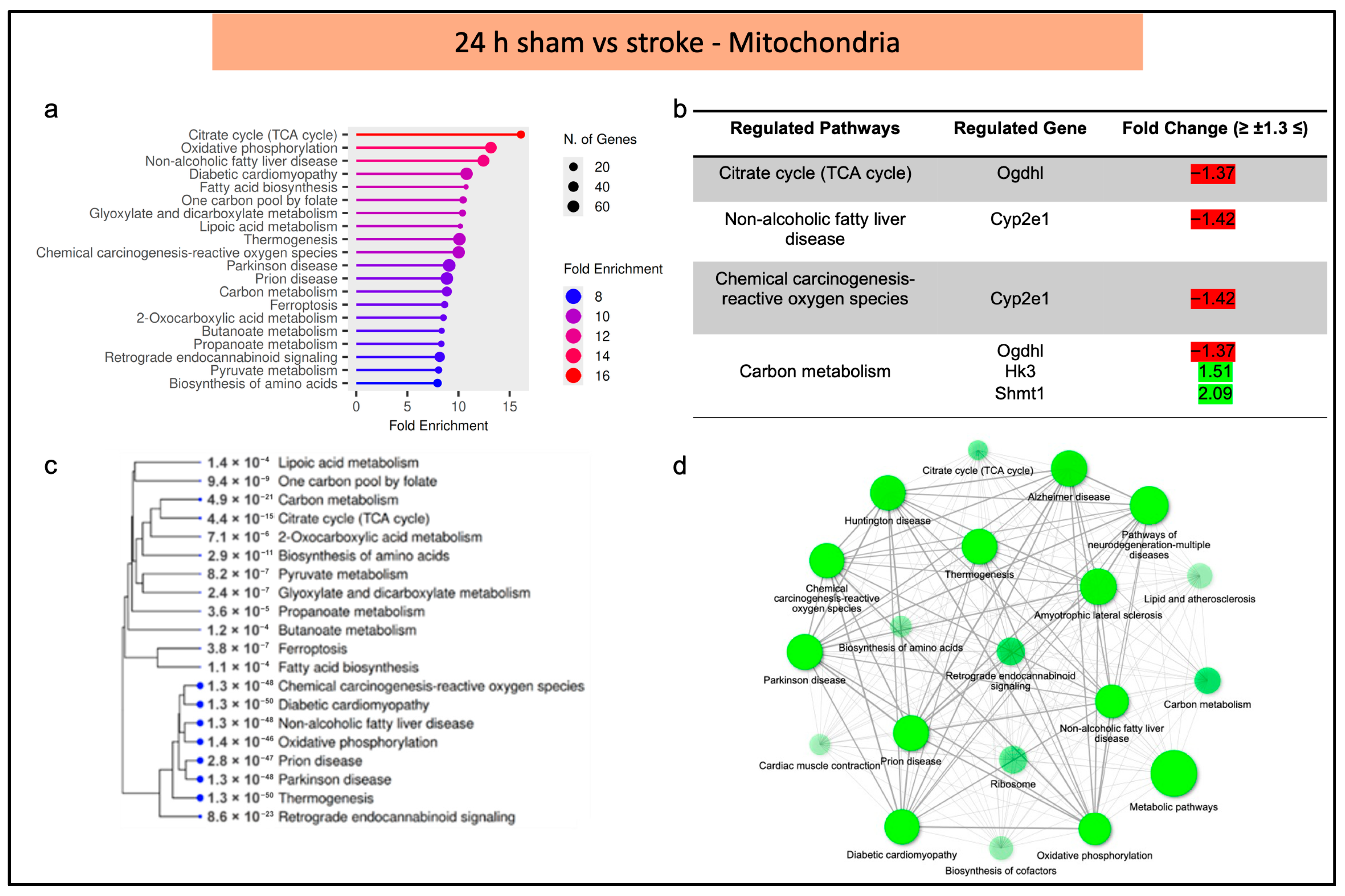
In the acute phase after stroke, brain microvessels exhibited pronounced transcriptional remodeling of mitochondria-associated pathways. ShinyGO analysis identified 20 significantly enriched mitochondrial pathways, with fold enrichment values ranging from approximately 8 to 16 and pathway gene counts between 20 and 60 genes (Figure 1a). Differential regulation within these pathways is summarized in Figure 1b (Supplementary Table S1), while hierarchical clustering (Figure 1c) and pathway interaction network analysis (Figure 1d) demonstrated strong interconnectivity, indicating coordinated mitochondrial regulation in response to ischemic stress. Among the most enriched pathways were those related to oxidative metabolism and redox regulation. Within the tricarboxylic acid (TCA) cycle, Oxoglutarate Dehydrogenase L (Ogdhl) was significantly downregulated (fold change [Fc] = −1.37; Figure 1b). The chemical carcinogenesis–reactive oxygen species (ROS) pathway exhibited marked downregulation of Cytochrome P450 2E1 (Cyp2e1) (Fc = −1.42), a pattern also observed in the non-alcoholic fatty liver disease pathway (Figure 1b). In contrast, selective upregulation of metabolic genes such as Hexokinase 3 (Hk3) (Fc = 1.51) and Serine hydroxymethyl transferase 1 (Shmt1) (Fc = 2.09) was observed within the carbon metabolism pathway, suggesting targeted metabolic reprogramming rather than uniform mitochondrial suppression.
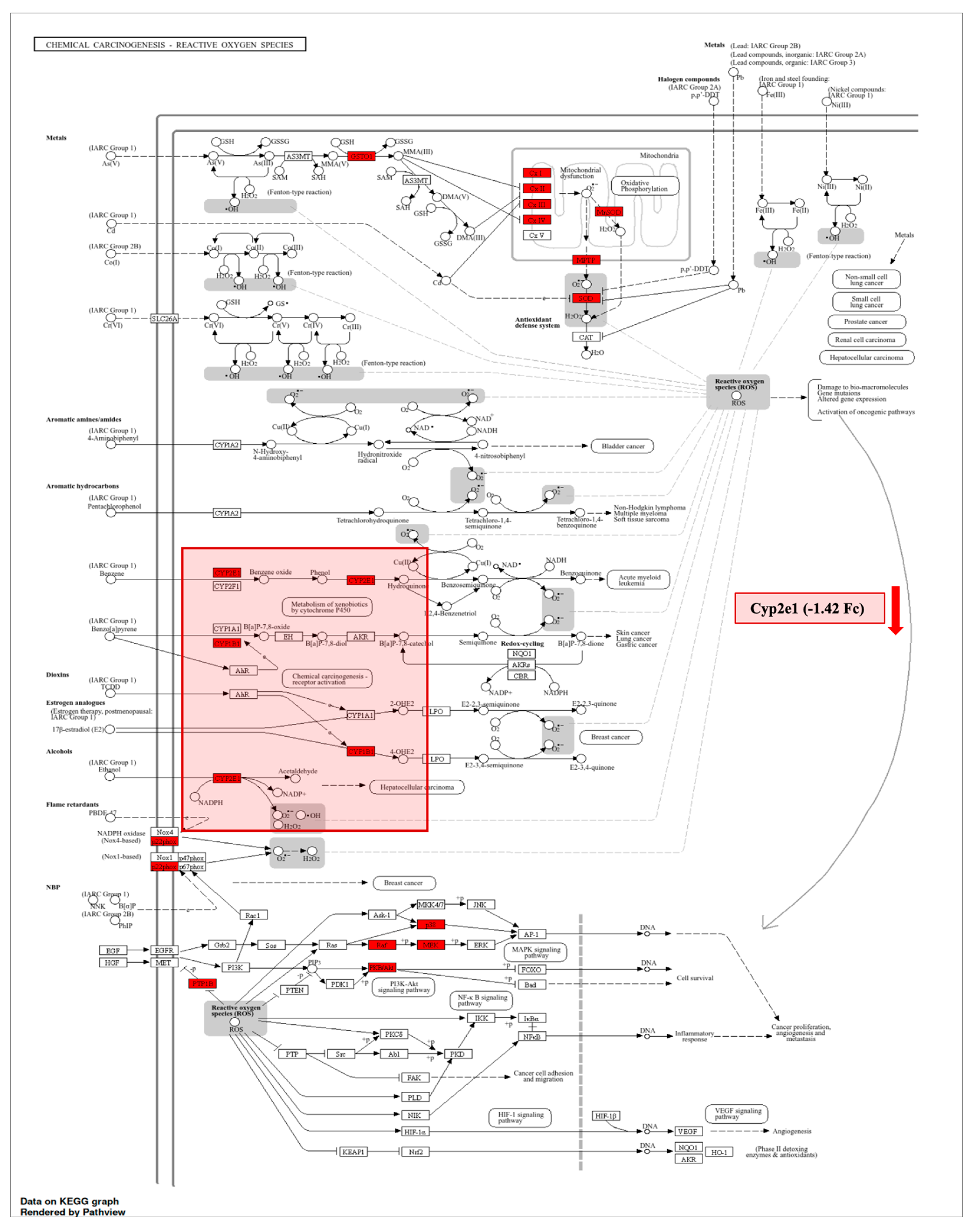
To further delineate mitochondrial redox alterations, KEGG pathway mapping of the chemical carcinogenesis–ROS pathway was performed. This analysis highlighted Cyp2e1 as a centrally downregulated component within a mitochondria-associated ROS pathway (Figure 2), suggesting modulation of oxidative metabolism in microvessels during early ischemic stress. Notably, no mitochondrial pathway enrichment was detected at 7 d post-stroke, suggesting that mitochondrial transcriptomic remodeling is mainly confined to the acute phase (24 h).

3.3. Phase Specific Remodeling of Endoplasmic Reticulum-Pathway Analysis Indicate Acute ER Activation Followed by Intermediate Phase Suppression

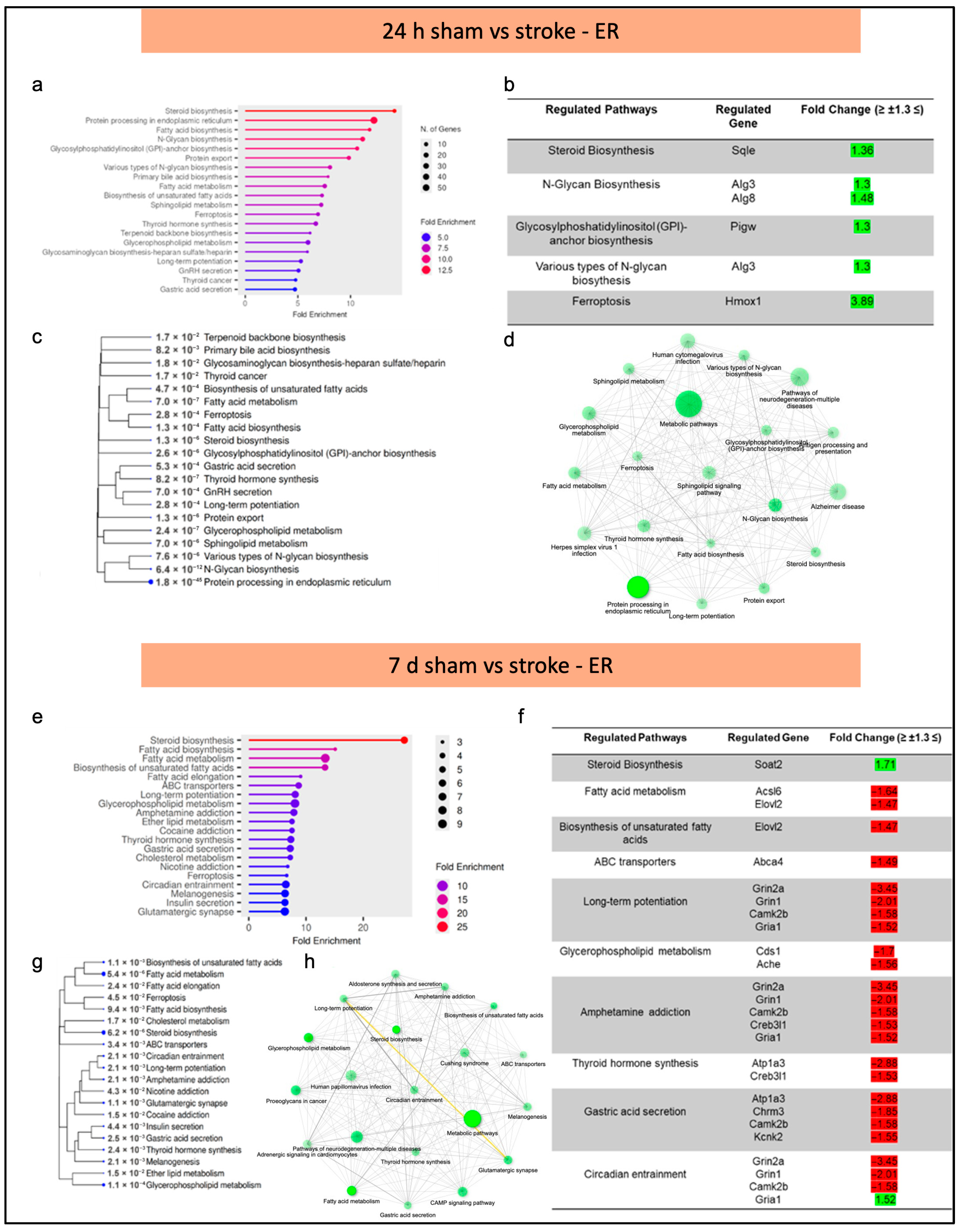
ER-associated pathways exhibited pronounced phase-specific regulation in brain microvessels following ischemic stroke. At 24 h post-stroke, ShinyGO analysis identified 20 significantly enriched ER-related pathways, with fold enrichment values ranging from approximately 5 to 12.5 (Figure 3a). All significantly regulated ER pathways at this time point showed predominant upregulation, as summarized in Figure 3b (Supplementary Table S2). Hierarchical clustering (Figure 3c) and pathway interaction network analysis (Figure 3d) revealed extensive functional connectivity among ER-associated pathways. Notably, the ferroptosis pathway exhibited the highest upregulation, with heme oxygenase 1 (Hmox1) showing a significant increase in expression (Fc = 3.89; Figure 3b), indicating activation of ER-linked oxidative stress responses. In contrast, the ER transcriptomic analysis at 7 d post-stroke was characterized by predominant downregulation across multiple functional categories, including lipid metabolism, neurotransmission, and intracellular signaling pathways (Figure 3e–h; Supplementary Table S3).
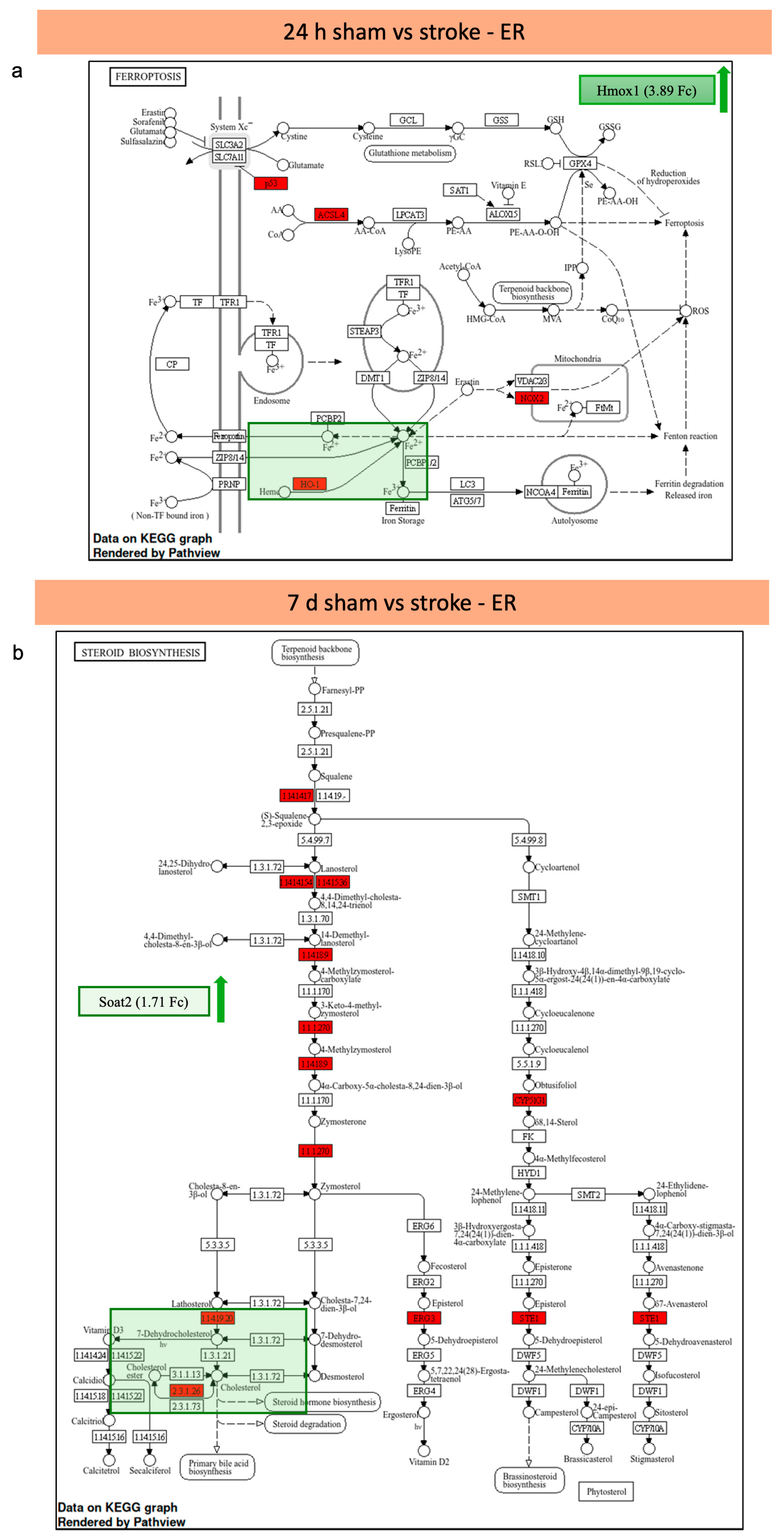
To directly illustrate this temporal transition, ER-associated KEGG pathway maps from both time points were combined. At 24 h post-stroke, KEGG mapping highlighted activation of the ferroptosis pathway, with Hmox1 positioned centrally within ER-associated redox signaling (Figure 4a).
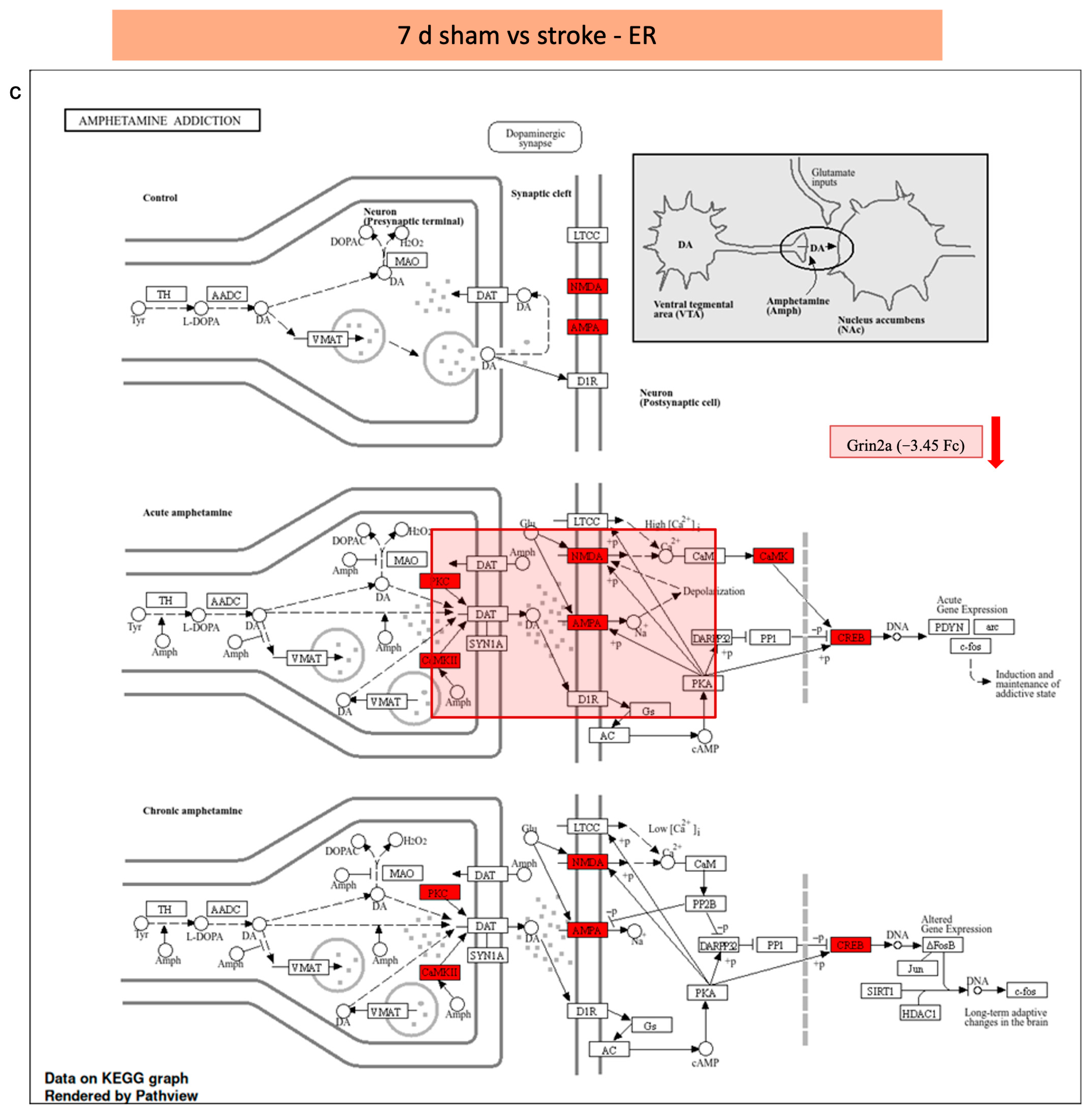
At 7 d post-stroke, KEGG mapping revealed selective upregulation of the steroid biosynthesis pathway, with Sterol O-Acyltransferase 2 (Soat2) (Fc = 1.71) as the main regulated gene, while the majority of ER-associated pathways were suppressed (Figure 4b). In addition, KEGG mapping of the amphetamine addiction pathway at 7 d post-stroke demonstrated significant downregulation of Grin2a (Fc = −3.45), reflecting impaired ER-associated neurotransmission signaling within brain microvessels (Figure 4c).

3.4. Temporal Regulation of Golgi Apparatus-Associated Pathways Characterized by Mixed Acute Responses and Sustained Intermediate-Phase Suppression

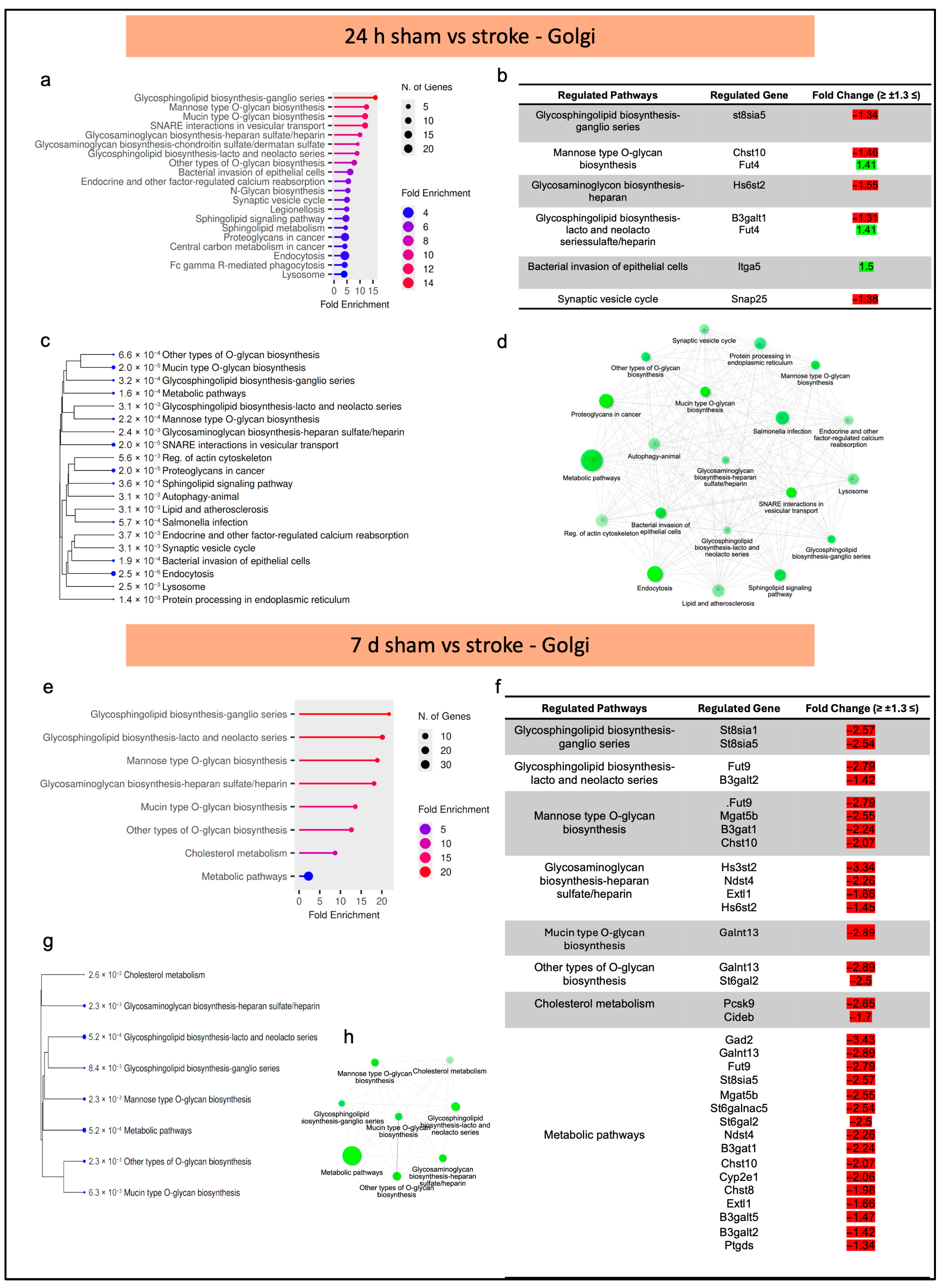
Golgi apparatus-associated pathways displayed a clear regulation pattern compared to mitochondrial or ER responses. At 24 h post-stroke, ShinyGO analysis identified 20 enriched Golgi-related pathways, showing a combination of upregulated and downregulated pathways (Figure 5a,b, Supplementary Table S4).
Hierarchical clustering (Figure 5c) and pathway interaction network analysis (Figure 5d) further reveal differential regulation and extensive functional connectivity among pathways linked to glycosphingolipid and glycosaminoglycan biosynthesis and synaptic vesicle cycle. At this time point, glycosylation-related pathways exhibited both suppression and activation. For example, ST8 Alpha-N-Acetyl-Neuraminide Alpha-2,8-Sialyltransferase 5 (St8sia5) (Fc = −1.34) and Heparan Sulfate 6-O-Sulfotransferase 2 (Hs6st2) (Fc = −1.55) were downregulated in glycosphingolipid and glycosaminoglycan biosynthesis pathways, respectively, whereas Integrin alpha-5 (Itga5) was significantly upregulated (Fc = 1.50) in the bacterial invasion of epithelial cells pathway. Conversely, the synaptic vesicle cycle pathway showed downregulation of Synaptosomal-Associated Protein 25 (Snap25) (Fc = −1.38), indicating early disruption of Golgi-dependent vesicular trafficking. In contrast, at 7 d post-stroke, Golgi-associated pathways showed consistent and significant downregulation (Figure 5e,f, Supplementary Table S5). Genes involved in glycosylation, Polypeptide N-Acetylgalactosaminyltransferase 13 (Galnt13), Fucosyltransferase 9 (Fut9), Alpha-1,6-Mannosylglycoprotein 6-Beta-N-Acetylglucosaminyltransferase B (Mgat5b), glycosphingolipid biosynthesis (St8sia1, B3galt2), and cholesterol metabolism Proprotein Convertase Subtilisin/Kexin type 9 (Pcsk9), (Fc = −2.65) were markedly downregulated at 7 d post-stroke (Figure 5f). This suppression across multiple Golgi-dependent biosynthetic and metabolic pathways indicates sustained Golgi apparatus dysfunction during the intermediate phase, emphasizing a notable shift towards functional suppression of Golgi-associated processes in post-ischemic brain microvessels, which was further evident in hierarchical clustering and pathway network analysis (Figure 5g,h).
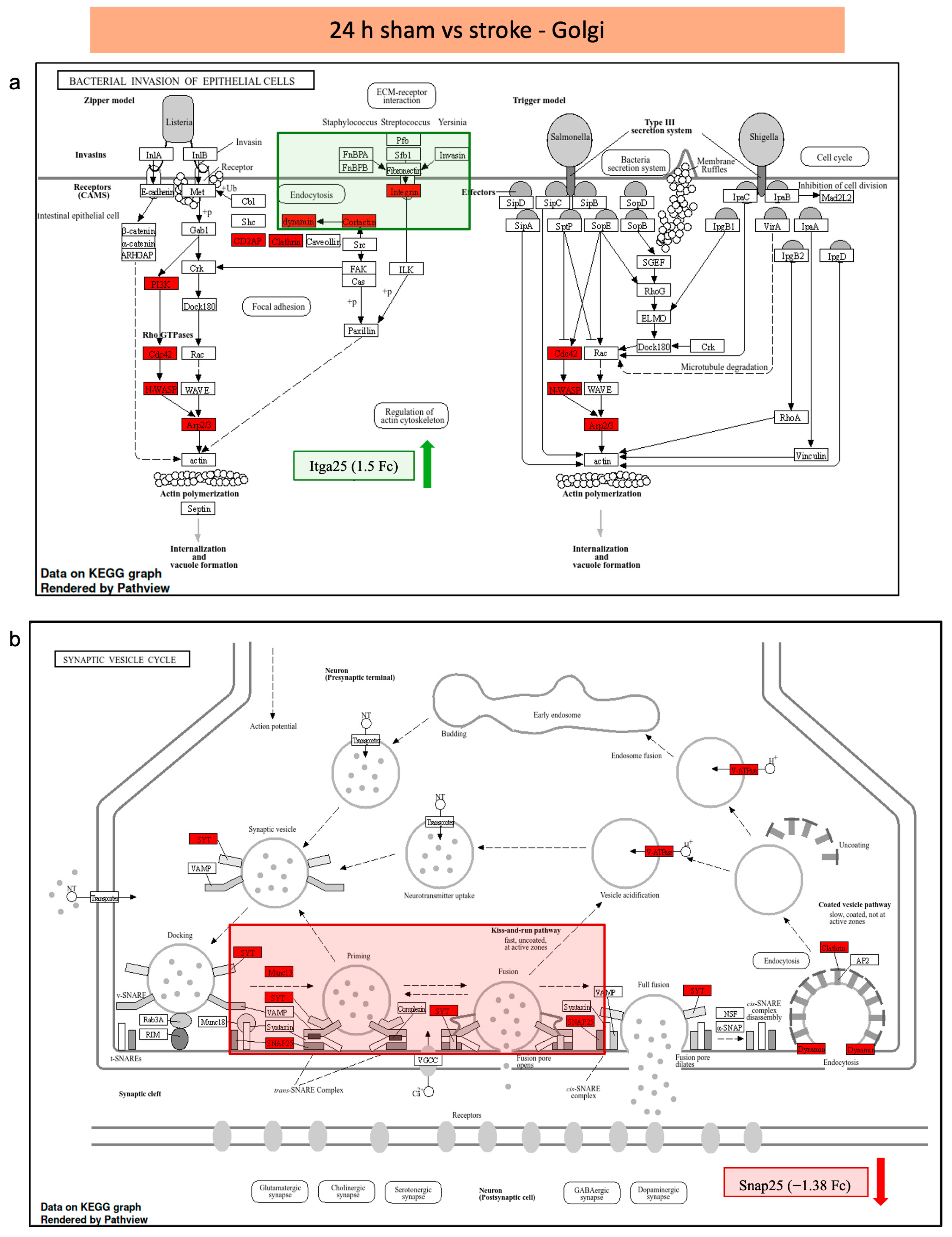
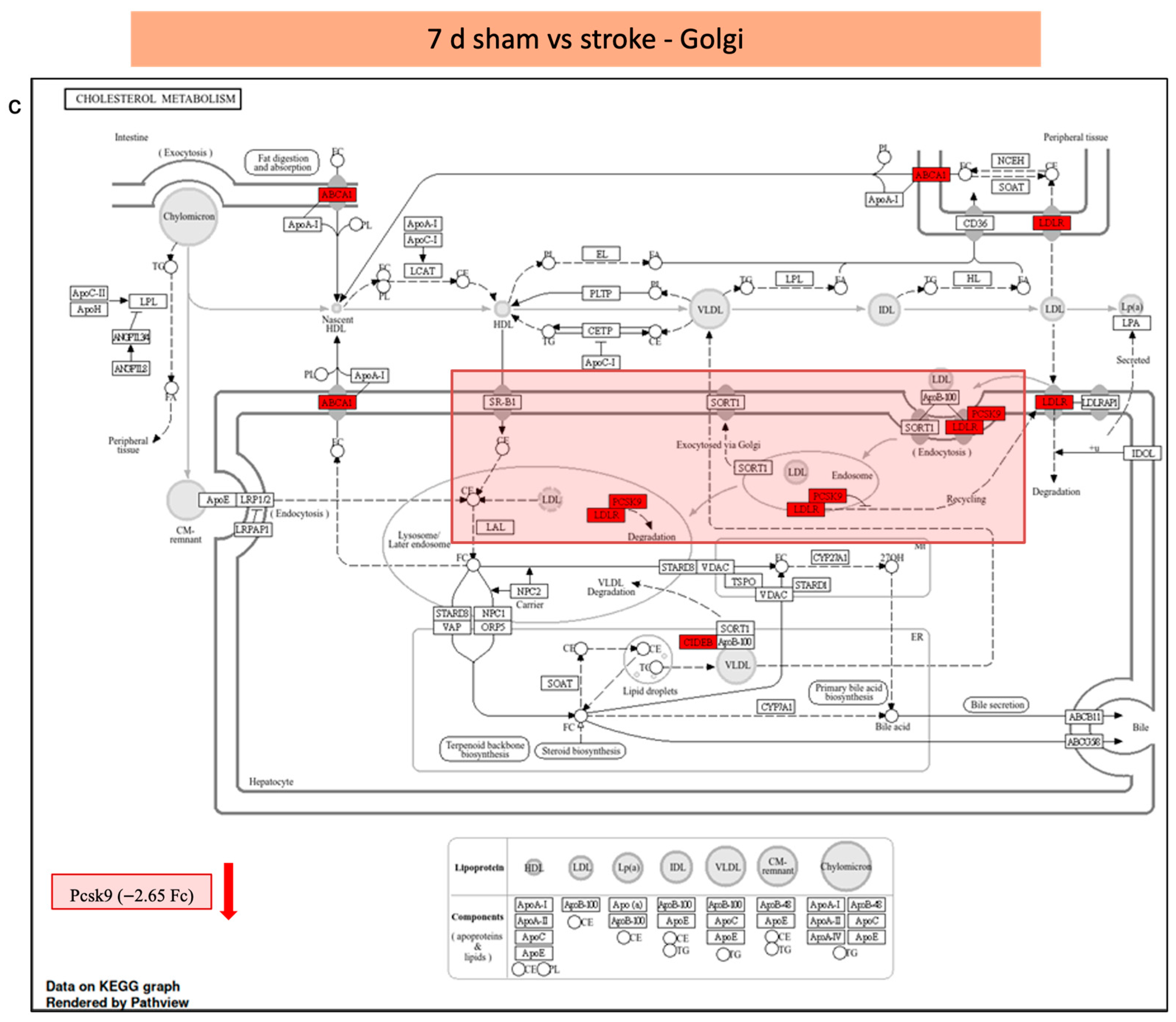
Further, KEGG pathway database mapping at 24 h post-stroke condition revealed a significant enrichment of the bacterial invasion of epithelial cells pathway, functionally implicating Itga25 (Fc = 1.5) as a critical regulator of cytoskeleton reorganization and integrin-mediated cell-matrix interaction, potentially contributing to neurovascular remodeling and cellular stress responses during the acute phase of ischemic injury (Figure 6a). Furthermore, a significant downregulation of the synaptic vesicle cycle protein, Snap25 (Fc = −1.38), underscoring its functional involvement in SNARE-mediated vesicle docking and fusion processes and indicating compromised synaptic transmission and presynaptic integrity under pathological conditions (Figure 6b). On the other hand, at 7 d post-stroke condition, KEGG pathway analysis revealed a pronounced attenuation of cholesterol metabolism, with Pcsk9 (Fc = −2.65) functionally implicated as a central modulator of lipid regulatory network via its role in promoting low density lipoprotein receptor catabolism (Figure 6c).
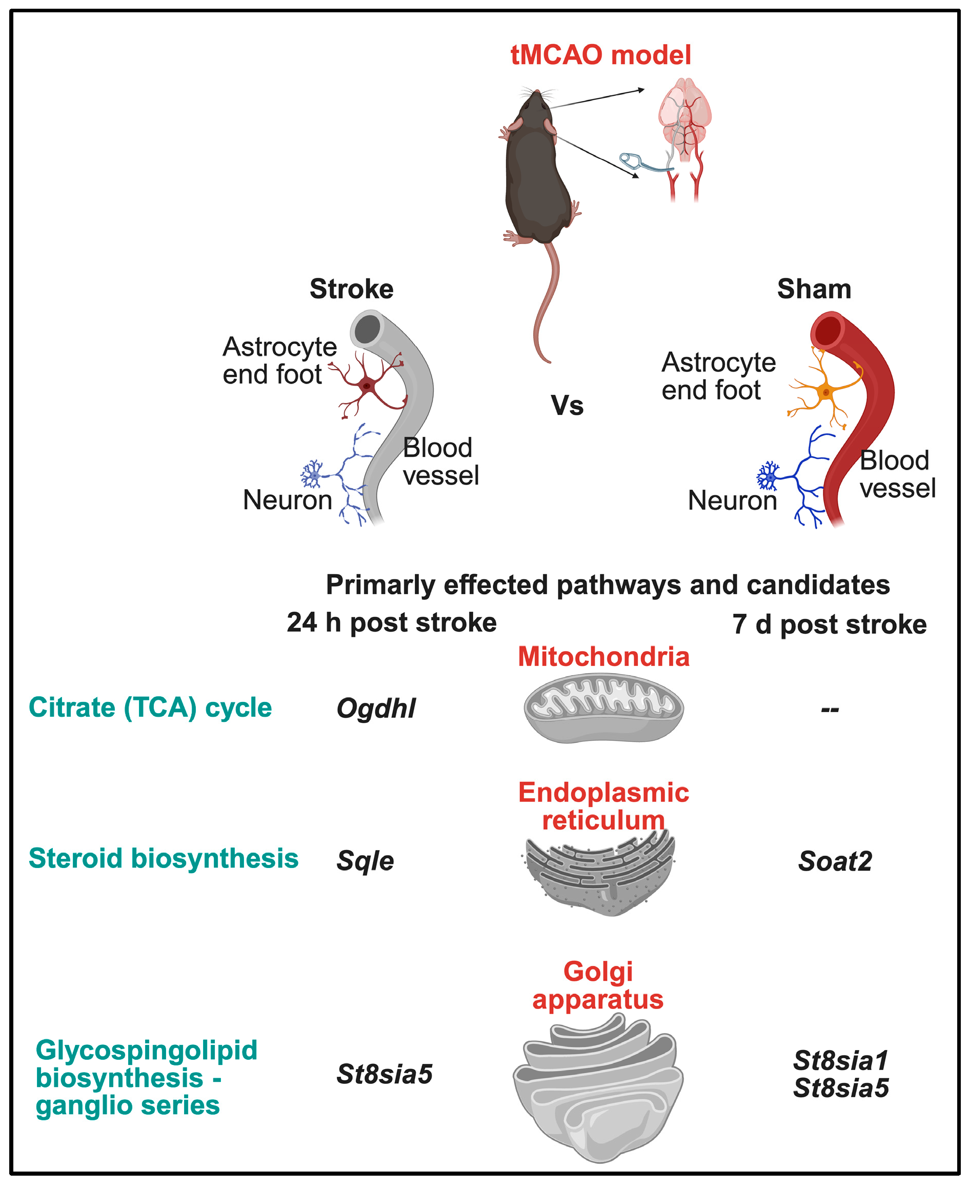
Taken together, our results indicate that ischemic stroke induces phase-dependent, organelle-specific molecular remodeling in brain microvessels. Mitochondrial transcriptomic alterations are restricted to the acute phase, whereas ER pathways transition from early activation to intermediate-phase suppression and Golgi-associated pathways shift from mixed acute regulation to predominant downregulation. These patterns identify sustained ER and Golgi dysfunction as prominent features of microvascular pathology during the intermediate post-stroke phase.

4. Discussion

This study provides an organelle-specific, phase-specific transcriptome analysis of mouse brain microvessels following ischemic stroke and elucidates distinct molecular remodeling patterns of different cell organelles during the acute and intermediate phases post-stroke (Figure 7). By focusing specifically on the microvascular compartment, the present work extends previous transcriptomic studies, which mainly analyzed whole-brain or bulk-tissue samples [6,11,12,25]. The findings support the hypothesis that ischemic stroke induces phase-specific organelle dysfunction within brain microvessels, characterized by coordinated early responses and sustained subcellular impairment at later stages. Both cellular and sub-cellular mechanisms shape the severity of injury and the capacity for tissue repair post-stroke.
During the acute phase (24 h), significant transcriptional changes were observed across mitochondria, ER, and Golgi-related pathways. We identified that mitochondrial pathway modulation was primarily linked to metabolic and redox processes, consistent with established evidence that ischemia rapidly disrupts mitochondrial energy metabolism and reactive oxygen species homeostasis [14,15,18,26]. The observed downregulation of genes such as Cyp2e1 may reflect an early adaptive response aimed at limiting oxidative stress within microvascular cells, a phenomenon previously suggested in vascular and endothelial models of ischemic injury [27,28].
Concomitantly, ER-associated pathways exhibited robust activation during the acute phase, including biosynthetic and stress-related signaling pathways such as ferroptosis. Upregulation of ferroptosis-associated genes, including Hmox1, suggests enhanced iron- and redox-dependent stress responses within microvessels, which may contribute to endothelial dysfunction and BBB disruption. While ER stress has been widely implicated in ischemic brain injury [29,30,31], most prior studies have focused on neuronal populations [32,33]. The present analysis highlights ER stress-related mechanisms specifically within the microvascular compartment, underscoring their potential contribution to early vascular pathology [34,35,36].
Golgi apparatus-associated pathways were also significantly altered during the acute phase, particularly those involved in vesicular trafficking, glycosylation, and synaptic vesicle cycling. Golgi stress has been reported in ischemic neurons and linked to impaired protein processing and trafficking [29,30,37]; however, its role in microvascular cells has received comparatively less attention. In the context of brain microvessels, disruption of Golgi-mediated pathways may influence endothelial surface protein expression, cell–matrix interactions, and intracellular transport processes critical for BBB maintenance [38,39].
In contrast, the intermediate post-stroke phase (7 d) was characterized by markedly reduced mitochondrial pathway involvement and predominant downregulation of ER- and Golgi-associated pathways. Initially, a strong regulation of mitochondrial pathways was observed at 24 h following the stroke induction, which likely reflects an acute stress response characterized by oxidative stress, metabolic reprogramming, and activation of other mitochondrial pathways (e.g., mitophagy and antioxidant responses). During the mid-term phase before reaching the intermediate phase of 7 days (not yet fibrotic phase) post-stroke, partial resolution of acute ischemic stress and clearance of severely damaged endothelial cells may reduce the transcriptional activation of the mitochondrial pathways, resulting in an apparent normalization of mitochondrial-related gene expression. Another possible mechanism is a shift in transcriptional regulation to post-transcriptional and even functional related responses. In addition, mitochondrial damage or dysfunction persists despite reduced gene expression through impaired electron transport chain activity, altered mitochondrial dynamics, or sustained oxidative damage that is not captured at the mRNA level, which argues for potential proteomics analysis.
Finally, as our data shows, suppression of mitochondrial genes at this stage is continued with the sequence of persistent ER and Golgi dysregulation. This may indicate a transition from acute energy/redox imbalance to chronic defects in protein processing, trafficking, and neurovascular signaling, consistent with incomplete tissue recovery. In our other studies, as well as in the literature, it is known that an irreversible glial cell scar occurs in the very long term, for example, at 30 days post stroke, where few cells survive.
This shift suggests a transition from acute metabolic adaptation toward sustained impairment of proteostasis, lipid metabolism, and intracellular trafficking within microvessels [40,41]. Persistent downregulation of ER-associated signaling and metabolic pathways may contribute to prolonged microvascular dysfunction, impaired neurovascular coupling, and delayed recovery. Similarly, widespread suppression of Golgi-associated glycosylation and cholesterol metabolism pathways, including reduced expression of Pcsk9, indicates long-lasting alterations in membrane composition and vesicle trafficking that may affect vascular remodeling and barrier integrity [42,43].

4.1. Limitations of the Present Study

Although our study provides valuable analysis of the publicly available dataset, the following limitations must be mentioned. First, the present study is based only on a single mRNA transcriptome dataset derived from isolated brain microvessels. Although this dataset is unique in its model, samples and time points, the findings should be interpreted within the context of secondary in silico analysis [16,32]. Second, as these are transcriptomic changes, they do not necessarily reflect protein changes or functional activity, and moreover, post-transcriptional regulatory mechanisms were not analyzed. Third, microvessels consist of multiple cell types, including endothelial cells, astrocytic end feet, glial cells and pericytes, and cell-type–specific contributions could not be resolved in the present analysis [44,45]. Finally, experimental validation of key pathways and genes was beyond the scope of this study.

4.2. Future Directions

Future studies integrating multi-omics, such as proteomics, single-cell or cell-specific analysis, will help to validate and extend these findings. Indeed, phase-specific drug–gene interaction analysis will further contextualize these findings. Acute-phase associations with thrombolytic and anticoagulant agents with established clinical interventions might be helpful [46,47,48]. Similarly, intermediate-phase associations with neuroprotective and synaptic-modulating compounds can suggest molecular pathways that may be relevant during recovery and secondary injury phases. Details about these associations and potential candidate pathways are targets for future experimental validation.

5. Conclusions

In conclusion, this study demonstrates that ischemic stroke induces distinct, phase-dependent organelle-specific molecular remodeling within brain microvessels, characterized by coordinated acute responses and sustained ER and Golgi dysfunction during the intermediate phase. By providing an organelle-resolved view of microvascular transcriptomic changes, these findings advance understanding of neurovascular pathology following stroke and identify subcellular pathways that may inform future biomarker development and stage-specific therapeutic strategies.

Supplementary Materials

The following supporting information can be downloaded at: https://www.mdpi.com/article/10.3390/biophysica6020033/s1, Table S1: 24 h Mitochondria; Table S2: 24 h ER; Table S3: 7 d ER; Table S4: 24 h Golgi; Table S5: 7 d Golgi.

Author Contributions

Conceptualization, R.V., A.A. and A.L.; methodology, S.I. and S.M.; software, S.M. and S.I.; validation, R.V., A.A. and A.L.; formal analysis, A.A., S.M. and S.I.; resources, A.L. and R.V.; data curation, S.I., S.M. and A.A.; writing—original draft preparation, S.I., S.M. and P.Y.; writing—review and editing, R.V., A.A. and A.L.; visualization, R.V., P.Y. and S.M.; supervision, A.A., R.V. and A.L.; project administration, A.L., A.A. and R.V.; funding acquisition, A.L. and R.V. All authors have read and agreed to the published version of the manuscript.

Funding

This research was funded by the German Research Foundation (SFB1039 for A.L., R.V.).

Institutional Review Board Statement

Not applicable.

Informed Consent Statement

Not applicable.

Data Availability Statement

The transcriptomic data analyzed in this study are publicly available and were obtained from the previously published dataset by Kestner et al. All data used in the present analysis can be accessed through the original publication, as already indicated in Section 2, and its associated public repositories. No new datasets were generated during this study.

Acknowledgments

The ChatGPT 5.2 AI tool has been used to improve only the language of the manuscript. The authors have reviewed and edited the output and take full responsibility for the content of this publication.

Conflicts of Interest

All the authors declare no conflicts of interest. The funders had no role in the design of the study; in the collection, analyses, or interpretation of data; in the writing of the manuscript; or in the decision to publish the results.

Abbreviations

The following abbreviations are used in this manuscript:
ABCATP-binding cassette
BBBBlood–brain barrier
ChEAChIP-X Enrichment Analysis
DEGsDifferentially expressed genes
DGIdbDrug–Gene Interaction Database
EREndoplasmic reticulum
GEOGene Expression Omnibus
GOGene Ontology
GPIGlycosylphosphatidylinositol
KEAKinase Enrichment Analysis
KEGGKyoto Encyclopedia of Genes and Genomes
MCODEMolecular Complex Detection
NVUNeurovascular unit
PPIProtein–protein interaction
ROSReactive oxygen species
STRINGSearch Tool for the Retrieval of Interacting Genes/Proteins
tMCAOTransient middle cerebral artery occlusion
UPRUnfolded protein response

References

  1. GBD 2019 Stroke Collaborators. Global, regional, and national burden of stroke and its risk factors, 1990–2019: A systematic analysis for the Global Burden of Disease Study 2019. Lancet Neurol. 2021, 20, 795–820. [Google Scholar] [CrossRef] [Scilit] [PubMed] [PubMed Central]
  2. GBD 2021 Stroke Risk Factor Collaborators. Global, regional, and national burden of stroke and its risk factors, 1990–2021: A systematic analysis for the Global Burden of Disease Study 2021. Lancet Neurol. 2024, 23, 973–1003. [CrossRef] [Scilit] [PubMed] [PubMed Central]
  3. Dirnagl, U.; Iadecola, C.; Moskowitz, M.A. Pathobiology of ischaemic stroke: An integrated view. Trends Neurosci. 1999, 22, 391–397. [Google Scholar] [CrossRef] [Scilit] [PubMed]
  4. Lo, E.H.; Dalkara, T.; Moskowitz, M.A. Mechanisms, challenges and opportunities in stroke. Nat. Rev. Neurosci. 2003, 4, 399–415. [Google Scholar] [CrossRef] [Scilit] [PubMed]
  5. Lucaciu, A.; Kuhn, H.; Trautmann, S.; Ferreirós, N.; Steinmetz, H.; Pfeilschifter, J.; Brunkhorst, R.; Pfeilschifter, W.; Subburayalu, J.; Vutukuri, R. A Sphingosine 1-Phosphate Gradient Is Linked to the Cerebral Recruitment of T Helper and Regulatory T Helper Cells during Acute Ischemic Stroke. Int. J. Mol. Sci. 2020, 21, 6242. [Google Scholar] [CrossRef] [Scilit] [PubMed] [PubMed Central]
  6. del Zoppo, G.J. The neurovascular unit in the setting of stroke. J. Intern. Med. 2010, 267, 156–171. [Google Scholar] [CrossRef] [Scilit] [PubMed]
  7. Hansen, L.; Lohfink, N.; Vutukuri, R.; Kestner, R.I.; Trautmann, S.; Hecht, M.; Wagner, P.V.; Spitzer, D.; Khel, M.I.; Macas, J.; et al. Endothelial Sphingosine-1-Phosphate Receptor 4 Regulates Blood-Brain Barrier Permeability and Promotes a Homeostatic Endothelial Phenotype. J. Neurosci. 2022, 42, 1908–1929. [Google Scholar] [CrossRef] [Scilit] [PubMed] [PubMed Central]
  8. Gurnik, S.; Devraj, K.; Macas, J.; Yamaji, M.; Starke, J.; Scholz, A.; Sommer, K.; Di Tacchio, M.; Vutukuri, R.; Beck, H.; et al. Angiopoietin-2-induced blood-brain barrier compromise and increased stroke size are rescued by VE-PTP-dependent restoration of Tie2 signaling. Acta Neuropathol. 2016, 131, 753–773. [Google Scholar] [CrossRef] [Scilit] [PubMed] [PubMed Central]
  9. Haley, M.J.; Lawrence, C.B. The blood-brain barrier after stroke: Structural studies and the role of transcytotic vesicles. J. Cereb. Blood Flow. Metab. 2017, 37, 456–470. [Google Scholar] [CrossRef] [Scilit] [PubMed] [PubMed Central]
  10. Iadecola, C. The Neurovascular Unit Coming of Age: A Journey through Neurovascular Coupling in Health and Disease. Neuron 2017, 96, 17–42. [Google Scholar] [CrossRef] [Scilit] [PubMed] [PubMed Central]
  11. Cipolla, M.J.; Crete, R.; Vitullo, L.; Rix, R.D. Transcellular transport as a mechanism of blood-brain barrier disruption during stroke. Front. Biosci. 2004, 9, 777–785. [Google Scholar] [CrossRef] [Scilit] [PubMed]
  12. Knowland, D.; Arac, A.; Sekiguchi, K.J.; Hsu, M.; Lutz, S.E.; Perrino, J.; Steinberg, G.K.; Barres, B.A.; Nimmerjahn, A.; Agalliu, D. Stepwise recruitment of transcellular and paracellular pathways underlies blood-brain barrier breakdown in stroke. Neuron 2014, 82, 603–617. [Google Scholar] [CrossRef] [Scilit] [PubMed] [PubMed Central]
  13. She, R.; Liu, D.; Liao, J.; Wang, G.; Ge, J.; Mei, Z. Mitochondrial dysfunctions induce PANoptosis and ferroptosis in cerebral ischemia/reperfusion injury: From pathology to therapeutic potential. Front. Cell Neurosci. 2023, 17, 1191629. [Google Scholar] [CrossRef] [Scilit] [PubMed] [PubMed Central]
  14. An, H.; Zhou, B.; Ji, X. Mitochondrial quality control in acute ischemic stroke. J. Cereb. Blood Flow Metab. 2021, 41, 3157–3170. [Google Scholar] [CrossRef] [Scilit] [PubMed] [PubMed Central]
  15. Sims, N.R.; Muyderman, H. Mitochondria, oxidative metabolism and cell death in stroke. Biochim. Biophys. Acta 2010, 802, 80–91. [Google Scholar] [CrossRef] [Scilit] [PubMed]
  16. Kovacheva, E.; Gevezova, M.; Koeva, M.; Mihaylova, V.; Kormova, V.; Kostadinova, E.; Kostadinova, Y.; Kazakova, M.; Sarafian, V. Mitochondrial Function, Oxidative Stress, Inflammation and Thrombolytic Treatment in Ischemic Stroke. Int. J. Mol. Sci. 2025, 26, 10289. [Google Scholar] [CrossRef] [Scilit] [PubMed] [PubMed Central]
  17. Nakka, V.P.; Gusain, A.; Raghubir, R. Endoplasmic reticulum stress plays critical role in brain damage after cerebral ischemia/reperfusion in rats. Neurotox. Res. 2010, 17, 189–202. [Google Scholar] [CrossRef] [Scilit] [PubMed]
  18. Paschen, W.; Mengesdorf, T. Endoplasmic reticulum stress response and neurodegeneration. Cell Calcium 2005, 38, 409–415. [Google Scholar] [CrossRef] [Scilit] [PubMed]
  19. Yuan, D.; Liu, C.; Hu, B. Dysfunction of Membrane Trafficking Leads to Ischemia-Reperfusion Injury After Transient Cerebral Ischemia. Transl. Stroke Res. 2018, 9, 215–222. [Google Scholar] [CrossRef] [Scilit] [PubMed] [PubMed Central]
  20. Nakagomi, S.; Barsoum, M.J.; Bossy-Wetzel, E.; Sütterlin, C.; Malhotra, V.; Lipton, S.A. A Golgi fragmentation pathway in neurodegeneration. Neurobiol. Dis. 2008, 29, 221–231. [Google Scholar] [CrossRef] [Scilit] [PubMed] [PubMed Central]
  21. Lu, W.; Xu, D.; Tu, R.; Hu, Z. Morphology of platelet Golgi apparatus and their significance after acute cerebral infarction. Neural Regen. Res. 2013, 8, 2134–2143. [Google Scholar] [CrossRef] [PubMed] [PubMed Central]
  22. Dergunova, L.V.; Filippenkov, I.B.; Stavchansky, V.V.; Denisova, A.E.; Yuzhakov, V.V.; Mozerov, S.A.; Gubsky, L.V.; Limborska, S.A. Genome-wide transcriptome analysis using RNA-Seq reveals a large number of differentially expressed genes in a transient MCAO rat model. BMC Genom. 2018, 19, 655. [Google Scholar] [CrossRef] [Scilit] [PubMed] [PubMed Central]
  23. Lv, Y.; Xi, Y.; Zhang, L.; Wang, J.; Wu, J. Cerebral ischemia-induced gene expression changes in diabetic mice from acute to subacute stage. Brain Res. 2024, 1825, 148737. [Google Scholar] [CrossRef] [Scilit] [PubMed]
  24. Kestner, R.-I.; Mayser, F.; Vutukuri, R.; Hansen, L.; Günther, S.; Brunkhorst, R.; Devraj, K.; Pfeilschifter, W. Gene Expression Dynamics at the Neurovascular Unit During Early Regeneration After Cerebral Ischemia/Reperfusion Injury in Mice. Front. Neurosci. 2020, 14, 280. [Google Scholar] [CrossRef] [Scilit] [PubMed] [PubMed Central]
  25. Maikap, S.; Lucaciu, A.; Chakraborty, A.; Kestner, R.I.; Vutukuri, R.; Annamneedi, A. Targeting the Neuro-vascular Presynaptic Signalling in STROKE: Evidence and Therapeutic Implications. Ann. Neurosci. 2025, 09727531241310048. [Google Scholar] [CrossRef] [Scilit] [PubMed] [PubMed Central]
  26. Doll, D.N.; Hu, H.; Sun, J.; Lewis, S.E.; Simpkins, J.W.; Ren, X. Mitochondrial crisis in cerebrovascular endothelial cells opens the blood-brain barrier. Stroke 2015, 46, 1681–1689. [Google Scholar] [CrossRef] [Scilit] [PubMed] [PubMed Central]
  27. Tindberg, N.; Baldwin, H.A.; Cross, A.J.; Ingelman-Sundberg, M. Induction of cytochrome P450 2E1 expression in rat and gerbil astrocytes by inflammatory factors and ischemic injury. Mol. Pharmacol. 1996, 50, 1065–1072. [Google Scholar] [CrossRef] [Scilit] [PubMed]
  28. Yu, J.; Zhu, H.; Kindy, M.S.; Taheri, S. Cytochrome P450 CYP2E1 Suppression Ameliorates Cerebral Ischemia Reperfusion Injury. Antioxidants 2021, 10, 52. [Google Scholar] [CrossRef] [Scilit] [PubMed] [PubMed Central]
  29. Choi, Y.K.; Kim, Y.M. Beneficial and Detrimental Roles of Heme Oxygenase-1 in the Neurovascular System. Int. J. Mol. Sci. 2022, 23, 7041. [Google Scholar] [CrossRef] [Scilit] [PubMed] [PubMed Central]
  30. Liu, Q.; Song, T.; Chen, B.; Zhang, J.; Li, W. Ferroptosis of brain microvascular endothelial cells contributes to hypoxia-induced blood-brain barrier injury. FASEB J. 2023, 37, e22874. [Google Scholar] [CrossRef] [Scilit] [PubMed]
  31. Li, Z.; Huang, K.; Cao, J.; Guo, M.; Dong, H.; Ye, W.; Zeng, S.; Wei, J.; Xi, Q. Silencing Hmox1 Attenuates Cerebral Ischemia/reperfusion Injury and Inhibits Inflammation and Ferroptosis Via the PPAR-γ/FABP4 Signaling Pathway. Mol. Neurobiol. 2025, 62, 11164–11178. [Google Scholar] [CrossRef] [Scilit] [PubMed]
  32. Wang, L.; Liu, Y.; Zhang, X.; Ye, Y.; Xiong, X.; Zhang, S.; Gu, L.; Jian, Z.; Wang, H. Endoplasmic Reticulum Stress and the Unfolded Protein Response in Cerebral Ischemia/Reperfusion Injury. Front. Cell Neurosci. 2022, 16, 864426. [Google Scholar] [CrossRef] [Scilit] [PubMed] [PubMed Central]
  33. Han, B.; Zhou, S.; Zhang, Y.; Chen, S.; Xi, W.; Liu, C.; Zhou, X.; Yuan, M.; Yu, X.; Li, L.; et al. Integrating spatial and single-cell transcriptomics to characterize the molecular and cellular architecture of the ischemic mouse brain. Sci. Transl. Med. 2024, 16, eadg1323. [Google Scholar] [CrossRef] [Scilit] [PubMed]
  34. Nan, D.; Jin, H.; Deng, J.; Yu, W.; Liu, R.; Sun, W.; Huang, Y. Cilostazol ameliorates ischemia/reperfusion-induced tight junction disruption in brain endothelial cells by inhibiting endoplasmic reticulum stress. FASEB J. 2019, 33, 10152–10164. [Google Scholar] [CrossRef] [Scilit] [PubMed]
  35. Xin, J.; Ma, X.; Chen, W.; Zhou, W.; Dong, H.; Wang, Z.; Ji, F. Regulation of blood-brain barrier permeability by Salvinorin A via alleviating endoplasmic reticulum stress in brain endothelial cell after ischemia stroke. Neurochem. Int. 2021, 149, 105093. [Google Scholar] [CrossRef] [Scilit] [PubMed]
  36. Fan, Q.; Takarada-Iemata, M.; Okitani, N.; Tamatani, T.; Ishii, H.; Hattori, T.; Kiryu-Seo, S.; Kiyama, H.; Hori, O. Brain injury triggers cell-type-specific and time-dependent endoplasmic reticulum stress responses. Glia 2023, 71, 667–681. [Google Scholar] [CrossRef] [Scilit] [PubMed]
  37. Kulkarni-Gosavi, P.; Makhoul, C.; Gleeson, P.A. Form and function of the Golgi apparatus: Scaffolds, cytoskeleton and signalling. FEBS Lett. 2019, 593, 2289–2305. [Google Scholar] [CrossRef] [Scilit] [PubMed]
  38. Sachdev, P.S.; Bentvelzen, A.C.; Gustafson, D.; Hansra, G.K.; Hosoki, S.; Jiang, J.; Lennon, M.J.; Moro, M.A.; Saks, D.G.; Samaras, K.; et al. Vascular Cognitive Impairment and Dementia: Clinical Features, Neuropathology, and Biomarkers. J. Am. Coll. Cardiol. 2026, 87, 52–76. [Google Scholar] [CrossRef] [Scilit] [PubMed]
  39. Xiang, Y.; Gu, Q.; Liu, D. Brain Endothelial Cells in Blood-Brain Barrier Regulation and Neurological Therapy. Int. J. Mol. Sci. 2025, 26, 5843. [Google Scholar] [CrossRef] [Scilit] [PubMed] [PubMed Central]
  40. Meng, J.; Zhang, J.; Fang, J.; Li, M.; Ding, H.; Zhang, W.; Chen, C. Dynamic inflammatory changes of the neurovascular units after ischemic stroke. Brain Res. Bull. 2022, 190, 140–151. [Google Scholar] [CrossRef] [Scilit] [PubMed]
  41. Moon, S.; Chang, M.S.; Koh, S.H.; Choi, Y.K. Repair Mechanisms of the Neurovascular Unit after Ischemic Stroke with a Focus on VEGF. Int. J. Mol. Sci. 2021, 22, 8543. [Google Scholar] [CrossRef] [Scilit] [PubMed] [PubMed Central]
  42. Zheng, Y.; Zhu, T.; Li, G.; Xu, L.; Zhang, Y. PCSK9 inhibitor protects against ischemic cerebral injury by attenuating inflammation via the GPNMB/CD44 pathway. Int. Immunopharmacol. 2024, 126, 111195. [Google Scholar] [CrossRef] [Scilit] [PubMed]
  43. Moustafa, B.; Oparowski, D.; Testai, S.; Guman, I.; Trifan, G. Efficacy and safety of PCSK9 inhibitors for stroke prevention: Systematic review and meta-analysis. J. Stroke Cerebrovasc. Dis. 2024, 33, 107633. [Google Scholar] [CrossRef] [Scilit] [PubMed]
  44. Rajput, P.; Brookshier, A.; Kothari, S.; Eckstein, L.; Chang, H.; Liska, S.; Lamb, J.; Sances, S.; Lyden, P. Differential Vulnerability and Response to Injury among Brain Cell Types Comprising the Neurovascular Unit. J. Neurosci. 2024, 44, e1093222024. [Google Scholar] [CrossRef] [Scilit] [PubMed] [PubMed Central]
  45. Kowiański, P.; Czuba-Pakuła, E.; Lietzau, G. The neurovascular unit response to cerebral ischemia as a promising area for translational studies—Comments on some opened questions. Exp. Neurol. 2026, 396, 115533. [Google Scholar] [CrossRef] [Scilit] [PubMed]
  46. Xiong, Y.; Wakhloo, A.K.; Fisher, M. Advances in Acute Ischemic Stroke Therapy. Circ. Res. 2022, 130, 1230–1251. [Google Scholar] [CrossRef] [Scilit] [PubMed]
  47. Greco, A.; Occhipinti, G.; Giacoppo, D.; Agnello, F.; Laudani, C.; Spagnolo, M.; Mauro, M.S.; Rochira, C.; Finocchiaro, S.; Mazzone, P.M.; et al. Antithrombotic Therapy for Primary and Secondary Prevention of Ischemic Stroke: JACC State-of-the-Art Review. J. Am. Coll. Cardiol. 2023, 82, 1538–1557. [Google Scholar] [CrossRef] [Scilit] [PubMed]
  48. Catanese, L.; Tarsia, J.; Fisher, M. Acute Ischemic Stroke Therapy Overview. Circ. Res. 2017, 120, 541–558. [Google Scholar] [CrossRef] [Scilit] [PubMed]
Figure 1. Mitochondrial pathway modulation in the acute phase following stroke (24 h sham vs. stroke). (a) Top significantly enriched mitochondrial pathways were ranked by fold enrichment through different colors. (b) Most significantly regulated mitochondrial-associated pathways with individual genes annotated by their corresponding fold change (Fc) values. Highlighted Fc values in red are downregulated ones and in green are upregulated. (c) Illustrating the correlation structure among enriched pathways by FDR value. (d) Pathway interactions, where nodes denote individual pathways and edges reflect shared gene composition, defined by cut off zero.
Biophysica 06 00033 g001
Figure 2. Mitochondria-associated chemical carcinogenesis-reactive oxygen species pathway as annotated in the KEGG database, highlighting significant downregulation of Cyp2e1. The highlighted pathway segment delineates Cyp2e1-dependent enzymatic reactions involved in xenobiotic metabolism and reactive oxygen species generation, emphasizing its potential contribution to altered mitochondrial redox homeostasis and oxidative stress regulation post stroke. Pathway enrichment analysis identified significant modulation of the chemical carcinogenesis-reactive oxygen species (ROS) pathway, as annotated in the KEGG database.
Biophysica 06 00033 g002
Figure 3. Phase-specific regulation of endoplasmic reticulum-associated pathways in brain microvessels following ischemic stroke. (a) Top enriched endoplasmic reticulum (ER)-associated pathways in brain microvessels at 24 h post-stroke, ranked by fold enrichment. (b) Differential regulation of genes (all upregulated and highlighted in green) within enriched ER-associated pathways at 24 h post-stroke. (c) Hierarchical clustering of enriched ER-associated pathways at 24 h based on FDR values. (d) Pathway interaction network of ER-associated pathways at 24 h post-stroke constructed based on shared gene composition. (e) Enriched ER-associated pathways in brain microvessels at 7 d post-stroke. (f) Differential regulation of genes within ER-associated pathways at 7 d post-stroke. Highlighted Fc values in red are downregulated ones and in green are upregulated. (g) Hierarchical clustering of ER-associated pathways at 7 d based on FDR values. (h) Pathway interaction network of ER-associated pathways at 7 d post-stroke.
Biophysica 06 00033 g003
Figure 4. Phase-specific KEGG mapping of endoplasmic reticulum-associated pathways in brain microvessels following ischemic stroke. (a) KEGG mapping of the ER-associated ferroptosis pathway at 24 h post-stroke, highlighting (in green) upregulation of Hmox1. (b) KEGG mapping of the ER-associated steroid biosynthesis pathway at 7 d post-stroke, highlighting upregulation (in green) of Soat2. (c) KEGG mapping of the ER-associated amphetamine addiction pathway at 7 d post-stroke, highlighting (in red) downregulation of Grin2a. Differentially expressed genes are indicated according to fold change.
Biophysica 06 00033 g004aBiophysica 06 00033 g004b
Figure 5. Directional regulation of Golgi apparatus-associated pathways in brain microvessels following ischemic stroke. (a) Top enriched Golgi apparatus-associated pathways in brain microvessels at 24 h post-stroke. (b) Differential regulation of genes within enriched Golgi-associated pathways at 24 h post-stroke. Highlighted Fc values in red are downregulated ones and in green are upregulated. (c) Hierarchical clustering of Golgi-associated pathways at 24 h based on FDR values. (d) Pathway interaction network of Golgi-associated pathways at 24 h post-stroke constructed based on shared gene composition. (e) Enriched Golgi-associated pathways in brain microvessels at 7 d post-stroke. (f) Differential regulation of genes (all downregulated and highlighted in red) within Golgi-associated pathways at 7 d post-stroke. (g) Hierarchical clustering of Golgi-associated pathways at 7 d based on FDR values. (h) Pathway interaction network of Golgi-associated pathways at 7 d post-stroke.
Biophysica 06 00033 g005
Figure 6. Phase-specific KEGG mapping of Golgi apparatus-associated pathways in brain microvessels following ischemic stroke. (a) KEGG mapping of the bacterial invasion of epithelial cells pathway, highlighting upregulation (in green) of Itga5 at 24 h post-stroke. (b) KEGG mapping of the synaptic vesicle cycle pathway, highlighting downregulation (in red) of Snap25 at 24 h post-stroke. (c) KEGG mapping of the cholesterol metabolism pathway, highlighting downregulation (in red) of Pcsk9 at 7 d post-stroke. Differentially expressed genes are indicated according to fold change.
Biophysica 06 00033 g006aBiophysica 06 00033 g006b
Figure 7. Mechanistic representation of temporal changes of different cell organelles and the affected pathways in the isolated microvessels post-stroke. No detectable changes (--) during 7 d post stroke in mitochondria. Image has been created using Biorender [Created in BioRender. Vutukuri, R. (2026) (https://BioRender.com/a67l808) accessed on 30 January 2026].
Biophysica 06 00033 g007
Disclaimer/Publisher’s Note: The statements, opinions and data contained in all publications are solely those of the individual author(s) and contributor(s) and not of MDPI and/or the editor(s). MDPI and/or the editor(s) disclaim responsibility for any injury to people or property resulting from any ideas, methods, instructions or products referred to in the content.