1. Introduction
Urinary stone disease is highly prevalent worldwide and is associated with an escalating clinical and economic burden. A recent Global Burden of Disease analysis estimated more than 100 million incident cases, with incident cases, deaths, and disability-adjusted life years all rising substantially between 2000 and 2021 despite modest declines in age-standardized rates [
1]. Stone formers are often young or middle-aged and frequently experience recurrence; epidemiologic evidence indicates 10-year recurrence rates of 30–50%, and nephrolithiasis is increasingly associated with obesity, diabetes, and cardiovascular disease [
2].
Emergency department (ED) utilization has expanded in parallel. In the United States, ED visits, imaging utilization, and pharmacotherapy related to urolithiasis increased markedly from the early 1990s through the 2010s, with computed tomography (CT) now performed in the majority of ED evaluations for suspected renal colic [
3]. This dependence on rapid imaging and timely intervention also exposed the vulnerability of stone-care pathways to system-level shocks [
4]. Non-contrast CT of the kidneys, ureters, and bladder (CT KUB) has emerged as the reference standard for the evaluation of acute flank pain because of its high sensitivity and specificity and its ability to delineate alternative diagnoses [
5]. Across many health systems, non-contrast CT KUB is widely employed to evaluate non-pregnant adults with suspected renal colic because of its high diagnostic accuracy and capacity to detect alternative diagnoses. Nevertheless, initial imaging recommendations differ across guidelines and practice environments, and an ultrasound (US)-first strategy is often favored for children, pregnant patients, and selected low-risk adults, with CT reserved for equivocal presentations or persistent clinical suspicion [
6,
7]. However, CT entails exposure to ionizing radiation, and recurrent stone formers may undergo repeated imaging across their lifespan; moreover, many pathways remain centered on binary determinations (“stone present?” “obstructing?”) rather than quantitative risk stratification for sepsis, urgent decompression, or recurrence.
Over the past decade, innovations in CT engineering and image analytics have begun to transform this landscape. Systematic reviews and meta-analyses indicate that low-dose and ultra-low-dose CT (LDCT and ULDCT) preserve excellent diagnostic accuracy while reducing radiation exposure by 40–80% relative to standard renal-protocol CT [
8,
9]. Photon-counting CT (PCCT) further enhances spatial resolution and contrast-to-noise performance and may enable dose-efficient urolithiasis protocols [
10]. Dual-energy CT (DECT) and radiomics approaches now facilitate noninvasive inference of stone composition, potentially informing therapeutic selection such as medical dissolution versus shock-wave lithotripsy or endourologic intervention [
11,
12,
13].
In parallel, artificial intelligence (AI) has rapidly penetrated stone imaging. Deep learning models already demonstrate strong performance for automated stone detection and volumetric segmentation on non-contrast CT [
14,
15], and analogous methods have been extended to plain radiography of the kidneys, ureters, and bladder (KUB) and US [
16,
17]. Beyond detection, radiomics and AI-derived imaging features are increasingly integrated with clinical variables in prediction models estimating risks of systemic inflammatory response syndrome (SIRS) or sepsis, need for ED admission or urologic intervention, and long-term recurrence [
18,
19,
20,
21,
22].
For long-term prognosis, the Recurrence of Kidney Stone (ROKS) nomogram provides individualized estimates of second symptomatic stone episodes using clinical and imaging predictors, and a revised ROKS tool has demonstrated that recurrence risk increases steeply with each successive episode [
18,
19]. In procedural decision-making, a similar shift toward quantification has occurred in endourology: preoperative scoring systems and nomograms leverage preoperative imaging and patient factors to estimate stone-free outcomes after flexible ureteroscopy and to facilitate individualized counseling and expectation setting [
20]. In acute care, nomograms predicting urosepsis among patients with ureteral calculi, including recent external validation and refinement studies, underscore how clinical, laboratory, and CT-derived features can be combined to identify high-risk patients [
21]. A broader review of machine learning (ML) applications in kidney stone disease catalogs diverse uses across diagnostics, therapeutics, and prognostication, while highlighting methodological heterogeneity and the imperative for rigorous external validation [
22,
23,
24,
25].
This review synthesizes these advances with a clinically oriented perspective, appraising LDCT/ULDCT and advanced CT technologies (DECT, PCCT, radiomics), summarizing AI models across CT, KUB, and US, and discussing predictive modeling and AI-enabled triage strategies. This review uniquely synthesizes: (i) dose-optimized CT acquisition protocols; (ii) spectral CT platforms (DECT/PCCT) and radiomics for robust stone characterization; (iii) AI-enabled detection and segmentation across CT, KUB radiography, and ultrasonography; and (iv) prediction and triage models, with emphasis on reporting standards, validation, and implementation considerations. By structuring these elements around real-world clinical pathways, this review delineates to delineate what is already clinically established versus what remains investigational, and to offer actionable guidance for translation into routine practice.
2. Literature Identification and Selection
A targeted literature search was conducted in PubMed. The search strategy combined terms related to urolithiasis/nephrolithiasis with LDCT, CT KUB, DECT, photon-counting CT, radiomics, deep learning, segmentation, and prediction models/triage. English-language human studies, meta-analyses, and key technical evaluations with clinical relevance were prioritized. The reference lists of included articles were also screened to identify additional relevant publications. The final narrative synthesis focused on publications that collectively represent the current evidence on dose-optimized CT, spectral characterization, AI-based detection/segmentation, and predictive modeling.
3. Current Imaging Workflow and Unmet Needs
In contemporary practice, CT KUB constitutes the mainstay of evaluation for adult patients with suspected renal colic across many health systems. Comprehensive reviews and guideline statements consistently indicate that CT offers the highest sensitivity and specificity for upper-tract stones and has become the de facto standard in the ED for non-pregnant adults [
5,
6,
7,
26,
27]. CT allows confirmation of ureteral calculi, accurate sizing and localization, assessment of obstruction, and identification of alternative diagnoses within a single encounter.
Guideline statements and expert reviews generally endorse non-contrast CT as the preferred diagnostic modality for many non-pregnant adults with suspected urolithiasis, reflecting its high sensitivity and specificity and its ability to evaluate obstruction and alternative diagnoses. Nevertheless, imaging pathways vary across professional societies and care settings, and US-first strategies are commonly adopted when radiation avoidance is prioritized or when clinical risk is low, with CT applied selectively when ultrasonography is non-diagnostic or symptoms persist [
6,
7,
28,
29]. Accordingly, in many EDs, adults with typical renal colic are triaged directly to CT KUB, whereas US or KUB is applied more selectively [
5]. By contrast, children, pregnant patients, and selected low-risk adults are often managed with a US-first strategy, with CT reserved for equivocal or negative US findings when clinical suspicion remains high [
5,
29].
For follow-up, guidelines and expert reviews generally favor US and/or KUB to confirm stone passage or surveil residual fragments, reserving CT for persistent symptoms, complex anatomy, or indeterminate findings [
5,
6,
7]. Nevertheless, real-world evidence demonstrates substantial CT utilization even during follow-up, and recurrent stone formers may accrue meaningful cumulative radiation exposure over years of imaging, particularly when standard-dose CT protocols are employed [
3,
5].
Several unmet needs emerge from this prevailing care pattern:
Radiation stewardship: balancing CT’s diagnostic performance against cumulative dose concerns in often young, recurrent patients.
Quantitative triage: progressing beyond binary detection to integrate imaging features (size, number, location, Hounsfield units, obstruction) into structured prediction of sepsis, need for urgent intervention, and recurrence.
Workflow efficiency and equity: addressing scanner access, reporting latency, and variability in expertise that can impede rapid, consistent triage.
Collectively, these gaps motivate broader implementation of LDCT/ULDCT, advanced spectral CT, and AI tools capable of extracting greater clinical value from each imaging study while mitigating harm.
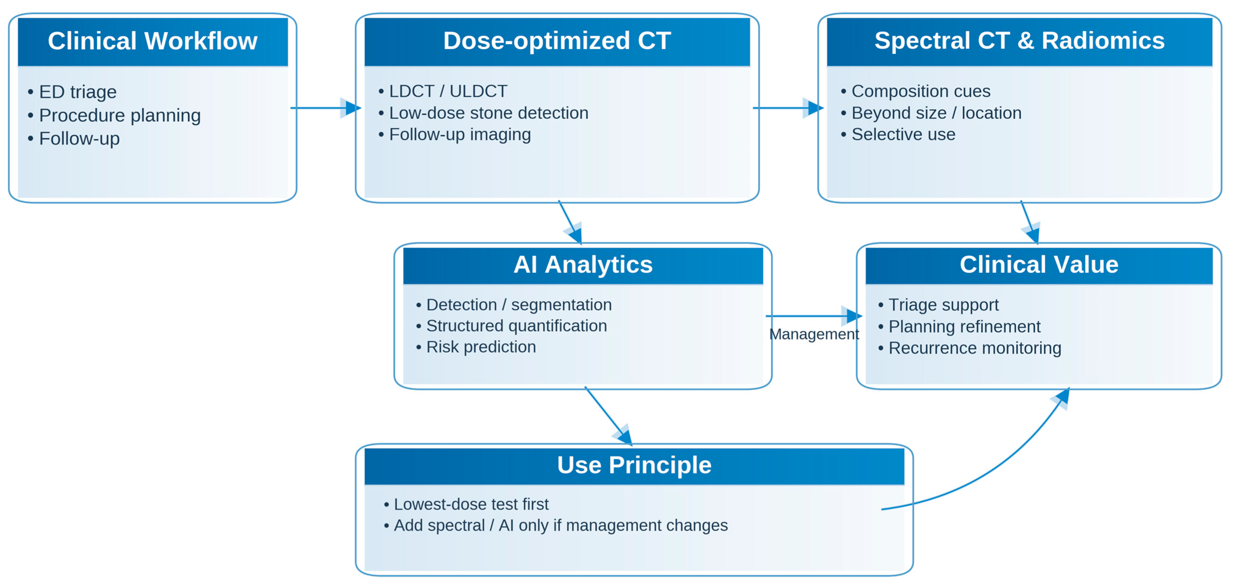
Figure 1 presents a workflow-oriented framework illustrating where dose-optimized CT, spectral CT/radiomics, and AI tools may add value across ED triage, procedural planning, and follow-up.
4. LD and ULD CT for Urolithiasis
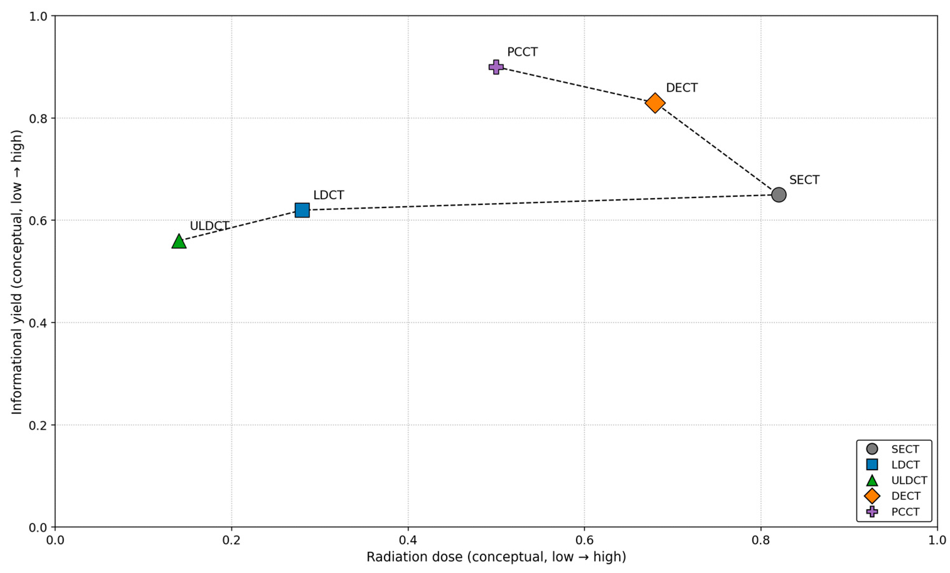
From a pragmatic perspective, CT strategies for urolithiasis lie on a dose–information continuum: dose-optimized protocols preserve diagnostic detectability, whereas spectral techniques add characterization that may influence management.
Table 1 provides a concise synthesis of this comparison, and
Figure 2 illustrates how these CT approaches relate to the broader clinical workflow of urinary stone disease.
Standard renal-protocol CT often delivers effective doses in the 8–16 millisievert (mSv) range, raising concerns about cumulative exposure, especially in young, recurrent stone formers [
5]. Early dose-reduction studies showed that substantial reductions in tube current and voltage were feasible, but meta-analytic data were needed to support widespread adoption [
30]. Nevertheless, real-world adoption of reduced-dose CT protocols remains variable across institutions and practice settings [
31].
Niemann et al. performed one of the first meta-analyses focused on LDCT for urolithiasis, pooling seven studies (1061 patients) that used protocols with effective dose < 3 mSv. They reported pooled sensitivity of 0.966 and specificity of 0.949 versus standard-dose CT and concluded that LDCT could reliably serve as an initial imaging technique for suspected renal colic, with some limitations in obese patients [
32]. Xiang et al. later conducted a systematic review and meta-analysis of LDCT KUB and similarly found that LDCT provided excellent diagnostic accuracy for clinically significant stones, with pooled sensitivity and specificity of 93.1% and 96.6%, respectively [
8].
Reported diagnostic performance of LDCT varies across studies for several reasons. Differences in patient body habitus and image noise, stone size and anatomic location, particularly small distal ureteral calculi, and the prevalence of mimics such as phleboliths can influence sensitivity and specificity. In addition, heterogeneity in acquisition parameters (kVp/mAs and dose targets), reconstruction approaches, reference standards, and outcome definitions limits direct cross-study comparability.
Rodger et al. [
9] extended this work by differentiating LDCT (<3.5 mSv) and ULDCT (<1.9 mSv) protocols in 1529 patients. They found diagnostic accuracies of 94.3% and 95.5% for LDCT and ULDCT, respectively, when standard-dose CT was the reference, with sensitivities of up to 98% and specificities up to 100% in individual series [
9]. Single-center studies support these findings. Klüner et al. showed that multislice ULDCT at doses comparable to abdominal KUB (male/female: 0.5/0.7 mSv) reliably detected renal and ureteral calculi [
33]. Moore et al. reported that reduced-dose CT in ED patients maintained high sensitivity and specificity for ureteral stones and detected nearly all stones requiring intervention within 90 days [
34].
More recently, ULDCT has been evaluated in the follow-up setting. Cheng et al. demonstrated that sub-millisievert ULDCT is highly effective for follow-up of ureterolithiasis, detecting small and distal ureteric stones often missed by KUB or US while maintaining a very low effective dose [
35]. Kandasamy et al. prospectively compared sub-mSv ultra-low-dose computed tomography of the kidneys, ureters, and bladder with X-ray KUB in patients with known urolithiasis and found that ULDCT had higher sensitivity for both intrarenal and ureteric stones at similar or lower dose, supporting ULDCT as a superior alternative to KUB for follow-up when CT is available [
36].
Dose reductions from low to ultra-low CT have been enabled by advances in reconstruction. Iterative reconstruction techniques reduce noise and allow 40–60% dose reductions without compromising stone detection or assessment of extra-urinary pathology [
37]. Deep learning reconstruction further improves noise–texture characteristics and can restore image quality at sub-mSv doses. Zhang et al. compared low-dose CT with hybrid iterative reconstruction to ULDCT reconstructed with deep learning reconstruction in patients with suspected renal calculi; ULDCT with deep learning reconstruction achieved similar detection rates at approximately 25% of the LDCT dose [
38].
In practice, LDCT/ULDCT perform best in patients with moderate-to-high pretest probability and without extreme obesity, and are particularly useful in follow-up where stone presence and location are known [
37,
38,
39]. Overall, these data support LDCT/ULDCT as central tools in a radiation-conscious imaging strategy for urinary stone disease (
Table 2).
5. Advanced CT Technologies: Spectral, Photon-Counting CT, and Radiomics
Stone composition is a key determinant of management: uric acid stones may be amenable to medical dissolution, whereas calcium oxalate, brushite, and cystine stones more commonly require endourologic intervention and show differential responsiveness to shock-wave lithotripsy.
DECT was the first major CT innovation designed for noninvasive compositional assessment. In a seminal in vivo study, Hidas et al. demonstrated that DECT could differentiate uric acid, cystine, and calcified stones using attenuation-ratio thresholds, achieving approximately 82% overall accuracy compared with X-ray diffraction [
11]. Eliahou et al. reviewed DECT applications in urinary stone disease and concluded that DECT reliably discriminates uric-acid from non-uric acid stones and often identifies cystine, thereby informing decisions regarding medical dissolution versus surgical management [
40]. Leng et al. extended DECT to mixed stones in a phantom study, showing that DECT can quantify uric-acid and non-uric acid fractions with low error across simulated body sizes, supporting more granular characterization than binary classification [
41]. Interpretation benefits from awareness of technique-specific pitfalls and stone mimics when applying DECT in routine practice [
42].
PCCT represents a newer detector technology that captures energy-resolved information at the photon level and provides improved spatial resolution and contrast-to-noise performance at equal or lower dose compared with conventional energy-integrating CT. General PCCT evaluations underscore its potential to deliver sharper images and reduce radiation dose in abdominal imaging [
43,
44]. Niehoff et al. assessed low-dose abdominal PCCT for urolithiasis in 98 examinations and reported that stepwise reductions in image-quality level yielded substantial dose savings without compromising quantitative metrics or radiologist confidence in stone detection [
10]. Collectively, these data suggest that PCCT may synergize with LD/ULDCT strategies by improving conspicuity for small stones and enabling richer spectral characterization without degrading diagnostic quality.
Radiomics adds an analytic layer to these imaging datasets [
45]. Kaviani et al. applied threshold-based segmentation and radiomics to single-energy non-contrast CT to classify uric-acid versus non-uric acid stones, achieving an area under the receiver operating characteristic curve (AUC) of 0.78 and demonstrating that composition-relevant signatures can be extracted from standard CT without dedicated DECT protocols [
12]. Huang et al. developed a radiomics model using conventional CT in 195 patients and reported an AUC of 0.81–0.89 for identifying uric acid stones, outperforming simple Hounsfield unit (HU)-threshold approaches [
13]. Hertel et al. leveraged PCCT-based spectral radiomics to classify six stone types in an ex vivo dataset, achieving robust multi-class discrimination and illustrating how PCCT combined with radiomics could enable fine-grained stone typing [
46].
Beyond composition, radiomics has also been explored for risk stratification. Lu et al. integrated CT radiomics with clinical variables to distinguish kidney stone-associated urinary tract infection from non-infected stones, achieving excellent discrimination and supporting the potential utility of radiomics-derived biomarkers for early identification of infected stones [
47]. Overall, DECT is sufficiently mature for routine composition typing in centers with access to the technology [
11,
41,
48], whereas PCCT and radiomics remain promising avenues for dose-efficient, information-rich imaging that can inform both treatment selection and risk stratification [
10,
12,
13,
46,
47].
6. AI for Automated Detection, Segmentation, and Quantification
Across published AI studies in urinary stone disease, publicly accessible imaging datasets remain limited, and most models have been developed using single-center retrospective cohorts. Dataset composition, including patient selection, the prevalence of mimicking conditions, scanner vendors, and dose and reconstruction settings, as well as annotation strategy and the presence or absence of external testing, materially influences reported performance and clinical generalizability.
Table 3 summarizes dataset characteristics and key elements of evaluation design, including training, validation, and testing splits, as well as external validation, as reported in representative studies.
CT-based AI has progressed rapidly in urinary stone disease. Parakh et al. developed a cascading convolutional neural network (CNN) for urinary stone detection on unenhanced CT and reported an AUC of 0.95, with high sensitivity for clinically significant stones across internal and external datasets [
14]. Elton et al. developed a deep learning system for automated kidney stone detection and volumetric segmentation on non-contrast CT; in their cohort, lesion-level sensitivity was 0.86, and automated volume estimates demonstrated strong agreement with manual reference standards [
15]. Together, these studies suggest that AI can approach radiologist-level detection performance while providing objective, reproducible stone-burden metrics for procedural planning and longitudinal follow-up.
Cui et al. integrated three-dimensional deep learning with thresholding to automate stone detection and compute the S.T.O.N.E. nephrolithometry score from CT, thereby linking AI-derived outputs to a validated percutaneous nephrolithotomy (PCNL) complexity grading framework [
49]. Kim et al. trained a deep-learning segmentation network on non-contrast CT and reported Dice coefficients of approximately 0.80 for stone masks, alongside high concordance in stone counts, supporting the feasibility of fully automated segmentation even in patients with multiple stones [
50].
AI has also been extended to lower-cost modalities. Kobayashi et al. developed a CNN-based computer-aided diagnosis system for upper-tract stones on KUB in 1017 patients; the model achieved high sensitivity and improved reader performance, particularly for proximal ureteral stones, compared with unaided interpretation [
16]. Preedanan et al. proposed a cascaded U-shaped network pipeline for urinary stone segmentation on abdominal KUB images, incorporating organ localization, stone-embedding data augmentation, and lesion-size reweighting to enhance recall for small stones [
51].
Liu et al. developed a deep learning-based computer-aided diagnosis system for urolithiasis detection on KUB images using 355 images from 104 patients, with additional preprocessing and augmentation for model development [
52]. Their work supports the feasibility of AI-assisted KUB interpretation in acute-care settings and highlights the potential of such systems to assist emergency physicians when rapid image review is required. Ahmed et al. applied a Visual Geometry Group 16-layer network-based classifier with gradient-weighted class activation mapping to identify stones on KUB radiographs, achieving approximately 95% accuracy while generating heatmaps that localize image regions driving model predictions [
53]. Obaid et al. developed an ensemble deep learning system to classify noisy kidney ultrasound images as normal versus stone-containing, demonstrating robust performance across noise conditions and underscoring the potential of AI to augment challenging US examinations [
17].
Collectively, these studies indicate that AI can automate detection, segmentation, volumetry, and scoring across CT, KUB, and US, and may reduce radiologist workload while delivering standardized quantitative outputs. These outputs include stone presence, number, size, volume, and composition surrogates that can be integrated into risk prediction models. However, most published models remain single-center and retrospective, are constrained by modest training datasets, and lack comprehensive external validation, highlighting the need for rigorous prospective evaluation and multi-center testing [
22].
Despite rapid progress, many stone-detection/segmentation studies remain retrospective and single-center, often with modest sample sizes and limited external validation. Reported performance can be sensitive to spectrum effects and to annotation conventions that vary across institutions. Moreover, metrics are often reported on curated test sets and may not reflect real-world prevalence of mimics or operational constraints. Accordingly, much of the current literature may be regarded as proof-of-concept and emphasize the need for multi-site external validation, calibration assessment where relevant, and prospective impact studies that quantify clinical utility and workflow integration. To improve reproducibility and clinical interpretability, reporting of dataset composition, reference standards, and evaluation design should follow established checklists such as the Checklist for Artificial Intelligence in Medical Imaging [
54].
7. Predictive Models and AI Triage in Urinary Stone Disease
Logistic regression models the probability of an outcome as p = 1/(1 + e−(β0 + βTx)), yielding an interpretable, odds-based estimate of risk derived from clinical and imaging features. Support vector machine (SVM) models define a decision boundary that maximizes the margin between classes and can accommodate non-linear relationships through kernel functions. Ensemble methods, such as gradient boosting, combine multiple weak learners to enhance discrimination and may be particularly useful when complex interactions exist among predictors. Regardless of the model family, calibration and clinically meaningful decision thresholds remain essential for safe implementation.
Prediction models in urinary stone disease span perioperative infection, ED disposition and intervention, and long-term outcomes. An overview of representative models, key inputs, and target endpoints is provided in
Table 4.
7.1. Perioperative Infectious Complications
SIRS and urosepsis after PCNL and ureteroscopy are key targets for risk prediction. Tang et al. developed a logistic-regression nomogram in 785 PCNL patients that used neutrophil-to-lymphocyte ratio, S.T.O.N.E. score, female sex, and positive urine culture to predict postoperative systemic infection (SIRS and urosepsis), achieving C-indices around 0.8 with good calibration [
55]. Zhang et al. compared logistic regression and multiple ML algorithms in 337 PCNL cases and found that an SVM model predicted SIRS with an AUC of 0.94, outperforming logistic regression; key predictors included inflammatory indices, stone characteristics, and operative factors [
56].
For patients with ureteral calculi, nomograms have also been developed to predict urosepsis risk. A recent
Diagnostics study externally validated Hu’s nomogram in a Japanese cohort and identified novel CT-based risk factors such as lower CT attenuation of calculi and higher hydronephrosis grade, yielding a refined model for early urosepsis prediction in ureteral stone patients [
21]. These models illustrate how clinical, laboratory, and CT features can be combined to identify high-risk patients who may benefit from early decompression, prolonged antibiotics, or intensive care unit-level monitoring.
7.2. ED Triage, Admission, and Intervention
In the ED, the immediate triage question is not only stone or no stone, but who needs admission or urgent urologic intervention. Daniels et al. analyzed 475 adults with CT-confirmed ureteral stones in a prospective ED cohort and found that only 20% were admitted and 17% underwent urologic intervention within 90 days [
57]. Predictors of admission and intervention included markers of symptom burden and clinical severity, as well as stone-related factors such as larger stone size and proximal ureteral location. These findings underscore variation in outcomes and the potential role of prediction tools to standardize decisions.
Haifler et al. applied ML to patients with ureteral stones ≤ 5 mm and showed that a combination of clinical and imaging features could predict justified surgical intervention with good discrimination [
58]. Influential variables included stone size, stone location, and selected clinical parameters reflecting acute presentation severity. Their work suggests that ML-enhanced triage could help avoid unnecessary admissions and unsafe discharges by identifying small-stone patients who are less likely to pass stones spontaneously under conservative management.
7.3. Long-Term Recurrence and Procedure Success
At longer time horizons, prediction models have focused on recurrence and procedure success. The original ROKS nomogram by Rule et al. used a population-based cohort of first-time stone formers in Olmsted County to estimate the risk of a second symptomatic episode, with 5-year recurrence risk ranging from <1% to nearly 50% depending on age, sex, body mass index (BMI), family history, stone type, and baseline factors [
18]. Vaughan et al. later created a revised ROKS tool using more than 3000 incident stone formers and multiple episodes, showing that recurrence risk escalates sharply with each successive episode and is further influenced by imaging features such as lower-pole location, number of stones, and largest diameter [
19]. These tools inform the intensity of preventive strategies and could be integrated with imaging-AI outputs in future follow-up algorithms.
In the surgical domain, nomograms and scoring systems for predicting stone-free rate (SFR) after endourological procedures have proliferated. A recent Japanese Urological Association-affiliated review outlines a step-by-step framework for developing, validating, and implementing nomograms for urological tumors, emphasizing principles that are equally applicable to stone-related prediction tools [
25]. More recently, Lee et al. developed and externally validated a machine learning model for SFR after retrograde intrarenal surgery in patients with lower pole renal stones; the Light Gradient Boosting Machine model achieved the highest predictive performance, and stone burden, HU, pelvic stone angle, and renal infundibular length emerged as the most influential features [
59]. These findings suggest that non-linear algorithms may better capture the interplay among stone burden, density, and pelvicalyceal anatomy, although prospective validation and broader generalizability remain to be established.
7.4. From Prediction Models to AI-Enabled Triage
Across these domains, several methodological themes are apparent. First, many models are derived from single-center cohorts with limited size and remain vulnerable to overfitting; independent external validation remains relatively uncommon [
22,
55,
56,
57,
58,
59]. Second, relatively few urolithiasis models incorporate image-derived features beyond size and location, despite evidence that CT attenuation, stone volume, and radiomics signatures carry prognostic information [
12,
13,
19,
21]. Third, clinical utility has rarely been tested prospectively using decision-curve analysis, workflow simulations, or impact studies [
60]. Risk of bias and applicability can be systematically appraised using the Prediction model Risk of Bias Assessment Tool and its recent artificial intelligence extension, complementing transparent reporting and facilitating safer translation [
61,
62].
Across the literature, no single ML model is uniformly superior, as performance depends on the specific task and underlying data structure. For imaging-centric tasks, such as stone detection and segmentation, deep learning approaches generally demonstrate the most consistent performance gains by learning hierarchical features directly from CT, KUB, or ultrasound images. For tabular prediction tasks, such as SIRS/sepsis or ED intervention, non-linear models such as SVM and gradient boosting may improve discrimination when complex interactions exist among clinical, laboratory, and imaging-derived predictors; however, direct comparisons remain limited by heterogeneity in endpoints, cohort composition, and external validation.
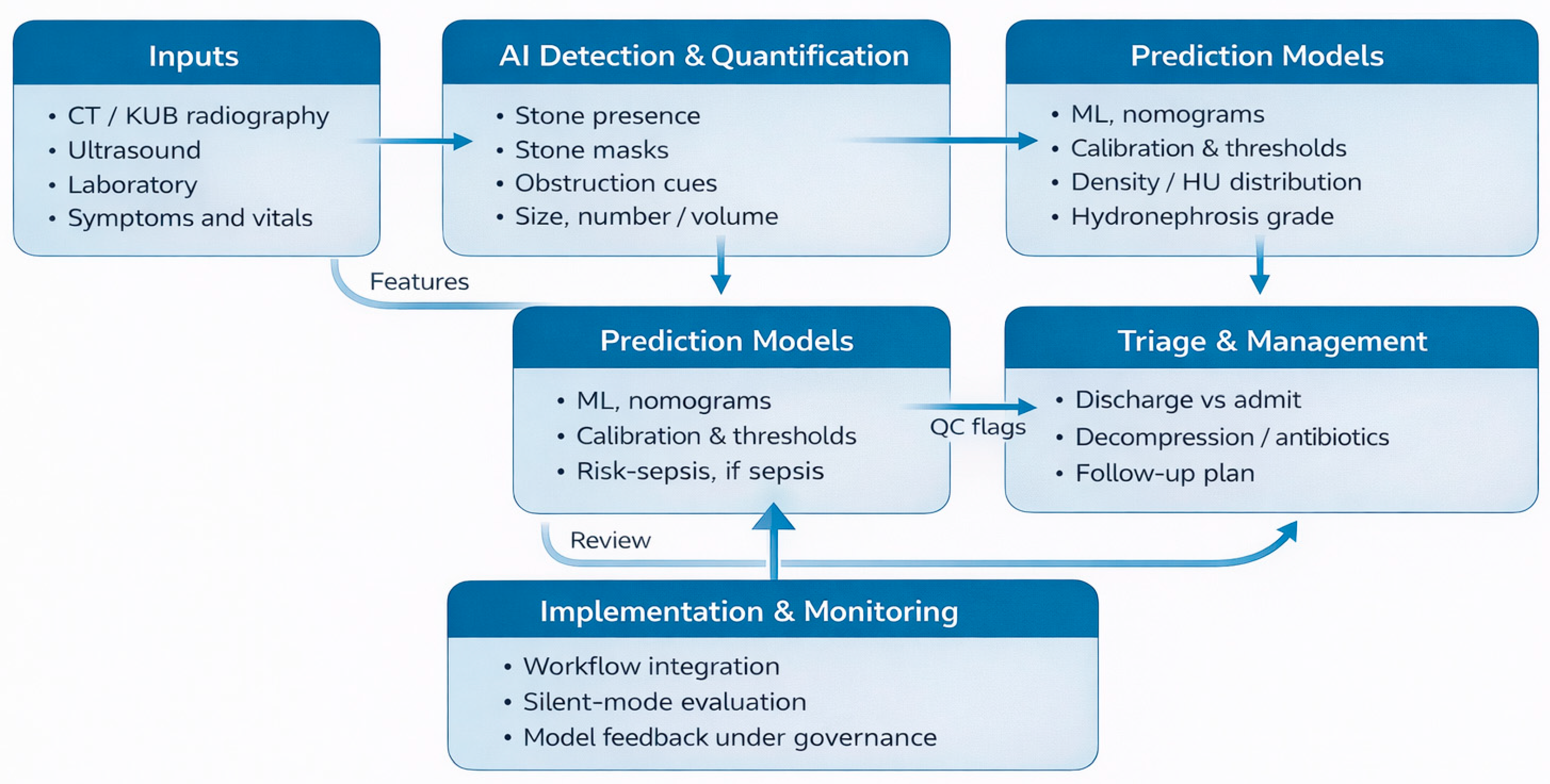
Conceptually, the path toward AI-enabled triage is clear. Automated imaging pipelines can output standardized descriptors, such as stone presence, number, largest diameter, total volume, HU distribution, and obstruction grade, that feed into prediction models for perioperative SIRS/sepsis, ED admission/intervention, and long-term recurrence. Logistic regression and ML models can then translate these features, together with clinical and laboratory data, into individualized risk estimates displayed at the point of care. TRIPOD and TRIPOD + AI provide frameworks to ensure such models are developed and reported transparently [
23,
24,
25].
Figure 3 illustrates a conceptual pipeline linking imaging inputs with AI-based detection, segmentation, and quantification, as well as calibrated prediction models, to point-of-care triage decisions and longitudinal management.
Among prediction and triage models in urolithiasis, direct comparison of performance metrics is limited by heterogeneity in outcome definitions and time horizons. Infection outcomes may range from SIRS to urosepsis, and ED endpoints vary from admission to intervention. Predictor definitions also differ, and calibration reporting remains inconsistent, with many studies focusing on discrimination alone. Consistent adoption of transparent reporting frameworks, explicit outcome definitions, and routine calibration assessment would improve interpretability, facilitate external validation, and support safer clinical translation.
8. Implementation, Regulation, and Future Directions
To facilitate practical interpretation, the reviewed technologies are categorized according to translational readiness, thereby helping clinicians and engineers align their expectations with real-world deployability.
Box 1 summarizes the translational readiness of the imaging and artificial intelligence approaches discussed in this review.
Box 1. Translation readiness map for imaging and AI in urolithiasis.
Clinically established
• LDCT protocols for suspected stones in appropriately selected adults, with attention to body habitus and protocol standardization.
• DECT-based differentiation of uric-acid-containing from calcium-containing stones in centers with appropriate acquisition and post-processing capability.
Emerging but promising
• PCCT applications for stone imaging and dose-efficient spectral characterization.
• CT-based radiomics for stone composition or fragility characterization, although external validation remains limited.
Experimental/proof-of-concept
• AI-enabled ED triage tools and automated detection/segmentation models without robust multi-site external validation.
• ML-based prediction models for complications or intervention that lack consistent outcome definitions, calibration reporting, or prospective impact evaluation.
Clinically, this framework may be used to align the clinical question of whether ED triage, procedural planning, or follow-up, with the lowest-dose and best-validated option, most likely to influence management. It may also help institutions separate near-term protocol standardization from technologies that still require prospective validation, workflow testing, or regulatory maturation before routine deployment.
AI-enabled imaging and prediction tools for urinary stone disease fall within the broader category of software as a medical device (SaMD). The U.S. Food and Drug Administration (FDA) AI/ML SaMD Action Plan and related guidance outline an oversight framework for AI/ML-enabled devices, emphasizing rigorous pre-market evaluation, post-market performance monitoring and model-update management, and mitigation of bias across demographic subgroups [
63]. The FDA also maintains a public registry of authorized AI/ML-enabled medical devices, many of which are radiology-focused.
In Europe, the combination of the Medical Device Regulation and the European Union Artificial Intelligence Act is expected to classify many medical AI systems as high-risk, necessitating risk-management systems, high-quality training data, transparency, and human oversight [
64]. Ethical guidance for AI in radiology emphasizes that systems should promote well-being, minimize harm, distribute benefits equitably, respect privacy and autonomy, remain transparent and reliable, and preserve human accountability [
65,
66]. In urinary stone disease, these principles translate into requirements for representative datasets (including diverse patient populations and scanner platforms), robust external validation, transparent reporting (TRIPOD+AI), and human-in-the-loop workflows in which clinicians retain ultimate responsibility for decisions.
Beyond experimental prototypes, commercially available post-processing platforms can already support key components of stone characterization in routine clinical practice. For example, dedicated applications may enable standardized quantification of stone burden (e.g., maximal diameter, volumetry, and attenuation-derived metrics) and generate visualization outputs (e.g., overlays) that enhance communication between radiology and urology. Moreover, spectral CT post-processing, particularly in DECT, can facilitate composition-relevant assessment, including discrimination of uric-acid from non-uric-acid calculi, provided that appropriate acquisition and processing pipelines are available. Collectively, these examples highlight a pragmatic pathway toward clinical translation; however, real-world impact hinges on harmonized imaging protocols, explicit intended-use definitions, interoperability with clinical information systems, and ongoing performance surveillance.
Implementation experience from other radiology AI tools suggests that seamless integration into picture archiving and communication systems and radiology information systems, automated background processing, and the delivery of concise, actionable outputs (e.g., stone overlays, standardized metrics, risk scores) are critical for adoption. Prospective silent-mode deployments, in which AI operates without influencing care while performance and failure modes are systematically monitored, are recommended before activating clinical decision support [
63,
65,
67].
Future priorities in urinary stone disease include:
Multimodal models integrating advanced imaging outputs (DECT/PCCT features, radiomics, AI segmentation) with clinical and laboratory data.
Rigorous, transparent evaluation of prediction models, including calibration, decision-curve analysis, and external validation, in accordance with TRIPOD+AI guidance [
23,
24].
Prospective impact studies to determine whether AI-assisted triage shortens time to decompression for obstructive infected stones, optimizes admission decisions, or reduces unnecessary imaging without increasing adverse events.
When AI-enabled tools are evaluated as clinical interventions, trial protocols and reports should follow the Standard Protocol Items: Recommendations for Interventional Trials–Artificial Intelligence (SPIRIT-AI) and Consolidated Standards of Reporting Trials–Artificial Intelligence (CONSORT-AI) extensions to ensure completeness and transparency [
68,
69].
Fairness and robustness assessments across sex, age, ethnicity, BMI, comorbidity, scanner type, and dose level.
Human–AI collaboration paradigms that prioritize augmentation of clinician performance and patient communication rather than automation alone [
22,
65,
66].
If these technical, regulatory, and ethical challenges are addressed, urinary stone disease is well positioned to become a model use-case for clinically meaningful integration of imaging engineering and AI.
9. Limitations
This review has several limitations. First, as a narrative review, study identification and selection were targeted rather than exhaustive, and relevant publications may have been overlooked. Second, heterogeneity in imaging protocols, reference standards, and outcome definitions limits direct cross-study comparisons and may constrain generalizability. Third, much of the AI literature remains based on single-center retrospective studies with modest sample sizes and incomplete external validation, which may overstate reported performance relative to real-world settings. Finally, regulatory and implementation considerations vary across jurisdictions and continue to evolve; accordingly, the present discussion should be interpreted within that context.
10. Conclusions
Urinary stone disease is highly prevalent, frequently recurrent, and intrinsically reliant on imaging for diagnosis and management. Standard-dose CT KUB has long served as the diagnostic workhorse, yet it raises concerns regarding cumulative radiation exposure and the underutilization of the abundant quantitative information embedded within imaging data. Accumulating evidence indicates that LDCT and ULDCT can maintain diagnostic performance while substantially reducing dose; spectral and photon-counting CT, in concert with radiomics, enable robust composition assessment and the derivation of novel imaging biomarkers; and AI models can automate detection, segmentation, and stone-burden quantification across CT, KUB, and ultrasound.
In parallel, prediction models can stratify risks of SIRS/sepsis, ED admission and intervention, and long-term recurrence. Nomogram development frameworks and TRIPOD/TRIPOD+AI guidance provide methodological scaffolding for constructing robust, clinically meaningful tools. The next logical step is to integrate these components: to channel standardized imaging outputs from advanced CT and AI pipelines into rigorously validated prediction models, and to deploy them within real-world ED and perioperative workflows under appropriate regulatory and ethical governance. If implemented thoughtfully, this integrated paradigm offers the prospect of safer imaging, more consistent triage, and more personalized longitudinal care for patients with urinary stone disease.
Author Contributions
Conceptualization, S.I. and T.U.; methodology, T.U.; software, T.S.; validation, T.N., Y.S. (Yuka Sugizaki), R.O. and T.E.; formal analysis, Y.S. (Yuta Suzuki); investigation, S.I. and T.U.; resources, R.I., N.I., Y.S. (Yuta Suzuki) and S.I.; data curation, R.I., N.I., Y.S. (Yuka Sugizaki), T.S. and T.E.; writing—original draft preparation, S.I.; writing—review and editing, T.U.; visualization, T.U.; supervision, N.K. and H.S.; project administration, N.K. and H.S. All authors have read and agreed to the published version of the manuscript.
Funding
This research received no external funding.
Institutional Review Board Statement
Not applicable.
Informed Consent Statement
Not applicable.
Data Availability Statement
The original contributions presented in this study are included in the article. Further inquiries can be directed to the corresponding author.
Conflicts of Interest
Hiroyoshi Suzuki reports research funding from Astellas, AstraZeneca, Bayer, Chugai, Eli Lilly, Janssen, MSD, Nihon Kayaku, and Sanofi; advisory fees from AstraZeneca, Bayer, Chugai-Roche, Eli Lilly, Ferring, Janssen, MSD, Novartis, Pfizer, and Sanofi; and lecture fees from Astellas, AstraZeneca, Bayer, Janssen, Novartis, Pfizer, and Sanofi. The authors declare no conflict of interest.
Abbreviations
The following abbreviations are used in this manuscript:
| AI | Artificial intelligence |
| AUC | Area under the receiver operating characteristic curve |
| BMI | Body mass index |
| CNN | Convolutional neural network |
| CT | Computed tomography |
| CT KUB | Computed tomography of the kidneys, ureters, and bladder |
| DECT | Dual-energy computed tomography |
| ED | Emergency department |
| FDA | Food and Drug Administration |
| HU | Hounsfield unit |
| KUB | Kidneys, ureters, and bladder |
| LDCT | Low-dose computed tomography |
| ML | Machine learning |
| mSv | Millisievert |
| NCCT | Non-contrast computed tomography |
| PCCT | Photon-counting computed tomography |
| PCNL | Percutaneous nephrolithotomy |
| QC | Quality control |
| ROKS | Recurrence of kidney stone |
| SaMD | Software as a medical device |
| SFR | Stone-free rate |
| SIRS | Systemic inflammatory response syndrome |
| SVM | Support vector machine |
| TRIPOD | Transparent Reporting of a Multivariable Prediction Model for Individual Prognosis or Diagnosis |
| TRIPOD+AI | Transparent Reporting of a Multivariable Prediction Model for Individual Prognosis or Diagnosis + Artificial Intelligence |
| ULDCT | Ultra-low-dose computed tomography |
| US | Ultrasound |
References
- GBD 2021 Urolithiasis Collaborators. The Global, Regional, and National Burden of Urolithiasis in 204 Countries and Territories, 2000–2021: A Systematic Analysis for the Global Burden of Disease Study 2021. eClinicalMedicine 2024, 78, 102924. [Google Scholar] [CrossRef] [Scilit] [PubMed]
- Ziemba, J.B.; Matlaga, B.R. Epidemiology and Economics of Nephrolithiasis. Investig. Clin. Urol. 2017, 58, 299–306. [Google Scholar] [CrossRef] [Scilit] [PubMed]
- Fwu, C.-W.; Eggers, P.W.; Kimmel, P.L.; Kusek, J.W.; Kirkali, Z. Emergency Department Visits, Use of Imaging, and Drugs for Urolithiasis Have Increased in the United States. Kidney Int. 2013, 83, 479–486. [Google Scholar] [CrossRef] [Scilit] [PubMed]
- Iijima, S.; Utsumi, T.; Ishitsuka, N.; Noro, T.; Suzuki, Y.; Sugizaki, Y.; Somoto, T.; Oka, R.; Endo, T.; Kamiya, N.; et al. Impact of COVID-19 on Ureteroscopy Management of Urolithiasis: Retrospective Comparative Study before and after Pandemic. Appl. Sci. 2025, 15, 6101. [Google Scholar] [CrossRef] [Scilit]
- Brisbane, W.; Bailey, M.R.; Sorensen, M.D. An Overview of Kidney Stone Imaging Techniques. Nat. Rev. Urol. 2016, 13, 654–662. [Google Scholar] [CrossRef] [Scilit]
- Pearle, M.S.; Goldfarb, D.S.; Assimos, D.G.; Curhan, G.; Denu-Ciocca, C.J.; Matlaga, B.R.; Monga, M.; Penniston, K.L.; Preminger, G.M.; Turk, T.M.T.; et al. Medical Management of Kidney Stones: AUA Guideline. J. Urol. 2014, 192, 316–324. [Google Scholar] [CrossRef] [Scilit]
- Bhojani, N.; Bjazevic, J.; Wallace, B.; Cajipe, C.; Chew, B.; De, S.; Hoar, N.; Kolla, S.B.; Razvi, H.; Stern, N.; et al. UPDATE—Canadian Urological Association Guideline: Evaluation and Medical Management of Kidney Stones. Can. Urol. Assoc. J. 2022, 16, 175–188. [Google Scholar] [CrossRef] [Scilit]
- Xiang, H.; Chan, M.; Brown, V.; Huo, Y.R.; Chan, L.; Ridley, L. Systematic Review and Meta-Analysis of the Diagnostic Accuracy of Low-Dose Computed Tomography of the Kidneys, Ureters and Bladder for Urolithiasis. J. Med. Imaging Radiat. Oncol. 2017, 61, 582–590. [Google Scholar] [CrossRef] [Scilit]
- Rodger, F.; Roditi, G.; Aboumarzouk, O.M. Diagnostic Accuracy of Low and Ultra-Low Dose CT for Identification of Urinary Tract Stones: A Systematic Review. Urol. Int. 2018, 100, 375–385. [Google Scholar] [CrossRef] [Scilit]
- Niehoff, J.H.; Carmichael, A.F.; Woeltjen, M.M.; Boriesosdick, J.; Michael, A.E.; Schmidt, B.; Panknin, C.; Flohr, T.G.; Shahzadi, I.; Piechota, H.; et al. Clinical Low-Dose Photon-Counting CT for the Detection of Urolithiasis: Radiation Dose Reduction Is Possible without Compromising Image Quality. Diagnostics 2023, 13, 458. [Google Scholar] [CrossRef] [Scilit]
- Hidas, G.; Eliahou, R.; Duvdevani, M.; Coulon, P.; Lemaitre, L.; Gofrit, O.N.; Pode, D.; Sosna, J. Determination of Renal Stone Composition with Dual-Energy CT: In Vivo Analysis and Comparison with X-Ray Diffraction. Radiology 2010, 257, 394–401. [Google Scholar] [CrossRef] [Scilit]
- Kaviani, P.; Primak, A.; Bizzo, B.C.; Ebrahimian, S.; Saini, S.; Dreyer, K.J.; Kalra, M.K. Performance of Threshold-Based Stone Segmentation and Radiomics for Determining the Composition of Kidney Stones from Single-Energy CT. Jpn. J. Radiol. 2023, 41, 194–200. [Google Scholar] [CrossRef] [Scilit] [PubMed]
- Huang, Y.; Li, N.; Han, X.; Xu, S.; Zhang, G.; Zhu, X. Radiomics Application Using Non-Contrast Computed Tomography for Predicting Uric Acid Kidney Stones. BMC Med. Imaging 2025, 25, 434. [Google Scholar] [CrossRef] [Scilit] [PubMed]
- Parakh, A.; Lee, H.; Lee, J.H.; Eisner, B.H.; Sahani, D.V.; Do, S. Urinary Stone Detection on CT Images Using Deep Convolutional Neural Networks: Evaluation of Model Performance and Generalization. Radiol. Artif. Intell. 2019, 1, e180066. [Google Scholar] [CrossRef] [Scilit] [PubMed]
- Elton, D.C.; Turkbey, E.B.; Pickhardt, P.J.; Summers, R.M. A Deep Learning System for Automated Kidney Stone Detection and Volumetric Segmentation on Noncontrast CT Scans. Med. Phys. 2022, 49, 2545–2554. [Google Scholar] [CrossRef] [Scilit]
- Kobayashi, M.; Ishioka, J.; Matsuoka, Y.; Fukuda, Y.; Kohno, Y.; Kawano, K.; Morimoto, S.; Muta, R.; Fujiwara, M.; Kawamura, N.; et al. Computer-Aided Diagnosis with a Convolutional Neural Network Algorithm for Automated Detection of Urinary Tract Stones on Plain X-Ray. BMC Urol. 2021, 21, 102. [Google Scholar] [CrossRef] [Scilit]
- Obaid, W.; Hussain, A.; Rabie, T.; Mansoor, W. Noisy Ultrasound Kidney Image Classifications Using Deep Learning Ensembles and Grad-CAM Analysis. AI 2025, 6, 172. [Google Scholar] [CrossRef] [Scilit]
- Rule, A.D.; Lieske, J.C.; Li, X.; Melton, L.J.; Krambeck, A.E.; Bergstralh, E.J. The ROKS Nomogram for Predicting a Second Symptomatic Stone Episode. J. Am. Soc. Nephrol. 2014, 25, 2878–2886. [Google Scholar] [CrossRef] [Scilit]
- Vaughan, L.E.; Enders, F.T.; Lieske, J.C.; Pais, V.M.; Rivera, M.E.; Mehta, R.A.; Vrtiska, T.J.; Rule, A.D. Predictors of Symptomatic Kidney Stone Recurrence after the First and Subsequent Episodes. Mayo Clin. Proc. 2019, 94, 202–210. [Google Scholar] [CrossRef] [Scilit]
- Sugizaki, Y.; Utsumi, T.; Ikeda, R.; Ishitsuka, N.; Noro, T.; Suzuki, Y.; Iijima, S.; Somoto, T.; Oka, R.; Endo, T.; et al. External Validation and Comparative Performance of the T.O.HO. and S.T.O.N.E. Scoring Systems for Predicting Stone-Free Outcomes Following Flexible Ureteroscopy: Toward Personalized Preoperative Counseling. J. Pers. Med. 2025, 15, 477. [Google Scholar] [CrossRef] [Scilit]
- Sugizaki, Y.; Utsumi, T.; Ishitsuka, N.; Noro, T.; Suzuki, Y.; Iijima, S.; Somoto, T.; Oka, R.; Endo, T.; Kamiya, N.; et al. Predicting Urosepsis in Ureteral Calculi: External Validation of Hu’s Nomogram and Identification of Novel Risk Factors. Diagnostics 2025, 15, 1104. [Google Scholar] [CrossRef] [Scilit] [PubMed]
- Sassanarakkit, S.; Hadpech, S.; Thongboonkerd, V. Theranostic Roles of Machine Learning in Clinical Management of Kidney Stone Disease. Comput. Struct. Biotechnol. J. 2023, 21, 260–266. [Google Scholar] [CrossRef] [Scilit] [PubMed]
- Collins, G.S.; Reitsma, J.B.; Altman, D.G.; Moons, K.G.M. Transparent Reporting of a Multivariable Prediction Model for Individual Prognosis or Diagnosis (TRIPOD): The TRIPOD Statement. Ann. Intern. Med. 2015, 102, 148–158. [Google Scholar] [CrossRef] [Scilit] [PubMed]
- Collins, G.S.; Moons, K.G.M.; Dhiman, P.; Riley, R.D.; Beam, A.L.; Van Calster, B.; Ghassemi, M.; Liu, X.; Reitsma, J.B.; Van Smeden, M.; et al. TRIPOD+AI Statement: Updated Guidance for Reporting Clinical Prediction Models That Use Regression or Machine Learning Methods. BMJ 2024, 385, e078378. [Google Scholar] [CrossRef] [Scilit]
- Utsumi, T.; Ishitsuka, N.; Noro, T.; Suzuki, Y.; Iijima, S.; Sugizaki, Y.; Somoto, T.; Oka, R.; Endo, T.; Kamiya, N.; et al. Development, Validation, and Clinical Utility of a Nomogram for Urological Tumors: How to Build the Best Predictive Model. Int. J. Urol. 2025, 32, 919–931. [Google Scholar] [CrossRef] [Scilit]
- American College of Radiology. ACR Appropriateness Criteria® Acute Onset Flank Pain—Suspicion of Stone Disease (Urolithiasis). 2023. Available online: https://acsearch.acr.org/Files/DownloadFile?fileId=1076&filename=Acute+Onset+Flank+Pain-Suspicion+of+Stone+Disease.pdf (accessed on 31 March 2026).
- European Association of Urology. EAU Guidelines on Urolithiasis 2025. 2025. Available online: https://d56bochluxqnz.cloudfront.net/documents/full-guideline/EAU-Guidelines-on-Urolithiasis-2025.pdf (accessed on 31 March 2026).
- Smith-Bindman, R.; Aubin, C.; Bailitz, J.; Bengiamin, R.N.; Camargo, C.A., Jr.; Corbo, J.; Dean, A.J.; Goldstein, R.B.; Griffey, R.T.; Jay, G.D.; et al. Ultrasonography versus Computed Tomography for Suspected Nephrolithiasis. N. Engl. J. Med. 2014, 371, 1100–1110. [Google Scholar] [CrossRef] [Scilit]
- National Institute for Health and Care Excellence. Renal and Ureteric Stones: Assessment and Management. NICE Guideline NG118. 2019. Available online: https://www.nice.org.uk/guidance/ng118 (accessed on 31 March 2026).
- Poletti, P.-A.; Platon, A.; Rutschmann, O.T.; Schmidlin, F.R.; Iselin, C.E.; Becker, C.D. Low-Dose versus Standard-Dose CT Protocol in Patients with Clinically Suspected Renal Colic. AJR Am. J. Roentgenol. 2007, 188, 927–933. [Google Scholar] [CrossRef] [Scilit]
- Weisenthal, K.; Karthik, P.; Shaw, M.; Sengupta, D.; Bhargavan-Chatfield, M.; Burleson, J.; Mustafa, A.; Kalra, M.K.; Moore, C.L. Evaluation of Kidney Stones with Reduced-Radiation Dose CT: Progress from 2011–2012 to 2015–2016—Not There Yet. Radiology 2018, 286, 581–589. [Google Scholar] [CrossRef] [Scilit]
- Niemann, T.; Kollmann, T.; Bongartz, G. Diagnostic Performance of Low-Dose CT for the Detection of Urolithiasis: A Meta-Analysis. AJR Am. J. Roentgenol. 2008, 191, 396–401. [Google Scholar] [CrossRef] [Scilit]
- Klüner, C.; Hein, P.A.; Gralla, O.; Hein, E.; Hamm, B.; Romano, V.; Rogalla, P. Does Ultra-Low-Dose CT with a Radiation Dose Equivalent to That of KUB Suffice to Detect Renal and Ureteral Calculi? J. Comput. Assist. Tomogr. 2006, 30, 44–50. [Google Scholar] [CrossRef] [Scilit]
- Moore, C.L.; Daniels, B.; Ghita, M.; Gunabushanam, G.; Luty, S.; Molinaro, A.M.; Singh, D.; Gross, C.P. Accuracy of Reduced-Dose Computed Tomography for Ureteral Stones in Emergency Department Patients. Ann. Emerg. Med. 2015, 65, 189–198.e2. [Google Scholar] [CrossRef] [Scilit] [PubMed]
- Cheng, R.Z.; Shkolyar, E.; Chang, T.C.; Spradling, K.; Ganesan, C.; Song, S.; Pao, A.C.; Leppert, J.T.; Elliott, C.S.; To’o, K.; et al. Ultra-Low-Dose CT: An Effective Follow-Up Imaging Modality for Ureterolithiasis. J. Endourol. 2020, 34, 139–144. [Google Scholar] [CrossRef] [Scilit] [PubMed]
- Kandasamy, M.; Chan, M.; Xiang, H.; Chan, L.; Ridley, L. Comparison of Diagnostic Accuracy of Ultra Low-Dose Computed Tomography and X-ray of the Kidneys, Ureters and Bladder for Urolithiasis in the Follow-Up Setting. J. Med. Imaging Radiat. Oncol. 2024, 68, 132–140. [Google Scholar] [CrossRef] [Scilit] [PubMed]
- den Harder, A.M.; Willemink, M.J.; de Ruiter, Q.M.B.; Wellenberg, R.H.H.; Sprooten, R.T.M.; Brink, M.; Das, M.; Krestin, G.P.; de Jong, P.A.; Schilham, A.M.R.; et al. Radiation Dose Reduction for CT Assessment of Urolithiasis Using Iterative Reconstruction: A Prospective Intraindividual Study. Eur. Radiol. 2018, 28, 2719–2728. [Google Scholar] [CrossRef] [Scilit]
- Zhang, X.; Zhang, G.; Xu, L.; Wang, Z.; Sun, H.; Zhang, Y.; Zhu, J.; He, D.; Jin, C.; Shuai, X. Application of Deep Learning Reconstruction of Ultra-Low-Dose Abdominal CT in the Diagnosis of Renal Calculi. Insights Imaging 2022, 13, 163. [Google Scholar] [CrossRef] [Scilit]
- Rob, S.; Bryant, T.; Wilson, I.; Somani, B.K. Ultra-Low-Dose, Low-Dose, and Standard-Dose CT of the Kidney, Ureters, and Bladder: Is There a Difference? Results from a Systematic Review of the Literature. Clin. Radiol. 2017, 72, 11–15. [Google Scholar] [CrossRef] [Scilit]
- Eliahou, R.; Hidas, G.; Duvdevani, M.; Sosna, J. Dual-Energy CT for Urinary Stone Disease. Semin. Ultrasound CT MRI 2010, 31, 315–320. [Google Scholar] [CrossRef] [Scilit]
- Leng, S.; Huang, A.; Montoya Cardona, J.; Duan, X.; Williams, J.C.; McCollough, C.H. Dual-Energy CT for Quantification of Urinary Stone Composition in Mixed Stones: A Phantom Study. AJR Am. J. Roentgenol. 2016, 207, 321–329. [Google Scholar] [CrossRef] [Scilit]
- Jepperson, M.A.; Cernigliaro, J.G.; Sella, D.; Ibrahim, E.; Thiel, D.D.; Leng, S.; Haley, W.E. Dual-Energy CT for the Evaluation of Urinary Calculi: Image Interpretation, Pitfalls and Stone Mimics. Clin. Radiol. 2013, 68, e707–e714. [Google Scholar] [CrossRef] [Scilit]
- Rajendran, K.; Petersilka, M.; Henning, A.; Shanblatt, E.R.; Ferrero, A.; Schmidt, B.; Flohr, T.; Jacobsen, M.C.; Leng, S.; Egger, C.; et al. First Clinical Photon-Counting Detector CT System: Technical Evaluation. Radiology 2022, 303, 130–138. [Google Scholar] [CrossRef] [Scilit]
- van der Bie, J.; van Straten, M.; Booij, R.; Bos, D.; Dijkshoorn, M.L.; Hirsch, A.; Sharma, S.P.; Oei, E.H.; Budde, R.P. Photon-Counting CT: Review of Initial Clinical Results. Eur. J. Radiol. 2023, 163, 110829. [Google Scholar] [CrossRef] [Scilit]
- Zwanenburg, A.; Vallières, M.; Abdalah, M.A.; Aerts, H.J.; Andrearczyk, V.; Apte, A.; Ashrafinia, S.; Bakas, S.; Beukinga, R.J.; Boellaard, R.; et al. The Image Biomarker Standardization Initiative: Standardized Quantitative Radiomics for High-Throughput Image-Based Phenotyping. Radiology 2020, 295, 328–338. [Google Scholar] [CrossRef] [Scilit]
- Hertel, A.; Froelich, M.F.; Overhoff, D.; Nestler, T.; Faby, S.; Jürgens, M.; Schmidt, B.; Vellala, A.; Hesse, A.; Nörenberg, D.; et al. Radiomics-Driven Spectral Profiling of Six Kidney Stone Types with Monoenergetic CT Reconstructions in Photon-Counting CT. Eur. Radiol. 2024, 35, 6414–6425. [Google Scholar] [CrossRef] [Scilit]
- Lu, J.; Zhu, K.; Yang, N.; Chen, Q.; Liu, L.; Liu, Y.; Yang, Y.; Li, J. Radiomics and Clinical Features for Distinguishing Kidney Stone–Associated Urinary Tract Infection: A Comprehensive Analysis of Machine Learning Classification. Open Forum Infect. Dis. 2024, 11, ofae581. [Google Scholar] [CrossRef] [Scilit] [PubMed]
- Euler, A.; Wullschleger, S.; Sartoretti, T.; Müller, D.; Keller, E.X.; Lavrek, D.; Donati, O. Dual-Energy CT Kidney Stone Characterization—Can Diagnostic Accuracy Be Achieved at Low Radiation Dose? Eur. Radiol. 2023, 33, 6238–6244. [Google Scholar] [CrossRef] [Scilit] [PubMed]
- Cui, Y.; Sun, Z.; Ma, S.; Zhang, X.; Wang, X.; Wang, Z.; Meng, Y.; Dong, G.; Xiao, Y.; Wang, Y. Automatic Detection and Scoring of Kidney Stones on Noncontrast CT Images Using S.T.O.N.E. Nephrolithometry: Combined Deep Learning and Thresholding Methods. Mol. Imaging Biol. 2021, 23, 436–445. [Google Scholar] [CrossRef] [Scilit] [PubMed]
- Kim, Y.I.; Song, S.H.; Park, J.; Youn, H.J.; Kweon, J.; Park, H.K. Deep-Learning Segmentation of Urinary Stones in Noncontrast Computed Tomography. J. Endourol. 2023, 37, 595–606. [Google Scholar] [CrossRef] [Scilit]
- Preedanan, W.; Suzuki, K.; Kondo, T.; Kumazawa, I. Urinary Stones Segmentation in Abdominal X-Ray Images Using Cascaded U-Net Pipeline with Stone-Embedding Augmentation and Lesion-Size Reweighting Approach. IEEE Access 2023, 11, 25702–25712. [Google Scholar] [CrossRef] [Scilit]
- Liu, Y.-Y.; Huang, Z.-H.; Huang, K.-W. Deep Learning Model for Computer-Aided Diagnosis of Urolithiasis Detection from Kidney–Ureter–Bladder Images. Bioengineering 2022, 9, 811. [Google Scholar] [CrossRef] [Scilit]
- Ahmed, F.; Abbas, S.; Athar, A.; Shahzad, T.; Khan, W.A.; Alharbi, M.; Khan, M.A.; Ahmed, A. Identification of Kidney Stones in KUB X-Ray Images Using VGG16 Empowered with Explainable Artificial Intelligence. Sci. Rep. 2024, 14, 6173. [Google Scholar] [CrossRef] [Scilit]
- Mongan, J.; Moy, L.; Kahn, C.E., Jr. Checklist for Artificial Intelligence in Medical Imaging (CLAIM): A Guide for Authors and Reviewers. Radiol. Artif. Intell. 2020, 2, e200029. [Google Scholar] [CrossRef] [Scilit] [PubMed]
- Tang, Y.; Zhang, C.; Mo, C.; Gui, C.; Luo, J.; Wu, R. Predictive Model for Systemic Infection after Percutaneous Nephrolithotomy and Related Factors Analysis. Front. Surg. 2021, 8, 696463. [Google Scholar] [CrossRef] [Scilit] [PubMed]
- Zhang, T.; Zhu, L.; Wang, X.; Zhang, X.; Wang, Z.; Xu, S.; Jiao, W. Machine Learning Models to Predict Systemic Inflammatory Response Syndrome after Percutaneous Nephrolithotomy. BMC Urol. 2024, 24, 140. [Google Scholar] [CrossRef] [Scilit] [PubMed]
- Daniels, B.; Schoenfeld, E.; Taylor, A.; Weisenthal, K.; Singh, D.; Moore, C.L. Predictors of Hospital Admission and Urological Intervention in Adult Emergency Department Patients with Computerized Tomography Confirmed Ureteral Stones. J. Urol. 2017, 198, 1359–1366. [Google Scholar] [CrossRef] [Scilit]
- Haifler, M.; Kleinmann, N.; Haramaty, R.; Zilberman, D.E. A Machine Learning Model for Predicting Surgical Intervention in Renal Colic Due to Ureteral Stone(s) < 5 mm. Sci. Rep. 2022, 12, 11788. [Google Scholar] [CrossRef] [Scilit]
- Lee, H.Y.; Tung, Y.-H.; Elises, J.C.; Wang, Y.-C.; Gauhar, V.; Cho, S.Y. Machine Learning-Based Prediction of Stone-Free Rate after Retrograde Intrarenal Surgery for Lower Pole Renal Stones. World J. Urol. 2025, 43, 433. [Google Scholar] [CrossRef] [Scilit]
- Vickers, A.J.; Elkin, E.B. Decision Curve Analysis: A Novel Method for Evaluating Prediction Models. Med. Decis. Mak. 2006, 26, 565–574. [Google Scholar] [CrossRef] [Scilit]
- Moons, K.G.M.; Wolff, R.F.; Riley, R.D.; Whiting, P.F.; Westwood, M.; Collins, G.S.; Reitsma, J.B.; Kleijnen, J.; Mallett, S. PROBAST: A Tool to Assess Risk of Bias and Applicability of Prediction Model Studies: Explanation and Elaboration. Ann. Intern. Med. 2019, 170, W1–W33. [Google Scholar] [CrossRef] [Scilit]
- Moons, K.G.M.; Damen, J.A.A.; Kaul, T.; Hooft, L.; Navarro, C.A.; Dhiman, P.; Beam, A.L.; Van Calster, B.; Celi, L.A.; Denaxas, S.; et al. PROBAST+AI: An Updated Quality, Risk of Bias, and Applicability Assessment Tool for Prediction Models Using Regression or Artificial Intelligence Methods. BMJ 2025, 388, e082505. [Google Scholar] [CrossRef] [Scilit]
- U.S. Food and Drug Administration. Artificial Intelligence and Machine Learning (AI/ML)-Enabled Medical Devices and the AI/ML SaMD Action Plan. 2021–2025. Available online: https://www.fda.gov/medical-devices/software-medical-device-samd/artificial-intelligence-software-medical-device (accessed on 15 November 2025).
- European Commission. Artificial Intelligence in Healthcare and the EU AI Act. 2024. Available online: https://health.ec.europa.eu/ehealth-digital-health-and-care/artificial-intelligence-healthcare_en (accessed on 15 November 2025).
- Geis, J.R.; Brady, A.P.; Wu, C.C.; Spencer, J.; Ranschaert, E.; Jaremko, J.L.; Langer, S.G.; Kitts, A.B.; Birch, J.; Shields, W.F.; et al. Ethics of Artificial Intelligence in Radiology: Summary of the Joint European and North American Multisociety Statement. Radiology 2019, 293, 436–440. [Google Scholar] [CrossRef] [Scilit]
- Pesapane, F.; Volonté, C.; Codari, M.; Sardanelli, F. Artificial Intelligence as a Medical Device in Radiology: Ethical and Regulatory Issues in Europe and the United States. Insights Imaging 2018, 9, 745–753. [Google Scholar] [CrossRef] [Scilit]
- Vasey, B.; Nagendran, M.; Campbell, B.; Clifton, D.A.; Collins, G.S.; Denaxas, S.; Denniston, A.K.; Faes, L.; Geerts, B.; Ibrahim, M.; et al. Reporting Guideline for the Early Stage Clinical Evaluation of Decision Support Systems Driven by Artificial Intelligence: DECIDE-AI. BMJ 2022, 377, e070904. [Google Scholar] [CrossRef] [Scilit]
- Cruz Rivera, S.; Liu, X.; Chan, A.-W.; Denniston, A.K.; Calvert, M.J.; Ashrafian, H.; Beam, A.L.; Collins, G.S.; Darzi, A.; Deeks, J.J.; et al. Guidelines for Clinical Trial Protocols for Interventions Involving Artificial Intelligence: The SPIRIT-AI Extension. Nat. Med. 2020, 26, 1351–1363. [Google Scholar] [CrossRef] [Scilit]
- Liu, X.; Cruz Rivera, S.; Moher, D.; Calvert, M.J.; Denniston, A.K. SPIRIT-AI and CONSORT-AI Working Group. Reporting Guidelines for Clinical Trial Reports for Interventions Involving Artificial Intelligence: The CONSORT-AI Extension. Nat. Med. 2020, 26, 1364–1374. [Google Scholar] [CrossRef] [Scilit]
| Disclaimer/Publisher’s Note: The statements, opinions and data contained in all publications are solely those of the individual author(s) and contributor(s) and not of MDPI and/or the editor(s). MDPI and/or the editor(s) disclaim responsibility for any injury to people or property resulting from any ideas, methods, instructions or products referred to in the content. |



 ,
,