Article

Reconstruction of Elbow Soft-Tissue Defects Using the Reverse Lateral Arm Flap: A Case Series

Clinic of Plastic Surgery, Department of Neuroscience, Azienda Ospedale Università di Padova, Via Giustiniani, 2, 35128 Padua, Italy
*
Author to whom correspondence should be addressed.
Surgeries 2026, 7(2), 60; https://doi.org/10.3390/surgeries7020060
Submission received: 14 March 2026 / Revised: 19 April 2026 / Accepted: 6 May 2026 / Published: 11 May 2026
(This article belongs to the Special Issue Feature Papers in Hand Surgery and Research)

Abstract

Background: Complex elbow soft-tissue defects often combine exposed critical structures, unstable scars, and high mechanical stress, making durable coverage and early mobilization challenging. Among regional options, the reverse lateral arm flap provides thin fasciocutaneous tissue based on a reliable collateral circulation and preserves major forearm vessels. The aim of this study was to report our single-center experience with the pedicled reverse lateral arm flap for elbow soft-tissue reconstruction, focusing on stable coverage, donor-site morbidity, and functional recovery. Methods: A retrospective single-center case series was conducted at the Division of Plastic Surgery, University Hospital of Padua, Italy. All consecutive patients treated between 2013 and 2023 with a pedicled reverse lateral arm flap for elbow soft-tissue defects were included. Recorded variables included defect etiology, donor-site management, complications, range of motion, and follow-up. Elbow flexion–extension was recorded clinically preoperatively and at last follow-up. Minimum follow-up was 12 months in all patients. Results: Seven patients underwent reconstruction. Defect etiology was burn-related in four cases, shotgun trauma in one, crush injury in one, and melanoma resection in one. All defects were covered with a pedicled reverse lateral arm flap. All flaps survived completely without partial necrosis or flap-related reoperation. Donor-site closure was primary in four patients and required split-thickness skin grafting in three. One patient developed donor-site keloid, and one had donor-site skin-graft partial loss with delayed healing. Elbow flexion–extension improved in all seven cases, with a median gain in arc of motion of 25° (range 15–41°). Conclusions: In this series, the reverse lateral arm flap provided complete coverage of selected elbow defects with preserved motion and limited donor-site morbidity at a minimum follow-up of 12 months. Our findings suggest that it may represent a useful regional option in selected posterior and lateral elbow defects, particularly in post-burn and traumatic settings where thin vascularized tissue is needed, and free-flap reconstruction may be avoidable.

1. Introduction

Soft-tissue defects of the elbow remain a demanding reconstructive problem because the region has limited local tissue, frequent exposure of bone, tendon, or joint structures, and is subjected to constant mechanical stress during flexion and extension. Unstable coverage predisposes to infection, stiffness, and wound breakdown, whereas overly bulky reconstruction can impair joint motion and function. Simple resurfacing is often insufficient when the defect is deep, scarred, unstable, or associated with exposed critical structures [1,2,3].
The reconstructive strategy depends on defect size, location, depth, wound-bed quality, and etiology. Local flaps, regional pedicled flaps, and free flaps all have a role. In small- and medium-sized defects, regional pedicled reconstruction remains attractive because it can provide vascularized tissue with lower operative burden than microsurgical transfer while preserving early mobilization of the elbow [1,2,3]. For clinical orientation in this manuscript, small- to medium-sized defects refer to defects generally amenable to regional pedicled coverage, often in the range of approximately 5 to 15 cm in greatest dimension, depending on location, depth, and tissue requirements.
The lateral arm flap is one of the established regional fasciocutaneous flaps for upper-limb reconstruction [3]. Its reverse pedicled variant is particularly suitable for elbow coverage because it provides thin and pliable tissue, has a consistent vascular basis, and preserves the major arteries of the forearm. The anatomical basis of this flap was described by Culbertson and Mutimer, who showed that retrograde perfusion through the periarticular collateral network could support transfer to the elbow region [4]. Hamdi and Coessens further defined the distally planned design and expanded its distal reach [5].
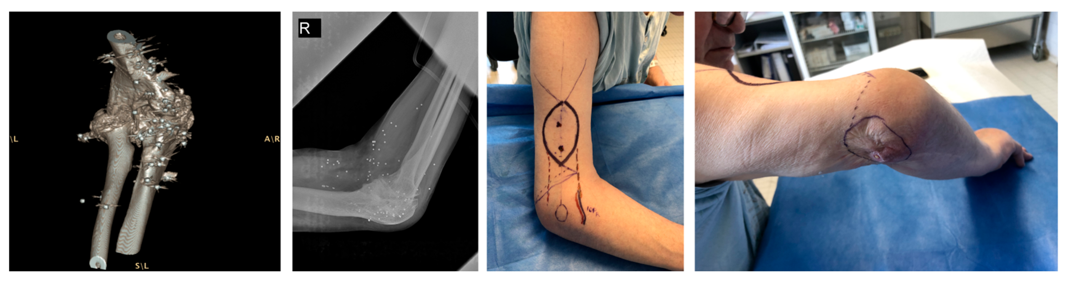
Since then, the reverse lateral arm flap has been used for post-burn defects, traumatic soft-tissue loss, posterior elbow wounds, and selected chronic defects around the olecranon and antecubital fossa [6,7,8,9,10,11]. Its continued use reflects a specific reconstructive profile: it offers regional vascularized tissue in one stage and avoids the sacrifice of a major forearm vessel. A representative high-energy shotgun injury with preoperative flap planning is shown in Figure 1.
Nevertheless, indications are often described broadly, and most series include heterogeneous defects without clearly defining when the reverse lateral arm flap should be preferred over perforator-based or muscular alternatives.
The aim of the present study was to report our single-center experience with the pedicled reverse lateral arm flap for elbow soft-tissue reconstruction and to evaluate flap survival, donor-site morbidity, and functional recovery in a consecutive case series. A further objective was to delineate the practical indications of this flap within a modern reconstructive armamentarium that includes local perforator, regional, and free-tissue options [1,2,3,12,13,14].

2. Materials and Methods

  • Study design and setting
This retrospective case series was conducted at the Division of Plastic Surgery, University Hospital of Padua, Italy. All consecutive patients who underwent elbow soft-tissue reconstruction with a pedicled reverse lateral arm flap between January 2013 and December 2023 were included.
  • Eligibility criteria
Patients were eligible when elbow soft-tissue loss was reconstructed with a pedicled reverse lateral arm flap during the study period. Patients requiring microsurgical free-flap reconstruction, local perforator flaps, or purely muscular flaps were not included. These patients were treated with alternative strategies according to defect characteristics and surgeon preference. A total of seven patients (n = 7) met these criteria. In our practice, the reverse lateral arm flap was preferentially considered for selected posterior or lateral elbow defects requiring thin and pliable fasciocutaneous coverage, with preserved collateral circulation, when local options were considered less suitable and free-flap reconstruction was not deemed necessary. This retrospective series reflects our practical indications for using the reverse lateral arm flap in selected elbow defects, rather than defining a comparative reconstructive algorithm.
  • Data collection
Clinical data were collected from hospital charts, operative reports, outpatient follow-up visits, and photographic documentation. Recorded variables included defect etiology, flap use, donor-site management, postoperative complications, and elbow range of motion. Elbow flexion-extension was recorded preoperatively and at last follow-up as part of routine clinical assessment; because this was a retrospective case series, no standardized goniometric protocol or validated elbow-specific functional score was uniformly available, and the rehabilitation dataset was not sufficiently standardized for comparative analysis.
  • Surgical technique
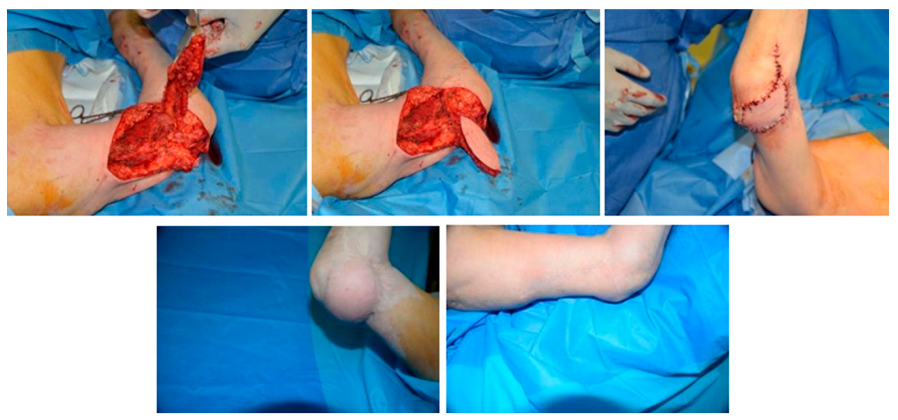
The patient was placed supine with the affected upper limb positioned on an arm table. A tourniquet was applied to the upper arm. The flap was designed along the line from the deltoid insertion toward the lateral epicondyle, centered on the lateral intermuscular septum. Dissection proceeded in the interval between brachialis-brachioradialis anteriorly and triceps posteriorly. The pedicle was raised on the posterior radial collateral system and transferred distally on the periarticular anastomotic network around the elbow. The flap was then rotated into the defect and inset without tension. Donor-site closure was performed primarily when feasible; otherwise, split-thickness skin grafting was used. Flap design, pedicle length, and arc of rotation were tailored to defect size and location in each case. This reverse-flow configuration allowed distal transfer to the elbow while preserving the major forearm vascular axes. A representative intraoperative sequence of flap elevation, distal transposition, and inset is shown in Figure 2.
  • Follow-up and outcomes
Minimum follow-up was 12 months in all patients. The duration of follow-up was calculated from the date of flap inset to the last recorded outpatient assessment. The primary outcome was stable soft-tissue coverage. Secondary outcomes were donor-site closure, postoperative complications, and elbow flexion-extension recovery. Functional recovery was therefore assessed descriptively on the basis of changes in flexion–extension arc, without formal functional or patient-reported outcome measures. Because of the small sample size, only descriptive statistics were used. Flexion-extension changes were summarized as median and range, calculated from the individual differences between preoperative assessment and last follow-up. No inferential statistical testing was performed.

3. Results

Seven patients underwent reconstruction with a pedicled reverse lateral arm flap. Four defects followed burns, one followed shotgun trauma, one followed crush injury with radial-sided soft-tissue loss, and one followed melanoma resection. In the shotgun case, preoperative angio-computed tomography showed preservation of the vascular axis before flap transfer. Case-level operative and functional data are summarized in Table 1.
All seven flaps survived completely. No partial necrosis was recorded. No patient required flap-related reoperation. Donor-site closure was primary in four cases and required split-thickness skin grafting in three. One patient developed a donor-site keloid. One patient had partial donor-site skin graft loss with delayed healing under conservative management.
Elbow flexion-extension improved in all cases. Recorded motion changed from 5 to 60 degrees to −5–85 degrees in case I, from 0 to 100 degrees to −1–140 degrees in case II, from 0 to 85 degrees to 0–100 degrees in case III, from 5 to 80 degrees to −1–105 degrees in case IV, from 3 to 95 degrees to −1–110 degrees in case V, from 4 to 90 degrees to −1–100 degrees in case VI, and from 5 to 95 degrees to 0–115 degrees in case VII. Overall, this corresponded to a gain in flexion–extension arc in every patient, with a median increase of 25° (range 15–41°).

4. Discussion

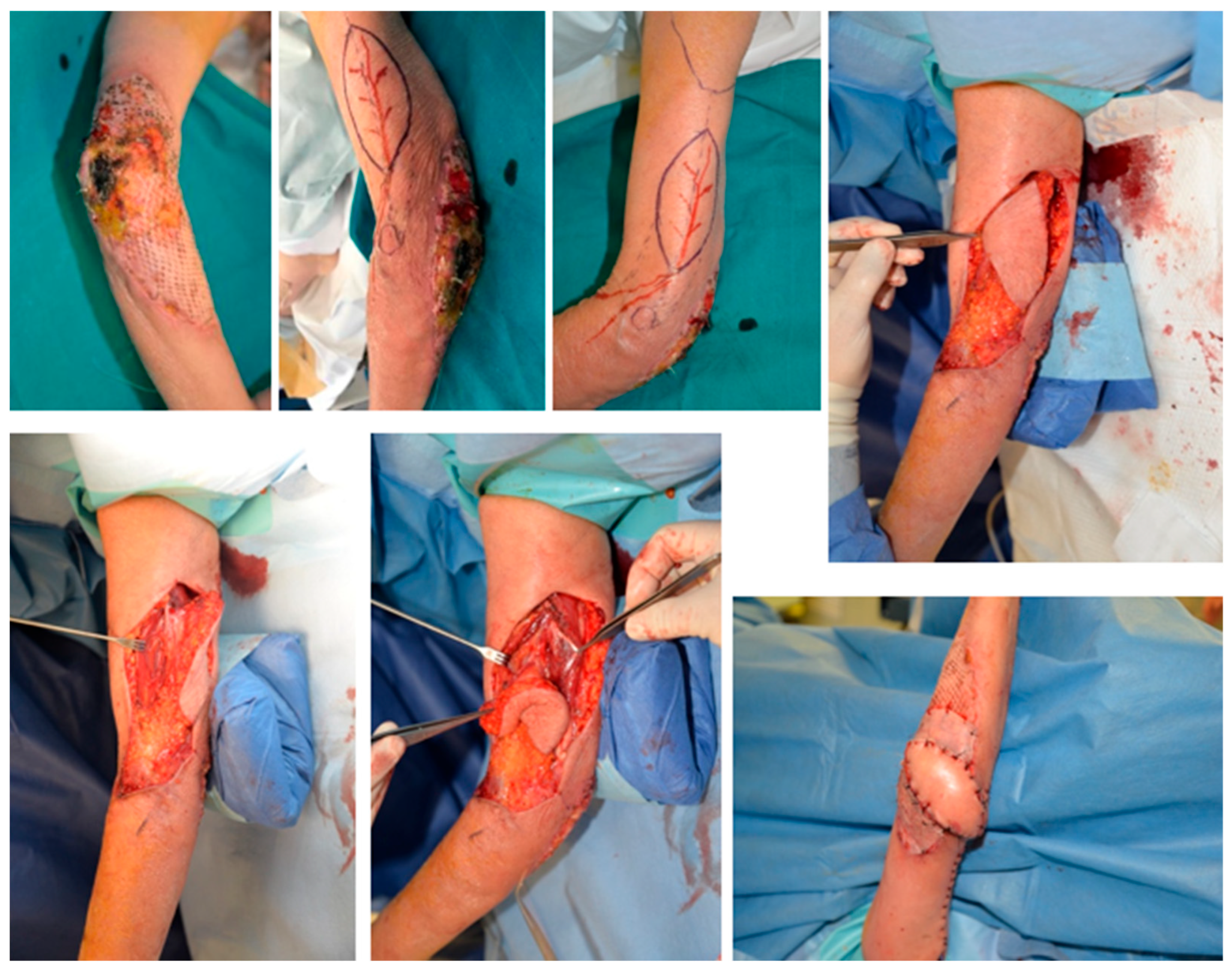
The present study aimed to evaluate the clinical utility of the pedicled reverse lateral arm flap for elbow soft-tissue reconstruction, with particular attention to stable coverage, donor-site morbidity, and postoperative motion. This series shows that the reverse lateral arm flap can provide stable elbow coverage across different etiologies, including burns, trauma, and oncologic resection, with complete flap survival and improvement of elbow flexion-extension in all seven patients. The complication profile was limited. Donor-site morbidity was limited to one case of partial donor-site graft loss and one keloid. These findings support technical feasibility and a favorable local experience when the defect requires vascularized, thin, and mobile tissue rather than simple resurfacing. In practical terms, its main advantages are the provision of thin and pliable vascularized tissue, one-stage regional transfer, and preservation of the major forearm vascular axes. These features are particularly relevant when stable coverage is required without the bulk of muscle flaps or the complexity of free-tissue transfer. A representative burn reconstruction case is shown in Figure 3.
However, the small sample size, retrospective design, and absence of standardized functional or patient-reported outcomes mandate cautious interpretation of these findings, which support feasibility and descriptive clinical utility rather than a comparative indication hierarchy versus perforator, muscle, or free flaps.
The present findings are consistent with previous reports using the same reconstructive strategy. Culbertson and Mutimer established the anatomical basis of the reverse lateral upper arm flap through the periarticular collateral network [4]. Hamdi and Coessens expanded this concept with the distally planned lateral arm flap, emphasizing improved distal reach and reduced bulk [5]. In a clinical series of seven posterior elbow defects, Tung et al. reported complete flap survival and recovery of the full range of motion at 6 months, although forearm paresthesias occurred in three patients and donor-site scar prominence in one [6]. Türegün et al. used the reverse lateral arm flap in 11 antecubital post-burn contractures; ten flaps survived completely, and one developed distal partial necrosis requiring skin grafting, while all patients improved functionally [7]. Prantl et al. later showed that the distal pedicled reversed upper arm flap can also be used for larger defects when early mobilization and durable coverage are required [8]. Morrison et al. extended its use to complex traumatic wounds with staged delay in three patients and obtained stable coverage in all, with only minor distal necrosis in one case [9]. Heidekrueger et al. reported no complete flap losses and full range of motion recovery in olecranon bursitis-related defects at long-term follow-up, supporting the flap even in chronically inflamed local fields [10]. Okamoto et al. further showed that reverse lateral upper arm flaps can reach large defects extending from the elbow to the forearm when the collateral circulation is preserved [11]. Taken together, these studies and our data confirm that the reverse lateral arm flap is a reliable option across a spectrum of posterior and peri-elbow defects, but they also highlight the heterogeneity of indications and the lack of comparative, prospective evidence. [4,5,6,7,8,9,10,11] The value of the flap becomes clearer when compared with competing reconstructive options. Modern elbow reconstruction is no longer organized around a single workhorse flap, but around defect site, defect size, defect depth, contamination, and tissue requirement [1,2,3]. For small and moderate posterior defects, local perforator options have become increasingly relevant. Nakao et al. reported 24 perforator-pedicled propeller flaps around the elbow, mainly for post-burn contracture release and trauma, with one partial necrosis and good postoperative range of motion at a mean follow-up of 10.1 months [12]. Di Summa et al. compared reverse-flow lateral arm flaps with radial collateral artery perforator propeller flaps and found shorter harvest time and shorter hospital stay in the perforator group, whereas distal venous congestion was more frequent in the reverse-flow group [13]. Çiftci et al. compared lateral arm and posterior interosseous artery flaps and found shorter operative time, better QuickDASH, and greater elbow range of motion in the posterior interosseous group; however, those patients had significantly smaller defects, which limits direct equivalence between techniques [14]. Other regional alternatives further narrow the indication. Fauconnet et al. proposed the descending superficial radial artery flap for small to moderate posterior defects requiring thin and flexible coverage [15]. Tiengo et al. described the proximal radial artery perforator flap as a local forearm option that avoids sacrifice of the radial artery [16]. Other vessel-sparing forearm options, including those based on the posterior interosseous artery, further broaden the regional reconstructive spectrum [17]. This stepwise selection is also consistent with a 25-year systematic review of flap survival and functional outcomes in elbow soft-tissue reconstruction [18]. The implication is not that the reverse lateral arm flap has been superseded, but that it should be selected for the right defect rather than used routinely. Within this landscape, our experience suggests that the reverse lateral arm flap is best reserved for small- to medium-sized posterior or lateral defects with preserved collateral circulation, where local perforator flaps are not feasible or would be at risk of tight inset and venous congestion, and free flap reconstruction would represent overtreatment. [12,13,14,15,16,17,18] Compared with free flaps, this regional option may reduce operative burden in appropriately selected defects; compared with local perforator-based solutions, it may offer a more reliable fasciocutaneous paddle when a wider skin island and tension-free inset are required. A selected narrative comparison with published reverse lateral arm flap series is provided in Table 2.
Its main indications remain clear. The reverse lateral arm flap is particularly useful for posterior and lateral elbow defects, and for selected antecubital defects after release, when the reconstruction needs supple fasciocutaneous tissue, preservation of major forearm vessels, and one-stage regional transfer. In burn reconstruction, this profile is especially relevant. Türegün et al. emphasized the conceptual advantage over skin grafting after contracture release: regional vascularized tissue, single-stage reconstruction, no sacrifice of a major artery, and no prolonged immobilization [7]. In defects where thickness matters more than skin paddle surface, Lai et al. described the reverse lateral arm adipofascial flap as a thinner variant that preserves contour and glide and still allows immediate graft coverage [19]. For small posterior wounds, however, a simpler solution may be preferable. Fleager and Cheung reported complete healing in 20 posterior wound complications managed with the anconeus slide, particularly in limited posterior defects and prosthetic settings [20]. Zampeli et al. showed that the brachioradialis muscle flap can be effective in infected or post-prosthetic posterior defects requiring vascularized muscle rather than a fasciocutaneous paddle [21]. In our series, the flap was selected for post-burn and traumatic defects requiring a larger, pliable skin paddle than local muscle or anconeus flaps could provide, but where free flaps would have added operative time and donor-site morbidity without clear functional benefit.

5. Study Limitations

This study is limited by its retrospective design, small sample size, heterogeneity of defect etiology, and absence of a comparator group. No validated patient-reported outcome measure was available. Furthermore, elbow motion was assessed clinically without standardized goniometric protocols or functional scoring systems such as the Mayo Elbow Performance Score or QuickDASH. The study therefore supports feasibility and clinical utility, but not superiority over alternative techniques.
These limitations likely bias the results towards favorable outcomes and should be considered when extrapolating our findings to broader patient populations.

6. Conclusions

The reverse lateral arm flap provided complete coverage in seven consecutive elbow reconstructions with preserved elbow motion and limited donor-site morbidity at a minimum follow-up of 12 months. In our experience, it may represent a useful regional option in selected posterior and lateral defects, particularly in post-burn and traumatic settings where thin vascularized coverage is required and free-flap reconstruction may not be necessary. Clinically, this flap may be considered when preservation of the major forearm vascular axes is desirable.

Author Contributions

Conceptualization, P.Z., L.F. and A.L.C.; methodology, P.Z., L.F. and A.L.C.; validation, A.L.C., C.T. and F.B.; investigation, P.Z., L.F., A.L.C., D.B. and F.M.; data curation, P.Z., L.F., D.B., F.M. and A.L.C.; writing—original draft preparation, P.Z., L.F. and A.L.C.; writing—review and editing, A.L.C., C.T., F.B., D.B. and F.M.; supervision, C.T. and F.B.; project administration, A.L.C. and C.T. All authors have read and agreed to the published version of the manuscript.

Funding

This research received no external funding.

Institutional Review Board Statement

Ethical review and approval were waived for this study due to its retrospective design and the use of fully anonymized clinical data, with no identifiable patient information or recognizable images.

Informed Consent Statement

Informed consent was obtained from all subjects involved in the study.

Data Availability Statement

The data presented in this study are available from the corresponding author upon reasonable request. The data are not publicly available due to privacy restrictions.

Conflicts of Interest

The authors declare no conflicts of interest.

Abbreviations

The following abbreviations are used in this manuscript:
LAFLateral arm flap

References

  1. Gandolfi, S.; Auquit-Auckbur, I.; Poirot, Y.; Bonmarchand, A.; Mouton, J.; Carloni, R.; Nseir, I.; Duparc, F. Focus on anatomical aspects of soft tissue coverage options in elbow reconstruction: An updating review. Surg. Radiol. Anat. 2018, 40, 943–954. [Google Scholar] [CrossRef]
  2. Ooi, A.; Ng, J.; Chui, C.; Goh, T.; Tan, B.K. Maximizing outcomes while minimizing morbidity: An illustrated case review of elbow soft tissue reconstruction. Plast. Surg. Int. 2016, 2016, 2841816. [Google Scholar] [CrossRef]
  3. Kokkalis, Z.T.; Papanikos, E.; Mazis, G.A.; Panagopoulos, A.; Konofaos, P. Lateral arm flap: Indications and techniques. Eur. J. Orthop. Surg. Traumatol. 2019, 29, 1427–1438. [Google Scholar] [CrossRef]
  4. Culbertson, J.H.; Mutimer, K. The reverse lateral upper arm flap for elbow coverage. Ann. Plast. Surg. 1987, 18, 62–68. [Google Scholar] [CrossRef] [PubMed]
  5. Hamdi, M.; Coessens, B.C. Distally planned lateral arm flap. Microsurgery 1996, 17, 375–379. [Google Scholar] [CrossRef]
  6. Tung, T.C.; Wang, K.C.; Fang, C.-M.; Lee, C.-M. Reverse pedicled lateral arm flap for reconstruction of posterior soft-tissue defects of the elbow. Ann. Plast. Surg. 1997, 38, 635–641. [Google Scholar] [CrossRef]
  7. Türegün, M.; Nisanci, M.; Duman, H.; Aksu, M.; Şengezer, M. Versatility of the reverse lateral arm flap in the treatment of post-burn antecubital contractures. Burns 2005, 31, 212–216. [Google Scholar] [CrossRef]
  8. Prantl, L.; Schreml, S.; Schwarze, H.; Eisenmann-Klein, M.; Nerlich, M.; Angele, P.; Jung, M.; Füchtmeier, B. A safe and simple technique using the distal pedicled reversed upper arm flap to cover large elbow defects. J. Plast. Reconstr. Aesthet. Surg. 2008, 61, 546–551. [Google Scholar] [CrossRef] [PubMed]
  9. Morrison, C.S.; Sullivan, S.R.; Bhatt, R.A.; Chang, J.T.; Taylor, H.O. The pedicled reverse-flow lateral arm flap for coverage of complex traumatic elbow injuries. Ann. Plast. Surg. 2013, 71, 37–39. [Google Scholar] [CrossRef] [PubMed]
  10. Heidekrueger, P.I.; Mueller, C.; Thiha, A.; Ehrl, D.; Weinschenk, F.; Herter, F.; Ninkovic, M.; Prantl, L. The lateral arm flap for reconstruction of tissue defects due to olecranon bursitis. J. Plast. Surg. Hand Surg. 2018, 52, 347–351. [Google Scholar] [CrossRef]
  11. Okamoto, H.; Kawaguchi, Y.; Miwa, S.; Aiba, H.; Senda, H.; Murakami, S.; Hayakawa, K.; Joyo, Y.; Murakami, H.; Kimura, H. Reverse lateral upper arm flaps for treating large soft tissue defects extending from the elbow to the forearm. Case Rep. Plast. Surg. Hand Surg. 2022, 9, 145–150. [Google Scholar] [CrossRef]
  12. Nakao, J.; Umezawa, H.; Ogawa, R.; Mateev, M.A. Reconstruction of elbow skin and soft tissue defects using perforator-pedicled propeller flaps. Microsurgery 2018, 38, 473–478. [Google Scholar] [CrossRef] [PubMed]
  13. Di Summa, P.G.; Sapino, G.; Guillier, D.; Dash, J.; Hart, A.; Raffoul, W. Reverse-flow versus perforator propeller lateral arm flap for elbow reconstruction. Ann. Plast. Surg. 2020, 84, 535–540. [Google Scholar] [CrossRef]
  14. Çiftci, S.; Odabaşı, E.; Ertas, E.S.; Tuğrul, A.İ.; Özdemir, A.; Acar, M.A. Comparison of lateral arm flap and posterior interosseous artery flap for soft tissue reconstruction of the elbow. Turk. J. Trauma Emerg. Surg. 2023, 29, 430–434. [Google Scholar] [CrossRef]
  15. Fauconnet, R.; Heitz, A.; Walch, A.; Druel, T.; Gazarian, A.; Cambon, A.; Mathieu, L. Elbow coverage by the descending superficial radial artery flap: A clinical series and literature review. Eur. J. Trauma Emerg. Surg. 2024, 50, 1733–1740. [Google Scholar] [CrossRef]
  16. Tiengo, C.; Macchi, V.; Porzionato, A.; Stecco, C.; Parenti, A.; Bassetto, F.; De Caro, R. The proximal radial artery perforator flap (PRAP-flap): An anatomical study for its use in elbow reconstruction. Surg. Radiol. Anat. 2007, 29, 245–251. [Google Scholar] [CrossRef]
  17. Costa, A.L.; Colonna, M.R.; Vindigni, V.; Bassetto, F.; Tiengo, C. Reverse posterior interosseous flap: Different approaches over the years—A systematic review. J. Plast. Reconstr. Aesthet. Surg. 2022, 75, 4023–4041. [Google Scholar] [CrossRef]
  18. Kahramangil, B.; Pires, G.; Ghaznavi, A.M. Flap survival and functional outcomes in elbow soft tissue reconstruction: A 25-year systematic review. J. Plast. Reconstr. Aesthet. Surg. 2022, 75, 991–1000. [Google Scholar] [CrossRef]
  19. Lai, C.S.; Tsai, C.C.; Liao, K.-B.; Lin, S.-D. The reverse lateral arm adipofascial flap for elbow coverage. Ann. Plast. Surg. 1997, 39, 196–200. [Google Scholar] [CrossRef]
  20. Fleager, K.E.; Cheung, E.V. The anconeus slide: Rotation flap for management of posterior wound complications about the elbow. J. Shoulder Elb. Surg. 2011, 20, 1310–1316. [Google Scholar] [CrossRef] [PubMed]
  21. Zampeli, F.; Spyridonos, S.; Fandridis, E. Brachioradialis muscle flap for posterior elbow defects: A simple and effective solution for the upper limb surgeon. J. Shoulder Elb. Surg. 2019, 28, 1476–1483. [Google Scholar] [CrossRef] [PubMed]
Figure 1. High-energy shotgun injury with severe elbow soft-tissue damage and ankylosis. Preoperative imaging showed retained bullet fragments and osteoarticular damage. The panels illustrate the clinical presentation and flap planning on the lateral arm before release and reconstruction, according to a reverse design for distal transposition to the elbow region.
Surgeries 07 00060 g001
Figure 2. Reverse lateral arm flap reconstruction in the shotgun injury case. The sequence shows flap elevation along the lateral intermuscular septum, distal transposition and inset into the elbow defect, early postoperative appearance, subsequent flap integration, and donor-site healing after split-thickness skin grafting.
Surgeries 07 00060 g002
Figure 3. Reverse lateral arm flap reconstruction for an acute third-degree burn of the dorsal elbow. The sequence shows the defect, flap design, flap elevation, inset, and early postoperative appearance with grafted donor site, highlighting the use of thin fasciocutaneous tissue for stable dorsal elbow coverage.
Surgeries 07 00060 g003
Table 1. Case-based operative and functional summary of the 7-patient series, including defect etiology, flap dimensions, reconstructive alternatives considered, donor-site closure strategy, complications, and preoperative/postoperative range of motion. Abbreviations: LAF, lateral arm flap; E/F, elbow flexion-extension; P/S, pronation-supination.
CaseElbow Defect EtiologyFlap Dimensions (cm)Reconstructive Alternatives ConsideredPrimary Donor-Site ClosureComplicationsPreoperative ROM (Degrees; E/F, P/S)Postoperative ROM (Degrees; E/F, P/S)
case IShotgun injury3 × 8-LAF
-reverse radial forearm flap
-free flap
YesNoE/F:5–60E/F:−5–85
case IIIII degree burn9 × 10-LAF
-free flap
NoNoE/F:0–100
P/S:50–75
E/F:−1–140
P/S:60–85
case IIICrush injury with loss of substance in the elbow region4 × 15-LAF
-reverse radial forearm flap
-free flap
-posterior ulnar flap
NoNoE/F:0–85
P/S:50–80
E/F:0–100
P/S:65–90
case IVSkin tumor (melanoma)10 × 15-LAF
-reverse radial forearm flap
-free flap
YesPartial donor-site skin graft lossE/F:5–80
P/S:70–80
E/F:−1–105
P/S:75–90
case VCicatricial outcomes of skin graft for III degree burn9 × 13-LAF
-reverse radial forearm flap
-free flap
YesDonor-site keloidE/F:3–95
P/S:65–70
E/F:−1–110
P/S:75–85
case VIIII degree burn3 × 7-LAF
-reverse radial forearm flap
-free flap
NoNoE/F:4–90
P/S:65–75
E/F:−1–100
P/S:75–90
case VIIIII degree burn4 × 10-LAF
-reverse radial forearm flap
-free flap
NoNoE/F:5–95
P/S:60–70
E/F:0–115
P/S:75–85
Table 2. Selected published reports of reverse lateral arm flap reconstruction for elbow and peri-elbow defects, including reported flap or defect size, donor-site management, and complications. When studies reported defect size rather than flap size, this is indicated explicitly. * Prantl et al. reported a mixed upper-arm series: 8 lateral upper-arm flaps and 2 medial upper-arm flaps.
ArticleStudy PopulationClinical SettingSize ReportedDonor-Site ManagementComplications
Culbertson and Mutimer1 casePost-burn antecubital release with acute cubital defectFlap 8 × 11 cmSplit-thickness skin graft to donor siteNone reported
Tung et al.7 casesPosterior elbow defects: infected bursa/subcutaneous olecranon, avulsion trauma, open fractureDefect size 4 × 6 to 5 × 13 cmPrimary donor-site closure in all casesAll flaps survived; forearm paresthesia in 3 patients; widened donor-site scar in 1
Türegün et al.11 casesPost-burn antecubital contracturesFlap 6 × 4 to 9 × 6 cm, mean 7.5 × 5.5 cmDonor wounds are generally closed primarily; the exact number requiring a graft is not specifiedDistal partial necrosis in 1 flap, later treated with skin graft
Prantl et al. *10 casesLarge elbow defects: chronic ulcer/bursitis/osteomyelitis, burn contracture, histiocytomaLateral flap up to 15 × 8 cm; wound size 4–10 cm, wound area 30–80 cm2Donor-site grafting not specifically reported; donor-site healing uneventfulNo flap loss; 1 wound-healing/venous drainage complication requiring secondary closure
Morrison et al.3 casesComplex traumatic elbow injuries with fracture and/or hardwareFlap 12 × 5 cm, 15 × 7.5 cm, 15 × 7 cmPrimary donor-site closure in all casesDistal superficial necrosis of 5% in 1 case
Okamoto et al.3 casesVery large defects extending from the elbow to the forearm after tumor resection or traumaFlap 19 × 6.5 cm, 20 × 7 cm, 17 × 7 cmPrimary closure in 2 cases; partial closure in 1 caseNo flap complications
Disclaimer/Publisher’s Note: The statements, opinions and data contained in all publications are solely those of the individual author(s) and contributor(s) and not of MDPI and/or the editor(s). MDPI and/or the editor(s) disclaim responsibility for any injury to people or property resulting from any ideas, methods, instructions or products referred to in the content.