
Review

From Spice to Scaffold: Design and Development of Curcumin Analogs to Combat Pancreatic Cancer
by
Mukund Jha
1,* and
Amitabh Jha
2
1
Department of Biology, Chemistry and Geography, Nipissing University, North Bay, ON P1B 8L7, Canada
2
Department of Chemistry, Acadia University, Wolfville, NS B4P 2R6, Canada
*
Author to whom correspondence should be addressed.
Organics 2026, 7(3), 30; https://doi.org/10.3390/org7030030
Submission received: 27 May 2026 / Revised: 22 June 2026 / Accepted: 26 June 2026 / Published: 13 July 2026

Abstract

Pancreatic ductal adenocarcinoma (PDAC) is considered as one of the most lethal malignancies, characterized by late diagnosis, aggressive local invasion, profound therapy resistance, and a suppressive tumor microenvironment. Currently known chemotherapy regimens for the treatment of PDAC are limited and typically depend on the stage of disease. For pre-surgery and post-surgery settings, modified combination of fluorouracil, leucovorin, irinotecan, and oxaliplatin are used. Gemcitabine/nab-paclitaxel is an alternative regimen used for the disease at advanced stage. However, modest efficacy and high toxicity are often associated with these treatments. Therefore, more efficacious, safer, and novel therapeutic options are urgently required. The natural product curcumin has been shown to exert promising anti-inflammatory, pro-apoptotic, and antimetastatic activities in PDAC models. Inspired by these initial reports, there has been a sustained effort in the medicinal chemistry community to develop chemotherapeutic agents for the treatment of PDAC based on the chemical architecture of curcumin. This review highlights recent developments of multiple classes of curcumin analogs as a credible and versatile class of investigational agents for addressing the unmet therapeutic needs of pancreatic cancer.

1. Introduction

Pancreatic cancer is a highly challenging form of cancer characterized by the development of cancer cells that are carcinogenic within the normal tissues of the pancreas, most often in the exocrine tissue. It is typically characterized by its stealthy onset, rapid progression, and extremely poor prognosis. Among various types of pancreatic cancer, pancreatic ductal adenocarcinoma (PDAC) is considered as the predominant and most lethal subtype of pancreatic cancer, representing more than 90% of all pancreatic malignancies [1]. This subtype generally presents itself at late stages when curative surgery is seldom possible. Globally, there were an estimated 510,992 new cases of pancreatic cancer in 2022, along with 467,409 deaths reported the same year due to this disease [2]. In the United States alone, projections for 2025 estimate about 67,440 new diagnoses and 51,980 deaths from pancreatic cancer [3]. Despite advances in oncology, the 5-year survival rate for all stages is only 10% [4]. Even for early-stage patients meeting the criteria have a dismal 5-year survival rate (<31%) [4]. The disease is more prevalent in North America, Europe, and other high-income regions of the world than in low- or middle-income countries [5]. This may correlate with risk factors, such as older age, obesity, smoking, diabetes, and better diagnostics [5]. These grim statistics underscore the urgency for improved early detection methods, novel therapeutics, and deeper mechanistic understanding to meaningfully shift the survival curve.
As alluded to above, PDAC is one of the most difficult cancers to treat successfully, and its clinical intractability reflects the combined influence of multiple interacting factors. Some of these are tumor intrinsic, meaning they originate within the cancer cells themselves. Genetic mutations in crucial genes, such as KRAS, CDKN2A, TP53, and SMAD4, drive aggressive tumor growth, promote metastasis, and confer resistance to conventional drugs [1]. In addition, PDAC cells often exhibit altered metabolism and enhanced DNA repair mechanisms, enabling them to survive therapeutic stress. Other contributors are microenvironmental factors arising from the tumor’s surrounding stroma. The PDAC microenvironment is characterized by a dense, fibrotic matrix that acts as a physical barrier, restricting the penetration of chemotherapeutic agents and immune effector cells. Moreover, it is enriched with immunosuppressive cells and signaling molecules that slow down antitumor immune responses. This combination of tumor-intrinsic adaptations and a protective microenvironment allow PDAC to evade both pharmacologic and immune-mediated attack. Consequently, these interdependent factors collectively produce a tumor that is biologically aggressive, hypoperfused, immunosuppressed, and resilient to conventional pharmacological and therapeutic approaches [1].
From a treatment perspective, PDAC is also one of the most difficult cancers to manage. Only a small percentage of patients are diagnosed at an early stage when the tumor can still be surgically removed [6]. For most individuals, the disease is discovered only after it has spread beyond the pancreas or has invaded nearby vital structures, making surgery no longer an option. In patients whose tumors are successfully removed surgically, relapses unfortunately occur in most cases within a few years, and a permanent cure remains elusive [6]. For patients with tumors that cannot be surgically removed, drug-based treatment becomes the main approach. Combination chemotherapy regimens, such as modified folfirinox or the pairing of gemcitabine with albumin-bound paclitaxel, can extend survival somewhat, but the improvement is modest [6]. These treatments are also quite toxic, often causing significant side effects, and they rarely lead to complete remission. More recent attempts to use targeted therapies and immune-based treatments have shown promise only in small groups of patients with specific genetic features. For example, tumors with breast cancer gene (BRCA) mutations may respond to poly(ADP-ribose) polymerase (PARP) inhibitors, and the very rare cases with high microsatellite instability (MSI-high) can sometimes benefit from immune checkpoint inhibitors [6]. Unfortunately, for the majority of patients, these newer strategies have not yet led to major improvements in overall survival rates.
The clinical realities associated with pancreatic cancers highlight three main areas of consideration for the development of novel chemotherapeutic agents. First, there is a need for drugs that can act on several tumor-promoting pathways at once, limiting the cancer’s ability to develop resistance. Second, new therapies must be able to penetrate the dense, fibrotic tissue and poor blood supply of PDAC tumors, which often prevent drugs from reaching cancer cells in sufficient amounts. Third, future treatments should be designed to work safely and effectively alongside existing therapies [7].
Turmeric (Curcuma longa) is a spice widely used in South Asian cuisine and traditional medicine. It has been used in Ayurvedic and traditional Chinese medicine for the treatment of inflammatory disorders, wounds, gastrointestinal, and other ailments for centuries [8]. The principal bioactive constituent of turmeric is curcumin (1), or (1E,6E)-1,7-bis(4-hydroxy-3-methoxyphenyl)hepta-1,6-diene-3,5-dione, which is also classified as a polyphenolic natural product [8]. Inspired by traditional knowledge, contemporary scientific research on turmeric, including reports from our group [9,10], has further substantiated that curcumin (1) is largely responsible for the broad spectrum of biological activities attributed to this spice. The chemical structure of 1 consists of a diarylheptanoid characterized by two phenolic rings connected by a conjugated diketone linker (Figure 1). Studies reveal that this conjugated structural feature is immensely crucial for the redox activity of curcumin (1), which is important for its ability to interact with multiple biological targets, consequently displaying a diverse array of biological activities [8]. Given these unique attributes, the chemical framework of 1 has been receiving special attention from medicinal chemists in the current literature, serving as a valuable lead structural template for the development of novel therapeutic agents.
Extensive preclinical studies have shown that curcumin (1) modulates key cellular signaling pathways involved in inflammation, cell proliferation, survival, and oxidative stress, including the nuclear factor-kappa B (NF-κB), signal transducer and transcription factor 3 (STAT3), activator protein-1 (AP-1), and nuclear factor erythroid 2 (Nrf2) [11]. Through these mechanisms, 1 exhibits anti-inflammatory, antioxidant, antiproliferative, and pro-apoptotic effects across a wide range of disease models, particularly cancer [11,12]. For instance, in vitro investigations reported antiproliferative activity and proapoptotic effects of curcumin (1) against diverse tumors [13,14,15]. Whereas 1 was found to suppress carcinogenesis of the skin [16,17,18,19], forestomach [20], breast [21], colon [22,23,24], and liver [25] in murine models.
The property that makes curcumin (1) truly remarkable is its ability to display a pleiotropic mode of action; that is, interacting with multiple molecular targets rather than a single defined enzyme or receptor [11,12]. This makes it an extremely appealing candidate for addressing the treatment of complex diseases, such as pancreatic cancer, where pathway redundancy and adaptive resistance often limit the efficacy of highly selective agents. Moreover, the traditional use of turmeric favorably establishes the selective toxicity profile (minimal toxicity to normal cells) of 1 in humans, which offers huge potential to be developed as a medicinal agent for PDAC management.

2. Molecular Mechanisms of Curcumin Intervention in Pancreatic Cancer

The first in vitro antitumor activity of curcumin against human pancreatic cell lines was reported by Li and co-workers [26]. Their findings demonstrated that curcumin exerts time- and dose-dependent growth-suppressive effects in pancreatic cancer cell lines through inhibition of NF-κB signaling. Subsequently, several studies have been undertaken to investigate the molecular mechanisms of curcumin (1) in pancreatic cancer [27]. Curcumin (1) exerts anti-pancreatic cancer effects mainly via following three mechanisms.
(a)
Signaling pathway modulation.
(b)
Cancer stem cell (CSC) targeting.
(c)
Tumor microenvironment modulation.

2.1. Signaling Pathway Modulation

Curcumin (1) appears to exhibit its anticancer effects through a coordinated inhibition of oncogenic transcription factors, suppression of chronic inflammatory signaling, modulation of oxidative stress responses, and disruption of pro-survival signaling networks. NF-κB pathway, a central regulator of inflammation and cell survival, represents one of the primary targets of 1 [28]. The natural product 1 has been shown to suppress NF-κB nuclear translocation by inhibiting phosphorylation and subsequent degradation of its inhibitor I-kappa-B-alpha, thereby attenuating the expression of pro-inflammatory cytokines and anti-apoptotic proteins. This mechanism is particularly relevant in pancreatic cancer, where constitutive NF-κB activation contributes to chemoresistance and tumor progression [29].
The protein kinase B (PI3K/AKT/mTOR) signaling cascade, another critical pathway in pancreatic cancer pathogenesis, is known to be effectively targeted by 1 [29]. The JAK/STAT3 pathway, frequently hyperactivated in PDAC and associated with poor prognosis, is similarly downregulated by curcumin (1) treatment, leading to decreased cell proliferation and enhanced apoptosis. Additionally, the diketone 1 affects the MAPK signaling pathway and Wnt/β-catenin pathway, both of which play crucial roles in pancreatic cancer cell survival and metastatic potential [29].

2.2. Cancer Stem Cell (CSC) Targeting

One of curcumin’s most promising attributes is its ability to target cancer stem cells (CSCs), a subpopulation of cells responsible for tumor initiation, metastasis, and resistance to conventional therapies [30]. Curcumin (1) has demonstrated efficacy in eliminating CSCs from various cancer origins, including pancreatic cancer, by modulating key stemness-maintaining factors, such as Nanog, Sox2, c-Myc, and Oct-4 [31]. The compound’s ability to inhibit sphere formation and reduce the expression of pluripotency markers represents a significant advancement, as CSC eradication is essential for achieving durable therapeutic responses and preventing disease recurrence [32].

2.3. Tumor Microenvironment Modulation

Pancreatic cancer is characterized by an extensive desmoplastic reaction resulting in a dense fibrotic stroma that creates both physical and biochemical barriers to drug delivery [1]. Curcumin (1) has been shown to target the tumor microenvironment by suppressing the sonic hedgehog (SHH) pathway and the oncogenic CXCR4/CXCL12 signaling axis, thereby inhibiting bidirectional tumor–stromal cell interactions [32]. This stromal modulation not only reduces fibrosis but also enhances the penetration and efficacy of co-administered chemotherapeutic agents such as gemcitabine [32]. The reduction in tumor stiffness, as measured by atomic force microscopy, further supports curcumin’s role in remodeling the tumor microenvironment to facilitate improved therapeutic outcomes.
However, despite its promising therapeutic potential, the clinical application of 1 is significantly hampered due to its inherent pharmacological limitations such as poor aqueous solubility, low chemical stability, rapid metabolism, and limited bioavailability following oral administration [33]. To address these fundamental challenges, the scientific community has pursued two complementary strategies: the development of synthetic curcumin analogs with improved pharmacokinetic properties and potency while retaining the compound’s safety and multitarget activity; and the design of sophisticated formulations for enabling tumor targeted delivery [34].

3. Development of Curcumin Analogs and Synthetic Derivatives

While curcumin (1) exhibits impressive anticancer activity in preclinical models, clinical trials have revealed limited efficacy against pancreatic cancer, primarily attributed to its poor bioavailability and suboptimal potency [33]. The inherent structural features of curcumin (1, Figure 1) play a dual role in determining its pharmacological behavior. Compound 1 is structurally classified as a diarylheptanoid characterized by two aromatic phenyl rings symmetrically connected through a seven-carbon linker (Figure 1). The phenyl rings, substituted with hydroxyl and methoxy groups, further contribute to curcumin’s reactivity and binding potential. The central β-diketone moiety and bis-α,β-unsaturated carbonyl groups are critical determinants of both its biological activity and its metabolic instability [35]. The enolic form 1a of β-diketone moiety predominates under physiological conditions (Figure 2). This not only enables 1a to act as a strong hydrogen donor but also a metal chelator, facilitating interactions with a wide range of biological targets, including transcription factors, kinases, and enzymes. These interactions underpin curcumin’s antioxidant, anti-inflammatory, and anticancer activities. However, the same β-diketone functionality also renders 1 chemically vulnerable to rapid hydrolysis and enzymatic reduction in biological environments. Similarly, the α,β-unsaturated carbonyl groups function as Michael acceptors, allowing curcumin (1) to form covalent or reversible adducts with nucleophilic residues, such as cysteine and lysine, in regulatory proteins [33]. This property is thought to be crucial in curcumin’s ability to modulate multiple signaling pathways, including NF-κB, STAT3, and AP-1, by directly targeting key redox-sensitive proteins. At the same time, these electrophilic centers contribute to metabolic instability by promoting rapid biotransformation through reduction, conjugation (Michael addition, glucuronidation, and sulfation), and oxidative metabolism [35].
Since the diarylheptanoid serves as the key pharmacophore responsible for the activity of curcumin (1), it has inspired the design of plethora of synthetic analogs aimed at improving stability, bioavailability, and anticancer efficacy. Furthermore, curcumin (1) analogs with a shortened central heptanoid chain to a five-carbon backbone, characterized as diarylpentanoids, have also been explored extensively. A summary of the synthetic curcumin (1) analogs and synthetic derivatives developed to date for the treatment of pancreatic cancer, together with their reported pharmacological activities, is presented below. For clarity, the chemical structures of curcumin (1) analogs discussed herein are classified as diarylheptanoids and diarylpentanoids.

3.1. Analogs of Curcumin

Initial efforts towards the development of anticancer compounds based on the framework of curcumin (1) focused on the truncation of diarylheptanoid moiety to either a single enone or a di-enone (diarylpentanoid) system [36,37]. These studies demonstrated that both mono- and di-enones possess promising anti-angiogenesis activity. Particularly, diarylpentanoid 1,5-bis(3,4-dimethoxyphenyl)-1,4-pentadiene-3-one (2) [38] and 3,5-Bis-(2-fluorobenzylidene)-piperidin-4-one (3) [39] were identified as lead compounds that possess superior anticancer activity (Figure 3) over 1. Remarkably, it was found that the latter, difluoroketone compound 3, belonging to diarylidenyl piperidone (DAP) class of curcumin analogs, exhibited significantly lower toxicity compared with the widely used chemotherapeutic agent cisplatin [37].
Inspired by these preliminary findings, Ohori and co-workers designed and synthesized four novel series of curcumin analogs, with a total of 51 compounds, and screened them for anticancer activity [38]. Their design strategy primarily focused on: (a) curcumin-type diarylheptanoids; (b) diarylpentanoids; (c) diarylpentanoids in which conformational flexibility was restricted through incorporation of a central cyclic ketone moiety; and (d) other related analogs (Figure 4). These efforts were aimed at identifying key structural motifs responsible for growth-suppressive activity in carcinogenesis while overcoming the inherent low bioavailability associated with curcumin (1, Figure 2) [38]. Overall, among various cancer cell lines tested, including pancreatic cancer (PK9), four compounds 47 were found to have up to 30 times higher growth-suppressive activity than curcumin (1) and other commonly used anticancer drugs. Importantly, the study outlined three key findings with regard to structure–activity relationships (SAR): (1) O-methylation of phenolic groups on diarylheptanoid series (series A) of compounds, e.g., 4, resulted in considerable enhancement in the growth suppressive activity compared to its parent compound 1; (2) a 5-carbon tether is superior to a 7-carbon tether in displaying anticancer activity in general, which appears to be consistent with previous findings [36,37]; and (3) conformational fixation around the enone subunit (Figure 4, series C) led to significant attenuation of the activity (e.g., activity of 2 >> 8) [38]. Each of the diarylpantanoids analogs identified in Figure 4 47 showed fundamentally stronger growth suppression activities in cancer lines than the lead dimethoxy diarylpentanoid analog 2 (Figure 3) as well. Moreover, it was noted that in vivo evaluations showed no adverse reactions with these new analogs.
Following these initial reports on curcumin analogs with improved anticancer properties, Friedman and co-workers synthesized two new monoketone diarylpentanoid analogs 9 and 10 (Figure 5) and evaluated their effects on pancreatic cancer lines in comparison with 1 [40]. The compounds were tested against five independent human pancreatic cell lines, namely PANC-1, BXPC-3, MIA-PANC-2, ASPC-1, and HPAC. The IC50 values of 9 and 10 in these different pancreatic cell lines were reported to be between 0.28 and 3.20 and between 0.91 and 3.43 μmol/L respectively, compared to 1 (IC50 values of between 8.67 and 20.35 μmol/l), which suggests 9 and 10 are substantially more potent than 1. The results further demonstrated that new analogs were effective in inhibiting STAT3 and AKT phosphorylation in human pancreatic cancer cells and exhibit greater inhibitory activity than curcumin (1). In addition, both analogs markedly reduce cell viability and effectively induce apoptosis in pancreatic cancer cells. Overall, the authors in this study concluded that 9 and 10 possess significant translational potential as novel therapeutic and/or chemopreventive agents for human pancreatic carcinoma [40].
It is well-established that STAT3 is aberrantly activated in a large proportion of pancreatic and breast cancers. Therapeutic strategies aimed at selectively inhibiting constitutive STAT3 signaling have demonstrated significant potential in reducing cancer cell viability. Lin et al. evaluated the effects of two novel diarylheptanoid analogs, 11 and 12 (Figure 6), in breast and pancreatic cancers using both in vitro and in vivo systems. These analogs were rationally designed to preferentially target the STAT3 signaling pathway [41].
The biological activity of curcumin is thought to be influenced by enolization of its central β-dicarbonyl moiety (Figure 2). To prevent enolization, the enolizable methylene hydrogens were replaced with geminal dimethyl substituents in 11 and a spiro-cyclohexyl ring in 12 (Figure 6). By eliminating the possibility of enolization, it was hypothesized that these analogs would enhance binding interactions with key residues within JAK2 and the SH2 dimerization domain of STAT3, relative to the parent curcumin (1) molecule. Furthermore, the 3,4-dimethoxy substitutions were retained in both analogs because the corresponding dimethoxy curcumin derivative 2 (Figure 3) had previously demonstrated improved stability, higher plasma concentrations, and enhanced anticancer efficacy compared with native curcumin (1) [38].
The study demonstrated that both 11 and 12 functioned as effective JAK2/STAT3 inhibitors and exhibited greater potency against pancreatic and breast cancer cell viability than commercially available JAK2 and STAT3 inhibitors, as well as curcumin (1). Specifically, both compounds inhibited STAT3 phosphorylation and suppressed DNA-binding activity and transcriptional transactivation in vitro, ultimately inducing apoptosis in pancreatic and breast cancer cells. Administration of 12 in vivo resulted in a significant reduction in tumor volume in mouse xenograft models. Notably, 12 displayed greater selectivity toward STAT3 and JAK2 than 11, likely due to additional stabilizing binding interactions conferred by its spiro-cyclohexyl moiety [41].
Around the same time, a new series of difluorinated diarylheptanoid analogs of curcumin, including diflurocurcumin (CDF) 13, their fluorinated Schiff bases 1417, and copper (II) complexes, which aimed at enhancing chemical stability and biological activity relative to native curcumin (1), was reported by Sarkar’s group (Figure 7) [42]. The rationale for designing difluorinated analogs was based on the fact that incorporation of fluorine atoms into the β-diketone moiety would greatly reduce metabolic degradation while preserving the key pharmacophoric features required for biological activity. The study reported that among all the synthesized compounds CDF 13 demonstrated markedly enhanced anticancer activity compared with curcumin (1) both in vitro as well as in vivo, largely due to its improved chemical stability and bioavailability. Most importantly, this difluorinated analog 13 was shown to be particularly superior in inducing apoptosis in BxPC-3 pancreatic cancer cells compared to 1. A follow-up study looking at the pharmacokinetic parameters of CDF 13 demonstrated that its concentration in the pancreas tissues was, in fact, found to be 10-fold higher that of curcumin (1) [43].
The potent anti-pancreatic cancer activity of CDF 13 prompted Sarkar and co-workers to investigate the underlying mechanisms responsible for CDF-mediated tumor growth inhibition [44,45]. They probed the histone methyltransferase EZH2, a central epigenetic regulator of cell survival, proliferation, and CSC function, in pancreatic cancer using the difluorinated analog 13. These studies found that CDF 13 decreased cancer cell survival, clonogenicity, formation of pancreatospheres, invasive cell migration, and CSC function in human pancreatic cells by targeting an EZH2-miRNA regulatory circuit for epigenetically controlled gene expression. Recognizing the remarkable potential of CDF 13 for the prevention of tumor progression and/or treatment of pancreatic cancer, the same group subsequently synthesized CDF inclusion complexes in an attempt to further enhance the systemic delivery of 13 in the target tissues and raise its clinical potential [46]. They successfully demonstrated that 1:2 CDF-β-cyclodextrin complex was a more potent anticancer agent than 13 alone or CDF:β-cyclodextrin 1:1 complex. The anticancer activity of 13 and the two complexes were tested against pancreatic (BxPC-3), breast (MDA-MB-231), and prostate (PC3) cancer cells. The IC50 values of CDF 13 against BxPC-3, MDA-MB-231, and PC3 cells were 350 nM, 325 nM, and 260 nM, whereas those of 1:2 CDF-β-cyclodextrin complex were 125 nM, 150 nM, and 120 nM, respectively. Furthermore, the analysis of serum concentrations further substantiated that the bioavailability of 1:2 conjugate is better compared to 13 and CDF:β-cyclodextrin 1:1 complex.
Another approach to enhance water solubility of CDF 13 involved engineering its self-assembling water-soluble nano-micelles using amphiphilic styrene–maleic acid (SMA) polymer and non-covalent hydrophobic interactions by Iyar and co-workers [47]. The SMA-CDF nano-micelles exhibited tunable loading of 13 from 5 to 15% with excellent aqueous solubility, stability, favorable hemocompatibility and sustained drug release characteristics in both in vitro and in vivo experiments. The size and surface characteristics of nano-micelles were found to be favorable for promoting efficient intracellular trafficking of 13, yielding pronounced antitumor response in pancreatic cancer lines (MiaPaCa-2 and AsPC-1). Although, the anticancer activity of SMA-CDF nano-micelles (IC50 230 ± 4.68 nM in MiaPaCa-2 and 710 ± 3.81 nM in AsPC-1) turned out to be marginally better than free CDF 13 (IC50 265 ± 2.57 nM in MiaPaCa-2 and 860 ± 6.21 nM in AsPC-1), the study highlighted that 13 could be efficiently trafficked into the cancer cells in its active form, even after encapsulation/fabrication, making it amenable for systemic administration for the management of pancreatic cancer.
More recently, Bhattacharyya et al. formulated CDF 13 into a 2-hydroxypropyl-β-cyclodextrin (HCD) inclusion complex (CDFHCD) to enhance water solubility and hydrolytic stability for pancreatic cancer treatment [48]. Both 13 and its HCD complex showed dose- and time-dependent antiproliferative activity against multiple PDAC cell lines, including MiaPaCa-2, PANC-1, Panc-01728, and S2-007, with CDFHCD exhibiting significantly improved efficacy compared to 13 alone. This was attributed to enhanced water solubility and a higher uptake of 13 into pancreatic cells through the formation of cyclodextrin inclusion complex. Both compounds reduced colony and spheroid formation while inducing cell cycle arrest and apoptosis. Mechanistically, these effects were associated with downregulation of key regulatory proteins, such as Cyclin D1, thereby limiting cell cycle progression. Apoptotic induction was evidenced by decreased expression of the anti-apoptotic protein Bcl-2, activation of caspases-3/7, and increased levels of cleaved PARP. Importantly, both 13 and CDFHCD reduced the expression of suppressed CSC markers, including CD44 and DCLK1, which are implicated in tumor initiation, metastasis, and chemoresistance. These findings further substantiated that 13 and its analogs with optimized delivery can markedly strengthen anticancer effects against pancreatic cancer cells.
Owing to excellent growth-suppressive potential of diarylpentanoid analog 6 (Figure 4) [38], Sato et al. set out to examine its inhibitory potential against NF-κB activation. The study was aimed at analyzing the effects of 6 on tumor cell growth and the mechanism responsible for its enhanced NF-κB inhibition compared to curcumin (1) [49]. It was found that compound 6 was a direct and potent inhibitor of kappa B kinase β (IKKβ) activity. Overall, NF-κB activation was suppressed to 8–62% in thyroid and pancreatic cancers, as well as in cholangiocarcinoma cells at one tenth concentration of 6 (1 μM) to that of 1 (10 μM). The 50% growth inhibition (IC50) concentrations of 6 ranged from one eleventh to one fourteenth to those of 1. Compound 6 displayed its ability to induce cell death comparable to 1 at a 10-fold lower concentration. Furthermore, the growth-inhibitory effect of 6, especially in pancreatic cancer cells, was found to be 4-fold greater than that of curcumin (1). With these outcomes the study emphasized that a combination therapy with cytotoxic agents and NF-κB inhibitors, such as 6, might be a synergistically beneficial chemotherapeutic approach for practical cancer chemotherapy, including pancreatic cancer, and compound 6 could serve as a good lead compound in that direction.
In another attempt to improve antiproliferative efficacy by addressing curcumin’s limited potency and bioavailability, Wei et al. synthesized a library of 61 curcumin-related analogs through systematic structural modifications of the parent curcumin scaffold [50]. These analogs incorporated variations in aryl substitution patterns and linker moieties while generally retaining the diarylpentanoid α,β-unsaturated ketone pharmacophore (Figure 8). When evaluated for inhibitory effects against human cancer cell lines, including prostate cancer (PC-3), pancreatic cancer (PANC-1), and colon cancer (HT-29), several compounds exhibited markedly enhanced cytotoxicity relative to curcumin (1) (IC50 < 1 µM across all three lines). Notably, analogs designated 18 and 19 were found to be 46- to 117-fold more potent than curcumin (1). Most importantly, the sulfur-containing DAP variants 20 and 21 showed sub-micromolar activity, particularly in PANC-1 cells. It was concluded that these active analogs were potent stimulators of apoptosis. Interestingly, in contrast to the findings of Ohori and co-workers [38], where conformational fixation of the enone subunit (Figure 4 and Figure 8) led to a significant loss of anticancer activity, analogs 1821 displayed substantially enhanced potency against pancreatic cancer cells. This observation suggests that the influence of conformational restriction on biological activity is highly dependent on the overall molecular scaffold and substitution pattern.
In a follow-up study, Wu et al. investigated the effects of DAP analog 21 in combination with gemcitabine on growth inhibition and apoptosis in human pancreatic cancer cells (PANC-1) [51]. It was found that compound 21 synergistically enhanced the cytotoxic effect of gemcitabine on gemcitabine-resistant PANC-1 cells, leading to greater suppression of cell proliferation and increased apoptosis compared with either agent alone. Mechanistically, it was shown that inhibition of cell growth and induction of apoptosis were associated with decrease in NF-κB and PI3K/Akt signaling pathways. These findings highlight a promising strategy for improving the anticancer efficacy of gemcitabine by combining it with 21 and effectively decreasing the resistance of pancreatic cancer to gemcitabine chemotherapy.
An indication of strong in vitro antiproliferative activity of diarylpentanoid 3,5-bis-(2-fluorobenzylidene)-piperidin-4-one (3, Figure 3), a DAP analog of 1, against pancreatic cells amongst others cancer cell lines was first reported by Subramaniam et al. [39]. Given the high potency of 3, Lagisetty et al. synthesized a hydrazinonicotinic acid conjugate of 3, 1-[2-aminoethyl-(6-hydrazinopyridine-3-carbamidyl)-3,5-bis-(2-fluorobenzylidene)-4-piperidone, as an imageable curcumin analog 22 (Figure 9) [52]. The study was designed to investigate image-derived knowledge of drug distribution in tumor tissues, which may offer an important tool in real-time management of cancer therapy. First, the piperidone derivative was efficiently labeled with a gamma-ray-emitting Tc-99m radionuclide 22. It was then evaluated in multiple cell lines in vitro, including pancreatic cancer cells (PANC-1). The results indicated that the antiproliferative activity of 22 was comparable to that of 3. Furthermore, in vivo antiproliferative efficacy of 22 was tested in a mouse model of xenograft PANC-1 tumors. A remarkable reduction in tumor size was observed after treatment with 22 compared to control. Overall, this study demonstrated that the chemical modification of 3 to 22 does not affect the antiproliferative activity both in vitro and in vivo. The Tc-99m-labeled compound 22 could be useful in providing qualitative information about biodistribution of drug from single-photon emission tomography (SPECT) in rat model.
Similarly, to address the challenges associated with the poor aqueous solubility and limited systemic bioavailability of 3 (Figure 3), Bisht et al. developed a nanoformulation of 3 into pegylated liposomes, Lipo-3, and subsequently evaluated its efficacy in preclinical in vitro and in vivo models of pancreatic cancer [53]. In vitro cell viability assay results demonstrate that Lipo-3 induced growth inhibition and apoptosis in human pancreatic cancer cell lines MIAPaCa and Pa03C. The extent of growth inhibition of both cell lines was found to be comparable to that of compound 3 in free form, whereas void liposomes showed negligible antiproliferative effects. In vivo studies showed monotherapy with Lipo-3 resulted in only a modest initial delay in tumor growth; however, a significant synergistic inhibition of tumor progression was observed in MIAPaCa xenograft models when Lipo-3 was administered in combination with the established chemotherapeutic agent gemcitabine. Thus, Lipo-3 was identified as a promising candidate to further evaluate in combinatorial regimens for pancreatic cancer chemotherapy.
In another endeavor, initial anticancer activity of ortho-fluoro substituted piperidone diarylpentanoid analog 3 led El-Rayes and co-workers to identify two new conformationally constrained pyridine-containing DAP analogs 23 and 24 (Figure 10) as promising compounds with higher potency against pancreatic cancer [54]. DNA methyltransferase-1 (DNMT-1) enzyme has been shown to be overexpressed in pancreatic cancer. Therefore, inhibition of DAMT-1 pathway is considered as a rational target for the prevention and therapy of pancreatic cancer. The study found that both, 23 and 24 inhibited pancreatic cancer cell growth in vitro and in vivo models similar to that of oxaliplatin and 5FU, two commonly used agents in pancreatic cancer. The authors concluded that, just like curcumin (1), 23 and 24 inhibit DAMT-1 activity and expression through multiple pathways; however, their potency is several folds higher than 1 due to their better solubility and bioavailability. It appears, the substitution of fluroaryl group with a heteroaromatic ring, such as pyridine, had a marked effect in improving the anticancer properties of these two compounds. Furthermore, the same group later reported antiangiogenic effects of 23 and 24 in pancreatic cancer through mediation of hypoxia-inducible factors (HIFs) and NF-κB, which are important in pancreatic cancer growth and resistance to therapy [55]. Collectively, these features make 23 and 24 promising compounds for clinical development in pancreatic cancer.
It is understood that the instability of curcumin (1) under physiological conditions is caused by the presence of active methylene and β-diketone moieties. In order to improve these challenges Andersson’s group designed and synthesized a new piperidone-based mono-carbonyl DAP analog, 25 (Figure 11), and evaluated its in vitro activity against pancreatic stellate cells (PSCs) [56]. PSCs are stromal components considered as key players in the desmoplastic response in pancreatic cancer. In this comparative study, the results indicated that 25 effectively inhibited PSCs proliferation and induced apoptosis analogous to curcumin (1) but at a 10-fold lower concentration.
Solano et al. developed a series of novel diarylheptanoid curcumin analogs featuring functionalized quaternary ammonium groups as potential anticancer agents, including against pancreatic cancer (Figure 12) [57]. These analogs were generated by reacting piperazinyl curcuminoids with Baylis–Hillman-derived allyl bromides. The authors envisioned that such structural modification would dramatically improve water solubility and biological potency of the synthesized compounds compared with native curcumin (1). These quaternary ammonium curcuminoids 26 and 27 demonstrated enhanced cytotoxicity across multiple cancer cell lines, including MIAPaCa-2 (human pancreatic cancer cells), MDA-MB-231 (human triple negative breast cancer cells), and 4T1 (metastatic murine breast cancer cells), with activity superior to curcumin (1). These results indicated that quaternization and introduction of cationic centers on the curcumin core led to several fold increase in anticancer efficacy for many of these derivatives in vitro. Compound 27 was found to be the most potent against MiaPaCa, exhibiting IC50 value of 2.7 ± 0.5 µM in contrast to 1 (IC50 = ~20 µM). It is worth noting that a change in methyl group from the ester moiety of 27 to longer alkyl groups led to decrease in cytotoxicity. The simplification of the N-substituent to allyl or benzyl resulted in strongly decreased cytotoxicity. The in vivo toxicity studies in mice showed that 27 was well tolerated, with normal body-weight gains comparable to controls. Furthermore, in tumor growth inhibition studies on a MIAPaCa-2 pancreatic cancer xenograft model, 27 inhibited tumor growth as a single agent and further enhanced antitumor efficacy when combined with the clinical agent gemcitabine. Overall, these results suggest that the quaternary ammonium modifications can translate to therapeutic benefit both in vitro and in vivo and merit further investigation as potent curcumin-based anticancer agents.
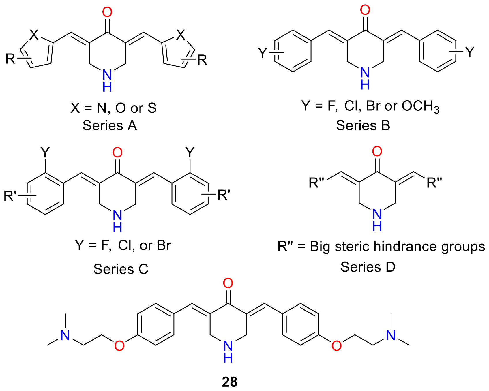
The inhibitory role of IKKβ protein in pancreatic cancer development and progression is believed to be critical. It has been observed that downregulation of IKKβ/NF-κB by small inhibitors successfully suppressed the growth, angiogenesis, and metastasis of pancreatic cancer. Thus, inhibitors targeting IKKβ are considered to be an important therapeutic avenue for treating pancreatic cancer. Given the paucity of clinically approved small molecule IKKβ inhibitors, Hu and co-workers set out to explore the development of novel IKKβ inhibitors [58]. The compounds considered in this study were designed based on stable diarylpentanoid leads 3 (Figure 3) and 23 (Figure 10), which are known IKKβ inhibitors [59,60] with potent anti-pancreatic cancer activity [48,50]. Since 4-piperidone serves as a common structural subunit in 3 and 23, the authors designed four new DAP series of compounds (series A–D) featuring a diarylidenepiperidin-4-one core scaffold in this study (Figure 13). In series A, the pyridine ring of 23 was replaced with various five-membered heteroaromatic rings. Series B and C were designed by modifying the 2-fluorophenyl moiety of 3 to incorporate other mono- and disubstituted phenyl groups, respectively. Finally, series D included substitutions intended to introduce significant steric bulk into the molecular framework. All designed compounds were chemically synthesized and evaluated for their ability to inhibit IKKβ kinase activity. The results indicated that replacement of the pyridine ring with five-membered heterocycles (series A) led to reduced activity. Notably, a few compounds from series B and C, particularly fluoro- and bromo-substituted analogs, demonstrated greater potency than their methoxy-substituted counterparts. However, the strongest IKKβ inhibition was exhibited by a phenoxyethanamine-substituted analog 28 belonging to series D. The enzyme inhibitory concentration (20) of 28 was found to be 0.8 μM, which was two times less than 3 (1.90 μM) and 23 (1.91 μM) and 25-fold less than curcumin (1) (20.5 μM). Molecular docking and molecular dynamics simulation studies performed on 28 suggested that it likely acts as a direct-binding inhibitor of IKKβ. Furthermore, in vitro antiproliferative effects on three pancreatic cancer cell lines, PNAC-1, MiaPaCa-2 and BxPC-3, showed that 28 possessed the greatest inhibitory activity among all tested compounds. Overall, the study identified compound 28 as a promising agent for the treatment of pancreatic cancer based on rational discovery of novel IKKβ inhibitors.
In earlier studies, novel curcumin DAP analogs, 29 and 30 (Figure 14), were identified as highly bioavailable diarylpentanoid derivatives exhibiting significant anticancer activity in human ovarian carcinoma [61]. In particular, 30 was rationally designed as a redox-active compound with antioxidant properties to selectively target malignant cells. Owing to its redox potential and potent anticancer effects, Hu et al. investigated its activity in human pancreatic cancer cell lines PANC-1 and BxPC-3 and explored its underlying mechanisms of action [62]. Compound 30 demonstrated marked antitumor activity at a concentration of 2 μmol/L in both cell lines. Mechanistic studies revealed that treatment with 30 reduced the expression of anti-apoptotic protein Bcl-2 and procaspase-3, while increasing cleaved PARP levels in a dose-dependent manner; however, Bax expression remained unchanged. Additionally, 30 significantly elevated intracellular reactive oxygen species (ROS) levels in PANC-1 and BxPC-3 cells, leading to enhanced expression of endoplasmic reticulum (ER) stress-related proteins. Co-treatment with N-acetylcysteine, a known ROS scavenger, partially attenuated compound 30-induced apoptosis, indicating a ROS-dependent component of its cytotoxicity. Furthermore, in a separate experiment 30 at 2 μmol/L concentration was also found to restrain the expression of P-STAT3, a key survival signaling protein implicated in resistance to apoptosis in pancreatic cancer cells. Collectively, these findings suggest that 30 exerts its anticancer effects in vitro through both ROS-mediated apoptosis and ROS-independent mechanisms, including direct inhibition of STAT3.
In a subsequent study, Kuppusamy’s group investigated the mechanisms of action of the DAP analogs 29 and 30 in AsPC-1 pancreatic cancer cells [63]. Although both compounds demonstrated significant cytotoxicity in proliferation assays, 29 was approximately 160-fold more potent than 30 in inhibiting colony formation. The reduced antiproliferative efficacy of 30 was attributed to the presence of the cytoprotective antioxidant N-hydroxypyrroline moiety. Because the DAP pharmacophore is known to induce ROS-mediated cytotoxicity in cancer cells, radical-scavenging activity conferred by N-hydroxypyrroline conjugation in 30 appeared to be counterproductive, thereby diminishing its antiproliferative activity. Mechanistic investigations revealed that both analogs induced G2/M cell cycle arrest and apoptosis in AsPC-1 cells, leading to cell death through mitochondrial dysfunction and inhibition of STAT3 phosphorylation.
In a study led by Li and co-workers, the effects of curcumin and its conformationally constrained cyclohexanone-based analog 31 (Figure 15) on enhancing gemcitabine activity in a combination therapy approach against pancreatic cancer were examined [64]. Initially, it was observed that increasing the curcumin (1) to gemcitabine ratio shifted the interaction from synergistic to antagonistic effects in pancreatic cancer cells. Accordingly, it was hypothesized that gemcitabine resistance may result from curcumin-mediated inhibition of equilibrative nucleoside transporter 1 (ENT1) at higher concentrations. Indeed, both curcumin (1) and 31 were found to concentration-dependently (2–20 μM) inhibit the intracellular accumulation of the ENT1 substrates uridine and gemcitabine in MIA PaCa-2 and PANC-1 pancreatic cancer cell lines, suggesting direct inhibition of ENT1 activity. It was concluded that 1 is unlikely to inhibit gemcitabine uptake in tumors; however, it may interfere with the oral absorption of ENT1 substrates owing to potentially high intestinal concentrations. Compared with 1, the synthetic analog 31 exhibited greater potency as an ENT1 inhibitor. Notably, this effect was observed only during co-incubation of curcumin (1) or 31 with gemcitabine. Sequential exposure 1 followed by gemcitabine or vice versa did not produce inhibitory effects. Interestingly, as a closely related DAP analog of 31, 3 bearing fluoro substitutions in place of methoxy and phenolic groups did not affect ENT1 activity, rendering it a more suitable adjuvant candidate in combination formulations.
Puskas and colleagues synthesized a library of twenty-three achiral Mannich-type curcumin analogs 32 (Figure 16), comprising diarylheptanoid derivatives and evaluated their antiproliferative and cytotoxic activities in human non-small-cell lung carcinoma (A549), hepatocellular carcinoma (HepG2), and pancreatic cancer (PANC-1) cell lines [65]. Six compounds bearing carboxylic acid, dihydroxyphenyl, or para-hydroxy substituents were found to be completely inactive across all tested cell lines, consistent with previously reported SAR findings. Three compounds 3335 exhibited significant cytotoxic activity. These lead compounds share a common C-4 chloroacetamidomethyl moiety, along with either meta-hydroxy or methoxy substituents on the aryl side chains, as shown in Figure 16. Notably, their antiproliferative activity, particularly against PANC-1 cells, was approximately 40-fold greater than that of curcumin (1). Mechanistic investigations revealed that these analogs accumulate in the ER, triggering ER stress and activation of the unfolded protein response. This cascade subsequently induces mitochondrial membrane depolarization, caspase-3 activation, and apoptosis.
An ethyl homolog of 25 (Figure 11), 36 (Figure 17), with known anticancer effects on lung cancer cells, encouraged Wan and co-workers to investigate its efficacy on PDAC [66]. The study demonstrated that 36 exhibits significant anticancer activity in pancreatic cancer cell growth via multiple targets and pathways. Moreover, it was found that this effect is markedly enhanced in the context of histone lysine N-methyltransferase 2D (KMT2D) deficiency, a histone methyltransferase frequently altered in cancer. Compound 36 suppressed PDAC cell proliferation by inducing apoptosis and ER stress pathway, with KMT2D-deficient cells showing heightened sensitivity to its cytotoxic effects. Mechanistically, loss of KMT2D appeared to exacerbate oxidative stress (significantly increased ROS levels) and disrupt survival signaling pathways in cells treated with 36. In vivo experiments further confirmed that KMT2D depletion significantly enhanced the antitumor efficacy of 36 in xenograft models. Collectively, these findings highlight 36 as a promising therapeutic candidate and suggest that epigenetic alterations such as KMT2D deficiency may serve as predictive biomarkers to improve the efficacy of curcumin analog-based strategies in pancreatic cancer.
A trifluoromethyl substituted diarylpentanoid analog of curcumin (1), 37 (Figure 18), was reported to be an inhibitor of protein kinase enzyme, c-Jum N-terminal Kinase (JNK), with excellent anti-inflammatory activity. These properties led Chen and co-workers to evaluate 37 for its role in the proliferation and migration of pancreatic cancer [67]. The study demonstrated that various inflammatory cytokines, such as IL-6, were more expressed in pancreatic cancer than in the matching normal tissue. Compound 37 was found to exert significant anticancer effects in pancreatic cancer by targeting inflammation-driven signaling pathways. It markedly inhibited pancreatic cancer cell proliferation, migration, and invasion while promoting apoptosis in vitro by significantly reducing the pro-inflammatory factors. The IC50 levels of its activity on PANC-1 and SW1990 were reported to be 113.4 and 91.83 μM, respectively. Mechanistically, its antitumor activity was primarily attributed to suppression of the JNK signaling pathway, resulting in reduced activation of downstream inflammatory mediators and protumorigenic cytokines. By attenuating JNK-mediated inflammatory signaling, 37 was found to disrupt a key pathway contributing to pancreatic cancer progression. Collectively, these findings identified 37 as a promising lead compound having anti-inflammatory and antitumor properties, supporting its potential development as a therapeutic strategy targeting inflammation-associated pancreatic tumorigenesis.
The inhibition of deubiquitinase (DBU) by N-acrylated DAP derivative 38 has been reported to be a new anticancer mode of action. The molecular skeleton of 38 is similar to that of 3 (Figure 3), which is known to be active against pancreatic cancer [39]. In an attempt to further enhance the potency of fluorinated benzylidene curcuminoid 3, Biersack and co-workers synthesized and evaluated a series of N-acrylamide derivatives of 3 and evaluated anticancer activity against pancreatic carcinoma cells [68]. A total of ten N-acryloylated 3,5-bisbenzylidene-4-piperidonones 39 having diverse substitutions on aryl group (Figure 19) were synthesized. The antiproliferative activity of the synthesized compounds were evaluated against human MiaPaCa-2 and PANC-1 pancreatic carcinoma cells. It was found that the presence of acrylamide functionality markedly improved the potency of compounds compared to 3 and the anticancer drug irinotecan except for 40 (IC50 = 4.14 ± 0.18 µM for MiaPaCa-2 and 7.37 ± 0.31 µM for PANC-1), which displayed 4–5-fold inferior activities than 3 (IC50 = 1.03 ± 0.19 µM for MiaPaCa-2 and 1.52 ± 0.25 µM for PANC-1). Three derivatives—bis-3-fluoro-4-methoxyphenyl, 41; bis-3,4-difluorophenyl, 42; and bis-4-trifluoromethylphenyl, 43—showing IC50 values in the low sub-micromolar range (0.29–0.77 µM) were identified to be most active against pancreatic carcinoma cells in this study (Figure 19). Mechanistically, these structural modifications appeared to enhanced apoptosis induction (via caspase-3 activation and PARP cleavage) and suppressed phospho-STAT3 signaling in pancreatic cancer cells. Furthermore, docking studies on STAT3, which is a reasonable target of 1 and structurally related compounds in pancreatic cancer, suggested a stronger STAT3 binding for select fluorinated acrylamides, illustrating how fluorination and N-acryloylation of the DAP core can significantly enhance anticancer efficacy relative to the lead scaffold 3.

3.2. Synthetic Derivatives of Curcumin

Curcumin (1) exhibits diverse pharmacological activities, including chemopreventive and anticancer effects. Despite being inexpensive, readily available, and relatively non-toxic, its clinical application is limited by poor aqueous solubility, low absorption, rapid metabolism, and fast glucuronidation and systemic elimination [35]. A few strategies based on chemically derivatizing 1 have been explored to improve water solubility and in vivo efficacy. A summary of those findings has been discussed below.
One of the major problems encountered in cancer chemotherapy is the severe toxic side effects of anticancer drugs designed to target rapidly dividing cells, including healthy ones. This sometimes necessitates dose reductions, treatment delays or discontinuance of therapy. To overcome these challenges, targeted anticancer drug delivery systems have been developed. Towards that end, Aggarwal and co-workers reported the development of a targeted anticancer conjugate composed of curcumin (1) linked to the luteinizing hormone-releasing hormone (LHRH) analog [DLys6]-LHRH, designed to selectively deliver active compound 1 to pancreatic cancer cells expressing LHRH receptors [69]. This approach is particularly important in pancreatic cancer, as pancreatic cancer cells express LHRH receptors while normal pancreatic cells do not. A glutarate ester linkage to link the phenolic group of curcumin with [DLys6]-LHRH (compound 44) was conveniently employed as it can be cleaved by esterases to deliver free curcumin in vivo (Figure 20). In contrast to curcumin (1), the synthetic [DLys6]-LHRH–curcumin conjugate 44 is readily soluble is water, enabling its systemic administration and its targeting to LHRH expressing cancer cells. In vitro tests showed 44 significantly inhibited pancreatic cancer cell proliferation in a dose-dependent manner. The conjugate also effectively induced apoptosis, as evidenced by caspase activation and PARP cleavage, indicating preservation of curcumin’s anticancer activity. Importantly, the antiproliferative effects of the conjugate were comparable to free curcumin (1) at equimolar concentrations, demonstrating that the bioactivity of the conjugate was not compromised. Competitive binding studies using [DLys6]-LHRH–curcumin conjugate 44 and [DLys6]-LHRH (free peptide) supported a receptor-specific mechanism of cellular uptake. In vivo, intravenous administration of the conjugate markedly suppressed tumor growth in pancreatic cancer xenograft models. In contrast, free curcumin (1) administered orally showed minimal antitumor efficacy, underscoring the advantage of targeted delivery and improved bioavailability. Histological analyses revealed increased tumor apoptosis without significant systemic toxicity. Collectively, this study highlights the potential of targeted peptide–drug conjugates to enhance the therapeutic utility of curcumin and represents a promising strategy for pancreatic cancer treatment.
Photodynamic therapy (PDT) is an emerging cancer treatment that involves administering a photosensitizer followed by irradiation with visible light of a specific wavelength. Light activation triggers interaction with molecular oxygen, generating cytotoxic ROS that induce cancer cell death through apoptosis and/or necrosis. Compared with conventional therapies, PDT is minimally invasive and offers tumor selectivity, as light can be precisely directed to the tumor site, limiting damage to surrounding healthy tissue. Chlorin e6, a derivative of chlorophyll, is a well-known second-generation photosensitizer with high sensitizing efficacy and rapid elimination time from the body. Kim and co-workers designed hybrid molecules by covalently linking chlorin e6 to curcumin (1) in order to combine the phototoxic effects of choline e6 with the intrinsic anticancer and anti-inflammatory properties of 1 (Figure 21) [70]. The conjugates were synthesized through amide/ester coupling strategies in the carboxylic acid groups of chlorin e6, preserving the conjugated porphyrinic chromophore while introducing the curcuminoid pharmacophore. These structural modifications were particularly aimed at enhancing cellular uptake, singlet oxygen generation, and tumor selectivity under light activation in the resultant hybrids. The synthesized molecules showed better physicochemical properties and higher singlet oxygen generation capability. The cellular uptake of these conjugates was found to be significantly better than free chlorin e6. In vitro evaluation against pancreatic cancer cell lines—AsPC-1, PaCa2, and PANC-1—demonstrated significantly enhanced phototoxicity compared with chlorin e6 alone, with low micromolar IC50 values under irradiation and minimal dark toxicity. Among the synthesized compounds, the polyethylene glycol (PEG) containing derivative 45 (Figure 21) displayed outstanding cytotoxicity against the three cell lines with IC50 values ranging from 34 to 41 nM. Remarkably, the dark/photo toxicity ratio for 45 ranged in between 2371 and 7500 and was found to be the highest among the synthesized conjugates. Mechanistically, the conjugates increased intracellular ROS production, leading to apoptosis-mediated cell death.
In a follow-up study, Kim and co-workers further sought to alter the conjugation sites in curcumin (1) [71]. 2,2’-(Ethane-1,2-diylbis(oxy))bis(ethan-1-amine) or a monoPEG and 3-[2-[2-(2-aminoethoxy)ethoxy]ethoxy]propanoic acid or a diPEG linkers were used for conjugating chlorin e6, either at the enone moiety or in the phenolic group of curcumin (1). The synthesized compounds were evaluated for their PDT efficacy against the same set of human pancreatic cancer lines in vitro and in vivo. Structural characterization confirmed successful conjugation and retention of key photophysical properties. Among the synthesized compounds, 46 (Figure 21) displayed remarkable cellular internalization capability and showed significant cytotoxicity against AsPC-1 (0.27 µM), MIA PaCA-2 (0.42 µM), and PANC-1 (0.21 µM) cell lines. These results indicate that compound 46 exhibited 101.18-, 26.26-, and 89.19-fold greater phototoxicity compared to its corresponding dark toxicity in these cell lines. The study further found that 46 caused cell death by intrinsic apoptosis in all tested cell lines though the downregulation of Bcl-2 and upregulation of the cytochrome C protein expression. Importantly, through SAR studies it was noted that the incorporation of additional methyl ester moieties and the conjugation with curcumin enone is crucial for displaying better cellular uptake and PDT efficacy. Moreover, compound 46 was found to significantly lower the tumor growth in vivo in the B16F10 murine melanoma mouse model, which might be due to the maximal blood concentration of 46 post 4h of injection and the absence of toxicity. These findings indicate that strategic integration of curcumin (1) into a chlorin photosensitizer framework can potentiate photodynamic anticancer effects, highlighting the promise of these hybrids as dual-function photosensitizers with both chemical and light-triggered cytotoxic mechanisms.
Platinum complexes have been known to exhibit anticancer properties. However, they are accompanied by severe side effects. Ruthenium (II)-based complexes have emerged as a promising alternative to Pt(II) anticancer agents due to lower softness and selective coordination of Ru(II) with soft nucleophiles. Particularly, Ru polypyridyl complexes have shown great promise in PDT photoactivated chemotherapy (PACT). PACT is nothing but photocaged complexes that cause molecular changes upon photoirradiation, offering several advantages over PCT. Mukherjee and co-workers reported the design, synthesis, and biological evaluation of novel Ru(II)-bis-bipyridyl hybrid complexes, coordinated to either native curcumin ([Ru(bpy)2(curcumin)]PF6) 47 or a morphocumin ligand ([Ru(bpy)2(morphocumin)]PF6) 48 to target pancreatic cancer (Figure 22) [72]. Morphocumin is specifically designed to be a physiologically stable, better soluble, lysosome targeting photoactive curcumin analog with pronounced phototherapeutic properties. The complexes were incorporated into a glucose-functionalized copolymer (P(G-EMA-co-MMA)) via reversible addition fragmentation chain transfer polymerization to exploit the Warburg effect for cancer-selective drug delivery. The glycopolymer is engineered to preferentially target the elevated glucose uptake characteristic of cancer cells, enhancing uptake and selectivity of the Ru complex. Among the two formulations, complex 48 displayed better aqueous-stability and found to release the active morphocumin unit in response to elevated H2O2 levels in the tumor microenvironment, with the complex itself promoting ROS accumulation to potentially amplify release through a synergistic feedback mechanism. Compared with its curcumin–Ru(II) counterpart 47, complex 48 demonstrated superior organelle specificity and cellular efficacy. Both complexes exhibited modest phototherapeutic activity under low-intensity visible light but greater chemotherapeutic effects in the dark, emphasizing their value as chemotherapeutic agents beyond light activation. Chemotherapeutic evaluations revealed that complex 48 encapsulated in the glucose polymer induced apoptosis via the intrinsic pathway and showed enhanced selectivity for pancreatic cancer cells, exhibiting roughly a nine-fold increase in selectivity for MIA PaCa-2 cells over non-cancerous HEK293 cells. Glucose deprivation in the culture medium further potentiated the effects of 48 by an additional 5-fold, consistent with exploitation of the Warburg effect. The enhanced selectivity and differential response in cancer versus normal cells underscore the promise of glycopolymer-assisted delivery of ROS-responsive ruthenium complexes for targeted chemotherapy. Collectively, this work highlights how rational metallodrug design combined with metabolic targeting strategies, like glycopolymer encapsulation, can improve both efficacy and selectivity in pancreatic cancer applications.

4. Structure–Activity Relationships of Curcumin Analogs in Pancreatic Cancer

The studies reviewed herein collectively demonstrate that the anticancer activity of curcumin analogs against PDAC is strongly influenced by strategic modifications aimed at improving chemical stability, metabolic resistance, target engagement, and bioavailability. Several recurring SAR trends emerge from these investigations which are summarized in Table 1. For instance, increased lipophilicity associated with O-methylation demonstrated improved stability, higher plasma concentrations, and enhanced anticancer efficacy [38]. The length of the central linker connecting the two aromatic rings also appears to play an important role. Many of the most potent analogs identified in the studies described herein belong to the five-carbon diarylpentanoid series [38,50,54,56,58,64,66,67,68]. Shortening the tether from seven to five carbons generally results in increased rigidity, improved molecular planarity, and eliminate the possibility of enolization to offer more favorable interactions with biological targets. Furthermore, contrary to the preliminary findings of Ohori and co-workers in the context of compound 8 [38], the conformational fixation of the enone-containing linker emerged as a particularly successful analog design strategy. Incorporation of cyclic ketone frameworks, such as piperidone [37,54,56,58,62,66,68], thiopyranone [38,50], and cyclohexanone [64,67] rings, were found to be among the most potent classes of curcumin-derived agents against pancreatic cancer. The restriction of rotational freedom perhaps locks the α,β-unsaturated carbonyl system into a favorable geometry, which might result in enhancement of target binding. Another major SAR trend concerning the replacement of the enolizable methylene unit with alkyl/cyclohexyl groups [41], cyclic ketones [37,50,56,58,62,64,66,67,68], and monocarbonyl [40] linkers resulted in marked improvement of chemical stability, pharmacological activity, and binding interactions. The superior performance these analogs strongly supports the importance of eliminating this metabolic liability present in curcumin (1) [35]. Extensive SAR studies also demonstrated the beneficial effects of halogen substitution on the aromatic rings. Fluorinated analogs repeatedly displayed enhanced potency, improved metabolic stability, and stronger inhibition of oncogenic signaling pathways [42,52,62,68]. Monofluoro-, difluoro-, and trifluoromethyl substituents generally outperformed unsubstituted phenyl analogs likely due to a combination of electronic effects, increased lipophilicity, and improved resistance to oxidative metabolism. The highly potent fluorinated N-acryloyl DAP derivatives provide particularly compelling examples of this trend [68]. Replacement of phenyl rings with pyridyl rings represents another important optimization strategy. In several cases, pyridyl-containing analogs exhibited improved pharmacological profiles and bioavailability relative to their phenyl counterparts, highlighting the value of heteroaromatic bioisosteric replacement [50,54]. Similarly, substitution of fluoroaryl groups with other heteroaromatic rings was found to modulate potency and selectivity. The incorporation of sulfur-containing heterocyclic frameworks also emerged as a productive strategy. Thiopyranone-containing DAP analogs and other sulfur-bearing derivatives generally demonstrated improved biological activity compared with analogous oxygen-containing systems [50].
Beyond structural modifications of the curcumin scaffold itself, numerous studies highlight the importance of formulation-based optimization strategies. The poor aqueous solubility and bioavailability of curcumin remain major barriers to clinical translation. Complexation of CDF 13 with cyclodextrins, particularly the 1:2 CDF–cyclodextrin inclusion complex, significantly improved aqueous solubility and systemic exposure [46,48]. Likewise, SMA-CDF nanoformulations led to enhanced tumor accumulation, pharmacokinetic properties, and therapeutic efficacy through nanoparticle-mediated delivery [47]. Additional inclusion complexes, polymeric carriers [53,70,71], peptide conjugates [69], and glucose-targeted delivery systems [72] further improved drug accumulation within tumors while reducing systemic toxicity. Quaternary ammonium-containing analogs and other permanently charged derivatives were also developed to enhance water solubility and cellular uptake [57]. Collectively, these modifications could synergistically improve stability, bioavailability, target engagement, and anticancer potency, providing a valuable framework for the future design of curcumin-based therapeutics for pancreatic cancer.

5. Biological Activity and Mode of Action of Curcumin Analogs

The mechanistic data summarized for curcumin analogs in this review were generated using different pancreatic cancer cell lines, experimental conditions, treatment durations, concentrations, and biological endpoints. Consequently, direct comparisons between studies are challenging, and some of the reported mechanistic differences may reflect variations in experimental design rather than true target selectivity arising from structural changes. Although individual curcumin analogs have been reported to modulate diverse targets, including STAT3, NF-κB, ROS, ER stress, and epigenetic regulators, caution should be exercised when attributing these effects solely to structural modifications. Differences in experimental models, assay conditions, and mechanistic endpoints may contribute to the observed variability. Nevertheless, many of these pathways are highly interconnected and appear to converge on common stress-response and survival networks that ultimately promote apoptotic cell death. A summary of the primary mode of action of various curcumin analogs is presented in Table 2.

6. Conclusions

In conclusion, pancreatic ductal adenocarcinoma (PDAC) remains a highly challenging and lethal malignancy characterized by late diagnosis, a dense fibrotic stroma, and profound resistance to conventional chemotherapeutic regimens. While the natural polyphenolic product curcumin (1) exhibits promising multi-targeted, pleiotropic anticancer mechanisms, its clinical translation is historically limited by poor aqueous solubility, rapid chemical degradation, and low systemic bioavailability. To overcome these inherent structural vulnerabilities, extensive medicinal chemistry efforts have yielded various optimized classes of diarylheptanoids and diarylpentanoids. Strategic modifications, such as substituting the enolizable methylene group to prevent tautomerism, integrating metabolic-shielding fluorine atoms, or constructing rigid diarylidenyl piperidone scaffolds, have significantly enhanced metabolic stability, heightened cellular accumulation, and augmented inhibitory potency against key oncogenic cascades, like NF-κB, STAT3, and IKKβ. Additionally, the synthesis of sophisticated delivery platforms, including polymeric nano-micelles, cyclodextrin inclusion complexes, receptor-specific peptide conjugates, and light-triggered dual-function photosensitizers, has effectively dismantled the desmoplastic barrier to optimize tumor-targeted accumulation. Collectively, these innovations substantiate curcumin analogs as a highly versatile and credible class of investigational candidates. Despite the encouraging preclinical activity of numerous curcumin analogs, several limitations continue to hinder their clinical translation. Most studies have been conducted in vitro or in xenograft models, with limited evaluation in clinically relevant genetically engineered or patient-derived models of pancreatic cancer. Furthermore, comprehensive pharmacokinetic, toxicity, and long-term safety studies remain scarce for many analogs, making it difficult to identify candidates suitable for clinical development. Future directions must focus on advancing these refined agents from preclinical platforms to rigorous clinical trials and evaluating their efficacy in targeted combinatorial frameworks with established chemotherapies to circumvent adaptive tumor resistance, minimize treatment toxicities, and ultimately improve the survival outcomes of pancreatic cancer patients.

Author Contributions

Conceptualization, M.J. and A.J.; literature review, M.J. and A.J.; writing—review and editing, M.J. and A.J.; funding acquisition, M.J. All authors have read and agreed to the published version of the manuscript.

Funding

This research was funded by the Natural Sciences and Engineering Research Council of Canada (NSERC), grant number 2021-03869.

Data Availability Statement

No new data were created or analyzed in this study. Data sharing is not applicable to this article.

Acknowledgments

The author (M.J.) gratefully acknowledges the financial support provided by the Natural Sciences and Engineering Research Council of Canada (NSERC) to conduct this research.

Conflicts of Interest

The authors declare no conflicts of interest.

References

  1. Park, W.; Chawla, A.; O’Reilly, E.M. Pancreatic cancer: A review. JAMA 2021, 326, 851–862. [Google Scholar] [CrossRef] [Scilit] [PubMed]
  2. World Cancer Research Fund. Available online: https://www.wcrf.org/preventing-cancer/cancer-statistics/pancreatic-cancer-statistics/ (accessed on 22 May 2026).
  3. American Cancer Society. Cancer Facts & Figures 2026; American Cancer Society: Atlanta, GA, USA, 2026. [Google Scholar]
  4. Wang, G.; Zou, X.; Chen, Q.; Nong, W.; Miao, W.; Luo, H.; Qu, S. The relationship and clinical significance of lactylation modification in digestive system tumors. Cancer Cell Int. 2024, 24, 246. [Google Scholar] [CrossRef] [Scilit] [PubMed]
  5. Simoes, P.K.; Olson, S.H.; Saldia, A.; Kurtz, R.C. Epidemiology of pancreatic adenocarcinoma. Chin. Clin. Oncol. 2017, 6, 24. [Google Scholar] [CrossRef] [Scilit] [PubMed]
  6. Mannucci, A.; Goel, A. Advances in pancreatic cancer early diagnosis, prevention, and treatment: The past, the present, and the future. CA Cancer J. Clin. 2026, 76, e70035. [Google Scholar] [CrossRef] [Scilit] [PubMed]
  7. Zhang, Y.; Yang, C.; Cheng, H.; Fan, Z.; Huang, Q.; Lu, Y.; Fan, K.; Luo, G.; Jin, K.; Wang, Z.; et al. Novel agents for pancreatic ductal adenocarcinoma: Emerging therapeutics and future directions. J. Hematol. Oncol. 2018, 11, 14. [Google Scholar] [CrossRef] [Scilit] [PubMed]
  8. Aggarwal, B.B.; Sung, B. Pharmacological basis for the role of curcumin in chronic diseases: An age-old spice with modern targets. Trends Pharmacol. Sci. 2009, 30, 85–94. [Google Scholar] [CrossRef] [Scilit] [PubMed]
  9. Jha, A.; Mohapatra, P.; AlHarbi, S.A.; Jahan, N. Curcumin: Not So Spicy after All. Mini-Rev. Med. Chem. 2017, 17, 1425–1434. [Google Scholar] [CrossRef] [Scilit] [PubMed]
  10. Jha, A.; Duffield, K.M. 3,5-Bis(arylmethylene)-4-piperidone Derivatives as Novel Anticancer Agents. Indian J. Chem. B 2006, 45, 2313–2320. [Google Scholar]
  11. Gupta, S.C.; Patchva, S.; Aggarwal, B.B. Therapeutic roles of curcumin: Lessons learned from clinical trials. AAPS J. 2013, 15, 195–218. [Google Scholar] [CrossRef] [Scilit] [PubMed]
  12. Kunnumakkara, A.B.; Anand, P.; Aggarwal, B.B. Curcumin inhibits proliferation, invasion, angiogenesis and metastasis of different cancers through interaction with multiple cell signaling proteins. Cancer Lett. 2008, 269, 199–225. [Google Scholar] [CrossRef] [Scilit] [PubMed]
  13. Kuo, M.L.; Huang, T.S.; Lin, J.K. Curcumin, an antioxidant and anti-tumor promoter, induces apoptosis in human leukemia cells. Biochim. Biophys. Acta. 1996, 1317, 95–100. [Google Scholar] [CrossRef] [Scilit] [PubMed]
  14. Chen, H.; Zhang, Z.S.; Zhang, Y.L.; Zhou, D.Y. Curcumin inhibits cell proliferation by interfering with the cell cycle and inducing apoptosis in colon carcinoma cells. Anticancer Res. 1999, 19, 3675–3680. [Google Scholar] [PubMed]
  15. Mehta, K.; Pantazis, P.; McQueen, T.; Aggarwal, B.B. Antiproliferative effect of curcumin (diferuloylmethane) against human breast tumor cell lines. Anti-Cancer Drugs 1997, 8, 470–481. [Google Scholar] [CrossRef] [Scilit] [PubMed]
  16. Lu, Y.P.; Chang, R.L.; Lou, Y.R.; Huang, M.T.; Newmark, H.L.; Reuhl, K.R.; Conney, A.H. Effect of curcumin on 12-O-tetradecanoylphorbol-13-acetate- and ultraviolet B light-induced expression of c-jun and c-fos in JB6 cells and in mouse epidermis. Carcinogenesis 1994, 15, 2363–2370. [Google Scholar] [CrossRef] [Scilit] [PubMed]
  17. Limtrakul, P.; Lipigorngoson, S.; Namwong, O.; Apisariyakul, A.; Dunn, F.W. Inhibitory effect of dietary curcumin on skin carcinogenesis in mice. Cancer Lett. 1997, 116, 197–203. [Google Scholar] [CrossRef] [Scilit] [PubMed]
  18. Huang, M.T.; Ma, W.; Yen, P.; Xie, J.G.; Han, J.; Frenkel, K.; Grunberger, D.; Conney, A.H. Inhibitory effects of topical application of low doses of curcumin on 12-O-tetradecanoylphorbol-13-acetate-induced tumor promotion and oxidized DNA bases in mouse epidermis. Carcinogenesis 1997, 18, 83–88. [Google Scholar] [CrossRef] [Scilit] [PubMed]
  19. Huang, M.T.; Lysz, T.; Ferraro, T.; Abidi, T.F.; Laskin, J.D.; Conney, A.H. Inhibitory effects of curcumin on in vitro lipoxygenase and cyclooxygenase activities in mouse epidermis. Cancer Res. 1991, 51, 813–819. [Google Scholar] [PubMed]
  20. Huang, M.T.; Lou, Y.R.; Ma, W.; Newmark, H.L.; Reuhl, K.R.; Conney, A.H. Inhibitory effects of dietary curcumin on forestomach, duodenal, and colon carcinogenesis in mice. Cancer Res. 1994, 54, 5841–5847. [Google Scholar] [PubMed]
  21. Huang, M.T.; Lou, Y.R.; Xie, J.G.; Ma, W.; Lu, Y.P.; Yen, P.; Zhu, B.T.; Newmark, H.; Ho, C.T. Effect of dietary curcumin and dibenzoylmethane on formation of 7,12-dimethylbenz[a]anthracene-induced mammary tumors and lymphomas/leukemias in sencar mice. Carcinogenesis 1998, 19, 1697–1700. [Google Scholar] [CrossRef] [Scilit] [PubMed]
  22. Kim, J.M.; Araki, S.; Kim, D.J.; Park, C.B.; Takasuka, N.; Baba-Toriyama, H.; Ota, T.; Nir, Z.; Khachik, F.; Shimidzu, N.; et al. Chemopreventive effects of carotenoids and curcumins on mouse colon carcinogenesis after 1,2-dimethylhydrazine initiation. Carcinogenesis 1998, 19, 81–85. [Google Scholar] [CrossRef] [Scilit] [PubMed]
  23. Rao, C.V.; Rivenson, A.; Simi, B.; Reddy, B.S. Chemoprevention of colon carcinogenesis by dietary curcumin, a naturally occurring plant phenolic compound. Cancer Res. 1995, 55, 259–266. [Google Scholar] [PubMed]
  24. Kawamori, T.; Lubet, R.; Steele, V.E.; Kelloff, G.J.; Kaskey, R.B.; Rao, C.V.; Reddy, B.S. Chemopreventive effect of curcumin, a naturally occurring anti-inflammatory agent, during the promotion/progression stages of colon cancer. Cancer Res. 1999, 59, 597–601. [Google Scholar] [CrossRef] [Scilit] [PubMed]
  25. Chuang, S.E.; Kuo, M.L.; Hsu, C.H.; Chen, C.R.; Lin, J.K.; Lai, G.M.; Hsieh, C.Y.; Cheng, A.L. Curcumin-containing diet inhibits diethylnitrosamine-induced murine hepatocarcinogenesis. Carcinogenesis 2000, 21, 331–335. [Google Scholar] [CrossRef] [Scilit] [PubMed]
  26. Li, L.; Aggarwal, B.B.; Shishodia, S.; Abbruzzese, J.; Kurzrock, R. Nuclear factor-κB and IκB kinase are constitutively active in human pancreatic cells, and their down-regulation by curcumin (diferuloylmethane) is associated with the suppression of proliferation and the induction of apoptosis. Cancer 2004, 101, 2351–2362. [Google Scholar] [CrossRef] [Scilit] [PubMed]
  27. Dhillon, N.; Aggarwal, B.B.; Newman, R.A.; Wolff, R.A.; Kunnumakkara, A.B.; Abbruzzese, J.L.; Ng, C.S.; Badmaev, V.; Kurzrock, R. Phase II trial of curcumin in patients with advanced pancreatic cancer. Clin. Cancer Res. 2008, 14, 4491–4499. [Google Scholar] [CrossRef] [Scilit] [PubMed]
  28. Cao, J.; Hang, K.; Zhang, H.; Xia, Q.; Zhang, X.; Men, J.; Tian, J.; Xia, Z.; Liao, D.; Li, K. Mechanistic insights curcumin’s anti-inflammatory in pancreatic cancer: Experimental and computational evidence implicating IL1B interference via IL10RA upregulation and NLRP3/TLR3 downregulation. Front. Cell Dev. Biol. 2025, 13, 1601908. [Google Scholar] [CrossRef] [Scilit] [PubMed]
  29. Cao, J.; Zhang, X.; Xia, Q.; Hang, K.; Men, J.; Tian, J.; Liao, D.; Xia, Z.; Li, K. Insights into curcumin’s anticancer activity in pancreatic ductal adenocarcinoma: Experimental and computational evidence targeting HRAS, CCND1, EGFR and AKT1. Bioorg. Chem. 2025, 157, 108264. [Google Scholar] [CrossRef] [Scilit] [PubMed]
  30. Zang, S.; Liu, T.; Shi, J.; Qiao, L. Curcumin: A promising agent targeting cancer stem cells. Anticancer Agents Med. Chem. 2014, 14, 787–792. [Google Scholar] [CrossRef] [Scilit] [PubMed]
  31. Li, Y.; Zhang, T. Targeting cancer stem cells by curcumin and clinical applications. Cancer Lett. 2014, 346, 197–205. [Google Scholar] [CrossRef] [Scilit] [PubMed]
  32. Subramaniam, D.; Kaushik, G.; Dandawate, P.; Anant, S. Targeting cancer stem cells for chemoprevention of pancreatic cancer. Curr. Med. Chem. 2018, 25, 2585–2594. [Google Scholar] [CrossRef] [Scilit] [PubMed]
  33. Anand, P.; Kunnumakkara, A.B.; Newman, R.A.; Aggarwal, B.B. Bioavailability of curcumin: Problems and promises. Mol. Pharm. 2007, 4, 807–818. [Google Scholar] [CrossRef] [Scilit] [PubMed]
  34. Mardaneh, P.; Lavian, S.; Bagherniya, M.; Roufogalis, B.D.; Sahebkar, A. Synthetic curcumin analogs in the treatment of cancer: A literature review. Curr. Med. Chem. 2025, 32, 3366–3388. [Google Scholar] [CrossRef] [Scilit] [PubMed]
  35. Martins, A.S.D.P.; Alves, M.D.C.; Araújo, O.R.P.D.; Camatari, F.O.D.S.; Goulart, M.O.F.; Moura, F.A. Curcumin in inflammatory bowel diseases: Cellular targets and molecular mechanisms. Biocell 2023, 47, 2547–2566. [Google Scholar] [CrossRef] [Scilit]
  36. Robinson, T.P.; Ehlers, T.; Hubbard, R.B., IV; Bai, X.; Arbiser, J.L.; Goldsmith, D.J.; Bowen, J.P. Design, synthesis, and biological evaluation of angiogenesis inhibitors: Aromatic enone and dienone analogues of curcumin. Bioorg. Med. Chem. Lett. 2003, 13, 115–117. [Google Scholar] [CrossRef] [Scilit] [PubMed]
  37. Adams, B.K.; Ferstl, E.M.; Davis, M.C.; Herold, M.; Kurtkaya, S.; Camalier, R.F.; Hollingshead, M.G.; Kaur, G.; Sausville, E.A.; Rickles, F.R.; et al. Synthesis and biological evaluation of novel curcumin analogs as anti-cancer and anti-angiogenesis agents. Bioorg. Med. Chem. 2004, 12, 3871–3883. [Google Scholar] [CrossRef] [Scilit] [PubMed]
  38. Ohori, H.; Yamakoshi, H.; Tomizawa, M.; Shibuya, M.; Kakudo, Y.; Takahashi, A.; Takahashi, S.; Kato, S.; Suzuki, T.; Ishioka, C.; et al. Synthesis and biological analysis of new curcumin analogues bearing an enhanced potential for the medicinal treatment of cancer. Mol. Cancer Ther. 2006, 5, 2563–2571. [Google Scholar] [CrossRef] [Scilit] [PubMed]
  39. Subramaniam, D.; May, R.; Sureban, S.M.; Lee, K.B.; George, R.; Kuppusamy, P.; Ramanujam, R.P.; Hideg, K.; Dieckgraefe, B.K.; Houchen, C.W.; et al. Diphenyl difluoroketone: A curcumin derivative with potent in vivo anticancer activity. Cancer Res. 2008, 68, 1962–1969. [Google Scholar] [CrossRef] [Scilit] [PubMed]
  40. Friedman, L.; Lin, L.; Ball, S.; Bekaii-Saab, T.; Fuchs, J.; Li, P.; Li, C.; Lin, J. Curcumin analogues exhibit enhanced growth suppressive activity in human pancreatic cancer cells. Anticancer Drugs 2009, 20, 444–449. [Google Scholar] [CrossRef] [Scilit] [PubMed]
  41. Lin, L.; Hutzen, B.; Zuo, M.; Ball, S.; Deangelis, S.; Foust, E.; Pandit, B.; Ihnat, M.A.; Shenoy, S.S.; Kulp, S.; et al. Novel STAT3 phosphorylation inhibitors exhibit potent growth-suppressive activity in pancreatic and breast cancer cells. Cancer Res. 2010, 70, 2445–2454. [Google Scholar] [CrossRef] [Scilit] [PubMed]
  42. Padhye, S.; Yang, H.; Jamadar, A.; Cui, Q.C.; Chavan, D.; Dominiak, K.; McKinney, J.; Banerjee, S.; Dou, Q.P.; Sarkar, F.H. New difluoro knoevenagel condensates of curcumin, their schiff bases and copper complexes as proteasome inhibitors and apoptosis inducers in cancer cells. Pharm. Res. 2009, 26, 1874–1880. [Google Scholar] [CrossRef] [Scilit] [PubMed]
  43. Padhye, S.; Banerjee, S.; Chavan, D.; Pandye, S.; Swamy, K.V.; Ali, S.; Li, J.; Dou, Q.P.; Sarkar, F.H. Fluorocurcumins as cyclooxygenase-2 inhibitor: Molecular docking, pharmacokinetics and tissue distribution in mice. Pharm. Res. 2009, 26, 2438–2445. [Google Scholar] [CrossRef] [Scilit] [PubMed]
  44. Bao, B.; Ali, S.; Banerjee, S.; Wang, Z.; Logna, F.; Azmi, A.S.; Kong, D.; Ahmad, A.; Li, Y.; Padhye, S.; et al. Curcumin analogue CDF inhibits pancreatic tumor growth by switching on suppressor microRNAs and attenuating EZH2 expression. Cancer Res. 2012, 72, 335–345. [Google Scholar] [CrossRef] [Scilit] [PubMed]
  45. Soubani, O.; Ali, A.S.; Logna, F.; Ali, S.; Philip, P.A.; Sarkar, F.H. Re-expression of miR-200 by novel approaches regulates the expression of PTEN and MT1-MMP in pancreatic cancer. Carcinogenesis 2012, 33, 1563–1571. [Google Scholar] [CrossRef] [Scilit] [PubMed]
  46. Dandawate, P.R.; Vyas, A.; Ahmad, A.; Banerjee, S.; Deshpande, J.; Swamy, K.V.; Jamadar, A.; Dumhe-Klaire, A.C.; Padhye, S.; Sarkar, F.H. Inclusion complex of novel curcumin analogue CDF and β-cyclodextrin (1:2) and its enhanced in vivo anticancer activity against pancreatic cancer. Pharm. Res. 2012, 29, 1775–1786. [Google Scholar] [CrossRef] [Scilit] [PubMed]
  47. Kesharwani, P.; Banerjee, S.; Padhye, S.; Sarkar, F.H.; Iyer, A.K. Parenterally administrable nano-micelles of 3,4-difluorobenzylidene curcumin for treating pancreatic cancer. Colloids Surf. B Biointerfaces 2015, 132, 138–145. [Google Scholar] [CrossRef] [Scilit] [PubMed]
  48. Bhattacharyya, S.; Ghosh, H.; Covarrubias-Zambrano, O.; Jain, K.; Swamy, K.V.; Kasi, A.; Hamza, A.; Anant, S.; VanSaun, M.; Weir, S.J. Anticancer activity of novel difluorinated curcumin analog and its inclusion complex with 2-hydroxypropyl-β-cyclodextrin against pancreatic cancer. Int. J. Mol. Sci. 2023, 24, 6336. [Google Scholar] [CrossRef] [Scilit] [PubMed]
  49. Sato, A.; Kudo, C.; Yamakoshi, H.; Uehara, Y.; Ohori, H.; Ishioka, C.; Iwabuchi, Y.; Shibata, H. Curcumin analog GO-Y030 is a novel inhibitor of IKKβ that suppresses NF-κB signaling and induces apoptosis. Cancer Sci. 2011, 102, 1045–1051. [Google Scholar] [CrossRef] [Scilit] [PubMed]
  50. Wei, X.; Du, Z.; Zheng, X.; Cui, X.; Conney, A.H.; Zhang, K. Synthesis and evaluation of curcumin-related compounds for anticancer activity. Eur. J. Med. Chem. 2012, 53, 235–245. [Google Scholar] [CrossRef] [Scilit] [PubMed]
  51. Wu, P.; Wang, X.; Ma, Y.; Xu, X.; Liu, W.; Sheng, Z.; Chen, M.; Zhou, R.; Zhang, K.; Goodin, S.; et al. (3E,5E)-3,5-bis(pyridin-3-methylene)-tetrahydrothiopyran-4-one enhances the inhibitory effect of gemcitabine on pancreatic cancer cells. Bioorg. Chem. 2020, 101, 104022. [Google Scholar] [CrossRef] [Scilit] [PubMed]
  52. Lagisetty, P.; Subramaniam, D.; Sahoo, K.; Anant, S.; Awasthi, V. Anticancer activity of an imageable curcuminoid 1-[2-aminoethyl-(6-hydrazinopyridine-3-carbamidyl)-3,5-bis-(2-fluorobenzylidene)-4-piperidone (EFAH). Chem. Biol. Drug Des. 2012, 79, 194–201. [Google Scholar] [CrossRef] [Scilit] [PubMed]
  53. Bisht, S.; Schlesinger, M.; Rupp, A.; Schubert, R.; Nolting, J.; Wenzel, J.ö; Holdenrieder, S.; Brossart, P.; Bendas, G.; Feldmann, G. A liposomal formulation of the synthetic curcumin analog EF24 (lipo-EF24) inhibits pancreatic cancer progression: Towards future combination therapies. J. Nanobiotechnol. 2016, 14, 57. [Google Scholar] [CrossRef] [Scilit] [PubMed]
  54. Nagaraju, G.P.; Zhu, S.; Wen, J.; Farris, A.B.; Adsay, V.N.; Diaz, R.; Snyder, J.P.; Mamoru, S.; El-Rayes, B. Novel synthetic curcumin analogues EF31 and UBS109 are potent DNA hypomethylating agents in pancreatic cancer. Cancer Lett. 2013, 341, 195–203. [Google Scholar] [CrossRef] [Scilit] [PubMed]
  55. Nagaraju, G.P.; Zhu, S.; Ko, J.E.; Ashritha, N.; Kandimalla, R.; Snyder, J.P.; Shoji, M.; El-Rayes, B. Antiangiogenic effects of a novel synthetic curcumin analogue in pancreatic cancer. Cancer Lett. 2015, 357, 557–565. [Google Scholar] [CrossRef] [Scilit] [PubMed]
  56. Gundewar, C.; Ansari, D.; Tang, L.; Wang, Y.; Liang, G.; Rosendahl, A.H.; Saleem, M.A.; Andersson, R. Antiproliferative effects of curcumin analog L49H37 in pancreatic stellate cells: A comparative study. Ann. Gastroenterol. 2015, 28, 391–398. [Google Scholar] [PubMed]
  57. Solano, L.N.; Nelson, G.L.; Ronayne, C.T.; Lueth, E.A.; Foxley, M.A.; Jonnalagadda, S.K.; Gurrapu, S.; Mereddy, V.R. Synthesis, in vitro, and in vivo evaluation of novel functionalized quaternary ammonium curcuminoids as potential anti-cancer agents. Bioorg. Med. Chem. Lett. 2015, 25, 5777–5780. [Google Scholar] [CrossRef] [Scilit] [PubMed]
  58. Xie, X.; Tu, J.; You, H.; Hu, B. Design, synthesis, and biological evaluation of novel EF24 and EF31 analogs as potential IκB kinase β inhibitors for the treatment of pancreatic cancer. Drug Des. Devel. Ther. 2017, 11, 1439–1451. [Google Scholar] [CrossRef] [Scilit] [PubMed]
  59. Kasinski, A.L.; Du, Y.; Thomas, S.L.; Zhao, J.; Sun, S.; Khuri, F.R.; Wang, C.; Shoji, M.; Sun, A.; Snyder, J.P.; et al. Inhibition of IkappaB kinase-nuclear factor-kappaB signaling pathway by 3,5-bis(2-flurobenzylidene)piperidin-4-one (EF24), a novel monoketone analog of curcumin. Mol. Pharmacol. 2008, 74, 654–661. [Google Scholar] [CrossRef] [Scilit] [PubMed]
  60. Olivera, A.; Moore, T.W.; Hu, F.; Brown, A.P.; Sun, A.; Liotta, D.C.; Snyder, J.P.; Yoon, Y.; Shim, H.; Marcus, A.I.; et al. Inhibition of the NF-κB signaling pathway by the curcumin analog, 3,5-bis(2-pyridinylmethylidene)-4-piperidone (EF31): Anti-inflammatory and anti-cancer properties. Int. Immunopharmacol. 2012, 12, 368–377. [Google Scholar] [CrossRef] [Scilit] [PubMed]
  61. Selvendiran, K.; Tong, L.; Bratasz, A.; Kuppusamy, M.L.; Ahmed, S.; Ravi, Y.; Trigg, N.J.; Rivera, B.K.; Kálai, T.; Hideg, K.; et al. Anticancer efficacy of a difluorodiarylidenyl piperidone (HO-3867) in human ovarian cancer cells and tumor xenografts. Mol. Cancer Ther. 2010, 9, 1169–1179. [Google Scholar] [CrossRef] [Scilit] [PubMed]
  62. Hu, Y.; Zhao, C.; Zheng, H.; Lu, K.; Shi, D.; Liu, Z.; Dai, X.; Zhang, Y.; Zhang, X.; Hu, W.; et al. A novel STAT3 inhibitor HO-3867 induces cell apoptosis by reactive oxygen species-dependent endoplasmic reticulum stress in human pancreatic cancer cells. Anticancer Drugs. 2017, 28, 392–400. [Google Scholar] [CrossRef] [Scilit] [PubMed]
  63. Mast, J.M.; Tse, D.; Shee, K.; Lakshmi Kuppusamy, M.; Kmiec, M.M.; Kálai, T.; Kuppusamy, P. Diarylidenylpiperidones, H-4073 and HO-3867, induce G2/M cell-cycle arrest, apoptosis and inhibit STAT3 phosphorylation in human pancreatic cancer cells. Cell Biochem. Biophys. 2019, 77, 109–119. [Google Scholar] [CrossRef] [Scilit] [PubMed]
  64. Revalde, J.L.; Li, Y.; Wijeratne, T.S.; Bugde, P.; Hawkins, B.C.; Rosengren, R.J.; Paxton, J.W. Curcumin and its cyclohexanone analogue inhibited human equilibrative nucleoside transporter 1 (ENT1) in pancreatic cancer cells. Eur. J. Pharmacol. 2017, 803, 167–173. [Google Scholar] [CrossRef] [Scilit] [PubMed]
  65. Szebeni, G.J.; Balázs, Á.; Madarász, I.; Pócz, G.; Ayaydin, F.; Kanizsai, I.; Fajka-Boja, R.; Alföldi, R.; Hackler, L., Jr.; Puskás, L.G. Achiral mannich-base curcumin analogs induce unfolded protein response and mitochondrial membrane depolarization in PANC-1 cells. Int. J. Mol. Sci. 2017, 18, 2105. [Google Scholar] [CrossRef] [Scilit] [PubMed]
  66. Li, S.; Jiang, W.; Xiao, W.; Li, S.; Jiang, W.; Xiao, W.; Li, K.; Zhang, Y.; Guo, X.; Dai, Y.; et al. KMT2D deficiency enhances the anti-cancer activity of L48H37 in pancreatic ductal adenocarcinoma. World J. Gastro. Oncol. 2019, 11, 599–621. [Google Scholar] [CrossRef] [Scilit] [PubMed]
  67. Chen, H.; Jiang, Y.; Liu, R.; Deng, J.; Chen, Q.; Chen, L.; Liang, G.; Chen, X.; Xu, Z. Curcumin derivative C66 suppresses pancreatic cancer progression through the inhibition of JNK-mediated inflammation. Molecules 2022, 27, 3076. [Google Scholar] [CrossRef] [Scilit] [PubMed]
  68. Ghosh, H.; Bhattacharyya, S.; Schobert, R.; Dandawate, P.; Biersack, B. Fluorinated and N-acryloyl-modified 3,5-di[(E)-benzylidene]piperidin-4-one curcuminoids for the treatment of pancreatic carcinoma. Pharmaceutics 2023, 15, 1921. [Google Scholar] [CrossRef] [Scilit] [PubMed]
  69. Aggarwal, S.; Ndinguri, M.W.; Solipuram, R.; Wakamatsu, N.; Hammer, R.P.; Ingram, D.; Hansel, W. DLys(6)]-luteinizing hormone releasing hormone-curcumin conjugate inhibits pancreatic cancer cell growth in vitro and in vivo. Int. J. Cancer 2011, 129, 1611–1623. [Google Scholar] [CrossRef] [Scilit] [PubMed]
  70. Jalde, S.S.; Chauhan, A.K.; Lee, J.H.; Chaturvedi, P.K.; Park, J.; Kim, Y. Synthesis of novel chlorin e6-curcumin conjugates as photosensitizers for photodynamic therapy against pancreatic carcinoma. Eur. J. Med. Chem. 2018, 147, 66–76. [Google Scholar] [CrossRef] [Scilit] [PubMed]
  71. Thapa Magar, T.B.; Lee, J.; Lee, J.H.; Jeon, J.; Gurung, P.; Lim, J.; Kim, Y. Novel chlorin e6-curcumin derivatives as a potential photosensitizer: Synthesis, characterization, and anticancer activity. Pharmaceutics 2023, 15, 1577. [Google Scholar] [CrossRef] [Scilit] [PubMed]
  72. Roy, S.; Paul, S.; Mukherjee, S.; De, P.; Mukherjee, A. Unraveling mechanism and enhancing selectivity of a Ru(II)-bis-bipyridyl-morphocumin complex with RAFT-generated glycopolymer exploiting Warburg effect in cancer. Chem. Eur. J. 2025, 31, e202403695. [Google Scholar] [CrossRef] [Scilit] [PubMed]
Figure 1. The chemical structure of curcumin (1).
Organics 07 00030 g001
Figure 2. Keto–enol tautomerism in curcumin (1).
Organics 07 00030 g002
Figure 3. The chemical structures of lead diarylpentanoid analogs, 2 and 3, with superior anticancer activity.
Organics 07 00030 g003
Figure 4. (I) General structure of various series of curcumin analogs designed and evaluated for their abilities to induce cancer cell growth suppression. (II) Analogs 47 displayed superior anticancer properties against several cancer cell lines, including pancreatic cancer (PK9), compared to curcumin (1) and the lead compound 2.
Organics 07 00030 g004
Figure 5. The chemical structures of monoketone diarylpentanoids 9 and 10.
Organics 07 00030 g005
Figure 6. The chemical structures of non-enolizable diarylheptanoid analogs 1, 11, and 12.
Organics 07 00030 g006
Figure 7. The chemical structures of fluorinated diarylheptanoid analogs of 1 and 1317.
Organics 07 00030 g007
Figure 8. The chemical structures of diarylpentanoid analogs 1821.
Organics 07 00030 g008
Figure 9. The chemical structure of Tc-99m-labeled 1-[2-aminoethyl-(6-hydrazinopyridine-3-carbamidyl)-3,5-bis-(2-fluorobenzylidene)-4-piperidone 22.
Organics 07 00030 g009
Figure 10. The chemical structures of pyridine-tethered DAP-type analogs 23 and 24.
Organics 07 00030 g010
Figure 11. The chemical structure of 1-methyl-3,5-bis(3,4,5-trimethoxybenzylidene)piperidin-4-one (25), a diarylpentanoid DAP analog of 1.
Organics 07 00030 g011
Figure 12. The chemical structures of quaternary ammonium curcuminoids 26 and 27.
Organics 07 00030 g012
Figure 13. The chemical structures of diarylpentanoid-based design of potential IKKβ inhibitors, series A–D. Compound 28 exhibited the most potent inhibitory activity among all tested compounds.
Organics 07 00030 g013
Figure 14. The chemical structures of DAP-based curcuminoids 29 and 30.
Organics 07 00030 g014
Figure 15. The chemical structure of conformationally constrained cyclohexanone-based analog of curcumin, 31.
Organics 07 00030 g015
Figure 16. A generic structure of achiral Mannich-type curcumin analogs 32. The chemical structure of lead compounds 3335, exhibiting antiproliferative activity 40-fold greater than that of curcumin against PANC-1 cells.
Organics 07 00030 g016
Figure 17. The chemical structure of diarylpentanoid analog 36.
Organics 07 00030 g017
Figure 18. The chemical structure of trifluoromethyl substituted diarylpentanoid analog 37.
Organics 07 00030 g018
Figure 19. The chemical structure of deubiquitinase (DBU) inhibitor 38, the generic structure of N-acryloylated 3,5-bisbenzylidene-4-piperidonones 39, and the chemical structure of N-acryloylated derivatives 4043 found to be most active against human pancreatic cell lines.
Organics 07 00030 g019
Figure 20. The chemical structure of [DLys6]-LHRH–curcumin conjugate 44.
Organics 07 00030 g020
Figure 21. The chemical structure of chlorin e6-conjugated curcumin derivatives 45 and 46.
Organics 07 00030 g021
Figure 22. The chemical structures of Ru(II)-bis-bipyridyl hybrid complexes coordinated to native curcumin (1) as ([Ru(bpy)2(curcumin)]PF6) 47 and morphocumin ligand ([Ru(bpy)2(morphocumin)]PF6) 48.
Organics 07 00030 g022
Table 1. Structure–activity relationships of curcumin analogs for pancreatic cancer.
Organics 07 00030 i001
EntryStructural ModificationRepresentative CompoundsEffect
1Phenolic O-methylation,
R = OMe
47, 11, 12, 18, 19, 25, 36Increased stability and bioavailability
2Use of 5C linker in place of 7C linkerDiarylpentanoid series of compounds, 57, 9, 10, 1825, 2831, 36, 37, 4043Increased potency
3Conformational fixation,
X = C, N, O, S
DAP and related series of compounds, 3, 1825, 2831, 36, 37, 4043Increased potency and target binding
4Prevention of enolizationDiarylheptanoid series of compounds 1113 and diarylpentanoid series of compounds, 57, 9, 10, 1825, 2831, 36, 37, 4043Increased chemical stability and improved binding interaction
5Florine substitutions,
R = F/CF3
3, 13, 22, 29, 30, 37, 4043Increased potency and metabolic stability
6Replacement of phenyl with pyridyl group,
aryl ring = pyridyl ring
20, 21, 23, 24Increased potency and bioavailability
7Thiopyranone substitution, X = S1921Increased potency
8Cyclodextrin/SMA formulations13Increased pharmacokinetics
Table 2. Summary of biological activity and mode of action of curcumin analogs.
Curcumin AnalogsMechanism of Action [References]Effect
9, 10Inhibitor of STAT3/PI3K-AKT [40]Cytotoxicity
11, 12Inhibitor of STAT3/PI3K-AKT [41]Inhibition of STAT3 phosphorylation
13Epigenetic modulation and cancer stem cell targeting [43]In vitro and in vivo cytotoxicity
1821Inhibition of NF-κB and PI3K/Akt survival signaling pathways [50,51]Cytotoxicity
23, 24Epigenetic DNMT1-mediated DNA methylation inhibition; at higher levels, antiangiogenic and tumor microenvironment-modulating activity via HIF and NF-κB inhibition [54,55]DNMT-1 inhibition and microenvironment-modulating activity
25Pancreatic tumor microenvironment modulation via PSC growth inhibition [56]Apoptosis
28NF-κB signaling inhibition via IKKβ blockade [58]Suppression of growth, angiogenesis, and metastasis of pancreatic cancer
29, 30Dual-action anticancer mechanism: ROS/ER stress-mediated apoptosis combined with STAT3 pathway inhibition [62]Cytotoxicity
31ENT1-mediated nucleoside transport inhibition (transporter-based anticancer mechanism) [64] Synergistic effect on cytotoxicity
3235Primary: ER stress and Unfolded Protein Response (UPR)-mediated apoptosis;
Secondary: Intrinsic (mitochondrial) apoptosis, caspase-dependent apoptosis, and cellular stress response signaling [65]
Cytotoxicity
36ROS-enhanced ER stress-mediated apoptosis in a KMT2D-deficient context [66]Cytotoxicity
37JNK signaling pathway inhibition leading to suppression of inflammatory and protumorigenic signaling [67]Cytotoxicity
3943STAT3 signaling inhibition combined with caspase-dependent apoptosis induction [68]Cytotoxicity
41Caspase-dependent apoptosis induction [69]Cytotoxicity
46ROS-mediated apoptosis [70]Phototoxicity
72ROS-mediated mitochondrial apoptosisPhototoxicity
Disclaimer/Publisher’s Note: The statements, opinions and data contained in all publications are solely those of the individual author(s) and contributor(s) and not of MDPI and/or the editor(s). MDPI and/or the editor(s) disclaim responsibility for any injury to people or property resulting from any ideas, methods, instructions or products referred to in the content.