Article

Size Your Valve: Sutureless Valve Size Recomendation System Using Machine Learning Algorithm

Adult Cardiac Surgery, Ospedale del Cuore Fondazione G. Monasterio, 54100 Massa, Italy
*
Author to whom correspondence should be addressed.
Hearts 2026, 7(2), 16; https://doi.org/10.3390/hearts7020016
Submission received: 27 January 2026 / Revised: 21 April 2026 / Accepted: 2 May 2026 / Published: 7 May 2026
(This article belongs to the Collection Feature Papers from Hearts Editorial Board Members)

Abstract

Background: Traditional intraoperative sizing for sutureless aortic valves, such as the Corcym Perceval Plus (CPP), often relies on subjective tactile feedback, which can lead to excessive over-sizing. Significant over-sizing is associated with complications like increased trans-prosthetic gradients, valve thrombosis, and conduction disturbances requiring permanent pacemakers. This study aims to develop an AI-driven predictive recommendation system using Multidetector Computed Tomography (MDCT) data to optimize valve sizing and improve patient outcomes. Methods: Data were collected from 380 consecutive patients who underwent aortic valve replacement with a CPP prosthesis between 2011 and 2026. Two machine learning models were trained using preoperative MDCT features, including annular area, perimeter, and diameters. The first model predicted “normal” clinical labels, while the second used “penalized” labels adjusted for postoperative hemodynamic performance to discourage over-sizing. The dataset was split into training (80%) and testing (20%) subsets. Results: The mean patient age was 77.6 years. The model using normal labels achieved an overall accuracy of 91.84% (68.75% on the test set). The penalized label model showed improved performance with an overall accuracy of 92.89% (72.16% on the test set). MDCT provided highly reproducible objective metrics superior to echocardiography for calculating optimal sizing. Conclusions: The AI-driven recommendation system proves to be a reliable and reproducible tool for preoperative planning. By transitioning from subjective tactile assessment to predictive modeling, surgeons can better select valve sizes that minimize complications, particularly in minimally invasive approaches.

1. Introduction

1.1. The Imperative for Predictive Intelligence in Aortic Valve Replacement

The practice of aortic valve replacement has been defined by a relentless pursuit of precision [1]. This evolution has carried us from the era of subjective, tactile assessment to an age of data-rich, high-resolution imaging. Yet, despite these remarkable advances, we have reached an inflection point where static anatomical measurements are no longer sufficient. This concept becomes of utmost importance when the procedure itself is used in minimally invasive approaches [2]. To conquer the final frontier of personalized surgical outcomes minimizing complications and maximizing long-term valve durability, we must transition from a descriptive to a predictive paradigm, a leap possible only through the integration of dynamic, AI-driven intelligence [3].

1.2. The Evolving Paradigm of Aortic Valve Assessment: From Tactile Feedback to Advanced Imaging

The methods for assessing the aortic valve have undergone a profound, technology-driven evolution over the past several decades [4,5]. This progression has been critical in enhancing the safety and efficacy of aortic valve replacement, yet it also reveals the inherent limitations of each technological stage, compelling the search for ever-greater predictive accuracy [1]. Historically, the definitive assessment of the aortic annulus occurred in the operating room, with the surgeon’s tactile feedback serving as the primary guide. After the excision of the diseased native valve, the annulus was measured directly using calibrated instruments such as Hegar dilators. While direct and practical, this method was inherently subjective, predicated on the surgeon’s tactile interpretation of annular resistance. While sufficient for the forgiving nature of traditional sutured valve replacement, this subjective approach proved inadequate for the exacting geometric demands of next-generation valve technologies. The paradigm shifted significantly with the advent of echocardiography, which offered the first noninvasive window into aortic root anatomy. Two-dimensional (2D) transthoracic (TTE) and transesophageal (TEE) imaging became standard practice, providing crucial functional and anatomical data. The subsequent development of three-dimensional echocardiography (3DE) represented another major advance, overcoming many of the geometric assumptions inherent in 2D imaging and allowing for a more comprehensive visualization of valve morphology and dynamics. Despite their revolutionary impact, established echocardiographic techniques possess critical limitations. The aortic annulus is not a simple circle but a complex, elliptical structure. As a result, 2D TEE, which captures only a single planar view, has a well-documented tendency to underestimate its true dimensions. Furthermore, both 2D and 3D echocardiography are fundamentally operator-dependent modalities. The quality and accuracy of the measurements are contingent on the imager’s skill, leading to potential inter-observer variability and diagnostic inaccuracies that can impact procedural planning. The rise in less invasive valve replacement procedures necessitated an even greater degree of preoperative precision, driving the adoption of more objective and comprehensive imaging modalities [6].

1.3. The Rise in Preoperative Planning and the Ascendancy of MDCT

The development of transcatheter and sutureless aortic valves fundamentally changed the nature of surgical planning, creating an urgent clinical need for highly accurate, objective, and reproducible preoperative data. No longer was it sufficient to size a valve intraoperatively; success now depends on the ability to predict, with millimeter accuracy, the complex interaction between a pre-manufactured device and the patient’s unique anatomical landscape. This shift elevated preoperative imaging from a diagnostic tool to a critical component of procedural strategy. Margarya et al. showed that even with limited data, it is still a good idea to create a predictive model that will guide the sizing process in preoperative planning [7]. Robotic cardiac surgery in the near future will require innovations; hence, the predictive value of MDCT will rise dramatically. The advent of procedures like Transcatheter Aortic Valve Replacement (TAVR) and the use of sutureless bio-prostheses, such as the Perceval valve, made precise preoperative sizing a primary determinant of success. Unlike traditional sutured valves, which could be meticulously secured to accommodate minor anatomical variations, these newer devices rely on radial force and precise anatomical coaptation for stability and sealing. Prosthetic mal-sizing became a critical failure point, with the potential for device migration, annular rupture, or significant para-valvular leakage—complications that were less forgiving than with traditional sutured valves. In this new context, Multidetector Computed Tomography (MDCT) has emerged as the modern gold standard for preoperative assessment. MDCT overcomes the critical limitations of echocardiography by providing an objective, high-resolution dataset free from operator dependency. This superiority is quantified by a significantly stronger correlation with intraoperative surgical sizing (r = 0.923) compared to 2D TEE (r = 0.523). This superior accuracy is primarily due to MDCT’s ability to precisely measure the complex, non-circular geometry of the true annular plane, often referred to as the “Virtual Basal Ring”—the plane connecting the three lowest points (nadirs) of the aortic cusp attachments where the inflow ring of a modern prosthesis is designed to anchor [8]. The data derived from MDCT has enabled the first generation of predictive sizing tools. Based on validated correlations, clinicians have developed linear regression models that can accurately predict the final prosthesis size required based on CT-derived diameters and perimeters. This represents a foundational form of predictive modeling in modern cardiac surgery, allowing surgical teams to select and prepare a prosthesis before the operation even begins [9]. Despite these remarkable advances, however, translating static anatomical data from an MDCT scan into a dynamic prediction of postoperative hemodynamic performance and complication risk remains a significant hurdle.

1.4. The Current Frontier: Justifying the Need for Predictive Artificial Intelligence Models

While modern imaging and sizing protocols have dramatically improved procedural safety, they are constrained by their descriptive, rather than predictive, nature. The static measurements offered by MDCT and the operator-dependent nature of echocardiography represent a ceiling on our predictive capabilities. The next leap forward in personalizing treatment, enhancing patient safety, and optimizing long-term outcomes hinges on the integration of predictive artificial intelligence, which can translate complex anatomical data into dynamic, patient-specific functional insights. The clinical imperative for this next step is underscored by the serious complications that continue to arise from sizing inaccuracies. Even with gold standard MDCT planning, suboptimal device–anatomy interaction remains a key challenge. The most significant documented complications are outlined below.

1.4.1. Para-Valvular Leak (PVL)

PVL is a frequent complication following TAVI and sutureless valve implantation. It is often linked to an improper seal between the prosthesis and the native annulus, which can be caused by sizing mismatch or the presence of heavy residual calcification that prevents the valve from seating completely. Suboptimal Hemodynamics and Structural Valve Deterioration (SVD): In cases of extreme over-sizing, the stent frame of a bio-prosthesis can be forced to buckle or “infold” [10]. This geometric distortion prevents the valve leaflets from coapting properly, leading to leaflet damage, elevated pressure gradients, and, ultimately, accelerated structural valve deterioration and premature valve failure. To mitigate these risks, we must move beyond our current predictive methods. While linear regression models are a valuable step, they cannot fully account for the complex, dynamic, and nonlinear interaction between a metallic stent frame and compliant, often calcified, human tissue. The persistent limitations of operator-dependent modalities like echocardiography and the labor-intensive nature of manual CT analysis further highlight the need for a more sophisticated, automated, and intelligent approach. Artificial intelligence (AI) offers transformative potential to overcome these challenges and usher in a new era of truly personalized surgical planning. Based on emerging research and capabilities, the specific advantages of AI-driven models are outlined below.

1.4.2. Overcoming Human Variability

AI and machine learning algorithms can perform automated segmentation and quantification of cardiac chambers and valvular structures from imaging data [11]. This overcomes the inter-observer variability inherent in operator-dependent modalities like echocardiography and streamlines the workflow of CT analysis, providing highly reproducible and reliable measurements. Enabling Dynamic Simulation: Advanced computational methods, such as Finite Element Analysis (FEA), can leverage patient-specific anatomical models created from CT scans to simulate the entire process of valve deployment. These simulations can predict the final “tensional state” of the expanded stent frame, identifying specific regions of high contact pressure on the aortic root. This allows for the preoperative identification of patients at high risk for complications like conduction block, enabling surgeons to adjust their sizing strategy accordingly. Achieving True Personalization: By integrating these powerful capabilities, AI-driven models can offer a truly personalized approach to aortic valve replacement [12]. This moves surgical planning beyond simple anatomical sizing to a holistic prediction of both hemodynamic performance and complication risk. With this level of predictive intelligence, we can finally complete the journey from subjective tactile feel and static anatomical images to a future defined by dynamic, patient-specific foresight, enabling surgeons to select the optimal therapeutic strategy for each individual patient in conventional and minimally invasive cardiac surgery [13].
The Corcym Perceval sutureless aortic bio-prosthesis (Corcym Inc.) is a self-expanding, nitinol stent (laser cut) without sutures, a true surgical sutureless valve [7,14,15]. This valve is particularly viable for older patient without excessive operative risk [16]. The anchoring and sealing of this valve rely on accurately sizing the stent frame compared with the native aortic annulus [17]. Cardiac computed tomography (CT) is considered the gold standard for measuring the aortic annulus in TAVI patients. The area of the aortic annulus, as measured by CT scans, can provide an estimate of the final cross-sectional area of the stent after deployment and help calculate the degree of over-sizing or under-expansion [18,19]. However, evidence suggests that significant over-sizing may lead to increased trans-prosthetic gradients and potentially negative outcomes such as valve thrombosis [19,20]. A universal approach to valve size prediction is lacking, especially when taking into account how over-sizing can reduce the lifespan of a sutureless valve [20].

1.5. Objective

The objective of this study was to develop a predictive recommendation system for Corcym’s Perceval valve sizing using CT scan data, with the aim of improving patient outcomes by reducing over-sizing and its associated risks based on our novel sizing technique [20].

2. Materials and Methods

2.1. Data Collection

From 1 2011 to 1 2026, 380 consecutive patients underwent aortic valve surgery (Corcym Perceval Plus, CPP). We implanted 4 available CPP sizes: S, M, L and XL. Patients’ consent was waived. At our institution, all patients scheduled for AVR using a minimally invasive approach undergo at least multi-detector row computed tomography (MDCT) without contrast enhancement for surgical planning. We identified patients who underwent MDCT with contrast enhancement. Subjects with renal impairment (glomerular filtration rate, <30 mL/min) did not undergo contrast-enhanced MDCT. We obtained CT scan images from 380 patients who underwent the implantation of a CPP sutureless aortic valve prosthesis at our institution. The aortic valve replacement (AVR) via minimally invasive cardiac surgery (MICS) procedure has been previously described [21,22]. Image acquisition and region of interest analysis have also been previously described [7]. Surgical valve intraoperative size selection was done using surgical sizers using a previously described procedure [20].

2.2. Data Pre-Processing

Data Cleaning and Feature Engineering

We removed any duplicate or inconsistent data points to ensure the accuracy of our analysis. We normalized the annular measurements by dividing them by the patient’s body surface area (BSA) to account for individual variations in size. A separate label variable was created, taking into account valve thermodynamics after intervention:
1.
If the valve had a high gradient (>20 mmHg) or para-valvular leakage with internal folding, the size was penalized down (for example XL to L).
2.
If the valve had more then mild para-valvular leakage (non-internal folding), its size was penalized up (for example, L to XL).
3.
Two marginal categories were created:
1.
No-s, where even the size S is big for a given annulus and can cause severe over-sizing (>22%, see [20]).
2.
No-xl, where even the size XL is small for a given annulus.

2.3. Statistical Analysis

Continuous variables are presented as the mean ± standard deviation (SD) for normally distributed data or as the median with the interquartile range (IQR) for non-normally distributed data. Categorical variables are reported as frequencies and percentages. The normality of data distribution was assessed using the Shapiro–Wilk test.
Comparisons between groups were performed using Student’s t-test or the Mann–Whitney U test for continuous variables and the Chi-square ( χ 2 ) test or Fisher’s exact test for categorical variables, as appropriate. Correlations between sizing parameters and outcomes were assessed using Pearson or Spearman correlation coefficients. Survival and time-to-event outcomes were analyzed using Kaplan–Meier curves and compared via the Log-rank test, where appropriate. All statistical tests were two-tailed, and a p-value of <0.05 was considered statistically significant. Analyses were performed using standard statistical software packages and languages (e.g., Python and/or R [23,24]).

2.3.1. Predictive Modeling and Feature Selection

We selected a set of relevant features from the CT scan data, including:
  • Phase of CT scan.
  • Maximal and minimal diameters of aortic annulus (for eccentricity calculation).
  • Annular area and perimeter.
  • Height.
  • Weight.
  • Prosthetic valve size with two marginal cases (no-s, small, medium, large, or extra-large, no-xl).
  • Left ventricular function in percentages.
  • Gradient on the prosthetic valve after 3 moths of implantation.
All measurements were taken by an experienced surgeon who has conducted at least 50 implantations of CPP valves. Cross-checking was guaranteed by three different operators (for details, see Table 1 and Figure 1).

2.3.2. Model Development and Evaluation

We trained two separate machine learning models using Python and sci-kit learn packages’ random RandomForests algorithms with 10-time cross-validation and native pipelines [24,25], as recommended by the literature [26]:
1.
One for original label prediction.
2.
One for penalized label prediction.
We evaluated the performance of both models using metrics such as the area under curve, accuracy, precision, recall, and F1-score.

2.3.3. Size Prediction Algorithm Development

Training Data
We used the pre-processed CT scan data to train a sizing algorithm that predicts the optimal valve size for each patient based on their annular measurements. A total of 80 % of data were used for the training set and 20 % for the test stet.
Validation Data
We validated the sizing algorithm using an independent dataset of 10% of patients (from training subset).
Model Deployment And Clinical Implementation: We planned to implement the predictive model and sizing algorithm in our clinical practice, with the aim of reducing over-sizing and improving patient outcomes.

3. Results

3.1. Clinical Outcomes

Patients’ Characteristics

The mean age was 77.6 ± 4.9 years, and 127 patients (51%) were female. The mean logistic EuroSCORE was 2 ± 1.6 %. For all other patients’ characteristics, see Table 2.
The most represented sizes were size M and L, and two marginal sizes were less represented with respect to all other sizes. For the size distribution, see Table 1.
The mean gradient on different valve sizes measured post-cooperatively is shown in Table 1. Other preoperative characteristics are presented in Table 1; intraoperative and procedure characteristics are provided in Table 2.

3.2. Model with Normal Labels

After final model selection, the performance of the model was tested (see Table 3).
Accuracy on the training set was 99.65 %; on the testing set, it was 68.75 %; and on all data, it was 91.84 %.

3.3. Model with Penalized Labels

After the selection of the final model, which was trained on the same data but with the use of penalized labels, it was then tested on real labeled data (the model should be created). Accuracy on the training set was 100.00%; on the testing set, it was 72.16%; and on all data, it was 92.89% (see Table 3). The penalized model performed better and had higher metrics in all domains (see Table 3).

3.4. Variable Importances

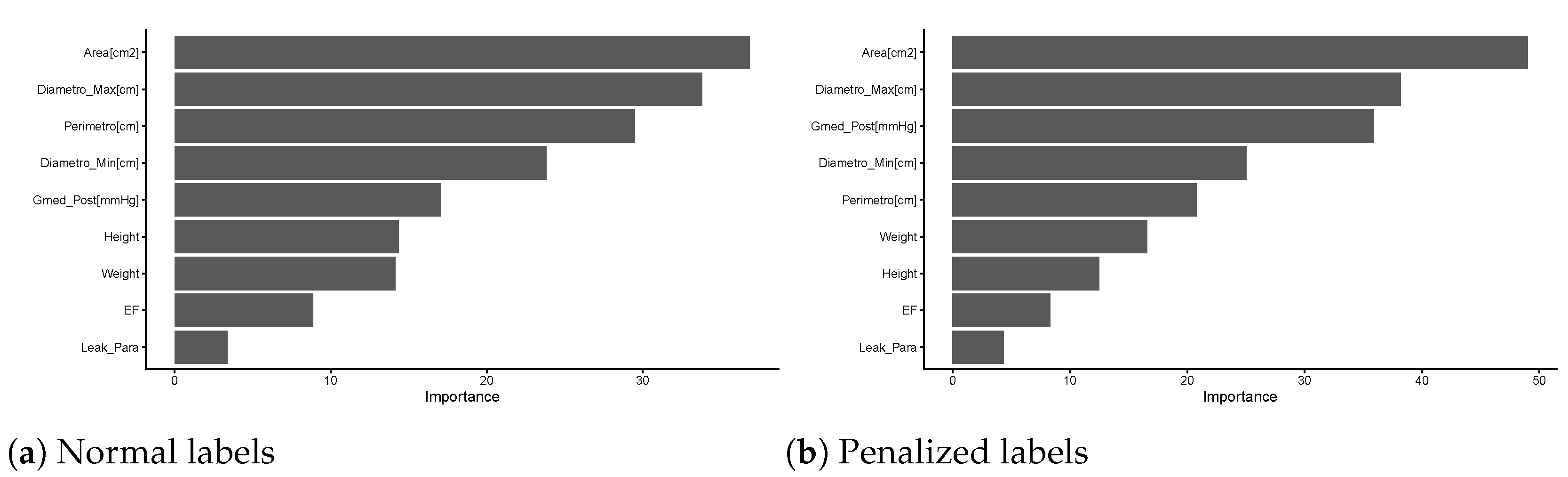
In the normal model, the most important variable contributing to a correct outcome was the aortic annulus area, followed by its maximum diameter (see Figure 2 and Table 1) and two other aortic measurements. The gradient on the prosthesis is the fifth most important. In the penalized model, the area and maximum diameter remain the first two most important variables, followed by the gradient on the prosthesis.

4. Discussion

In this study, we attempted to solve a scientific question which has an important clinical implication, especially in minimally invasive cardiac surgery. The predictive algorithm based on our data enabled a reliable, safe and concrete prediction recommendation system that can be used in clinical settings. MDCT is the best modality for this kind of task, and once again, it proved to be the most advanced, easy, fast and reliable modality for complete preoperative planning. The accurate prediction of prosthetic size for the Perceval sutureless valve (Corcym) has emerged as a critical determinant of clinical success, distinguishing it from conventional sutured valves. Unlike sutured prostheses, the Perceval valve relies on the radial force of a self-expanding nitinol stent for anchoring and sealing. Consequently, the margin for sizing error is narrow, and the literature advocates for a paradigm shift from subjective intraoperative assessment to objective, imaging-based predictive modeling to mitigate complications ranging from hemodynamic failure to conduction disturbances. Moreover, being a true sutureless valve, it is implanted into the aortic annulus after native valve removal, but this is not the case for transcatheter procedures. Margaryan et al. [7] addressed this problem with limited data and an initial sizing experience. Glauber et al. [17] extensively explored the implantation technique and sizing algorithm suggested by a manufacturer. In a recent publication [20], we provided sizing data collected since 2017, which is simplified and permits under-sizing with respect to manufacturer recommendations. With this study, we summarize, to our knowledge, the results of the largest CT scan cohort used for Corcym Perceval valve sizing, with all the valve sizes available, and even more importantly, marginal observations are present in this dataset. This provides a robust idea of how much aortic annulus measurements correlate with clinical cases (see Table 1). This modeling architecture could become an important recommendation system. The normal labels (see Table 1) and penalized labels (see Table 4) shed light on the measurements of the native annulus, and by themselves, they could represent an important guide.
The accurate prediction of prosthetic valve size is a pivotal determinant of clinical success in both Transcatheter Aortic Valve Implantation (TAVI) and surgical aortic valve replacement (SAVR), particularly with sutureless and rapid deployment bio-prostheses [27]. The literature reveals a critical transition from subjective intraoperative assessment to objective, multimodal imaging-based predictive modeling to mitigate life-threatening complications [28].
As evidence suggests [20], there is a conflict between traditional manufacturer recommendations and emerging evidence favoring imaging-based predictive modeling.

4.1. The Conflict: Manufacturer vs. Evidence-Based Sizing

Standard sizing for the Perceval valve relies on intraoperative obturators, where the correct size is selected when the “transparent” obturator passes easily but the “white” obturator meets resistance. This method is criticized as being subjective and prone to excessive over-sizing because it depends on tactile feedback, which varies by surgeon and annular calcification. Consequently, a “novel” sizing technique has been proposed where the white obturator passes with “slight friction,” effectively downsizing the valve compared to original instructions.

4.2. The Role of CT and Advanced Imaging

There is a consensus that Multidetector Computed Tomography (MDCT) provides the most reproducible and objective sizing metrics, superior to the often-underestimating 2D echocardiography [29]. While 3D TEE and MRI are emerging as accurate alternatives [30], CT-based “virtual basal ring” measurements allow for the precise calculation of over-sizing percentages. Automated software and 3D modeling further standardize this process [29,31]. The literature strongly supports the adoption of Multidetector Computed Tomography (MDCT) as the gold standard for Perceval sizing, moving away from the subjectivity of intraoperative obturators. MDCT allows for the measurement of the “virtual basal ring” (VBR) and calculation of the area-derived diameter, which correlates more accurately with the optimal prosthesis size than 2D echocardiography or visual surgical estimates. Studies confirm that MDCT-based sizing can predict the correct valve size in the majority of cases and helps identify patients with specific anatomical risks, such as a high sinotubular junction (STJ) ratio (>1.3), which is a contraindication for Perceval implantation.

4.3. The Dangers of Over-Sizing

While some over-sizing is requisite for anchoring and sealing, excessive over-sizing (often defined as >22.6% [20]) is detrimental. A degree of over-sizing is necessary for anchoring, but recent evidence identifies excessive over-sizing as a primary mechanism of valve failure [19]. Traditional manufacturer instructions—selecting a size where the “white” obturator does not pass—have been criticized for promoting over-sizing. Studies indicate that an over-sizing of the native annulus greater than 22.6% is significantly associated with increased trans-valvular gradients, thrombosis and early structural valve degeneration (SVD) [32]. Excessive radial force prevents the nitinol stent from achieving a circular expansion, leading to stent infolding and “pinwheeling” or distortion of leaflets. This geometrical distortion impairs leaflet coaptation, increases mechanical stress, and may predispose the valve to thrombosis and fibrosis. Conversely, a “novel” sizing technique, which involves downsizing to allow the white obturator to pass with “slight friction,” has been shown to reduce discharge gradients and SVD rates without increasing migration risk, as other authors also declare [33].
This study has limitations: the number of patients is limited, and for good ML and AI models, more observations are important. This study is single-center-based, but for a better understanding, multi-center data and experience are necessary.

5. Conclusions

To date, the studied approach has proven to be safe, reliable and reproducible. It can guarantee preoperative planning and is especially valuable for minimally invasive approaches like right minithoracotomy. The generalization of this algorithm can help young specialists and groups in terms of achieving better valve size selection and outcomes.

Author Contributions

Conceptualization, R.M.; Formal analysis, R.M.; Investigation, R.M. and G.C.; Methodology, R.M. and G.C.; Project administration, R.M.; Supervision, M.S.; Writing—review, G.C., G.B. and M.S.; Writing—review and editing, R.M. All authors have read and agreed to the published version of the manuscript.

Funding

This research received no external funding.

Institutional Review Board Statement

This study was conducted in accordance with the Declaration of Helsinki and approved by the Ethics Committee of Ospedale Del Cuore Fondazione G. Monasterio (Studio n587/2015, approval date: 15 July 2015).

Informed Consent Statement

Patients’ consent was waived because medical records were audited with no direct interaction with subjects who received standard care.

Data Availability Statement

The data presented in this study are available from the corresponding author on reasonable request for fruitful research or collaboration.

Conflicts of Interest

The authors declare no conflicts of interest.

References

  1. Guimaron, S.; De Brux, J.; Verhoye, J.; Guihaire, J. Surgical aortic valve replacement in the modern era: Insights from the French Registry EPICARD. J. Card. Surg. 2021, 36, 4573–4581. [Google Scholar] [CrossRef] [PubMed]
  2. Solinas, M.; Bianchi, G.; Chiaramonti, F.; Margaryan, R.; Kallushi, E.; Gasbarri, T.; Santarelli, F.; Murzi, M.; Farneti, P.; Leone, A.; et al. Right anterior mini-thoracotomy and sutureless valves: The perfect marriage. Ann. Cardiothorac. Surg. 2020, 9, 305–313. [Google Scholar] [CrossRef] [PubMed]
  3. Sulague, R.M.; Beloy, F.J.; Medina, J.R.; Mortalla, E.D.; Cartojano, T.D.; Macapagal, S.; Kpodonu, J. Artificial intelligence in cardiac surgery: A systematic review. World J. Surg. 2024, 48, 2073–2089. [Google Scholar] [CrossRef]
  4. Herrmann, H.C.; Pibarot, P.; Wu, C.; Hahn, R.T.; Tang, G.H.; Abbas, A.E.; Playford, D.; Ruel, M.; Jilaihawi, H.; Sathananthan, J.; et al. Bioprosthetic Aortic Valve Hemodynamics: Definitions, Outcomes, and Evidence Gaps. J. Am. Coll. Cardiol. 2022, 80, 527–544. [Google Scholar] [CrossRef]
  5. Kanschik, D.; Haschemi, J.; Heidari, H.; Klein, K.; Afzal, S.; Maier, O.; Piayda, K.; Binneboesssel, S.; Oezaslan, G.; Bruno, R.R.; et al. Feasibility, Accuracy, and Reproducibility of Aortic Valve Sizing for Transcatheter Aortic Valve Implantation Using Virtual Reality. J. Am. Heart Assoc. 2024, 13, e034086. [Google Scholar] [CrossRef] [PubMed]
  6. Italiano, G.; Belli, M.; Lassandro Pepe, F. 3D echocardiography to plan aortic valve surgery and evaluate the results. Vessel. Plus 2025, 9, 12. [Google Scholar] [CrossRef]
  7. Margaryan, R.; Kallushi, E.; Gilmanov, D.; Micelli, A.; Murzi, M.; Solinas, M.; Cerillo, A.G.; Glauber, M. Sutureless Aortic Valve Prosthesis Sizing Estimation and Prediction Using Multidetector-Row Computed Tomography. Innovations 2015, 10, 230–235. [Google Scholar] [CrossRef]
  8. Dashkevich, A.; Blanke, P.; Siepe, M.; Pache, G.; Langer, M.; Schlensak, C.; Beyersdorf, F. Preoperative Assessment of Aortic Annulus Dimensions: Comparison of Noninvasive and Intraoperative Measurement. Ann. Thorac. Surg. 2011, 91, 709–714. [Google Scholar] [CrossRef]
  9. Montisci, A.; Palmieri, V.; Vietri, M.T.; Sala, S.; Maiello, C.; Donatelli, F.; Napoli, C. Big Data in cardiac surgery: Real world and perspectives. J. Cardiothorac. Surg. 2022, 17, 277. [Google Scholar] [CrossRef]
  10. Pfeiffer, S.; Wilbring, M.; Kappert, U.; Santarpino, G. The ‘entangled’ stent: A preventable cause of paravalvular leak of the Perceval bioprosthesis. Interact. Cardiovasc. Thorac. Surg. 2017, 25, 987–989. [Google Scholar] [CrossRef]
  11. Sharobeem, S.; Le Breton, H.; Lalys, F.; Lederlin, M.; Lagorce, C.; Bedossa, M.; Boulmier, D.; Leurent, G.; Haigron, P.; Auffret, V. Validation of a Whole Heart Segmentation from Computed Tomography Imaging Using a Deep-Learning Approach. J. Cardiovasc. Transl. Res. 2021, 15, 427–437. [Google Scholar] [CrossRef] [PubMed]
  12. Nedadur, R.; Bhatt, N.; Liu, T.; Chu, M.W.; McCarthy, P.M.; Kline, A. The Emerging and Important Role of Artificial Intelligence in Cardiac Surgery. Can. J. Cardiol. 2024, 40, 1865–1879. [Google Scholar] [CrossRef]
  13. Wah, J.N.K. Revolutionizing surgery: AI and robotics for precision, risk reduction, and innovation. J. Robot. Surg. 2025, 19, 47. [Google Scholar] [CrossRef]
  14. Raja, S. Sutureless Aortic Valve Replacement Using Perceval S Valve. Recent Patents Cardiovasc. Drug Discov. 2013, 8, 75–80. [Google Scholar] [CrossRef]
  15. Müller, H.; Szalkiewicz, P.; Benedikt, P.; Ratschiller, T.; Schachner, B.; Schröckenstein, S.; Zierer, A. Single-center real-world data and technical considerations from 100 consecutive patients treated with the Perceval aortic bioprosthesis. Front. Cardiovasc. Med. 2024, 11, 1417617. [Google Scholar] [CrossRef]
  16. Deblier, I.; De Bock, D.; Rodrigus, I.; Mistiaen, W. The Use of Perceval Valves in Older Patients: A Systematic Review. Rev. Cardiovasc. Med. 2025, 26, 39463. [Google Scholar] [CrossRef]
  17. Glauber, M.; Miceli, A.; Bacco, L.d. Sutureless and rapid deployment valves: Implantation technique from A to Z—the Perceval valve. Ann. Cardiothorac. Surg. 2020, 9, 33040–33340. [Google Scholar] [CrossRef] [PubMed]
  18. Concistrè, G.; Chiaramonti, F.; Bianchi, G.; Cerillo, A.; Murzi, M.; Margaryan, R.; Farneti, P.; Solinas, M. Aortic Valve Replacement with Perceval Bioprosthesis: Single-Center Experience with 617 Implants. Ann. Thorac. Surg. 2018, 105, 40–46. [Google Scholar] [CrossRef] [PubMed]
  19. Cerillo, A.G.; Amoretti, F.; Mariani, M.; Cigala, E.; Murzi, M.; Gasbarri, T.; Solinas, M.; Chiappino, D. Increased Gradients After Aortic Valve Replacement With the Perceval Valve: The Role of Oversizing. Ann. Thorac. Surg. 2018, 106, 121–128. [Google Scholar] [CrossRef] [PubMed]
  20. Margaryan, R.; Concistre, G.; Bianchi, G.; Solinas, M. Novel Perceval Sizing Technique: Single Center Experience. medRxiv 2024. [Google Scholar] [CrossRef]
  21. Gilmanov, D.; Miceli, A.; Bevilacqua, S.; Farneti, P.; Solinas, M.; Ferrarini, M.; Glauber, M. Sutureless Implantation of the Perceval S Aortic Valve Prosthesis Through Right Anterior Minithoracotomy. Ann. Thorac. Surg. 2013, 96, 2101–2108. [Google Scholar] [CrossRef]
  22. Glauber, M.; Miceli, A.; Bevilacqua, S.; Farneti, P.A. Minimally invasive aortic valve replacement via right anterior minithoracotomy: Early outcomes and midterm follow-up. J. Thorac. Cardiovasc. Surg. 2011, 142, 1577–1579. [Google Scholar] [CrossRef] [PubMed]
  23. R Core Team. R: A Language and Environment for Statistical Computing; R Foundation for Statistical Computing: Vienna, Austria, 2024. [Google Scholar]
  24. Pedregosa, F.; Varoquaux, G.; Gramfort, A.; Michel, V.; Thirion, B.; Grisel, O.; Blondel, M.; Prettenhofer, P.; Weiss, R.; Dubourg, V.; et al. Scikit-learn: Machine Learning in Python. J. Mach. Learn. Res. 2011, 12, 2825–2830. [Google Scholar]
  25. Breiman, L. Random Forests. Mach. Learn. 2001, 45, 5–32. [Google Scholar] [CrossRef]
  26. Allgaier, J.; Pryss, R. Practical approaches in evaluating validation and biases of machine learning applied to mobile health studies. Commun. Med. 2024, 4, 76. [Google Scholar] [CrossRef]
  27. Meng, X.; Sun, Y.; Bai, W.; Li, Y.; Tuo, S.; Cao, L.; Du, M.; Liu, Y.; Jin, P.; Yang, J.; et al. Application research of three-dimensional transesophageal echocardiography in predicting prosthetic valve size for transcatheter aortic valve implantation. Ann. Transl. Med. 2022, 10, 84. [Google Scholar] [CrossRef] [PubMed]
  28. Higuchi, R.; Saji, M.; Hagiya, K.; Takamisawa, I.; Shimizu, J.; Tobaru, T.; Iguchi, N.; Takanashi, S.; Takayama, M.; Isobe, M. Transcatheter aortic valve implantation-related futility: Prevalence, predictors, and clinical risk model. Heart Vessel. 2020, 35, 1281–1289. [Google Scholar] [CrossRef]
  29. Kempfert, J.; Van Linden, A.; Lehmkuhl, L.; Rastan, A.J.; Holzhey, D.; Blumenstein, J.; Mohr, F.W.; Walther, T. Aortic annulus sizing: Echocardiographic versus computed tomography derived measurements in comparison with direct surgical sizing. Eur. J. Cardio-Thorac. Surg. 2012, 42, 627–633. [Google Scholar] [CrossRef]
  30. Piazza, N.; de Jaegere, P.; Schultz, C.; Becker, A.E.; Serruys, P.W.; Anderson, R.H. Anatomy of the Aortic Valvar Complex and Its Implications for Transcatheter Implantation of the Aortic Valve. Circ. Cardiovasc. Interv. 2008, 1, 74–81. [Google Scholar] [CrossRef]
  31. Pouch, A.M.; Vergnat, M.; McGarvey, J.R.; Ferrari, G.; Jackson, B.M.; Sehgal, C.M.; Yushkevich, P.A.; Gorman, R.C.; Gorman, J.H. Statistical Assessment of Normal Mitral Annular Geometry Using Automated Three-Dimensional Echocardiographic Analysis. Ann. Thorac. Surg. 2014, 97, 71–77. [Google Scholar] [CrossRef]
  32. Oezpeker, U.C.; Feuchtner, G.; Bonaros, N. Cusp thrombosis of a self-expandable sutureless aortic valve treated by valve-in-valve transcatheter aortic valve implantation procedure: Case report. Eur. Heart J.—Case Rep. 2018, 2, yty117. [Google Scholar] [CrossRef]
  33. Nezafati, P.; Yadav, S. How to avoid oversizing Perceval sutureless aortic valve: Technique. ARYA Atheroscler. J. 2025, 21, 1–3. [Google Scholar] [CrossRef]
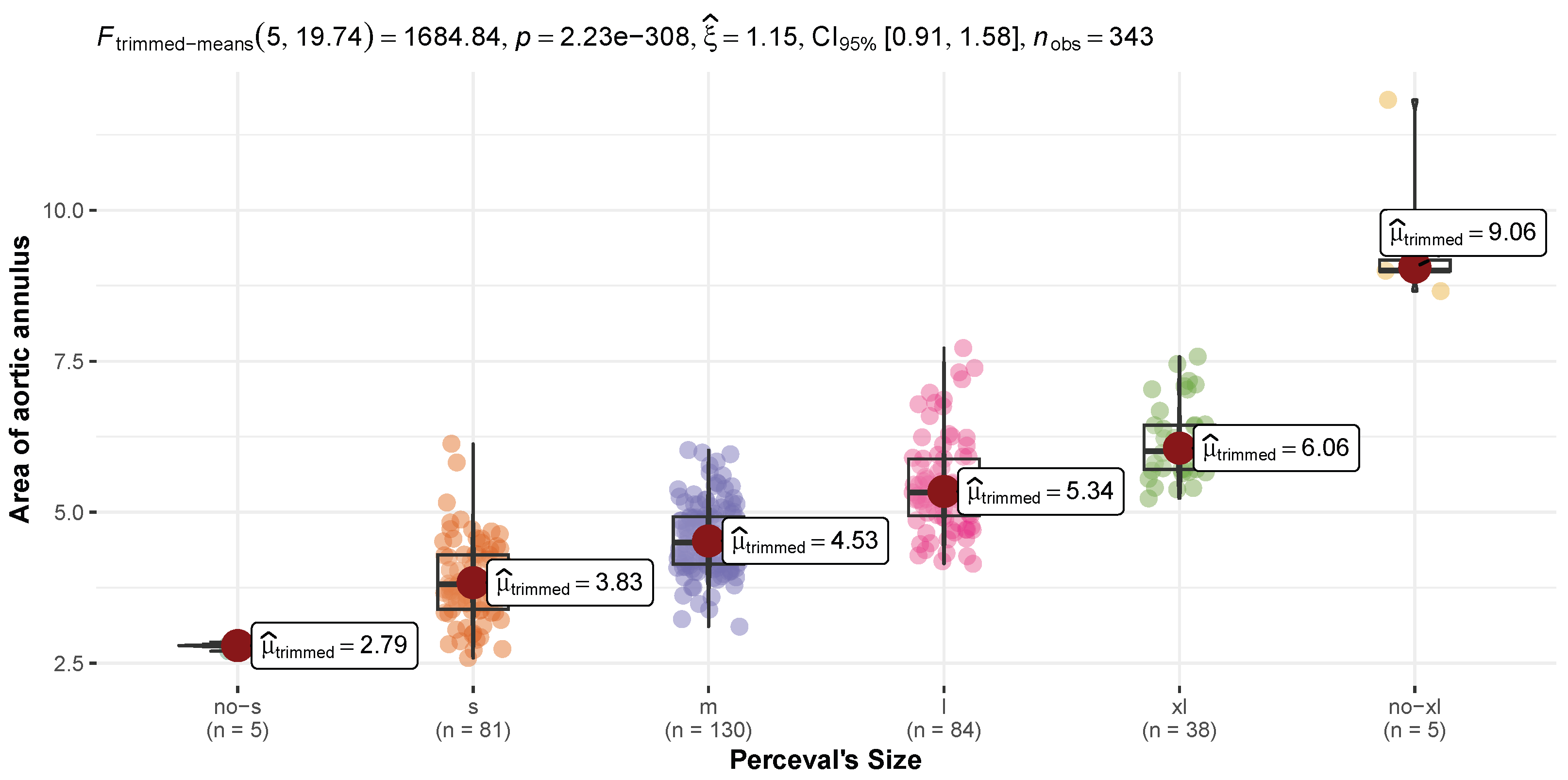
Figure 1. Perceval size diameter plot with original labels.
Hearts 07 00016 g001
Figure 2. Variable importance plot for two models.
Hearts 07 00016 g002
Table 1. Aortic annular measurements and dynamics on all sizes of Perceval valves.
CharacteristicNo-S N = 5S N = 102M N = 139L N = 91XL N = 38No-XL N = 5
Maximum Diameter2.03 ± 0.102.38 ± 0.212.61 ± 0.202.86 ± 0.203.05 ± 0.223.19 ± 0.62
Minimum Diameter1.62 ± 0.041.94 ± 0.192.11 ± 0.162.29 ± 0.232.46 ± 0.152.71 ± 0.54
Perimeter6.07 ± 0.057.01 ± 0.627.63 ± 0.538.30 ± 0.628.91 ± 0.5110.13 ± 1.30
Area2.79 ± 0.063.83 ± 0.664.54 ± 0.585.40 ± 0.776.16 ± 0.619.53 ± 1.30
Pre-Op Ejection Fraction59 ± 261 ± 560 ± 558 ± 660 ± 553 ± 12
Post-Op Ejection Fraction55 ± 559 ± 560 ± 658 ± 761 ± 652 ± 8
Post-Op Mean Gradient12.8 ± 2.814.7 ± 6.112.9 ± 4.011.8 ± 4.411.4 ± 3.59.6 ± 1.7
Post-Op Para-valvular Leakage
No4 (80%)94 (92%)123 (88%)86 (95%)35 (92%)4 (80%)
Mild0 (0%)6 (5.9%)12 (8.6%)5 (5.5%)2 (5.3%)0 (0%)
Moderate1 (20%)2 (2.0%)4 (2.9%)0 (0%)0 (0%)1 (20%)
Severe0 (0%)0 (0%)0 (0%)0 (0%)1 (2.6%)0 (0%)
Table 2. Patients’ characteristics.
CharacteristicN = 380
Age77.6 ± 4.9
BSA1.83 ± 0.35
BMI27.6 ± 4.4
Sex
Female127 (51%)
Male120 (49%)
CPB109 ± 38
X-clamp67 ± 24
Hospital Stay8.0 ± 4.4
Hospital Mortality0 (0%)
Approach
Right Minithoracotomy92 (38%)
Median Sternotomy58 (24%)
Mini-Sternotomy91 (38%)
Perceval Type
Lancelot65 (17%)
Perceval S114 (30%)
Perceval Plus201 (53%)
Table 3. Metric table.
MetricAll NormalTrain NormalTest NormalAll PenalizedTrain PenalizedTest Penalized
Accuracy0.91842110.99647890.68750000.928947410.7216495
Balanced Accuracy0.91200050.99663800.69262620.956341710.8318185
F1-Score0.88367810.99575320.68677350.939965610.7519111
Precision0.94499820.99758450.68935440.953520110.8258066
Recall0.84321790.99404760.45929950.929617010.7294673
Sensitivity0.84321790.99404760.45929950.929617010.7294673
Specificity0.98078310.99922840.92595290.983066410.9341696
Table 4. Penalized label distribution.
CharacteristicNo-S N = 30S N = 88M N = 132L N = 87XL N = 35No-XL N = 8
Maximum Diameter
Mean ± SD2.28 ± 0.212.40 ± 0.222.62 ± 0.192.87 ± 0.203.05 ± 0.233.14 ± 0.48
Minimum Diameter
Mean ± SD1.85 ± 0.211.96 ± 0.192.11 ± 0.172.31 ± 0.222.45 ± 0.152.67 ± 0.42
Perimeter
Mean ± SD6.70 ± 0.627.12 ± 0.607.63 ± 0.518.34 ± 0.628.87 ± 0.519.83 ± 1.08
Area
Mean ± SD3.48 ± 0.663.95 ± 0.654.54 ± 0.555.46 ± 0.776.11 ± 0.598.51 ± 1.73
Disclaimer/Publisher’s Note: The statements, opinions and data contained in all publications are solely those of the individual author(s) and contributor(s) and not of MDPI and/or the editor(s). MDPI and/or the editor(s) disclaim responsibility for any injury to people or property resulting from any ideas, methods, instructions or products referred to in the content.