1. Introduction
Radiotherapy is a cornerstone in the management of nasopharyngeal carcinoma (NPC) and many sinonasal malignancies because of the anatomical complexity of the region and the proximity of tumors to critical neurovascular structures [
1]. Modern techniques such as intensity-modulated radiotherapy (IMRT) and volumetric modulated arc therapy (VMAT) have improved tumor control while reducing several severe toxicities previously associated with conventional radiation delivery [
2,
3]. Nevertheless, a substantial proportion of long-term survivors experience persistent sinonasal symptoms following treatment. These symptoms are frequently attributed to chronic rhinosinusitis, yet their relationship to radiation exposure is often underappreciated in routine oncologic practice.
Radiation-induced rhinosinusitis (RIR) represents a clinically relevant but insufficiently characterized late toxicity of head-and-neck radiotherapy. In patients treated for NPC, imaging and clinical studies have reported sinonasal inflammatory changes in a considerable proportion of cases during follow-up, with reported rates varying widely depending on study design and diagnostic criteria. For example, radiologic evidence of sinus mucosal disease after NPC radiotherapy has been widely reported, with MRI-based cohorts demonstrating post-treatment sinusitis in more than 80 to 90% of patients at follow-up, indicating that sinonasal injury represents a common sequela rather than a rare complication [
4].
The mechanisms underlying RIR are multifactorial. Ionizing radiation can disrupt epithelial integrity, impair mucociliary clearance, and induce long-term inflammatory and fibrotic changes in sinonasal tissues. Experimental and clinical observations indicate that radiation exposure disrupts the structure and function of ciliated epithelial cells within the nasal mucosa, leading to impaired mucociliary transport and subsequent retention of mucus. This stagnation fosters microbial colonization and drives persistent inflammatory responses in the paranasal sinuses. This radiation-induced pathological changes parallel the mechanisms underlying chronic rhinosinusitis, although in this context they arise as a direct consequence of therapeutic irradiation rather than primary inflammatory disease [
5].
RIR is rarely discussed as a dose-limiting toxicity in head-and-neck oncology. Most radiation planning paradigms prioritize protection of organs such as the spinal cord, brainstem, salivary glands, and optic apparatus. In contrast, the paranasal sinuses and associated mucosal structures receive relatively little attention during treatment planning, even though they are often located within high-dose radiation fields for NPC and sinonasal tumors. Consequently, sinonasal morbidity may emerge as a survivorship issue long after tumor control has been achieved.
Although post-radiotherapy sinonasal symptoms are often managed as chronic rhinosinusitis, RIR differs from ordinary inflammatory sinus disease because it develops in a mucosa exposed to radiation-related epithelial injury, impaired mucociliary clearance, vascular damage, fibrosis, crusting, and altered local defense [
6]. For this reason, RIR should be considered not only as a survivorship complaint but also as a potential dose-limiting toxicity when uninvolved sinonasal mucosa or drainage pathways are exposed to high doses.
Persistent post-treatment sinus opacification should not be assumed to mimic recurrence in isolation. In contemporary practice, inflammatory changes are interpreted together with clinical examination, endoscopic findings, serial imaging, and, when indicated, biopsy. However, post-radiotherapy mucosal thickening, retained secretions, and focal inflammatory change may still create interpretive uncertainty during surveillance, particularly when unilateral, progressive, or adjacent to the primary tumor bed. Post-treatment imaging after NPC radiotherapy can be challenging because expected radiation-related changes may overlap with features of residual or recurrent disease, requiring correlation with clinical, endoscopic, MRI, and PET/CT findings [
7,
8,
9,
10,
11,
12,
13].
This narrative review focuses primarily on RIR after radiotherapy for NPC, where the evidence base is strongest. Evidence from selected sinonasal malignancies is included where it informs sinonasal dose distribution, radiation-related tissue response, or management principles. The review summarizes current evidence on epidemiology, mechanisms, dosimetric predictors, imaging considerations, prevention, management, and future reporting needs. Its main contribution is to frame RIR as a clinically meaningful sinonasal field-of-injury toxicity and to propose a harmonized framework for future outcome reporting.
2. Methods
This article is a narrative review rather than a systematic review or meta-analysis. A narrative design was selected because the available literature on RIR is heterogeneous in tumor site, radiotherapy technique, diagnostic criteria, follow-up timing, and endpoint definition. The review was therefore intended to synthesize clinically relevant themes rather than to provide pooled prevalence estimates or a formal quality assessment of individual studies.
Relevant literature was identified through targeted searches of major biomedical databases and review of reference lists from key articles. Search terms were selected to capture studies addressing radiation-induced rhinosinusitis, post-radiotherapy sinusitis, nasopharyngeal carcinoma, sinonasal toxicity, mucociliary dysfunction, paranasal sinus dosimetry, IMRT, VMAT, and proton therapy. Priority was given to studies that informed the epidemiology, pathophysiology, imaging characteristics, dosimetric predictors, prevention, or management of sinonasal injury after radiotherapy.
Because this was not a systematic review, no formal risk-of-bias tool, PRISMA flow diagram, or meta-analysis was performed. The findings were synthesized qualitatively, with emphasis on recurring observations, areas of uncertainty, and practical implications for radiation planning and survivorship care. The review focuses primarily on NPC because most available clinical and dosimetric data come from this population. Evidence from sinonasal cancers is discussed selectively where it contributes to understanding radiation-related sinonasal injury.
3. Epidemiology and Clinical Burden of Radiation-Induced Rhinosinusitis
RIR is increasingly recognized as a frequent sequela of radiotherapy for NPC and, less consistently, for selected sinonasal malignancies. Its true prevalence remains uncertain because studies use different endpoints, including radiologic sinus opacification, MRI-based Lund–Mackay scores, endoscopic inflammatory findings, symptom questionnaires, and composite clinical definitions. Across available NPC cohorts, imaging-defined sinonasal inflammatory changes are commonly reported in approximately 30% to more than 70% of patients [
14,
15]. Some longitudinal MRI-based studies have reported higher rates, including estimates exceeding 80%, but these generally reflect broader radiologic definitions of sinus mucosal disease rather than uniformly symptomatic or clinically active RIR [
4]. For this reason, reported prevalence should be interpreted as a spectrum that depends on the diagnostic endpoint, follow-up interval, baseline sinonasal status, and imaging modality used.
Clinically, RIR may present with nasal obstruction, rhinorrhea or mucopurulent discharge, crusting, postnasal drip, facial pressure, hyposmia, recurrent infectious exacerbations, or persistent sinonasal dryness. However, symptoms and imaging changes do not always align. Some patients develop extensive mucosal thickening or sinus opacification with limited symptoms, whereas others experience substantial sinonasal morbidity despite less striking imaging findings [
4,
16]. A plausible, practical conceptual model can therefore separate RIR into three overlapping endpoint domains: radiologic RIR, defined by mucosal thickening or opacification on CT or MRI; symptomatic RIR, defined by patient-reported sinonasal burden; and clinically active RIR, defined by symptoms plus endoscopic evidence of inflammation, crusting, purulence, synechiae, ostial obstruction, or mucosal atrophy.
The maxillary and ethmoid sinuses are most frequently emphasized in the available dosimetric and imaging literature because of their proximity to the nasopharynx, nasal cavity, and ostiomeatal drainage pathways. In VMAT-treated NPC cohorts, higher cumulative incidences of maxillary and ethmoid sinusitis have been associated with high-dose exposure, including V70 thresholds for the maxillary and ethmoid sinuses [
14]. MRI-based comparative studies have similarly categorized the maxillary and anterior ethmoid sinuses as high-risk sites for post-irradiation sinus mucosa disease, while the posterior ethmoid and sphenoid sinuses appear to represent intermediate-risk sites and the frontal sinus a lower-risk site [
17]. Sphenoid sinus involvement remains clinically relevant in nasopharyngeal carcinoma because treatment volumes commonly include the skull base and sphenoid region, and recent long-term data identified tumor invasion of the sphenoidal base as an independent predictor of radiation-induced rhinosinusitis [
15]. Frontal sinus involvement is less consistently reported and appears less prominent in several imaging series, likely reflecting both anatomic distance from high-dose NPC target volumes and variable reporting across imaging-based scoring systems [
5,
17]. Future studies should therefore report sinus-specific outcomes rather than relying only on global Lund–Mackay or aggregate sinus scores. Although many studies rely on imaging findings, symptomatic disease also contributes to the clinical burden of RIR. Patients may report persistent nasal obstruction, mucopurulent discharge, facial pressure, or recurrent infections that resemble chronic rhinosinusitis. These symptoms can significantly impair quality of life and may require ongoing medical management, including saline irrigation, intranasal corticosteroids, or intermittent antibiotic therapy. Importantly, sinonasal dysfunction may coexist with other radiation-related toxicities such as xerostomia or mucosal dryness, which can further compromise nasal physiology.
Despite its frequency, radiation-induced sinonasal injury remains underrepresented in oncology toxicity reporting frameworks. Most radiation toxicity scales used in head-and-neck cancer trials focus on structures such as the salivary glands, pharynx, or larynx, while sinonasal morbidity is often grouped under nonspecific categories or not reported at all [
18]. As survivorship improves in NPC and other head-and-neck malignancies, greater attention to these chronic but impactful complications is warranted. Recognizing RIR as a clinically meaningful outcome is an important step toward integrating sinonasal structures into radiotherapy planning and survivorship care.
4. Pathophysiology of Radiation-Induced Sinonasal Injury
RIR is best understood as a disorder of epithelial injury, impaired clearance, and maladaptive tissue repair rather than as a simple extension of ordinary infectious sinusitis. The sinonasal mucosa lies close to the target volume in nasopharyngeal and many sinonasal cancer plans, so it is often exposed to substantial incidental doses. Clinical and translational studies suggest that the earliest effects of radiation include mucosal edema, epithelial disruption, and loss of normal ciliary architecture, changes that can then evolve into chronic mucus retention and persistent inflammatory disease [
5,
19].
A central mechanism is mucociliary dysfunction. In a prospective cohort of patients undergoing radiotherapy for head and neck cancer, mucociliary clearance time was prolonged in most patients by the end of treatment, and dysfunction remained evident at 3 months in those who completed follow-up [
20]. That observation is biologically plausible because radiation damages proliferative epithelial and submucosal tissues and reduces the integrity of the ciliated respiratory lining that is required for effective mucus transport. Once clearance is impaired, secretions stagnate, sinus ostia are more easily obstructed, and secondary inflammatory changes become more likely [
19,
21].
Histopathologic work supports this model. The recent mechanistic review by Zheng et al. [
19] summarized post-radiation nasal mucosal findings, including lamina propria fibrosis, cilia malformation or loss, and vacuole change, together with squamous metaplasia of the respiratory epithelium. These changes matter because metaplastic epithelium no longer provides normal ciliary function or goblet-cell physiology, so the mucosa loses much of its capacity for coordinated clearance and surface defense. The same review argues that radiation may also impair the regenerative capacity of nasal epithelial stem cells, which could help explain why some patients recover partially, whereas others develop persistent disease. These findings are compelling, although most direct mechanistic data remain limited and are not yet well linked to standardized clinical phenotypes.
The subsequent phase of radiation-induced sinonasal injury appears to be characterized not merely by transient inflammation but by a shift toward chronic tissue remodeling. Radiation injury in multiple organ systems is well documented to trigger sustained oxidative stress, endothelial dysfunction, cytokine dysregulation, and progressive fibrosis, processes that are plausibly applicable to the sinonasal mucosa as well [
22]. In clinical practice, such chronic remodeling may present as persistent mucosal thickening, narrowing of sinonasal drainage pathways, crusting, and long-lasting sinus opacification on imaging [
23]. Importantly, these radiologic abnormalities can endure even after acute mucositis has resolved, indicating that late-phase toxicity likely reflects structural remodeling in addition to residual inflammation [
7]. However, a study by Hemmi et al. [
24], which evaluated post-treatment CT imaging, argued that head-and-neck cancer treatment was not an apparent risk factor for developing sinonasal mucosal disease, though its conclusions were limited by reliance on imaging findings alone without assessment of clinical symptoms.
There is also reason to think that radiation alters the local microbial and immune environment [
25], although the evidence in HNC treatment is still incomplete. Chronic rhinosinusitis following radiotherapy appears to be multifactorial, with mucosal damage, altered mucus transport, and impaired local defense all contributing [
26]. This interpretation is clinically useful because it explains why symptoms may persist even when no acute bacterial infection is demonstrable and why management often requires more than antibiotics alone. Still, direct microbiome studies in irradiated sinonasal tissue remain sparse, so this part of the model should be regarded as plausible rather than confirmatory.
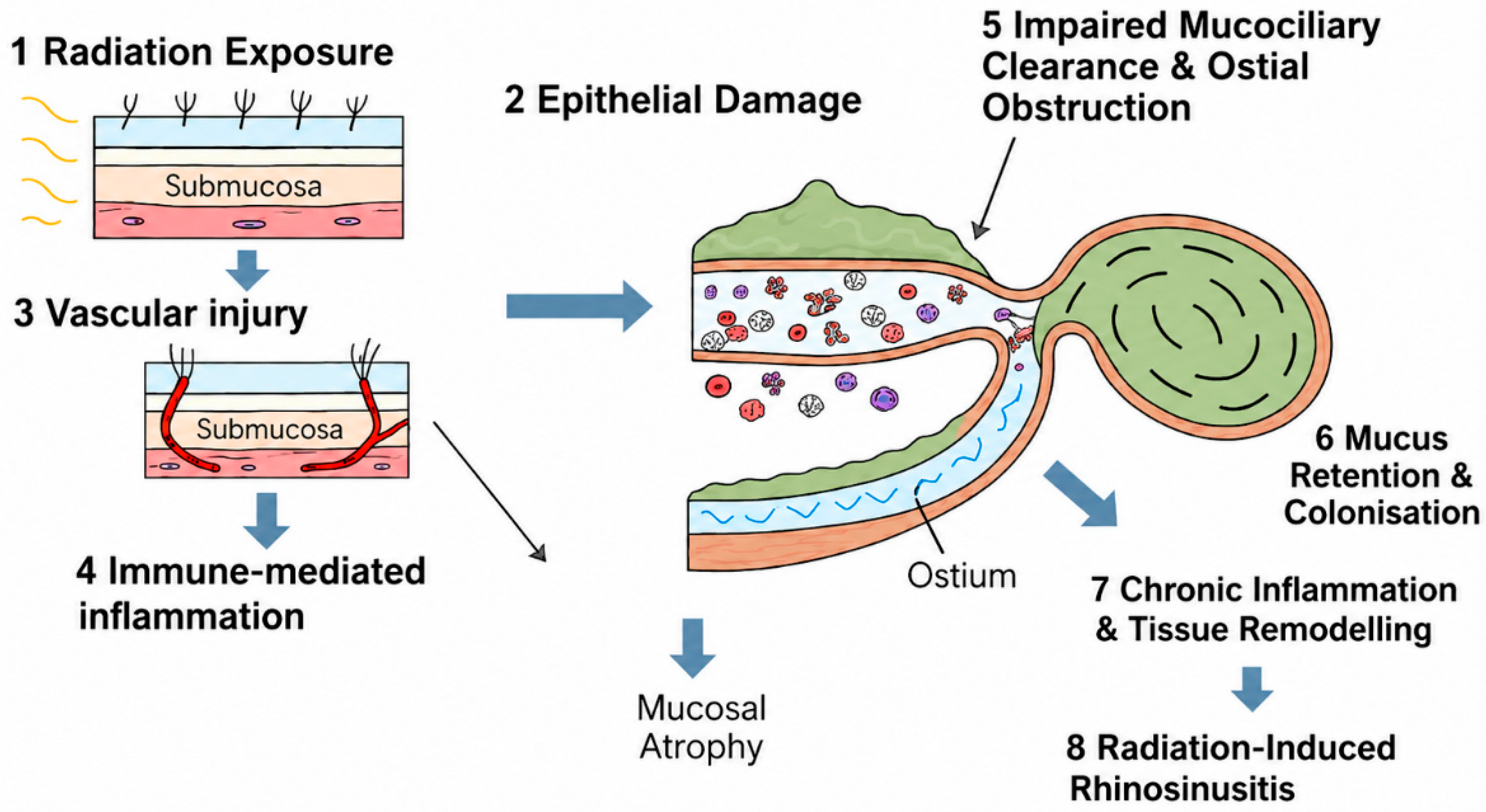
Taken together, available evidence supports a model in which radiation damages the ciliated epithelium, slows mucociliary clearance, promotes mucus stasis, worsens sinus ventilation, and contributes to chronic inflammatory-fibrotic remodeling, as summarized in
Figure 1.
5. Dosimetric Predictors of Radiation-Induced Rhinosinusitis
RIR is closely related to the dose distribution delivered to the paranasal sinuses and adjacent drainage pathways, yet these structures are rarely considered explicit organs-at-risk during radiotherapy planning. Most treatment plans for nasopharyngeal carcinoma inevitably expose the maxillary, ethmoid, and sphenoid sinuses to moderate or high doses because of their proximity to the primary tumor and regional lymphatic targets [
27]. Consequently, several studies have attempted to determine whether specific dosimetric parameters correlate with the development of post-radiotherapy sinonasal disease.
Growing evidence from imaging-based cohorts indicates that increasing radiation dose to the paranasal sinuses correlates directly with a heightened risk of mucosal injury and sinus opacification. In a retrospective analysis of 196 patients with nasopharyngeal carcinoma treated with definitive VMAT, Bao et al. [
14] reported that high-dose sinus exposure, including maxillary sinus V70 and maximum dose (Dmax), was associated with subsequent RIR. Their recommendation suggests the incorporation of these constraints into VMAT planning and close surveillance with early preventive interventions within 3–6 months post-RT for higher-risk patients. Complementing these findings, a large prospective study of 1410 NPC patients by Chen et al. [
15] confirmed that RIR is a common late effect of treatment and is strongly influenced by delivered sinus dose, baseline mucosal status, and patterns of tumor extension. Patients receiving higher mean sinus doses were considerably more likely to develop persistent mucosal thickening and sinus opacification during follow-up, reinforcing a clear dose–response relationship between radiation exposure and sinonasal toxicity.
Subsequent investigations have explored similar relationships in the era of intensity-modulated radiotherapy. Although IMRT improves conformity around critical neural structures [
28], the paranasal sinuses often remain within the high-dose region. A retrospective analysis of NPC patients treated with IMRT demonstrated that post-treatment sinusitis remained common despite the use of modern conformal techniques, with the maxillary and anterior ethmoid sinuses showing the highest rates of persistent mucosal abnormalities, findings that underscore that enhanced targeting precision does not necessarily reduce sinonasal toxicity when these structures remain within high-dose regions [
29]. Furthermore, in a separate analysis of 230 IMRT-treated NPC patients, Huang et al. [
30] found that post-radiation sinusitis was a significant predictor of local recurrence, reinforcing its clinical relevance as more than a benign radiologic finding.
Beyond mean sinus dose, attention has increasingly focused on the ostiomeatal complex and sinus drainage pathways, which play a central role in sinus ventilation and mucociliary clearance. Radiation exposure to these narrow anatomical channels may promote obstruction through mucosal edema and fibrosis, thereby predisposing patients to chronic sinus disease even when the sinus cavity itself receives moderate doses [
19]. Although this concept is biologically plausible, quantitative dose constraints for these structures have not yet been standardized.
Another challenge is that many available studies rely on radiologic rather than clinical endpoints. Imaging findings such as mucosal thickening or sinus opacification are frequently used as markers of radiation-induced injury, but these changes do not always correlate with patient-reported symptoms. Consistent with prior observations in chronic rhinosinusitis, there is often a poor correlation between clinical symptoms and radiologic severity, with some patients remaining asymptomatic despite marked sinus opacification, while others report significant sinonasal morbidity in the presence of only modest imaging changes [
31]. This discrepancy complicates the interpretation of dose–response relationships and highlights the need for studies that integrate imaging, endoscopic findings, and validated symptom measures.
The current evidence, therefore, supports the existence of a dose-dependent component of radiation-induced sinonasal injury, but important uncertainties remain. Dose thresholds for individual sinuses have not been clearly established, and the relative importance of dose to sinus cavities versus drainage pathways is still poorly defined. As survivorship outcomes receive greater attention in head-and-neck oncology, incorporating sinonasal structures into dosimetric analyses may help identify strategies to reduce this under-recognized complication of radiotherapy. Key studies evaluating clinical and dosimetric predictors of radiation-induced rhinosinusitis are summarized in
Table 1.
6. Imaging Findings and Diagnostic Considerations
Imaging is central to the evaluation of RIR, but post-radiotherapy sinonasal abnormalities should be interpreted within the broader oncologic context. In patients treated for NPC or sinonasal cancers, imaging must distinguish treatment-related mucosal inflammation from findings suspicious for persistent or recurrent tumor. Diffuse or stable sinus mucosal thickening is often compatible with inflammatory change, particularly when supported by clinical examination and nasal endoscopy. However, focal, progressive, asymmetric, mass-like, or clinically discordant abnormalities may require closer radiologic follow-up, endoscopic assessment, or biopsy [
7,
8,
9,
10,
11,
12,
13]. This distinction is especially important because RIR studies often use imaging-defined endpoints that may not correspond directly to symptomatic disease.
Computed tomography (CT) remains the most practical and widely used first-line modality for evaluating post-radiotherapy sinonasal disease, given its ability to depict mucosal thickening, sinus opacification, fluid levels, retained secretions, and chronic bony changes with high reliability [
7,
33]. Evidence from imaging-based studies of NPC confirms that CT consistently identifies the maxillary sinus, anterior ethmoid sinus, and ostiomeatal complex as the regions most affected after irradiation, reinforcing its value in assessing structural and inflammatory sequelae of treatment [
15,
34]. Recent reviews of radiation-induced rhinosinusitis likewise emphasize CT as a central tool for evaluating post-treatment mucosal inflammation, noting that most RIR research relies on CT-based scoring rather than symptom-only assessment because of its objectivity and reproducibility [
5,
19]. Broader imaging guidelines support this practice, with national radiology recommendations identifying CT as the preferred initial imaging modality for sinonasal inflammatory conditions due to its superior visualization of both mucosal and bony abnormalities [
35]. Availability of CT imaging in outpatient clinic settings may be crucial for identifying RIR. The expansion of in-clinic cone-beam CT use in rhinology practice reflects a broader shift toward greater dependence on imaging for sinonasal disease assessment, which has important implications in post-radiotherapy settings where persistent mucosal changes frequently require repeated imaging and may complicate differentiation between treatment-related inflammation and tumor recurrence [
36].
Magnetic resonance imaging (MRI) offers a different advantage: better soft-tissue characterization and a more detailed assessment of the post-treatment nasopharynx, skull base, orbit, and adjacent spaces. This matters because inflammatory mucosal thickening, fibrosis, and retained secretions can all mimic recurrent disease on surveillance imaging. An MRI-based study of RIR in nasopharyngeal carcinoma used serial MRI scoring to track the evolution of post-radiotherapy sinus disease and concluded that MRI can characterize the distribution and temporal pattern of these changes [
4], while older and newer literature on post-radiotherapy NPC imaging continues to emphasize MRI’s value when recurrence is part of the differential diagnosis [
8,
9].
Interpreting post-radiotherapy imaging in nasopharyngeal carcinoma is complicated by the fact that radiologic abnormalities do not consistently correspond to symptom burden or underlying pathology. As established in sinonasal disease literature, there is often a poor correlation between imaging severity and clinical symptoms, with some patients demonstrating extensive sinus opacification while remaining minimally symptomatic, and others reporting significant congestion or facial pressure despite relatively modest radiologic changes. This discordance is further amplified after radiotherapy, where benign post-treatment inflammatory or fibrotic changes can closely mimic recurrent tumors, creating substantial diagnostic uncertainty [
10,
11]. Imaging studies have consistently shown that post-radiotherapy changes, such as chronic mucositis, mucosal atrophy, ulceration, and soft-tissue thickening, can closely mimic malignant recurrence on both CT and MRI, thereby complicating interpretation. In a cohort study, Lee et al. [
12] demonstrated that MRI alone is insufficient for determining recurrence following radiotherapy, with 5 of 13 radiologically suspected recurrences ultimately found to be false positives. This limitation is supported by more recent comparative data: a 2024 meta-analysis reported higher sensitivity for FDG-PET than MRI in detecting residual or recurrent disease at the primary site [
13]. Subsequent cohort studies have described MRI-suspicious post-treatment abnormalities that were later disproven on biopsy or serial follow-up, including cases of skull-base osteitis related to sphenoid sinusitis [
9]. These findings highlight the limitations of structural imaging alone and support a multimodal approach when evaluating suspected recurrence in the irradiated nasopharynx.
For that reason, imaging should not be interpreted in isolation. Endoscopy remains important for correlating symptoms with visible mucosal findings such as edema, crusting, purulent drainage, synechiae, or ostial obstruction. Delayed sinonasal complications after nasopharyngeal radiotherapy, including chronic sinusitis, choanal stenosis, and intranasal synechiae, are likely underestimated in part because nasal endoscopy is not performed routinely in many survivorship settings. In practice, the most reliable assessment combines imaging, endoscopic examination, symptom history, and oncologic context rather than relying on any single modality. A biopsy may be performed for lesions with red flags following extensive examinations.
A further limitation in the literature is the lack of standardized imaging endpoints. Some studies define radiation-induced rhinosinusitis by radiologic mucosal disease alone, while others require symptoms or endoscopic confirmation. This heterogeneity makes cross-study comparisons difficult and may partly explain why reported incidence varies so widely. It also weakens efforts to derive clinically meaningful dose constraints because an imaging-defined endpoint may not reflect the same burden as symptomatic chronic rhinosinusitis. The heterogeneity in outcome definitions used across studies is summarized in
Table 2. A more useful framework for future studies would integrate imaging severity with patient-reported symptoms and endoscopic findings, especially in longitudinal cohorts treated with modern radiotherapy techniques. This is addressed subsequently in this review.
7. Prevention Strategies During Radiotherapy
Prevention of radiation-induced rhinosinusitis begins with a simple premise: sinonasal structures should be treated as functionally relevant normal tissues, even if they are not yet formal organs-at-risk in most planning workflows. At present, however, prevention is constrained by the anatomy of nasopharyngeal and sinonasal cancers. In many cases, the primary tumor and elective target volumes lie immediately adjacent to the maxillary, ethmoid, sphenoid, and posterior nasal cavity mucosa, so complete sparing is not realistic without compromising coverage. The more practical goal is therefore dose reduction where feasible, especially to uninvolved sinus mucosa and drainage pathways, rather than the pursuit of absolute avoidance. Recent precision-radiotherapy studies in NPC emphasize that modern planning has substantially improved conformity, but toxicity reduction depends on which normal tissues are explicitly prioritized during optimization [
39,
40].
The strongest prevention strategy is likely better dosimetric stewardship, although the evidence base remains incomplete. Retrospective NPC studies consistently suggest that post-radiotherapy sinusitis is associated with dose to the paranasal sinuses [
14], and more recent work has linked risk not only to dose but also to baseline sinus mucosal status and local tumor invasion [
15]. That pattern is clinically important because it argues against a one-size-fits-all prevention model: patients with pre-existing mucosal disease or direct tumor extension into sinonasal structures may remain at high risk even under otherwise conformal plans. The implication is that future planning studies should move beyond whole-sinus dose metrics and examine whether sparing the ostiomeatal complex, sphenoethmoidal recess, and posterior nasal cavity is both technically feasible and clinically meaningful. At present, validated dose constraints for these structures are still lacking.
Whether proton therapy changes the toxicity profile is an important but still unsettled question. The theoretical argument is strong: proton therapy can reduce exit dose and may better spare adjacent normal tissues when anatomy permits. In NPC, a comparative study by Wu et al. [
17] found lower incidence and severity of post-irradiation sinus mucosa disease after IMPT than after VMAT, with more rapid regression of mucosal disease in the proton cohort. Systematic reviews of proton therapy in NPC also report a generally improved toxicity profile compared with photon-based treatment, although these analyses were not designed specifically around sinonasal endpoints [
12,
41]. The caution is that most data remain retrospective, single-center, and potentially confounded by selection effects. Proton therapy may reduce sinonasal injury in some settings, but the current evidence does not justify a blanket claim that it prevents radiation-induced rhinosinusitis across all head-and-neck indications.
By contrast, evidence for prophylactic medical prevention during radiotherapy is still limited. Supportive care measures such as humidification and saline-based nasal care are biologically plausible because they may reduce crusting, improve mucus clearance, and mitigate surface irritation, but high-quality trials specifically preventing radiation-induced rhinosinusitis are scarce. A clinical study by Ouyang et al. [
42] on “early nasal intervention” in NPC suggests that proactive local care may reduce radiation-associated sinusitis, but this literature is still too early and too narrow to support firm recommendations beyond low-risk supportive measures. It is reasonable to consider saline irrigation early in treatment in patients with emerging nasal dryness or crusting, but stronger claims about prevention would run ahead of the data.
There is also a case for pre-treatment risk stratification. Patients with baseline sinus mucosal disease, large tumors abutting the sinus drainage pathways, or direct sinonasal invasion appear more likely to develop post-radiotherapy sinus complications [
15]. That does not mean radiotherapy should be modified at the expense of oncologic control, but it does support documenting baseline sinonasal status before treatment and monitoring high-risk patients more deliberately during follow-up. In practical terms, prevention may depend as much on anticipation and early survivorship planning as on dosimetry alone. The literature increasingly supports the view that radiation-induced rhinosinusitis is not simply an unavoidable nuisance toxicity; it is a complication whose burden may be reduced by a combination of better planning, more explicit recognition of high-risk anatomy, and earlier supportive care. The challenge now is to define which of those steps has the greatest clinical impact.
8. Management of Radiation-Induced Rhinosinusitis
8.1. Medical Management of Radiation-Induced Rhinosinusitis
Management of RIR overlaps with standard chronic rhinosinusitis care but should not be regarded as identical. In non-radiation-induced chronic rhinosinusitis, treatment is usually directed toward inflammatory control, infection management when present, and restoration of drainage. In RIR, these principles still apply, but the underlying mucosa has been altered by radiation-related epithelial injury, impaired mucociliary clearance, dryness, crusting, vascular compromise, and fibrosis [
5,
43]. Consequently, treatment should emphasize long-term supportive care, hydration of the mucosal surface, restoration of clearance, cautious use of topical anti-inflammatory therapy, and selective antibiotics only for suspected bacterial exacerbations. Surgical intervention should be reserved for carefully selected refractory cases, particularly where fixed obstruction, synechiae, chronic retention, or osteoradionecrosis contributes to symptoms.
8.1.1. Nasal Saline Irrigation
Saline irrigation is widely used as a first-line supportive intervention in patients with sinonasal dysfunction after head-and-neck radiotherapy. Regular irrigation may improve mucus clearance, reduce crusting, and help maintain nasal mucosal hydration, which is often compromised after radiation exposure. Evidence supporting this approach derives primarily from chronic rhinosinusitis literature [
26], but observational studies in patients treated for nasopharyngeal carcinoma suggest that nasal irrigation can alleviate nasal obstruction and discharge during survivorship [
14]. Given its low cost and minimal risk, irrigation is generally recommended as part of supportive care in symptomatic patients following radiotherapy.
8.1.2. Intranasal Corticosteroids
Corticosteroid therapy plays an important role in mitigating post-radiotherapy sinonasal inflammation, as these agents reduce mucosal edema and suppress inflammatory mediator activity, thereby improving sinus ventilation and drainage. Clinical evidence in nasopharyngeal carcinoma demonstrates that intranasal corticosteroids significantly lessen nasal obstruction, headache, and endoscopic inflammatory findings compared with irrigation alone, reflecting their ability to reverse radiation-associated mucosal swelling and promote more effective mucus clearance [
37]. Broader systematic reviews of corticosteroid use in chronic rhinosinusitis similarly confirm reductions in mucosal inflammation and improved symptomatic relief, further supporting their utility in restoring sinonasal function in settings of inflamed or impaired mucosa [
44]. However, evidence specifically addressing steroid therapy in radiation-induced disease is limited. Most recommendations are extrapolated from chronic rhinosinusitis management guidelines rather than randomized studies in post-radiotherapy populations. Clinicians should also consider the possibility of mucosal fragility in irradiated tissue when prescribing topical medications.
8.1.3. Antibiotic Therapy
Antibiotics have a limited role in the management of RIR, as the underlying pathology is typically driven by impaired mucociliary clearance and chronic mucosal dysfunction rather than persistent bacterial infection. Current evidence from chronic rhinosinusitis literature indicates that routine or long-term antibiotic therapy is not supported and should be avoided unless there are clear signs of acute bacterial superinfection, such as purulent discharge, fever, or worsening facial pain. When these features are present, short-course antibiotics may be appropriate, but therapy must be used judiciously and integrated with supportive measures that address the primary mucociliary impairment rather than relying on antimicrobials alone [
45].
8.1.4. Adjunctive Supportive Therapies
Additional supportive measures may be beneficial in selected patients. Humidification and nasal moisturizers can help counteract dryness associated with post-radiation mucosal damage [
19]. In some patients, mucolytics or topical therapies aimed at improving mucus viscosity may reduce symptom burden, although robust evidence for these approaches remains limited. Current prevention and management strategies for radiation-induced rhinosinusitis are summarized in
Table 3.
Overall, current medical management strategies for RIR remain largely symptom-directed, reflecting the limited number of controlled studies focused specifically on radiation-induced sinonasal disease. Most available evidence derives from small observational cohorts or extrapolation from chronic rhinosinusitis research. As survivorship improves in nasopharyngeal and sinonasal cancers, prospective studies evaluating targeted medical therapies for radiation-induced sinonasal dysfunction will be important to define more effective management strategies.
8.2. Surgical Management
Most patients with radiation-induced rhinosinusitis can be managed with supportive medical therapy. However, a subset develops persistent, refractory sinonasal disease that does not respond adequately to irrigation, topical corticosteroids, or treatment of infectious exacerbations. In such cases, surgical intervention may be considered to restore sinus ventilation and improve mucus clearance. The most common procedure is endoscopic sinus surgery (ESS), which aims to enlarge obstructed drainage pathways and remove inflammatory tissue while preserving as much normal mucosa as possible [
19].
Patients most likely to require surgical evaluation appear to be those in whom RIR is driven by fixed anatomic or structural abnormalities rather than reversible mucosal edema alone. Potential risk factors include pre-existing chronic rhinosinusitis or baseline sinus mucosal disease, tumor extension into sinonasal structures, high radiation dose to the maxillary or ethmoid sinuses and drainage pathways, persistent ostial obstruction, synechiae, choanal stenosis, chronic crusting, osteoradionecrosis, recurrent bacterial exacerbations, and persistent symptoms despite optimized saline irrigation and topical therapy [
14,
15,
32,
46,
47]. These factors should be interpreted cautiously because surgical outcome data remain limited and mostly observational. Nevertheless, their presence may justify earlier rhinology referral and closer post-treatment surveillance.
Evidence for surgery in radiation-associated sinonasal disease is limited and derives largely from small case series or observational studies. Nevertheless, available reports suggest that ESS can improve symptoms in carefully selected patients, particularly when chronic sinus obstruction or synechiae contribute to impaired drainage [
46,
47]. In patients treated for nasopharyngeal carcinoma, endoscopic approaches have been used to address persistent sinusitis, choanal stenosis, and intranasal adhesions that develop after radiotherapy. Although symptom improvement has been reported, the evidence base remains heterogeneous and does not yet allow reliable prediction of which patients benefit most from surgery.
Surgical management in previously irradiated tissue presents several technical and biological challenges. Radiation may induce mucosal atrophy, fibrosis, and impaired vascular supply, which can compromise wound healing and increase susceptibility to infection [
19]. In addition, distorted anatomy and scar formation may complicate endoscopic access. These factors underscore the importance of careful patient selection and surgical planning. Surgeons should also consider that the underlying mucociliary dysfunction may persist even after anatomical obstruction has been corrected, meaning that surgery may alleviate drainage problems without fully resolving sinonasal symptoms.
Another concern is the need to distinguish chronic inflammatory disease from tumor recurrence or post-radiation tissue changes before undertaking surgical intervention. Imaging and endoscopic evaluation should therefore be interpreted within the broader oncologic context. In some situations, surgical exploration may also serve a diagnostic purpose, particularly when imaging findings remain ambiguous after radiotherapy.
Despite these challenges, ESS remains a valuable option for patients with refractory disease [
46]. Reports from head-and-neck survivorship clinics suggest that surgical intervention can improve nasal obstruction, reduce infection frequency, and facilitate postoperative irrigation in selected cases [
48]. However, the literature emphasizes that surgery should be viewed as adjunctive rather than curative, given that radiation-related mucosal dysfunction may persist long after structural drainage pathways have been restored [
46].
Future research should focus on defining clearer indications for surgery in radiation-induced rhinosinusitis and evaluating outcomes using standardized symptoms and quality-of-life measures. Prospective studies comparing medical therapy alone with combined medical–surgical approaches would be particularly valuable, as current evidence remains insufficient to establish definitive treatment algorithms.
9. Proton Therapy and Emerging Radiation Techniques
Advances in radiation delivery have raised an important question in survivorship research: can newer techniques reduce radiation-induced sinonasal toxicity without compromising tumor control? Modern head-and-neck radiotherapy increasingly relies on highly conformal approaches such as IMRT, VMAT, and proton therapy. While these techniques were initially developed to protect critical neural structures and salivary glands, they also have the potential to modify radiation exposure to sinonasal mucosa and drainage pathways.
Intensity-modulated radiotherapy represented a major step forward in reducing toxicity compared with conventional two-dimensional radiation techniques [
17]. IMRT allows more precise shaping of radiation dose around complex anatomy, which has substantially improved organ preservation and quality of life in nasopharyngeal carcinoma survivors. Nevertheless, sinonasal structures often remain within the treatment field, particularly when tumors involve the posterior nasal cavity or skull base. Consequently, several studies have reported that chronic sinusitis remains common after IMRT despite overall improvements in toxicity profiles [
19].
Proton therapy has attracted growing interest because of its unique physical properties. Unlike photon-based radiation, protons deposit most of their energy at the Bragg peak, after which the dose rapidly declines [
49,
50]. This property can reduce exit dose and theoretically spare adjacent normal tissues. In the context of nasopharyngeal carcinoma, IMPT has been associated with lower radiation exposure to several surrounding structures compared with photon-based techniques [
49]. Early comparative studies suggest that IMPT may also reduce the incidence and severity of post-radiotherapy sinus mucosal disease, with some reports indicating faster recovery of sinus mucosa abnormalities in proton-treated cohorts [
17].
Despite these promising observations, the available evidence remains limited. Most studies comparing proton and photon therapy are retrospective and derived from single institutions, which introduces potential selection bias. In addition, sinonasal toxicity is rarely a primary endpoint in these comparisons, making it difficult to draw definitive conclusions regarding dose–response relationships or long-term clinical benefit. Proton therapy may therefore reduce sinonasal radiation exposure in selected anatomical situations, but it cannot be assumed to eliminate radiation-induced rhinosinusitis altogether.
Other emerging strategies may also influence sinonasal toxicity. Adaptive radiotherapy, which adjusts treatment plans in response to anatomical changes during therapy, could potentially reduce unnecessary radiation to sinonasal mucosa as tumor volume decreases [
51,
52]. Similarly, advances in image-guided radiotherapy and improved contouring practices may enable more consistent identification of sinonasal structures during treatment planning [
53,
54]. However, these approaches have not yet been systematically evaluated with sinonasal toxicity endpoints.
An important implication of these developments is that sinonasal structures may eventually need to be incorporated into routine dosimetric planning frameworks. At present, most treatment plans focus on traditional organs-at-risk such as the brainstem, spinal cord, optic nerves, and salivary glands. If future studies confirm meaningful dose–response relationships for sinus toxicity, it may become feasible to establish planning constraints that reduce the risk of radiation-induced rhinosinusitis without compromising tumor coverage.
Overall, modern radiation techniques have improved the therapeutic ratio in head-and-neck oncology, but their impact on sinonasal survivorship outcomes remains incompletely defined. Proton therapy and adaptive planning approaches offer potential advantages, yet robust prospective studies with standardized sinonasal endpoints are still needed to determine whether these technologies meaningfully alter the risk of radiation-induced rhinosinusitis.
10. Knowledge Gaps and Future Research
Several uncertainties limit the prevention and management of RIR. The first is definitional. Across the literature, post-radiotherapy sinonasal injury is described using heterogeneous endpoints, including radiologic sinus opacification, endoscopic inflammation, patient-reported symptoms, or formal chronic rhinosinusitis criteria. This heterogeneity explains why incidence estimates vary widely and why studies are difficult to compare directly. A harmonized composite framework that integrates symptoms, endoscopy, imaging, timing, baseline sinonasal status, and dosimetry would be more clinically informative than radiology alone and would better support future dose–response modeling. This proposed framework is outlined in
Table 4.
A second gap concerns dose–response modeling. Although several studies suggest that a higher radiation dose to the maxillary, ethmoid, or sphenoid sinuses is associated with post-treatment mucosal disease, validated dose constraints for sinonasal structures have not been established. This is particularly important because whole-sinus dose may not be the most relevant metric. The ostiomeatal complex, posterior nasal cavity, and sphenoethmoidal recess may have greater functional importance than the sinus cavity dose alone, yet these regions are rarely contoured prospectively or analyzed in a standardized way [
14]. Future planning studies should therefore examine whether sparing drainage pathways, rather than simply reducing mean sinus dose, is associated with better long-term outcomes.
A third limitation is the relative absence of prospective survivorship data integrating imaging findings with patient-reported symptom burden and endoscopic assessment. Much of the existing RIR literature remains retrospective and imaging-based, with limited use of validated quality-of-life measures, symptom scores, or longitudinal endoscopic findings. This matters because imaging severity and clinical burden do not always align. In chronic rhinosinusitis, recent studies have shown that patient-reported symptom scores may correlate weakly or inconsistently with objective CT, endoscopic, or polyp-severity measures, indicating that radiologic abnormalities alone may not reliably capture patient morbidity [
55,
56,
57]. A similar mismatch is plausible in RIR: extensive mucosal thickening or sinus opacification may be clinically mild if drainage and airflow remain adequate, whereas modest imaging abnormalities may cause substantial symptoms when they involve narrow drainage pathways, coexist with crusting or mucosal atrophy, or occur alongside impaired mucociliary clearance, dryness, altered mucosal sensation, or concurrent xerostomia. Future studies should therefore use combined endpoints that include patient-reported symptoms, endoscopy, imaging, timing, baseline sinonasal status, and dosimetry so that treatment-related sinonasal injury can be measured in a way that is meaningful both to patients and to radiation planners.
The evidence base for prevention and treatment is also thin. Saline irrigation and intranasal corticosteroids are commonly used in practice [
42], but high-quality trials specific to irradiated sinonasal mucosa are lacking. Likewise, surgery is used for selected refractory cases, yet comparative outcome data are sparse. There is a need for prospective studies that evaluate whether early local intervention, intensified surveillance in high-risk patients, or structured post-radiotherapy sinonasal care pathways can reduce chronic morbidity. Without such data, current management remains largely extrapolated from chronic rhinosinusitis rather than tailored to radiation injury.
The role of proton therapy and other emerging techniques also remains incompletely defined. Early comparative data suggest that IMPT may reduce post-irradiation sinus mucosal disease relative to VMAT in NPC [
17], but these findings require confirmation in multicenter prospective cohorts using standardized sinonasal endpoints. Future studies should determine whether improved dosimetry translates into clinically meaningful reductions in symptoms, medication burden, infection frequency, or surgical intervention rather than imaging changes alone.
11. Conclusions
RIR is a clinically meaningful but underreported toxicity after radiotherapy for NPC, with selected relevance to sinonasal cancers. Current evidence suggests that RIR reflects radiation-related epithelial injury, impaired mucociliary clearance, altered local defense, and chronic mucosal remodeling rather than infection alone. Reported prevalence varies widely because studies use heterogeneous radiologic, endoscopic, and symptom-based endpoints; imaging-defined rates are often higher than clinically defined disease. Available data support a dose-influenced component, particularly in relation to paranasal sinus and drainage-pathway exposure, but validated dose constraints remain lacking. Future progress will depend on prospective studies using harmonized composite endpoints that link symptoms, endoscopy, imaging, baseline sinonasal status, and dosimetry to outcomes that matter to patients.